Advanced Surfaces for Optimizing Cell Growth & Differentiation · Applications • BD Falcon™...
52
BD Biosciences January 19, 2011 Advanced Surfaces for Optimizing Cell Growth & Differentiation Paula Flaherty
Transcript of Advanced Surfaces for Optimizing Cell Growth & Differentiation · Applications • BD Falcon™...

BD Biosciences
January 19, 2011
Advanced Surfaces for Optimizing Cell Growth & DifferentiationPaula Flaherty

2
Topics for Discussion
Overview of Cell Culture Surfaces and Representative Applications
• BD Falcon™ Tissue Culture (TC)-Treated
• BD Primaria™
• Extracellular BD Matrigel™ matrix & BD BioCoat™
• BD PureCoat™ Surfaces
3
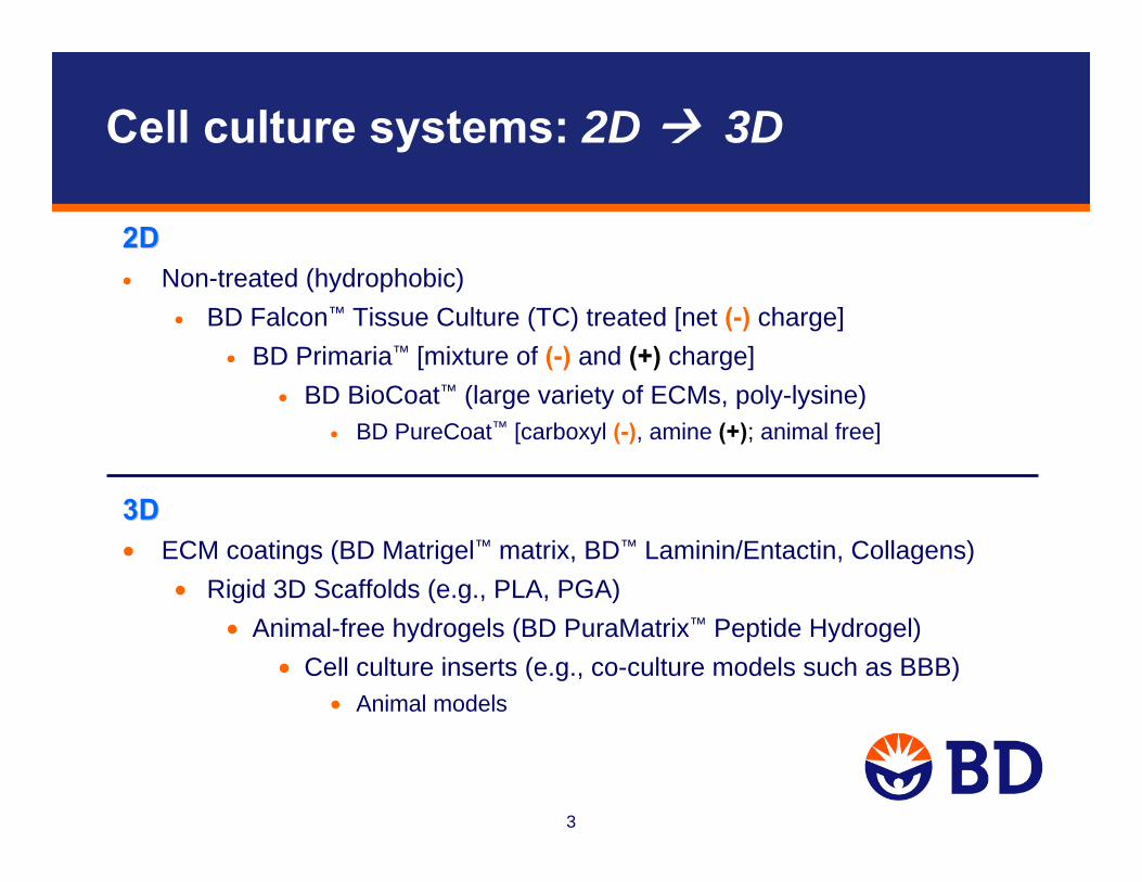
Cell culture systems: 2D 3D
2D2D• Non-treated (hydrophobic)
• BD Falcon™ Tissue Culture (TC) treated [net (-) charge]• BD Primaria™ [mixture of (-) and (+) charge]
• BD BioCoat™ (large variety of ECMs, poly-lysine) • BD PureCoat™ [carboxyl (-), amine (+); animal free]
3D3D• ECM coatings (BD Matrigel™ matrix, BD™ Laminin/Entactin, Collagens)
• Rigid 3D Scaffolds (e.g., PLA, PGA)• Animal-free hydrogels (BD PuraMatrix™ Peptide Hydrogel)
• Cell culture inserts (e.g., co-culture models such as BBB)• Animal models

4
Cell Culture Models Support or Promote Cell Behavior
Cell behaviors (differentiation, functionality) influenced by cues in the microenvironment:
• Cell morphology (structure, phenotype)
• Polarity (functional directionality)
• Growth (proliferation)
• Cell motility (migration, invasion)
• Neurite outgrowth
• Signal transduction (surface receptor function)
• Gene and protein expression (different cell types can express different genes/proteins; liver vs. heart vs. brain)
• Biochemical activities (proteins, enzymes)
5
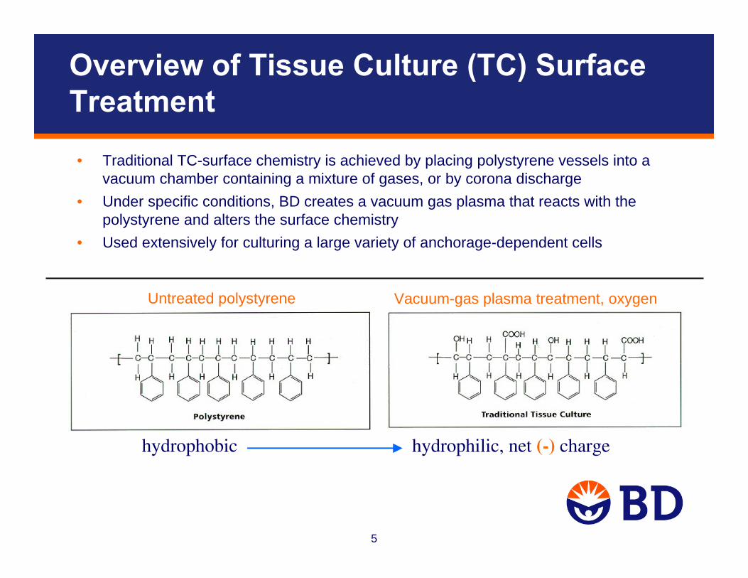
Overview of Tissue Culture (TC) Surface Treatment
• Traditional TC-surface chemistry is achieved by placing polystyrene vessels into a vacuum chamber containing a mixture of gases, or by corona discharge
• Under specific conditions, BD creates a vacuum gas plasma that reacts with the polystyrene and alters the surface chemistry
• Used extensively for culturing a large variety of anchorage-dependent cells
hydrophobic hydrophilic, net (-) charge
Vacuum-gas plasma treatment, oxygen Untreated polystyrene

6
BD Primaria Cultureware
BD Primaria™ is prepared by treating polystyrene vessels with vacuum-gas plasmamixture containing oxygen and ammonia. The treated surface is comprised of oxygen- and nitrogen-containing functional groups.
Applications:
• Attachment and differentiation of certain fastidious cell types such as primary neurons, hepatocytes, endothelial cells, & cardiomyocytes
• Low to moderate scale for cell expansion (up to 75 cm2 flask)
• Low to moderate throughput for cell-based assays (up to 96-well)
hydrophilic, (-) and (+) charge

7
NIH-3T3 Cells Exhibit Enhanced Growth on BD Primaria
BD Falcon™
Non-treated
BD Falcon TC-treated
BD Primaria
NIH-3T3 24 hr post-seeding(in serum-free media)
0
0.2
0.4
0.6
0.8
1
1.2
1.4
1.6
Non-treated TC-treated BD Primaria™
MTS
Sig
nal (
OD
@ 4
90nm
)
8
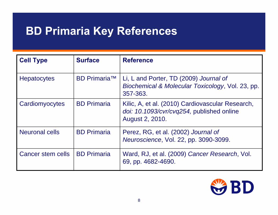
BD Primaria Key References
Perez, RG, et al. (2002) Journal of Neuroscience, Vol. 22, pp. 3090-3099.
BD PrimariaNeuronal cells
Kilic, A, et al. (2010) Cardiovascular Research, doi: 10.1093/cvr/cvq254, published online August 2, 2010.
BD PrimariaCardiomyocytes
Ward, RJ, et al. (2009) Cancer Research, Vol. 69, pp. 4682-4690.
BD PrimariaCancer stem cells
Li, L and Porter, TD (2009) Journal of Biochemical & Molecular Toxicology, Vol. 23, pp. 357-363.
BD Primaria™Hepatocytes
ReferenceSurfaceCell Type

9
The Extracellular Matrix
• Complex mixture containing glycoproteins, collagens, and proteoglycans
• Forms structural framework that stabilizes tissues and provides mechanical support for cell attachment
• Plays important role in cell proliferation, migration,shape, orientation, and differentiation (e.g., signal transduction, gene expression, enzymatic activities)

10
The Basal Lamina: A Thin ‘Mat’ that Underlies Epithelial Cell Sheets and Tubes
basal lamina = basement membrane
BD Matrigel™ matrix = reconstituted basement membrane
Figure: Molecular Biology of the Cell (3rd Edition)

11
ECM Provides a Physiological Growth Substrate
• Although tissue culture plastic is used for many cell types, this environment is not physiological
• ECM-based growth substrates provide a physiological environment that supports and promotes key cell functions

12
ECM Contributes to Intracellular Signaling Pathways
• ECM molecules interact with cell surface receptors (e.g., regulation of integrin signaling by fibronectin:integrin interactions)
• The ECM appears to function in the storage and presentation of growth factors
13
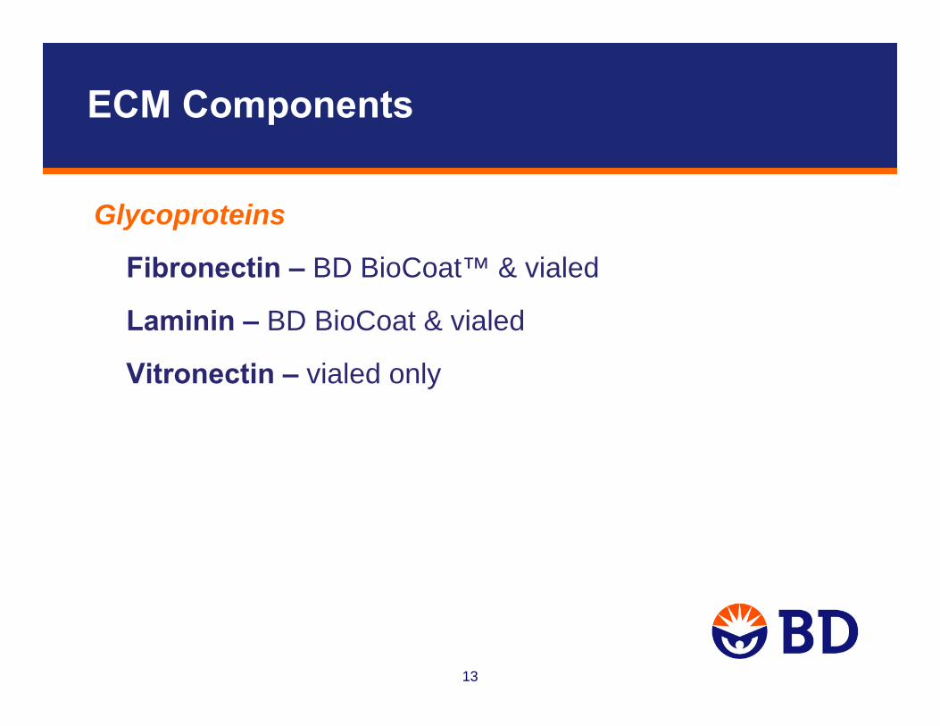
ECM Components
Glycoproteins
Fibronectin – BD BioCoat™ & vialed
Laminin – BD BioCoat & vialed
Vitronectin – vialed only

14
Fibronectin
Figure: Molecular Biology of the Cell (4th Edition)
• Large dimeric protein (multiple isoforms)
• Contributes to matrix organization
• Cell receptors (integrins) bind to FN ‘RGD motif’
• Promotes cell differentiation and functionality (e.g., cell migration, integrin signaling, gene expression)
R
RR
R
RGD

15
Laminin
cell binding
cell binding
entactin binding col IV binding
col IV binding
heparin binding
Laminin
• Large heterotrimeric proteins (11 isoforms)
• Primarily found in basal lamina
• Major structural component of basal lamina
• Cell receptors (integrins) bind to multiple sites on LM
• Promotes cell differentiation and functionality (e.g., neurite outgrowth, receptor signaling, gene expression)
Figure: Molecular Biology of the Cell (4th Edition)

16
Collagens
• Most ubiquitous ECM molecules (at least 16 types)– subunits of collagen ‘trimer’ encoded by multiple genes
• Fibrous proteins that provide structure and resiliency to tissues
• Major component of skin and bone
• Most abundant protein in mammals (~ 25% of total protein mass)
Type Tissue Distribution
Fibrillar I, V bone, skin, tendon, (polymerized fibrils) cornea, internal organs
II cartilage, notochord
III skin, muscle, blood vessels
Network-Forming IV all basal laminaes

17
Collagen Fibrils in Connective Tissue of Skin
Figure: Molecular Biology of the Cell (3rd Edition)

18
BD Matrigel™ Matrix: Reconstituted Basement Membrane
Composition
Laminin ~ 60%
Collagen IV ~ 30%
Entactin ~ 8%
Heparan sulfate proteoglycan (perlecan)
Growth factors (e.g., PDGF, EGF, TGF-β)
Matrix metalloproteinases
19
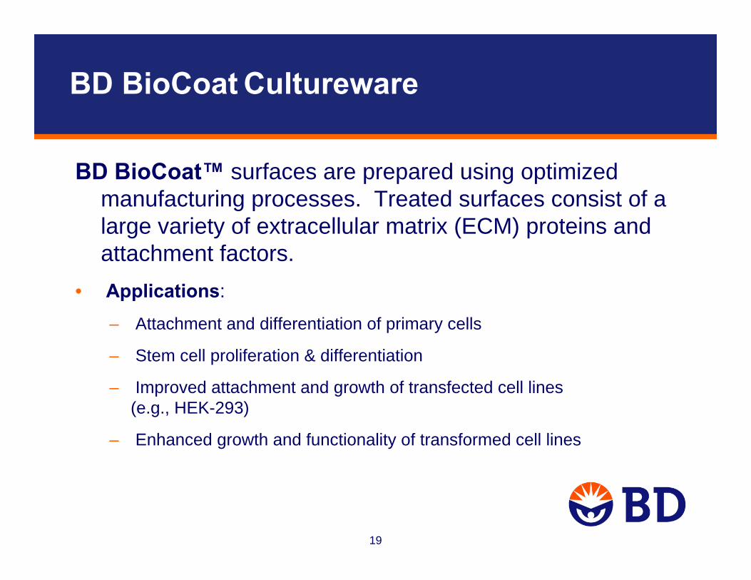
BD BioCoat Cultureware
BD BioCoat™ surfaces are prepared using optimized manufacturing processes. Treated surfaces consist of a large variety of extracellular matrix (ECM) proteins and attachment factors.
• Applications:
– Attachment and differentiation of primary cells
– Stem cell proliferation & differentiation
– Improved attachment and growth of transfected cell lines (e.g., HEK-293)
– Enhanced growth and functionality of transformed cell lines

20
BD BioCoat™ Cultureware
• Gelatin
• Poly-D-Lysine
• Poly-L-Lysine
• Poly-D-Lysine/Laminin
• Poly-L-Ornithine/Laminin
• Custom coatings
• BD Matrigel™ matrix
• Laminin
• Fibronectin
• Laminin/Fibronectin
• Collagen I
• Collagen IV

21
Primary Endothelial Cells Exhibit Enhanced Growth on BD BioCoat Collagen I
BD Falcon™
TC-treated
BD BioCoat™Collagen I
HUVEC Fetal Bovine Heart

22
Saline Control 100 µM Glutamate
Rat Cortical Neurons Exhibit Differentiated Morphology and Function on BD BioCoat™ Laminin/Fibronectin

23
HTS Analysis of PC-12 Cell Neurite Outgrowth Using the BD Pathway™ Bioimager
Control 200 ng/ml NGF
Neurite Total Length
0
40
80
120
160
0.01 0.1 1.0
NGF Dose (µM, on log scale)
Res
pons
e Le
vel
β-tubulin staining, 20x objective

24
After Wash, Before Wash After Wash Calcein AM Staining
BD Falcon™
TC-treated
BD BioCoat™Poly-D-Lysine
• 384-well b/c plates; serum-free conditions for 24 hours; washed with 384-well plate washer
• EcoPack2-293: transformed cell line derived from HEK-293 (Clontech)
Transfected EcoPack™2-293 cells Exhibit Strong Adherence to BD BioCoat Poly-D-Lysine

25
BD Matrigel Matrix for Feeder-Free hES Culture
hES cells cultured on BD Matrigel™ matrix:
• Maintain normal karyotype
• Demonstrate a stable proliferation rate and high telomerase activity
• Express characteristic undifferentiated hES cell markers
• Form embryoid bodies when transferred to low attachment substrate
• Form teratomas in severe combined immunodeficient (SCID) mice and differentiate into cells from all three germ layers

26
Undifferentiated hES Cell Colonies
• Compact and dense H9 colonies on MEF feeders• Spread-out and monolayer-like colonies on BD Matrigel™ matrix
MEF-conditioned mediahES mediaMEF feeder layer BD Matrigel hESC-qualified matrix BD Matrigel hESC-qualified matrix
mTeSR™1

27
SSEA-4
OCT-4
BD™ Laminin/Entactin complex BD Matrigel™ hESC-qualified Matrix
H9 Cells Cultured in mTeSR™1 Medium Express Markers Specific for Undifferentiated hES Cells
28
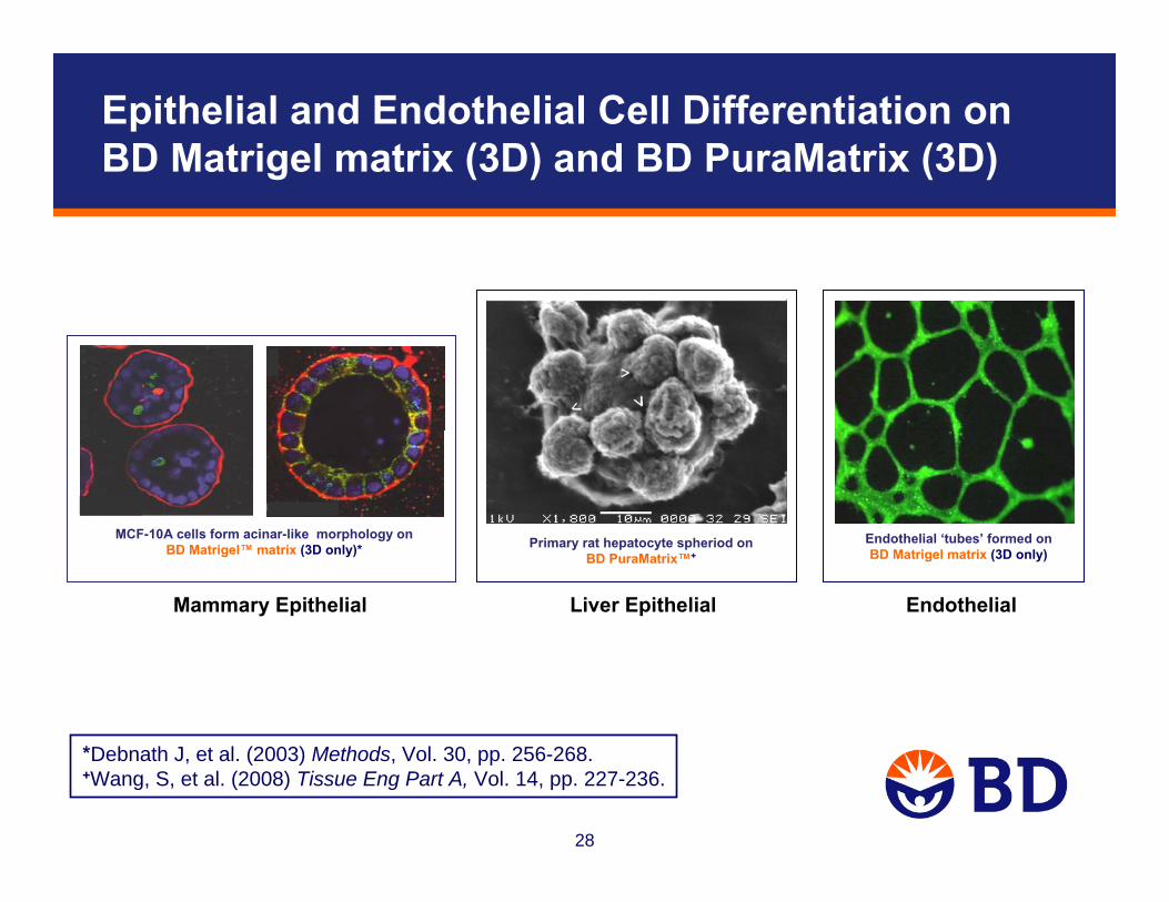
Epithelial and Endothelial Cell Differentiation on BD Matrigel matrix (3D) and BD PuraMatrix (3D)
MCF-10A cells form acinar-like morphology on BD Matrigel™ matrix (3D only)*
Endothelial ‘tubes’ formed on BD Matrigel matrix (3D only)
*Debnath J, et al. (2003) Methods, Vol. 30, pp. 256-268.+Wang, S, et al. (2008) Tissue Eng Part A, Vol. 14, pp. 227-236.
Primary rat hepatocyte spheriod on BD PuraMatrix™+
Mammary Epithelial Liver Epithelial Endothelial

29
Calcein AM staining, 4x confocal
[Suramin]
HTS Analysis of Endothelial Cell Tube Formation using the BD Pathway™ Bioimager
0 160 μM

30
BD BioCoat™, 3D Cell CultureKey References
Cote, MC, et al. (2010) Journal of Biological Chemistry, Vol. 285, pp. 8013-8021.
BD Matrigel Matrix (3D)
Endothelial (tube formation)
Debnath, J, et al. (2003) Methods, Vol. 30, pp. 256-268.BD Matrigel™Matrix (3D)
Mammary epithelial cells
Gupta, MK, et al. (2009) Molecular Pharmacology, Vol. 76, pp. 314-326.
PDL-Laminin (BD BioCoat)
Neuronal cells
Wang, S, et al. (2008) Tissue Engineering Part A, Vol. 14, pp. 227-236.
BD™ PuraMatrix™
(3D)Hepatocytes
Liu, X, et al. (2010) American Journal of Pathology, Vol. 176, pp. 504-515.
Fibronectin(BD BioCoat)
Endothelial progenitors
ReferenceSurfaceCell Type

31
• Chemically defined, animal-free surfaces
• Manufactured using a proprietary thin-film coating technology
- BD PureCoat™ amine
- BD PureCoat carboxyl
BD PureCoat Surfaces

32
Plasma
Radiofrequency Power Applied to System
Monomer Vapour Flows into Chamber
Plasma Chamber (under vacuum)
Substrate(dish, plate, flask)
The Coating is Applied Using Plasma Polymerization

33
BD PureCoat™ Surface Properties
• The amine surface is nitrogen rich, and positively charged
• The carboxyl surface is oxygen rich, and negatively charged
• The thin-film coating is applied to TC-treated vessels
• The detailed chemical composition of the monomers used in the manufacturing process is proprietary
• The charged chemical groups are covalently bound to the plastic

34
BD PureCoat™ – Representative Applications
• Cell attachment and proliferation in serum-free or serum-reduced conditions
• Attachment & differentiation of primary neurons andastrocytes
• Attachment, growth, and differentiation of mesenchymal stem cells
• Cell-based HTS assays– GPCR (e.g., HEK-293), cAMP, proteasome-inhibition
• Cell transfection
• Recovery of cells from cryopreservation

35
Increased BHK-21 Cell Proliferation in Reduced Serum (1% FBS) on BD PureCoat Amine
Tissue Culture
BD PureCoat™ amine
• > 100% increase in proliferation vs. TC and Competitor C
• 3d growth assay (96-well format), n = 10 wells/surface
0
0.5
1
1.5
2
2.5
3
Rel
ativ
e A
bsor
banc
e U
nits
#1 #3#2#1 #3#2#1 #3#2
TCCompetitor CBD PureCoat amine
6400 cells/well 3200 cells/well 1600 cells/well
Initial seeding density

36
Tissue Culture Competitor C BD PureCoat™ carboxyl
Images captured 24h post seeding (100x magnification)
~25-45% increase compared to TC and
Competitor C surfaces
Improved recovery from cryopreservation: LnCAPProstate Cancer Cells on BD PureCoat Carboxyl

37
Anti-β-tubulin III Anti-PGP 9.5
Differentiation of Primary Rat CerebellarGranule Cells on BD PureCoat™ Amine

38
Differentiation of Rat Cerebellar Granule Cells on BD PureCoat Amine
Rat cerebellar granule (RCG) cells cultured on BD PureCoat™ amine for 24 and 48 hours; immunostained with anti-tubulin IIIβ
• RCG cells exhibit extensive neurite outgrowth• Neurite length increases with time
24 hours post-isolation 48 hours post-isolation

39
TC-treated BD PureCoat amine
• Astrocytes were cultured for 24 hours, and then stained with antibody directed against Glial Fibrillary Acidic Protein (GFAP)
Differentiation of Rat Primary Astrocyteson BD PureCoat™ amine

40
• 384-well b/c plates; serum-free conditions for 24 hours; washed with 384-well plate washer
• EcoPack™2-293: transformed cell line derived from HEK-293 (Clontech)
Before wash
After wash
Tissue Culture Competitor C BD PureCoat™amine
EcoPack™2-293 cells exhibit strong attachment to BD PureCoat Amine

41
Analysis of Adult-Derived Stem Cells
• Human bone marrow-derived mesenchymal stem cells (hMSCs, Lonza)
– Cell source: posterior iliac crest of pelvic bone, normal donors
– hMSC maintenance medium: MSCGM™ (Lonza)
– hMSC differentiation:
• Adipogenic induction medium (Lonza)
• Osteogenic induction medium (Lonza)

42
Analysis of hMSC Growth
Methodology
• Prepare cell suspension using MSC growth medium (MSCGM, Lonza)
• Seed 6,000 cells/cm2 on TC-treated and BD PureCoat™ amine 6-well plates
• Culture cells for multiple passages
• Sample analysis:– Growth kinetics: Collect samples at multiple time points (day 1 - day 4)
for each passage number and assess cell confluence using the IncuCyte™ Plus (Essen BioScience)
– Cell yield: Samples from day 4 of each passage number were analyzed using the Vi-CELL™ automated cell counter (Beckman Coulter)

43
0
20
40
60
80
100
passage 3
0
20
40
60
80
100
passage 4
day 1 day 2 day 3 day 4
0
20
40
60
80
100
passage 2
Donor 2
passage 4
passage 2Donor 1
TC-treated
BD PureCoat™ amine
Mea
n ce
ll co
nflu
ence
(%)/
wel
l
0
20
40
60
80
100
day 1 day 2 day 3
0
20
40
60
80
100
day 1 day 2 day 3 day 4
Donor 1
passage 2
passage 4
Mea
n ce
ll co
nflu
ence
(%)/
wel
lhMSCs Exhibit an Enhanced Growth Rate on BD PureCoat™ Amine
44
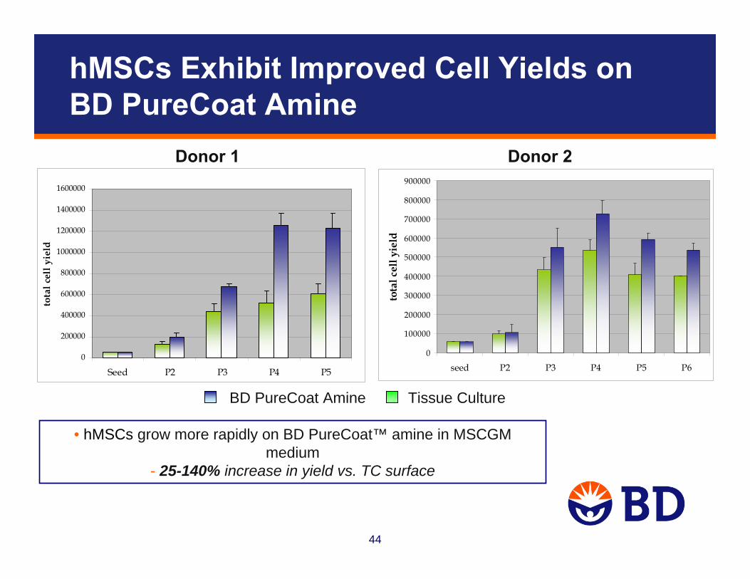
hMSCs Exhibit Improved Cell Yields on BD PureCoat Amine
0
200000
400000
600000
800000
1000000
1200000
1400000
1600000
Seed P2 P3 P4 P5
tota
l cel
l yie
ld
Donor 1 Donor 2
0
100000
200000
300000
400000
500000
600000
700000
800000
900000
seed P2 P3 P4 P5 P6
tota
l cel
l yie
ldBD PureCoat Amine Tissue Culture
• hMSCs grow more rapidly on BD PureCoat™ amine in MSCGM medium
- 25-140% increase in yield vs. TC surface

45
hMSCs Cultured on BD PureCoat™ Amine Express Characteristic Surface Markers
CD29+
CD44+
CD90+
CD34-
CD45-
Cells (P4) were grown on 6-well plates, collected and resuspended in buffer (DPBS/5% serum), stained with antibodies, and then analyzed using a BD FACSCaliber™
flow cytometer.
46
Analysis of hMSC Differentiation
Methodology
Osteogenic lineage:
1. Culture hMSCs (P3) on BD PureCoat™ amine (6-well), and then sub-culture (3,000 cells/cm2) to BD PureCoat amine, BD PureCoat carboxyl, and TC-treated (data not shown) surfaces (6-well)
2. On day 2, induce to differentiate using Osteogenic Induction Medium (Lonza) or feed with MSCGM™ (non-induced control)
3. Re-feed every 3-4 days for 2-3 weeks
4. Wash with 1x PBS, fix with 4% paraformaldehyde, and stain with Alizarin Red

47
A. BD PureCoat™ Amine to Amine B. BD PureCoat Amine to Carboxyl
C. Alizarin Red staining
• Both BD PureCoat surfaces support osteogenic differentiation of hMSCs
• Calcium deposits in induced cultures were stained with Alizarin Red dye
Induced
Non-Induced
hMSCs Expanded on BD PureCoat Amine Retain Osteogenic Differentiation Potential
48
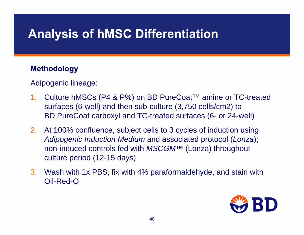
Analysis of hMSC Differentiation
Methodology
Adipogenic lineage:
1. Culture hMSCs (P4 & P%) on BD PureCoat™ amine or TC-treated surfaces (6-well) and then sub-culture (3,750 cells/cm2) to BD PureCoat carboxyl and TC-treated surfaces (6- or 24-well)
2. At 100% confluence, subject cells to 3 cycles of induction usingAdipogenic Induction Medium and associated protocol (Lonza); non-induced controls fed with MSCGM™ (Lonza) throughout culture period (12-15 days)
3. Wash with 1x PBS, fix with 4% paraformaldehyde, and stain with Oil-Red-O

49
BD PureCoat™ amine TC-treated BD PureCoat amine carboxyl TC-treated TC-treated
Indu
ced
Non
-Ind
uced
• Intracellular lipid vacuoles present in induced cultures- stained with Oil-Red-O
hMSC Expanded on BD PureCoat Amine Retain Adipogenic Differentiation Potential
50
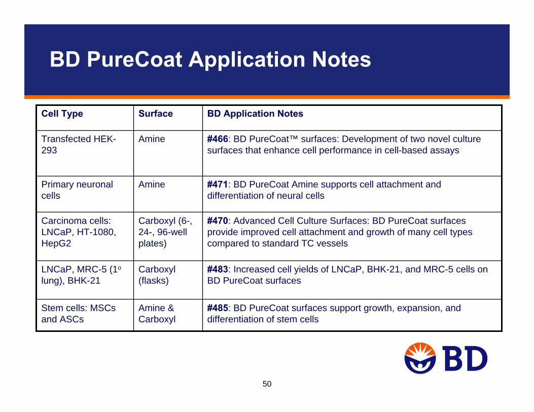
BD PureCoat Application Notes
#470: Advanced Cell Culture Surfaces: BD PureCoat surfaces provide improved cell attachment and growth of many cell types compared to standard TC vessels
Carboxyl (6-, 24-, 96-well plates)
Carcinoma cells: LNCaP, HT-1080, HepG2
#483: Increased cell yields of LNCaP, BHK-21, and MRC-5 cells on BD PureCoat surfaces
Carboxyl (flasks)
LNCaP, MRC-5 (1o
lung), BHK-21
#485: BD PureCoat surfaces support growth, expansion, and differentiation of stem cells
Amine & Carboxyl
Stem cells: MSCsand ASCs
#471: BD PureCoat Amine supports cell attachment and differentiation of neural cells
AminePrimary neuronal cells
#466: BD PureCoat™ surfaces: Development of two novel culture surfaces that enhance cell performance in cell-based assays
AmineTransfected HEK-293
BD Application NotesSurfaceCell Type

51
Uncoated Collagen I PDL
Immunofluorescence Analysis of EcoPack™ Cells (derived from HEK-293) Stained with Integrin αv

52
Questions?
Contact information:Paula Flahertye-mail: [email protected]
Technical Support:tel: 877.232.8995e-mail: [email protected]/webinarsFor research use only. Not intended for use in diagnostic or therapeutic procedures. BD, BD Logo, and all other trademarks are property of Becton, Dickinson and Company. ©2010 BD