Advanced anatomy of lateral nasal wall For the endoscopic ... · Advanced anatomy of lateral nasal...
8
From the SelectedWorks of Balasubramanian iagarajan September 2012 Advanced anatomy of lateral nasal wall For the endoscopic sinus surgeon Contact Author Start Your Own SelectedWorks Notify Me of New Work Available at: hp://works.bepress.com/drtbalu/17
Transcript of Advanced anatomy of lateral nasal wall For the endoscopic ... · Advanced anatomy of lateral nasal...
From the SelectedWorks of BalasubramanianThiagarajan
September 2012
Advanced anatomy of lateral nasal wall For theendoscopic sinus surgeon
ContactAuthor
Start Your OwnSelectedWorks
Notify Meof New Work
Available at: http://works.bepress.com/drtbalu/17
3/30/13 Advanced anatomy of lateral nasal wall – Ent Scholar
entscholar.wordpress.com/article/advanced-anatomy-of-lateral-nasal-wall/ 1/7
Advanced anatomy of lateral nasal wallFor the endoscopic sinus surgeon
September 19, 2012 · Rhinology
Introduction:
Anatomy of the lateral nasal wall is highly complex and variable. With the popularity of endoscopic
sinus surgery a through knowledge of this complex anatomy is very vital. Highly variable anatomy and
paucity of standard landmark makes this region vulnerable for complications during endoscopic sinus
surgery. The learning curve for endoscopic sinus surgery is made rather steep by this highly variable
anatomy . Study of anatomy of lateral nasal wall dates back to Galen (AD 130-201). He described the
porosity of bones in the head. Davinci in his classical anatomical drawings has illustrated maxillary
sinus antrum. He also described maxillary sinus as cavities in the bone that supports the cheek .
Highmore (1651) described maxillary sinus anatomy. Hence it is also known as antrum of Highmore .
It was during the 19th century that Zuckerkandl came out with the first detailed description of maxillary
sinus and its surrounding anatomy. Paranasal sinuses are four air filled cavities situated at the
entrance of the upper airway. Each of these sinuses are named after the skull bone in which it is
located .
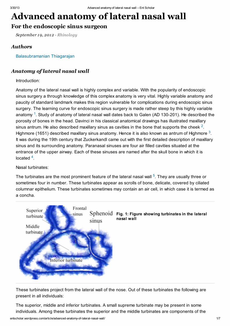
Nasal turbinates:
The turbinates are the most prominent feature of the lateral nasal wall . They are usually three or
sometimes four in number. These turbinates appear as scrolls of bone, delicate, covered by ciliated
columnar epithelium. These turbinates sometimes may contain an air cell, in which case it is termed as
a concha.
Fig. 1: Figure showing turbinates in the lateralnasal wall
These turbinates project from the lateral wall of the nose. Out of these turbinates the following are
present in all individuals:
The superior, middle and inferior turbinates. A small supreme turbinate may be present in some
individuals. Among these turbinates the superior and the middle turbinates are components of the
Anatomy of lateral nasal wall
1
2
3
4
5
Authors
Balasubramanian Thiagarajan

3/30/13 Advanced anatomy of lateral nasal wall – Ent Scholar
entscholar.wordpress.com/article/advanced-anatomy-of-lateral-nasal-wall/ 2/7
ethmodial complex where as the inferior turbinate is a separate bone. Commonly a prominence may
be seen at the attachment of the middle turbinate.
lateral wall of nose after removal of turbinates
This prominence is known as the agger nasi cell. This prominence varies in size in different
individuals. These agger nasi cells overlie the lacrimal sac, separated from it just by a thin layer of
bone. Infact this agger nasi cell is considered to be a remnant of naso turbinal bones seen in animals.
When the anterior attachment of the inferior and middle turbinates are removed, the lacrimal drainage
system and sinus drainage system can be clearly seen.
The inferior turbinate is a separate bone developed embryologically from the maxilloturbinal bone.
The inferior meatus is present between the inferior turbianate and the lateral nasal wall. The nasal
opening of the naso lacrimal duct opens in the anterior third of the inferior meatus. This opening is
covered by a mucosal valve known as the Hassner’s valve. The course of the naso lacrimal duct from
the lacrimal sac lie under the agger nasi cell.
The middle meatus lie between the middle turbinate and the lateral nasal wall. The middle turbinate is
part of the ethmoidal complex. The sinuses have been divided into the anterior and posterior groups.
The anterior group of sinuses are frontal, maxillary and anterior ethmoidal sinuses. These sinuses
drain into the middle meatus, i.e. under the middle turbinate. The middle meatus hosts from anterior to
posterior the following structures:
1. Agger nasi
2. Uncinate process
3. Hiatus semilunaris
4. Ethmoidal bulla
5. Sinus lateralis
6. Posterior fontanelle
Uncinate process: actually forms the first layer or lamella of the middle meatus. This is the most stable
landmark in the lateral nasal wall. It is a wing or boomerang shaped piece of bone. It attaches
anteriorly to the posterior edge of the lacrimal bone, and inferiorly to the superior edge of the inferior
turbinate . Superior attachment of the uncinate process is highly variable, may be attached to the
lamina palyracea, or the roof of the ethmoid sinus, or sometimes to the middle turbinate. The
configuration of the ethmoidal infundibulum and its relationship to the frontal recess depends largely
6

3/30/13 Advanced anatomy of lateral nasal wall – Ent Scholar
entscholar.wordpress.com/article/advanced-anatomy-of-lateral-nasal-wall/ 3/7
on the behavior of the uncinate process. The Uncinate process can be classified into 3 types
depending on its superior attachment.
The anterior insertion of the uncinate process cannot be identified clearly because it is covered with
mucosa which is continuous with that of the lateral nasal wall. Sometimes a small groove is visible over
the area where the uncinate attaches itself to the lateral nasal wall. The anterior convex part forms the
anterior boundary of the ostiomeatal complex. It is here the maxillary, anterior ethmoidal and frontal
sinuses drain. Uncinate process can be displaced medially by the presence of polypoidal tissue, or
laterally against the orbit in
individuals with maxillary sinus hypoplasia . Removing of this piece of bone is the most important step
in Endoscopic sinus surgery.
Type I uncinate: Here the uncinate process bends laterally in its upper most portion and inserts into
the lamina papyracea. Here the ethmoidal infundibulum is closed superiorly by a blind pouch called
the recessus terminalis (terminal recess). In this case the ethmoidal infundibulum and the frontal
recess are separated from each other so that the frontal recess opens into the middle meatus medial
to the ethmoidal infundibulum, between the uncinate process and the middle turbinate. The route of
drainage and ventilation of the frontal sinus run medial to the ethmoidal infundibulum.
Type I uncinate insertion
Type II uncinate: Here the uncinate process extends superiorly to the roof of the ethmoid. The frontal
sinus opens directly into the ethmoidal infundibulum. In these cases a disease in the frontal recess
may spread to involve the ethmoidal infundibulum and the maxillary sinus secondarily. Sometimes the
superior end of the uncinate process may get divided into three branches one getting attached to the
roof of the ethmoid, one getting attached to the lamina papyracea, and the last getting attached to the
middle turbinate.
Type II uncinate insertion
7
3/30/13 Advanced anatomy of lateral nasal wall – Ent Scholar
entscholar.wordpress.com/article/advanced-anatomy-of-lateral-nasal-wall/ 4/7
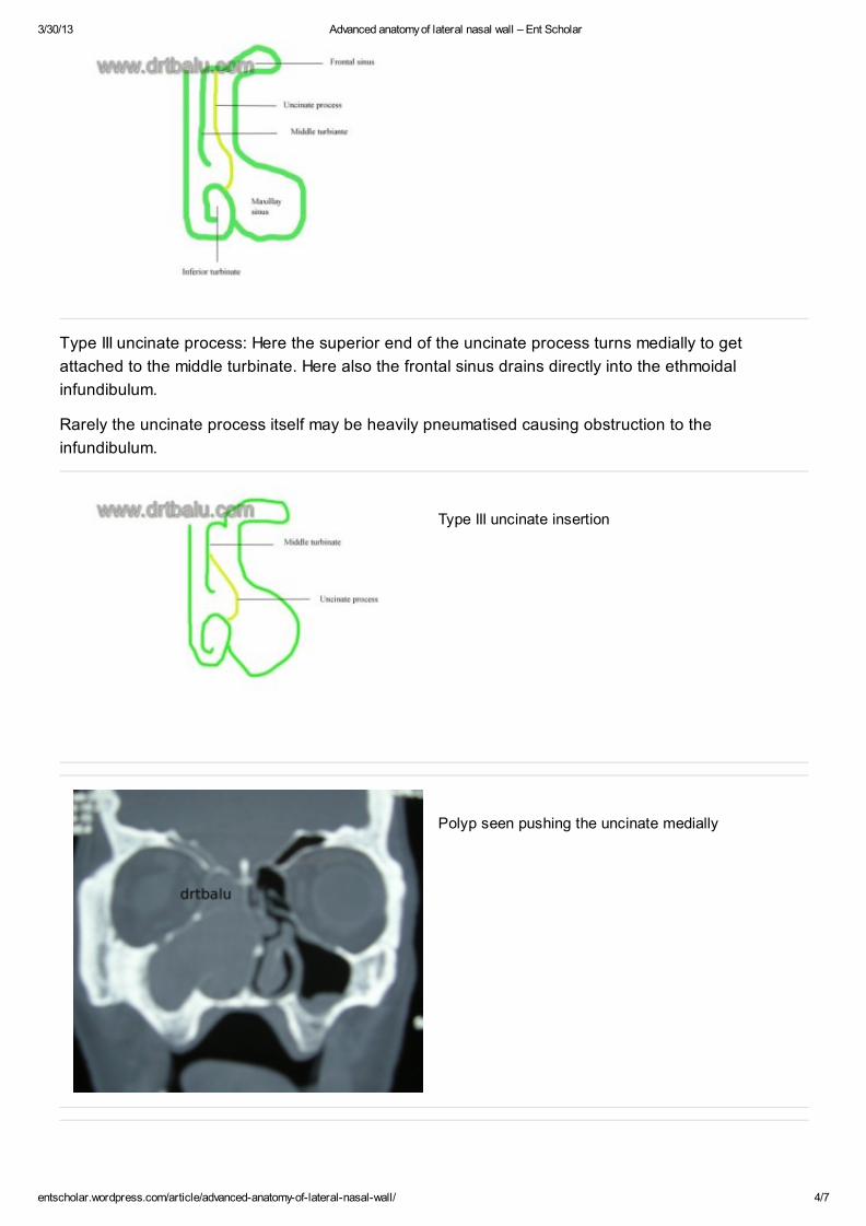
Type III uncinate process: Here the superior end of the uncinate process turns medially to get
attached to the middle turbinate. Here also the frontal sinus drains directly into the ethmoidal
infundibulum.
Rarely the uncinate process itself may be heavily pneumatised causing obstruction to the
infundibulum.
Type III uncinate insertion
Polyp seen pushing the uncinate medially

3/30/13 Advanced anatomy of lateral nasal wall – Ent Scholar
entscholar.wordpress.com/article/advanced-anatomy-of-lateral-nasal-wall/ 5/7
Hypoplasia of maxillary sinus seen pushing the uncinate laterally
Image showing uncinate process
Removal of uncinate process reveals the natural ostium of the maxillary sinus. This is another vital
landmark in the lateral nasal cavity. The superior wall of the natural ostium of the maxillary sinus is at
the level of floor of the orbit.Agger nasi: This is a latin word for “Mound”. This area refers to the most
superior remnant of the first ethmoturbinal which presents as a mound anterior and superior to the
insertion of middle turbinate.Depending on the pneumatization of this area may reach up to the level
of lacrimal fossa thereby causing narrowing of frontal sinus outflow tract. Ethmoidal infundibulum: is a
cleft like space, which is three dimensional in the lateral wall of the nose. This structure belongs to the
anterior ethmoid. This space is bounded medially by the uncinate process and the mucosa covering it.
Major portion of its lateral wall is bounded by the lamina papyracea, and the frontal process of maxilla
to a lesser extent. Defects in the medial wall of the infundibulum is covered with dense connective
tissue and periosteum. These defects are known as anterior and poterior fontanelles. Anteriorly the
ethmoidal infundibulum ends blindly in an acute angle.
Figure showing large agger nasi air cell
Bulla ethmoidalis: This is derived from Latin. Bulla means a hollow thin walled bony prominence. This

3/30/13 Advanced anatomy of lateral nasal wall – Ent Scholar
entscholar.wordpress.com/article/advanced-anatomy-of-lateral-nasal-wall/ 6/7
is another landmark since it is the largest and non variant of the aircells belonging to the anterior
ethmoidal complex. This aircell is formed by pneumatization of bulla lamella (second ethmoid basal
lamella). This air cell appears like a bleb situated in the lamina papyracea. Some authors consider this
to be a middle ethmoid cell. If bulla extends up to the roof of ethmoid it can form the posterior wall of
frontal recess. If it does not reach up to the level of skull base then a recess can be formed between
the bulla and skull base. This recess is known as suprabullar recess. If the posterior wall of bulla is not
in contact with basal lamella then a recess is formed between bulla and basal lamella. This recess is
known as retrobullar recess / sinus lateralis. This retrobullar recess may communicate with the
suprabullar recess. Osteomeatal complex: This term is used by the surgeon to indicate the area
bounded by the middle turbinate medially, the lamina papyracea laterally, and the basal lamella
superiorly and posteriorly. The inferior and anterior borders of the osteomeatal complex are open.
The contents of this space are the aggernasi, nasofrontal recess (frontal recess), infundibulum, bulla
ethmoidalis and the anterior group of ethmoidal air cells.This is infact a narrow anatomical region
consisting of : 1. Multiple bony structures (Middle turbinate, uncinate process, Bulla ethmoidalis) 2. Air
spaces (Frontal recess, ethmoidal infundibulum, middle meatus) 3. Ostia of anterior ethmoidal,
maxillary and frontal sinuses. In this area, the mucosal surfaces are very close, sometimes even in
contact causing secretions to accumulate. The cilia by their sweeping movements pushes the nasal
secretions. If the mucosa lining this area becomes inflamed and swollen the mucociliary clearance is
inhibited, eventually blocking the sinuses. Some authors divide this osteomeatal complex into anterior
and posterior. The classic osteomeatal complex described already has been described as the anterior
osteomeatal complex, while the space behind the basal lamella containing the posterior ethmoidal
cells is referred to as the posterior ethmoidal complex, thus recognising the importance of basal
lamella as an anatomical landmark to the posterior ethmoidal system. Hence the anterior and the
posterior osteomeatal complex has separate drainage systems. So when the disease is limited to the
anterior compartment of the osteomeatal complex, the ethmoid cells can be opened and diseased
tissue removed as far as the basal lamella, leaving the basal lamella undisturbed minimising the risk
during surgery.Hiatus semilunaris: Lies between the anterior wall of the Bulla and the free posterior
margin of the uncinate process. This is infact a two dimensional space. Through this hiatus a cleft like
space can be entered. This is known as the ehtmoidal infundibulum. This ethmoidal infundibulum is
bounded medially along its entire length by the uncinate process and its lining mucosa. The lateral
wall is formed by the lamina papyracea of the orbit, with participation from the frontal process of the
maxilla and the lacrimal bone. The anterior group of sinuses drain into this area. Infact this area acts
as a cess pool for all the secretions from the anterior group of sinuses.
Osteomeatal complex
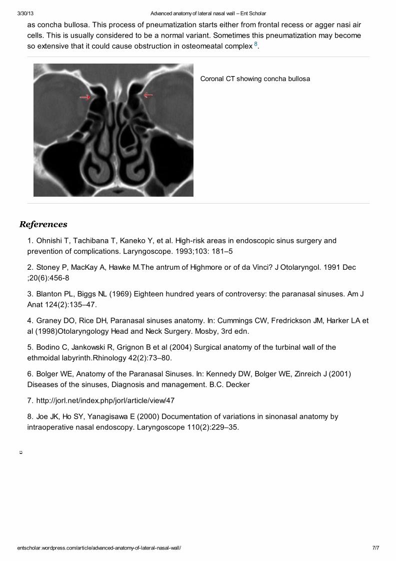
Concha bullosa: Sometimes middle turbinate may become pneumatized. This pneumatization is known
3/30/13 Advanced anatomy of lateral nasal wall – Ent Scholar
entscholar.wordpress.com/article/advanced-anatomy-of-lateral-nasal-wall/ 7/7
as concha bullosa. This process of pneumatization starts either from frontal recess or agger nasi air
cells. This is usually considered to be a normal variant. Sometimes this pneumatization may become
so extensive that it could cause obstruction in osteomeatal complex .
Coronal CT showing concha bullosa
1. Ohnishi T, Tachibana T, Kaneko Y, et al. High-risk areas in endoscopic sinus surgery and
prevention of complications. Laryngoscope. 1993;103: 181–5
2. Stoney P, MacKay A, Hawke M.The antrum of Highmore or of da Vinci? J Otolaryngol. 1991 Dec
;20(6):456-8
3. Blanton PL, Biggs NL (1969) Eighteen hundred years of controversy: the paranasal sinuses. Am J
Anat 124(2):135–47.
4. Graney DO, Rice DH, Paranasal sinuses anatomy. In: Cummings CW, Fredrickson JM, Harker LA et
al (1998)Otolaryngology Head and Neck Surgery. Mosby, 3rd edn.
5. Bodino C, Jankowski R, Grignon B et al (2004) Surgical anatomy of the turbinal wall of the
ethmoidal labyrinth.Rhinology 42(2):73–80.
6. Bolger WE, Anatomy of the Paranasal Sinuses. In: Kennedy DW, Bolger WE, Zinreich J (2001)
Diseases of the sinuses, Diagnosis and management. B.C. Decker
7. http://jorl.net/index.php/jorl/article/view/47
8. Joe JK, Ho SY, Yanagisawa E (2000) Documentation of variations in sinonasal anatomy by
intraoperative nasal endoscopy. Laryngoscope 110(2):229–35.
8
References