Advance Publication by J-STAGE Journal of Reproduction and ...
23
Advance Publication by J-STAGE Journal of Reproduction and Development Accepted for publication: December 8, 2020 Advanced Epub: January 29, 2021
Transcript of Advance Publication by J-STAGE Journal of Reproduction and ...

Advance Publication by J-STAGE
Journal of Reproduction and Development
Accepted for publication: December 8, 2020
Advanced Epub: January 29, 2021

1
Intravaginal administration of estradiol benzoate capsule for estrus synchronization in goats 1
2
Sayaka Matsumoto1, Tomomi Tanaka1, and Natsumi Endo1* 3
4
1Laboratory of Veterinary Reproduction, Tokyo University of Agriculture and Technology, 3-5-8 5
Saiwai-cho, Fuchu-city, Tokyo, 183-8509, Japan 6
7
*Corresponding author: N. Endo, Laboratory of Veterinary Reproduction, Tokyo University of 8
Agriculture and Technology, 3-5-8 Saiwai-cho, Fuchu-city, Tokyo, 183-8509, Japan 9
Tel.: +81-42-367-5782 10
Fax: +81-42-366-4062 11
Email: [email protected] 12
13
Running head: Intravaginal estradiol administration 14
15

2
Abstract 16
Estrus synchronization requires multiple treatments of hormonal drugs, requiring considerable 17
time and cost. The aim of the present study was to develop an estrus synchronization protocol using 18
intravaginal administration of estradiol benzoate (EB) capsules in goats. Two types of capsules were 19
prepared: an EB capsule that melted immediately after administration and a sustained-release (SR) EB 20
capsule that dissolved slowly and reached a peak after 24 h. Goats with functional corpus lutea were 21
intramuscularly treated with prostaglandin F2 (PG). At 24 h after PG administration, goats were 22
administered 1 mg of EB solution intramuscularly (PG+24IM; n = 6) or 1 mg of EB capsule 23
intravaginally (PG+24EB; n = 6). The SR EB capsule was administered intravaginally at the time of 24
PG administration (PG+SR; n = 6). The control group (n = 6) received only PG. All groups showed 25
estrus within 72 h after PG administration. The onset of estrus did not differ significantly between the 26
PG+24IM and PG+SR groups but was earlier than in the control group. Estradiol concentration in the 27
PG+SR group peaked at 11.5 ± 6.1 h after EB and PG administration. Peak estradiol concentrations 28
were not significantly different between the PG+24IM and PG+SR groups (78.0 ± 25.8 and 64.0 ± 29
38.1 pg/ml respectively), and were higher than the PG+24EB and control groups (27.3 ± 8.8 and 14.6 30
± 6.1 pg/ml; respectively). These results suggest that intravaginal administration of an EB capsule 31
with a sustained-drug release base is applicable for estrus synchronization, as an alternative to 32
intramuscular administration. 33
34
Key words: estradiol benzoate, estrus synchronization, intravaginal administration 35
Declarations of interest: none.36

3
37
1. Introduction 38
As herd sizes increase, visual observation of individual cows is not practical within the 39
available time of herd managers, resulting in unobserved estrus and substantial economic losses [1]. A 40
decline in reproductive efficiency due to weak estrus and ovulation disorders in high-producing dairy 41
cows has also been noted [2, 3]. Therefore, there is an increasing demand for synchronized estrus and 42
ovulation using a hormone preparation to conduct fixed-time artificial insemination (TAI). Current 43
estrus synchronization protocols employ a combination of several hormones, i.e.,such as 44
prostaglandin F2α (PG) for inducing luteolysis, an intravaginal progesterone device used to artificially 45
extend the luteal phase, and estradiol sodium or gonadotropin-releasing hormone for the induction of 46
estrus and/or ovulation [4]. However, these protocols involving multiple hormonal treatments require 47
repeated tasks, such as holding and treating cows. 48
As an alternative to intramuscular administration, the vagina is a potential route for systemic 49
drug delivery. Suppositories, creams, gels, tablets, and vaginal rings are commonly used to administer 50
hormones and antimicrobial agents. There are many advantages to intravaginal administration; this 51
route avoids gastrointestinal absorption and hepatic first-pass metabolism of the drug [5], and direct 52
delivery to the site of action results in reduced systemic side effects [6]. However, the vaginal mucus 53
that coats the vaginal walls can significantly affect penetration, distribution, and residence time of the 54
drug administered via this route [7]. Mucoadhesive polymers are sometimes used in tablet 55
formulations to increase the vaginal residence time of microbicides [8]. Recent studies have shown 56
that the use of mucoadhesive polymers such as polyacrylic acid bases and cellulose derivatives can 57
improve the efficacy of drug release and absorption in the vagina [16, 17]. We reported that 58
intravaginal administration of a progesterone capsule formulated using a mixture of a mucoadhesive 59
polymer and silicone fluid could maintain a plasma progesterone (P4) concentration similar to that in 60
the natural luteal phase for 9 d in goats [9]. In this way, a reduction in the administration frequency 61
using intravaginal sustained drug release can reduce the time and cost of treatment and reduce pain in 62
animals. 63
In cattle, estradiol benzoate (EB) is used at the beginning of various estrus synchronization 64
protocols to synchronize the follicular wave or at the end of the protocol to induce estrus and 65
synchronize ovulation for TAI [10–12]. In studies on estrus synchronization using EB, the drug was 66
administered intramuscularly at 24 or 48 h after PG treatment to induce luteolysis [12–16]. The aim of 67

4
the present study was to develop a new estrus synchronization protocol using the intravaginal route 68
for EB administration in goats. We used goats as an experimental model of cattle because of their 69
similar reproductive characteristics [17, 18]. This study investigated the effect of vaginal 70
sustained-release (SR) EB capsules on the circulating profiles of estradiol and the expression of estrus 71
in goats. 72
73

5
74
2. Materials and methods 75
This study was approved by the University Committee for the Use and Care of Animals at the 76
Tokyo University of Agriculture and Technology (no. 31–52). 77
78
2.1. Preparation of intravaginal estradiol capsules 79
Two types of intravaginal capsules were prepared. The EB capsule was designed to melt 80
immediately after administration and was formulated using commercially available hard fat (Hosco 81
S-55, Maruishi Pharmaceutical Co., Osaka) as the suppository base. The SR EB capsule was designed 82
to melt gradually to reach a peak 24 h after administration and was formulated using an SR base 83
developed previously in our laboratory [9]. 84
The EB capsule was prepared by adding 1 mg of EB powder (Tokyo Chemical Industry Co., Ltd., 85
Japan) and 0.8 ml of hard fat to a gelatin capsule (#00, volume of 1.01 ml, Matsuya Co., Osaka, 86
Japan). For the SR capsule, 1 mg of EB powder and hard fat was placed in a small capsule (#2, 87
volume of 0.37 ml, Matsuya Co., Osaka, Japan) and placed in the center of a large capsule (#00) filled 88
with a SR base containing silicone fluid (Q7-9120, Dow Corning, MI, USA) as a dispersing agent and 89
acrylic starch (SANWET®, Sanyo Chemical Co. Ltd., Kyoto, Japan) as a mucoadhesive polymer [9] 90
(Figure 1). The capsules were then stored at 4C in a refrigerator until administration. 91
92
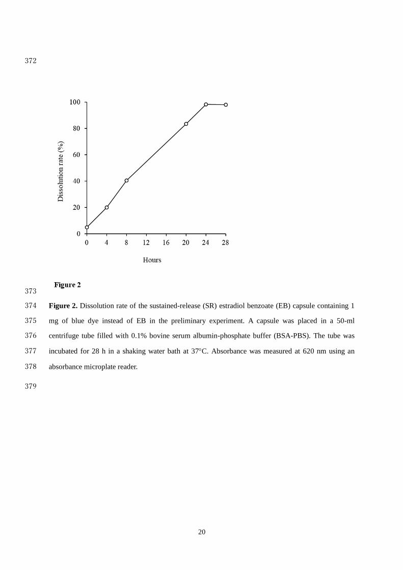
2.2 Dissolution test 93
One milligram of blue dye (Brilliant Blue FCF CI42090, Fuji Film Wako Pure Chemical Industries, 94
Japan) was used instead of EB for in vitro dissolution testing of the SR capsule. For this, a capsule 95
was placed in a 50-ml centrifuge tube filled with 0.1% bovine serum albumin phosphate buffer 96
(BSA-PBS). The pH was adjusted to 7.1, which is the average pH of the cervical mucus during the 97
luteal phase [19]. The tube was then incubated for 28 h in a shaking water bath at 37C. Samples of 1 98
ml were withdrawn at 4, 8, 20, 24, and 28 h and replaced with the same volume of 0.1% BSA-PBS. 99
Absorbance was measured at 620 nm using an absorbance microplate reader (Multiskan FC, Thermo 100
Fisher Scientific, Tokyo, Japan). Dissolution rates of the SR capsule in the preliminary experiment are 101
shown in Figure 2. 102
103
2.3. Animals 104
6
Fifteen female Shiba goats (age, 71.4 ± 31.9 [mean ± SD] months; body weight, 29.0 ± 5.8 kg) 105
maintained at the Tokyo University of Agriculture and Technology were used. The goats were housed 106
in outdoor paddocks with sheltered areas and were fed alfalfa hay cubes (350 g) twice a day. Clean 107
water and mineralized salt were provided ad libitum. All goats were confirmed to be clinically healthy 108
and in good condition and to have normal estrous cycles before beginning the study from September 109
2018 to February 2019. 110
111
2.4. Treatments 112
All goats were checked for estrus daily. During 7-14 d after estrus, all goats with functional 113
corpus lutea were treated with PG (2 mg of dinoprost, intramuscular; Pfizer, Tokyo, Japan) to induce 114
luteolysis (day 0: the day of PG administration). The control group (n = 6) did not receive any further 115
treatment. However, the PG+24IM (n = 6) and PG+24EB (n = 6) groups received 1 mg of EB 116
(Ovahormone®; ASKA Pharmaceutical Co., Tokyo, Japan) intramuscularly or EB capsule 117
intravaginally at 24 h after PG treatment, respectively. Goats in the PG+SR group (n = 6) were 118
administered an SR capsule at the same time as PG treatment (day 0). The administered intravaginal 119
EB and SR capsules were attached to a Y-shaped silicone device [9] to prevent the capsule from 120
flowing out of the vagina. The device was removed on day 2, and by then, it was confirmed that all 121
capsules had melted in the vagina. 122
123
2.5. Blood sampling 124
Blood samples were collected via jugular venipuncture into 5 ml syringes containing heparin. 125
Samples were obtained just before PG administration, and once daily at 24-h intervals for 5 d. In the 126
PG+24IM and PG+24EB groups, additional samples were obtained at 9 h after EB administration. In 127
the PG+SR group, additional samples were obtained at 9 and 33 h after SR capsule administration. 128
Goats in the control group were sampled in the same manner as those in the PG+SR group. Blood 129
samples were placed in iced water and then centrifuged immediately at 3000 rpm at 4C for 20 min. 130
Following separation, the plasma was stored at -20C until subsequent assays were performed. 131
132
2.6. Estrus detection 133
Estrus detection was performed using a male goat at the time of blood collection for 5 d. Standing 134
estrus meant that the female goat stood still and allowed a nearby male goat to mount. The onset of 135

7
estrus was defined as the first observation time point at which the goat showed standing estrus. The 136
end of estrus was defined as the first observation time point at which the goat showed no standing 137
estrus. The duration of estrus was calculated as the interval between the onset and the end of estrus. 138
139
2.7. Hormone assays 140
Plasma concentrations of estradiol and P4 were measured via an enzyme immunoassay after 141
extraction using dichloromethane and diethyl ether, respectively. Plasma estradiol concentrations were 142
measured using a commercial assay kit (Estradiol ELISA Kit, Cayman Chemical, USA), following the 143
manufacturer’s instructions. Plasma P4 concentrations were measured according to the method 144
reported by Prakash et al. [20] with some modifications. The intra- and inter-assay coefficients of 145
variation for E₂ were 33.0% and 9.0%, respectively, with a sensitivity of 0.55 pg/ml. The intra- and 146
inter-assay coefficients of variation for P₄ were 7.6% and 4.1%, respectively, with a sensitivity of 0.78 147
ng/ml. 148
149
2.8. Statistical analysis 150
Data are presented as means ± standard deviations. All data were analyzed using a statistical 151
software (Excel Statistics, Social Information Services, Japan). Comparisons of the estrus 152
characteristics among groups were made using one-way analysis of variance (ANOVA), followed by 153
Tukey’s multiple comparison test. Continuous variables such as E2 and P4 concentrations were 154
compared using two-way repeated-measures ANOVA, followed by Tukey’s multiple comparison test. 155
Blood sampling at 9 h after PG administration was performed only in the PG+SR and control groups. 156
Therefore, the data at this time point were analyzed separately using Student’s t-test. Differences were 157
considered significant at P < 0.05. 158
159

8
3. Results 160
3.1. Estrus 161
All goats showed estrous behavior (Table 1). The onset of estrus did not differ significantly 162
between the PG+24IM and PG+SR groups, but was earlier than that in the control group (P < 0.05). 163
The duration of estrus varied widely among the animals (range, 15–96 h for all groups), but no 164
significant differences were found in the duration and end of estrus among groups. These results were 165
calculated from the estrus detection, which was performed once daily at 24-h intervals. 166
167
3.2. Plasma concentrations of steroid hormones 168
Plasma P4 concentrations in all animals declined to less than 1 ng/ml during 24 h after PG 169
administration. There was no significant difference among groups in progesterone concentrations after 170
PG administration during the blood sampling period (P > 0.1). 171
Plasma estradiol concentrations until 96 h after EB administration were compared among the 172
PG+24IM, PG+24EB, and PG+SR groups (Figure 3). There were no significant differences in 173
estradiol concentration at 0 h among the PG+24IM, PG+24EB, and PG+SR groups (6.1 ± 1.6, 10.2 ± 174
5.5, and 5.8 ± 3.8 pg/ml, respectively). The mean estradiol concentration in the PG+24IM group 175
reached a peak of 74.6 ± 30.0 pg/ml at 24 h after administration. In contrast, the mean estradiol 176
concentrations in PG+24EB and PG+SR groups peaked at 9 h after administration. The concentration 177
at 9 h after administration in the PG+24IM and PG+SR groups (57.0 ± 29.1 and 61.0 ± 41.6 pg/ml, 178
respectively) was not significantly different but was significantly higher than that in the PG+24EB 179
group (24.5 ± 8.3 pg/ml, P > 0.05). Estradiol concentrations in the PG+24IM group at 24, 48, and 72 h 180
after administration were significantly higher than those in the PG+24EB and PG+SR groups at 181
respective sampling times. 182
Plasma estradiol concentrations at different time points after PG administration in all groups are 183
shown in Figure 4. Goats in the PG+SR group received the SR capsule along with PG. Consequently, 184
the mean estradiol concentration in the PG+SR group reached a peak at 9 h after PG administration, 185
which was earlier than that in the other groups. In the PG+24IM and PG+24EB groups, EB was 186
administered at 24 h after PG administration. The mean estradiol concentration in the PG+24IM group 187
was higher at 33–96 h after PG administration than that in the other three groups. Estradiol 188
concentrations in the PG+24EB group were significantly higher at 33 and 48 h after PG 189
administration than in the PG+SR and control groups, and they returned to levels similar to those in 190

9
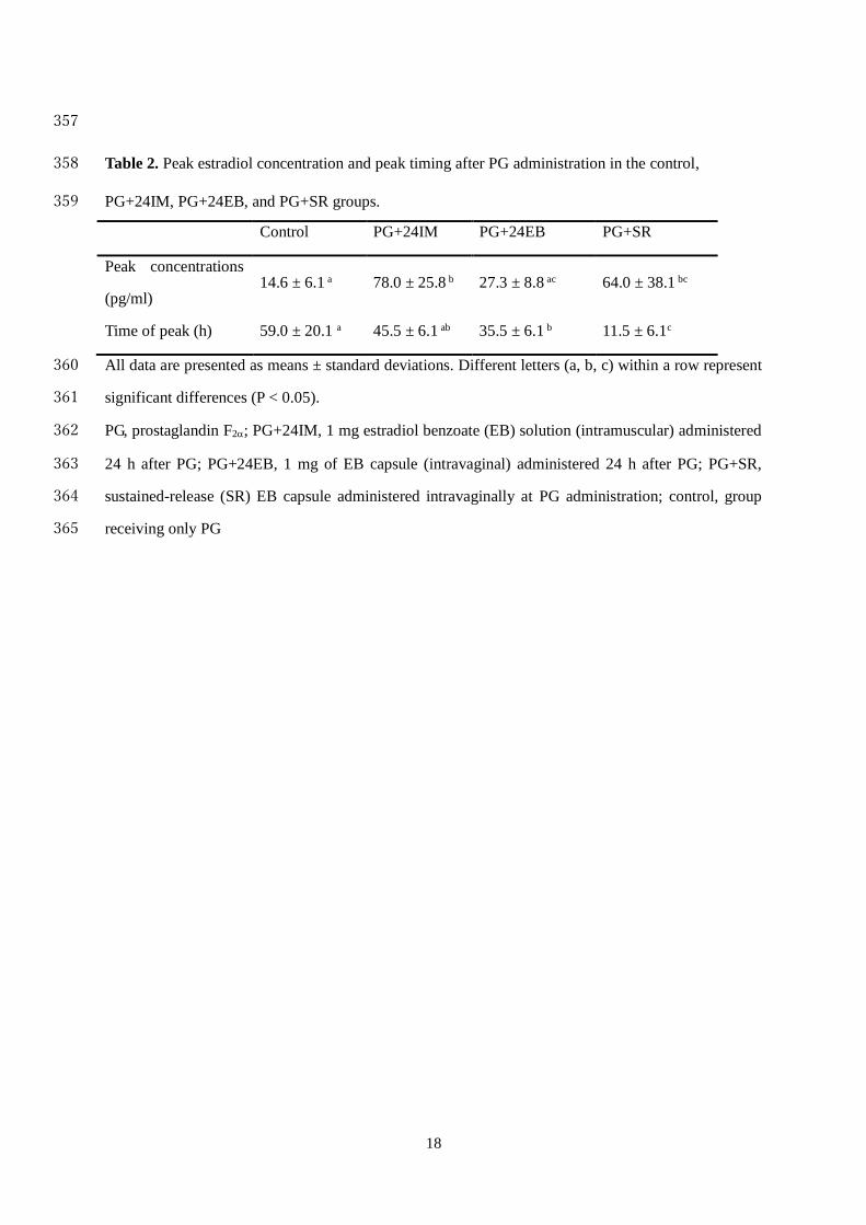
the PG+SR and control groups at 72 h after PG administration. Peak estradiol concentrations and 191
timing to reach the peak after PG treatment are shown in Table 2. The peak timings were calculated 192
from the sampling times, which were once daily at 24-h intervals. The peak estradiol concentration 193
was not significantly different between the PG+24IM and SR groups, but the time to reach the peak 194
concentration in the SR group was earlier than that in the other three groups. 195
Table 2, Figure 3, and Figure 4 show results of analyzing the same estradiol concentration data 196
from different viewpoints. 197
198

10
4. Discussion 199
Administration of PG to goats and other ruminants with a functional corpus luteum is the simplest 200
method for inducing estrus. In the present study, administration of PG induced estrus in all goats, 201
regardless of EB treatment. The mean interval from PG administration to the onset of estrus in goats 202
was reported to be 42–47 h [21] or 57 h [22]. In the present study, the interval from PG administration 203
to the onset of estrus in the control group was comparable to that in the aforementioned studies. 204
Estradiol is the primary hormone that induces behavioral estrus. It was proposed that once a threshold 205
of estradiol is achieved, estrus is induced, and additional amounts of estradiol above the threshold do 206
not further enhance the estrous response, duration, or intensity of the estrus [23]. In agreement with 207
this, the results of the present study showed that intramuscular administration of EB at 24 h after PG 208
administration or intravaginal administration of an SR capsule at the same time as PG administration 209
affected the onset of estrus but not the duration or end of estrus. In contrast, intravaginal 210
administration of an EB capsule prepared using a hard-fat suppository base did not advance the onset 211
of estrus unlike that in the control group. These differences could be attributed to the estradiol 212
concentrations after EB administration. The increase in estradiol concentration after 9 h after in the 213
PG+24EB group was less than half that of the PG+24IM and PG+24SR groups. It was considered that 214
this increase was not sufficient to induce estrus immediately after capsule administration, and an 215
additional amount of estradiol secreted from the pre-ovulatory follicles was needed for estrus to 216
commence in some cases. 217
An estrus synchronization protocol of 1 mg intramuscularly administered EB is generally used in 218
cows, while equine chorionic gonadotropin (eCG) has been used for goats. However, some recent 219
studies indicated the effectiveness of EB as an alternative to eCG [24, 25]. In these studies, 220
administration of 0.2 mg EB effectively induced estrus and ovulation, similar to eCG in goats. In the 221
present study, goats were administered 1 mg of EB to determine differences in estradiol profiles 222
caused by changing the method of administration (intramuscular vs. intravaginal) and the suppository 223
base (hard fat vs. SR formulation). A previous study examined the effect of EB dose and route on 224
plasma estradiol concentrations in ovariectomized heifers. It showed that intravaginal administration 225
of 10-mg EB powder via a gelatin capsule increased the estradiol concentration to 4.1 pg/ml, which 226
was not significantly different from the concentration after intramuscular administration of 0.5-mg EB 227
[26]. This result implies that an approximately 20-fold intravaginal dose of EB powder was required 228
to attain estradiol profiles similar to those attained by intramuscular administration. Another study 229

11
reported that, to achieve similar estrus, a gelatin capsule containing four times more EB in powered 230
form than in the intramuscular injection form was required [27]. However, the study did not measure 231
plasma estradiol concentrations. In the present study, when EB was administered intravaginally to 232
goats in a hard-fat suppository base, the increase in plasma estradiol concentration was less than half 233
of that attained following PG administration intramuscularly. In contrast, the concentration was 234
significantly higher in the PG+SR group than in the PG+24EB group, and the increase at 9 h after 235
administration was close to that in the PG+24IM group. The efficacy of drug therapy using the 236
vaginal route may be restricted by the short residence time of the drug within the vagina owing to the 237
vaginal fluid that coats the mucosal tissue. The use of a mucoadhesive polymer such as a polyacrylic 238
acid base and cellulose derivatives can promote a prolonged and intimate contact with the vaginal 239
mucus, enhancing the delivery of drugs to the underlying tissue and their sustained release [28–30]. 240
We consider that the use of polyacrylic starch in the SR capsule formulation increased the adhesion 241
and retention of EB on the vaginal wall, preventing EB from leaking out of the vagina. 242
The plasma estradiol concentration in the PG+SR group was expected to peak at 24 h after 243
administration, based on an in vitro test of the SR capsule. This formulation was based on a recently 244
developed estrus and ovulation synchronization protocol for cows, in which intramuscular 245
administration of EB was performed 24 h after PG treatment and TAI was performed 24–28 h after 246
EB administration [13]. This protocol can minimize the cost and time of treatment, while the 247
pregnancy rate after TAI was satisfactory in comparison with conventional protocols. However, in the 248
present study, the peak estradiol concentration was observed at an average of 11.5 h after SR capsule 249
administration, which was earlier than our expected time. It is possible that the SR capsule 250
disintegrated earlier in the vagina than in vitro owing to the pressure and movement of the vaginal 251
wall. Furthermore, physiological factors such as changes in the volume, viscosity, and pH of the 252
vaginal fluid can affect the efficacy of drug delivery systems [28]. The present study was limited to 253
examining two types of intravaginal EB capsules in terms of estrus response and blood estradiol 254
profiles in a goat model. Modification of the size of the surrounding SR suppository base and/or inner 255
EB capsule is one way to extend the drug-release profile and retard the estradiol peak. To validate the 256
effectiveness of intravaginal EB as an estrus synchronization protocol, more in vivo clinical data, 257
including the estrus response, blood hormone profiles, follicular development, and ovulation as well 258
as the effect on the pregnancy rate after TAI, will be required in future studies. As intravaginal 259
administration of the SR capsule to goats resulted in a similar estrus onset time and peak estradiol 260
12
concentration as that following intramuscular administration, estrus synchronization with the SR 261
capsule can be applied to other domestic animals, including cattle. 262
In conclusion, intravaginal administration of an EB capsule prepared using a mucoadhesive 263
polymer and a sustained-release base at the same time as PG treatment effectively induced estrus in 264
goats. The effect was comparable to that of intramuscular administration of EB at 24 h after PG 265
administration. This novel method of intravaginal EB administration can be applied for estrus 266
synchronization in goats, as an alternative to intramuscular administration. 267

13
Acknowledgements 268
This study was supported by JSPS KAKENHI (Grant Number JP19K06400). 269
270
14
References 271
1. Senger PL. The Estrus Detection Problem: New Concepts, Technologies, and Possibilities. 272
J Dairy Sci 1994; 77: 2745-2753. 273
2. Walsh SW, Williams EJ, Evans ACO. A review of the causes of poor fertility in high milk 274
producing dairy cows. Anim Reprod Sci 2011; 123: 127-138. 275
3. Dobson H, Smith R, Royal M, Knight C, Sheldon I. The high-producing dairy cow and 276
its reproductive performance. Reprod Domest Anim 2007; 42 Suppl 2: 17-23. 277
4. Pursley JR, Mee MO, Wiltbank MC. Synchronization of Ovulation in Dairy-Cows Using 278
Pgf(2-Alpha), and Gnrh. Theriogenology 1995; 44: 915-923. 279
5. Roumen FJ, Dieben TO. Comparison of uterine concentrations of ethinyl estradiol and 280
etonogestrel after use of a contraceptive vaginal ring and an oral contraceptive. Fertil Steril 281
2006; 85: 57-62. 282
6. Major I, McConville C. Vaginal drug delivery for the localised treatment of cervical cancer. 283
Drug Deliv Transl Res 2017; 7: 817-828. 284
7. Ensign LM, Cone R, Hanes J. Nanoparticle-based drug delivery to the vagina: a review. J 285
Control Release 2014; 190: 500-14. 286
8. Ndesendo VMK, Pillay V, Choonara YE, Buchmann E, Bayever DN, Meyer LCR. A 287
review of current intravaginal drug delivery approaches employed for the prophylaxis of 288
HIV/AIDS and prevention of sexually transmitted infections. Aaps Pharmscitech 2008; 9: 289
505-520. 290
9. Endo N, Rahayu LP, Yamamura T, Tanaka H, Tanaka T. Intravaginal administration of 291
progesterone using a new technique for sustained drug release in goats. J Reprod Devel 2020; 292
10. Kasimanickam R, Cornwell JM, Nebel RL. Fertility following fixed-time AI or 293
insemination at observed estrus in Ovsynch and Heatsynch programs in lactating dairy cows. 294
Theriogenology 2005; 63: 2550-2559. 295
11. Dailey RA, James RE, Inskeep EK, Washburn SP. Synchronization of Estrus in Dairy 296

15
Heifers with Prostaglandin-F-2-Alpha with or without Estradiol Benzoate. J Dairy Sci 1983; 297
66: 881-886. 298
12. Dailey RA, Price JC, Simmons KR, Meisterling EM, Quinn PA, Washburn SP. 299
Synchronization of Estrus in Dairy Cows with Prostaglandin F2α and Estradiol Benzoate1. J 300
Dairy Sci 1986; 69: 1110-1114. 301
13. Bandai K, Kusaka H, Miura H, Kikuchi M, Sakaguchi M. A simple and practical 302
short-term timed artificial insemination protocol using estradiol benzoate with prostaglandin 303
F2 alpha in lactating dairy cows. Theriogenology 2020; 141: 197-201. 304
14. Melo LF, Monteiro PLJ, Jr., Surjus RS, Drum JN, Wiltbank MC, Sartori R. 305
Progesterone-based fixed-time artificial insemination protocols for dairy cows: 306
Gonadotropin-releasing hormone versus estradiol benzoate at initiation and estradiol 307
cypionate versus estradiol benzoate at the end. J Dairy Sci 2016; 99: 9227-9237. 308
15. Ryan DP, Snijders S, Yaakub H, O'Farrell KJ. An evaluation of estrus synchronization 309
programs in reproductive management of dairy herds. J Anim Sci 1995; 73: 3687-95. 310
16. Miura H, Kotani S, Kohiruimaki M, Ohtsuka H, Kikuchi M, Ohnami Y. Relationships 311
between the Conception Rate of Estrus Synchronization Using Estradiol Benzoate and CIDR 312
(Progesterone) and Other Parameters in Holstein Lactating Dairy Cows. J Reprod Devel 313
2008; 54: 214-216. 314
17. Mori Y, Kano Y. Changes in plasma concentrations of LH, progesterone and oestradiol in 315
relation to the occurrence of luteolysis, oestrus and time of ovulation in the Shiba goat (Capra 316
hircus). J Reprod Fertil 1984; 72: 223-30. 317
18. Orita J, Tanaka T, Kamomae H, Kaneda Y. Ultrasonographic observation of follicular 318
and luteal dynamics during the estrous cycle in Shiba goats. J Reprod Devel 2000; 46: 31-37. 319
19. Mori J, Tomizuka T, Hiroki M, Kariya T. In vivo pH and electric conductivity of 320
cer-vical mucus of cows during estrous cycle. The Japanese Journal of Animal Reproduction 321
1979; 25: 6-11. 322
16
20. Prakash BS, Meyer HH, Schallenberger E, van de Wiel DF. Development of a sensitive 323
enzymeimmunoassay (EIA) for progesterone determination in unextracted bovine plasma 324
using the second antibody technique. J Steroid Biochem 1987; 28: 623-7. 325
21. Bretzlaff KN, Ott RS, Weston PG, Hixon JE. Doses of prostaglandin F2α effective for 326
induction of estrus in goats. Theriogenology 1981; 16: 587-591. 327
22. Romano JE, Alkar A, Amstalden M. Onset of luteolytic action of exogenous 328
prostaglandinF-2α during estrous cycle in goats. Theriogenology 2017; 92: 45-50. 329
23. Allrich RD. Endocrine and neural control of estrus in dairy cows. J Dairy Sci 1994; 77: 330
2738-44. 331
24. Menchaca A, Miller V, Salveraglio V, Rubianes E. Endocrine, luteal and follicular 332
responses after the use of the Short-Term Protocol to synchronize ovulation in goats. Anim 333
Reprod Sci 2007; 102: 76-87. 334
25. Menchaca A, Rubianes E. Pregnancy rate obtained with short-term protocol for timed 335
artificial insemination in goats. Reprod Domest Anim 2007; 42: 590-593. 336
26. O'Rourke M, Diskin MG, Sreenan JM, Roche JF. The effect of dose and route of 337
oestradiol benzoate administration on plasma concentrations of oestradiol and FSH in 338
long-term ovariectomised heifers. Anim Reprod Sci 2000; 59: 1-12. 339
27. Smith JF, Mcgowan LT. Estrogen and the Prid. Proc New Zeal Soc An 1982; 42: 87-89. 340
28. Vanić Ž, Škalko-Basnet N. Nanopharmaceuticals for improved topical vaginal therapy: 341
Can they deliver? Europ J Pharmac Sci 2013; 50: 29-41. 342
29. Khutoryanskiy VV. Advances in mucoadhesion and mucoadhesive polymers. Macromol 343
Biosci 2011; 11: 748-64. 344
30. Hombach J, Bernkop-Schnurch A. Mucoadhesive drug delivery systems. Handb Exp 345
Pharmacol 2010; 251-66. 346
347

17
348
Table 1. Estrus detection rate, onset, and end of estrus after PG administration as well as estrus 349
duration in the control, PG+24IM, PG+24EB, and PG+SR groups 350
Control PG+24IM PG+24EB PG+SR
Estrus detection rate
(%) 6/6 (100) 6/6 (100) 6/6 (100) 6/6 (100)
Onset of estrus (h) 60.0 ± 13.1a 35.5 ± 6.1bc 49.5 ± 12.5ab 30.0 ± 4.6c
Duration of estrus (h) 32.0 ± 12.4 46.0 ± 15.3 26.5 ± 11.1 50.0 ± 27.4
End of estrus (h) 88.0 ± 19.6 80.0 ± 19.6 76.0 ± 23.6 80.0 ± 24.8
All data are presented as means ± standard deviations. Different letters (a, b, c) within a row represent 351
significant differences (P < 0.05). 352
PG, prostaglandin F2; PG+24IM, 1 mg estradiol benzoate (EB) solution (intramuscular) administered 353
24 h after PG; PG+24EB, 1 mg of EB capsule (intravaginal) administered 24 h after PG; PG+SR, 354
sustained-release (SR) EB capsule administered intravaginally at PG administration; control, group 355
receiving only PG 356
18
357
Table 2. Peak estradiol concentration and peak timing after PG administration in the control, 358
PG+24IM, PG+24EB, and PG+SR groups. 359
Control PG+24IM PG+24EB PG+SR
Peak concentrations
(pg/ml) 14.6 ± 6.1 a 78.0 ± 25.8 b 27.3 ± 8.8 ac 64.0 ± 38.1 bc
Time of peak (h) 59.0 ± 20.1 a 45.5 ± 6.1 ab 35.5 ± 6.1 b 11.5 ± 6.1c
All data are presented as means ± standard deviations. Different letters (a, b, c) within a row represent 360
significant differences (P < 0.05). 361
PG, prostaglandin F2; PG+24IM, 1 mg estradiol benzoate (EB) solution (intramuscular) administered 362
24 h after PG; PG+24EB, 1 mg of EB capsule (intravaginal) administered 24 h after PG; PG+SR, 363
sustained-release (SR) EB capsule administered intravaginally at PG administration; control, group 364
receiving only PG 365

19
366
367
Figure 1. Schematic diagram of the estradiol benzoate (EB) capsule (upper panel) and 368
sustained-release (SR) EB capsule (lower panel). 369
370
371
20
372
373
Figure 2. Dissolution rate of the sustained-release (SR) estradiol benzoate (EB) capsule containing 1 374
mg of blue dye instead of EB in the preliminary experiment. A capsule was placed in a 50-ml 375
centrifuge tube filled with 0.1% bovine serum albumin-phosphate buffer (BSA-PBS). The tube was 376
incubated for 28 h in a shaking water bath at 37C. Absorbance was measured at 620 nm using an 377
absorbance microplate reader. 378
379

21
380
381
382
Figure 3. Plasma estradiol concentrations until 96 h after estradiol benzoate (EB) administration in 383
goats intramuscularly administered 1 mg of EB (PG+24IM group, n = 6) or intravaginally 384
administered an EB capsule (PG+24EB group, n = 6) or sustained-release EB capsule (PG+SR group, 385
n = 6). Different letters (a, b, c) represent a significant difference (P < 0.05) at each time point. 386
387
388

22
389
390
391
Figure 4. Plasma estradiol concentrations until 96 h after prostaglandin F2 (PG) administration in 392
goats intramuscularly administered 1 mg of EB (PG+24IM group, n = 6), intravaginally administered 393
an EB capsule (PG+24EB group, n = 6), or a sustained-release EB capsule (PG+SR group, n = 6). 394
Different letters (a, b, c) represent a significant difference (P < 0.05) at each time point. Blood 395
sampling at 9 h after PG administration was performed only in the PG+SR and control groups, and the 396
data at this time point were analyzed separately via Student’s t-test. 397