Adult Rhinosinusitis: Diagnosis and Management · Acute rhinosinusitis has a relatively rapid...
8
JANUARY 1, 2001 / VOLUME 63, NUMBER 1 www.aafp.org/afp AMERICAN FAMILY PHYSICIAN 69 and no commonly accepted disease staging system was in place. In consultation with other medical disciplines, the American Academy of Otolaryngology–Head and Neck Surgery established baseline parameters in 1996 for discerning the subtypes of rhinosi- nusitis (acute, recurrent acute, subacute and chronic). These parameters have since become widely adopted by researchers and by health maintenance organizations. 6,7 Symptoms associated with rhinosinusitis are divided into major and minor groups (Table 1), 7 and specific combinations of these symptoms (Table 2) 6 provide a diagnosis based on patient history, with the sole physical finding that may be included being nasal purulence, either viewed by anterior rhinos- copy or as a postnasal discharge on pharyn- geal examination. The emphasis on obtaining a patient history and performing a limited physical examination is based on the fact that most patients can be effectively treated (med- ically and cost-wise) without the necessity of nasal endoscopy, radiographic studies or bac- terial cultures. S inusitis has a self-reported inci- dence of 135 per 1,000 of the pop- ulation per year and was the princi- ple reason for almost 12 million physician office visits during 1995. 1-4 Sinusitis significantly impacts quality of life measures (e.g., the Medical Outcomes Study SF-36) with decrements in general health perception, vitality and social func- tioning comparable with that observed in patients who have angina or chronic obstruc- tive pulmonary disease. 5 Sinusitis is one of the main reasons for which an antibiotic is prescribed and for lost productivity in the work force. Classification Rhinosinusitis is a more accurate term for what is commonly termed sinusitis, because the mucous membranes of the nose and sinuses are contiguous and subject to the same disease processes. Sinusitis without rhinitis is rare. Until a few years ago, no diag- nostic criteria existed for distinguishing among the various subtypes of rhinosinusitis, Rhinosinusitis can be divided among four subtypes: acute, recurrent acute, subacute and chronic, based on patient history and a limited physical examination. In most instances, therapy is initiated based on this classification. Antibiotic therapy, supple- mented by hydration and decongestants, is indicated for seven to 14 days in patients with acute, recurrent acute or subacute bacterial rhinosinusitis. For patients with chronic disease, the same treatment regimen is indicated for an additional four weeks or more, and a nasal steroid may also be prescribed if inhalant allergies are known or suspected. Nasal endoscopy and computed tomography of the sinuses are reserved for circumstances that include a failure to respond to therapy as expected, spread of infection outside the sinuses, a question of diagnosis and when surgery is being considered. Laboratory tests are infrequently necessary and are reserved for patients with suspected allergies, cystic fibrosis, immune deficiencies, mucociliary disorders and similar disease states. Findings on endoscopically guided microswab culture obtained from the middle meatus correlate 80 to 85 percent of the time with results from the more painful antral puncture technique and is performed in patients who fail to respond to the initial antibiotic selection. Surgery is indicated for extranasal spread of infection, evidence of mucocele or pyocele, fungal sinusitis or obstructive nasal polyposis, and is often performed in patients with recurrent or per- sistent infection not resolved by drug therapy. (Am Fam Physician 2001;63:69-76.) Adult Rhinosinusitis: Diagnosis and Management J. DAVID OSGUTHORPE, M.D., Medical University of South Carolina, Charleston, South Carolina This article exemplifies the AAFP 2001 Annual Clinical Focus on allergies and asthma.
Transcript of Adult Rhinosinusitis: Diagnosis and Management · Acute rhinosinusitis has a relatively rapid...
JANUARY 1, 2001 / VOLUME 63, NUMBER 1 www.aafp.org/afp AMERICAN FAMILY PHYSICIAN 69
and no commonly accepted disease stagingsystem was in place. In consultation withother medical disciplines, the AmericanAcademy of Otolaryngology–Head and NeckSurgery established baseline parameters in1996 for discerning the subtypes of rhinosi-nusitis (acute, recurrent acute, subacute andchronic). These parameters have sincebecome widely adopted by researchers and byhealth maintenance organizations.6,7
Symptoms associated with rhinosinusitisare divided into major and minor groups(Table 1),7 and specific combinations of thesesymptoms (Table 2)6 provide a diagnosisbased on patient history, with the sole physicalfinding that may be included being nasalpurulence, either viewed by anterior rhinos-copy or as a postnasal discharge on pharyn-geal examination. The emphasis on obtaininga patient history and performing a limitedphysical examination is based on the fact thatmost patients can be effectively treated (med-ically and cost-wise) without the necessity ofnasal endoscopy, radiographic studies or bac-terial cultures.
Sinusitis has a self-reported inci-dence of 135 per 1,000 of the pop-ulation per year and was the princi-ple reason for almost 12 millionphysician office visits during
1995.1-4 Sinusitis significantly impacts qualityof life measures (e.g., the Medical OutcomesStudy SF-36) with decrements in generalhealth perception, vitality and social func-tioning comparable with that observed inpatients who have angina or chronic obstruc-tive pulmonary disease.5 Sinusitis is one ofthe main reasons for which an antibiotic isprescribed and for lost productivity in thework force.
ClassificationRhinosinusitis is a more accurate term for
what is commonly termed sinusitis, becausethe mucous membranes of the nose andsinuses are contiguous and subject to thesame disease processes. Sinusitis withoutrhinitis is rare. Until a few years ago, no diag-nostic criteria existed for distinguishingamong the various subtypes of rhinosinusitis,
Rhinosinusitis can be divided among four subtypes: acute, recurrent acute, subacuteand chronic, based on patient history and a limited physical examination. In mostinstances, therapy is initiated based on this classification. Antibiotic therapy, supple-mented by hydration and decongestants, is indicated for seven to 14 days in patientswith acute, recurrent acute or subacute bacterial rhinosinusitis. For patients withchronic disease, the same treatment regimen is indicated for an additional fourweeks or more, and a nasal steroid may also be prescribed if inhalant allergies areknown or suspected. Nasal endoscopy and computed tomography of the sinuses arereserved for circumstances that include a failure to respond to therapy as expected,spread of infection outside the sinuses, a question of diagnosis and when surgery isbeing considered. Laboratory tests are infrequently necessary and are reserved forpatients with suspected allergies, cystic fibrosis, immune deficiencies, mucociliarydisorders and similar disease states. Findings on endoscopically guided microswabculture obtained from the middle meatus correlate 80 to 85 percent of the time withresults from the more painful antral puncture technique and is performed in patientswho fail to respond to the initial antibiotic selection. Surgery is indicated forextranasal spread of infection, evidence of mucocele or pyocele, fungal sinusitis orobstructive nasal polyposis, and is often performed in patients with recurrent or per-sistent infection not resolved by drug therapy. (Am Fam Physician 2001;63:69-76.)
Adult Rhinosinusitis:Diagnosis and ManagementJ. DAVID OSGUTHORPE, M.D., Medical University of South Carolina, Charleston, South Carolina
This article exemplifies the AAFP2001 Annual ClinicalFocus on allergies andasthma.

Acute rhinosinusitis has a relatively rapidonset, is normally of four weeks’ duration orless and symptoms totally resolve. Most casesare viral in origin. Resolution of symptomsusually occurs within five to seven days, andmost patients recover without medical inter-vention.6 The subcategory of acute bacterialrhinosinusitis is more likely to evolve intochronic disease or to spread outside thesinuses to the orbital area or to the meninges.Acute bacterial rhinosinusitis is suggested bysymptoms including purulent drainage thatworsens after five days or persists beyond 10 days, and/or symptoms that are out of pro-portion to those typically associated with aviral upper respiratory process.6 Recurrentacute rhinosinusitis is defined as four or moreepisodes of acute disease within a 12-monthperiod, with resolution of symptoms betweeneach episode (each episode of at least sevendays’ duration). Subacute rhinosinusitis is
70 AMERICAN FAMILY PHYSICIAN www.aafp.org/afp VOLUME 63, NUMBER 1 / JANUARY 1, 2001
TABLE 1
Signs and Symptoms Associated with the Diagnosis of Rhinosinusitis
MajorFacial pain/pressure/fullness*Nasal obstruction/blockageNasal or postnasal discharge/purulence
(by history or physical examination)Hyposmia/anosmiaFever (in acute rhinosinusitis only)†
*—Facial pain/pressure alone does not constitute a suggestive history in theabsence of another finding listed in the Major category.†—Fever in acute rhinosinusitis alone does not constitute a suggestive history inthe absence of another finding listed in the Major category.
Adapted with permission from Hadley JA, Schaefer SD. Clinical evaluation of rhi-nosinusitis: history and physical examination. Otolaryngol Head Neck Surg 1997;117(3 pt 2):S8-S11.
TABLE 2
Classification of Adult Rhinosinusitis
Classification Duration History, examination Special notes
Acute Up to four weeks The presence of two or more Major Fever or facial pain/pressure does not constitute a signs and symptoms; one Major and suggestive history in the absence of other nasal two or more Minor signs or symptoms; signs and symptoms. Consider acute bacterial or nasal purulence on examination* rhinosinusitis if symptoms worsen after five days,
if symptoms persist for 10 days or with symptoms out of proportion to those typically associated with viral infection.
Subacute Four to < 12 weeks Same Complete resolution after effective medical therapy.
Recurrent Four or more episodes Same —acute per year with each
episode of at least seven days’ duration; absence of intervening signs and symptoms
Chronic 12 weeks or more Same Facial pain/pressure does not constitute a suggestive history in the absence of other nasal signs and symptoms.
*—See Table 1 for listing of Major and Minor signs and symptoms.
Adapted with permission from Lanza D, Kennedy DW. Adult rhinosinusitis defined. Otolaryngol Head Neck Surg 1997;117(3 pt 2):S1-7.
Minor HeadachesFever (other than acute rhinosinusitis)HalitosisFatigueDental painCoughEar pain/pressure/fullness

basically a low-grade continuum of acuteinfection of more than four weeks’ but lessthan 12 weeks’ duration. Chronic rhinosi-nusitis is distinguished by symptoms thatpersist for 12 weeks or more.
EtiologyViral upper respiratory infection is the
most common precursor to bacterial rhinosi-nusitis, followed by sinus obstruction fromthe mucosal edema of inhalant allergies andby anatomic factors.1,7-9 Air pollution, mostcommonly tobacco smoke, can be an impor-tant co-factor.1 Less frequent causes includenasal polyps (e.g., the “aspirin triad” ofaspirin sensitivity, asthma and nasal polyps),the hormone-based turbinate edema associ-ated with pregnancy, medication side effects(e.g., rhinitis medicamentosa from abuse oftopical vasoconstrictors or cocaine, mucosaledema from use of oral antihypertensivedrugs, antiosteoporosis agents or hormonereplacement sprays), and mucociliary dys-function associated with cystic fibrosis andimmune deficiencies.1,7
Diagnostic TestsNasal endoscopy is indicated in patients
with rhinosinusitis who do not respond totherapy as expected, when anatomic factorspreclude adequate examination by anteriorrhinoscopy, in younger children in whom amedical history is deemed unreliable, as anobjective monitoring of patients hospitalizedfor spread of infection outside of the sinusesand in perioperative nasal inspection andcleaning.2,7,10 Endoscopy is usually performedby otolaryngologists using rigid opticalscopes; however, the flexible nasopharyngo-scopes used in many primary care offices forpharyngeal and laryngeal examinations arequite adequate for those with appropriatetraining. Endoscopy is performed after spray-ing the nose with a topical nasal vasoconstric-tor such as phenylephrine (Neo-Synephrine)or oxymetazoline (Afrin), and an analgesicsuch as pontocaine or lidocaine.
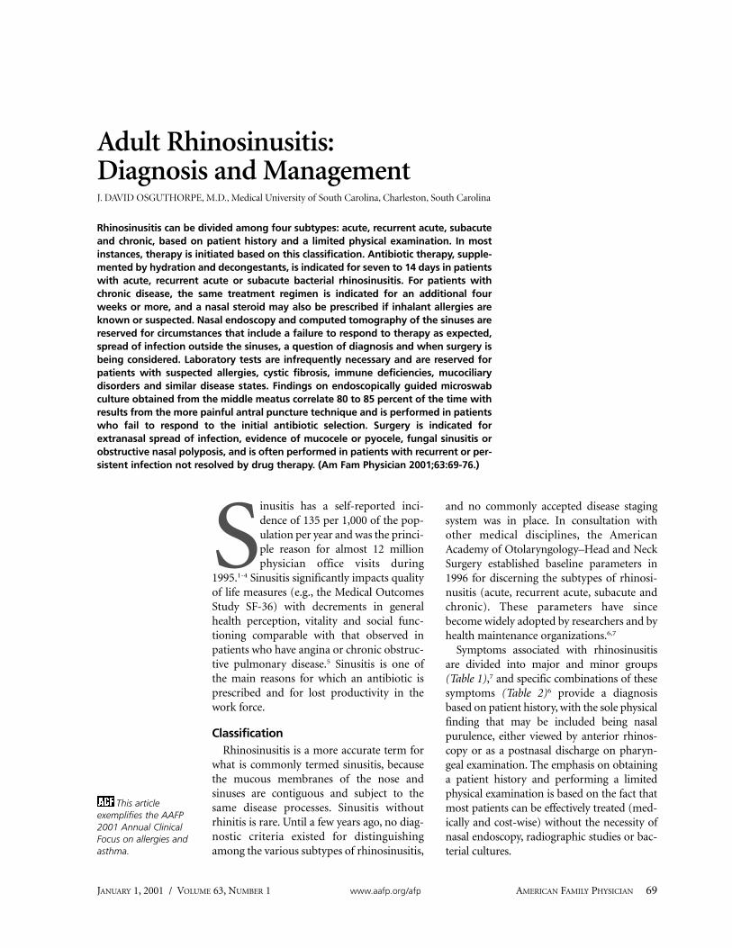
The most crucial area to examine is the hia-tus semilunaris, a crescent-shaped groove thatmeasures approximately 2 cm in length by 3 to4 mm in width and is located just lateral to theanterior third of the middle turbinate (Figure1). The frontal, maxillary and most of the eth-moid sinuses drain into this small channel. Inpatients with bacterial rhinosinusitis, pus usu-ally emanates from this area (Figure 2)and sources of obstruction (small polyps notvisible by anterior rhinoscopy, septal orturbinate abnormalities, and fungal concre-tions) should be sought. In patients who havenot responded to the initial choice of antibi-otic therapy, those with extrasinus spread ofinfection and those with chronic rhinosinusi-tis, an endoscopically directed microswabculture of the hiatus semilunaris should beobtained. Protecting the microswab from con-tamination by the nasal vibrissa using thenasal speculum or by painting the vibrissawith povidone-iodine solution yields cultureaccuracy rates of 80 to 85 percent of cultureresults obtained by the more painful, directantral puncture technique.1,2,10
Plain film radiographs of the sinuses arerarely necessary. Although such studies candisclose sinus opacification or reveal an airfluid level associated with maxillary, frontalor sphenoid disease, the most commonlyinfected area is the anterior ethmoid regionwhich is poorly visualized on plain film radi-ographs. Simple plain films may be indi-cated when the physician has questionsabout the diagnosis in a patient with anequivocal history, or to follow the responseto therapy in a hospitalized patient withsevere sinus disease. Computed tomo-graphic (CT) scan, performed in a coronalplane with cuts of 4 mm or less, is consid-ered the gold standard radiographic delin-
Rhinosinusitis
JANUARY 1, 2001 / VOLUME 63, NUMBER 1 www.aafp.org/afp AMERICAN FAMILY PHYSICIAN 71
Most cases of acute rhinosinusitis are viral in origin andsymptoms usually resolve within five to seven days.

eation of sinus disease1,9,11 (Figure 3). Mag-netic resonance imaging (MRI) is overlysensitive to the transient mucosal changesassociated with a normal nasal cycle. A false-positive rate for sinus abnormalities of up to40 percent has been reported; whereas, CTscan correlates with symptoms and nasalendoscopic findings in at least 75 percent ofpatients.2,7,11 However, the costly CT delin-eation of sinus anatomy and any associateddisease should be reserved for uncommoncircumstances of extrasinus manifestation ofinfection spread, such as that associated withpatients hospitalized for intravenous ther-apy, and the far more common circumstance
of patients with recurrent acute or chronicrhinosinusitis who have not responded toappropriate medical therapy and for whomsurgery is being contemplated.
Laboratory studies are unnecessary in theevaluation and management of most patientswith uncomplicated rhinosinusitis.1,2,10,12 Ifthe history suggests an allergic origin to sinusobstruction, an in vitro IgE screen (eight to 12regionally adjusted allergens) will identify94 to 96 percent of patients in whom aggres-sive antiallergy pharmacotherapy and/or fur-ther allergy testing and immunotherapy isneeded.2,12 When the history is equivocal, nasalcytology to detect the presence of eosinophilsassociated with allergic rhinitis or to detect theleukocytic infiltrates associated with acute bac-terial or viral infections is indicated. Tests formucociliary transport, nasal mucosal biopsiesand immunologic evaluations, for example,should be reserved for appropriately indicatedcircumstances.12
72 AMERICAN FAMILY PHYSICIAN www.aafp.org/afp VOLUME 63, NUMBER 1 / JANUARY 1, 2001
Endoscopically directed microswab cultures of the hiatussemilunaris yield a culture accuracy rate of 80 to 85 percentin cases of bacterial rhinosinusitis.
FIGURE 1. Schematic representation of the lateral wall of the nasal cavity, with the turbinatesremoved to expose the sinus ostia.
ILLU
STR
ATI
ON
BY
CH
RIS
TY K
RA
MES
Posterior ethmoid ostia
Anterior and middleethmoid ostia
Sphenoid sinus
Maxillary sinus ostium
Frontal sinus ostium
Hiatus semilunaris
Orifice of lacrimal duct
Inferior turbinate (cut)
. ...
.. .
..
.

Nasal/sinus bacterial cultures should bereserved for the circumstances previously out-lined. The most common organisms isolatedfrom patients with acute rhinosinusitis includeStreptococcus pneumoniae, Haemophilus in-fluenzae, Moraxella catarrhalis and Staphylo-coccal species.9,12-16 Staphylococcal species,especially Staphylococcus aureus, are muchmore common in chronic bacterial infections,as are anaerobes and multiple organism infec-tions. Beta-lactamase resistance, occurring inless than 30 percent of patients with acute bac-terial rhinosinusitis, increases to the 40 to 50percentile in patients with chronic disease.17,18
Allergic fungal sinusitis accounts for 2 to 4percent of patients with chronic rhinosinusitisand is suggested by characteristic findings onCT scan (Figure 4). These characteristic find-ings include opacified sinuses with inspissatedmucus containing small calcium granules.However, allergic fungal sinusitis must be con-firmed by obtaining a sinus culture (Asper-gillus, Alternaria and Bipolaris are the mostcommon fungi species). A culture can usuallybe obtained only during surgery because the
Rhinosinusitis
JANUARY 1, 2001 / VOLUME 63, NUMBER 1 www.aafp.org/afp AMERICAN FAMILY PHYSICIAN 73
FIGURE 2. Nasal endoscopic view of pus ema-nating from the hiatus semilunaris (H) locatedjust behind the uncinate process (U) in the“middle meatus” lateral to the anterior endof the middle turbinate (T) in a patient withethmoid rhinosinusitis (S = septum).
FIGURE 3. Unenhanced coronal computedtomographic scan of midface at level of hia-tus semilunaris in a patient with left maxil-lary (M) rhinosinusitis and mucosal thicken-ing in an adjacent anterior ethmoid cell (E).Note the infundibulum, (the small narrowpathway marked with small arrows bilater-ally) through which the maxillary sinus (M)drains into the hiatus semilunaris. The direc-tion of mucous drainage from the maxillarysinus through the narrow infundibulum intothe hiatus semilunaris is marked by largearrows.
FIGURE 4. Unenhanced coronal computedtomographic scan of midface at level ofhiatus semilunaris in patient with allergicfungal sinusitis; note the characteristic fun-gal concretion filling the right ethmoid (E),and the thickened mucosa and fluid in theright maxillary sinus (M). The right nasalcavity is obstructed by the reactive nasalpolyps (P), deviating the septum (S). Thesmall arrows point to the septal bone thathas been “bowed” into the contralateralnasal cavity.

fungi tend to remain within the bony confinesof the sinuses rather than extruding into thenasal cavities.15,16
TreatmentThe general goals of therapy for patients
with bacterial rhinosinusitis are to controlinfection, diminish tissue edema and reversesinus ostial obstruction so the mucopus candrain.1,10,17,19-21 Maintaining patient hydration
by adequate oral fluid intake, supplementedby the use of saline nasal sprays as desired, isrecommended. Although study results areonly suggestive, some studies8,17,19,20 used amucolytic (supersaturated potassium iodide[SSKI] solution, guaifenesin), and mostfavored the use of an oral decongestant (pseu-doephedrine [Sudafed]), for patients withsevere nasal/sinus obstruction. This can besupplemented with the addition of a topicaldecongestant (e.g., phenylephrine, oxymeta-zoline) for three to five days. Oral antibioticsare recommended for seven to 14 days inpatients with acute, recurrent acute or suba-cute bacterial rhinosinusitis1,2,10,17,22 (Figure 5).Antibiotics labeled by the U.S. Food and DrugAdministration (FDA) for the treatment ofpatients with acute rhinosinusitis includeamoxicillin-clavulanate potassium (Aug-mentin) and most of the newer generation
74 AMERICAN FAMILY PHYSICIAN www.aafp.org/afp VOLUME 63, NUMBER 1 / JANUARY 1, 2001
Antibiotic Therapy in Acute Rhinosinusitis
For community-acquired infec-tions in healthy adults, high-dose amoxicillin or folic acidinhibitor for seven to 14 days
Continue symptomatictherapy and observation.
For adults who fail to improveafter two to three days, consider broad spectrum andbeta-lactamase–resistant antibiotics for seven to 14 days.
Symptoms and signs worsen after five days,last more than 10 days or are out of proportion to those of the typical viral URI;therefore, bacterial infection is likely.
For hospitalized adults or those withchronic respiratory disease, diabetesmellitus, renal failure, immune compromise, cystic fibrosis, or thosewith recurrent acute infections treatedwith antibiotics within the previous sixto eight weeks, consider broad spectrum and beta-lactamase–resistantantibiotics for seven to 14 days.
Symptoms and signs begin to wane in five to sevendays; therefore, viral infection is likely.
Symptomatic therapy, hydration and observation
FIGURE 5. Algorithm for determining the use of antibiotic therapy in acute rhinosinusitis. (URI =upper respiratory infection)
The Author
J. DAVID OSGUTHORPE, M.D., is professor of otolaryngology and communicative sci-ences at the Medical University of South Carolina, Charleston. He received his medicaldegree from the University of Utah School of Medicine, Salt Lake City, and pursuedspecialty training at the University of California, Los Angeles.
Address correspondence to J. David Osguthorpe, M.D., Department of Otolaryngologyand Head and Neck Surgery, 207 St. Francis Annex, 150 Ashley Ave., Charleston, SC29425. Reprints are not available from the author.

cephalosporins, macrolides and fluoroquino-lones. Because the list of antibiotics changesfrequently as newer drugs are brought to themarket, up-to-date information about themost recent antibiotic releases and their indi-cations can be viewed on the FDA Center forDrug Evaluation and Research Web site athttp://www.fda.gov/cder/.
The federal Agency for Healthcare Researchand Quality (formerly the Agency for HealthCare Policy and Research) recently analyzedthe world’s literature and concluded thatamoxicillin or folate inhibitors (most com-monly trimethoprim-sulfamethoxazole [Bac-trim, Septra]) are appropriate choices as initialtherapy in an otherwise healthy adult popula-tion with uncomplicated, acute bacterial rhi-nosinusitis.17,22 Final recommendations maywell mirror the antibiotic regimen for thetreatment of patients with acute otitismedia—amoxicillin, 80 to 90 mg per kg perday as initial therapy, reserving the broaderspectrum and more expensive antibiotics forthose patients who fail to improve after threedays or whose symptoms worsen.18
In selecting an appropriate antibiotic forpatients with rhinosinusitis, physicians mustkeep in mind the incidence of drug-resistantbacteria in their community and consider thepatient’s overall health status. Special attentionshould be given to diseases that could impedenormal recovery from infection and/or predis-pose to complications (e.g., diabetes mellitus,chronic pulmonary disease, asthma, cysticfibrosis and immune deficiencies).
Some experts favor a short course of oralsteroid therapy in patients with acute (viral orbacterial) rhinosinusitis to diminish tissueedema around the sinus ostia and to providesymptomatic relief.1,2,10 This is controversial,given the potential for immunosuppressionprolonging the underlying disease process.Topical steroids are commonly used in symp-tomatic patients with subacute or chronic rhi-nosinusitis and who have or are suspected ofhaving inhalant sensitivities; however, thesepreparations are relatively ineffective in
patients with acute rhinosinusitis because theypoorly penetrate the associated rhinorrhea.
Oral antihistamines have not been docu-mented to exhibit a positive effect in patientswith rhinosinusitis and are usually avoidedbecause of the concern that the mucousthickening effects of all but the latest genera-tion of antihistamines could impede muco-pus drainage from the sinuses.1,2,10 Data onwhether topical antihistamines or anticholin-ergic preparations have a deleterious effect onthe course of bacterial rhinosinusitis areinsufficient; however, sinus penetration ofthese preparations (and, hence, an effect onmucopus drainage) is minimal. Anticholiner-gic agents are often used to control the rhin-orrhea associated with acute rhinosinusitis,which is more commonly viral than bacterialin origin.
Approximately 175,000 persons undergo si-nus surgery in the United States each year.1,23,24
The absolute indications for surgery includeextrasinus spread of sinus infection, mucoceleor pyocele, fungal sinusitis or massive nasalpolyps that obstruct the sinuses. The relativeindications for surgery that constitute the rea-sons for a preponderance of sinus operationsinclude: (1) recurrent acute bacterial rhinosi-nusitis in which a persisting area of obstruc-tion to sinus aeration or of recurring diseasehas been identified by nasal endoscopy and/orCT; or (2) chronic rhinosinusitis that hasfailed to resolve after an appropriate course ofmedical therapy as documented by thepatient’s symptoms or findings on nasalendoscopy and/or CT scan. The long-termsuccess of any particular sinus surgerydepends on the cause of the original disease.
At least 93 percent of patients who developchronic bacterial suprainfection from a bout of
Rhinosinusitis
JANUARY 1, 2001 / VOLUME 63, NUMBER 1 www.aafp.org/afp AMERICAN FAMILY PHYSICIAN 75
Beta-lactamase resistance occurs more frequently withchronic rhinosinusitis than with acute bacterial rhinosinusitis,but is relatively common in both.

Rhinosinusitis
influenza and who have no other predisposingfactors for sinus disease achieve alleviation ofsymptoms with a single surgical interven-tion.24,25 In patients with significant predispos-ing factors (e.g., inhalant sensitivities, polyps,asthma or continuing tobacco use), the surgi-cal success rate is 80 to 85 percent but dimin-ishes to less than 50 percent in patients withaspirin triad, immune deficiencies, cysticfibrosis or allergic fungal sinusitis.24 For sinussurgery taken as a whole, the surgical revisionrate is 18 to 20 percent with a 0.2 to 0.6 serioussurgical complication percentage.24
REFERENCES
1. Kaliner MA, Osguthorpe JD, Fireman P, Anon J,Georgitis J, Davis ML, et al. Sinusitis: bench to bed-side. Current findings, future directions. J AllergyClin Immunol 1997;99(6 pt 3):S829-48 [Publishederratum appears in J Allergy Clin Immunol 1997;100:510].
2. Osguthorpe JD, Hadley JA. Rhinosinusitis: currentconcepts in diagnosis and management. Med ClinNorth Am 1999;83:27-42.
3. United States Department of Health and HumanServices, Public Health Service, Agency for HealthCare Policy and Research, Center for GeneralHealth Services Intramural Research. Annual ex-penses and sources of payment for health care ser-vices. Washington D.C.: Government PrintingOffice, 1994; AHCPR publication 93-0007.
4. Benson V, Marano M. Current estimates from theNational Health Interview Survey, 1993. VitalHealth Stat 1994;10:1-221.
5. Gliklich R, Metsen R. The health impact of chronicsinusitis in patients seeking otolaryngologic care.Otolaryngol Head Neck Surg 1995;113:104-9.
6. Lanza D, Kennedy DW. Adult rhinosinusitisdefined. Otolaryngol Head Neck Surg 1997;117 (3 pt 2):S1-7.
7. Hadley JA, Schaefer SD. Clinical evaluation of rhi-nosinusitis: history and physical examination. Oto-laryngol Head Neck Surg 1997;117(3 pt 2):S8-11.
8. Fergusen BJ, Johnson JT. Allergic rhinitis and rhinos-inusitis. Is there a connection between allergy andinfection? Postgrad Med 1999;105:55-8,61,64.
9. Settipane RA. Complications of allergic rhinitis.Allergy Asthma Proc 1999;20:209-13.
10. Benninger MS, Anon J, Mabry RL. The medicalmanagement of rhinosinusitis. Otolaryngol HeadNeck Surg 1997;117(3 pt 2):S41-9.
11. Zinreich S. Rhinosinusitis: radiologic diagnosis. Oto-larygnol Head Neck Surg 1997;117(3 pt 2):S27-34.
12. Fergusen BJ, Mabry RL. Laboratory diagnosis. Oto-laryngol Head Neck Surg 1997;117(3 pt 2):S12-6.
13. Brook I. Microbiology and management of sinusi-tis. J Otolaryngol 1996;25:249-56.
14. Gwaltney JM. Acute community-acquired sinusitis.Clin Infect Dis 1996;23:1209-23.
15. Nadel DM, Lanza DC, Kennedy DW. Endoscopicallyguided cultures in chronic sinusitis. Am J Rhinol1998;12:233-41.
16. Corey JP, Delsupehe KG, Ferguson BJ. Allergic fun-gal sinusitis: allergic, infectious, or both? Otolaryn-gol Head Neck Surg 1995;113:110-9.
17. Benninger MS, Sedory Holzer SE, Lau J. Diagnosisand treatment of uncomplicated acute bacterialrhinosinusitis: summary of the Agency for HealthCare Policy and Research evidence-based report.Otolaryngol Head Neck Surg 2000;122:1-7.
18. Antimicrobial treatment guidelines for acute bacte-rial rhinosinusitis. Sinus and Allergy Health Partner-ship. Otolaryngol Head Neck Surg 1997;117:S50-2.
19. Evans KL. Diagnosis and management of sinusitis.BMJ 1994;309:1415-22.
20. Wagner W. Changing diagnostic and treatmentstrategies for chronic sinusitis. Cleve Clin J Med1996;63:396-405.
21. Fagnan LJ. Acute sinusitis: a cost-effective ap-proach to diagnosis and treatment. Am Fam Physi-cian 1998;58:1795-802,805-6.
22. Klein JO. Review of consensus reports on manage-ment of acute otitis media. Pediatr Infect Dis J1999;18:1152-5.
23. Anand VK, Osguthorpe JD, Rice D. Surgical man-agement of adult rhinosinusitis. Otolaryngol HeadNeck Surg 1997;117(3 pt 2):S50-2.
24. Osguthorpe JD. Surgical outcomes in rhinosinusitis:what we know. Otolaryngol Head Neck Surg1999;120:451-3.
25. Kennedy DW. A 48-year-old man with recurrentsinusitis. JAMA 2000;283:2143-50.
76 AMERICAN FAMILY PHYSICIAN www.aafp.org/afp VOLUME 63, NUMBER 1 / JANUARY 1, 2001