Adrenaline and Stress-Induced Cardiomyopathies: Three Competing … · 2019-05-06 · Adrenaline...
36
In: Adrenaline ISBN: 978-1-63321-084-4 Editor: Alfred Bennun © 2014 Nova Science Publishers, Inc. Chapter 4 Adrenaline and Stress-Induced Cardiomyopathies: Three Competing Hypotheses for Mechanism(s) of Action Candice N. Baker, Rebekah Katsandris, Chaunhi Van and Steven N. Ebert Burnett School of Biomedical Sciences, College of Medicine, University of Central Florida, Orlando, FL, US Abstract Stress-induced cardiomyopathies such as Tako-Tsubo Syndrome (also known as “Broken-Heart Syndrome”) primarily affect post- menopausal women who have experienced a sudden emotional shock. Clinical presentation includes symptoms mimicking a myocardial infarction (severe chest pain and S-T elevation on EKG), but do not show significant occlusion of the coronary arteries. Instead, patients display left ventricular (LV) dysfunction characterized by hypo- or a-kinetic regions that appear to ―balloon-out‖, particularly in the region near the apex, and thus is sometimes referred to as “Apical-Ballooning Syndrome”. Some patients have been reported with high circulating levels of adrenaline, and symptoms have been effectively managed in many cases by treatment with beta-adrenergic receptor blockers. It is not entirely clear, however, why only specific regions of the LV were affected in these patients, nor is No part of this digital document may be reproduced, stored in a retrieval system or transmitted commercially in any form or by any means. The publisher has taken reasonable care in the preparation of this digital document, but makes no expressed or implied warranty of any kind and assumes no responsibility for any errors or omissions. No liability is assumed for incidental or consequential damages in connection with or arising out of information contained herein. This digital document is sold with the clear understanding that the publisher is not engaged in rendering legal, medical or any other professional services.
Transcript of Adrenaline and Stress-Induced Cardiomyopathies: Three Competing … · 2019-05-06 · Adrenaline...
In: Adrenaline ISBN: 978-1-63321-084-4
Editor: Alfred Bennun © 2014 Nova Science Publishers, Inc.
Chapter 4
Adrenaline and Stress-Induced Cardiomyopathies: Three
Competing Hypotheses for
Mechanism(s) of Action
Candice N. Baker, Rebekah Katsandris, Chaunhi Van
and Steven N. Ebert Burnett School of Biomedical Sciences, College of Medicine,
University of Central Florida, Orlando, FL, US
Abstract
Stress-induced cardiomyopathies such as Tako-Tsubo Syndrome
(also known as “Broken-Heart Syndrome”) primarily affect post-
menopausal women who have experienced a sudden emotional shock.
Clinical presentation includes symptoms mimicking a myocardial
infarction (severe chest pain and S-T elevation on EKG), but do not show
significant occlusion of the coronary arteries. Instead, patients display left
ventricular (LV) dysfunction characterized by hypo- or a-kinetic regions
that appear to ―balloon-out‖, particularly in the region near the apex, and
thus is sometimes referred to as “Apical-Ballooning Syndrome”. Some
patients have been reported with high circulating levels of adrenaline, and
symptoms have been effectively managed in many cases by treatment
with beta-adrenergic receptor blockers. It is not entirely clear, however,
why only specific regions of the LV were affected in these patients, nor is
No part of this digital document may be reproduced, stored in a retrieval system or transmitted commercially in any form or by any means. The publisher has taken reasonable care in the preparation of this digital document, but makes no expressed or implied warranty of any kind and assumes no responsibility for any errors or omissions. No liability is assumed for incidental or consequential damages in connection with or arising out of information contained herein. This digital document is sold with the clear understanding that the publisher is not engaged in rendering legal, medical or any other professional services.

Candice Baker, Rebekah Katsandris, Chaunhi Van et al. 82
it understood why postmenopausal women are so susceptible relative to
the rest of the population. With respect to the first question, we will
review three competing, though not necessarily mutually exclusive,
hypotheses to explain how adrenaline plays a key role in precipitating
stress-induced cardiomyopathies. The first of these is a vascular
microspasm hypothesis which focuses on stress-induced changes in the
coronary microvasculature feeding the LV, leading to microspasms,
interrupted regional blood-flow, and corresponding myocardial
dysfunction in affected areas of the LV (Sato et al., 1990 and Dote et al.,
J Cardiol 1991;21:203) [6, 7]. The second hypothesis will be referred to
as the differential β-receptor expression hypothesis, which postulates
there is a higher density of β-adrenergic, (especially β2-adrenergic)
receptors in the apical region of the LV compared to other regions,
thereby making it more sensitive to adrenergic overload and myocardial
stunning due to agonist-mediated switch from Gs to Gi coupling of the
β2-adrenergic receptors in this region relative to other regions of the heart
(Lyon et al., Nature Clin Pract Cardiovasc Med 2008;5:22) [10]. A third
hypothesis focuses on differential local production of adrenergic
hormones within the left myocardium itself (Kume et al., Circ J 2008;
72:106 and Osuala et al., PLoS One 2011;8:e22811) [1, 3]. From this
third hypothesis, selective myocardial stunning in the LV results from
local overload of adrenergic stimulation due to autocrine/paracrine
actions of adrenaline (epinephrine) and noradrenaline (norepinephrine) in
addition to sympathetic stimulation and circulating catecholamines in
periods of stress. The evidence for each hypothesis is critically evaluated,
with discussion of potential future directions for work in this field in
relation to the role of gender (sex), age, and menopausal status.
Introduction
The impacts of emotional stress on health have been generally recognized
since ancient times, but only in relatively recent years have scientists and
clinicians started to gain an understanding of the pathophysiological
mechanisms linking emotional stress to specific dysfunctions within the
cardiovascular system. In contrast, biological manifestations of physical stress
are much more extensively characterized, and broadly include hypertension,
ischemic heart disease (including myocardial infarction and its consequences),
myocarditis, cardiomyopathies, heart failure, arrhythmias, and sudden cardiac
death [11]. While emotional stress may also contribute to these conditions, the
specific pathophysiologic manifestations of emotional stress are only
beginning to emerge.

Adrenaline and Stress-Induced Cardiomyopathies 83
Takotsubo Syndrome is a prototypical stress-induced cardiomyopathy.
The first clinical cases of this stress cardiomyopathy were reported in Japan in
1990 [7]. Early reports described a transient hypokinesis or akinesis in the
apex of the LV in patients suffering from acute emotional stress [12-14]. The
unusual shape of the heart in these patients bore a striking resemblance to a
Japanese octopus trap, and hence, it was named ―takotsubo‖, meaning octopus
trap in Japanese [6, 7, 13] (Figure 1). Takotsubo Syndrome is also sometimes
referred to as ―Broken Heart Syndrome‖ (due to the emotional stress
connection), ―Ampulla Cardiomyopathy‖ (due to the peculiar shape of the
heart), or ―Apical Ballooning Syndrome‖ (to describe one of the most
pronounced clinical features commonly observed in these patients) [14].
Takotsubo Syndrome is also referred to as a ―Stress-Induced Cardiomyopathy‖
or more simply, ―Stress Cardiomyopathy‖. These terms have become
essentially interchangeable when describing this syndrome [15]. For the
purpose of this review, we will use the historical ―Takotsubo Cardiomyopathy
(TTC)‖ designation as the prototype clinical form of stress-induced
cardiomyopathies. It is important to note, however, that stress induction for
TTC need not be exclusively emotional or psychological, but can also be
precipitated by physical stressors such as pharmacological challenge with
adrenaline and other adrenergic agonists [16, 17], complications from other
diseases (e.g., pheochromocytoma) [18, 19], physical trauma from injury or
surgery, and a wide variety of other physical stressors [20, 21].
Figure 1. X-ray image of a heart showing typical apical ballooning during systole in a
TTC patient. This characteristic shape resembles the shape of clay pots used in Japan
to trap octopus. The first reported case was in Japan and the investigators named this
peculiar condition ―Tako-Tsubo‖, which is Japanese for octopus trap. Reprinted with
permission from the Portuguese Journal of Cardiology [8], Copyright Elsevier (2012).

Candice Baker, Rebekah Katsandris, Chaunhi Van et al. 84
The common feature of both physical and psychological stress responses
in these contexts is elevated plasma catecholamines [1, 22-24]. Indeed, studies
examining plasma catecholamine concentrations in TTC patients have shown
high levels of circulating catecholamines compared to those from control
groups [22, 25]. The fact that exogenously administered catecholamines can
induce TTC-like symptoms in patients and animal models further supports
adrenergic mediation of this syndrome [16, 26-28]. As mentioned above, TTC
symptoms have also been reported in some patients with pheochromocytoma,
which results in abnormally high concentrations of circulating endogenous
catecholamines secreted from adrenal medullary tumors [29]. TTC symptoms
have responded favorably to pharmacological intervention with beta-blockers
in some cases [12]. Taken together, these observations strongly implicate the
major peripheral catecholamines, adrenaline and noradrenaline, as key players
in acute precipitation of TTC.
The central objective of this review is to critically evaluate three distinct
hypotheses put forward to describe the pathophysiology and mechanism of the
clinical phenotypes associated with TTC. For the sake of simplicity, we have
grouped the three main hypotheses into general categories that will be referred
to as (i) Vascular Microspams [6, 22], (ii) Differential β-adrenergic receptor
distribution [2, 10], and (iii) Differential Regional Adrenergic Stimulation [1,
3]. We define and discuss each of these hypotheses in separate sections that
follow, but first briefly describe the main clinical characteristics, treatment and
history of TTC.
Clinical Presentation
TTC is commonly misdiagnosed as Acute Coronary Syndrome (ACS) or
ST-segment elevation myocardial infarction (STEMI) due to similarities in
their clinical presentation [30, 31]. As a result of growing awareness of this
issue, the United States and international guidelines include TTC as a
differential diagnosis for ACS [32]. Ischemic chest pain and dyspnea are the
two most common presenting symptoms of TTC [33]. Other symptoms
reported include palpitations, syncope, shock, respiratory arrest, abdominal
pain, myalgia, aphasia, ataxia, and sudden death [33-36].
One of the main clinical features of TTC is akinesis or dyskinesis of the
LV. Originally, akinesis of the apex was described as causing an ―apical-
ballooning‖ phenomena during systole. The apical wall motion abnormality is
considered the ―typical‖ TTC variant but other morphologies have been
described (Figure 2) [4]. In the mid-ventricular variant, for example, the areas

Adrenaline and Stress-Induced Cardiomyopathies 85
around the mid-ventricles do not contract adequately during systole. A reverse
TTC variant affects the base of heart instead of the apex [4]. During systole,
the basal akinesis or hypokinesis cause an apparent ―ballooning‖ of the
myocardium at the base because the rest of the ventricle contracts as usual, but
the affected areas do not and, hence, remain in relaxed condition during
contraction, which gives the appearance of ballooning during systole when
viewed using in vivo imaging techniques such as angiography,
echocardiography, and magnetic resonance imaging (MRI). Other localized
variants affect limited regions of the LV and cannot be categorized into one of
the three previously described morphologies [4]. These areas of wall motion
abnormalities are not within the distribution of a single coronary artery [12].
Figure 2. Different forms of TTC. The most commonly reported form is the Takotsubo
type. Reprinted with permission from Journal of Cardiology [4]. Copyright, Elsevier
(2006).
Many TTC patients present with a low left ventricular ejection fraction
[37]. One review of case studies found that mean ejection fraction in TTC
patients ranged from 20-49% compared with the normal range of 60-76%
upon follow-up [38]. EKG changes were reported in a many cases. ST-
segment elevation was the most common EKG abnormality followed by T-
wave inversion [33, 39]. Other changes seen less frequently are ST-segment
depression, peaked T-wave, flattened T-waves, QT-prolongation, and Q-waves
[6, 12, 36, 39]. These changes are transient and usually resolve within a few
months. Cardiac enzymes, such as troponins, creatine kinase, and brain
natriuretic peptide (BNP), are usually elevated [33, 39]. One review of case
studies found 86% of patients had elevated troponins [38].
Another literature review found only 10.7% of patients had normal or
absent enzymes [39]. EKG changes in TTC cannot be readily distinguished
from EKG changes found during a myocardial infarction (MI) in the absence
Figure 2. Different forms of TTC. The most commonly
reported form is the Takotsubo type. Reprinted with
permission from Journal of Cardiology [4] -pending.

Candice Baker, Rebekah Katsandris, Chaunhi Van et al. 86
of coronary angiography. In some cases, however, cardiac enzymes in TTC
can be differentiated from MI by the amount detected with milder elevations in
enzymes seen in TTC compared to an MI [39].
Another distinction of TTC is a preceding stress-related event, though this
may not be the sole distinguishing feature since stress has also been reported
to trigger MI and other cardiovascular events [40].
Physical and emotional stress such as death of a loved one, public
speaking, motor vehicle accidents, arguments, natural disasters, alcohol, and
surprise parties have been reported before the patient experiences symptoms of
TTC [12, 13, 41]. TTC patients have a higher prevalence of anxiety disorders,
which may predispose them to developing this disease [36]. Medical
procedures, acute medical illnesses, and opiate withdrawal can also trigger the
cardiomyopathy [12, 35].
Specific pathophysiological mechanisms leading to TTC are still a matter
of debate, though there has been a suggestion of an underlying genetic
component since only a subset of postmenopausal women are susceptible and
because familial cases have been reported [42, 43]. The American Heart
Association, however, has classified TTC as a primary acquired
cardiomyopathy [44], which deemphasizes the genetic component of this
disease.
Formal diagnostic criteria for TTC have been proposed [36]. Of these, the
Mayo Clinic diagnostic criteria appear to be the most widely accepted [45].
These criteria are defined as follows:
1. ―Transient hypokinesis, akinesis, or dyskinesis of the left ventricular
mid segments with or without apical involvement. The regional wall-
motion abnormalities extend beyond a single epicardial vascular
distribution.
2. Absence of obstructive coronary disease or angiographic evidence of
acute plaque rupture.
3. New ECG abnormalities (either ST-segment elevation and/or T –wave
inversion) or elevated cardiac troponin.
4. Absence of:
Recent significant head trauma
Intracranial bleeding
Pheochromocytoma
Myocarditis
Hypertrophic cardiomyopathy‖

Adrenaline and Stress-Induced Cardiomyopathies 87
Diagnostic Evaluation
The ―gold-standard‖ for diagnosing TTC is coronary angiography and left
ventriculography [46]. Cardiac catheterization and angiography is helpful in
determining ejection fraction and stenosis of vessels. As the criteria proposed
by Prasad states, TTC patients typically do not have coronary artery disease, or
if there is mild to moderate atherosclerosis it cannot account for the wall
motion abnormalities [12, 45, 47]. If severe stenosis is present and there is
evidence of coronary artery disease (CAD), acute coronary syndrome will
more likely be an appropriate diagnosis. Cardiac catheterization has also found
vessel spasms and aberrant coronary microcirculatory function at the time of
presentation. The significance of the spasm and abnormal circulation has not
been determined [12, 39]. Left ventriculography allows visualization of wall
motion abnormalities [35, 38, 48]. EKG and cardiac enzymes levels should be
obtained in patients describing ischemic chest pain to rule out major cardiac
conditions such as ACS, STEMI, and arrhythmias. Since one of the diagnostic
criterion for TTC is transient changes in EKG and cardiac enzymes, it is also
important to perform these tests to establish a baseline and observe any
changes for diagnosis [12].
In an emergency setting, echocardiograms are the preferred imaging
modality. Echocardiograms are non-invasive and can assess potential
complications. Similar to the left ventriculograph, it shows apical ballooning
or other dyskinesis of the ventricular walls [38, 46, 48]. Cardiac magnetic
resonance imaging (CMRI) with gadolinium contrast can help to discriminate
between ACS, myocarditis, and TTC in obscure cases, such as those with
elevated cardiac enzymes and EKG changes [46]. In TTC, CMRI will not
show contrast enhancement whereas ACS and myocarditis will, due to
myocardial edema [49]. CMRI is also useful in detecting a thrombus missed
on echocardiogram [50].
Complications and Prognosis
One retrospective data analysis looked at the clinical course of 107 TTC
patients and analyzed their cardiac complications [51]. Cardiac complications
found in these patients included left ventricular outflow obstruction, mitral
regurgitation, pericardial effusion, coronary artery stenosis (that did not
account for the abnormal wall motion abnormalities), atrial fibrillation, atrial
flutter, cardiac death, pump failure, ventricular tachycardia, ventricular

Candice Baker, Rebekah Katsandris, Chaunhi Van et al. 88
fibrillation, and atrioventricular (AV) block [51]. Apical thrombus is another
cardiac complication that has been reported in a different study. Thrombi form
in 2.5-9% and thromboemboli form in 0.8-14% of cases and are thought to be
due to hypokinesis or stasis of the left ventricle. Thromboembolism can also
lead to stroke and neurological deficits [36]. Cardiogenic shock and
thromboembolism are the most common causes of death in these cases [33].
Rupture of the septum, free wall, and papillary muscles can also occur [12,
36]. In one-third of patients right ventricle dysfunction as well as LV is
observed [12]. These patients have a worse prognosis with more severe heart
failure, longer hospital stays, and greater hemodynamic instability [12]. Non-
cardiac complications include pulmonary edema, found in 0-44% of cases, and
pneumothorax, which is rare [33, 52].
TTC is transient and has a relatively good outcome with low in-hospital
mortality. Recovery of left ventricular function and ejection fraction occurs
over days to months. Mean ejection fraction on follow-up ranged from 60-76%
[13, 38]. Prognosis appears to be dependent on ventricular function [36]. T-
wave inversion, elevated white blood cells (WBC), BNP, and c-reactive
protein (CRP) has been associated with poor outcome [36, 51]. Recurrence
rate is 2-10% within the first few years.
Treatment
There has yet to be a clinical trial for treatment of TTC; therefore, no
standardized treatment protocol is currently in place. Current practice is thus
highly variable, but appears to mostly be aimed at supportive and preventative
care. For example, in patients that were hemodynamically unstable, intra-
aortic balloon counterpulsation, fluids, beta-blockers or vasopressors have
been used [53]. Anticoagulation is recommended to reduce risk of thrombus
formation and embolization. Echocardiograms should be performed if heart
failure is worsening, before discharge, and on follow-up if left ventricular
function had not normalized at discharge [12, 36].
Epidemiology and Prevalence
A disproportionate number of patients affected by TTC are
postmenopausal women [12, 52, 54]. According to one review, 82-100% of
patients affected by TTC were women [12]. The average age of these patients

Adrenaline and Stress-Induced Cardiomyopathies 89
ranged from 61-76 years, and only a small percentage (2.7-3%) were under the
age of 50 [36, 38, 52]. Reported cases, however, ranged from 10 to 91 years
old [12, 38]. In cases that reported race, the majority of patients diagnosed
were Asian women followed by Caucasian women [33]. Caucasians more
commonly had T-wave inversion on EKG, were younger than their Asian
counterparts, presented with chest pain, and were more sensitive to emotional
stress. While Asians tended to have ST-elevations, were older (70.4 years old),
less likely to have preceding stress, and had greater mortality rate [33].
Although early cases of TTC were reported predominantly in Japanese
literature, cases have been reported worldwide [12, 33]. It is estimated that
approximately two percent of patients diagnosed with ACS may actually be
patients with TTC cardiomyopathy [55]. One study following intensive care
unit patients admitted for non-cardiac diagnosis found 26 of 92 patients had
decreased ejection fraction, and increased incidence of apical ballooning [56].
Role of Sex (Gender), Age, and Hormonal Status
As mentioned above, TTC is predominately found in female patients who
have undergone menopause [33]. In conglomerate, it has been estimated that
approximately 90% of documented TTC cases were postmenopausal women
[12, 37, 48, 50, 57-60]. Perhaps not surprisingly, this has led researchers to
suspect a connection between female hormone status and TTC susceptibility
[61-65]. Ovarian failure, when follicles in the ovaries have been eradicated
and the female can no longer enter ovulation, is a characteristic sign of
menopause. Due to the lack of follicles the female can no longer produce
estrogen and progesterone [66].
Studies in animal models have suggested that estrogen may provide some
protection from TTC [67, 68]. For example, estrogen supplementation to
ovariectomized rats has been shown to attenuate some of the symptoms
associated with TTC [63-65, 67, 68]. So far, all of these studies have been
conducted in rats by the Ueyama group. Some primate studies have been done
to examine TTC, however, these did not evaluate the role of gender or female
sex hormones (only male cynomoglus monkeys were used) [28]. Thus, there is
a need for additional animal model studies to help elucidate mechanisms.
Primates are expensive and there are many ethical concerns about their use for
stress research. Alternative large animal models such as pigs, dogs, or sheep
may prove useful, but to the best of our knowledge, these have not yet been
developed for TTC. Mid-sized animal models like the rabbit have proved
Candice Baker, Rebekah Katsandris, Chaunhi Van et al. 90
useful for examining gender-related cardiovascular differences [69-73]. Mice
may also serve as an attractive complement to rats as a TTC model since
genetic components can be relatively easily manipulated in this model. Several
studies have shown that estrogen has significant influence over cardiac ion
channels and function in this model [74-77]. Although the effects of gender
and estrogen have not yet been carefully examined in the mouse model, Shao
et al. [27] have recently described TTC-like induction in mice following
administration of a single high dose of the beta-agonist, isoproterenol, and
have made the novel observation that lipotoxicity may be a contributing factor,
though the doses of isoproterenol required to induce TTC were extraordinarily
high. This caveat notwithstanding, there has been a reported association
between hyperlipidemia as a risk factor for TTC in human patients [78], and
the observed lipotoxicity in this mouse model may aid our understanding of
how dysregulation of lipid metabolism may affect TTC outcomes. Further
development of these and related animal models should ultimately prove
useful for elucidating the underlying mechanisms leading to the onset and
severity of TTC.
In contrast to the paucity of animal model data, there have been many
retrospective clinical studies on TTC and gender differences [79]. According
to one study, 93.5% of the patients diagnosed with TTC were female [33],
though the authors noted that once a TTC attack has taken place, the clinical
outcome appeared similar regardless of gender [33]. Male and female patients
displayed similar complications during TTC [79]. The differences concerning
TTC seem to take place before an attack occurs, and less so afterwards [80],
though this has not been uniformly established due to lack of carefully
controlled studies. Retrospective studies also suffer from relatively few male
patients for comparison. Nevertheless, the mortality rates measured between
genders showed no significant differences despite the fact there were certainly
individual differences from both gender categories [80]. Clearly, additional
research is needed to determine the true impact and consequences of TTC in
males relative to females.
A study done on TTC compared differences between male and female
patients [21]. There were no differences in the psychiatric health of the patients
for this study. The psychiatric health parameters reviewed included anxiety,
migraines, and depression. Despite this, males and females appeared to have
different triggers for TTC. Male patients with TTC previously had some type
of physical stress stimulus (surgery, embolism, or pancreatitis) [21]. On the
other hand, female TTC patients usually experienced a specific emotional
event (death of relative, divorce, or notice of debts) in their lives [33]. Another

Adrenaline and Stress-Induced Cardiomyopathies 91
interesting note made through the study was the difference between treatment
regime when comparing males and females. Males presented with lower
ejection fractions than females affected by TTC. This influenced the reason(s)
males had an increase in mechanical ventilation (replaces or assistance in
natural respiration) as a treatment option during the attack. A review of 224
TTC patients in the United States indicated that male patients were able to
endure longer amounts of time with increased catecholamines before
developing TTC, while female patients developed TTC much more quickly
brought on by an apparent surge of circulating catecholamines [21].
An anatomical difference between males and females relating to TTC is
the size of the LV [33]. Males have a larger LV than females on average. This
size difference could be one reason that females are more affected by TTC
than males. When catecholamines increase in circulation the female LV is
more prone to an outflow tract obstruction than the male LV. This could cause
the base of the LV to hypercontract resulting in left ventricular outflow tract
(LVOT) obstruction. It remains to be determined how prevalent these issues
are in males and females since LVOT obstructions have been documented in
only a very small number of TTC cases to date [33].
To summarize this section, there is strong clinical evidence showing the
vast majority of TTC cases are postmenopausal women. Men and
premenopausal women tend to have different precipitating factors compared to
postmenopausal women who typically had specific emotional stressors that
triggered the TTC episode(s). Hormonal status is a likely risk factor for TTC,
and some studies have shown correlations between declining female sex
steroid hormone levels and TTC incidence. This hypothesis is supported from
animal model (rat) data showing that estrogen can attenuate TTC. Despite
much progress and attention in recent years, there remain many mysteries
regarding the roles of biological sex and sex steroid hormones with respect to
TTC. Some of the key unanswered questions include the following:
1. Does the loss of estrogen and/or progesterone truly lead to increased
TTC susceptibility?
2. Conversely, can estrogen and/or progesterone provide protection from
TTC?
3. Does testosterone provide protection from TTC?
4. How do sex steroid hormones influence TTC susceptibility? (i.e.,
what are the targets?)
5. Are TTC ―triggers‖ truly more emotionally-induced in
postmenopausal women compared to men or premenopausal women?

Candice Baker, Rebekah Katsandris, Chaunhi Van et al. 92
6. Are age and sex (gender) independent risk factors (e.g., from
menopausal status) for TTC?
Answers to these important questions will require additional research.
New and improved preclinical animal models of TTC would also help to
address these issues. Most clinical studies focused on TTC to date have all
been retrospective, which is a current limitation of our knowledge in this area.
In silico models hold promise for testing TTC mechanistic hypotheses, but
these work best in conjunction with patient data and/or relevant experimental
data from animal models. Aside from the issues of risk factors as they relate to
sex, menopausal status, and emotional triggers, there are even more
compelling unanswered questions regarding the acute pathophysiological
mechanisms responsible for the peculiar cardiovascular effects seen in TTC
patients. In the following section, we critically examine three distinct
competing hypotheses that have been put forward to explain the biological
basis for TTC.
Hypothesis I: Vascular Microspasms
The pathogenesis of TTC has been debated since the description of this
unique cardiovascular condition. One early hypothesis has become
controversial since it was first proposed by Sato et al., 1990 [7] stating
microvasculature dysfunction is the precipitating factor that causes TTC [6]. In
this study they showed spontaneous multi-vessel spasms in two patients, and
another two showed similar symptoms following an ergonovine provocation
test [6]. Ergonovine, as well as acetylcholine (ACh) provocation tests, are
commonly performed to diagnose patients with coronary artery spasms that
present without occlusion of coronary arteries [81]. Briefly, the tests are
performed by administering graded doses (25-100 µg) of the drug into the left
coronary artery and then the right coronary artery. If no chest pain, EKG
change, or coronary spasm is observed, the dose is increased until the
maximum dose (100 µg) is administered, followed by infusion with
nitroglycerin to ameliorate symptoms. Coronary spasm is typically defined by
reduction of the epicardial arteries by greater than 75% (Figure 3) [82].
After the initial description of coronary spasms in the affected areas of
TTC multiple case studies were published supporting this hypothesis. One
such study produced vascular spasms in 10 of 14 patients, with four patients
experiencing single epicardial coronary spasms and six experiencing
multivessel coronary spasms [60]. Using myocardial contrast
echocardiography, 11 of 14 patients were assessed for and diagnosed with a

Adrenaline and Stress-Induced Cardiomyopathies 93
myocardial perfusion defect within the left ventricular apical myocardium that
was ameliorated by the infusion of adenosine [83].
Figure 3. Cartoon depiction of Ach-induced coronary vasospasm test showing how it
may lead to apical (P1) or mid (P2) ventricular ballooning. Reprinted with permission
from Texas Heart Institute Journal [9]. Copyright 2010 by the Texas Heart Institute,
Houston..
Other methods used to evaluate coronary vascular dysfunction include
Thrombolysis in Myocardial Infarction (TIMI) flow grade. This methods
utilizes the number of cineframes it takes for a dye to reach a location during
angiography [84]. In a case study of patients presenting with TTC at the Mayo
Clinic, Rochester, MN (Jan 2002-Dec 2003) all 16 patients had larger mean
TIMI frame counts in the left anterior descending, left circumflex and right
coronary arteries than matched controls (p less 0.001 for each) [85]. Similarly,
a study including 28 patients showed significantly higher TIMI frame counts
in all three arteries when evaluated with a coronary angiogram [59]. Moreover,
assessment of the TIMI myocardial perfusion grade (TMPG), dysregulation of
perfusion was measured in 29 of 42 patients (69%) in a retrospective study
conducted at Mayo Clinic, Rochester, MN [86].
Using an alternative method to evaluate coronary microvascular function,
coronary flow reserve (CFR) quantification was obtained during transthoracic
doppler echocardiography in 20 consecutive patients [87]. It was found that
CFR increased by a mean of 40% in patients between the acute phase of TTC
and recovery phase [87]. Not unlike CFR, coronary flow velocity reserve
(CFVR) was measured using a doppler guidewire in patients in the acute phase
Candice Baker, Rebekah Katsandris, Chaunhi Van et al. 94
of TTC and at follow-up. In all patients (n=8) CFVR was increased in the
three coronary arteries at follow-up appointments as compared to initial
presentation [88].
Although these early studies proposed coronary microvascular spasms as
an underlying pathophysiological mechanism as the cause for TTC, many
subsequent reports have failed to provide substantial supporting evidence for
this hypothesis. One example of this was observed when only three of 212
(1.4%) patients examined demonstrated spontaneous multivessel epicardial
spasms, and a mere 24 of 84 (28.6%) demonstrated multivessel epicardial
spasms even after a provocation test (ergonovine and/or acetylcholine) [38].
Abe et al evaluated coronary microvascular spasms using acetylcholine
provocation testing, however, of the seven patients evaluated only one patient
showed coronary vasospasm and four demonstrated diffuse vasoconstriction.
This led the group to conclude that, ―coronary vasospasm does not contribute
to the etiology‖ of TTC [89]. Others have repeated provocation tests with
similar results, demonstrating only two of seven patients experienced coronary
spasms and a more drastic study showed zero of 47 cases displayed
vasospasms in the epicardial coronary arteries [80, 90].
These contradicting reports have dampened early enthusiasm supporting
the hypothesis of microvascular coronary spasms as the pathogenesis of the
observed apical ballooning. While this hypothesis was the prominent etiology
most favored initially, it has declined in popularity due to discrepancies
between studies indicating that coronary vasospasms may not be the most
probable mechanism for the induction of TTC. This does not mean, however,
that coronary microspasms are not contributory in TTC. It is nearly impossible
to continuously record and capture early triggering events in patients, and so it
is also not possible to say for certain that vasospasms do not occur in most
cases. Consequently, the vasospasm hypothesis remains viable despite its
apparent decline in popularity. Further work is required to determine the extent
to which vasospasms may or may not underlie TTC mechanisms.
Hypothesis II: Differential Regional β2-receptor Expression and
Signaling (Gs/Gi Switch)
An alternative theory to the pathogenesis of TTC proposed that increased
expression and stimulation of β2-adrenergic receptors in the left ventricular
apex as compared to right ventricle and the basal portions of the heart may
lead to selective myocardial stunning [10]. This hypothesis is supported, in
part, by a study that examined β-receptors in the canine heart and found there
was greater β-adrenergic receptor sensitivity in the apex due to significantly

Adrenaline and Stress-Induced Cardiomyopathies 95
greater cAMP responses to challenge with noradrenaline or a forskolin
derivative (stimulates adenylate cyclase) were observed compared to basal
regions. Further, β-receptor densities were found to be significantly (p<0.05)
higher in the apical regions of the heart as compared to the base (Bmax =
455±45 vs. 341±35, respectively, n=5) [91]. The authors speculated the
increased receptor density in apical regions may be a physiological adaptation
to compensate for lower sympathetic nerve input to the apex. Highest nerve
terminal densities in the ventricle are concentrated at the base [92]. Similar
differential apical-base responsiveness to isoproterenol was also observed in
the feline, rat, and rabbit models [93-95]. These studies suggested enhanced β-
adrenergic receptor expression and/or sensitivity in apical regions of the LV
may be a conserved feature of mammalian cardiac physiology. These studies
did not examine β-receptor subtype distribution, so the relative ratio of β1 to
β2-adrenergic receptors in apex versus base was not resolved in these studies.
A recent study by Paur et al. [2] did measure β2-adrenergic receptor
binding in myocytes isolated from base versus apex in the rat model, and
found that apical myocytes had significantly higher β2 densities than those
isolated from the base. This study also examined an adrenaline-induced shift
from Gs (stimulatory G protein signaling) to Gi (inhibitory G protein signaling)
in the rat heart model, and found that adrenaline, unlike noradrenaline,
produced a negative inotropic response in apical and mid, but not basal regions
of the LV [2].
The authors hypothesized this negative inotropic effect was the result of
high-dose adrenaline stimulation of β2-adrenergic receptors thereby causing
myocardial stunning (sometimes referred to as ―neurogenic stunning‖), a
condition whereby the myocytes cease to contract and, in fact, become
refractory to further stimulation. Under these conditions, the β2-adrenergic
receptors are thought to be phosphorylated at specific residues by both protein
kinase A (PKA) and G-protein receptor kinase (GRKs), resulting in a switch
from Gs to Gi. This was shown by pretreatment with pertussis toxin (PTX) to
prevent the Gs to Gi shift, resulting in complete ablation of the negative
inotropic effects in apical and mid-regions of the LV following adrenaline
injection [2, 26].
The authors suggest that circulating adrenaline, which is typically elevated
in TTC patients, induces TTC through hyperactivation of β2-adrenergic
receptors preferentially in apical and sometimes mid-regions of the left
ventricular wall, leading to selective myocardial stunning in these areas due to
the switch from Gs to Gi coupling mechanisms, as originally postulated in a
2008 review by Lyon et al. [10].

Candice Baker, Rebekah Katsandris, Chaunhi Van et al. 96
This hypothesis helps to explain how stress-induced myocardial stunning
may preferentially affect apical myocardium in the LV to produce TTC-like
symptoms. There are, however, a number of inconsistencies and perplexities
that call into question the general validity of this hypothesis. One of the most
striking of these are a number of recent reports from the clinical literature
showing that accidental adrenaline overdose (e.g., from EpiPen® or similar)
can produce TTC-like symptoms, but these are characterized by hypokinesis
of the base rather than apex, leading investigators to refer to this variation as
―Reverse or Inverted TTC‖ [17, 96-98]. Studies have reported a higher percent
of the inverted TTC patients reported were significantly younger than mid and
apical cases of TTC [99, 100]. If there is truly a higher β2-adrenergic receptor
density at the apex, then why is basal hypokinesis seen in adrenaline-induced
cases?
Another challenge to this hypothesis stems from case reports showing that
infusion of dobutamine, a β1-selective adrenergic agonist, led to TTC as seen
in Figure 4 [5, 16]. According to the Gs/Gi theory, β1-adrenergic stimulation
does not induce the switch to Gi, and therefore should not lead to myocardial
stunning.
The results from the rat model showed that β1-selective adrenergic
receptor stimulation did not induce TTC-like symptoms [2]. This may be a
limitation of the model system, but clearly there are clinical situations where
human patients develop TTC-like symptoms with dobutamine. Interestingly,
there was some evidence of coronary vasospasms in the presence of
dobutamine [5, 101], suggesting it may be working through a different type of
mechanism, perhaps more akin to that described in the first hypothesis
discussed. This raises the question that there may be more than one
mechanism that can produce TTC.
Another study showed that metoprolol, a β1-selective adrenergic receptor
antagonist, effectively attenuated symptoms associated with adrenaline-
induced TTC in cynomolgus monkeys [28]. Others have shown the increase in
heart rate after isoproterenol infusion occurs in primarily a β1-adrenergic
receptor-dependent fashion [102]. In addition, cardiac β1-receptors were
shown to be increased in ovariectomized (post-menopausal) rats [103]. As
stated previously, TTC affects primarily post-menopausal women, and
upregulation of β1-adrenergic receptors in the heart may contribute to this
susceptibility, but it is not yet known if this happens in human patients.
Nevertheless, when considered in sum, clinical evidence certainly suggests
that β1-adrenergic receptors likely play a significant role in the development of
TTC.

Adrenaline and Stress-Induced Cardiomyopathies 97
Consequently, the question of which adrenergic receptor subtype is the
key mediator for TTC remains open. Relatively few studies have even reported
adrenergic receptor expression or binding data along the apex-base axis, and to
the best of our knowledge, none of these studies have examined this in the
human heart. As mentioned earlier, there was one classic study done in a
relatively small number (five) of canine hearts that showed significant
increases in β-adrenergic receptor densities at the apex versus base of the LV
[91].
Figure 4. Case of dobutamine-induced TTC. Reprinted with permission from
International Journal of Cardiology [5]. Copyright Elsevier (2006).
This study did not examine β-receptor subtypes. At present, only one
study has shown higher densities of β2-adrenergic receptors in apical myocytes
compared to those from the base, and this was done in the rat model using
isolated myocytes rather than in situ receptor binding [2]. Thus, there is a
scarcity of hard data on the anatomical distribution of β1- versus β2-adrenergic
receptor distributions along the apical-base axis of the LV, though it is well-
established that both receptor subtypes are present and functional in the heart,
with β1-adrenergic receptors accounting for the majority (~67%) of these
[104]. Key questions will be their relative anatomical densities, sensitivities,
and functions, and how these may differ depending on age, sex, and/or
hormonal status.
Another consideration that should not be overlooked is the involvement of
α-adrenergic receptors. The blockade of the α1-receptor, in combination with
β1-receptor blockers, was shown to diminish the effects on immobilization
stress (IMO)-induced cardiomyopathy in a rat model [61, 67, 105]. Further, a
calcium channel blocker, azelnidipine, has been reported to prevent TTC in a

Candice Baker, Rebekah Katsandris, Chaunhi Van et al. 98
rat IMO model [106]. Although these are isolated reports, they nevertheless
demonstrate that TTC is complex and there may be multiple mechanisms
involved.
Hypothesis III: Differential Regional Cardiac Catecholamine
Release/Overload
A third distinct hypothesis that has been put forward to explain TTC is
that local release of catecholamines could overload adrenergic receptor
signaling systems in specific regions of the heart [1, 3]. During stress, there is
input from sympathetic nerves as well as circulating catecholamines, but there
are also autocrine/paracrine actions of catecholamines from stores within the
heart itself [3, 107-113]. As mentioned earlier, sympathetic nerve input is not
uniform throughout the LV. There is much greater nerve terminal density in
the base compared to mid or apical sections of the LV [114, 115]. Kume et al.
[1] measured catecholamines from two locations representing base (aortic root,
Ao) and apex (coronary sinus, CS) in five confirmed cases of TTC. In all five
cases, concentrations of noradrenaline and dopamine were elevated in CS
compared to Ao, whereas adrenaline concentrations were unchanged or
slightly decreased (Figure 5) [1]. These results suggested there may indeed be
differential local catecholamine concentrations in apical and basal regions of
the left ventricular myocardium.
Figure 5. Catecholamine concentrations measured in 5 TTC patients from regions in
the base (aortic root, Ao) or apex (coronary sinus, CS). Reprinted with permission
from Circulation Journal [1].
Another piece of evidence comes from recent studies in the mouse heart
showing cells marked by expression of the adrenaline biosynthetic enzyme,

Adrenaline and Stress-Induced Cardiomyopathies 99
phenylethanolamine n-methyltransferase (Pnmt), were found to be
concentrated on the left side of the adult heart [3]. More specifically, Pnmt+
cells were localized to swaths or finger-like projections into left ventricular
myocardium at the apex, mid, and basal regions as shown in Figure 6 [3].
These data suggest there may be an anatomical substrate for regional
catecholamine production differences in the heart. Nearly 90% of Pnmt-
derived cells were found to be localized to the left side of the heart, which
could, in theory, help to explain why left ventricular function is selectively
influenced in TTC. Moreover, the regional variations within the LV could
likewise help to explain how those areas could be susceptible to local surges in
catecholamines. It is also possible that local presence of catecholamines in
these regions during development may influence the expression and functional
sensitivities of adrenergic receptor subtype distributions in the LV. Although
this has not been explicitly demonstrated in the heart, there are numerous
studies showing that innervation and adrenergic receptor expression is
influenced by catecholamine exposure [116-118].
Figure 6. (a) Adrenergic (Pnmt+) myocardium is stained blue with XGAL in the
mouse heart. (b) Three-dimensional reconstruction of adrenergic cell staining
throughout the LV. Note the concentrations of blue finger-like projections into the
apex, mid, and base regions of the LV (arrows). Reprinted with permission from Plos
One [3].
Candice Baker, Rebekah Katsandris, Chaunhi Van et al. 100
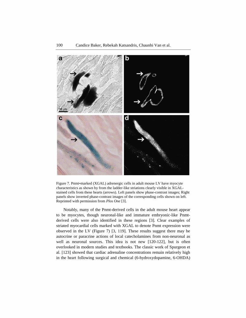
Figure 7. Pnmt-marked (XGAL) adrenergic cells in adult mouse LV have myocyte
characteristics as shown by from the ladder-like striations clearly visible in XGAL-
stained cells from these hearts (arrows). Left panels show phase-contrast images; Right
panels show inverted phase-contrast images of the corresponding cells shown on left.
Reprinted with permission from Plos One [3].
Notably, many of the Pnmt-derived cells in the adult mouse heart appear
to be myocytes, though neuronal-like and immature embryonic-like Pnmt-
derived cells were also identified in these regions [3]. Clear examples of
striated myocardial cells marked with XGAL to denote Pnmt expression were
observed in the LV (Figure 7) [3, 119]. These results suggest there may be
autocrine or paracrine actions of local catecholamines from non-neuronal as
well as neuronal sources. This idea is not new [120-122], but is often
overlooked in modern studies and textbooks. The classic work of Spurgeon et
al. [123] showed that cardiac adrenaline concentrations remain relatively high
in the heart following surgical and chemical (6-hydroxydopamine, 6-OHDA)

Adrenaline and Stress-Induced Cardiomyopathies 101
denervation, while noradrenaline concentrations were nearly completely
eradicated by these procedures. The authors concluded that cardiac
noradrenaline content was mostly neuronal while only ~50% of the adrenaline
content was neuronal. They hypothesized that cardiac adrenaline ―is held in
non-neuronal stores either in chromaffin cells, in the specialized cells
themselves or in cardiac analogs of chromaffin cells‖ [123]. There is ample
evidence for these from a wide variety of species [124-127], including humans
[128], suggesting their presence is highly conserved and, therefore, likely
critical for survival. It remains to be determined if these cells truly play a
significant role in TTC, but they certainly represent a potential local source of
adrenaline that could contribute to regional variations in adrenergic
stimulation within the LV.
There are still many unanswered questions about this hypothesis. We do
not know, for example, if there are changes in cardiac adrenaline content or
regional differences in the distribution or concentrations of local
catecholamine production in the heart in response to aging, sex, or hormonal
status. One conundrum with this hypothesis is the heaviest concentrations of
Pnmt-marked adrenergic cells are located at the base of the LV, yet the apical
and mid sections of the LV are much more frequently affected in clinical cases
of TTC. The base concentrations of adrenergic cells could conceivably
contribute to inverted forms of TTC, but it is still a mystery why sometimes
base and other times the mid or apex are most affected. Clearly, more work is
required to elucidate these mechanisms.
In addition, no-one has yet demonstrated that non-neuronal sources of
cardiac catecholamines are secreted during emotional or physical stress. It is
still unclear how these cells are regulated, and how they influence their local
environment, though there are several reports in rodent models showing that
cardiac Pnmt expression is upregulated by glucocorticoid hormones, which
themselves are typically elevated in the circulation during stress responses
[129, 130]. Similar concerns exist for stimulation of sympathetic nerves and
the intrinsic cardiac nervous system, which in the human heart is estimated to
contain at least 14,000 neurons [113]. Further research is required to delineate
how local sources of catecholamines from autocrine/paracrine or neuronal
inputs may impact regional myocardial function at baseline and under different
types of stressful conditions.

Candice Baker, Rebekah Katsandris, Chaunhi Van et al. 102
Figure 8. Schematic illustrations of three distinctive models proposed to explain TTC
mechanisms. 1. Coronary Microspasms: In this model, stress-induced surges in
catecholamines trigger coronary microspasms, which then lead to decreased blood
flow and regional myocardial inactivity. 2. Increased β2-Receptor Density: This model
proposes that β2-adrenergic receptors are more concentrated at the apex compared to
the base. High concentrations of adrenaline cause these receptors to switch from Gs to
Gi, which results in myocardial stunning in affected regions that would appear as
akinetic or hypokinetic sections of LV during systole. 3. Increased Local
Catecholamine Release: In this hypothesis, adrenaline and/or noradrenaline are
released locally in the apex, mid, and/or basal regions of the LV from non-neuronal
resident adrenergic cell populations (depicted by the highlighted blue areas). This local
release overstimulates adrenergic receptors that are already near saturation from
circulating adrenaline and sympathetic nerves (NA). Abbreviations: HPA,
Hypothalamic-Pituitary-Adrenal (axis); A, adrenaline; NA, noradrenaline.
Summary, Conclusion and
Future Directions
TTC is a complex clinical condition characterized by left ventricular
regional akinesis/hypokinesis resulting in severe chest pain and ST-elevation
on EKG. Episodes are typically precipitated by acute emotional or physical
stress, and the vast majority of TTC cases have been reported in

Adrenaline and Stress-Induced Cardiomyopathies 103
postmenopausal women. There is strong evidence implicating elevated
catecholamine levels as a critical factor in TTC induction, but it is not yet clear
if they are coming from the circulation (adrenal secretion), sympathetic
innervation, and/or local autocrine/paracrine sources. The relative inputs from
these different sources may vary from individual to individual, and will likely
differ depending on the type of stress impacting the individual patient. Hence,
one could imagine different sorts of ―catecholamine storms‖ [131] that could
trigger different types of TTC.
Each of the three major hypotheses discussed in this chapter offer credible
explanations for how adrenergic hormones may influence the development of
TTC. For comparative purposes, they are each illustrated in cartoon form in
Figure 8.
Although all of these hypotheses provide reasonable explanations for
some aspects of TTC, none of them provide a full explanation of the
underlying mechanisms leading to TTC. Each is distinctive in its features, yet
they are not mutually exclusive. For example, regional variations in β2-
adrenergic receptor concentrations and/or sensitivities could result in local
fluctuations inducing vasospasms in nearby coronary vessels. These could also
be influenced by local release of catecholamines either from sympathetic nerve
terminals or non-neuronal autocrine/paracrine sources that could result in
regional overstimulation of adrenergic receptors leading to myocardial
stunning, perhaps as a result of a switch from Gs to Gi coupling primarily at β2-
adrenergic receptors. Further study is required determine how these different
mechanistic components of the cardiac stress response system interact to in the
context of TTC.
After nearly 25 years of research since the first formal description of TTC
in the published literature, we still have only a rudimentary picture of how this
peculiar stress cardiomyopathy materializes. Although it was ―discovered‖
only relatively recently, TTC has existed as an undiagnosed or misdiagnosed
clinical condition for countless years. Because of the relative ―newness‖ of
TTC, many patients and some physicians are still largely unaware of the
conditions and potential severity of stress cardiomyopathies, yet TTC
continues to represent a real and persistent danger to susceptible individuals.
In many and perhaps even most cases, TTC can often resolve on its own
without interventional therapy within a matter of a few weeks [48]. This is not
to say, however, that dangerous complications do not arise. On the contrary,
TTC patients with manifest coronary vasospasms are increased risk for heart
attacks and strokes from thrombolytic embolisms [36]. Other studies have
demonstrated significant lengthening of the Q-T interval on EKGs from TTC

Candice Baker, Rebekah Katsandris, Chaunhi Van et al. 104
patients [132]. Long QT intervals are known risk factor for dangerous and life-
threatening forms of ventricular arrhythmias known as Torsade de Pointes
[133-135]. Several other serious complications from TTC have been noted in
the medical and scientific literature, so it is by no means a benign disease [136,
137]. Continued research and critical evaluation of the results and ideas
emanating from these exercises will surely help to refine our understanding of
the mechanisms underlying TTC. Some of key outstanding questions include
but are not limited to the following:
1. How is emotional stress ―physiologically‖ transmitted to the heart?
2. Do different types of stress have different impacts on cardiovascular
function (i.e., does it matter if the stressor elicits selective autonomic
nerve stimulation versus systemic adrenaline surges)?
3. Do local non-neuronal stores of catecholamines play a part in TTC? If
so, then what regulates their activities?
4. Why are only certain regions of the LV primarily affected in TTC?
5. What controls regional variations in adrenergic receptor subtype
expression patterns?
These are just a few of the general questions about TTC that remain
answered. We have also highlighted a number of questions earlier in this
chapter pertaining to sex, age, and hormonal status. In addition, we have
discussed the merits and limitations of three independent models of TTC
mechanisms. Thus, while much progress has been made in the last 25 years on
this subject, there is still much work to be done before we have a full
understanding of the pathophysiological mechanisms responsible for this
stress-induced cardiomyopathy. The three mechanistic models reviewed here
offer strong frameworks for specific hypothesis testing, and thus each should
be useful for designing new experiments to address some of the many still
outstanding questions associated with TTC.
References
[1] Kume, T., et al., Local release of catecholamines from the hearts of
patients with tako-tsubo-like left ventricular dysfunction. Circ J, 2008.
72(1): p. 106-8.
[2] Paur, H., et al., High levels of circulating epinephrine trigger apical
cardiodepression in a beta2-adrenergic receptor/Gi-dependent manner: a

Adrenaline and Stress-Induced Cardiomyopathies 105
new model of Takotsubo cardiomyopathy. Circulation, 2012. 126(6): p.
697-706.
[3] Osuala, K., et al., Distinctive left-sided distribution of adrenergic-
derived cells in the adult mouse heart. PLoS One, 2011. 6(7): p. e22811.
[4] Shimizu, M., et al., [Recurrent episodes of takotsubo-like transient left
ventricular ballooning occurring in different regions: a case report]. J
Cardiol, 2006. 48(2): p. 101-7.
[5] Kumar, A., et al., Transient left ventricular apical ballooning during
dobutamine myocardial perfusion imaging. Int J Cardiol, 2008. 124(3):
p. 378-80.
[6] Dote, K., et al., [Myocardial stunning due to simultaneous multivessel
coronary spasms: a review of 5 cases]. J Cardiol, 1991. 21(2): p. 203-14.
[7] H. Sato, H.T., K. Dote, T. Uchida, M. Ishihara, Tako-tsubo-like left
ventricular dysfunction due to multivessel coronary spasm, in Clinical
aspect of myocardial injury: from ischemia to heart failure, K.H. K.
Kodama, M. Hori, Editor. 1990, Kagakuhyoronsha Publishing Co.:
Tokyo.
[8] Cesario, V., Loureiro, M.J., and H. Pereira, Takotsubo cardiomyopathy
in a cardiology department. Portuguese J of Cardiol, 2012. 31(9): p.
603-8.
[9] Angelini, P., Takotsubo cardiomyopathy: what is behind the octopus
trap? Tex Heart Inst J, 2010. 37(1): p. 85-7.
[10] Lyon, A.R., et al., Stress (Takotsubo) cardiomyopathy--a novel
pathophysiological hypothesis to explain catecholamine-induced acute
myocardial stunning. Nat Clin Pract Cardiovasc Med, 2008. 5(1): p. 22-
9.
[11] Steptoe, A. and M. Kivimaki, Stress and cardiovascular disease. Nat Rev
Cardiol, 2012. 9(6): p. 360-70.
[12] Bybee, K.A. and A. Prasad, Stress-related cardiomyopathy syndromes.
Circulation, 2008. 118(4): p. 397-409.
[13] Tsuchihashi, K., et al., Transient left ventricular apical ballooning
without coronary artery stenosis: a novel heart syndrome mimicking
acute myocardial infarction. Angina Pectoris-Myocardial Infarction
Investigations in Japan. J Am Coll Cardiol, 2001. 38(1): p. 11-8.
[14] Hurst, R.T., et al., Takotsubo cardiomyopathy: a unique cardiomyopathy
with variable ventricular morphology. JACC Cardiovasc Imaging, 2010.
3(6): p. 641-9.
[15] Sharkey, S.W., et al., Why not just call it tako-tsubo cardiomyopathy: a
discussion of nomenclature. J Am Coll Cardiol, 2011. 57(13): p. 1496-7.

Candice Baker, Rebekah Katsandris, Chaunhi Van et al. 106
[16] Abraham, J., et al., Stress cardiomyopathy after intravenous
administration of catecholamines and beta-receptor agonists. J Am Coll
Cardiol, 2009. 53(15): p. 1320-5.
[17] Litvinov, I.V., M.A. Kotowycz, and S. Wassmann, Iatrogenic
epinephrine-induced reverse Takotsubo cardiomyopathy: direct evidence
supporting the role of catecholamines in the pathophysiology of the
"broken heart syndrome". Clin Res Cardiol, 2009. 98(7): p. 457-62.
[18] Marcovitz, P.A., et al., Pheochromocytoma presenting with Takotsubo
syndrome. J Interv Cardiol, 2010. 23(5): p. 437-42.
[19] Naderi, N., et al., Pheochromocytoma-induced reverse tako-tsubo with
rapid recovery of left ventricular function. Cardiol J, 2012. 19(5): p.
527-31.
[20] Ennezat, P.V., et al., Transient left ventricular basal dysfunction without
coronary stenosis in acute cerebral disorders: a novel heart syndrome
(inverted Takotsubo). Echocardiography, 2005. 22(7): p. 599-602.
[21] Patel, S.M., et al., Distinctive clinical characteristics according to age
and gender in apical ballooning syndrome (takotsubo/stress
cardiomyopathy): an analysis focusing on men and young women. J
Card Fail, 2013. 19(5): p. 306-10.
[22] Wittstein, I.S., et al., Neurohumoral features of myocardial stunning due
to sudden emotional stress. N Engl J Med, 2005. 352(6): p. 539-48.
[23] Akashi, Y.J., et al., 123I-MIBG myocardial scintigraphy in patients with
"takotsubo" cardiomyopathy. J Nucl Med, 2004. 45(7): p. 1121-7.
[24] Ito, K., et al., Assessment of Takotsubo (ampulla) cardiomyopathy using
99mTc-tetrofosmin myocardial SPECT--comparison with acute
coronary syndrome. Ann Nucl Med, 2003. 17(2): p. 115-22.
[25] Yoshida, T., et al., A pathophysiologic study of tako-tsubo
cardiomyopathy with F-18 fluorodeoxyglucose positron emission
tomography. Eur Heart J, 2007. 28(21): p. 2598-604.
[26] Shao, Y., et al., Novel rat model reveals important roles of beta-
adrenoreceptors in stress-induced cardiomyopathy. Int J Cardiol, 2013.
[27] Shao, Y., et al., A mouse model reveals an important role for
catecholamine-induced lipotoxicity in the pathogenesis of stress-induced
cardiomyopathy. Eur J Heart Fail, 2013. 15(1): p. 9-22.
[28] Izumi, Y., et al., Effects of metoprolol on epinephrine-induced
takotsubo-like left ventricular dysfunction in non-human primates.
Hypertens Res, 2009. 32(5): p. 339-46.

Adrenaline and Stress-Induced Cardiomyopathies 107
[29] Kim, S., et al., Inverted-Takotsubo pattern cardiomyopathy secondary to
pheochromocytoma: a clinical case and literature review. Clin Cardiol,
2010. 33(4): p. 200-5.
[30] Crea, F. and G. Liuzzo, Pathogenesis of acute coronary syndromes. J Am
Coll Cardiol, 2013. 61(1): p. 1-11.
[31] Windecker, S., et al., Future treatment strategies in ST-segment
elevation myocardial infarction. Lancet, 2013. 382(9892): p. 644-57.
[32] Sealove, B.A., S. Tiyyagura, and V. Fuster, Takotsubo cardiomyopathy.
J Gen Intern Med, 2008. 23(11): p. 1904-8.
[33] Donohue, D. and M.R. Movahed, Clinical characteristics, demographics
and prognosis of transient left ventricular apical ballooning syndrome.
Heart Fail Rev, 2005. 10(4): p. 311-6.
[34] Sardar, M.R., et al., Recurrent takotsubo cardiomyopathy in the setting
of transient neurological symptoms: a case report. J Med Case Rep,
2011. 5: p. 412.
[35] Teramachi, Y., et al., Takotsubo cardiomyopathy in a senile female
patient after transcatheter coil embolization of patent arterial duct.
Geriatr Gerontol Int, 2013. 13(4): p. 1077-9.
[36] Castillo Rivera, A.M., M. Ruiz-Bailen, and L. Rucabado Aguilar,
Takotsubo cardiomyopathy--a clinical review. Med Sci Monit, 2011.
17(6): p. RA135-47.
[37] Eshtehardi, P., et al., Transient apical ballooning syndrome--clinical
characteristics, ballooning pattern, and long-term follow-up in a Swiss
population. Int J Cardiol, 2009. 135(3): p. 370-5.
[38] Gianni, M., et al., Apical ballooning syndrome or takotsubo
cardiomyopathy: a systematic review. Eur Heart J, 2006. 27(13): p.
1523-9.
[39] Sanchez-Jimenez, E.F., Initial clinical presentation of Takotsubo
cardiomyopathy with-a focus on electrocardiographic changes: A
literature review of cases. World J Cardiol, 2013. 5(7): p. 228-41.
[40] Jiang, W., et al., Mental stress--induced myocardial ischemia and cardiac
events. JAMA, 1996. 275(21): p. 1651-6.
[41] Subban, V., et al., Apical ballooning syndrome in first degree relatives.
Indian Heart J, 2012. 64(6): p. 607-9.
[42] Kumar, G., D.R. Holmes, Jr., and A. Prasad, "Familial" apical
ballooning syndrome (Takotsubo cardiomyopathy). Int J Cardiol, 2010.
144(3): p. 444-5.
[43] Pison, L., P. De Vusser, and W. Mullens, Apical ballooning in relatives.
Heart, 2004. 90(12): p. e67.

Candice Baker, Rebekah Katsandris, Chaunhi Van et al. 108
[44] Maron, B.J., et al., Contemporary definitions and classification of the
cardiomyopathies: an American Heart Association Scientific Statement
from the Council on Clinical Cardiology, Heart Failure and
Transplantation Committee; Quality of Care and Outcomes Research
and Functional Genomics and Translational Biology Interdisciplinary
Working Groups; and Council on Epidemiology and Prevention.
Circulation, 2006. 113(14): p. 1807-16.
[45] Prasad, A., Apical ballooning syndrome: an important differential
diagnosis of acute myocardial infarction. Circulation, 2007. 115(5): p.
e56-9.
[46] Bossone, E., et al., Takotsubo cardiomyopathy: an integrated multi-
imaging approach. Eur Heart J Cardiovasc Imaging, 2013.
[47] Eitel, I., et al., Differential diagnosis of suspected apical ballooning
syndrome using contrast-enhanced magnetic resonance imaging. Eur
Heart J, 2008. 29(21): p. 2651-9.
[48] Bybee, K.A., et al., Systematic review: transient left ventricular apical
ballooning: a syndrome that mimics ST-segment elevation myocardial
infarction. Ann Intern Med, 2004. 141(11): p. 858-65.
[49] Dec, G.W., Recognition of the apical ballooning syndrome in the United
States. Circulation, 2005. 111(4): p. 388-90.
[50] Sharkey, S.W., et al., Natural history and expansive clinical profile of
stress (tako-tsubo) cardiomyopathy. J Am Coll Cardiol, 2010. 55(4): p.
333-41.
[51] Murakami, T., et al., Characterization of predictors of in-hospital cardiac
complications of takotsubo cardiomyopathy: Multi-center registry from
Tokyo CCU Network. J Cardiol, 2013.
[52] Akashi, Y.J., et al., Takotsubo cardiomyopathy: a new form of acute,
reversible heart failure. Circulation, 2008. 118(25): p. 2754-62.
[53] Bietry, R., A. Reyentovich, and S.D. Katz, Clinical management of
takotsubo cardiomyopathy. Heart Fail Clin, 2013. 9(2): p. 177-86, viii.
[54] Sharma, V., et al., Stress cardiomyopathy: case series and the review of
literature. J Emerg Med, 2013. 45(4): p. e95-8.
[55] Kurowski, V., et al., Apical and midventricular transient left ventricular
dysfunction syndrome (tako-tsubo cardiomyopathy): frequency,
mechanisms, and prognosis. Chest, 2007. 132(3): p. 809-16.
[56] Park, J.H., et al., Left ventricular apical ballooning due to severe
physical stress in patients admitted to the medical ICU. Chest, 2005.
128(1): p. 296-302.

Adrenaline and Stress-Induced Cardiomyopathies 109
[57] Milinis, K. and M. Fisher, Takotsubo cardiomyopathy: pathophysiology
and treatment. Postgrad Med J, 2012. 88(1043): p. 530-8.
[58] Nobrega, S. and D. Brito, [The "broken heart syndrome": state of the
art]. Rev Port Cardiol, 2012. 31(9): p. 589-96.
[59] Kurisu, S. and Y. Kihara, Tako-tsubo cardiomyopathy: clinical
presentation and underlying mechanism. J Cardiol, 2012. 60(6): p. 429-
37.
[60] Kurisu, S., et al., Tako-tsubo-like left ventricular dysfunction with ST-
segment elevation: a novel cardiac syndrome mimicking acute
myocardial infarction. Am Heart J, 2002. 143(3): p. 448-55.
[61] Ueyama, T., Emotional stress-induced Tako-tsubo cardiomyopathy:
animal model and molecular mechanism. Ann N Y Acad Sci, 2004. 1018:
p. 437-44.
[62] Kuo, B.T., R. Choubey, and G.M. Novaro, Reduced estrogen in
menopause may predispose women to takotsubo cardiomyopathy. Gend
Med, 2010. 7(1): p. 71-7.
[63] Ueyama, T., et al., Chronic estrogen supplementation following
ovariectomy improves the emotional stress-induced cardiovascular
responses by indirect action on the nervous system and by direct action
on the heart. Circ J, 2007. 71(4): p. 565-73.
[64] Ueyama, T., et al., Cardiac and vascular gene profiles in an animal
model of takotsubo cardiomyopathy. Heart Vessels, 2011. 26(3): p. 321-
37.
[65] Ueyama, T., et al., Estrogen attenuates the emotional stress-induced
cardiac responses in the animal model of Tako-tsubo (Ampulla)
cardiomyopathy. J Cardiovasc Pharmacol, 2003. 42 Suppl 1: p. S117-9.
[66] Col, N.F., et al., In the clinic. Menopause. Ann Intern Med, 2009.
150(7): p. ITC4-1-15; quiz ITC4-16.
[67] Ueyama, T., et al., Emotional stress induces transient left ventricular
hypocontraction in the rat via activation of cardiac adrenoceptors: a
possible animal model of 'tako-tsubo' cardiomyopathy. Circ J, 2002.
66(7): p. 712-3.
[68] Ueyama, T., et al., Catecholamines and estrogen are involved in the
pathogenesis of emotional stress-induced acute heart attack. Ann N Y
Acad Sci, 2008. 1148: p. 479-85.
[69] Katchman, A.N., et al., Comparative evaluation of HERG currents and
QT intervals following challenge with suspected torsadogenic and
nontorsadogenic drugs. J Pharmacol Exp Ther, 2006. 316(3): p. 1098-
106.

Candice Baker, Rebekah Katsandris, Chaunhi Van et al. 110
[70] Liu, X.K., et al., In vivo androgen treatment shortens the QT interval and
increases the densities of inward and delayed rectifier potassium currents
in orchiectomized male rabbits. Cardiovasc Res, 2003. 57(1): p. 28-36.
[71] Liu, X.K., et al., Gender difference in the cycle length-dependent QT
and potassium currents in rabbits. J Pharmacol Exp Ther, 1998. 285(2):
p. 672-9.
[72] Wang, W.X., et al., "Conventional" antihistamines slow cardiac
repolarization in isolated perfused (Langendorff) feline hearts. J
Cardiovasc Pharmacol, 1998. 32(1): p. 123-8.
[73] Kim, J.J., et al., Bradycardia alters Ca(2+) dynamics enhancing
dispersion of repolarization and arrhythmia risk. Am J Physiol Heart
Circ Physiol, 2013. 304(6): p. H848-60.
[74] Saito, T., et al., Estrogen contributes to gender differences in mouse
ventricular repolarization. Circ Res, 2009. 105(4): p. 343-52.
[75] Brouillette, J., et al., Functional properties of K+ currents in adult mouse
ventricular myocytes. J Physiol, 2004. 559(Pt 3): p. 777-98.
[76] Trepanier-Boulay, V., et al., Gender-based differences in cardiac
repolarization in mouse ventricle. Circ Res, 2001. 89(5): p. 437-44.
[77] Wu, Y. and M.E. Anderson, Reduced repolarization reserve in
ventricular myocytes from female mice. Cardiovasc Res, 2002. 53(3): p.
763-9.
[78] Deshmukh, A., et al., Prevalence of Takotsubo cardiomyopathy in the
United States. Am Heart J, 2012. 164(1): p. 66-71 e1.
[79] Stollberger, C. and J. Finsterer, Why does takotsubo ("broken heart
syndrome") affect more females than males? Int J Cardiol, 2011. 147(1):
p. 175-6.
[80] Song, B.G., et al., Clinical characteristics, ballooning pattern, and long-
term prognosis of transient left ventricular ballooning syndrome. Heart
Lung, 2010. 39(3): p. 188-95.
[81] Group, J.C.S.J.W., Guidelines for diagnosis and treatment of patients
with vasospastic angina (coronary spastic angina) (JCS 2008): digest
version. Circ J, 2010. 74(8): p. 1745-62.
[82] Okumura, K., et al., Sensitivity and specificity of intracoronary injection
of acetylcholine for the induction of coronary artery spasm. J Am Coll
Cardiol, 1988. 12(4): p. 883-8.
[83] Galiuto, L., et al., Reversible coronary microvascular dysfunction: a
common pathogenetic mechanism in Apical Ballooning or Tako-Tsubo
Syndrome. Eur Heart J, 2010. 31(11): p. 1319-27.

Adrenaline and Stress-Induced Cardiomyopathies 111
[84] Gibson, C.M., et al., TIMI frame count: a quantitative method of
assessing coronary artery flow. Circulation, 1996. 93(5): p. 879-88.
[85] Bybee, K.A., et al., Clinical characteristics and thrombolysis in
myocardial infarction frame counts in women with transient left
ventricular apical ballooning syndrome. Am J Cardiol, 2004. 94(3): p.
343-6.
[86] Elesber, A., et al., Myocardial perfusion in apical ballooning syndrome
correlate of myocardial injury. Am Heart J, 2006. 152(3): p. 469 e9-13.
[87] Meimoun, P., et al., Transient impairment of coronary flow reserve in
tako-tsubo cardiomyopathy is related to left ventricular systolic
parameters. Eur J Echocardiogr, 2009. 10(2): p. 265-70.
[88] Kume, T., et al., Assessment of coronary microcirculation in patients
with takotsubo-like left ventricular dysfunction. Circ J, 2005. 69(8): p.
934-9.
[89] Abe, Y., et al., Assessment of clinical features in transient left
ventricular apical ballooning. J Am Coll Cardiol, 2003. 41(5): p. 737-42.
[90] Kawai, S., et al., Ampulla cardiomyopathy ('Takotusbo'
cardiomyopathy)--reversible left ventricular dysfunction: with ST
segment elevation. Jpn Circ J, 2000. 64(2): p. 156-9.
[91] Mori, H., et al., Increased responsiveness of left ventricular apical
myocardium to adrenergic stimuli. Cardiovasc Res, 1993. 27(2): p. 192-
8.
[92] Kawano, H., R. Okada, and K. Yano, Histological study on the
distribution of autonomic nerves in the human heart. Heart Vessels,
2003. 18(1): p. 32-9.
[93] Lathers, C.M., R.M. Levin, and W.H. Spivey, Regional distribution of
myocardial beta-adrenoceptors in the cat. Eur J Pharmacol, 1986.
130(1-2): p. 111-7.
[94] Heather, L.C., et al., Isoproterenol induces in vivo functional and
metabolic abnormalities: similar to those found in the infarcted rat heart.
J Physiol Pharmacol, 2009. 60(3): p. 31-9.
[95] Mantravadi, R., et al., Autonomic nerve stimulation reverses ventricular
repolarization sequence in rabbit hearts. Circ Res, 2007. 100(7): p. e72-
80.
[96] Khoueiry, G., et al., Reverse Takotsubo cardiomyopathy in the setting of
anaphylaxis treated with high-dose intravenous epinephrine. J Emerg
Med, 2013. 44(1): p. 96-9.

Candice Baker, Rebekah Katsandris, Chaunhi Van et al. 112
[97] Collen, J., W. Bimson, and P. Devine, A variant of Takotsubo
cardiomyopathy: a rare complication in the electrophysiology lab. J
Invasive Cardiol, 2008. 20(11): p. E310-3.
[98] Bonnemeier, H., et al., "The artichoke heart": the inverse counterpart of
left ventricular apical ballooning. Resuscitation, 2007. 72(3): p. 342-3.
[99] Song, B.G., et al., The clinical characteristics, laboratory parameters,
electrocardiographic, and echocardiographic findings of reverse or
inverted takotsubo cardiomyopathy: comparison with mid or apical
variant. Clin Cardiol, 2011. 34(11): p. 693-9.
[100] Ramaraj, R. and M.R. Movahed, Reverse or inverted takotsubo
cardiomyopathy (reverse left ventricular apical ballooning syndrome)
presents at a younger age compared with the mid or apical variant and is
always associated with triggering stress. Congest Heart Fail, 2010.
16(6): p. 284-6.
[101] Brewington, S.D., et al., Reproducible microvascular dysfunction with
dobutamine infusion in Takotsubo cardiomyopathy presenting with ST
segment elevation. Catheter Cardiovasc Interv, 2006. 68(5): p. 769-74.
[102] Whalen, E.J. and S.J. Lewis, In vivo evidence that isoproterenol may
increase heart rate in the rat by mechanisms in addition to activation of
cardiac beta(1)- or beta(2)-adrenoceptors. Eur J Pharmacol, 1999.
382(3): p. 207-10.
[103] Thawornkaiwong, A., S. Preawnim, and J. Wattanapermpool,
Upregulation of beta 1-adrenergic receptors in ovariectomized rat hearts.
Life Sci, 2003. 72(16): p. 1813-24.
[104] Bristow, M.R., et al., Beta 1- and beta 2-adrenergic-receptor
subpopulations in nonfailing and failing human ventricular myocardium:
coupling of both receptor subtypes to muscle contraction and selective
beta 1-receptor down-regulation in heart failure. Circ Res, 1986. 59(3):
p. 297-309.
[105] Ishikura, F., Y. Takano, and T. Ueyama, Acute effects of beta-blocker
with intrinsic sympathomimetic activity on stress-induced cardiac
dysfunction in rats. J Cardiol, 2012. 60(6): p. 470-4.
[106] Takano, Y., T. Ueyama, and F. Ishikura, Azelnidipine, unique calcium
channel blocker could prevent stress-induced cardiac dysfunction like
alpha.beta blocker. J Cardiol, 2012. 60(1): p. 18-22.
[107] Ebert, S.N., et al., Expression of phenylethanolamine n-
methyltransferase in the embryonic rat heart. J Mol Cell Cardiol, 1996.
28(8): p. 1653-8.

Adrenaline and Stress-Induced Cardiomyopathies 113
[108] Ebert, S.N. and D.G. Taylor, Catecholamines and development of
cardiac pacemaking: an intrinsically intimate relationship. Cardiovasc
Res, 2006. 72(3): p. 364-74.
[109] Ebert, S.N. and R.P. Thompson, Embryonic epinephrine synthesis in the
rat heart before innervation: association with pacemaking and
conduction tissue development. Circ Res, 2001. 88(1): p. 117-24.
[110] Huang, M.H., et al., An intrinsic adrenergic system in mammalian heart.
J Clin Invest, 1996. 98(6): p. 1298-1303.
[111] Papka, R.E., A study of catecholamine-containing cells in the hearts of
fetal and postnatal rabbits by fluorescence and electron microscopy. Cell
Tissue Res, 1974. 154(4): p. 471-84.
[112] Elayan, H.H., B.P. Kennedy, and M.G. Ziegler, Cardiac atria and
ventricles contain different inducible adrenaline synthesising enzymes.
Cardiovasc Res, 1990. 24(1): p. 53-6.
[113] Armour, J.A., et al., Gross and microscopic anatomy of the human
intrinsic cardiac nervous system. Anat Rec, 1997. 247(2): p. 289-98.
[114] Pierpont, G.L., E.G. DeMaster, and J.N. Cohn, Regional differences in
adrenergic function within the left ventricle. Am J Physiol, 1984. 246(6
Pt 2): p. H824-9.
[115] Angelakos, E.T., Regional Distribution of Catecholamines in the Dog
Heart. Circ Res, 1965. 16: p. 39-44.
[116] Clarke, G.L., et al., ss-adrenoceptor blockers increase cardiac
sympathetic innervation by inhibiting autoreceptor suppression of axon
growth. J Neurosci, 2010. 30(37): p. 12446-54.
[117] Lau, C., S.P. Burke, and T.A. Slotkin, Maturation of sympathetic
neurotransmission in the rat heart. IX. Development of transsynaptic
regulation of cardiac adrenergic sensitivity. J Pharmacol Exp Ther,
1982. 223(3): p. 675-80.
[118] Habecker, B.A., N.M. Malec, and S.C. Landis, Differential regulation of
adrenergic receptor development by sympathetic innervation. J
Neurosci, 1996. 16(1): p. 229-37.
[119] Ebert, S.N., et al., Catecholamine-synthesizing cells in the embryonic
mouse heart. Ann N Y Acad Sci, 2008. 1148: p. 317-24.
[120] Kloberg, A.J. and R. Fritsche, Catecholamines are present in larval
Xenopus laevis: a potential source for cardiac control. J Exp Zool, 2002.
292(3): p. 293-303.
[121] Kimura, K., M. Ieda, and K. Fukuda, Development, maturation, and
transdifferentiation of cardiac sympathetic nerves. Circ Res, 2012.
110(2): p. 325-36.

Candice Baker, Rebekah Katsandris, Chaunhi Van et al. 114
[122] Slavikova, J., et al., Catecholaminergic neurons in the rat intrinsic
cardiac nervous system. Neurochem Res, 2003. 28(3-4): p. 593-8.
[123] Spurgeon, H.A., et al., Catecholamines associated with conductile and
contractile myocardium of normal and denervated dog hearts. J
Pharmacol Exp Ther, 1974. 190(3): p. 466-71.
[124] Ellison, J.P. and R.G. Hibbs, Catecholamine-containing cells of the
guinea pig heart: an ultrastructural study. J Mol Cell Cardiol, 1974. 6(1):
p. 17-26.
[125] Padbury, J.F., et al., Ontogenesis of tissue catecholamines in fetal and
neonatal rabbits. J Dev Physiol, 1981. 3(5): p. 297-303.
[126] Abrahamsson, T., A.C. Jonsson, and S. Nilsson, Catecholamine
synthesis in the chromaffin tissue of the African lungfish, Protopterus
aethiopicus. Acta Physiol Scand, 1979. 107(2): p. 149-51.
[127] Forsgren, S., M. Moravec, and J. Moravec, Catecholamine-synthesizing
enzymes and neuropeptides in rat heart epicardial ganglia; an
immunohistochemical study. Histochem J, 1990. 22(12): p. 667-76.
[128] Dail, W.G., Jr. and G.C. Palmer, Localization and correltion of
catecholamine-containing cells with adenyl cyclase nd
phosphodiesterase activities in the human fetal heart. Anat Rec, 1973.
177(2): p. 265-87.
[129] Krizanova, O., et al., Existence of cardiac PNMT mRNA in adult rats:
elevation by stress in a glucocorticoid-dependent manner. Am J Physiol
Heart Circ Physiol, 2001. 281(3): p. H1372-9.
[130] Kvetnansky, R., et al., Localization and regulation of
phenylethanolamine N-methyltransferase gene expression in the heart of
rats and mice during stress. Ann N Y Acad Sci, 2004. 1018: p. 405-17.
[131] Legriel, S., et al., Recurrent takotsubo cardiomyopathy triggered by
convulsive status epilepticus. Neurocrit Care, 2008. 9(1): p. 118-21.
[132] Behr, E.R. and S. Mahida, Stress cardiomyopathy and the acquired long
QT syndrome. Heart Rhythm, 2011. 8(4): p. e1; author reply e1-2.
[133] Al-Khatib, S.M., et al., What clinicians should know about the QT
interval. JAMA, 2003. 289(16): p. 2120-7.
[134] Valbusa, A., et al., Takotsubo cardiomyopathy and torsade de pointes in
myasthenic crisis: be aware of QT prolongation. Am J Emerg Med, 2013.
31(12): p. 1717-8.
[135] Kurisu, S., et al., Torsade de pointes associated with bradycardia and
takotsubo cardiomyopathy. Can J Cardiol, 2008. 24(8): p. 640-2.

Adrenaline and Stress-Induced Cardiomyopathies 115
[136] Rotondi, F. and F. Manganelli, Takotsubo cardiomyopathy and
arrhythmic risk: the dark side of the moon. Eur Rev Med Pharmacol Sci,
2013. 17(1): p. 105-11.
[137] Salazar-Mendiguchia, J., et al., An unusual complication of a Takotsubo
cardiomyopathy: a not so benign disease? Int J Cardiol, 2011. 150(3): p.
348-9.