Adolescent Intermittent Alcohol Exposure: Persistence … and Functional Hippocampal Abnormalities...
9
Adolescent Intermittent Alcohol Exposure: Persistence of Structural and Functional Hippocampal Abnormalities into Adulthood Mary-Louise Risher, Rebekah L. Fleming, W. Christopher Risher, K. M. Miller, Rebecca C. Klein, Tiffany Wills, Shawn K. Acheson, Scott D. Moore, Wilkie A. Wilson, Cagla Eroglu, and H. S. Swartzwelder Background: Human adolescence is a crucial stage of neurological development during which etha- nol (EtOH) consumption is often at its highest. Alcohol abuse during adolescence may render individu- als at heightened risk for subsequent alcohol abuse disorders, cognitive dysfunction, or other neurological impairments by irreversibly altering long-term brain function. To test this possibility, we modeled adolescent alcohol abuse (i.e., intermittent EtOH exposure during adolescence [AIE]) in rats to determine whether adolescent exposure to alcohol leads to long-term structural and functional changes that are manifested in adult neuronal circuitry. Methods: We specifically focused on hippocampal area CA1, a brain region associated with learn- ing and memory. Using electrophysiological, immunohistochemical, and neuroanatomical approaches, we measured post-AIE changes in synaptic plasticity, dendritic spine morphology, and synaptic struc- ture in adulthood. Results: We found that AIE-pretreated adult rats manifest robust long-term potentiation, induced at stimulus intensities lower than those required in controls, suggesting a state of enhanced synaptic plasticity. Moreover, AIE resulted in an increased number of dendritic spines with characteristics typi- cal of immaturity. Immunohistochemistry-based analysis of synaptic structures indicated a significant decrease in the number of co-localized pre- and postsynaptic puncta. This decrease is driven by an over- all decrease in 2 postsynaptic density proteins, PSD-95 and SAP102. Conclusions: Taken together, these findings reveal that repeated alcohol exposure during adoles- cence results in enduring structural and functional abnormalities in the hippocampus. These synaptic changes in the hippocampal circuits may help to explain learning-related behavioral changes in adult animals preexposed to AIE. Key Words: Hippocampus, Long-Term Potentiation, Dendritic Spines, Adolescence, Ethanol. A DOLESCENCE IS A critical period for cognitive, emotional, and social maturation (Choudhury et al., 2006) that is accompanied by the pruning of synapses, refine- ment of neural circuitry, and changes in receptor expression and sensitivity (Kilb, 2012). These processes contribute to the normal maturation of cognitive processes crucial for suc- cessful adult function, including planning, inhibitory control, and working memory (Paus, 2005). Adolescence is also a period during which alcohol con- sumption is often initiated and sustained at high levels (Sque- glia et al., 2012). While it has become clear that adolescents respond differently than adults to the acute effects of ethanol (EtOH) on learning, sedation, and motor function (Little et al., 1996; Markwiese et al., 1998; Spear, 2000), the enduring consequences of repeated EtOH exposure during this develop- mental period have only recently begun to be addressed. In humans, chronic excessive alcohol use during adoles- cence has been associated with cognitive deficits manifesting in adulthood, particularly in the domain of memory function (Brown et al., 2000; Hanson et al., 2011). In animal models used to reflect human levels of consumption, behavioral stud- ies have shown that adolescent intermittent EtOH (AIE) exposure in rats results in long-lasting changes in EtOH sensi- tivity, consumption, and aversion (Diaz-Granados and Gra- ham, 2007; Matthews et al., 2008; Risher et al., 2013). With respect to learning and memory in particular, it has been reported that AIE, but not chronic intermittent EtOH (CIE) From the Durham VA Medical Center (M-LR, RLF, KM, RK, SKA, SDM, WAW, HSS), Duke University Medical Center, Durham, North Carolina; Department of Psychiatry and Behavioral Sciences (M-LR, RLF, KM, RK, SKA, SDM, HSS), Duke University Medical Center, Durham, North Carolina; Department of Cell Biology (WCR, CE), Duke University Medical Center, Durham, North Carolina; Department of Molecular Physiology & Biophysics (TW), Vanderbilt University, Nashville, Tennessee; Social Sciences Research Institute (WAW), Duke University Medical Center, Durham, North Carolina; and Department of Psychology and Neuroscience (HSS), Duke University Medical Center, Durham, North Carolina. Received for publication February 2, 2015; accepted March 12, 2015. Reprint requests: H. Scott Swartzwelder, Department of Psychiatry and Behavioral Sciences, Duke University Medical Center and Durham VA Medical Center, Building 15, VAMC, 508 Fulton Street, Durham, NC 27705; Tel.: 919-971-0964; Fax: 919-286-6811; E-mail: [email protected] Copyright © 2015 by the Research Society on Alcoholism. DOI: 10.1111/acer.12725 Alcohol Clin Exp Res, Vol **, No *, 2015: pp 1–9 1 ALCOHOLISM:CLINICAL AND EXPERIMENTAL RESEARCH Vol. **, No. * ** 2015
-
Upload
nguyenphuc -
Category
Documents
-
view
215 -
download
2
Transcript of Adolescent Intermittent Alcohol Exposure: Persistence … and Functional Hippocampal Abnormalities...

Adolescent Intermittent Alcohol Exposure: Persistence ofStructural and Functional Hippocampal Abnormalities into
Adulthood
Mary-Louise Risher, Rebekah L. Fleming, W. Christopher Risher, K. M. Miller,Rebecca C. Klein, Tiffany Wills, Shawn K. Acheson, Scott D. Moore, Wilkie A. Wilson,
Cagla Eroglu, and H. S. Swartzwelder
Background: Human adolescence is a crucial stage of neurological development during which etha-nol (EtOH) consumption is often at its highest. Alcohol abuse during adolescence may render individu-als at heightened risk for subsequent alcohol abuse disorders, cognitive dysfunction, or otherneurological impairments by irreversibly altering long-term brain function. To test this possibility, wemodeled adolescent alcohol abuse (i.e., intermittent EtOH exposure during adolescence [AIE]) in ratsto determine whether adolescent exposure to alcohol leads to long-term structural and functionalchanges that are manifested in adult neuronal circuitry.
Methods: We specifically focused on hippocampal area CA1, a brain region associated with learn-ing and memory. Using electrophysiological, immunohistochemical, and neuroanatomical approaches,we measured post-AIE changes in synaptic plasticity, dendritic spine morphology, and synaptic struc-ture in adulthood.
Results: We found that AIE-pretreated adult rats manifest robust long-term potentiation, inducedat stimulus intensities lower than those required in controls, suggesting a state of enhanced synapticplasticity. Moreover, AIE resulted in an increased number of dendritic spines with characteristics typi-cal of immaturity. Immunohistochemistry-based analysis of synaptic structures indicated a significantdecrease in the number of co-localized pre- and postsynaptic puncta. This decrease is driven by an over-all decrease in 2 postsynaptic density proteins, PSD-95 and SAP102.
Conclusions: Taken together, these findings reveal that repeated alcohol exposure during adoles-cence results in enduring structural and functional abnormalities in the hippocampus. These synapticchanges in the hippocampal circuits may help to explain learning-related behavioral changes in adultanimals preexposed to AIE.
Key Words: Hippocampus, Long-Term Potentiation, Dendritic Spines, Adolescence, Ethanol.
ADOLESCENCE IS A critical period for cognitive,emotional, and social maturation (Choudhury et al.,
2006) that is accompanied by the pruning of synapses, refine-ment of neural circuitry, and changes in receptor expressionand sensitivity (Kilb, 2012). These processes contribute to
the normal maturation of cognitive processes crucial for suc-cessful adult function, including planning, inhibitory control,and working memory (Paus, 2005).Adolescence is also a period during which alcohol con-
sumption is often initiated and sustained at high levels (Sque-glia et al., 2012). While it has become clear that adolescentsrespond differently than adults to the acute effects of ethanol(EtOH) on learning, sedation, and motor function (Littleet al., 1996;Markwiese et al., 1998; Spear, 2000), the enduringconsequences of repeatedEtOH exposure during this develop-mental period have only recently begun tobe addressed.In humans, chronic excessive alcohol use during adoles-
cence has been associated with cognitive deficits manifestingin adulthood, particularly in the domain of memory function(Brown et al., 2000; Hanson et al., 2011). In animal modelsused to reflect human levels of consumption, behavioral stud-ies have shown that adolescent intermittent EtOH (AIE)exposure in rats results in long-lasting changes in EtOH sensi-tivity, consumption, and aversion (Diaz-Granados and Gra-ham, 2007; Matthews et al., 2008; Risher et al., 2013). Withrespect to learning and memory in particular, it has beenreported that AIE, but not chronic intermittent EtOH (CIE)
From the Durham VAMedical Center (M-LR, RLF, KM, RK, SKA,SDM, WAW, HSS), Duke University Medical Center, Durham, NorthCarolina; Department of Psychiatry and Behavioral Sciences (M-LR,RLF, KM, RK, SKA, SDM, HSS), Duke University Medical Center,Durham, North Carolina; Department of Cell Biology (WCR, CE), DukeUniversity Medical Center, Durham, North Carolina; Department ofMolecular Physiology & Biophysics (TW), Vanderbilt University,Nashville, Tennessee; Social Sciences Research Institute (WAW), DukeUniversity Medical Center, Durham, North Carolina; and Department ofPsychology and Neuroscience (HSS), Duke University Medical Center,Durham, North Carolina.
Received for publication February 2, 2015; accepted March 12, 2015.Reprint requests: H. Scott Swartzwelder, Department of Psychiatry
and Behavioral Sciences, Duke University Medical Center and DurhamVAMedical Center, Building 15, VAMC, 508 Fulton Street, Durham, NC27705; Tel.: 919-971-0964; Fax: 919-286-6811; E-mail: [email protected]
Copyright© 2015 by the Research Society on Alcoholism.
DOI: 10.1111/acer.12725
Alcohol Clin Exp Res,Vol **, No *, 2015: pp 1–9 1
ALCOHOLISM: CLINICAL AND EXPERIMENTAL RESEARCH Vol. **, No. *** 2015

exposure in adulthood, results in greater sensitivity to the spa-tial memory-impairing effects of acute EtOH in the radialarm maze, in the absence of an effect on baseline learningability (Risher et al., 2013; White et al., 2000). Interestingly,AIE has also been shown to reduce the efficacy of EtOH toimpair spatial learning in the Morris water maze 24 hoursafter the last EtOH dose (Silvers et al., 2003, 2006), althoughit is noteworthy that the effect may have been driven by with-drawal, tolerance, or both, given the brief delay betweenrepeated EtOH exposure and testing. Aside from the subse-quent responsiveness to EtOH challenge, Sircar and Sircar(2005) reported thatAIE caused deficits inMorris watermazeperformance up to 25 days after the last EtOH treatment,and fear retention deficits have also been observed 25 daysfollowingAIE exposure (Broadwater and Spear, 2013).
At the neuronal level in similar AIE rodent models (whichachieve equivalent blood EtOH concentrations [BECs]),enduring functional effects are produced that are not appar-ent when comparable EtOH exposure is administered inadulthood. For example, AIE has been shown to reduceA-type potassium current (IA) in GABAergic hippocampalinterneurons (Li et al., 2013), GABAA receptor-mediatedtonic current in dentate granule cells (Fleming et al., 2012,2013) in adulthood, and protein levels of delta and alpha-4GABA receptors in whole hippocampus (Centanni et al.,2014), while comparable EtOH exposure during adulthooddid not produce equivalent long-lasting changes in these cel-lular functions or receptor proteins. Thus, not only does ado-lescent EtOH exposure promote long-lasting changes inhippocampal cellular function, but that adolescence is also aperiod of distinctive vulnerability to the long-term effects ofEtOH, enduring even after an extended period of abstinence.
While these findings support heightened vulnerability tothe long-term consequences of repeated alcohol exposure dur-ing adolescence, the extent to which AIE alters subsequenthippocampal neuronal, synaptic, and behavioral processes isunclear. We used hippocampal slices to assess the long-termeffects of AIE (an established model of intermittent EtOHexposure) on CA1 structure and function. The CA1 area ofthe hippocampus was selected because of its role in learning/memory processes and because it has been a focus of studiesin the emerging literature on the enduring effects of AIE.Here, we assessed the effects of AIE on long-termpotentiation(LTP), dendritic spine morphology (to determine whetherAIE alters synaptic structure that in turn can influence synap-tic function), and postsynaptic density proteins; the latter ofwhich has been found to influence dendritic spine morphol-ogy and play a critical role in the recruitment and stabilizationof excitatory synapses and synaptic function (B�e€ıque et al.,2006; El-Husseini et al., 2000; Zheng et al., 2012). Thisapproach represents a comprehensive analysis combiningphysiological, biochemical, and morphological assessmentsthat support the hypothesis that AIE results in neuronalchanges that persist into adulthood, emphasizing the vulnera-bility of this critical developmental period to insults that canpotentially alter the trajectory of brain development.
MATERIALS ANDMETHODS
All of the procedures used in this study were conducted in accor-dance with the guidelines of the American Association for theAccreditation of Laboratory Animal Care and the NationalResearch Council’s Guide for Care and Use of Laboratory Animalsand were approved by the Durham VA Medical Center and theDuke University IACUCs.
Male postnatal day (PND) 25 Sprague-Dawley rats (CharlesRiver, Raleigh, NC) were double housed and maintained in a tem-perature- and humidity-controlled room with ad libitum access tofood and water. Animals were dosed using modified methods previ-ously described (Risher et al., 2013). Briefly, animals were allowedto acclimatize for 5 days on a reverse 12-hour:12-hour light:darkcycle (lights off at 9:00 AM) prior to beginning AIE or saline admin-istration. All animals were exposed to AIE or saline beginning onPND 30 and consisting of 10 doses of 5 g/kg EtOH (35% v/v in sal-ine at 18.12 ml/kg; VWR, Suwanee, GA) or isovolumetric salineadministered by intragastric gavage (i.g.) using a 2 days on, 1 dayoff, 2 days on, and 2 days off intermittent schedule for 16 days, fol-lowed by a 24- to 29-day washout period, thus allowing all animalsto reach adulthood prior to sacrifice. EtOH doses were selected toproduce BECs that are consistent with adolescent human BECs dur-ing binge drinking episodes.
Blood EtOHConcentration
To avoid the possible confounds associated with stress andstress/EtOH interactions in experimental animals, a group of ani-mals (n = 7) were dosed in parallel with the electrophysiology ani-mals to assess BECs during the intermittent EtOH administration.Animals were dosed (i.g.) on the intermittent schedule describedabove with 5 g/kg EtOH (35% v/v in normal saline) beginning onPND 30. Approximately 150 ll of blood was drawn from the lateralsaphenous vein at 60 minutes post-EtOH administration on the firstand last day of administration. Serum was collected from centri-fuged samples and stored at�80°C. Samples were analyzed in tripli-cate using an Analox GL5 alcohol analyzer (Analox Instruments,Lunenburg, MA).
Electrophysiology
Twelve rats exposed to AIE and 12 controls were used for theseelectrophysiological experiments. Extracellular field recordings wereperformed in the CA1 area of hippocampal slices using modifiedtechniques described previously (Bourne and Harris, 2011; Kleinet al., 2014; Swartzwelder et al., 1995). Briefly, rats (PND 70 to 75)were anesthetized with isoflurane, decapitated, and the brain quicklyremoved. One hemisphere was randomly selected and prepared forGolgi-Cox staining (as described in a later section), while the otherhemisphere was placed in ice-cold artificial cerebral spinal fluid(aCSF) consisting of (in mM) 116.4 NaCl, 5.4 KCl, 1 NaH2PO4,26.2 NaHCO3, 10 D-glucose, 3.2 CaCl2, 1.6 MgSO4 and bubbledwith a gas mixture of 95% O2 to 5% CO2. Coronal sections(400 lm) were cut using a vibratome and incubated at room tem-perature for 15 minutes. Slices were then transferred to a holdingchamber and maintained at 30°C for a minimum of 90 minutesprior to recording. Slices were maintained at 30°C in the recordingchamber and perfused with aCSF at a flow rate of 4 ml/min. A glassmicropipette (recording tip 2 lm, 2 to 4 MO containing 120 mMNaCl) was placed in the CA1 stratum radiatum and field excitatorypostsynaptic potentials (fEPSPs) were elicited by stimulating theSchaffer collateral fibers with a concentric bipolar electrode (FHC,Bowdoin, ME). An Axopatch 200B amplifier (10 kHz low-passfilter) and pClamp software (Sunnyvale, CA, RRID: rid_000085,10 kHz sampling rate) were used to record all data. Input/outputcurves were generated in all slices, and the subsequent baseline stim-
2 RISHER ET AL.

ulus intensity was set at a level that elicited 40% of maximal fEPSPslope. Baseline fEPSPs were recorded every 60 seconds for 25 min-utes and LTP was induced using a theta burst stimulation (TBS)protocol consisting of 2 stimulus trains, each consisting of ten4-pulse 100 Hz bursts with a 200-ms interburst interval. The stimu-lus trains were delivered 30 seconds apart, at a stimulus intensity of20, 30, or 40% of maximal fEPSP slope. fEPSPs were then evokedwith baseline level stimulus pulses every 60 seconds for 60 minutes(n = 8 to 12/treatment group). LTP was defined as >15% potentia-tion 60 minutes after TBS induction. Any slices that failed to main-tain a stable baseline (more than � 5% of baseline for 5consecutive time points) were removed from the analysis.
Golgi-Cox Staining
Rats were handled and dosed with EtOH or saline as describedabove (n = 5 per treatment group). Following the 24- to 29-daywashout period, Golgi-Cox staining was performed as previouslydescribed (Risher et al., 2014). The animals (PND 70 to 75) weredeeply anesthetized with isoflurane, decapitated, and the brain wasquickly removed. One hemisphere was randomly selected, quicklyrinsed in distilled water, and immersed in a 1:1 mixture of solutionsA and B (Rapid Golgi Stain Kit; FDNeurotechnologies, Baltimore,MD). The other hemisphere was placed in ice-cold aCSF in prepara-tion for electrophysiology (as described above). After 2 weeks ofimpregnation in solutions A and B, brains were transferred to solu-tion C for 48 hours, then removed and frozen in tissue freezing med-ium (Electron Microscopy Sciences, Hatfield, PA). Coronal slices(100 lm) were sectioned using a cryostat (Microm HM 505E; Ther-moFisher Scientific, Waltham, MA) and mounted onto 2% gelatin-coated slides (LabScientific Inc., Livingston, NJ). Sections werestained with a mixture containing solutions D and E, dehydrated,cleared, and coverslipped with Permount.
Dendritic Spine Analysis. Golgi-impregnated neurons were visu-alized using the Neurolucida system (MBF Bioscience, Williston,VT, RRID: nif-000-10382), and image stacks were generated usinga 1009 oil immersion lens. Each image stack was extracted usingImageJ software (NIH, Bethesda, MD) and subsequently importedinto RECONSTRUCT software (available from http://syn-apses.clm.utexas.edu/tools/index.stm; Fiala, 2005) for analysis asdescribed in Risher and colleagues (2014) with modifications.Briefly, secondary and tertiary dendritic branches of CA1 hippo-campal neurons were analyzed using an unbiased rating system bymeasuring the length and width of each protrusion with visible con-nections to the dendritic shaft from dendritic segments 10 lm inlength. Average spine densities were calculated using 2 to 3 separatedendrites from at least 4 to 5 separate image stacks per animal.Spine types were determined on the basis of the ratio of the width(W) of the spine head to the length (L) of the spine neck and classi-fied as (in lm): filopodia (L > 1.5), thin/long thin (L < 1.5 & L:W > 1), stubby (L:W < 1), and mushroom (W > 0.6), also see Fig.2B (Klein et al., 2014; Risher et al., 2014).
Dendritic Branching Analysis. Analysis was performed using aNeurolucida system and NeuroExplorer: NeurophysiologicalData Analysis Package (MBF Bioscience, Williston, VT, RRID:nif-000-10382). Neurons were visualized using a 409 oil immersionlens. CA1 hippocampal neurons that were clearly visible and not sev-ered in the sectioning and mounting process were randomly selectedand traced, taking into account both the length and width of all dis-cernible dendritic branches. Final calculations for dendritic branchnumber, length, and Sholl analysis were made from Neurolucida-derived numerical measurements based on 3 dendrite tracings peranimal. All Golgi-Cox images were prepared using a custom MAT-LAB code (available from [email protected] upon request).
Immunohistochemistry
To probe for changes in the number of structurally mature syn-apses, VGlut1 was used as a presynaptic marker, and 2 members ofthe postsynaptic membrane-associated guanylate kinases(MAGUKs) family, the postsynaptic density protein-95 (PSD-95)and synapse-associated protein 102 (SAP102), were used to identifythe postsynaptic partners. Rats were handled and dosed with AIEor saline as described above (n = 5 per treatment group). Immuno-histochemistry was performed as previously described in Ippolitoand Eroglu (2010) with modifications. Primary (guinea pig anti-VGlut1, EMD Millipore, Billerica, MA, cat#AB5905, RRID:AB_2301751; rabbit anti-PSD-95, Invitrogen, Life Technologies,Grand Island, NY, cat#51-6900, RRID:AB_87705; and rabbit anti-SAP102, Abcam, Cambridge, MA, cat#ab3438, RRID:AB_303802) and secondary antibodies (goat anti-guinea pig IgG488 Alexa Fluor, Life Technologies, cat#A11073, RRID:AB_142018 and goat anti-rabbit IgG 594 Alexa Fluor, Life Tech-nologies, cat#A11012, RRID:AB_141359) were prepared in 5%normal goat serum, 0.2% triton in Tris-buffered saline solution.Rats (PND 70) were deeply anesthetized with isoflurane and tran-scardially perfused with phosphate-buffered saline (PBS), pH 7.4,containing 25 U/ml heparin, followed by 4% paraformaldehyde inPBS. Brains were removed and postfixed for 24 hours at 4°C in 4%paraformaldehyde in PBS. The brains were rinsed in PBS andplaced in 30% sucrose in PBS. Upon sinking, brains were removedand frozen in tissue freezing medium (Electron Microscopy Sci-ences, Hatfield, PA) and stored at �80°C. Sagittal sections (20 lm)were cut using the Leica CM 3000 (Leica Microsystems, BuffaloGrove, IL) and placed in glycerol:Tris-buffered saline solution.Glycerol was removed and slices were blocked with 5% normalgoat serum, 0.2% triton for 1 hour at room temperature. Sliceswere incubated in primary antibodies for 3 days at 4°C, followedby secondary antibodies for 2 hours at room temperature. Sectionswere rinsed and coverslipped with Vectashield mounting media(Vector Laboratories, Burlingame, CA). Confocal z-stacks (5 lmthick, optical section depth 0.33 lm, 15 sections/z-stack,1,024 9 1,024 image size) of the synaptic zone in area CA1 wereimaged at 639 magnification on a Leica SP5 confocal laser-scan-ning microscope. Maximum projections of 3 consecutive opticalsections (corresponding to 1 lm total depth) were generated fromthe original z-stack. The Puncta Analyzer plugin (available uponrequest from [email protected]) for ImageJ was used tocount the number of co-localized, pre- and postsynaptic puncta (Ip-polito and Eroglu, 2010).
Statistical Analysis
Comparisons between treatments were made using independent-samples Student’s t-tests for Golgi-Cox and immunohistochemistry.Analysis of covariance was used for the Sholl analysis. Repeatedmeasures analysis of variance and independent-samples Student’s t-tests were used for analysis of electrophysiological data. Degrees offreedom were corrected using the Greenhouse-Geisser procedurewhere the assumption of sphericity was not met. All analyses wereconducted using SPSS (v.21/22; Chicago, IL, RRID: rid_000042).Statistical significance was assessed using an alpha level of 0.05. Alldata are presented in figures as the mean � SEM.
RESULTS
Blood EtOHConcentrations
We assessed BECs to confirm blood EtOH levels consis-tent with those observed during human drinking episodes.
HIPP STRUCTUREAND FUNCTION AFTER AIE 3

Animals that received 5 g/kg EtOH achieved average BECs(in mg/dl � SEM) of 199.7 � 19.9 sixty minutes after thefirst dose and 172.8 � 13.3 sixty minutes after the last dose.These BECs are consistent with those in our previous studies(Acheson et al., 2012) and achieved by adolescent humansduring binge drinking episodes (NIAAA, 2004).
Elevated Propensity for LTP Induction After AIE
Prior to inducing LTP, we assessed whether AIE influ-enced the baseline stimulation/response–input/output (I/O)curves; an indication of AMPA receptor function. We found
that AIE had no effect on the I/O curve prior to TBS(p = 0.237), suggesting that AIE does not appreciably alterfast excitatory synaptic transmission driven by AMPA recep-tor function (Fig. 1C). Assessment of LTP after low intensityTBS (20% of maximum fEPSP slope) showed that potentia-tion was elicited in both AIE and saline-treated animals overthe first 4 minutes (control: 121.67% � 2.33 and AIE:130.92 � 1.98; Fig. 1D). This potentiation rapidly decayedin saline control slices but persisted for 60 minutes in slicesfrom AIE animals, thus reaching criteria for LTP (>15%potentiation 60 minutes after TBS induction). However,following strong TBS trains (40% and 30% maximum
A
B
C
D
E
F
Fig. 1. Adolescent intermittent ethanol (AIE) increases the propensity for long-term potentiation (LTP) induction in adulthood. Representative image ofa typical hippocampal slice showing the location of the STIM (stimulating electrode) and the REC (recording electrode) within CA1 (A). Schematic of thehippocampal slice showing the major pathways projecting throughout the slice in relation to the STIM and REC (B). The fEPSP slope was plotted againststimulus intensity to generate input/output curves for AIE- and saline-treated animals, F(1, 68) = 501.879, p = 0.474 (C). Time course of the averaged ini-tial fEPSP slope during 25 minutes baseline and after 20% theta burst stimulation (TBS) (D). Saline-treated animals (filled squares) failed to maintain theirpotentiated state beyond 4 minutes post-TBS, while AIE animals (open squares) maintained their potentiated state throughout the 60 minutes, thusmeeting our criteria for LTP, F(1, 19) = 946.079, *p = 0.045). Thirty percent TBS (E) and 40% TBS (F) protocols elicited robust, stable LTP in saline (filledsquares)- and AIE (open squares)-treated animals , F(1, 21) = 581.748, p = 0.542, and F(1, 16) = 1,240.989, p = 0.596, analysis of variance, respectively.Arrows indicate onset of TBS. Representative images of pre- and post-TBS accompany all quantified data; black lines indicate pre-TBS traces, while redlines indicate post-TBS traces (far right panel). *p < 0.05, n = 8 to 12 animals/treatment.
4 RISHER ET AL.

fEPSP slope), robust LTP was observed in slices from bothAIE and saline control animals, with no AIE-dependentchange in the magnitude of LTP (Fig. 1E,F). These datashow that AIE results in an elevated propensity for LTPinduction by low intensity TBS in adulthood, suggesting thatAIE induces a hyperplastic state in hippocampal circuits,which could interfere with further memory acquisition dueto enhanced metaplasticity (Brun et al., 2001; Moser et al.,1998).
AIE Increased the Number of Immature Dendritic Spines andDecreased the Number of Mature Dendritic Spines in AreaCA1 in Adulthood
Dendritic spine density and structural diversity are usefulindices of excitatory synapse number and maturity, respec-tively (Bourne and Harris, 2007, 2008; Matsuzaki, 2007).Therefore, in order to determine whether changes in spinemorphology could contribute to the changes in LTP thresh-old observed after AIE, we conducted Golgi-Cox staining ofarea CA1. We categorized and assessed 4 spine types repre-senting stages of maturation throughout the life of the spine:mushroom and stubby spines, generally considered to bemature; thin/long spines with smaller spine heads, which rep-resent the more intermediate to immature spine types; andfilopodia-like structures, which are considered to be the mostimmature phenotype and are highly expressed during devel-opment (representative images are shown in Fig. 2A,C).Given the increase in functional plasticity that we observedin the LTP experiment, we hypothesized that AIE wouldinduce an increase in immature spine morphology in adult-
hood that may contribute to the adolescent-like thresholdfor LTP.As predicted, AIE resulted in a shift toward a greater
proportion of immature spines in adulthood. Specifically, weobserved an increase in filopodia (6.87% � 0.01 vs. 11.36%� 0.01, saline vs. AIE) and long/thin spines (47.15% � 0.03vs. 51.37% � 0.031, saline vs. AIE) and a decrease in thenumber of stubby (10.93% � 0.014 vs. 6.18% � 0.01) andmushroom spines (35.04% � 0.02 vs. 31.09% � 0.017)(Fig. 2C,D). In addition, AIE subtly but significantlyincreased the length of all spine types (overall length [in lm]saline: 0.8857 � 0.0159; AIE: 0.9238 � 0.0148; t(842) = �1.758, p = 0.04). Despite the significant changes inspine type and length, AIE had no effect on overall spinewidth or density (data not shown). Thus, AIE drives a shiftin spine type toward an immature phenotype, and takentogether with our functional analysis, these findings indicatethat AIE leads to enhanced structural and functional plastic-ity within CA1 neuronal circuitry.
AIE Reduces Postsynaptic Proteins (PSD) in CA1 Neurons
The presence of immature-like dendritic spines after AIEsuggests alteration of postsynaptic features (Tada and Sheng,2006). Therefore, we assessed changes in the abundance of amember of the postsynaptic MAGUKs family, the PSD-95.We chose PSD-95 because it is known to have mechanisticinfluence over postsynaptic stability, excitatory receptorinsertion (B�e€ıque et al., 2006; Zheng et al., 2012), and spinemorphology (El-Husseini et al., 2000).Using immunohistochemistry, we determined that
AIE reduced the punctate PSD-95 staining in the CA1region of the hippocampus, t(27) = 2.249, p = 0.017(Fig. 3B,E), driving a decrease in co-localized PSD-95/VGlut1 puncta, t(27) = 2.385, p = 0.012 (Fig. 3B,C) in theabsence of an effect on presynaptic VGlut1, t(27) = 1.604,p = 0.06 (Fig. 3B,D). These data suggest that AIE not onlyenhances the induction of hippocampal synaptic plasticity,but may do so through sustaining the presence of imma-ture synapses into adulthood, as reflected by an increase inimmature dendritic spines and a reduction in PSD-95. Toexplore this, further, we assessed SAP102, which is also aMAGUK postsynaptic protein that interacts with a multi-tude of membrane and cytoplasmic proteins including theNR2 subunit, the same region that is required for the local-ization of excitatory receptor subunits to the PSD (Yuste,2009). SAP102 was chosen because it is highly expressed inimmature spines (relative to mature spines) and has beenshown to not only compensate for a loss of PSD-95 butalso to be involved in excitatory receptor subunit traffick-ing and clustering (Elias et al., 2008).Using immunohistochemistry, we determined that AIE
actually reduced the number of co-localized SAP102/VGlut1puncta, t(27) = 2.363, p = 0.013 (Fig. 4A,B), which was dri-ven by a reduction in SAP102 puncta, t(27) = 2.826,p = 0.004 (Fig. 4A,D) as no changes in presynaptic VGlut1
A
B
C
D
Fig. 2. Adolescent intermittent ethanol (AIE) results in an immature-likedendritic spine phenotype in adulthood. Representative image of Golgi-Cox staining within CA1. Or = stratum oriens, CA1 s. rad = stratum radia-tum (A). Examples of the dendritic spine types analyzed (from left to right):filopodia, thin/long thin, stubby, and mushroom; the criteria for each spinetype are listed (B). Representative images of saline- and AIE-treated den-dritic sections (C). Quantification of spine type (D). Independent t-testsshowed a significant difference in spine type with AIE promoting the devel-opment of more immature spines and less mature spines when comparedto controls: *p < 0.001, filopodia and stubby; p = 0.017, mushroom;p = 0.041, long thin; 10 to 15 samples per animal (t-test), *p < 0.05, n = 5animals/treatment.
HIPP STRUCTUREAND FUNCTION AFTER AIE 5

were observed, t(27) = 2.410, p = 0.186 (Fig. 4A,C). Thus,after AIE, SAP102 is not increased as would be expected inimmature spines, and SAP102 is not elevated to compensatefor a loss of PSD-95, indicating that the changes in LTP andspine morphology may indicate spine/neuronal pathologyrather than immaturity per se. Nevertheless, these resultsdemonstrate that AIE results in significant reduction in theco-localized pre- and postsynaptic proteins (VGlut1 andPSD-95 or SAP102) indicating a significant change in thesynaptic structures as a long-term consequence of AIE.
AIE Does Not Produce Changes in Gross DendriticMorphology
Although the AIE-induced decrease in PSD-95 andSAP102 puncta does not appear to be due to a decrease inspine density (Fig. 2), it is possible that a decrease in overalldendritic branching or overall dendritic number could drive
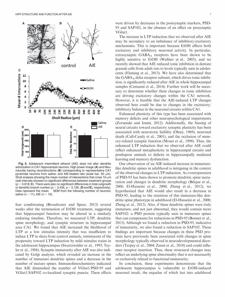
the decrease in PSD-95 and SAP102 within CA1 (i.e., fewerbranches = fewer synapses). Therefore, we assessed changesin dendritic branch number and length and conducted aSholl analysis. There was no significant change in the Shollanalysis, F(1, 1,076) = 0.249, p = 0.618 (Fig. 5), dendriticbranch length/total outgrowth, t(22) = 0.168, p = 0.434, orbranch number, t(22) = 1.18, p = 0.126 (Fig. 5), suggestingthat gross changes in neuronal morphology are not drivingthe changes in PSD-95 and SAP102 puncta observed inFigs 3 and 4.
DISCUSSION
The long-term impact of repeated EtOH exposure duringadolescence has become a topic of intensive interest andrecent investigation. Because EtOH is well known to affectmemory and hippocampal function, and to do so morepotently during adolescence than adulthood (Spear andSwartzwelder, 2014; White and Swartzwelder, 2004), theenduring effects of AIE on memory and hippocampal func-tion are of particular interest. Previous studies have shownthat AIE alters hippocampal synaptic function and plasticity(Sabeti and Gruol, 2008), but those studies did not addresswhether the effects of AIE persist into adulthood. Studies ofthe more enduring effects of AIE have shown impairment ofspatial learning (Sircar and Sircar, 2005) and retention of
A
B C
D
E
Fig. 3. Adolescent intermittent ethanol (AIE) reduces co-localized pre-and postsynaptic VGlut1/PSD-95 in adulthood. Schematic of a hippocam-pal slice showing the major projecting pathways in relation to the region ofinterest (ROI) in CA1 (A). Representative images of immunolabeling ofCA1 hippocampal neurons in control and AIE animals for pre-(VGlut1) andpost-(PSD-95) (B) synaptic markers. Arrows indicate yellow co-localizedpre-(VGlut1) and post-(PSD-95) synaptic puncta. AIE significantlydecreased co-localized pre- and postsynaptic puncta (*p = 0.012, t-test)(C), indicative of a decrease in synapse formation. This co-localization wasunaffected by the number of VGlut1 puncta staining after AIE (p = 0.06, t-test) (D) but rather was influenced by a significant decrease in the numberof PSD-95 puncta (*p = 0.017, t-test) (E). Three image stacks/animal andn = 5 animals/treatment, *p < 0.05. Scale bar represents 10 lm.
A B
C
D
Fig. 4. Adolescent intermittent ethanol (AIE) reduces co-localized pre-and postsynaptic VGlut1/SAP102 in adulthood. Representative images ofimmunolabeling of CA1 hippocampal neurons in control and AIE animalsfor pre-(VGlut1) and post-(SAP102) (A) synaptic markers. Arrows indicateyellow co-localized pre-(VGlut1) and post-(SAP102) puncta. AIE signifi-cantly decreased co-localized pre- and postsynaptic puncta (*p = 0.013, t-test) (B). This co-localization was unaffected by the number of presynapticVGlut1 puncta staining after AIE (p = 0.19, t-test) (C) but rather was influ-enced by a significant decrease in the number of postsynaptic SAP102puncta (*p = 0.005, t-test) (D). Three image stacks/animal and n = 5 ani-mals/treatment, *p < 0.05. Scale bar represents 10 lm.
6 RISHER ET AL.
fear conditioning (Broadwater and Spear, 2013) severalweeks after the termination of EtOH treatment, suggestingthat hippocampal function may be altered in a similarlyenduring timeline. Therefore, we measured LTP, dendriticspine morphology, and synaptic structure in hippocampalarea CA1. We found that AIE increased the likelihood ofLTP at a low stimulus intensity that was insufficient toinduce LTP in slices from control animals, reminiscent of thepropensity toward LTP induction by mild stimulus trains inthe adolescent hippocampus (Swartzwelder et al., 1995; Tey-ler et al., 1988). Synaptic immaturity after AIE was also indi-cated by Golgi analysis, which revealed an increase in thenumber of immature dendritic spines and a decrease in thenumber of mature spines. Immunohistochemistry indicatedthat AIE diminished the number of VGlut1/PSD-95 andVGlut1/SAP102 co-localized synaptic puncta. These effects
were driven by decreases in the postsynaptic markers, PSD-95 and SAP102, in the absence of an effect on presynapticVGlut1.The increase in LTP induction that we observed after AIE
may be secondary to an imbalance of inhibitory/excitatorymechanisms. This is important because EtOH affects bothexcitatory and inhibitory neuronal activity. In particular,extrasynaptic GABAA receptors have been shown to behighly sensitive to EtOH (Wallner et al., 2003), and werecently showed that AIE reduced tonic inhibition in dentategranule cells from adult rats to levels typically seen in adoles-cents (Fleming et al., 2013). We have also determined thatthe GABAA delta receptor subunit, which drives tonic inhibi-tion, is significantly reduced after AIE in whole hippocampalsamples (Centanni et al., 2014). Further work will be neces-sary to determine whether these changes in tonic inhibitionare driving excitatory changes within the CA1 network.However, it is feasible that the AIE-induced LTP changesobserved here could be due to changes in the excitatory/inhibitory balance in the neuronal circuits within CA1.Enhanced plasticity of this type has been associated with
memory deficits and other neuropsychological impairments(Zorumski and Izumi, 2012). Additionally, the biasing ofneural circuits toward excitatory synaptic plasticity has beenassociated with neurotoxic liability (Olney, 1969), neuronaldeath (Cull-Candy et al., 2001), and the occlusion of mem-ory-related synaptic function (Moser et al., 1998). Thus, theenhanced LTP induction that we observed after AIE couldreflect enhanced metaplasticity in hippocampal circuits andpredispose animals to deficits in hippocampally mediatedlearning and memory dysfunction.Our observation of an AIE-induced increase in immature-
like dendritic spines in adulthood is intriguing in the contextof the observed changes in LTP induction. As overexpressionof PSD-95 has been shown to promote dendritic spine matu-ration and changes in dendritic morphology (B�e€ıque et al.,2006; El-Husseini et al., 2000; Zheng et al., 2012), wehypothesized that AIE would also result in a decrease inPSD-95, leading to the retention of this immature-like den-dritic spine phenotype in adulthood (El-Husseini et al., 2000;Zheng et al., 2012). Also, if these dendritic spines were trulyimmature, and not just abnormal, they would contain moreSAP102: a PSD protein typically seen in immature spinesthat can compensate for reductions in PSD-95 (Bonnet et al.,2013). Although we found a reduction in PSD-95, indicativeof immaturity, we also found a reduction in SAP102. Thesefindings are important because changes in these PSD pro-teins have previously been associated with changes in spinemorphology typically observed in neurodevelopmental disor-ders (Tarpey et al., 2004; Zanni et al., 2010) and could influ-ence receptor insertion. Thus, these structural changes mayreflect an underlying spine abnormality that is not necessarilyor exclusively related to functional immaturity.In conclusion, these experiments demonstrate that the
adolescent hippocampus is vulnerable to EtOH-inducedneuronal insult, the sequelae of which last into adulthood
A B
C
D E
Fig. 5. Adolescent intermittent ethanol (AIE) does not alter dendriticarborization in CA1 hippocampal neurons. High power image (A) and Neu-rolucida tracing reconstructions (B) corresponding to representative CA1pyramidal neurons from saline- and AIE-treated rats (scale bar, 50 lm).Sholl analysis showing the mean number of intersections that cross 10-lmradii intervals showed no significant differences between treatment groups(p = 0.618;C). There were also no significant differences in total outgrowthor dendritic branch number (p = 0.434, p = 0.126, (D and E), respectively).Data represent the mean � SEM from the following number of neurons:saline (n = 11), AIE (n = 10).
HIPP STRUCTUREAND FUNCTION AFTER AIE 7
and include morphological changes, alterations in memory-related synaptic plasticity, and the expression of postsynapticMAGUKs. These findings support the emerging hypothesisthat AIE induces an array of pathological neural changesthat can manifest with immature-like characteristics intoadulthood.
ACKNOWLEDGMENTS
Many thanks to Dana Morin, Hannah Sexton, JenniferBourne, Jonnathan Singh Alvarado, and Alexander Alvara-do Singh for their technical support. Thanks to Lesa Hall forthe artwork. The work was supported by U01AA019925NADIA (to HSS), UO1AA020938 and BX-001271-02 (toSDM), DA031833 (to CE), 2T32NS51156-6 and 1F32NS083283-01A1 (toWCR), U.S. Department of Veterans Affairs,Senior Research Career Scientist award (to HSS), BX-002128-01 (to RLF), VA CDAs IK2BX001267 (to SKA) and2-010-10S (to RLF), Institute for Medical Research (toRCK), DVAMC VSN 6MIRECC (to SDM and RCK), andK99-AA22651 (to TW). CE was an Esther and Joseph Klin-genstein Fund Fellow andAlfred P. Sloan Fellow.
REFERENCES
Acheson SK, Bearison C, Risher ML, Abdelwahab SH, Wilson WA, Swar-
tzwelder HS (2012) Effects of acute or chronic ethanol exposure during
adolescence on behavioral inhibition and efficiency in a modified water
maze task. PLoS ONE 8:1–15.B�e€ıque J-C, Lin D-T, Kang M-G, Aizawa H, Takamiya K, Huganir R
(2006) Synapse-specific regulation of AMPA receptor function by PSD-95.
Proc Natl Acad Sci USA 103:19535–19540.Bonnet SA, Akad DS, Samaddar T, Liu Y, Huang X, Dong Y, Schl€uter OM
(2013) Synaptic state-dependent functional interplay between postsynaptic
density-95 and synapse-associated protein 102. J Neurosci 33:13398–13409.
Bourne J, Harris K (2007) Do thin spines learn to be mushroom spines that
remember? Curr Opin Neurobiol 17:381–386.Bourne J, Harris K (2008) Balancing structure and function at hippocampal
dendritic spines. Annu RevNeurosci 31:47–67.Bourne J, Harris K (2011) Coordination of size and number of excitatory
and inhibitory synapses results in a balanced structural plasticity along
mature hippocampal CA1 dendrites during LTP. Hippocampus 21:354–373.
Broadwater M, Spear LP (2013) Consequences of ethanol exposure on
cued and contextual fear conditioning and extinction differ depending
on timing of exposure during adolescence or adulthood. Behav Brain
Res 256:10–19.Brown S, Tapert S, Granholm E, Delis D (2000) Neurocognitive functioning
of adolescents: effects of protracted alcohol use. Alcohol Clin Exp Res
24:164–171.Brun VH, Ytterbo K, Morris RG, Moser MB, Moser EI (2001) Retrograde
amnesia for spatial memory induced by NMDA receptor-mediated long-
term potentiation. J Neurosci 21:356–362.Centanni SW, Teppen T, Risher ML, Fleming RL, Moss JL, Acheson
SK, Mulholland PJ, Pandey SC, Chandler LJ, Swartzwelder HS (2014)
Adolescent alcohol exposure alters GABAA receptor subunit expression
in adult hippocampus. Alcohol Clin Exp Res 38:2800–2808.Choudhury S, Blakemore S-J, Charman T (2006) Social cognitive develop-
ment during adolescence. Soc Cogn Affect Neurosci 1:165–174.Cull-Candy S, Brickley S, Farrant M (2001) NMDA receptor subunits:
diversity, development and disease. Curr Opin Neurobiol 11:327–335.
Diaz-Granados J, Graham D (2007) The effects of continuous and intermit-
tent ethanol exposure in adolesence on the aversive properties of ethanol
during adulthood. Alcohol Clin Exp Res 31:2020–2027.El-Husseini A, Schnell E, Chetkovich D, Nicoll R, Bredt D (2000) PSD-95
involvement in maturation of excitatory synapses. Science 290:1364–1368.Elias GM, Elias LA, Apostolides PF, Kriegstein AR, Nicoll RA (2008) Dif-
ferential trafficking of AMPA and NMDA receptors by SAP102 and
PSD-95 underlies synapse development. Proc Natl Acad Sci USA
105:20953–20958.Fiala JC (2005) Reconstruct: a free editor for serial section microscopy.
J Microsc 218:52–61.Fleming R, Acheson S, Moore S, Wilson W, Swartzwelder H (2012) In the
rat, chronic intermittent ethanol exposure during adolescence alters the
ethanol sensitivity of tonic inhibition in adulthood. Alcohol Clin Exp Res
36:279–285.Fleming R, Li Q, Risher M-L, Sexton H, Moore S, Wilson W, Acheson S,
Swartzwelder H (2013) Binge-pattern ethanol exposure during adoles-
cence, but not adulthood, causes persistent changes in GABAA receptor-
mediated tonic inhibition in dentate granule cells. Alcohol Clin Exp Res
37:1154–1160.Hanson K,MedinaK, Padula C, Tapert S, Brown S (2011) Impact of adoles-
cent alcohol and drug use on neuropsychological functioning in young
adulthood: 10-year outcomes. J Child Adolesc Subst Abuse 20:135–154.Ippolito D, Eroglu C (2010) Quantifying synapses: an immunocytochemis-
try-based assay to quantify synapse number. J Vis Exp 45: 1–8.Kilb W (2012) Development of the GABAergic system from birth to adoles-
cence. Neuroscientist 18:613–630.Klein RC, Saini S, Risher M-L, Acheson SK, Fleming RL, Sexton HG,
Swartzwelder HS, Moore SD (2014) Regional-specific effects of ovarian
hormone loss on synaptic plasticity in adult human APOE targeted
replacement mice. PLoS ONE 9:e94071.
Li Q, Fleming R, Acheson S, Madison R, Moore S, Risher M-L, Wilson W,
Swarztwelder H (2013) Long-term modulation of A-type K(+) conduc-tances in hippocampal CA1 interneurons in rats after chronic intermittent
ethanol exposure during adolescence or adulthood. Alcohol Clin Exp Res
37:2074–2085.Little P, Kuhn C, Wilson W, Swartzwelder H (1996) Differential effects of
ethanol in adolescent and adult rats. Alcohol Clin Exp Res 20:1346–1351.Markwiese B, Acheson S, Levin E,WilsonW, Swartzwelder H (1998) Differ-
ential effects of ethanol on memory in adolescent and adult rats. Alcohol
Clin Exp Res 22:416–421.Matsuzaki M (2007) Factors critical for the plasticity of dendritic spines and
memory storage. Neurosci Res 57:1–9.Matthews D, Tinsley K, Diaz-Granados J, Tokunaga S, Silvers J (2008)
Chronic intermittent exposure to ethanol during adolescence produces tol-
erance to the hypnotic effects of ethanol in male rats: a dose-dependent
analysis. Alcohol 42: 617–621.Moser E, Krobert K, Moser M, Morris R (1998) Impaired spatial learning
after saturation of long-term potentiation. Science 281:2038–2042.NIAAA (2004) NIAAA council approves definition of binge drinking. NIA-
AA Newsletter . Available at: http://pubs.niaaa.nih.gov/publications/
Newsletter/winter2004/Newsletter_Number3.pdf. Accessed April 12,
2015.
Olney J (1969) Brain lesions, obesity, and other disturbances in mice treated
with monosodium glutamate. Science 164:719–721.Paus T (2005) Mapping brain maturation and cognitive development during
adolescence. Trends Cogn Sci 9:60–68.RisherM-L, Fleming R, Boutros N, Semenova S,WilsonW, Levin E,Mark-
ou A, Swartzwelder H, Acheson S (2013) Long-term effects of chronic
intermittent ethanol exposure in adolescent and adult rats: radial-arm
maze performance and operant food reinforced responding. PLoS ONE 8:
e62940.
Risher WC, Ustunkaya T, Singh Alvarado J, Eroglu C (2014) Rapid Golgi
analysis method for efficient and unbiased classification of dendritic spines.
PLoS ONE 9:1–8.Sabeti J, Gruol D (2008) Emergence of NMDAR-independent long-term
potentiation at hippocampal CA1 synapses following early adolescent
8 RISHER ET AL.

exposure to chronic intermittent ethanol: role for sigma-receptors. Hippo-
campus 18:148–168.Silvers JM, Tokunaga S, Mittleman G, Matthews DB (2003) Chronic inter-
mittent injections of high-dose ethanol during adolescence produces meta-
bolic, hypnotic, and cognitive tolerance in rats. Alcohol Clin Exp Res
27:1606–1612.Silvers JM, Tokunaga S,Mittleman G, O’Buckley T,Morrow AL,Matthews
DB (2006) Chronic intermittent ethanol exposure during adolescence
reduces the effect of ethanol challenge on hippocampal allopregnanolone
levels andMorris water maze task performance. Alcohol 39:151–158.Sircar R, Sircar D (2005) Adolescent rats exposed to repeated ethanol treat-
ments show lingering behavioral impairments. Alcohol Clin Exp Res
29:1402–1410.Spear L (2000) The adolescent brain and age-related behavioral manifesta-
tions. Neurosci Biobehav Rev 24:417–463.Spear LP, Swartzwelder HS (2014) Adolescent alcohol exposure and persis-
tence of adolescent-typical phenotypes into adulthood: a mini-review.
Neurosci Biobehav Rev 45:1–8.Squeglia LM, Pulido C, Wetherill RR, Jacobus J, Brown GG, Tapert SF
(2012) Brain response to working memory over three years of adolescence:
influence of initiating heavy drinking. J Stud Alcohol Drugs 73:749.
Swartzwelder H, Wilson W, Tayyeb M (1995) Age-dependent inhibition of
long-term potentiation by ethanol in immature versus mature hippocam-
pus. Alcohol Clin Exp Res 19:1480–1485.Tada T, Sheng M (2006) Molecular mechanisms of dendritic spine morpho-
genesis. Curr Opin Neurobiol 16:95101.
Tarpey P, Parnau J, BlowM,Woffendin H, Bignell G, Cox C, Cox J, Davies
H, Edkins S, Holden S, Korny A, Mallya U, Moon J, O’Meara S, Parker
A, Stephens P, Stevens C, Teague J, Donnelly A, Mangelsdorf M, Mulley
J, Partington M, Turner G, Stevenson R, Schwartz C, Young I, Easton D,
Bobrow M, Futreal PA, Stratton MR, Gecz J, Wooster R, Raymond FL
(2004) Mutations in the DLG3 gene cause nonsyndromic X-linked mental
retardation. Am J HumGenet 75:318–324.Teyler TJ, Perkins AT, Harris KM (1988) The development of long-term
potentiation in hippocampus and neocortex. Neuropsychologia 27:31–39.
Wallner M, Hanchar HJ, Olsen RW (2003) Ethanol enhances a4b3r and
a6b3r ϒ-aminobutyric acid type A receptors at low concentrations known
to affect humans. Proc Natl Acad Sci USA 100: 1518–1523.White A, Ghia A, Levin E, Swartzwelder H (2000) Binge pattern ethanol
exposure in adolescent and adult rats: differential impact on subsequent
responsiveness to ethanol. Alcohol Clin Exp Res 24: 1251–1256.White AM, Swartzwelder HS (2004) Hippocampal function during adoles-
cence: a unique target of ethanol effects. Ann N Y Acad Sci 1021:206–220.
Yuste R (2009) Dendritic Spines. MIT Press Books, Cambridge, MA.
Zanni G, van Esch H, Bensalem A, Saillour Y, Poirier K, Castelnau L, Rop-
ers HH, de Brouwer AP, Laumonnier F, Fryns J-PP, Chelly J (2010) A
novel mutation in the DLG3 gene encoding the synapse-associated protein
102 (SAP102) causes non-syndromic mental retardation. Neurogenetics
11:251–255.Zheng S, Gray E, Chawla G, Porse B, O’Dell T, Black D (2012) PSD-95 is
post-transcriptionally repressed during early neural development by
PTBP1 and PTBP2. Nat Neurosci 15:381–388, S1.Zorumski C, Izumi Y (2012) NMDA receptors and metaplasticity: mecha-
nisms and possible roles in neuropsychiatric disorders. Neurosci Biobehav
Rev 36:989–1000.
HIPP STRUCTUREAND FUNCTION AFTER AIE 9