Adherus Dural and Spinal Sealant as Adjuncts to … were no adverse test article- ... smooth,...
12
HyperBranch Medical Technology, Inc. 2010-TR-04876-04876 R1 Adherus Dural and Spinal Sealant as Adjuncts to Sutured Dural Repair in a Canine Lumbar Durotomy Repair Model Anthony L. Asher, MD, FACS 1 , Michael A Carnahan, PhD 2 , Robert B. Boyd, DVM 3 , Eric L. Adams 3 , Mark T. Butt, DVM, DACVP 4 1. Carolina Neurosurgery & Spine Associates, Charlotte, NC 2. HyperBranch Medical Technology, Inc, Durham, NC 3. Northern Biomedical Research, Inc. Muskegon, MI 4. Tox Path Specialists, LLC, Hagerstown, MD SUMMARY: The objective of this GLP study was to evaluate the safety and effectiveness of Adherus Dural Sealant and Adherus Spinal Sealant when used to achieve watertight dural closure in a canine lumbar durotomy repair model. Another important objective of this study was to determine whether Adherus Dural and Adherus Spinal Sealants would inhibit the formation of peridural fibrosis and dural adhesions as normal healing occurred. The formulations were easy to apply, setting in the expected time frame to form an approximate 2 mm hydrogel film over the durotomy site. Application of Adherus Dural Sealant or Adherus Spinal Sealant provided 100% water-tight closure at time of surgery, as well as at four or five days, two months and four months post-operatively. There were no adverse test article- related effects in clinical observations, body weight, food consumption, physical or neurological parameters, clinical pathology parameters, CSF total cell count or chemistry parameters during this study. In addition, there was no histopathological evidence that application of Adherus Dural Sealant or Adherus Spinal Sealant had any adverse effects on the adjacent tissues, including the spinal cord and spinal nerve roots. Furthermore, the formulations did not appear to impede healing of the surgical site. Adherus Dural and Adherus Spinal Sealant, in general, significantly limited dura adhesions at both the two month and four month necropsy intervals. Peridural fibrosis and dura thickening/fibrosis was also reduced in animals that received Adherus Dural or Spinal treatment over that in control animals which did not receive either of the test articles. INTRODUCTION Two of the prevalent difficulties associated with spinal procedures are the prevention of cerebrospinal fluid (CSF) leaks and the prevention of adhesions between epidural scar tissue and the dura. CSF leaks are either the result of an incidental dural tear or an intentional durotomy and, if not properly managed, may significantly increase hospitalization times and the cost of patient treatment. 1 Furthermore, as the surgical site heals, peridural scar tissue is produced, often blending almost imperceptibly with the dura in the months following the procedure. The formation of this scar tissue may lead to complications such as radicular and low back pain. A variety of adjunctive treatments have been used to either augment dural closure or to minimize dural adhesions. These products typically lack the mechanical and adhesive strength to provide confident water-tight closure or lack the residence time and durability to serve as a barrier against the formation of dural adhesions. Additionally, some of the existing products have the tendency to expand in situ, creating the risk of symptoms or physical deficit from compression of neural structures. Adherus Dural and Spinal Sealants both have been designed specifically to both limit postoperative CSF leaks and inhibit dural adhesions. The products form densely crosslinked
Transcript of Adherus Dural and Spinal Sealant as Adjuncts to … were no adverse test article- ... smooth,...

HyperBranch Medical Technology, Inc. 2010-TR-04876-04876 R1
Adherus Dural and Spinal Sealant as Adjuncts to Sutured Dural Repair in a Canine
Lumbar Durotomy Repair Model
Anthony L. Asher, MD, FACS1, Michael A Carnahan, PhD
2, Robert B. Boyd, DVM
3, Eric L. Adams
3,
Mark T. Butt, DVM, DACVP4
1. Carolina Neurosurgery & Spine Associates, Charlotte, NC 2. HyperBranch Medical Technology, Inc, Durham,
NC 3. Northern Biomedical Research, Inc. Muskegon, MI 4. Tox Path Specialists, LLC, Hagerstown, MD
SUMMARY: The objective of this GLP study was to evaluate the safety and effectiveness of Adherus
Dural Sealant and Adherus Spinal Sealant when used to achieve watertight dural closure in a canine
lumbar durotomy repair model. Another important objective of this study was to determine whether
Adherus Dural and Adherus Spinal Sealants would inhibit the formation of peridural fibrosis and dural
adhesions as normal healing occurred. The formulations were easy to apply, setting in the expected time
frame to form an approximate 2 mm hydrogel film over the durotomy site. Application of Adherus
Dural Sealant or Adherus Spinal Sealant provided 100% water-tight closure at time of surgery, as well
as at four or five days, two months and four months post-operatively. There were no adverse test article-
related effects in clinical observations, body weight, food consumption, physical or neurological
parameters, clinical pathology parameters, CSF total cell count or chemistry parameters during this
study. In addition, there was no histopathological evidence that application of Adherus Dural Sealant or
Adherus Spinal Sealant had any adverse effects on the adjacent tissues, including the spinal cord and
spinal nerve roots. Furthermore, the formulations did not appear to impede healing of the surgical site.
Adherus Dural and Adherus Spinal Sealant, in general, significantly limited dura adhesions at both the
two month and four month necropsy intervals. Peridural fibrosis and dura thickening/fibrosis was also
reduced in animals that received Adherus Dural or Spinal treatment over that in control animals which
did not receive either of the test articles.
INTRODUCTION
Two of the prevalent difficulties associated with
spinal procedures are the prevention of
cerebrospinal fluid (CSF) leaks and the prevention
of adhesions between epidural scar tissue and the
dura. CSF leaks are either the result of an
incidental dural tear or an intentional durotomy
and, if not properly managed, may significantly
increase hospitalization times and the cost of
patient treatment.1 Furthermore, as the surgical
site heals, peridural scar tissue is produced, often
blending almost imperceptibly with the dura in the
months following the procedure. The formation
of this scar tissue may lead to complications such
as radicular and low back pain.
A variety of adjunctive treatments have been
used to either augment dural closure or to
minimize dural adhesions. These products
typically lack the mechanical and adhesive
strength to provide confident water-tight closure
or lack the residence time and durability to serve
as a barrier against the formation of dural
adhesions. Additionally, some of the existing
products have the tendency to expand in situ,
creating the risk of symptoms or physical deficit
from compression of neural structures.
Adherus Dural and Spinal Sealants both have
been designed specifically to both limit
postoperative CSF leaks and inhibit dural
adhesions. The products form densely crosslinked

HyperBranch Medical Technology, Inc. 2010-TR-04876-04876 R1
hydrogels that swell minimally and adhere
persistently to the dura while forming a robust
barrier to both prevent CSF leaks and dural
adhesions. Degradable ester linkages allow the
product to degrade after sufficient time has lapsed
to permit the dura to heal and the initial healing
response to subside.
To date, these sealants have undergone extensive
in vitro biocompatibility and preclinical safety
and effectiveness testing2. These studies indicate
that Adherus Dural and Spinal sealants are safe
and perform well as adjuncts to standard methods
of dural repair. In a pre-clinical canine cranial
durotomy repair model3 there were no adverse
clinical effects related to the application of
Adherus Dural Sealant and physical and
neurological evaluations were normal. During
this study, Adherus Dural Sealant was 100%
effective in sealing cranial CSF leaks
intraoperatively and postoperatively at six months
when challenged at CSF pressures up to 44 mm
Hg. In addition, Adherus Dural Sealant allowed
the dura to heal without complication over the
following months. Monthly MRI evaluations
demonstrated a consistent degradation profile over
approximately three months. Histopathological
examination following the two necropsy time
points revealed no adverse changes in the brain,
calvarium, dura, meninges or non-nervous system
organs associated with the test article in any of the
animals.
The following study extends the use of Adherus
Dural Sealant to spinal procedures and examines
the use of a new but similar Adherus Spinal
Sealant formulation. Adherus Spinal Sealant
contains all of the same components and the same
ratio of crosslinking components as Adherus
Dural Sealant, but differs in the overall
crosslinker content (10 wt% versus 15 wt% in
Adherus Dural Sealant) and set time
(approximately 30-35 seconds versus 1-2 seconds
for Adherus Dural Sealant)
MATERIALS and METHODS
PEG-Based Surgical Sealants
Adherus Dural Sealant
The Adherus Dural Sealant system (HyperBranch
Medical Technology, Inc.) is a synthetic hydrogel
sealant designed for use as an adjunct to standard
methods of dural repair to provide watertight
closure. At time of use, the crosslinking
components, an activated polyethylene glycol
(PEG) and polyethyleneimine (PEI), are
reconstituted with their respective reconstitution
buffers and withdrawn into 5 mL syringes. The
two syringes are then coupled to a Micromedics
Spray Applicator Kit.
To dispense the sealant system, the user depresses
the plunger cap which forces equal amounts of
each solution down separate cannulas inside the
applicator toward the mix tip. As the solutions
pass through the mix tip, the solution paths merge
immediately prior to exiting the tip. The mixed
solution is expressed as a spray and deposited on
the surgical site where it will rapidly polymerize
to form a compliant, well adhered film. Once the
plunger assembly is released, the flow of solution
ceases.
Adherus Spinal Sealant
The Adherus Spinal Sealant system (HyperBranch
Medical Technology, Inc.) is a synthetic hydrogel
sealant designed for use an adjunct to standard
methods of dural repair to provide watertight
closure, and as an adhesion barrier for the
inhibition of post surgical peridural fibrosis and
dural adhesion. The applicator for Adherus
Spinal Sealant is ideally designed to deliver the
formulation to tight surgical sites and allow for
precise application of the mixed formulation into

HyperBranch Medical Technology, Inc. 2010-TR-04876-04876 R1
smaller spaces which can be encountered during
spinal surgery.
At time of use, the polyethyleneimine (PEI)
crosslinker is first reconstituted by the buffer
within the same syringe. Following
reconstitution, the syringe is coupled to the vial
adapter assembly, the PEG powder in the glass
vial is attached to the vial adapter spike and an
angled applicator tip is also attached, via luer
lock, to the other end of the three-way valve. At
this stage, the device is ready for use.
To apply Adherus Spinal Sealant, the solution in
the syringe must be pushed through the syringe
chambers, through the three-way valve and into
the glass vial. This action reconstitutes the PEG
powder in the glass vial, allowing for the mixing
of the two components and the initiation of a
crosslinking reaction. The syringe plunger is then
allowed to recoil, pulling the solution out of the
vial and back into the syringe assembly. The PEG
is fully reconstituted by depressing and
withdrawing the syringe plunger one additional
time. Following an approximate 10 second
reconstitution, the vial adapter and vial are
removed, automatically turning the three way
valve to allow solution to flow down the
applicator tip. The mixed solution is expressed by
depressing the syringe plunger to deliver the
desired amount of the crosslinking formulation.
Once delivered, the formulation will set within
approximately thirty seconds.
Adherus Sealants
Once applied, the Adherus Sealants adhere
tenaciously to the underlying tissue providing a
smooth, lubricous coating which prevents
cerebrospinal fluid leaks and minimizes or
prevents adhesions to bone and other tissue
structures. The hydrogels are resorbed as the
underlying tissue heals, establishing a competent
CSF barrier and separating the dura from other
tissues to prevent dural adhesions. The
degradable ester linkages are designed to allow
controlled degradation of the hydrogel over the
course of eight to twelve weeks. Due to the
structure of the hydrogel, the Adherus Sealant
systems swell minimally after implantation,
exhibiting at most only a 6% dimensional change
in any axis. The degradation byproducts (mainly
PEG) are water-soluble and are cleared through
the renal and hepatic pathways. No toxic
byproducts are created and the sealant does not
interfere with tissue healing.
Surgical Procedure
Group Treatment
Number of Animals /
Necropsy Interval
2 Month 4 Month
1 Control 3 3
2 Adherus Dural
Sealant 3 3
3 Adherus Spinal
Sealant 3 3
Table 1 Treatment groups and number of animals at
each necropsy interval.
This study was conducted at Northern Biomedical
Research, Inc, in accordance with the United
States Food and Drug Administration (FDA)
Good Laboratory Practice Regulations (GLP)
(21CFR Part 58), the Japanese Ministry of Health,
Labor, and Welfare (MHLW) Good Laboratory
Practice Standards Ordinance 21, and the
Organization for Economic Cooperation and
Development (OECD) Principles of Good
Laboratory Practice [C (97) 186/Final]. Eighteen
male beagle dogs (Covance Research Products,
Inc., Kalamazoo, Michigan) were utilized for this
study. The dogs were approximately 9-18 months
old and weighed 10.0 to 13.7 kg. The animals

HyperBranch Medical Technology, Inc. 2010-TR-04876-04876 R1
were randomly assigned to three treatment groups
as illustrated in Table 1.
All animals were pretreated with atropine sulfate
SQ (0.04 mg/kg). Approximately fifteen minutes
later, an IV dose of sodium thiopental (16 mg/kg)
was provided to induce sedation. If necessary, an
anesthetic mask (4% isoflurane, 1 liter/min
oxygen) was used to aid in sedation. Animals
were intubated and maintained on approximately
1 liter/minute of oxygen and approximately 2.0%
isoflurane. Prednisolone sodium succinate IV (30
mg/kg) and flunixin meglumine IM (2 mg/kg)
were administered prior to surgery.
In surgery under aseptic conditions, a midline
lumbar incision was made and the skin and the
musculature reflected from the L3 spinous process
and lamina. A hemilaminectomy was made at L3
using a cutting burr drill bit. A 0.9 mm OD x 0.5
mm ID polyurethane catheter was inserted into the
subarachnoid space for collection of CSF and a
baseline CSF pressure reading. For the first
control animal, a purse string suture (6-0 nylon)
was used to secure the catheter to the dorsal dura.
Sutures were not used to secure the catheter in
subsequent surgeries due to the technical
difficulty of suture placement inside the
laminectomy defect. Dental acrylic, lightly
applied within the laminectomy defect, was
subsequently used to secure the catheter in the
remainder of the experimental animals. The
catheter was connected to an intracranial pressure
(ICP) transducer (Codman Microsensor ICP
Transducer) and three way stop cock via a
stainless steel blunt-tipped needle. Once the
catheter was implanted, a baseline CSF pressure
was recorded using the pressure transducer. After
obtaining the baseline reading, additional
musculature was reflected over the L2 spinous
process and lamina. The dorsal aspect of the L2
lamina was removed with a burr drill bit and the
dura was exposed. The dura and arachnoid were
Figure 1 Intraoperative view of the approximate 1 cm
dural incision closed with 4 interrupted 6-0 nylon
sutures.
Figure 2 Intraoperative view of the approximate 1 cm
dural incision closed with 4 interrupted 6-0 nylon
sutures and approximately 2 mm of Adherus Spinal
Sealant over the suture line.
incised to a length of approximately 1.0 cm in the
parasagittal plane and CSF was allowed to freely
egress. The dural incision was then re-
approximated with four evenly spaced simple
interrupted 6-0 nylon sutures and blotted dry
(Figure 1). In group 2 and 3 animals, one of the
two test articles was then applied to the sutured
dural closure to a thickness of approximately 2
mm over the defect (Figure 2). No test article was
applied to the Group 1 control animals. The same
procedure was followed for the L5 vertebral
segment. Adequacy of dural closure was
evaluated intraoperatively by infusing saline
through the previously implanted intradural
catheter and monitoring the ICP with the pressure
transducer. The maximum intradural pressure
reached and maintained for 30 seconds in both
control and experimental animals was recorded.
The intradural catheter was then tied off in all
animals. The paraspinous muscle and connective
tissues were closed in layers with absorbable

HyperBranch Medical Technology, Inc. 2010-TR-04876-04876 R1
sutures, and the skin was closed with absorbable
sutures and tissue adhesive. The animals were
recovered from anesthesia, placed in a protective
jacket, administered butorphanol IM (0.05 mg/kg)
and placed on a post surgical antibiotic (ceftiofur
sodium IM (5 mg/kg) b.i.d., one injection during
or immediately prior to surgery followed by three
injections post operatively).
Following surgery, clinical observations were
performed daily, food consumption was
monitored daily after the first week, body weights
were monitored weekly, clinical pathology was
collected prior to necropsy, CSF samples were
collected before necropsy, and neurological and
physical examinations were performed at one
week and at necropsy.
Immediately prior to necropsy at the specified
endpoints the animals were sedated with sodium
thiopental IV (16 mg/kg) taken to surgery,
maintained on inhalant anesthesia (approximately
1 L/min of oxygen and 2-5% halothane or
isoflurane) and the durotomy sites were surgically
re-explored. A midline lumbar incision was made
and the musculature was reflected to expose the
durotomy sites. At both the two month and four
month necropsy, nearly all of the surgical sites
were found to have significant connective tissue
overgrowth. The sites were not further explored
and the animals were immediately euthanized and
given an IV bolus of heparin sodium, 200 IU/kg.
The animals were subsequently perfused via the
left cardiac ventricle with 0.001% sodium nitrite
in saline followed by 10% neutral buffered
formalin fixative. Tissues were then procured
from all animals at necropsy and maintained in
10% neutral buffered formalin. The surgical sites
were removed en bloc in order that those areas
could be scored for fibrosis and adhesions
histologically.
Durotomy Site Evaluations by MRI
On study Day 5 or 6, the animals were dosed with
atropine SQ, 0.04 mg/kg and thiopental sodium
IV, 16 mg/kg (50 mg/mL), intubated and
maintained on inhalant anesthesia (isoflurane). A
20 or 22 gauge IV catheter coated with heparin
was inserted into the cephalic vein and attached to
a slow drip of 0.9% sodium chloride. The animals
were subsequently taken for MRI evaluation on a
Philips Achieva 3T MRI Scanner. T2 sagittal
(DRIVE and FLAIR), pre-contrast sagittal T1-
weighted turbo spin echo (TSE), axial turbo field
echo and post-contrast sagittal T1-weighted
sequences were performed for the evaluation.
Gadolinium IV (gadodiamide) was the contrast
agent used at a dose of 0.1 mmol/kg (0.2 ml/kg).
Histopathology
Histopathologic evaluations were completed on
all designated tissues harvested at necrospy. At
the L2 and L5 surgical sites, the vertebrae/spinal
cord/dura specimen was decalcified. The section
was then cut as close to the middle of the surgical
site as could be determined in the wet tissue. The
cranial side of the site was embedded in one
block. The caudal side of the site was embedded
in the following block. Dorsal nerve roots were
examined along with the spinal cord sections. All
sections were embedded in paraffin and stained
with hematoxylin and eosin.
At each surgical site, the bone, muscle/soft tissue,
peridural tissue/space, dura, spinal cord, catheter
track (for the indwelling pressure recording
catheter) and spinal nerve roots were examined.
Sections of the caudal cervical spinal cord and
cauda equina were examined in order to
investigate the possibility of distant spinal cord
effects. Multiple sections at each surgical site
were examined.
Both peridural adhesions and fibrosis were
evaluated histologically. For the purposes of this

HyperBranch Medical Technology, Inc. 2010-TR-04876-04876 R1
study, adhesions are defined as the extent of
attachments between the dura and other tissues in
the epidural space. Fibrosis is defined by the
extent of fibrous connective tissue formation in
the epidural space. The grading scheme used to
evaluate dural adhesions is outlined in Table 2.
For the determination of dural adhesions, the
pathologist was blinded to treatment groups. The
grading scheme used to record peridural fibrosis is
outlined in Table 3.
Grade Characterization Description*
0 0-25% Adhesions present on small
portion of the dura
1 25-50% Adhesions present on up to half
of the dura
2 50-75% Adhesions present on a majority
of the dura
3 75-100% Adhesions present on all of the
dura
*Percentages based upon the portion of the dura exposed or
adjacent to the surgical site. If a complete bone roof had formed
over the dura, the scar extent was considered a 0.
Table 2 Grading scheme for dural adhesions.
Table 3 Grading scheme for peridural fibrosis.
Statistical Analysis
Body weights, body weight changes, clinical
pathology data (hematology, serum chemistry and
coagulation), CSF total cell count, CSF chemistry,
heart rate, body temperature, respiration, and food
consumption data were analyzed by a one-way
analysis of variance and comparison of the control
group to the treated groups by Dunnett’s test.
Analysis was two-tailed for significance levels of
5% and 1%. A probability value <0.5 was
considered statistically significant.
For dural adhesion and peridural fibrosis
analysis, an F test was performed to determine if
the variances in the two test populations were
equivalent. In instances where the variances were
determined to be equivalent, a t-test for equivalent
variances was used to determine statistical
significance of the difference in means. Likewise,
in cases where the F test showed unequal
variances, a t-test for unequal variances was used
to determine statistical significance of the
difference in means.
RESULTS
Clinical and Neurological Evaluations
All animals recovered from the surgical
procedure, and the animals remained
neurologically intact throughout the course of the
study. The surgical sites healed as expected and
showed no signs of infection or delay in healing.
Furthermore, the application of Adherus Dural or
Adherus Spinal Sealant produced no adverse test
article-related effects in clinical observations,
body weight, food consumption, clinical
pathology parameters or CSF total cell count or
chemistry parameters during this study.
Pressure Testing at Surgery
At surgery, baseline CSF pressures (before
durotomy) were recorded for each animal in
Grade Description
0 No tissue affected
1
Severity grade slight; A severity grade of 1
denotes a very slight change, barely
discernable in the section, and affecting less
than 1% of the potentially affected tissue.
2
Severity grade minimal; A severity grade of 2
denotes a microscopic change slightly more
pronounced that a grade 1, but still affecting a
very small portion (less than 5%) of the
potentially affected tissue.
3
Severity grade mild; A severity grade of 3
denotes a microscopic change slightly more
pronounced that a grade 2, still limited in
distribution, but readily apparent in the section.
4
Severity grade moderate; A severity grade of 4
denotes a microscopic change slightly more
pronounced that a grade 3, affecting a majority
of the potentially affected tissue or space,
readily apparent in the section.
5 Severity grade severe; A severity grade of 5
denotes the change was among the most
pronounced observed in the study.
HyperBranch Medical Technology, Inc. 2010-TR-04876-04876 R1
Groups 1 through 3. The individual durotomy
sites were tested for CSF leakage following dural
closure and maximum intradural pressure
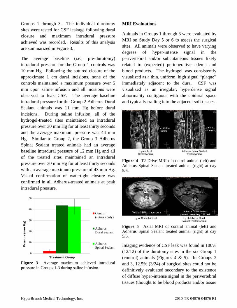
achieved was recorded. Results of this analysis
are summarized in Figure 3.
The average baseline (i.e., pre-durotomy)
intradural pressure for the Group 1 controls was
10 mm Hg. Following the sutured closure of the
approximate 1 cm dural incisions, none of the
controls maintained a maximum pressure over 5
mm upon saline infusion and all incisions were
observed to leak CSF. The average baseline
intradural pressure for the Group 2 Adherus Dural
Sealant animals was 11 mm Hg before dural
incisions. During saline infusion, all of the
hydrogel-treated sites maintained an intradural
pressure over 30 mm Hg for at least thirty seconds
and the average maximum pressure was 44 mm
Hg. Similar to Group 2, the Group 3 Adherus
Spinal Sealant treated animals had an average
baseline intradural pressure of 12 mm Hg and all
of the treated sites maintained an intradural
pressure over 30 mm Hg for at least thirty seconds
with an average maximum pressure of 43 mm Hg.
Visual confirmation of watertight closure was
confirmed in all Adherus-treated animals at peak
intradural pressure.
0
10
20
30
40
50
Treatment Group
Pre
ssu
re (
mm
Hg)
Control
(sutures only)
Adherus
Dural Sealant
Adherus
Spinal Sealant
Figure 3 Average maximum achieved intradural
pressure in Groups 1-3 during saline infusion.
MRI Evaluations
Animals in Groups 1 through 3 were evaluated by
MRI on Study Day 5 or 6 to assess the surgical
sites. All animals were observed to have varying
degrees of hyper-intense signal in the
perivertebral and/or subcutaneous tissues likely
related to (expected) perioperative edema and
blood products. The hydrogel was consistently
visualized as a thin, uniform, high signal “plaque”
immediately adjacent to the dura. CSF was
visualized as an irregular, hyperdense signal
abnormality contiguous with the epidural space
and typically trailing into the adjacent soft tissues.
Figure 4 T2 Drive MRI of control animal (left) and
Adherus Spinal Sealant treated animal (right) at day
5/6.
Figure 5 Axial MRI of control animal (left) and
Adherus Spinal Sealant treated animal (right) at day
5/6.
Imaging evidence of CSF leak was found in 100%
(12/12) of the durotomy sites in the six Group 1
(control) animals (Figures 4 & 5). In Groups 2
and 3, 12.5% (3/24) of surgical sites could not be
definitively evaluated secondary to the existence
of diffuse hyper-intense signal in the perivertebral
tissues (thought to be blood products and/or tissue

HyperBranch Medical Technology, Inc. 2010-TR-04876-04876 R1
edema). In contrast to unequivocal radiographic
examples of CSF fistula in the control animals,
this imaging feature was not contiguous with the
subdural space, presented above the hydrogel and
lacked homogenous signal characteristics. In the
21 evaluable experimental surgical sites, there
was no radiographic evidence (0%) of CSF leak.
(Figures 4 and 5).
Histopathology
Two Month Necropsy
There were no significant histological differences
between the control and test article treated groups
at the level of the cervical spinal cord or cauda
equina. Between the surgical sites (i.e., at L3/L4),
slight to minimal nerve fiber degeneration was
observed and was likely due to local spinal cord
damage occurring secondary to the surgical
procedure (laminectomy) or (more likely) the
placement/presence of the catheter used to
monitor cerebrospinal fluid pressure.
The dura was observed to be intact without any
visible gaps (healed) in all treatment groups at
both surgical sites (L2 and L5). Foamy
macrophages were noted at the periphery of the
site of test article application in the Adherus-
treated groups, but were not observed at the
control sites. Overall, there was no evidence that
application of Adherus Dural Sealant or Adherus
Spinal Sealant had any detrimental/adverse effects
on the local tissues (including the spinal cord) or
impeded healing at the surgical site.
As compared to the control animals in which
adhesions in three of six sites were graded as a 3
on the adhesion scale, two sites were graded as a
2, and one site lacked any adhesions,
administration of Adherus Dural Sealant to a
durotomy site appeared to essentially eliminate
dura adhesions based on microscopic examination
of the surgical sites two months post-surgery
(average scores presented in Figure 8). Although
Figure 6 Representative photomicrographs of
histology slides from Group 1 (A), Group 2 (B), and
Group 3 (C).at 2 month necropsy. Figure 6A shows
the spinal cord, dura and peridural tissue are all
adhered and prominent peridural fibrosis is evident in
the control while Figure 6B and Figure 6C show little
peridural fibrosis and no dural adhesions in Adherus-
treated animals. In figure 6B, the space to the right of
the dura was most likely occupied by the Adherus
sealant. In Figure 6C, bone has formed a nearly
complete shelf over the dura. Bar = 500 m.
A
B A
C

HyperBranch Medical Technology, Inc. 2010-TR-04876-04876 R1
the hydrogel typically does not survive
histological processing, in five of six sites that
were treated with Adherus Dural Sealant, an
approximate 1.5 mm thick space adjacent to the
dura and/or persistent material (thought to be
Adherus Dural Sealant) appeared to completely
block the formation of dura adhesions. One site,
which appeared to lack the same epidural space as
previously described, was graded as a 3.
Dura adhesions were also decreased in the
Adherus Spinal Sealant treated animals. Three of
six sites had no adhesions, one site was graded as
an adhesion score 1, and two other sites were
graded as a 2 (average score presented in Figure
8). At microscopic examination, the presence or
absence of the test material or an epidural space
was not as noticeable when compared to the
Adherus Dural Sealant-treated sites.
There was, in general, also a decrease in peridural
fibrosis in the Adherus Dural Sealant and Adherus
Spinal Sealant groups as compared to the controls
(average scores presented in Figure 9). The
difference was most notable in the Adherus Dural
Sealant group in which three of six sites had no
peridural fibrosis, one site was graded as a 1 on
the fibrosis scale, and two other sites were graded
as a 2 as compared to the control animals in which
five of six sites were graded as a 4 and one site
was graded as a 3. In the Adherus Spinal Sealant
group, one of six sites was graded as a 1, two sites
were graded as a 2, and three sites were graded as
a 3.
Four Month Necropsy
Again, there were no significant histological
differences between the control and test article
treated groups at the cervical spinal cord or cauda
equina. Slight to minimal nerve fiber
degeneration was observed at the L 3/4 level
secondary to the laminectomy and/or
Figure 7 Representative photomicrographs of
histology slides from Group 1 (A), Group 2 (B), and
Group 3 (C) at 4 month necropsy. Figure 7A shows
the dura completely adhered to the overlying
connective tissue while Figure 7B and Figure 7C show
little peridural fibrosis and no dural adhesions in
Adherus-treated animals. In each photomicrograph,
the spinal cord is visible on the left hand side. Bar =
500 m.
A
C
B

HyperBranch Medical Technology, Inc. 2010-TR-04876-04876 R1
placement/presence of the catheter used to
monitor intradural cerebrospinal fluid pressure.
The dura was observed to be healed in all
treatment groups at both durotomy sites (L2 and
L5). Foamy macrophages in the peridural tissue,
noted at the two month sacrifice in the Adherus-
treated animals, were nearly (Adherus Dural
Sealant) or totally (Adherus Spinal Sealant)
absent in both treatment groups by the four month
sacrifice. Furthermore, there was no evidence that
application of Adherus Dural Sealant or Adherus
Spinal Sealant had any detrimental/adverse effects
on the local tissues (including the spinal cord) or
impeded healing at the surgical site.
As compared to the control animals (Group 1),
dura adhesions were markedly decreased (on
average) in the Adherus Dural Sealant and
Adherus Spinal Sealant groups. For the control
sites, four of six sites were graded as a 3 on the
adhesion grading scale and two sites did not have
adhesions. Three of six Adherus Dural Sealant
treatment sites did not have adhesions, one site
was graded as a 1 and both sites on one animal
were graded as a 3. Five of the six Adherus
Spinal Sealant treatment sites did not have
adhesions and one site was graded as a 2 (average
scores presented in Figure 8).
Figure 8 Dural adhesion scoring for all treatment
groups at two and four months.
Peridural fibrosis was also somewhat decreased at
4 months in both Adherus treated groups as
compared to controls. In contrast to the two
month sacrifice group, the distinct space above
the dura/peridural tissue at the surgical site in the
Adherus Dural Sealant animals was not apparent
at four months. This observation likely indicates
resorption of the test article during the four month
period followed by connective tissue filling the
space.
During peridural fibrosis evaluation, two of the
six control sites were each graded as either a 2, 3,
or 4. Two of the six Adherus Dural Sealant sites
were each graded as either a 1 or 2 respectively on
the fibrosis scale while one site was graded as a 3
and one site was graded as a 4. The Adherus
Spinal Sealant group was observed to have one
site free of peridural fibrosis, two sites graded as a
1, two sites graded as a 2 and one site graded as a
3 (average scores presented in Figure 9).
Figure 9 Peridural fibrosis scoring for all treatment
groups at two and four months.
DISCUSSION
Following spine surgery, Adherus Dural and
Spinal Sealants successfully and safely
established a watertight seal at all sites. As the
dura healed, the sealants were able to maintain
this watertight seal and served as a physical
barrier between the dura and the epidural space,
limiting dural adhesions.

HyperBranch Medical Technology, Inc. 2010-TR-04876-04876 R1
Pressure testing was performed at surgery
following closure of the durotomy sites. This test
was performed to rigorously test the hydrogel’s
strength intraoperatively and is intended to
simulate the pressures produced within the lumbar
cistern after patients assume an upright posture in
the peri-operative period.4 In the Group 1 control
animals 0/6 animals were able to maintain the
protocol stated pressure of 30 mm of Hg for 30
seconds. Following dural incision and primary
repair, all control animals leaked at a pressure at
or below 5 mm of Hg. The Group 2 and Group 3
animals, treated with Adherus Dural Sealant and
Adherus Spinal Sealant respectively, all were able
to withstand CSF pressures over 30 mm Hg for 30
seconds. This suggests that Adherus sealants can
significantly augment standard dural closures and
provide watertight seals that can withstand supra-
physiological pressures for sustained periods.
MRI analysis was utilized to determine if the test
articles were able to maintain a watertight seal on
Study Day 5 or 6. The control animals were also
examined to determine whether the untreated
durotomy sites still had evidence of CSF leakage.
All control animals (Group 1) were found to have
evidence of spontaneous CSF leaks at both
surgical sites. Animals treated with Adherus
Dural Sealant (Group 2) or the Adherus Spinal
Sealant (Group 3) showed no evidence of
spontaneous CSF leaks, although three sites could
not be definitively evaluated based on extensive
hyper-intense signal at or near the surgical sites.
As noted previously, varying degrees of hyper-
intense signal within the perivertebral or
subcutaneous tissues was observed in all animals
and is believed to be secondary to post surgical
effects (likely tissue edema and/or blood
products). Individual variation and slight
differences in surgical technique could account
for the more prominent soft tissue signal found in
three of the animals. Although the presence of a
CSF leak cannot be ruled out at these three sites,
the lack of communication between this
radiographic finding and the subdural space
suggest that a CSF leak was not present in these
few animals.
Although precise comparisons cannot be made
between intraoperative photographs of the
Adherus hydrogels within the confines of the
laminectomy site at time zero (which document
approximately 2 mm of hydrogel over the incision
and approximately 3 mm near the edges of the
laminectomy site) and subsequent post operative
MRIs, it is of interest that MRI images at day 5-6
suggest no appreciable change in the size of the
hydrogel within that timeframe. This dimensional
stability appears to be confirmed with MRI
images obtained at post-op days five, 14, and 28
during a pilot laminectomy animal study of the
same design5. Necropsy at the 1 month time
interval during this pilot laminectomy study also
confirmed that the Adherus hydrogel does not
undergo any appreciable dimensional changes5.
These findings are confirmed by in vitro data
which has documented minimal swelling of the
Adherus hydrogel over time.6 This finding is of
great clinical importance as volumetric expansion
within the epidural space runs the risk of inducing
neurological deficit. This possibility is of more
than theoretical concern. Other hydrogel dural
sealants possessing less favorable dimensional
stability have been observed to create
neurological signs and symptoms secondary to
volumetric expansion in both spine and cranial
surgeries7,8
.
Clinically, there were no test article-related
changes in hematology, serum chemistry and
coagulation parameters throughout the study.
There were also no test article-related changes in
CSF total cell counts or CSF chemistry.
During histopathological examination, foamy
macrophages were present at the periphery of the
site of test article application in the Adherus Dural

HyperBranch Medical Technology, Inc. 2010-TR-04876-04876 R1
Sealant and Adherus Spinal Sealant groups. It
was presumed that the macrophages were
recruited in response to the test articles. The
presence of the foamy macrophages indicated
phagocytosis of the test article.
Histopathologic examination and grading also
indicated that both hydrogel sealants successfully
limited dural adhesions and epidural fibrosis
while not impeding the rate of dural healing.
Administration of Adherus Dural Sealant to a
durotomy site essentially eliminated dura
adhesions when examined two months post
surgery. At four months following the application
of Adherus Dural Sealant, dura adhesions were
decidedly decreased as compared to control
animals that had a durotomy but were not treated
with a sealant. Administration of Adherus Spinal
Sealant to a durotomy site dramatically reduced
dura adhesions when examined two months post
surgery. When examined at four months post
surgery, Adherus Spinal Sealant essentially
eliminated dura adhesions as 5 of 6 Adherus
Spinal Sealant treated surgical sites examined
were without any visible dura adhesions.
Peridural fibrosis and dura thickening/fibrosis in
general were reduced at two and four months post
surgery following the application of Adherus
Dural Sealant and Adherus Spinal Sealant.
Finally, there was no evidence that application of
Adherus Dural Sealant or Adherus Spinal Sealant
had any adverse effects on the adjacent tissues,
including the spinal cord and spinal nerve roots.
Also, Adherus Dural Sealant or Adherus Spinal
Sealant application did not appear to impede
healing of the surgical site.
CONCLUSIONS
Combined results from cranial and spine studies
suggest that the Adherus Dural and Spinal
Sealants are ideal adjuncts to standard methods of
dural repair to provide watertight closure.
Adherus Dural and Adherus Spinal Sealants
appear to lack neuro or systemic toxicity,
consistently seal the dura to prevent CSF leaks (in
both the intraoperative and postoperative time
periods) and allow normal healing of the dura
while reducing dural adhesions and epidural
fibrosis.
DISCLOSURE
Dr. Asher is a compensated consultant to HyperBranch
Medical Technology, Inc. and has received compensation in
the form of consultant fees, stock and stock options.
REFERENCES
1 Grotenhuis JA.: Costs of postoperative cerebrospinal fluid
leakage: 1-year, retrospective analysis of 412 consecutive
nontrauma cases. Surg Neurol 64: 490-494, 2005.
2 Data on file at HyperBranch Medical Technology, Inc.
3 Data on file at HyperBranch Medical Technology, Inc. and
summarized in 2010-CS-04875-04875 R1
4Bono F, Giliberto C, Lavano A, Quattrone A: Posture-
related cough headache and orthostatic drop in lumbar CSF
pressure. J. Neurol 252: 237-238, 2005
5 Data on file at HyperBranch Medical Technology, Inc.
6 Data on file at HyperBranch Medical Technology, Inc. and
summarized in 2010-CS-04867-04867 R1
7 Mulder M, Crosier J, Dunn R: Cauda Equina compression
by hydrogel dural sealant after a laminotomy and
discectomy. Spine 34: E144-E148, 2009
8 Blackburn SL, Smyth MD: Hydrogel-induced
cervicomedullary compression after posterior fossa
decompression for Chiari malformation. J Neurosurg (4
Suppl Pediatrics) 106: 302-304, 2007