Adaptation to oxidative stress by Gram-positive … the cell must be maintained in order to avoid...
10
Adaptation to oxidative stress by Gram-positive bacteria: the redox sensing system HbpS-SenS-SenR from Streptomyces reticuli Matthew R. Groves 1 , and Darío Ortiz de Orué Lucana 2* 1 EMBL Hamburg Outstation, c/o DESY, Building 25A, Notkestrasse 85, 22603 Hamburg, Germany. 2 University of Osnabrueck, Faculty of Biology and Chemistry, Department of Applied Genetics of Microorganisms, Barbarastr. 13, 49069 Osnabrueck, Germany. *Corresponding author. E-mail: [email protected] , Tel.: 00495419693439 In common with all other living organisms, Gram-positive bacteria must continuously deal with stress situations in vivo. Such stress conditions may include changes in environmental temperature, pH, humidity, etc. In the case of many pathogens - including Mycobacterium tuberculosis, Staphylococcus aureus, Corynebacterium diphtherieae, Enterococcus phaecalis, Streptococcus pneumoniae or Bacillus anthracis - such changes include an efficient response to changing environments during both infection and virulence within the host. Other non-pathogenic and soil-dwelling bacteria, such as Streptomyces reticuli, Bacillus subtillis, Corynebacterium glutamicum or Mycobacterium snegmatis, must also respond to a wide variety of environmental stimuli including, UV-radiation or nutritional stress. All these bacteria have developed sensory systems that facilitate adaptation to changes in the environmental conditions. Recently, the three-component signalling system HbpS-SenS-SenR from the cellulose degrader Streptomyces reticuli has been reported as an example of a redox sensing pathway in bacteria. Using a combination of structural biology and in vivo and in vitro experiments it was demonstrated that the extracellular oligomer-forming protein HbpS acts in concert with the two-component system SenS- SenR in the sensing and reaction of the bacteria to heme- and iron-mediated oxidative stress. HbpS can bind and degrade heme via a non-enzymatic pathway known as coupled oxidation and it has been proposed that HbpS communicates the extracellular presence of heme and oxidative stress to the bacteria through the membrane-embedded sensor histidine kinase SenS using HbpS-bound iron ions. SenS subsequently phosphorylates the response regulator SenR, acting as a transcriptional regulator of a number of genes encoding for redox active proteins that are involved in the adaptation of the bacteria to oxidative stress. In this report, we analyze the sensing of signals by Gram-positive bacteria upon oxidative stress. We also focus further on the HbpS-SenS-SenR system and provide new insights into molecular mechanism used by Gram-positive bacteria to monitor and react to changes in environmental conditions. A knowledge of the interaction and reaction of bacteria to changes in their environment will open new avenues of understanding that could have many applications in fields as diverse as agriculture, biotechnology, ecosystem monitoring as well as human medicine. Keywords: Streptomyces; soil-dwelling bacteria; biopolymer degradation; oxidative stress; sensory systems, environmental signalling 1. Introduction 1.1 Oxidative stress While in vitro cultured bacteria have well defined and relatively static conditions under which growth can proceed, bacteria in vivo must respond efficiently and rapidly to any potentially damaging changes in their immediate environment, such as temperature, salinity, osmotic pressure, pH, the presence or absence of nutrient sources, etc. In addition, bacteria are rarely found in isolation and have developed complex mechanisms to allow each bacterium to compete with other nearby species for resources. This has given rise to the production of natural antibiotics by some species (particularly from the order Actinomycetales: streptomycin, platensimycin, chloramphenicol, erythromycin, vancomycin, etc) to provide an edge in competition for resources [1,2]. Hemin is one such natural antibiotic and has the effect of creating oxidative stress conditions, which may provide a basis for one bacterial species to outcompete its immediate neighbours. Oxidative stress is defined as interference in the balance between the production of reactive oxygen species (ROS), including free radicals, oxides and peroxides (Table 1), and the ability of biological systems to readily detect their presence and detoxify ROS or repair the resulting damage. A common feature among the different ROS types is their capacity to cause oxidative damage to macromolecules - proteins, DNA, and lipids - leading to an increased rate of mutagenesis, and cell death. In humans for example, oxidative stress is involved in many diseases, such as atherosclerosis, Parkinson's disease, heart failure, myocardial infarction, or Alzheimer's disease. The manipulation of ROS can also be beneficial, as they are used by the human immune system to attack and kill pathogens. ROS are also used in cell signaling, where it is known as redox- signalling. In order to prevent damage to essential macromolecules living organisms generally maintain a reducing environment within their cells. To preserve this state, they have developed highly complex nonenzymatic and enzymatic protection, repair and detoxification mechanisms [3-6]. Most of the ROS naturally present in the environment are generated endogenously as a product of aerobic metabolism, or of enzymatic reactions (e.g. NO synthase). However, exogenous _______________________________________________________________________________________
Transcript of Adaptation to oxidative stress by Gram-positive … the cell must be maintained in order to avoid...

Adaptation to oxidative stress by Gram-positive bacteria: the redox
sensing system HbpS-SenS-SenR from Streptomyces reticuli
Matthew R. Groves1, and Darío Ortiz de Orué Lucana
2*
1 EMBL Hamburg Outstation, c/o DESY, Building 25A, Notkestrasse 85, 22603 Hamburg, Germany. 2 University of Osnabrueck, Faculty of Biology and Chemistry, Department of Applied Genetics of Microorganisms,
Barbarastr. 13, 49069 Osnabrueck, Germany. *Corresponding author. E-mail: [email protected], Tel.: 00495419693439
In common with all other living organisms, Gram-positive bacteria must continuously deal with stress situations in vivo. Such stress conditions may include changes in environmental temperature, pH, humidity, etc. In the case of many pathogens - including Mycobacterium tuberculosis, Staphylococcus aureus, Corynebacterium diphtherieae, Enterococcus
phaecalis, Streptococcus pneumoniae or Bacillus anthracis - such changes include an efficient response to changing environments during both infection and virulence within the host. Other non-pathogenic and soil-dwelling bacteria, such as Streptomyces reticuli, Bacillus subtillis, Corynebacterium glutamicum or Mycobacterium snegmatis, must also respond to a wide variety of environmental stimuli including, UV-radiation or nutritional stress. All these bacteria have developed sensory systems that facilitate adaptation to changes in the environmental conditions. Recently, the three-component signalling system HbpS-SenS-SenR from the cellulose degrader Streptomyces reticuli has been reported as an example of a redox sensing pathway in bacteria. Using a combination of structural biology and in vivo and in vitro experiments it was demonstrated that the extracellular oligomer-forming protein HbpS acts in concert with the two-component system SenS-SenR in the sensing and reaction of the bacteria to heme- and iron-mediated oxidative stress. HbpS can bind and degrade heme via a non-enzymatic pathway known as coupled oxidation and it has been proposed that HbpS communicates the extracellular presence of heme and oxidative stress to the bacteria through the membrane-embedded sensor histidine kinase SenS using HbpS-bound iron ions. SenS subsequently phosphorylates the response regulator SenR, acting as a transcriptional regulator of a number of genes encoding for redox active proteins that are involved in the adaptation of the bacteria to oxidative stress. In this report, we analyze the sensing of signals by Gram-positive bacteria upon oxidative stress. We also focus further on the HbpS-SenS-SenR system and provide new insights into molecular mechanism used by Gram-positive bacteria to monitor and react to changes in environmental conditions. A knowledge of the interaction and reaction of bacteria to changes in their environment will open new avenues of understanding that could have many applications in fields as diverse as agriculture, biotechnology, ecosystem monitoring as well as human medicine.
Keywords: Streptomyces; soil-dwelling bacteria; biopolymer degradation; oxidative stress; sensory systems, environmental signalling
1. Introduction
1.1 Oxidative stress
While in vitro cultured bacteria have well defined and relatively static conditions under which growth can proceed, bacteria in vivo must respond efficiently and rapidly to any potentially damaging changes in their immediate environment, such as temperature, salinity, osmotic pressure, pH, the presence or absence of nutrient sources, etc. In addition, bacteria are rarely found in isolation and have developed complex mechanisms to allow each bacterium to compete with other nearby species for resources. This has given rise to the production of natural antibiotics by some species (particularly from the order Actinomycetales: streptomycin, platensimycin, chloramphenicol, erythromycin, vancomycin, etc) to provide an edge in competition for resources [1,2]. Hemin is one such natural antibiotic and has the effect of creating oxidative stress conditions, which may provide a basis for one bacterial species to outcompete its immediate neighbours. Oxidative stress is defined as interference in the balance between the production of reactive oxygen species (ROS), including free radicals, oxides and peroxides (Table 1), and the ability of biological systems to readily detect their presence and detoxify ROS or repair the resulting damage. A common feature among the different ROS types is their capacity to cause oxidative damage to macromolecules - proteins, DNA, and lipids - leading to an increased rate of mutagenesis, and cell death. In humans for example, oxidative stress is involved in many diseases, such as atherosclerosis, Parkinson's disease, heart failure, myocardial infarction, or Alzheimer's disease. The manipulation of ROS can also be beneficial, as they are used by the human immune system to attack and kill pathogens. ROS are also used in cell signaling, where it is known as redox-signalling. In order to prevent damage to essential macromolecules living organisms generally maintain a reducing environment within their cells. To preserve this state, they have developed highly complex nonenzymatic and enzymatic protection, repair and detoxification mechanisms [3-6]. Most of the ROS naturally present in the environment are generated endogenously as a product of aerobic metabolism, or of enzymatic reactions (e.g. NO synthase). However, exogenous
_______________________________________________________________________________________

sources of ROS include: a) The exposure of living organisms to ionizing (γ) and nonionizing irradiation (UV); which leads to the production of a number of radical and peroxide species from ionization of intracellular water, b) Air pollutants such as car exhaust, cigarette smoke, and industrial contaminants, c) Different drugs including narcotic drugs and anesthetizing gases, d) Xenobiotics and chemicals produce ROS as a by-product of their metabolism in vivo, or e) Food. (f) It has also been suggested that the generation of ROS is induced by bactericidal antibiotics to kill bacteria in a complex pathway, in which iron-ions and the Fenton reaction play a role [7]. Generation of ROS has been also detected in a wide range of plant or human pathogen interactions. For example, a natural defense mechanism is the “oxidative burst” generated during phagocytosis. In this mechanism, material imported from the environment during phagocytosis is rapidly oxidised and any potential threats, such as pathogens, may be neutralised. However, a number of the more successful human pathogens have developed defense strategies against this - as will be described in more detail below. ROS can be also produced during the degradation of natural existing biopolymers (cellulose, chitin or xylan) by microorganisms [8]. Table 1. List of some reactive oxygen species (ROS); the table has been adapted from [9,4].
ROS Structure Description ROS controlling family
Hydrogen peroxide H2O2 Formed by dismutation of O2•- or by direct reduction of O2. Lipid soluble and thus able to diffuse across membranes. Substrate of the Fenton reaction.
Catalase-peroxidases (cf .1.4.1) eg. CpeB
Superoxide anion O2•- Formed in many autooxidation reactions and by electron transport chain. Undergoes dismutation to form H2O2 spontaneously or by enzymatic catalysis and is a precursor for metal-catalyzed OH• formation.
Superoxide dismutases (cf. 1.4.2) eg. SodA, SodB
Hydroxyl radical OH• Formed by the Fenton reaction and decomposition of peroxynitrite. Extremely reactive.
Catalase-peroxidases (cf .1.4.1) eg. CpeB
Hypochlorous acid HOCl
Formed from H2O2 by myeloperoxidase. Lipid soluble and highly reactive. Will readily oxidize protein constituents, including thiol groups, amino groups and methionine.
Not discussed in this review
Nitric oxide NO
Formed endogenously from arginine and oxygen by various nitric oxide synthases (NOS) and by reduction of inorganic nitrate. Reacts with O2•- to produce peroxynitrite. Highly reactive.
Not discussed in this review
Peroxynitrite anion ONOO- Formed in a rapid reaction between O2•- and NO•. Lipid soluble and similar in reactivity to HOCl•. Protonation forms peroxynitrous acid, which can undergo homolytic cleavage to form OH• and NO2.
Not discussed in this review
Organic hydroperoxide
ROOH Formed by radical reactions with cellular components such as lipids and nucleobases.
Alkylhydroperoxide reductases (cf. 1.4.3) eg. AhpC and AhpD
1.2 Iron-mediated oxidative stress
Iron is the fourth most abundant element in the Earth`s crust and is an essential trace mineral for nearly all known organisms. Under physiological conditions, it exists predominantly in the oxidized Fe3+ (ferric) form. However, reduced Fe2+ (ferrous) is also present within biological systems, but is tightly regulated and controlled due to its high oxidative potential. With a redox potential ranging from -300 to +700 mV, iron is an excellent “all-round” prosthetic component that can be incorporated into proteins either as a mono- or binuclear species, or in a more complex form as part of iron-
_______________________________________________________________________________________

sulfur clusters or heme groups. In the reduced form it plays a crucial role in many biological processes, as photosynthesis, N2 fixation, H2 production and consumption, respiration, oxygen transport and gene regulation [10-12]. However, in the presence of oxygen, ferrous ions frequently result in oxidative stress through the generation of ROS. Oxidation of ferrous iron by molecular oxygen (Reaction 1) yields O2
•- that can undergo the dismutation reaction (Reaction 2) to form H2O2. Hydrogen peroxide in turn can react with the ferrous iron via the Fenton reaction (Reaction 3), generating a hydroxyl radical. Hydroxyl radicals are also generated via the Haber-Weiss reaction (Reaction 4). Thus, the presence of even small quantities of iron can have a major effect upon the oxidative state of the local environment. Fe2+ + O2 → Fe3+ + O2
•- (Reaction 1) 2 O2
•- + 2 H+ → H2O2 + O2 (Reaction 2) Fe2+ + H2O2 → Fe3+ + OH• + OH- (Reaction 3) O2
•- + H2O2 → OH• + OH- + O2 (Reaction 4)
1.3 Oxidative stress in Gram-positive pathogenic bacteria
As mentioned above, the interaction of pathogens with plants or human hosts leads to the generation of ROS. Most of the human pathogenic bacteria are Gram-positive organisms, including those from the genera: Staphylococcus, Streptococcus, Enterococcus, Bacillus, Corynebacterium, Nocardia, Clostridium, Listeria and Actinobacteria. These pathogens must be able to deal with the phagocyte-derived ROS, which is part of the burst of oxygen consumption that is associated with microbial killing [13,14]. For instance, the generation of reactive oxygen and nitrogen intermediates upon macrophage activation via interferon-γ is damaging to Mycobacterium tuberculosis [13]. To reduce the ROS effect of the phagocytic burst Mycobacterium tuberculosis has developed multiple strategies, including the production and excretion of a catalase-peroxidase (KatG), superoxide dismutases (SodA and SodC), and alkylhydroperoxide reductase (Ahp) [14-16]. While oxidative stress is a major component of cellular defense mechanisms, the reducing enviroment within the cell must be maintained in order to avoid macromolecular damage. Thus, other small proteins, such as thioredoxins or low molecular-weight thiols including glutathione and mycothiol, play a key role in maintaining a reducing environment both in the cell and in controlling the extent of the oxidative burst [17,18].
1.4 Enzymes detoxifying ROS
As can be seen from the section above, the control of ROS is not only necessary for pathogens, but is an important feature of almost all living organisms. It is perhaps not surprising that a wide variety of molecules have evolved in order to detoxify ROS (detailed in Table 1). Such enzymes include catalase-peroxidases, superoxide dismutases and alkylhydroperoxide reductases and a brief summary of the main properties of each of these families is given below:
1.4.1 Catalase-peroxidase (CpeB)
Catalases promote the disportionation of H2O2, contain heme as a prosthetic group and are homotetramers, while peroxidases use H2O2 to oxidize a number of compounds. The bifunctional catalase-peroxidases comprise varying ratios of these two enzymatic activities. The studied bacterial catalase-peroxidases usually contain 726-753 amino acids per subunit and consist of two highly homologous halves, each of which shares significant amino acid identity with eukaryotic monomeric peroxidases, including cytochrome c peroxidase (CCP) [19]. Amongst the catalase-peroxidases, CpeB from Streptomyces reticuli has also been shown to be involved in the mechanism of degradation of crystalline cellulose, the most abundant polysaccharide in soil [20]. It has been assumed that CpeB can participate in the initial attack on the insoluble cellulose during the degradation processes. CpeB might also protect Streptomyces reticuli against ROS that is known to be generated during biopolymer degradation [8]. CpeB shows a high degree of amino acid identity (62 % identity, 73 % similarity) to the catalase-peroxidase KatG from Mycobacterium tuberculosis. KatG plays an essential role in the survival of the organism in the environment of the phagocyte oxidative burst. Interestingly, the front-line anti-tuberculosis drug isoniazid (isonicotinic acid hydrazide) requires KatG activation before exerting a lethal effect [21].
1.4.2 Superoxide dismutase (SOD)
The substrate of SODs is the superoxide radical anion O2•- which is generated by the transfer of one electron to molecular oxygen (Table 1). This anion is involved in the generation of further ROS. SODs maintain the concentration of superoxide radicals in low limits through the catalysis of the dismutation of two molecules of O2•- (2 O2•- + 2H+ → O2 + H2O2). SODs are generally classified according to the metal species located at the redox-active centre. For example, Mycobacterium tuberculosis possesses both an Mn2+,Fe2+ SOD (SodA) as well as Cu2+, Zn2+-SOD (SodC), and both have also been associated with the detoxification of ROS during phagocytosis. A Ni2+-SOD has been characterized in some Streptomyces sp. [22].
_______________________________________________________________________________________

1.4.3 Alkylhydroperoxide reductase (Ahp)
These antioxidant proteins have a redox-active cysteine (the peroxidatic cysteine) that can be oxidised to a sulfenic acid by the peroxide substrate. The expression of Mycobacterium tuberculosis Ahp has been associated with the protection of bacilli against the toxic effects of organic peroxides during infection. Interestingly, it was observed that mutations leading to an over-expression of Ahp could compensate the loss of KatG activity in katG mutants [23].
2. Oxidative stress regulatory sensory systems
To stimulate the expression of proteins involved in the anti-oxidative stress response, bacteria have evolved different regulatory sensory systems. They are substantially diverse in terms of mode of action or number of implicated components. For example, there are single transcription regulators (e.g. OxyR, PerR or FurS) which display a sensory and response domain within itself [24-26]. Other encompass two different proteins responsible for either sensing or response, such two-component systems (e.g. ChrS-ChrA or VicR-VicK) sense in general an external stimulus through a membrane protein kinase which in turn activates/inactivates its cognate response regulator [27,28]. Recently, more sophisticated systems encompassing an additional third component have been described and characterised (e.g. HbpS-SenS-SenR or MtrAB-LpqB). This third component is able to modulate the activity of a two-component sensor kinase [29,30].
2.1 Single trancriptional regulators: OxyR and FurS
OxyR is an H2O2 sensing transcriptional activator that has been described in a number of Gram-negative as well as Gram-positive bacteria [31]. It is activated by an intramolecular disulfide bond (S-S) formation between two cysteine residues. Using crystal structure analyses of the E. coli OxyR it has been shown that the S-S formation between redox-sensitive cysteines provokes a major structural change in the regulator that affects the specificity of DNA recognition [32]. The S-S form of OxyR acts as a transcriptional activator of several genes involved in the antioxidative defence. oxyR genes have also been identified in Gram-positive bacteria, including Mycobacteria and Streptomyces. For instance, the OxyR protein from Streptomyces coelicolor A3(2) is involved in the regulation of the alkyl hydroperoxide reductase system (AhpC and AhpD). The corresponding genes are located directly downstream from oxyR and form an operon that is divergently transcribed than oxyR [24]. FurS is a zinc-containing redox regulator that in contrast to OxyR represses the transcription of the catalase-peroxidase cpeB gene in Streptomyces reticuli in its thiol-reduced (SH) form upon binding of an operator located upstream of the furS-cpeB operon [26]. However, under oxidative stress conditions an internal S-S bridge is formed within FurS, leading to the suggestion that it undergoes conformational changes. The S-S form of FurS is not able to bind the operator, losing the ability to block the transcription of furS-cpeB, which in turn leads to a high production of CpeB under oxidative stress conditions [33]. FurS contains six C-terminally located cysteine residues, four of them contained within two C-X-X-C motifs. Such a motif can be found in a number of redox-active proteins, including thioredoxin, glutaredoxins and thiol-disulfide oxidoreductases [34]. FurS is closely related to the Mycobacterium
tuberculosis FurA, which regulates katG transcription [35] and is considerably divergent from the global E. coli Fur regulator [36].
2.2 Two-component systems
Two component signal transduction systems (TCS) are one of the most important mechanisms by which bacteria sense, respond, and adapt to changes in their environment or in their intracellular state. These signalling systems typically consist of a sensor histidine kinase (SK), which is autophosphorylated upon signal recognition, and one or more cognate response regulators (RR) which alter their DNA binding behaviour upon phosphorylation [37]. A single bacterium may encode for ten to fifty and sometimes even up to two-hundred TCS [38]. Sequence analysis of the Streptomyces
coelicolor A3(2) genome revealed 84 SK- and 80 RR-genes, respectively [39] whereas that of Streptomyces avermitilis revealed 67 SK- and 68 RR-genes [40], suggesting that Streptomyces sp. are well equipped to respond to highly variable
environmental conditions. In comparison, the genome of the Gram-negative bacterium Escherichia coli encodes only 32 SKs and 23 RRs and five hybrid SKs [41]. The large number of different two-component systems within Actinobacteria appears to reflect their responsiveness to a wide range of rapidly changing environmental stimuli. Redox sensing systems are often related to oxygen (O2) sensing mechanisms and are widely distributed along bacteria, yeast and metazoans [42]. The TCS ChrS-ChrA from Corynebacterium diphteriae and the HssS-HssR system of Staphylococcus aureus are for example involved in the sensing of heme-mediated signals [27,43]. The exact mechanisms by which ChrS and HssS sense the corresponding signals are to date still unknown. A well-studied TCS is Dos/DevS-DosR from Mycobacterium tuberculosis. Unlike many pathogens, Mycobacterium tuberculosis can persist for several years within the host in a clinically latent state. Latency is often linked to hypoxic conditions. The heme-based SKs DosT and DevS act as triggers of the Mycobacterium tuberculosis hypoxia/latency response by direct sensing of O2. The deoxy form is the active kinase, and conversion to the oxy form switches off activity by preventing
_______________________________________________________________________________________
an initial autophosphorylation [44]. The RR DosR - known also as DevR - is a transcription factor that governs nearly all genetic responses to hypoxia [45]. DosR is activated by DosT and/or DevS [46].
2.3 Three-componen systems
We define three-component systems those ones consisting of a typical TCS and a third protein component without kinase, phosphatase or DNA-binding activities. This third component acts as an accessory protein that senses environmental conditions and thereby influences the kinase activity of its cognate SK. To date, a direct interaction and the kind of influence have only been described or postulated in a few cases. The majority of them are not involved in sensing of oxidative stress. One of the best-studied TCS with accessory proteins is YycFG, which has been shown to be essential for cell viability. It is highly conserved and specific to low-G+C content bacteria, such as Bacillus subtilis, Streptococcus pneumoniae, Staphylococcus aureus, and Enterococcus faecalis [47]. The activity of the SK YycG from Bacillus subtilis is modulated by two other proteins with N-terminal transmembrane helices, YycH and YyvI. Interestingly, all corresponding four genes are clustered and located in the same operon [48]. The role as accessory protein has been postulated for the lipoprotein (LpqB) that together with the TCS MtrA-MtrB might form an actinobacterial three-component system. The genes mtrA, mtrB and lpqB are also clustered and located as one transcriptional unit [30]. Recently, it was experimentally shown that the Mycobacterium tuberculosis LpqB interacts with the extracellular domain of the SK MtrB and affects the phosphorylation status of the RR MtrA, supporting that LpqB functions as an accessory protein that modulates activity of the MtrAB system in controlling homeostasis of the cell wall and cell division [49]. However, the nature of the environmental signal sensed by this system has been not yet described.
3. The redox-sensing system HbpS-SenS-SenR from Streptomyces reticuli
Streptomycetes are Gram-positive mycelium-forming soil bacteria that are the major producers of a large variety of chemically different antibiotics [1,2]. Streptomyces sp. are also able to degrade many macromolecules such as proteins (including keratin), lipids, nucleic acids, chitin and starch. Streptomyces reticuli (S. reticuli) hydrolyses efficiently the most abundant biopolymer in the nature - cellulose (Avicel). A number of extracellular or mycelium-associated enzymes, including cellulases, β-glucosidases and proteases, are involved in the degradation processes [50]. Interestingly, the catalase-peroxidase CpeB has been shown to be produced during cellulose degradation, in which CpeB participates either in the oxidation of cellulose facilitating a subsequent degradation or protection of S. reticuli from ROS that have been generated during cellulose degradation. We have previously identified and characterized the TCS SenS-SenR in S. reticuli that governs the transcription of the furS-cpeB operon encoding for the redox regulator FurS and the above mentioned CpeB [51]. The SK SenS comprises a membrane protein composed of five transmebrane regions. SenS has been shown to autophosphorylate at a conserved histidine residue and is able to transfer the phosphate group to its cognate response regulator, SenR, which possesses a C-terminal domain with a predicted helix-turn-helix (HTH) DNA binding motif. Upon phosphorylation SenR binds with high affinity to the upstream regions of the senS-senR operon and the gene hbpS, which encodes for a secreted heme-binding protein (HbpS). The phosphatase activity of SenS leads to the dephosphorylation of SenR, which binds upstream of the furS-cpeB operon [52]. Further comparative physiological and biochemical studies allowed the identification of two additional proteins, which are under the control of SenS-SenR [53]. One of these proteins shows significant amino acid identity to xylanase B from Streptomyces lividans, the other to a tyrosinase from Streptomyces
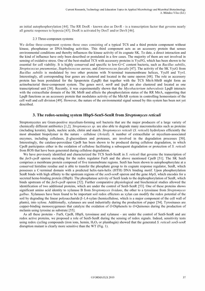
galbus. Xylanases have been found to be important soil redox effectors as xylan can modify the redox potential of the soil by degrading the linear polysaccharide β-1,4-xylan (hemicellulose, which is a major component of the cell wall of plants), into xylose. Additionally, xylanases are used industrially during the production of paper [54]. Tyrosinases are copper-binding monooxygenases that catalyze the oxidation of O-Diphenols to O-Quinones during the production of melanin using tyrosine as substrate [55]. As all these proteins - FurS, CpeB, HbpS, tyrosinase and xylanase - are under the control of SenS-SenR and are redox active proteins, we proposed a role of SenS-SenR during the sensing of redox signals. Indeed, sensitivity tests using redox cycling compounds (iron ions, hemin, H2O2 or plumbagin) showed that the generated S. reticuli senS-senR disruption mutant is clearly more sensitive than the WT (Fig. 1).
_______________________________________________________________________________________

Fig. 1. Live/dead viability assays. S. reticuli wild-type (WT), S. reticuli senS-senR disruption mutant (∆senS-senR) and S. reticuli hbpS disruption mutant (∆hbpS) were cultivated in the presence of the redox cycling compound plumbagin. Samples were subjected to a viability assay [56] and subsequently examined under UV using filter sets for FITC and Texas red. Living mycelia exhibited green fluorescence, whereas dead mycelia were seen in red. Almost identical results were obtained using the stressors: hemin, iron ions, or H2O2, respectively.
3.1 HbpS is an accessory protein of SenS-SenR
HbpS is an extracellular heme-binding protein, which has been shown to be secreted in a Tat (twin-arginine translocation) dependent manner [57,58]. Mutational analyses revealed that the presence of HbpS increases the synthesis of the highly active catalase-peroxidase CpeB in vivo and provides S. reticuli with resistance against different redox cycling compounds (Fig. 1). We have therefore proposed that HbpS interacts with an extracellular, membrane-associated or membrane-integrated protein(s) involved in a signal transduction cascade regulating cpeB transcription and redox stress resistance. Protein-protein interaction studies revealed that both native S. reticuli HbpS and recombinantly produced HbpS interact specifically with the SK SenS [51]. In addition, it was shown that the SenS N-terminal domain (containing predicted extracytoplasmic and transmembrane regions) is essential for the interaction [53]. Phosphorylation analyses of SenS in the presence of HbpS revealed that high quantities of heme-free HbpS inhibit the autophosphorylation of the sensor kinase under non-oxidative stress conditions. Thus, an interaction with heme-free HbpS is predicted to lead to an inactive conformation of SenS. However, HbpS significantly enhanced SenS autokinase activity in the presence of HbpS and heme alone or heme in combination with DTT or H2O2. Fe2+ and H2O2 are precursors of the Fenton reaction that catalyze the formation of hydroxyl radicals and Fe3+ [59]. Additional autokinase activity tests have shown that HbpS was also able to considerably enhance SenS autophosphorylation upon iron-mediated oxidative stress [53].
3.2 HbpS assembly and implications on SenS modulation
As mentioned above HbpS is secreted in a TAT-dependent manner. The most remarkable characteristic of the Tat pathway is that it functions to transport folded proteins of variable dimensions and oligomeric states across the cytoplasmic membrane. In most cases, the TAT substrates bind their corresponding cofactors in the cytoplasm and are thus already folded prior export. Such proteins function predominantly in respiratory and photosynthetic electron transport chains and are vital for many types of bacterial energy metabolism [60]. HbpS was immunologically detected in vivo in culture filtrates from Streptomyces as an oligomeric and heme-binding protein. Replacements of the two adjacent arginines - in the twin arginine motif (SRRTRV) within its signal sequence - by lysines abolish the secretion of HbpS [57], indicating that HbpS should be a substrate of the TAT pathway. To analyze HbpS in more detail, it was isolated as recombinant heme-free protein from an E. coli transformant [61]. Further spectroscopic analyses revealed that the recombinant HbpS specifically interacts with heme. Size exclusion chromatography combined with static light scattering (SLS) analyses allowed the identification of HbpS as an octomer in vitro. Moreover, we have elucidated the high resolution crystal structures (1.6 Å and 2.3 Å) of HbpS crystallized in the presence or absence of heme, both crystal structures revealed an octomeric assembly. The observed extensive interactions between the N-terminal residues within the HbpS octomer are suggested to control the oligomerization process. A detailed analysis of the crystal structure with subsequent mutagenesis and SLS analyses allowed the identification of two N-terminal residues (Ser26 and His28) that are essential for the octomeric assembly. Further electron density analysis from the crystal structure obtained in the presence of heme revealed the presence of a bound iron. Subsequently, spectroscopic and biochemical studies demonstrated that HbpS can degrade heme through a nonenzymatic H2O2-dependent mechanism known as coupled oxidation, leading to free iron [62]. Coupled oxidation has previously been demonstrated for other heme-binding proteins, such as myoglobin [63]. These data have resulted in
_______________________________________________________________________________________

the hypothesis that HbpS not only binds heme, but that HbpS supports coupled oxidation of the bound heme and then captures and displays the released iron atom. The captured iron is coordinated by a lysine residue (at position 108, Lys108) of HbpS that is located on the surface of the protein (Fig. 2).
Fig. 2. HbpS structures. Monomer (left), octomer without iron (middle) and octomer with iron (green sphere; right). The figure was produced using PyMol (DeLano Scientific; www.pymol.org).
As mentioned above the extracellular HbpS acts as an accessory protein within the three-component system HbpS-SenS-SenR, via the interaction with the membrane-embedded SK SenS. Protein-protein interactions and in vitro phosphorylation studies demonstrated that the octomeric assembly is a pre-requisite for efficient interaction with SenS and modulation of its phosphorylation state [62]. Previous data demonstrated that HbpS significantly enhanced SenS autokinase activity under heme-mediated oxidative stress conditions [53]. We propose that under such condition the coupled oxidation of heme, leading to a free iron, plays an important role. Interestingly, mutation of the Lys108 that binds the released iron significantly reduced HbpS-mediated autokinase activity of SenS in response to heme and oxidative stress. As HbpS modulates SenS autophosphorylation under heme/iron-mediated oxidative stress conditions it could be expected that these processes are in part controlled by conformational changes in HbpS. Indeed, using HbpS wild-type and mutants as well as different biochemical and biophysical approaches, including fluorescence resonance energy transfer (FRET) and CD-spectroscopy, we have also shown that iron-mediated oxidative stress induces both secondary structure and overall intrinsic conformational changes within HbpS. During these processes HbpS is oxidatively modified, leading to the generation of highly reactive carbonyl groups and tyrosine-tyrosine bonds. Oxidative modifications causing structural and conformational changes should be therefore responsible for the control of SenS and hence of the HbpS-SenS-SenR signalling cascade [64].
3.3 HbpS-SenS-SenR homolog systems in other Gram-positive bacteria
Analysis of the HbpS primary sequence using BlastP [65] and Pfam [66] yielded a number of putative homologues encoded within Gram-positive and Gram-negative bacterial genomes. Interestingly, each of the deduced HbpS-like proteins showed a predicted domain with unknown function termed “DUF336”. This domain is also present in other uncharacterised sequences, including several GlcG proteins and contains many conserved motifs that are suggestive of cofactor binding and enzymatic activity [61,57]. The majority of these homologs represent hbpS-like genes which are located in the direct vicinity of genes encoding for sensor kinases and response regulators [29]. Remarkably, only the hbpS-like genes from a number of Gram-positive bacteria (including Streptomyces coelicolor A3(2), Streptomyces
kasugaensis, Arthrobacter aurescens, Streptomyces griseus, Rhodococcus sp. RH1) are clustered with the hbpS, senS and senR genes in the same relative transcriptional orientation (Fig. 3). We would like to emphasize that for example the S. kasugaensis hbpS-senS-senR-like genes are localized in the direct vicinity of the “kas” cluster involved in the production of the aminoglycoside kasugamycin (EMBL: BAF79711.1). It would be interesting to explore if this system participates somehow in the biosynthesis or resistance against kasugamycin. Also the clarifying of the role of HbpS-SenS-SenR-like systems for the biology of Arthrobacter auresences TC1 or Rhodococcus sp. RH1 merit further interest, as both bacteria have enormous ecological importance. For instance, Arthrobacter aurescens TC1 has been shown to metabolize s-triazine compounds, which are common starting materials for the synthesis of pesticides, plastic resins, dyes, and explosives. Rhodococcus sp. RH1 is able to metabolize harmful environmental pollutants such as toluene, naphtalene, herbicides, PCBs. Thus, these soil bacteria are of high importance in bioremediation.
_______________________________________________________________________________________

Fig. 3. Relative location and transcriptional orientation of hbpS-, senS- and senR-like genes on different Gram-positive bacterial genomes. The S. reticuli hbpS (yellow), senS (orange) and senR (blue) gene products were analysed using BlastP. The corresponding homologs hbpS-like (white), senS-like (grey) and senR-like (black) are shown.
4. Conclusions and perspectives
To date, the HbpS-SenS-SenR system is the only three-component system involved in the detection of heme/iron-mediated oxidative stress signals characterised among Gram-positive bacteria. These signals induce a number of changes in HbpS, including the binding and display of an iron atom as well as conformational changes, leading to the recognition by its interaction partner - the histidine sensor kinase SenS. SenS is then activated and autophosporylates, subsequently transferring the phosphate group to the response regulator that then activates the transcription of genes (e.g. hbpS and cpeB) involved in anti-oxidative stress response. HbpS has been shown not only to bind heme but also to degrade it in H2O2-dependent manner, known as coupled oxidation. While the monomeric form of HbpS still showed activity in the degradation of heme, and thus the subsequent release of ferrous iron, these mutants were unable to transmit the changes in ROS concentrations to SenS. This strongly suggests that an assembly mechanism in HbpS is essential for the correct functioning of HbpS-SenS-SenR signalling cascade, but that octomeric assembly is not required for the coupled-oxidation activity. HbpS-like proteins are widespread in a number of Gram-positive bacteria that are involved for example in the production of antibiotics (Streptomyces coelicolor A3(2), Streptomyces kasugaensis, Streptomyce griseus), in the interaction with plants (Streptomyces lividans, Leifsonia xyli) or in bioremediation (Arthrobacter aurescens,
Rhodococcus sp. RH1). The structure of gene clusters of corresponding hbpS-like genes is similar and highly conserved in all - suggesting the significant overlaps may exist in the detection and reaction of bacteria to external antibiotic attack form other neighbouring species as well as to other existing environmental stressors. Their analysis is highly relevant to understand the biology of these bacteria particularly in the manner in which they evolve responses to changes in their environmental conditions. In addition, using transcriptomics and proteomics other genes and proteins, respectively, belonging to the HbpS-SenS-SenR regulon should be identified.
Acknowledgements We are very grateful in particular to Prof. Dr. H. Schrempf, Dr. G. Siedenburg, née Bogel, and Dr. P. Zou. We would also thank all collaborating students and members of the department of Applied Genetics of Microorganisms (University of Osnabrueck). The investigations were supported by the “Deutscheforschungsgemeinschaft” (DFG) and EMBL, Hamburg Outstation.
References
[1] Clardy J, Fischbach MA, Walsh CT. New antibiotics from bacterial natural products. Nat Biotechnol. 2006;24:1541-1550. [2] Baltz RH. Molecular engineering approaches to peptide, polyketide and other antibiotics. Nat Biotechnol. 2006;24:1533-1540. [3] Halliwell B, Gutteridge JM. Oxygen toxicity, oxygen radicals, transition metals and disease. Biochem J. 1984;219:1-14. [4] Kohen R, Nyska A. Oxidation of Biological Systems: Oxidative Stress Phenomena, Antioxidants, Redox Reactions, and Methods
for Their Quantification. Toxicol Pathol. 2002;30:620-650. [5] Apel K, Hirt H. REACTIVE OXYGEN SPECIES: Metabolism, Oxidative Stress, and Signal Transduction. Annu Rev Plant
Physiol Plant Mol Biol. 2004;55:373-399. [6] Zuber P. Management of Oxidative Stress in Bacillus. Annu Rev Microbiol. 2009;63:575-597.
_______________________________________________________________________________________

[7] Kohanski MA, Dwyer DJ, Hayete B, Lawrence CA, Collins JJ. A common mechanism of cellular death induced by bactericidal antibiotics. Cell. 2007;130:797-810.
[8] Baldrian P, Valášková V. Degradation of cellulose by basidiomycetous fungi. FEMS Microbiol Rev. 2008;32:501-521. [9] Lee Y, Shacter E. Oxidative stress inhibits apoptosis in human lymphoma cells. J Biol Chem. 1999;274:19792-13798. [10] Andrews SC, Robinson AK, Rodríguez-Quiñones F. Bacterial iron homeostasis. FEMS Microbiol Rev. 2003;27:215-237. [11] Baker HM, Anderson BF, Baker EN. Dealing with iron: common structural principles in proteins that transport iron and heme.
Proc Natl Acad Sci USA. 2003;100:3579-3583. [12] Rudolph G, Hennecke H, Fischer HM. Beyond the Fur paradigm: iron-controlled gene expression in rhizobia. FEMS Microbiol
Rev. 2006;30:631-648. [13] MacMicking JD, North RJ, Lacourse R, Mudgett JS; Shah SK, Nathan CF. Identification of nitric oxide synthase as a protective
locus against tuberculosis. Proc. Natl. Acad. Sci. USA. 1997;94:5243-5248. [14] Pieters J. Mycobacterium tuberculosis and the macrophage: maintaining a balance. Cell Host Microbe. 2008;3:399-407. [15] Piddington DL, Fang FC, Laessig T, Cooper AM, Orme IM, Buchmeier NA. Cu,Zn superoxide dismutase of Mycobacterium
tuberculosis contributes to survival in activated macrophages that are generating an oxidative burst. Infect Immun. 2001;69:4980-4987.
[16] Bryk R, Griffin P, Nathan C. Peroxynitrite reductase activity of bacterial peroxiredoxins. Nature. 2000;407:211-215. [17] Zeller T, Klug G. Thioredoxins in bacteria: functions in oxidative stress response and regulation of thioredoxin genes.
Naturwissenschaften. 2006;93:259-266. [18] Rawat M, Av-Gay Y. Mycothiol-dependent proteins in actinomycetes. FEMS Microbiol Rev. 2007;31:278-292. [19] Zou P, Schrempf H. The heme-independent manganese-peroxidase activity depends on the presence of the C-terminal domain
within the Streptomyces reticuli catalase-peroxidase CpeB. Eur J Biochem. 2000;267:2840-2849. [20] Zou P, Borovok I, Ortiz de Orué Lucana D, Müller D, Schrempf H. The mycelium-associated Streptomyces reticuli catalase-
peroxidase, its gene and regulation by FurS. Microbiology. 1999;145:549-559. [21] Zhang Y, Heym B, Allen B, Young D, Cole S. The catalase-peroxidase gene and isoniazid resistance of Mycobacterium
tuberculosis. Nature. 1992;358:591-593. [22] Youn HD, Kim EJ, Roe JH, Hah YC, Kang SO. A novel nickel-containing superoxide dismutase from Streptomyces spp.
Biochem J. 1996;318:889-896. [23] Sherman DR, Mdluli K, Hickey MJ, Arain TM, Morris SL, Barry CE 3rd, Stover CK. Compensatory ahpC gene expression in
isoniazid-resistant Mycobacterium tuberculosis. Science. 1996;272:1641-1643. [24] Hahn JS, Oh SY, Roe JH. Role of OxyR as a peroxide-sensing positive regulator in Streptomyces coelicolor A3(2). J Bacteriol.
2002;184:5214-5222. [25] Duarte V, Latour JM. PerR vs OhrR: selective peroxide sensing in Bacillus subtilis. Mol Biosyst. 2010;6:316-323. [26] Ortiz de Orué Lucana D, Schrempf H. The DNA-binding characteristics of the Streptomyces reticuli regulator FurS depend on
the redox state of its cysteine residues. Mol Gen Genet. 2000;264:341-353. [27] Schmitt MP. Identification of a two-component signal transduction system from Corynebacterium diphteriae that activates gene
expression in response to the presence of heme and hemoglobin. J Bacteriol. 1999; 81:5330-5340. [28] Malpica R, Franco B, Rodriguez C, Kwon O, Georgellis D. Identification of a quinone-sensitive redox switch in the ArcB sensor
kinase. Proc Natl Acad Sci USA. 2004;101:13318-13323. [29] Ortiz de Orué Lucana D, Groves MR. The three-component signalling system HbpS-SenS-SenR as an example of a redox
sensing pathway in bacteria. Amino Acids, 2009;37:479-486. [30] Hoskisson PA, Hutchings MI. MtrAB-LpqB: a conserved three-component system in actinobacteria? Trends Microbiol.
2006;14: 444-449. [31] Zheng M, Åslund F, Storz G. Activation of the OxyR transcription factor by reversible disulphide bond formation. Science.
1998;279:1718-1721. [32] Choi HJ, Kim SJ, Mukhopadhyay P, Cho S, Woo JR, Storz G, Ryu SE. Structural basis of the redox switch in the OxyR
transcription factor. Cell. 2001;105:103-113. [33] Ortiz de Orué Lucana D, Tröller M, Schrempf H. Amino acid residues involved in reversible thiol formation and zinc ion
binding in the Streptomyces reticuli redox regulator FurS. Mol Genet Genomics. 2003;268:618-627. [34] Rietsch A, Beckwith J. The genetics of disulfide bond metabolism. Annu Rev Genet. 1998;32:163-184. [35] Sala C, Forti E, Di Florio F, Canneva F, Milano A, Riccardi G, Ghisotti D. Mycobacterium tuberculosis FurA autoregulates its
own expression. J Bacteriol. 2003;185:5357-5362. [36] Hantke K. Iron and metal regulation in bacteria. Curr Opin Microbiol. 2001;4:172-177. [37] Hoch JA, Varughese KI. Keeping signals straight in phosphorelay signal transduction. J Bacteriol. 2001;183:4941-4949. [38] Laub MT, Goulian M. Specificity in two component signal transduction pathways. Annu Rev Genet. 2007;41:121-145. [39] Hutchings MI, Hoskisson PA, Chandra G, Buttner MJ. Sensing and responding to diverse extracellular signals? Analysis of the
sensor kinases and response regulators of Streptomyces coelicolor A3(2). Microbiology. 2004;150:2795-2806. [40] Wei W, Wang W, Cao Z, Yu H, Wang X, Zhao J, Tan H, Xu H, Jiang W, Li Y Comparative analysis of two-component signal
transduction system in two streptomycete genomes. Acta Biochim Biophys Sin (Shanghai). 2007;39: 317-325. [41] Mizuno T. Compilation of all genes encoding two-component phosphotransfer signal transducers in the genome of Escherichia
coli. DNA Res . 1997;4:161-168. [42] Cash TP, Pan Y, Simon MC. Reactive oxygen species and cellular oxygen sensing. Free Radic Biol Med. 2007;43:1219-1225. [43] Stauff DL, Torres VJ, Skaar EP. Signaling and DNA-binding activities of the Staphylococcus aureus HssR-HssS two-
component system required for heme sensing. J Biol Chem. 2007;282:26111-26121. [44] Sousa EH, Tuckerman JR, Gonzalez G, Gilles-Gonzalez MA. DosT and DevS are oxygen-switched kinases in Mycobacterium
tuberculosis. Protein Sci. 2007;16:1708-1719. [45] Park HD, Guinn KM, Harrell MI, Liao R, Voskuil MI, Tompa M, Schoolnik GK, Sherman DR. Rv3133c/dosR is a transcription
factor that mediates the hypoxic response of Mycobacterium tuberculosis. Mol Microbiol. 2003;48:833-843.
_______________________________________________________________________________________

[46] Roberts DM, Liao RP, Wisedchaisri G, Hol WG, Sherman DR. Two sensor kinases contribute to the hypoxic response of Mycobacterium tuberculosis. J Biol Chem. 2004;279:23082-23087.
[47] Szurmant H, Nelson K, Kim EJ, Perego M, Hoch JA. YycH regulates the activity of the essential YycFG two-component system in Bacillus subtilis. J Bacteriol. 2005;187:5419-5426.
[48] Szurmant H, Bu L, Brooks CL 3rd, Hoch JA. An essential sensor histidine kinase controlled by transmembrane helix interactions with its auxiliary proteins. Proc Natl Acad Sci USA. 2008;105:5891-5896.
[49] Nguyen HT, Wolff KA, Cartabuke RH, Ogwang S, Nguyen L. A lipoprotein modulates activity of the MtrAB two-component system to provide intrinsic multidrug resistance, cytokinetic control and cell wall homeostasis in Mycobacterium. Mol
Microbiol. 2010;76:348-364. [50] Schrempf H, Walter S. The cellulolytic system of Streptomyces reticuli. Int J Biol Macromol. 1995;17:353-355. [51] Ortiz de Orué Lucana D, Zou P, Nierhaus M, Schrempf H. Identification of a novel two-component system SenS/SenR
modulating the production of the catalase-peroxidase CpeB and the haem-binding protein HbpS in Streptomyces reticuli. Microbiology. 2005;151:3603-3614.
[52] Bogel G, Schrempf H, Ortiz de Orué Lucana D. DNA-binding characteristics of the regulator SenR in response to phosphorylation by the sensor histidine autokinase SenS from Streptomyces reticuli. FEBS Journal. 2007;274:3900-3913.
[53] Bogel G, Schrempf H, Ortiz de Orué Lucana D. The heme-binding protein HbpS regulates the activity of the Streptomyces
reticuli iron-sensing histidine kinase SenS in a redox-dependent manner. Amino Acids. 2009;37:681-691. [54] Polizeli ML, Rizzatti AC, Monti R, Terenzi HF, Jorge JA, Amorim DS. Xylanases from fungi: properties and industrial
applications. Appl Microbiol Biotechnol. 2005;67:577-591 [55] Claus H, Decker H. Bacterial tyrosinases. Syst Appl Microbiol. 2006;29: 3-14. [56] Siemieniewicz KW, Schrempf H. Concerted responses between the chitin-binding protein secreting Streptomyces olivaceoviridis
and Aspergillus proliferans. Microbiology. 2007;153:593-600. [57] Ortiz de Orué Lucana D, Schaa T, Schrempf H. The novel extracellular Streptomyces reticuli haem-binding protein HbpS
influences the production of the catalase-peroxidase CpeB. Microbiology. 2004;150:2575-2585. [58] Schaerlaekens K, Schierova M, Lammertyn E, Geukens N, Anné J, van Mellaert L. Twin-arginine translocation pathway in
Streptomyces lividans. J Bacteriol. 2001;183:6727-6732. [59] Fenton HJH. Oxidation of tartaric acid in the presence of iron. J Chem Soc. 1986;65:899-910. [60] Berks BC, Sargent F, Palmer T. The Tat protein export pathway. Mol Microbiol. 2000;35:260-274. [61] Zou P, Groves MR, Viale-Bouroncle SD, Ortiz de Orué Lucana D. Cristallization and preliminary characterization of a novel
haem-binding protein of Streptomyces reticuli. Acta Crystallogr Sect F Struct Biol Cryst Commun. 2008;64:386-390. [62] Ortiz de Orué Lucana D, Bogel G, Zou P, Groves MR. The oligomeric assembly of the novel haem degrading protein HbpS is
essential for interaction with its cognate two-component sensor kinase. J Mol Biol. 2009;386 .1108-1122. [63] Nagababu E, Rifkind JM. Heme degradation by reactive oxygen species. Antioxid Redox Signal. 2004;6:967-978. [64] Ortiz de Orué Lucana D, Roscher M, Honigmann A, Schwarz J. Iron-mediated oxidation induces conformational changes within
the redox-sensing protein HbpS. J Biol Chem. 2010;285:28086-28096. [65] Altschul SF, Madden TL, Schäffer AA, Zhang J, Zhang Z, Miller W, Lipmann DJ. Gapped BLAST and PSI-BLAST: a new
generation of protein database search programs. Nucleic Acids Res. 1997;25:3389-3402. [66] Sonnhammer ELL, Eddy SR, Durbin R. Pfam: a comprehensive database of protein families based on seed alignments. Proteins.
1997;28:405-420.
_______________________________________________________________________________________