
AD Award Number: W81XWH-12-1-0416 TITLE: Translational ... · The purpose of this proposal is to...
15
AD_________________ Award Number: W81XWH-12-1-0416 TITLE: Translational Studies of GALGT2 Gene Therapy for Duchenne Muscular Dystrophy PRINCIPAL INVESTIGATOR: Paul T. Martin, PhD CONTRACTING ORGANIZATION: The Research Institute at Nationwide Children’s REPORT DATE: October 2014 TYPE OF REPORT: Annual PREPARED FOR: U.S. Army Medical Research and Materiel Command Fort Detrick, Maryland 21702-5012 DISTRIBUTION STATEMENT: Approved for Public Release; Distribution Unlimited The views, opinions and/or findings contained in this report are those of the author(s) and should not be construed as an official Department of the Army position, policy or decision unless so designated by other documentation.
Transcript of AD Award Number: W81XWH-12-1-0416 TITLE: Translational ... · The purpose of this proposal is to...

AD_________________
Award Number: W81XWH-12-1-0416 TITLE: Translational Studies of GALGT2 Gene Therapy for Duchenne Muscular Dystrophy PRINCIPAL INVESTIGATOR: Paul T. Martin, PhD CONTRACTING ORGANIZATION: The Research Institute at Nationwide Children’s Hospital Columbus, OH 43205 REPORT DATE: October 2014 TYPE OF REPORT: Annual PREPARED FOR: U.S. Army Medical Research and Materiel Command Fort Detrick, Maryland 21702-5012 DISTRIBUTION STATEMENT: Approved for Public Release; Distribution Unlimited The views, opinions and/or findings contained in this report are those of the author(s) and should not be construed as an official Department of the Army position, policy or decision unless so designated by other documentation.

REPORT DOCUMENTATION PAGE Form Approved
OMB No. 0704-0188 Public reporting burden for this collection of information is estimated to average 1 hour per response, including the time for reviewing instructions, searching existing data sources, gathering and maintaining the data needed, and completing and reviewing this collection of information. Send comments regarding this burden estimate or any other aspect of this collection of information, including suggestions for reducing this burden to Department of Defense, Washington Headquarters Services, Directorate for Information Operations and Reports (0704-0188), 1215 Jefferson Davis Highway, Suite 1204, Arlington, VA 22202-4302. Respondents should be aware that notwithstanding any other provision of law, no person shall be subject to any penalty for failing to comply with a collection of information if it does not display a currently valid OMB control number. PLEASE DO NOT RETURN YOUR FORM TO THE ABOVE ADDRESS.
1. REPORT DATE
October 2014
31/08/20
14
2. REPORT TYPE: ANNUAL
3. DATES COVERED 30 Sep 2013 – 29 Sep 2014
4. TITLE AND SUBTITLE
Translational Studies of GALGT2 Gene Therapy for Duchenne Muscular Dystrophy 5a. CONTRACT NUMBER
5b. GRANT NUMBER
W81XWH-12-1-0416
5c. PROGRAM ELEMENT NUMBER
6. AUTHOR(S)
Paul T. Martin, PhD 5d. PROJECT NUMBER
5e. TASK NUMBER
Email: [email protected]
5f. WORK UNIT NUMBER 7. PERFORMING ORGANIZATION NAME(S) AND ADDRESS(ES)
8. PERFORMING ORGANIZATION REPORT NUMBER
The Research Institute at Nationwide Children’s Hospital Columbus, OH 43205
9. SPONSORING / MONITORING AGENCY NAME(S) AND ADDRESS(ES) 10. SPONSOR/MONITOR’S ACRONYM(S)
U.S. Army Medical Research and Materiel Command
Fort Detrick, Maryland 21702-5012
11. SPONSOR/MONITOR’S REPORT
NUMBER(S)
12. DISTRIBUTION / AVAILABILITY STATEMENT
Approved for Public Release; Distribution Unlimited
13. SUPPLEMENTARY NOTES 14. ABSTRACT The purpose of this proposal is to perform translational studies in support of developing GALGT2 gene therapy for use in Duchenne Muscular dystrophy patients. In year 2, we have completed Specific Aims 1 and 2 of the proposal, and now have begun the systemic delivery experiments described in Specific Aim 3. We now have completed the tasks of dosing all cohorts of experimental animals to be used in Years 1 and 2 (Wild type or WT, Cmah-/-, mdx and Cmah-/-mdx) via intramuscular injection with the two therapeutic gene therapy vectors to be tested (AAVrh74.MCK.GALGT2 and AAVrh74.MHCK7.GALGT2) and have analyzed all of the results from those experiments. This work demonstrates that Cmah-/- animals have more expression from rAAVrh74.(MCK or MHCK7).GALGT2 injection at the same dose compared to WT and that the human GALGT2 gene may show greater potency in DMD patients than its does in mdx mice due to the altered sialic acid repertoire that exists in humans. In addition, we show loss of GALGT2 expression in mdx muscles when injected at 2 weeks of age, likely the result of muscle damage prior to when GALGT2 expression would allow therapeutic effects. This stands in contrast to our studies showing robust inhibition of muscle damage when this vector is injected into adult mdx animals. We have done additional experiments to demonstrate that this lack of expression when vector is injected at two weeks in part is due to lack of time for GALGT2 overexpression to stabilize dystroglycan protein expression, as co-injection of dystroglycan cDNA with GALGT2 at 2 weeks of age allows for large scale GALGT2 overexpression using the same experimental paradigm. In summary, these studies suggest that use of the MHCK7 promoter in gene therapy vectors will be more potent in DMD patients, and that dystroglycan expression aids in GALGT2’s therapeutic effects. Measures of mRNA and CT glycan expression also have defined the level of mRNA overexpression of GALGT2 required to saturate myofiber expression of functional CT glycan. Another major advance has occurred in the transition plan. We have now completed all in-life toxicology studies for use of rAAVrh74.MCK.GALGT2 and have now filed and obtained an IND its use in DMD patients from the FDA. Another major advance in the transition plan is that we have now begun cGMP manufacturing of rAAVrh74.MCK.GALGT2 for its potential use in the first ever DMD clinical trial. 15. SUBJECT TERMS- Gene Therapy, Duchenne Muscular Dystrophy, 16. SECURITY CLASSIFICATION OF:
17. LIMITATION OF ABSTRACT
18. NUMBER OF PAGES
19a. NAME OF RESPONSIBLE PERSON USAMRMC
a. REPORT
U b. ABSTRACT
U c. THIS PAGE
U
UU 14
19b. TELEPHONE NUMBER (include area
code)

TABLE OF CONTENTS
Front Cover Page 1
Standard Form (SF) 298 Page 2
Table of Contents Page 3
Introduction Page 4
Body Page 4
Key Research Accomplishments Page 12
Reportable Outcomes Page 13
Conclusion Page 13
References Page 14
Appendices NA
Supporting Data NA

4
Introduction:
Galgt2 overexpression in skeletal myofibers has been demonstrated to protect both wild
type and dystrophic muscles from injury and to inhibit the development of muscular dystrophy in
three mouse models of human disease, including the mdx mouse model of Duchenne Muscular
Dystrophy (DMD)1-5. We have developed two gene therapy vectors for use in human DMD
clinical trials that allow expression of the human GALGT2 gene driven by a skeletal muscle-
specific promoter (AAV(rh.74)-MCK-GALGT2) or cardiac and skeletal muscle-specific promoter
(AAV(rh.74)-MHCK7-GALGT2). Using these AAV8-like gene therapy vectors, which can cross
the vascular barrier, we can effectively deliver GALGT2 transgene to skeletal muscles via the
bloodstream, providing functional correction in mdx mice1. In order for such studies to have
reference to clinical meaning in human trials, additional dose response studies will be done in
this proposal using the mdx mouse and the more severe DMD-like Cmah-/-mdx mouse, a mouse
with a humanized sialoglycome6. The objective of the proposed work is to provide pre-clinical
data in support of a planned IND application to use GALGT2 gene therapy to treat Duchenne
muscular dystrophy.
Body:
We have now completed milestones 1 and 2 (tasks 1-8), which is the entirety of the work
proposed in Years 1 and 2 of the grant. In addition, we have begun milestone 3, which involves
the experiments described in Year 3 (the forthcoming year of the proposal). Figure 1 shows
dose response curves for intramuscular (IM) injection of rAAVrh74.MCK.GALGT2 and
rAAVrh74.MHCK7.GALGT2 at doses of 1x10¹⁰, 1x10¹¹ and 5x10¹¹ vector genomes (vg) in four
different genotypes of mice, wild type (WT), Cmah-/-, mdx and Cmah-/-mdx. The percentage of
myofibers overexpressing the CT carbohydrate, a functional readout of GALGT2
overexpression, is shown. In each instance, mice were injected at 2-3 weeks of age and

5
analyzed at 3 months post-treatment. We have learned three important things from theses
studies. First, at the low dose shown (1x10¹⁰vg), for both viral vectors, Cmah-/- muscles
showed increased percentages of transduced myofibers compared to WT muscles. Thus,
GALGT2 overexpression is more potent, on a per dose basis, in mouse muscles with a
humanized sialoglycome (Cmah-/-) compared to wild type mouse muscles. Since GALGT2, as
an enzyme, requires sialic acid on its substrates for its enzymatic activity7, these data suggest
that human GALGT2 enzyme may be more potent at inducing muscle glycosylation when
human sialic acids are present than when mouse sialic acids are present. This is a very
encouraging finding as regards our previous definition of the minimally effective dose for
functional muscle correction after rAAVrh74.MCK.GALGT2 treatment in mdx mouse muscle.
The second thing we have learned is that the MHCK7 promoter is more potent at inducing
GALGT2 glycosylation than the MCK promoter. At the 1x10¹⁰vg dose,
rAAVrh74.MHCK7.GALGT2 induced higher levels of muscle transduction in both WT and Cmah-
/- muscles than did rAAVrh74.MCK.GALGT2. This is in keeping with the original studies on this
promoter that demonstrate that it is more effective than MCK in skeletal muscle8. Last, when
injected at 2-3 weeks and analyzed 3 months later, both mdx and Cmah-/-mdx muscles showed
very poor levels of GALGT2 gene expression, suggesting that these transduced muscles were
damaged prior to analysis. This stands in contrast to the clear effect of
rAAVrh74.MCK.GALGT2 in protecting mdx muscles when injected into adult animals1. We
suggest that this is due to the fact that the majority of the TA and EDL muscle turns over in mdx
mice between 3 and 6 weeks of age due to a severe bout of muscle damage. As rAAV vectors
require 3 weeks to induce maximal gene expression, this likely means that the kinetics of
therapeutic gene induction are slower at this time point than the kinetics of muscle damage.
Beyond 6 weeks, the rate of muscle damage subsides and so this may not be as much of an
issue when injected vectors at later time points.

6
Figure 1. Dose-response curve for rAAVrh74.MCK.GALGT2 and
rAAVrh74.MHCK7.GALGT2 treatment. Wild type (WT), Cmah-/-, mdx or Cmah-/-mdx mice
were given an intramuscular injection of AAV as indicated in the TA/EDL muscle. Muscles were
analyzed for CT glycan overexpression 12 weeks after treatment. Errors are standard errors of
the mean for n=10 animals per condition. vg : vector genomes. Statistical comparisons were
done by ANOVA with post-hoc t test. *P<0.05, **P<0.01, ***P<0.001.
We took advantage of this finding to see if we could understand something about the
kinetics of GALGT2’s therapeutic mechanism in mdx skeletal muscle. As the principal target of
GALGT2 glycosylation is α dystroglycan, and as α dystroglycan is reduced and destabilized in
mdx muscles, and as we have previously shown that overexpression of dystroglycan does not
ameliorate disease in mdx muscles, we co-injected identical doses of human dystroglycan
(DAG1) and human GALGT2 using the MCK promoter in mdx muscles at 2 weeks and analyzed
these at 3 months (Figure 2). Here, we now accelerated the therapeutic effects of GALGT2

7
overexpression, seeing 60% of the muscle transduced with CT glycan by a dose of 5x10¹¹vg.
These data strongly suggest that GALGT2 is indeed therapeutic in mdx muscles, even at 2
weeks, but that the kinetics of gene expression do not allow for that effect to be seen when the
single gene is injected at 3 months post-infection.
Figure 2. Co-injection of equivalent doses of dystroglycan (DAG1) and GALGT2 driven
by the MHCK7 promoter. Errors are SD for n=3 per group. ***P<0.001
rAAVrh74.MHCK7.GALGT2 injection into mdx muscles at 2-3 weeks of age with analysis
of CT glycan expression at 3 months post-treatment showed very low levels of sustained
muscle transduction, however, co-injection of rAAVrh74.MHCK7.GALGT2 with an equivalent
dose of rAAVrh74.MHCK7.DAG1 allowed for sustained GALGT2 induction of glycosylation. As

8
a dystroglycan is the principal target for GALGT2 and as a dystroglycan is down-regulated in
mdx muscles, this suggest that the reduced dystroglycan expression in mdx muscles, coupled
with the 3 week time for maximal gene expression induced by a single-stranded AAV GALGT2
vector4, did not allow GALGT2 to be therapeutic in the 3-6 week window in mdx muscles, a time
at which severe muscle turnover occurs9.
We analyzed mRNA expression in all muscles where CT glycan overexpression was
significant (all WT and Cmah-/- muscles) to compare MCK and MHCK7 promoter induction of
GALGT2 gene expression (Fig. 3). As expected based on the glycosylation data shown in
Figure 1, MHCK7 promoter induced a statistically significant increased in GALGT2 gene
expression compared to MCK promoter in both WT and Cmah-/- muscles at all doses used.
Figure 3. qRT-PCR measurement of induction of GALGT2 gene expression by
rAAVrh74.MCK.GALGT2 and rAAVrh74.MHCK7.GALGT2 in Wild type (WT) and Cmah-/-
skeletal muscles. Errors are SEM for n=10 per group. *P<0.05, **P<0.01, ***P<0.001

9
Figure 4 shows the relationship between GALGT2 mRNA induction and the percentage
of myofibers overexpressing CT glycan for all experiments for WT and Cmah-/- mice. As most
doses saturated CT glycan in muscles, this allowed for us to take the left-most portion of this
curve and determine whether there was a threshold of GALGT2 mRNA induction required to
saturate CT glycan expression. While not the most beautiful line, this data suggests that a 430-
fold increase in GALGT2 mRNA expression should be competent to induce near saturating
levels of CT glycan overexpression. Such threshold data will be important when GALGT2 is
injected IM in DMD patients, as we can measure the extent of mRNA overexpression and
determine the level of CT glycan overexpression is what we would expect based on this work in
the mouse
Figure 4. Scatter plot comparing level of GALGT2 mRNA induction to the percentage of
myofibers overexpressing CT glycan. Open circles indicate muscle injected with various
doses of rAAVrh74.MCK.GALGT2, closed circles indicated muscles injected with various doses
of rAAVrh74.MHCK7.GALGT2.
10
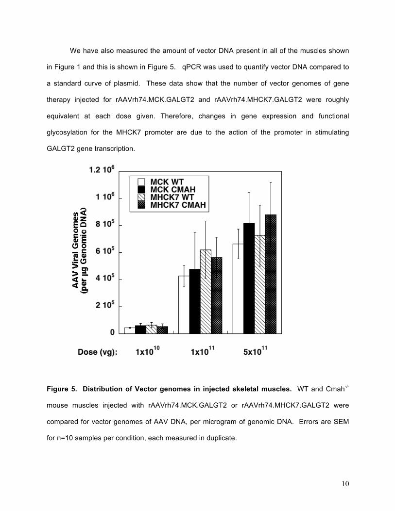
We have also measured the amount of vector DNA present in all of the muscles shown
in Figure 1 and this is shown in Figure 5. qPCR was used to quantify vector DNA compared to
a standard curve of plasmid. These data show that the number of vector genomes of gene
therapy injected for rAAVrh74.MCK.GALGT2 and rAAVrh74.MHCK7.GALGT2 were roughly
equivalent at each dose given. Therefore, changes in gene expression and functional
glycosylation for the MHCK7 promoter are due to the action of the promoter in stimulating
GALGT2 gene transcription.
Figure 5. Distribution of Vector genomes in injected skeletal muscles. WT and Cmah-/-
mouse muscles injected with rAAVrh74.MCK.GALGT2 or rAAVrh74.MHCK7.GALGT2 were
compared for vector genomes of AAV DNA, per microgram of genomic DNA. Errors are SEM
for n=10 samples per condition, each measured in duplicate.

11
With the assistance of our qualified optional collaborator, Dr. Paul Janssen, we were able to
measure changes in specific maximal tetanic force of isolated EDL muscles, measured ex vivo,
after 3 months of therapy with either rAAVrh74.MCK.GALGT2 or rAAVrh74.MHCK7.GALGT2.
Here, we show changes in specific force that occurred in wild type and Cmah-/- muscles when
the muscles were saturated for GALGT2 overexpression at the 5x10¹¹vg dose.
Figure 6. Percentage change in specific force after GALGT2 treatment of wild type (WT)
and Cmah-/- skeletal muscles. High dose rAAV.MCK.GALGT2 and rAAV.MHCK7.GALGT2 in
some cases led to increased specific force after 12 weeks administration in the extensor
digitorum longus muscle measured ex vivo. Errors are SEM for n=10 muscles per condition.
In addition to completing milestones 1 and 2, we have begun dosing animals for
milestone 3, including mdx and Cmah-/-mdx animals at day 1 (3 each).

12
Last, we would note that we have accomplished a major milestones in our
transition plan to move rAAVrh74.MCK.GALGT2 to a clinical trial for DMD. We have
filed an application for use of rAAVrh74.MCK.GALGT2 with the Recombinant DNA
Advisory Committee (RAC) at the NIH. RAC met on November 1, 2013 and approved
the proposal without further comment. We have now also completed in-life GLP
biodistribution and toxicology studies for IM and IA delivery of rAAVrh74.MCK.GALGT2.
This report is certified and was included with an IND application to the FDA for a Phase
1 DMD clinical trial on September 23, 2014. The IND, 16175, was approved on
October 17 and includes the IM portion of the phase I clinical trial. The data supporting
an IA trial is included in this IND, but an amendment will be needed to proceed to that
portion of the trial. Last, through other sources, we have obtained funds to begin cGMP
clinical manufacturing of rAAVrh74.MCK.GALGT2 and that manufacturing process has
begun now at the cGMP AAV Manufacturing facility at Nationwide Children’s Hospital.
Key Research Accomplishments:
1. Completion of the study demonstrating human GALGT2 overexpression is more
potent in mouse muscles when the Cmah gene, which is normally absent in
humans but is expressed in mice, is deleted. This deletion better mimics the
sialic acid substrates found in human muscle, suggesting that the human
GALGT2 gene has more activity when the sialic acid substrates available mimic
those found in humans.
2. Completion of the study demonstrating that MHCK7 promoter drives stronger
GALGT2 expression than the MCK promoter in mouse skeletal muscle and

13
would therefore be more optimal for use in patients. This suggests that MHCK7
may be useful as a second generation promoter for GALGT2 gene therapy
studies.
3. Demonstration that co-injection of dystroglycan with GALGT2 in mdx mice leads
to higher levels of GALGT2 induced glycosylation. This suggest that lack of
expression at two weeks is due to down-regulation of dystroglycan in mdx
muscles such that GALGT2 glycosylation of α dystroglycan cannot be achieved
prior to the onset of massive muscle degeneration in the 3-6 week time period.
4. Review and approval of rAAVrh74.MCK.GALGT2 clinical study by the
Recombinant DNA Advisory Committee (RAC) of the NIH.
5. Completion of in-life cGMP biodistibution and toxicology studies for planned
clinical trial for IM and IA delivery of rAAVrh74.MCK.GALGT2 in DMD patients.
6. Filing of IND with the FDA for use of rAAVrh74.MCK.GALGT2 in DMD patients.
7. Approval of IND (16175) for Phase 1 DMD clinical trial.
Reportable Outcomes:
1. DOD acknowledged in two presentations given by the PI this past year.
2. Data from this grant was used to support two grants submitted to the NIH.
3. Data from this grant was used to support the IND filing to the FDA for
rAAVrh74.MCK.GALGT2 therapy, which was approved in October of 2014.
Conclusion: We have made excellent progress in Year 2 of this 3-year award, having
now completed the milestones for Year 1 and Year 2. We have demonstrated that

14
humanizing mouse sialic acids allows the human GALGT2 gene to be more potent.
This suggests that the minimally effective dose reported in the mouse for our IND
application will be above or at the level we might expect in human muscle. We have
demonstrated that the MHCK7 promoter drives stronger GALGT2 expression than the
MCK promoter. This suggests that the use of the MHCK7 promoter may be more
efficacious than the currently planned MCK promoter in patient trials for DMD. We have
identified the level of mRNA overexpression required of GALGT2 to saturate muscle
expression of the CT glycan. We have also demonstrated that co-expression of
GALGT2 with dystroglycan drives better glycosylation at early time points in mdx
muscles. In our transition plan, we have met three major milestones for
rAAVrh74.MCK.GALGT2, having passed review by RAC, having completed and
certified in-life GLP biodistribution and toxicology safety studies, and having filed and
obtained an IND (16175) from the FDA for use of rAAVrh74.MCK.GALGT2 in DMD
patients.
References:
1. Martin, P.T., et al. Overexpression of Galgt2 in skeletal muscle prevents injury resulting from eccentric contractions in both mdx and wild-type mice. Am J Physiol Cell Physiol 296, C476-488 (2009).
2. Nguyen, H.H., Jayasinha, V., Xia, B., Hoyte, K. & Martin, P.T. Overexpression of the cytotoxic T cell GalNAc transferase in skeletal muscle inhibits muscular dystrophy in mdx mice. Proc Natl Acad Sci U S A 99, 5616-5621 (2002).
3. Xu, R., Camboni, M. & Martin, P.T. Postnatal overexpression of the CT GalNAc transferase inhibits muscular dystrophy in mdx mice without altering muscle growth or neuromuscular development: evidence for a utrophin-independent mechanism. Neuromuscul Disord 17, 209-220 (2007).
4. Xu, R., Chandrasekharan, K., Yoon, J.H., Camboni, M. & Martin, P.T. Overexpression of the cytotoxic T cell (CT) carbohydrate inhibits muscular dystrophy in the dyW mouse model of congenital muscular dystrophy 1A. Am J Pathol 171, 181-199 (2007).

15
5. Xu, R., DeVries, S., Camboni, M. & Martin, P.T. Overexpression of Galgt2 reduces dystrophic pathology in the skeletal muscles of alpha sarcoglycan-deficient mice. Am J Pathol 175, 235-247 (2009).
6. Chandrasekharan, K., et al. A human-specific deletion in mouse Cmah increases disease severity in the mdx model of Duchenne muscular dystrophy. Sci Transl Med 2, 42ra54 (2010).
7. Smith, P.L. & Lowe, J.B. Molecular cloning of a murine N-acetylgalactosamine transferase cDNA that determines expression of the T lymphocyte-specific CT oligosaccharide differentiation antigen. J Biol Chem 269, 15162-15171 (1994).
8. Salva, M.Z., et al. Design of tissue-specific regulatory cassettes for high-level rAAV-mediated expression in skeletal and cardiac muscle. Mol Ther 15, 320-329 (2007).
9. De la Porte, S., Morin, S. & Koenig, J. Characteristics of skeletal muscle in mdx mutant mice. Int Rev Cytol 191, 99-148 (1999).