Acute Stroke Care— - Medical Mastermind...
113
Version: November 29, 2004 Ken Uchino, M.D. Jennifer K. Pary, M.D. James C. Grotta, M.D. Acute Stroke Care— A Manual from the University of Texas- Houston Stroke Team -1-
Transcript of Acute Stroke Care— - Medical Mastermind...
UT Stroke Service Handb
Ken Uchino, M.DJennifer K. Pary, James C. Grotta,
AcuteCa
A ManuUniversi
HoustoT
Stroke re—
al from the
. MM
tyn
e
ook
Version: November 29, 2004
.D. .D.
of Texas- Stroke
am
-1-
UT Stroke Service Handbook
Introduction This handbook has been compiled from the day-to-day experiences of the University of Texas Houston Stroke Team in caring for acute stroke patients on a dedicated in-patient stroke service. It describes the options and underlying rationale for making treatment decisions for stroke patients in the Emergency Department, Stroke Unit, Neurological Critical Care Unit, and pre-rehabilitation setting. It is evidence-based where evidence exists, but some of what is included reflects our best interpretation of what should be done in the absence of conclusive data. It is intended as a practical guide to be used for medical students, house officers, and other clinicians with first hand responsibility for the “nuts and bolts” care of these patients. The handbook has been arranged generally in order of the things one should consider chronologically in assessing and treating the patient in the Emergency Department, then the Stroke Unit, and then on discharge or transfer to a rehabilitation facility. The Appendix contains useful “nuts and bolts” reference information that is hard to remember, such as dosing algorithms and conversion factors, standing orders, drug protocols, various stroke scales, and detailed description of imaging sequences and brainstem syndromes. In the text an asterisk “*” marks where there is sufficient evidence to make a strong recommendation based on randomized trials or consensus statements.
-2-
UT Stroke Service Handbook
Table of Contents 1. Stroke in the Emergency Department 2. Ischemic Stroke Care
The Four Components of Stroke Care 1) Acute therapy, such as t-PA, and optimization of
neurological status 2) Etiological work-up for secondary prevention. 3) Prevention of medical complications 4) Recovery and rehabilitation
Tissue Plasminogen activator (t-PA) protocol Neurologic deterioration Ischemic Stroke Prevention General measures; Risk factor reduction Atrial Fibrillation Carotid Stenosis Carotid Occlusion Lacunar Strokes Arterial Dissection Patent Foramen Ovale
3. Transient Ischemic Attack 4. Intracerebral Hemorrhage (ICH) 5. Subarachnoid Hemorrhage (SAH) 6. Organization of Stroke Care Appendix Numbers and Calculations Sample Admission Orders Routine Post- t-PA ICH
-3-

UT Stroke Service Handbook
Sample Discharge Summary Stroke Radiology CT MRI Transcranial Doppler Ultrasound (TCD) Drug Protocols Heparin drip Insulin drip Insulin sliding scale Medical Complications Deep venous thrombosis (DVT) Aspiration pneumonia Urinary tract infection (UTI) Heparin induced Thrombocytopenia Brainstem Syndromes Unusual Causes of Ischemic Stroke and Mimics Brain Death Criteria Neurologic Scales Glasgow Coma Scale The ICH Score Hunt-Hess Scale of SAH WFNS Scale of SAH Mini-Mental State Examination Modified Rankin Scale NIH Stroke Scale Recommended Reading
-4-

UT Stroke Service Handbook
Abbreviations: ACA Anterior cerebral artery AHA American Heart Association ARR Absolute risk reduction ASA American Stroke Association CBC Complete blood count CN Cranial nerve CSF Cerebrospinal fluid DBP Diastolic blood pressure DVT Deep venous thrombosis ED Emergency department GCS Glasgow Coma Scale HOB Head of bed ICA Internal carotid artery ICH Intracerebral hemorrhage INR International normalized ratio IV Intravenous LDL Low density lipoprotein LMB Lower motor neuron MAP Mean arterial pressure MCA Middle cerebral artery NIHSS National Institute of Health Stroke Scale NS Normal saline NNT Number needed to treat NPO None per oral PCA Posterior cerebral artery PO per oral PTT Partial thromboplastin time SAH Subarachnoid hemorrhage SBP Systolic blood pressure SC Subcutaneous TCD Transcranial Doppler ultrasound TIA Transient ischemic attack t-PA tissue plasminogen activator
-5-

UT Stroke Service Handbook
Stroke in the Emergency Department 1. Is this a stroke? If so, what is the time of onset of stroke
symptoms? Stroke mimics: • Seizures • Migraine • Syncope • Hypoglycemia • Metabolic encephalopathy • Drug overdose • Central nervous system tumor • Other neurologic diseases: subdural hematoma, peripheral
compression neuropathy, Bell’s palsy, benign positional vertigo.
• Conversion disorder-ALWAYS assume that your patient has a true neurologic illness first.
2. What type of stroke?
Ischemic Stroke Intracerebral Hemorrhage (see ICH section) Subarachnoid hemorrhage (see SAH section) Do a non-contrast head CT immediately!
The subsequent management will focus on ischemic stroke. 3. Airway-Breathing-Circulation (ABCs)?
O2 via nasal cannula (oxygen delivery in ischemia might be good).* Consider putting the head of bed (HOB) flat. This can significantly help cerebral perfusion. Consider Normal saline bolus 250-500cc if blood pressure is low.
4. How bad are the symptoms now? What was the time of onset?
-6-
UT Stroke Service Handbook
These are the questions to ask keeping t-PA in mind (see t-PA protocol). Try to get this information from a reliable source who witnessed the episode. If the time is unclear, try to be a detective and get as close to the time as possible (i.e., Ask them what TV show they were watching or how long it takes for them to drive from the grocery store to their home, etc).
5. Try to get the artery open if patient meets criteria (see t-PA protocol). This is the only effective treatment for ischemic stroke. For simplicity, the use of t-PA is detailed in its own section starting on page 20.
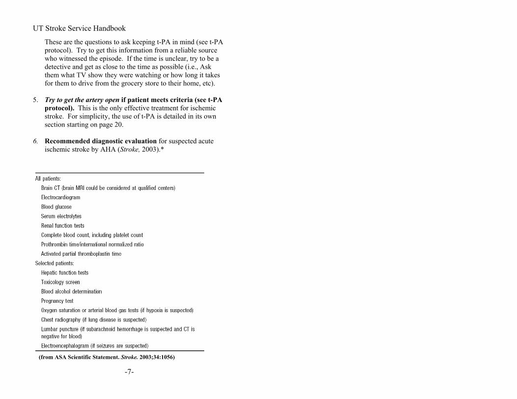
6. Recommended diagnostic evaluation for suspected acute ischemic stroke by AHA (Stroke, 2003).*
(from ASA Scientific Statement. Stroke. 2003;34:1056)
-7-

UT Stroke Service Handbook
Ischemic Stroke Care There are four components to caring for people with acute ischemic stroke. At every point, one should be thinking about the four issues.
1. Acute therapy, such as t-PA, and optimization of neurological status
2. Etiological work-up for secondary prevention. 3. Prevention of medical complications 4. Recovery and rehabilitation
On a daily basis think about the following that are part of the four components… • Is the patient neurologically stable or improving? Neurologic
deterioration? o Avoid dehydration of dysphagic patients with limited
oral intake. o Avoid diuretics in patients receiving IV fluids.
• Is the patient medically stable (e.g., congestive heart failure, infection)? medical complication?
• Is the blood pressure coming down slowly? neurologic deterioration? Stroke Prevention?
• Is the patient eating safely? medical complication? • Is the patient comfortable and sleeping well? medical
complication? Neurologic deterioration? o Ask yourself why the patient STILL gets blood drawn
qAM for blood count, chemistry, calcium…. • What is the mechanism of the stroke? Stroke prevention
o Is the work-up appropriate and complete? • What are we doing to prevent another stroke?
o Ask yourself why the patient is NOT on antiplatelets, statins, ACE-inhibitors, because most patients on stroke service would be…except people with ICH or on anticoagulation.
• What are we doing to promote recovery? • What are we doing to prevent complications from the stroke?
o Don’t forget DVT prophylaxis.
-8-

UT Stroke Service Handbook
o Ask yourself why the patient STILL has a Foley catheter and IV fluids if the patient is being discharged soon.
• What is the disposition? stroke recovery • Think about disposition early: o Consult Physical Therapy, Occupational Therapy and
Rehabilitation o Contact primary care provider for follow-up. o Arrange home health if indicated
General timeline Stroke Unit for 1-3 days. On floor to finish work-up and disposition determination. Discharge by day 2-5. This chapter discusses the 4 components in brief and then there are longer discussions on the following topics:
• t-PA therapy • Neurologic Deterioration • Stroke Prevention
1) Acute Therapy and Optimization of Neurological Status See sample admission orders (appendix) and also see the t-PA Protocol and Neurologic Deterioration sections The main goal of therapy is to get the artery open and reestablish blood flow. You should always ask yourself if you are doing everything possible to optimize blood flow to regions of cerebral ischemia. IV t-PA is the only FDA approved treatment for stroke in the U.S. It is approved under safety monitoring in the European Union. We are using intra-arterial thrombolysis as a rescue therapy investigationally. We are also investigating a variety of neuroprotective agents (hypothermia, other drugs) to try to decrease infarct size, but none are FDA approved at this time.
-9-

UT Stroke Service Handbook
Knowing the stroke mechanism helps to guide therapy and detection of large artery occlusion or stenosis is also helpful. Acute transcranial doppler (TCD) can be performed to detect changes in arterial flow of the large intracranial arteries in real-time. Other institutions use CT angiography more routinely. MRI/MRA acutely also helps, but it takes more time and the images are of varying quality. The need to do acute studies depends on a balance of availability of therapy, time requirement, clinical suspicion, and cost. Detection and localization of large artery occlusion would help in planning acute recanalization strategies and risk stratification for recurrent stroke or neurologic deterioration. Maintenance of cerebral perfusion. To maximize brain perfusion through stenoses and collateral vessels, we maintain euvolemia, support blood pressure, and put the head of the bed flat. Do not treat hypertension acutely until MAP >130 *, UNLESS: 1) the patient was treated with t-PA
2) symptomatic of hypertension (Congestive heart failure, myocardial infarction, hypertensive encephalopathy).
If you are going to treat hypertension, consider using a short acting agent that will wear off quickly or be turned off in case BP drops too much, such as…
• Labetalol (Trandate, Normodyne) 10-20 mg IV * • Nicardipine (Cardene) 5 mg/hr IV infusion as initial dose;
titrate to desired effect by increasing 2.5 mg/hr every 5 min to maximum of 15 mg/hr *
Goal: Blood pressure reduction by 10-15%. We use Nicardipine most commonly in the ED and first 24 hours to smoothly titrate blood pressure to desired levels.
-10-

UT Stroke Service Handbook
ASA Scientific Statement. Stroke. 2003;34:1056 Other Options for Maintenance of Cerebral Perfusion:
• Normal saline for IV fluids to maintain euvolemia and because it is isotonic and will not cause fluid shifts.
• Consider normal saline 500 cc bolus over 20-30 min. • Consider Hetastarch (Hespan, Hextend) for volume
expansion • Hetastarch 500cc over 1 hour. Then consider hetastarch 250
cc IV q8h. Monitor jugular venous pressure and input/output. Watch for fluid overload.
• Consider phenylephrine (Neo-Synephrine) drip in ICU for induced hypertension.
-11-
UT Stroke Service Handbook
Antiplatelet vs. Anticoagulant Therapy as an Acute Treatment for Ischemic Stroke Acute Antiplatelet Therapy: Aspirin (ASA) for acute stroke has been shown to be effective, though only marginally when studied in thousands of patients (see Lancet, 1997, for CAST and IST trials)*. Antiplatelet treatment beyond aspirin is driven by evidence from acute cardiovascular trials until there are more stroke data available, remembering of course the greater propensity of the brain to develop hemorrhagic complications.
We give ASA 325mg to all patients. In many patients, particularly those who have had strokes or TIAs while already on antiplatelet therapy, who have a fluctuating neurological course, or who have a heavy burden of atherosclerotic risk factors or atherosclerotic lesions, we will often orally load the patient in the Emergency Department with clopidogrel (Plavix) 375 mg, and then ASA 81 mg and clopidogrel 75 mg once daily for the first few days. The idea of an oral load stems from studies in patients undergoing coronary procedures who have less peri-procedural ischemic complications if they receive a load pre-procedure. We then switch to Aspirin/dipyridamole combination (Aggrenox) * or aspirin alone * or clopidogrel alone * if the patient is going home on antiplatelets. Try to convert patients to medications that they will be going home on to make sure they tolerate it prior to discharge, and take into account cost issues. If the patient cannot afford it, they will not take it. Acute Anticoagulant Therapy: Anticoagulation for acute ischemic stroke has never been shown to be effective (The International Stroke Trial, Lancet, 1997) *. Even among those with atrial fibrillation, the stroke recurrence rate is 5~8% in the first 14 days, which is not reduced by early acute anticoagulation (HAEST, Lancet, 2000; Hart et al, Stroke, 2002) *. Anticoagulation is mostly used for long-term secondary prevention in patients with atrial fibrillation and cardioembolic stroke at this point. In certain cases, like patients with a cardioembolic condition at high risk for recurrence (thrombus on
-12-

UT Stroke Service Handbook
valves or mural thrombus), venous thrombosis, or arterial dissection, patients may be started on heparin acutely and transitioned to warfarin (Coumadin). If ordering heparin, use weight adjusted algorithm with NO bolus *. Enoxaparin (Lovenox) at 1 mg/kg SC Q12h may be used in place of heparin .
How long should one wait before starting anticoagulation? There are no clear data on this topic. There is concern that the risk of hemorrhagic conversion is increased with anticoagulation, particularly in patients with large strokes. Hemorrhagic transformation is frequent in the evolution of large infarcts, especially those that have been reperfused by either spontaneous recanalization or with thrombolytics. One should be particularly careful about early anticoagulation in these patients. One generally waits 2-14 days before starting anticoagulation, the specific duration depending on the urgency of the indication vs the risks. You must carefully weigh the risks and benefits on a case by case basis, and never start anticoagulants without obtaining brain imaging first. Hyperglycemia is known to worsen stroke outcome. The mechanism by which and level at which hyperglycemia worsens stroke is not known. However, there are data that show even glucose in high 100s enlarges eventual stroke size. Therefore, treat glucose aggressively. (See Appendix for insulin algorithm). Hyperthermia has been correlated with poor outcome. Experimentally, increasing the body temperature of animals increases metabolic demand and infarct size. Therefore, treat hyperthermia aggressively with acetaminophen (Tylenol) and cooling blankets if necessary *. Drug therapy in the first 72 hours: (Those most commonly
started in our Stroke Unit)
Antiplatelets Aspirin (ASA) 81 mg once daily*, or Clopidogrel (Plavix) 75 mg PO once daily *, or ASA 25 mg/dipyridamole 200 mg extended release
(Aggrenox/Asasantine) twice daily *
-13-

UT Stroke Service Handbook
DVT prophylaxis
Heparin 5,000 u s.c. q 12 h *, or Enoxaparin (Lovenox, Clexane) 40 mg s.c. once daily, or Dalteparin (Fragmin) 5000 u s.c. once daily Sequential compression devices (non-drug) TEDs stockings
Anticoagulants for cardioembolic stroke Weight adjusted heparin (see appendix) Warfarin (Coumadin) (start with 5-10 mg day) * Insulin if needed (see appendix) * Temperature control with acetaminophen if needed * HMG CoA Reductase inhibitors with goal of LDL <100 * Oral antihypertensive agents * ACE-Inhibitors
• Perindopril (Aceon, Coversyl) 4 mg PO once daily
• Ramipril (Altace) starting at 2.5-5 mg /day target 10 mg PO once daily
Angiotensin Receptor Blockers (ARBs) • Losartan (Cozaar)
Diuretics • Hydrochlorothiazide (HCTZ), Chlorthalidone
(Hygroton) Beta-blockers
• Metoprolol (Lopressor, Toprol) Calcium channel blockers
-14-

UT Stroke Service Handbook
2) Etiological Work-up for Secondary Prevention See Stroke Prevention section for details. With imaging and vascular work-up we try to find a specific etiology such as cardioembolic source, arterial stenosis, etc. At the same time, we look for reversible risk factors for recurrent stroke such as hypertension, diabetes, hypercholesterolemia, and smoking/substance abuse that will need to be addressed. Ischemic Stroke can be thought of in various ways. Mechanisms (TOAST Classification, Adams HP et al, Stroke, 1993)—
• Large artery atherosclerosis Intracranial, Extracranial
(carotid, aortic arch) • Cardioembolic
Atrial fibrillation, segmental wall akinesis, paradoxical embolus, etc.
• Small Vessel: Lacunar Infarction-usually related to diabetes and hypertension
• Other unusual causes (dissection, venous thrombosis, drugs, etc.)
• Unknown (Cryptogenic) Albers et al, Chest, 2001 Screening for arterial stenosis/obstruction: MR angiography shows arterial stenosis intracranially and extracranially and excludes large aneurysms and vascular malformations. It is a good screening tool. Recent data indicate that contrast enhanced MRA might be the most reliable of non-invasive tests (U-King-Im JM, et al, Neurology, 2004). In our hands, Carotid ultrasound is better at estimating the degree of
-15-

UT Stroke Service Handbook
internal carotid artery (ICA) stenosis at the bifurcation. TCD complements other vascular imaging and can also be used to follow changes over time. One often focuses on the origins of the internal carotid arteries, but one should not forget the vertebral artery origins and intracranial arteries that are often the locations of atherosclerosis. Digital subtraction angiography (DSA) is considered the gold standard for visualizing the arteries, but is not without risk. CT angiography can give you sufficient detail and can be done emergently, instead of DSA. For determining degree of arterial stenoses, seeing arterial dissection, other vascular abnormalities, DSA is still the best. Cardiac evaluation: EKG should be done to exclude atrial fibrillation and to rule out silent myocardial infarction or ischemia which may occur as a consequence of the stroke. If atrial fibrillation or other important arrhythmia is suspected, cardiac telemetry or Holter monitor is needed. Echocardiogram is helpful in looking for a cardioembolic source and right-to-left shunts. A transthoracic echocardiogram (TTE) can show wall motion abnormalities (anterior wall akinesis carries high embolic risk), low left ventricular ejection fraction (<20-30% generally agreed upon as a cut-off), valvular abnormalities, and a patent foramen ovale (PFO). Transesophageal echocardiogram (TEE) can show the atria better. Left atrial appendage clot, size of PFO, PFO associated with atrial septal aneurysm, aortic arch atheroma, and spontaneous echo contrast are some of the findings associated with increased risk for ischemic stroke. Long-term anticoagulation with warfarin is considered to be the best prevention strategy for cardioembolic sources, but for many of the etiologies, it is still controversial whether warfarin is better than antiplatelets.
-16-

UT Stroke Service Handbook
TCD with bubble is as sensitive as TEE for detection of R to L shunt (see appendix for further discussion of how to do a “bubble study”). Risk Factor Screening
• Monitor blood pressure* • Obtain fasting lipid panel* • Screen for diabetes • Screen for hyperhomocysteinemia (Though a risk factor,
whether or not screening and therapy are beneficial is controversial)
• Smoking cessation counseling, if applicable.* 3) Prevention of medical complications See appendix for specific protocols
• Deep Vein Thrombosis (DVT) prophylaxis (pharmacologic, devices, and pt. mobilization)
• Aspiration precautions (swallowing assessment and nursing supervision before allowing the patient to eat).
• Gastrointestinal ulcer prophylaxis • Take out indwelling urinary (Foley) catheter as soon as
possible. 4) Stroke Recovery and Rehabilitation Physical therapy (PT), occupational therapy (OT), and speech pathology should get involved EARLY!* Patients who are eating (after swallowing assessment by speech pathology) make happy patients and family members. The sooner you get them involved, the earlier you will be able to begin working on placement at the appropriate location (home, rehabilitation, skilled nursing facility (SNF), nursing home, long term acute care facility (LTAC), etc.). The rehabilitation team is the key to determining disposition. The only times when PT/OT would not be involved early is when the patient is obtunded or needs to lie flat in bed in an attempt to maximize cerebral perfusion. It is very important to get the patient
-17-
UT Stroke Service Handbook
mobilized with out of bed (OOB) order (e.g.,, out of bed with meals, with PT, etc.). Mobilization also prevents complications. Ischemic Stroke Outcome Depends on stroke severity, size, mechanism, age, premorbid functional status, whether and when the patient received t-PA, and if they are cared for in a Stroke Unit. Overall, ~30% mortality in the first year, 40-50% in 5 years (data
from Rochester Epidemiology Project and NOMASS; Petty et al, Neurology, 1998; Hartman et al, Neurology, 2001).
From Medicare database (age >=65 years, Bravata et al, Stroke, 2003): -Once you survive an ischemic stroke hospitalization, 26.4% mortality in 1 year, 60% mortality after 5 years. -After surviving a TIA hospitalization, 15% mortality in 1 year, 50% mortality in 5 years.
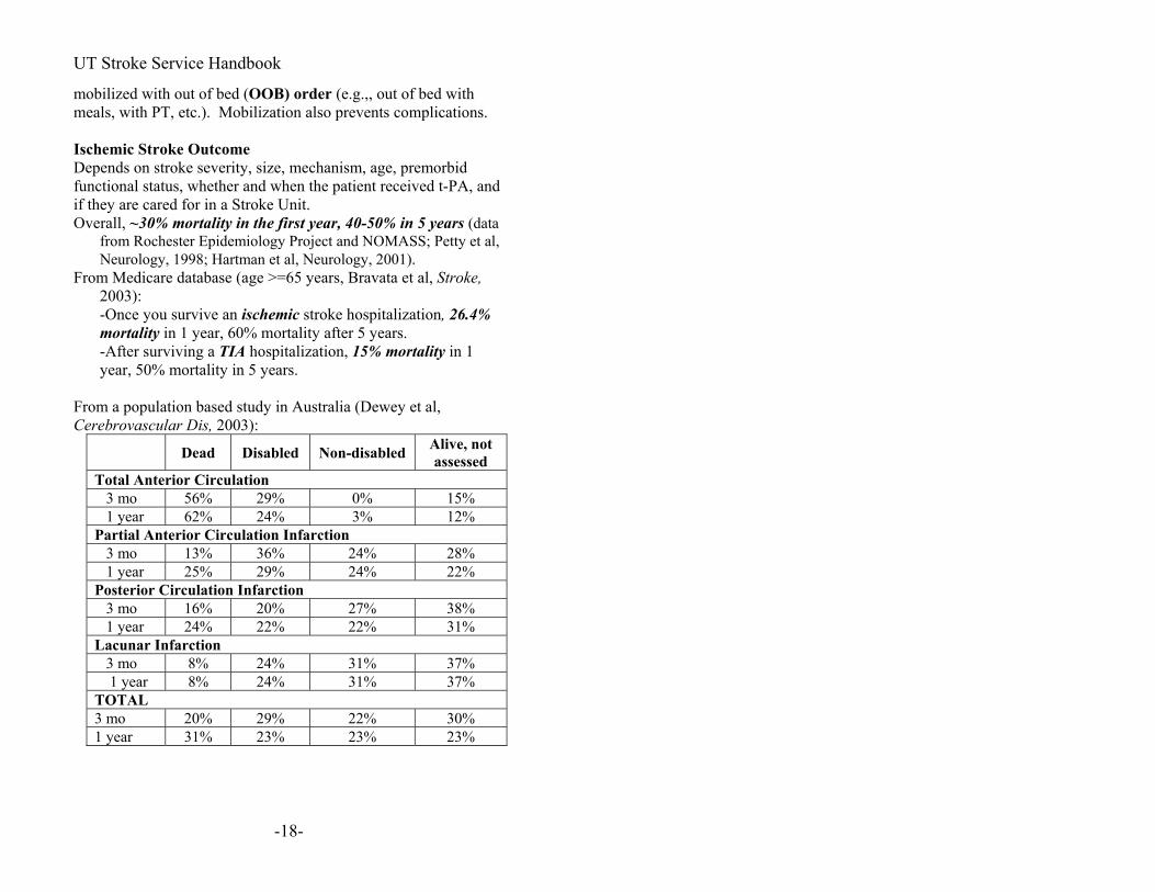
From a population based study in Australia (Dewey et al, Cerebrovascular Dis, 2003):
Dead Disabled Non-disabled Alive, not assessed
Total Anterior Circulation 3 mo 56% 29% 0% 15% 1 year 62% 24% 3% 12% Partial Anterior Circulation Infarction 3 mo 13% 36% 24% 28% 1 year 25% 29% 24% 22% Posterior Circulation Infarction 3 mo 16% 20% 27% 38% 1 year 24% 22% 22% 31% Lacunar Infarction 3 mo 8% 24% 31% 37% 1 year 8% 24% 31% 37% TOTAL 3 mo 20% 29% 22% 30% 1 year 31% 23% 23% 23%
-18-

UT Stroke Service Handbook
At patient discharge---Go over the following:
• What is the stroke location and mechanism? • What strategies are we using to prevent another stroke? • Is the patient on any antihypertensives, in particular ACE-I? • Is the patient on antiplatelets (e.g., aspirin,
asprin/dypridamole, or clopidogrel)? • Is the patient’s LDL<100 and is he or she on a statin? • Let’s get rid of unnecessary drugs. • Is the follow-up plan established? Who is following the INR if
necessary? It is important to communicate in some way with the primary care providers as they are the ones who will be managing the risk factors on a long term basis.
• Dictate a discharge summary including the above thought process (see Appendix for sample)
-19-

UT Stroke Service Handbook
t-PA (alteplase, Activase/Actilyse) Protocol* IV t-PA is the only FDA-approved therapy for acute ischemic stroke (NEJM, 1995; 333: 1581-7).* Indications:
< 3 hours since symptom onset to onset of therapy Stroke of more than minimal severity (in most, but not all,
cases, NIH Stroke Scale score >5). We use the criteria—“Would it be disabling if the deficit were to persist?”
Contraindications:
• Uncontrolled hypertension at the start of treatment (SBP >185 mm Hg or > 110 mm Hg)
• Evidence of intracranial hemorrhage on pretreatment CT • Significant mass effect on pretreatment CT • Suspicion of subarachnoid hemorrhage • Recent intracranial surgery, serious head trauma, or stroke • Recent major surgery or lumbar or arterial puncture where
a bleeding complication could not be controlled • History of intracranial hemorrhage • Seizure at the onset of stroke that clouds stroke evaluation • Significant active internal bleeding associated with
decreased hemoglobin • Intracranial neoplasm, untreated arteriovenous
malformation (AVM) or aneurysm that has bled or is at risk of bleeding
• Known bleeding diathesis. If recently on warfarin (Coumadin), INR >1.6
• Heparin within the last 48 hours and abnormal PTT • Platelet count <100,000 • Glucose >400 mg/dL (if high bring it down) • Glucose <50 mg/dL (treat the hypoglycemia and see if
symptoms resolve) Blood pressure control is very important to prevent complications.
-20-

UT Stroke Service Handbook
Before treatment, the goal is <185/<110 mm Hg. Labetalol (Trandate, Normodyne) 10-20 mg IV or a nicardipine (Cardene) drip (start at 5 mg/h and titrate up to a maximum of 15 mg/h) may be given to lower the blood pressure. If you are unable to keep the BP in the specified range with a few doses or a reasonable rate, the risk of hemorrhage is too high and the patient should not receive t-PA . Procedure: (FAST! Remember: Time is brain! Best results occur with treatment started within 2 hours of symptom onset.)
• Check to make sure laboratory tests have been sent and EKG ordered (2 mins)
o Glucose, hematocrit, and platelets are the only ones required before treatment
o Glucose can be by fingerstick o Complete blood count (CBC) o Coagulation studies (PTT, PT, INR) if patient is
on anticoagulants or coagulopathy is suspected o Some centers now have a fingerstick INR o Urine pregnancy test if appropriate
• Examine patient-(5 mins) o Establish clear time of onset o Obtain pertinent historical details (e.g., past
medical history, medications) o NIH Stroke Scale
• Obtain non-contrast head CT (Maximum ED arrival to CT time should be 30 mins)
• Talk to patient and family to explain risks/benefits • Obtain the patient’s weight—ask the patient or family
member(s) or estimate o If the patient weighs over 220 lbs or 100 kg-they
will get the maximum dose and it is not important to figure out the exact weight
• Think again, go over indications/contraindications, • Check BP again • Pre-Rx: two peripheral IV lines
o Foley catheter (optional) • Door to needle time goal is <40 mins, but max is 60
mins. -21-

UT Stroke Service Handbook
Dose: t-PA (alteplase, Activase/Actilyse) 0.9 mg/kg up to a maximum of 90 mg total
10% given IV bolus over 1 minute, remaining 90% infused over 1 hour.
Note: Only alteplase (Activase) has been approved for the treatment of stroke. Other drugs that may be given to patients with MI may NOT be used for stroke (i.e., reteplase (Retavase) , Tenecteplase (TNKase, META lyse), streptokinase (Streptase), etc.). Make sure to double check the name of the drug because there are some hospitals that may not carry t-PA, or nurses may reach for one of the other thrombolytic drugs due to their comfort with them for use in acute myocardial infarction. Also, the dosing for stroke and acute myocardial infarction are different.
Sample post t-PA orders (see Appendix) t-PA-related Intracranial Hemorrhage STOP t-PA infusion if still running. Goal Fibrinogen level >100 mg/dL with cryoprecipitate
1. Type and Cross 2. Check Fibrinogen level immediately and every 6 hours 3. Give 10-20 units of Cryoprecipitate before level returns (1
unit raises fibrinogen by 5-10 mg/dl. Assume there is no fibrinogen and adjust dose when level is back).
4. Repeat cryoprecipitate if needed. 5. May use fresh frozen plasma (FFP) in case of no
cryoprecipitate (1 unit of cryoprecipitate is made from 1 bag of FFP).
6. May give platelet concentrate if low Oropharyngeal Angioedema Management Protocol
1. Repeatedly examine oropharynx watching for edema (may be subtle swelling of lip or tongue just on one side).
2. If angioedema is suspected, immediately call for personnel experienced in intubation and airway management.
3. Choose from the following medication options:
-22-

UT Stroke Service Handbook
a. Epinephrine 0.5 ml via nebulizer or 0.3 ml of 0.1% solution subcutaneously
i. May repeat 2x as tolerated b. Diphenhydramine (Benadryl) 50 mg IV followed
by 25 mg every 6 h x4 doses c. Methylprednisolone (Solu-Medrol) 100 mg IV
i. May follow with 20-80 mg IV daily for 3-5 days depending on degree and course of angioedema
d. Famotidine 20 mg IV followed by 20 mg IV Q12h x2 doses.
4. If further increase in oropharyngeal angioedema is seen or there is airway compromise…
a. If tongue is edematous, but oral intubation is possible—perform urgent orotracheal intubation.
b. If tongue is too edematous for orotracheal intubation—perform fiberoptic nasotracheal intubation.
c. If there is severe stridor or impending airway obstruction—perform tracheostomy or cricothyrotomy and consider reversing t-PA
Facts about NINDS t-PA trial (NEJM, 1995; 333: 1581-7) What’s the Risk? 6.4 % (=“1 in 16” 95% CI 3.5-9.2%) symptomatic intracranial
hemorrhage rate vs. 0.6% in placebo There have been cases of angioedema. In a retrospective series, it was reported to occur at a rate of 5.1% (95% CI 2.3-9.5%), but this is probably an over-estimate (see above for treatment options). What is the Benefit? The odds ratio of good outcome was 1.7 (95% confidence interval 1.2-2.6). Patients treated with t-PA were 30% more likely (relative risk increase) to be have minimal or no disability at three months.
-23-

UT Stroke Service Handbook
3 = number needed to treat to result in 1 patient with better outcome than if not treated.
33 = approximate number needed to treat to result in 1 patients with worse outcome than if not treated Who benefits? Subgroups:
• All stroke subtypes showed benefit • Elderly subgroups >75 years old benefit but have
increased mortality regardless of treatment. • Patients with early ischemic changes on CT still benefit if
they meet all other criteria. • Time to treatment is key to improved chance of recovery
(Marler et al, Neurology 2000) ”Time is brain!” Who is more likely to bleed?
• Patients with more severe stroke • Patients with extensive CT changes, elevated BP, glucose
and temperature, and advanced age • BUT even those with severe strokes, early CT changes,
and advanced age were likely to benefit overall. Even accounting for the chances of bleeding, without treatment the severe strokes were going to do poorly (NINDS Investigators, Stroke, 1997; Patel et al, JAMA, 2001).
When to Consider Intra-Arterial (IA) Therapy and IV t-PA beyond 3 hours
• Remember, IA therapy, and IV t-PA given beyond 3 hours, remain un-approved and therefore still investigational. IA therapy refers to IA thrombolytics given directly into the clot, mechanical clot disruption, or both. In general, the trend is toward more mechanical methods, particularly in those patients who have already received IV t-PA or who have contraindications to IV thrombolytics.
•
• Remember also, if a patient qualifies within 3 hours for IV t-PA, but you think might also benefit from IA therapy, do not withhold IV therapy in favor of IA therapy. If you choose to proceed to IA, still treat with IV conventional
-24-

UT Stroke Service Handbook
dose first. Generally, in these cases, we are using more mechanical means to get the artery open.
o There are several reasons for this recommendation, but mainly we don’t want to deprive someone of proven effective therapy in favor of something that remains unproven and unapproved by the FDA. Also, we have found that when we decided to go directly to IA, some patients may never get treated or their treatment will be delayed because of logistic reasons (mobilizing the angiography team, equipment failure, difficulty with catheterization, etc). In others who we treated with IV thrombolytics first, we found that the clot was already lysed by the IV drug by the time we got the artery catheterized, so that had we not given IV t-PA, lysis would not have occurred as soon. Finally, the IMS study experience of IV t-PA followed by IA therapy has shown that his approach is no more risky than either IV or IA alone (IMS Study Investigators, Stroke, 2004).
If a patient with a distal ICA, M1 segment of MCA, proximal M2 segment of MCA, or basilar lesion on TCD, has received IV t-PA, and has not recanalized by the time they get to the angio suite, and still has a disabling deficit IA therapy.
•
•
•
If the patient qualifies for IV t-PA within 3 hours but has had recent major surgery, or INR > 1.6, and develops a devastating stroke IA therapy. If patient is outside the 3 hour window, has a significant perfusion/diffusion mismatch on MRI, arterial occlusion on MRA or TCD, and disabling neurological deficit, with no other contraindication IA therapy.
• If patient is outside the 3 hour window but within 6 hours of onset of symptoms, has a severe stroke (NIHSS >10), and limited or no ischemic changes on CT. In these cases, we consider either IV t-PA or IA therapy depending on whether we can identify a large artery occlusion on TCD etc, and the availability of the endovascular team to
-25-
UT Stroke Service Handbook
quickly mobilize. We often push the window further if the patient has a suspected basilar occlusion because doing nothing would be uniformly fatal.
Neurological Deterioration in Acute Ischemic Stroke Although classically stroke symptoms are maximal at onset and patients gradually recover over days, weeks, and months, patients can deteriorate. People have termed the phenomenon stroke progression, stroke in evolution, stroke deterioration, and symptom fluctuation. There is no consistent terminology. The phenomenon occurs from different causes and is incompletely understood. The probable causes are the following:
1. Stroke enlargement (e.g., arterial stenosis or occlusion and worsening perfusion)
2. Drop in perfusion pressure 3. Recurrent stroke (not common) 4. Edema and mass effect 5. Hemorrhagic conversion. 6. Symptom fluctuation without good cause
(inflammation?) 7. Metabolic problem (decreased O2 saturation,
decreased cardiac output, increased glucose, decreased sodium, fever, sedative drugs, etc)
8. The patient is not feeling like cooperating (sleepy, drugs)
1. Stroke Enlargement
This clearly occurs when there is arterial stenosis or occlusion and the hemodynamics change for whatever reason. There are no data to support that anticoagulation prevents this, though
-26-

UT Stroke Service Handbook
many jump to it! Probably the best treatment is to treat the underlying stenosis/occlusion early. The approach to prevent (rather than treat after deterioration) should be to do early imaging to detect large artery stenosis/occlusion by TCD, CTA, or MRI. (i.e., find the high risk patients early). Patients with minor deficits but abnormal TCD are at highest risk of progression. Perfusion imaging may indicate areas of tissue at risk. Even without a perfusion study, diffusion weighted MRI and the clinical exam compared to the TCD or arterial imaging allow an educated guess. In such patients, you might want to consider early intervention, such as IV thrombolysis despite low NIHSS score, intra-arterial therapy, carotid endarterectomy, or carotid stenting. Remember, though, that intra-arterial thrombolysis and carotid stenting are still investigational.
2. Drop in perfusion pressure Since autoregulation is lost in ischemic brain, any reduction in blood pressure will reduce flow to penumbral regions thereby potentially worsening the clinical deficit. This is true in both cortical and subcortical strokes. The latter have particularly poor collateral flow and may be at greatest risk for hypo-perfusion-related deterioration. As a rule of thumb, MAP should be kept at pre-stroke levels (as a general guideline, around 130 mmHg in hypertensive patients, and 110 in normotensive patients) in the first 24 hours, and if MAPs drop below this level, and the patient deteriorates, the MAP should be increased by a fluid bolus and possibly a pressor.
3. Recurrent stroke Unfortunately some go on to have recurrent stroke. There are no data that immediate or “early” anticoagulation helps, even in the setting of atrial fibrillation because (see #5) it can lead to hemorrhagic complications. Among atrial fibrillation patients, the stroke recurrence risk has been reported to be 5-8% in the first two weeks, which is not reduced by
-27-

UT Stroke Service Handbook
anticoagulation (HAEST, Lancet, 2000; Saxena et al, Stroke, 2001; Hart et al, Stroke 2002).* In a recent study, MRI detected 34% stroke recurrence in the first week, while clinically, only 2% stroke recurrence was noted (Kang et al, Ann Neurol, 2003). In a larger population-based study, large artery atherosclerosis was associated with highest risk of stroke recurrence (see Figure, Lovett et al, Neurology, 2004). Stroke Mechanisms and risk of early recurrence in % (95% CI)
Mechanism At 1 week At 1 month At 3 months Large Artery Atherosclerosis
4.0% (0.2-8) 12.6% (6-19) 19.2% (11-27.)
Cardioembolism 2.5% (0.1-5) 4.6% (1.3-7.9) 11.9% (6-17) Small vessel Ischemia
0% 2.0% (0-4%) 3.4% (0.5-6)
Undetermined 2.3% (0.5-4) 6.5% (3-10) 9.3% (6-13)
4. Cerebral Edema This is a worry with large strokes, such as big MCA strokes involving the basal ganglia, often with some involvement of the ACA or PCA territories as well, and with large cerebellar strokes. It is a worry with young patients who do not have
-28-

UT Stroke Service Handbook
much atrophy and thus not much room to swell inside the skull. Monitor for any neurologic change, decline in level of consciousness, rising blood pressure, periodic breathing, hiccups, headache, new cranial nerve abnormalities, and pupils (late phenomenon). Medical Treatment:
• Steroids do not help *(grade A recommendation)! • Osmotherapy (i.e., mannitol) and hyperventilation are
only temporizing. • Cerebrospinal fluid drainage does not do much.
Definitive Therapy:
• Consult neurosurgery early and do early hemicraniectomy. The skull is taken off (and put in freezer) and dural incision is made so that the brain can swell out rather than compress the brainstem (see below).
• For cerebellar stroke, posterior fossa decompression and cerebellectomy.
• With both procedures, a common error is not to remove enough bone and do an adequate decompression. Be sure the neurosurgeon knows the anatomical guidelines for decompression.
This is a life-saving measure. It is highly recommended for cerebellar strokes as people can be quite functional without a large part of their cerebellum. However, with respect to large MCA strokes, talk with family about quality of life after stroke survival vs. death. Many do not perform the procedure for large left MCA strokes as the patient is likely to be aphasic. Best results occur with early intervention in young patients with non-dominant hemisphere strokes. Criteria for including patients into ongoing evaluations of hemicraniectomy are:
• < 5hrs; > 50% MCA territory hypodense
-29-

UT Stroke Service Handbook
< 48 hrs; complete MCA territory hypodense • • •
• • • • •
> 7.5 mm midline shift > 4 mm midline shift with lethargy
Other criteria include: • age < 60 • 145 cc infarct volume on MRI or 240
cc on CT
Guidelines for adequate surgical decompression are: • Anterior: frontal to mid-pupillary line
Posterior: 4 cm posterior to external auditory canal Superior: superior sagittal sinus Inferior: floor of middle cranial fossa Durotomy over the entire region of decompression Dural grafting
Other criteria include:
• 12 cm diameter craniectomy
-30-

UT Stroke Service Handbook
5. Hemorrhagic Conversion
It should be clearly visible on non-contrast head CT. Most of the time, the patient is asymptomatic from the hemorrhagic conversion, unless it is large or in a critical location. Usually there isn’t much you can do or should do, except to stop the antiplatelets and anticoagulants. Radiographically, hemorrhagic conversion is divided into 4 categories (Fiorelli et al, Stroke, 1999). • Hemorrhagic infarct-1 and 2 (HI-1 and HI-2) represent
petechial bleeding into the area of infarct and are almost never symptomatic. Parenchymal hemorrhage-1 and 2 (PH-1 and PH-2) represent confluent bleeding. If the bleeding takes up more than 30% of the infracted area and produces mass effect (PH-2), it usually produces neurological deterioration.
•
The risk of developing PH-2 is the main reason why anticoagulation is not recommended immediately after cardioembolic stroke, and without repeat brain imaging first.
6. Symptom fluctuations without a good cause.
This is a poorly understood phenomenon. It is commonly seen with subcortical strokes. Usually in the first 3 days the
-31-
UT Stroke Service Handbook
symptoms worsen. It can occur up to 2 weeks after stroke onset. The mechanism is unknown. Local hyperperfusion? Inflammation? Neurochemical or neurotransmitter changes? Apoptosis? Treatment is mainly supportive (euvolemic, check blood pressure to make sure it didn’t drop, put the head of bed flat). Anti-inflammatory and anti-apoptotic neuroprotective therapies are under evaluation.
7. Metabolic problems These are pretty self-explanatory. Remember that a sick brain is more sensitive to the effects of metabolic perturbations so that mild fever or changes in sodium or glucose may have an exaggerated clinical effect. Reduced cardiac output is a particularly bad co-morbidity resulting in worse clinical outcome, and should be carefully avoided by optimizing fluid and inotropic therapy. Remember also that “if the lips are blue, the brain is too”. Sedative drugs interfere with rapid transition to rehabilitation mode and also have been associated with worse outcome, decreased mobilization with attendant increased DVT, etc. Sedating drugs should be avoided as much as possible.
Evaluation of patients with neurologic deterioration:
a. Get a STAT non-contrast head CT (to evaluate for hemorrhage, new stroke, etc.)
b. Talk to and examine the patient. Is the patient sleepy (because it’s 3 am or because there is mass effect)? Is there a pattern of symptoms (global worsening vs. focal worsening)?
c. Check the ABCs, laboratory tests, vital signs. Is the patient hypotensive or hypoxic?
d. Review medications (antihypertensives, sedatives) e. Consider MRI or TCD.
-32-

UT Stroke Service Handbook
Ischemic Stroke Prevention --or why we do the things we do. A thorough Ischemic “Stroke workup” is not just CTMRIMRAECHOCAROTIDSTCDLIPIDSHGA1C There should be some thought behind it which is based on finding out the cause of stroke in order to optimally prevent another. Initial acute CT*
• To r/o ICH and other causes, to assess for old strokes, size, …
MRI/MRA • To localize the lesion • To try to understand the mechanism (lacune, large artery
atherosclerosis, or embolic, etc.) • To say what’s acute and what’s old • You can see many things, including incidental findings • To look for intracranial and extracranial stenosis (athero,
dissection…), aneurysm, arteriovenous malformation (AVM)…
Repeat CT • To localize the lesion, if patient is unable to have an MRI • To assess mass effect/edema • To look for hemorrhagic conversion • To look for stroke recurrence
CT angiogram • To look for arterial stenosis, dissection, aneurysm
(especially. if the patient is unable to have an MRI) Transthoracic Echocardiogram (TTE) (order with “bubble study”)
• To assess for embolic source (anterior wall or apical akinesis, clot, large PFO)
• Low ejection fraction at 20-30% is generally agreed upon as a cutoff, where thromboembolic risk increases significantly due to stasis.
Transesophageal Echocardiogram (TEE) (order with “bubble study”)
-33-

UT Stroke Service Handbook
• To assess for embolic source (smaller PFO, aortic atheroma, left atrial appendage clot, atrial septal aneurysm, spontaneous echo contrast, endocarditis…)
• If PFO is found, usually will do a bilateral lower extremity Duplex and pelvic MR venogram to look for venous clot
Carotid Duplex • To assess for internal carotid stenosis, occlusion • Shows you direction of vertebral artery flow • BUT do you need it if you have a good MRA?
Transcranial Doppler (with or without bubble study) • To assess for intracranial stenosis/occlusion of major
arteries (complements MRA but cheaper) • Emboli monitoring • Look for PFO with bubble study. TCD is the most
sensitive and least expensive/invasive way to screen for right to left shunting.
• Hemodynamic reserve (breath holding index, vasomotor reactivity)
Digital Subtraction Angiography (DSA) • Gold standard for determining degree narrowing
Only way to definitively identify aneurysms or AVMs, dissection, vasculitis, or other arteriopathies
•
Fasting Lipids (target LDL <100) • Look for high total cholesterol, triglycerides, LDL • Look for low HDL
Hemoglobin A1C (HgA1C) • Screen for Diabetes
MR Spectroscopy • Tells you what the lesion may be (ischemia vs. tumor vs.
infection vs. demyelination…) • BUT can be non-specific
MR Perfusion, and other blood flow studies • This test, or other studies of cerebral perfusion (single
photon emission computed tomography {SPECT} with and without Diamox challenge, Xenon enhanced CT, CT perfusion, PET), can help you plan the need for revascularization procedures in patients with extracranial occlusive disease.
-34-

UT Stroke Service Handbook
Ischemic Stroke Prevention General Measures: Educate your patients so they can take an active role in their health care. 1. Control risk factors:
• Hypertension (SHEP trial, etc.)* See subsequent section for more detail
o ACE inhibitors (HOPE, PROGRESS trials)* o Diuretics and calcium channel blockers, especially
in African Americans (ALLHAT trial)* • Elevated lipids
o Statins (several trials incl. MRC Heart Protection Study)
o Target LDL < 70 according to the latest guidelines in high risk cardiovascular patients (Grundy et al, Circulation, 2004)
o Be sure to get baseline liver functions before starting statin therapy
• Smoking o Cessation counseling and pharmacotherapy
• Diabetes o Identification o Treatment, including diet
• Hyperhomocysteinemia o Folic acid ?? So far, no evidence for vitamins
including folic acid for stroke prevention in general (VISP study).
o Therefore, since there is no effective treatment for this risk factor for stroke, routine screening for hyperhomocysteinemia is probably not cost effective.
• Estrogen use (WEST trial, NEJM 2001; Women’s Health Initiative)*
o Avoid in most cases • Others
o Vasoactive drugs such as phenylpropanolamine, cocaine, amphetamines
o Sedentary lifestyle
-35-

UT Stroke Service Handbook
2. Antithrombotic or anticoagulant medications • ASA (many studies).* 20% relative risk reduction of
secondary stroke/other vascular events. • ASA/dipyridamole ER (Aggrenox, Asasantine)--ESPS-1
and 2 trials; 30% better than aspirin alone *. PROFESS is a trial comparing it to Clopidogrel (Plavix) (ongoing)
• Clopidogrel (Plavix)--CAPRIE trial. Slightly better than aspirin and better tolerated *.
• ASA/clopidogrel combination?—Bleeding rate are too high with long-term use (MATCH).
• Warfarin (Coumadin) for o Atrial fibrillation (SPAFs)*—this is the only
indication having class I evidence o Critical extracranial carotid stenosis—string sign
or occlusion (experience but no good data) o Basilar thrombosis/stenosis (non-randomized
data) o Arterial dissection *(experience, case series, and
consensus statements) o Other “embologenic” cardiac conditions (LV
akinesis) aortic atheroma > 4mm (SPAF III subgroup analysis from Blackshear et al, Am J Cardiol, 1999)
o Embolic-looking cryptogenic stroke (WARSS subgroup analysis: Mohr JP, Cleve Clin J Med. 2004)
o Warfarin not indicated for intracranial disease (WASID), or routine stroke (WARSS: Mohr et al, NEJM, 2001), most stroke patients with positive antiphospholipid antibody (APASS--WARSS substudy: Levine SR et al. JAMA, 2004), or PFO (PICCS--WARSS substudy: Homma et al, Circulation, 2002) *.
3. Lifestyle modification: stop smoking, better diet, exercise may reduce the above mentioned risk factors, but also underlying inflammation leading to atherosclerosis. 4. Blood Pressure Control*
-36-

UT Stroke Service Handbook
Hypertension is the single most important modifiable risk factor. JNC-7 reports that “the risk of cardiovascular disease, beginning at 115/75 mm Hg, doubles with each increment of 20/10 mm Hg” (JAMA, 2003). -Multiple large randomized controlled trials have shown efficacy of antihypertensive treatment in primary and secondary prevention of stroke. The selection of antihypertensives remains unsettled and controversial. -Many drugs have been shown to reduce stroke in primary prevention (beta-blocker in SHEP, diuretic in SHEP and ALLHAT, Calcium channel blocker in ALLHAT, ACE inhibitor in HOPE and PROGRESS, ARB in LIFE). -A combination of perindopril (Aceon), a tissue-specific ACE-I, and indapamide (Lozol), a diuretic, have been shown to reduce stroke in secondary prevention even among non-hypertensive patients (PROGRESS). Whether this effect is due to tissue-specific ACE-I rather than ACE-I class effect, or whether ACE-I needs to be used in combination with a diuretic remains controversial. Probably the most important point is blood pressure reduction, not the specific drug. For primary prevention, diuretic seems to be effective and cheap. Recent meta-analysis seems to support superiority of diuretics (Psaty et al, JAMA, 2003). JNC-7 also recommends thiazide diuretics as a first-line pharmacologic therapy, though it recognizes that more than 1 drug is commonly needed. But in hospital setting, especially after a stroke, patient’s fluid intake may be poor. A diuretic while on IV fluids does not make sense. Start a diuretic in stroke inpatients only if the patient is drinking fluids consistently. Bring down BP slowly by oral antihypertensives after acute ischemic stroke.
-37-

UT Stroke Service Handbook
According to JNC-7, the goal blood pressure are the following: <140/90 mm Hg <130/80 mm Hg for patients with diabetes or chronic kidney disease
But remember, there is a continuous increase in risk of stroke with increase in blood pressure. There is no biological cut-off point.
Atrial Fibrillation (A fib) Atrial fibrillation is a cause of stroke about which a lot is known about prevention. For a good review see Hart et al, Ann Int Med, 2003. In the acute stroke setting:
• Acute stroke recurrence rate estimates vary.
-38-
• Therefore, no reason to rush to anticoagulate after A Fib related stroke. Should wait 48-96 hrs after a major stroke and repeat the CT (or MRI) first.
• From International Stroke Trial 3.9% stroke recurrence rate in 14 days (Stroke 2001; 32:2333).

UT Stroke Service Handbook
Natural History:
• Valvular Atrial fibrillation: risk ~17x that of controls • Non-valvular Atrial fibrillation: 6x controls or ~5% year • “Lone” Atrial fibrillation: age <60 (no coronary artery
disease, hypertension, diabetes mellitus, hyperthyroidism or COPD): 0.5% per year
• Prior stroke/TIA/embolism: >10% per year stroke risk • Age>80: >7% per year
Primary Stroke Prevention with Warfarin (Coumadin)*:
• Major bleeding risk: 1.5%-2% per year. o Increased risk with recent hemorrhage,
alcohol binge, closed head injury, liver disease, aspirin, NSAIDs, cancer, age, previous stroke, uncontrolled hypertension
• ICH risk: 0.2-0.4% per year o Age=<75: 0.5% per year (data from SPAFII) o Age >75: 1.8% per year
• Stroke risk to ~2% per year, or 62% relative risk reduction.
Primary Stroke Prevention with aspirin*:
• Major bleeding risk: 0.3-0.9 % per year. • ICH risk: 0-0.3% per year • Stroke Risk: ~7.9% per year or 22% relative risk
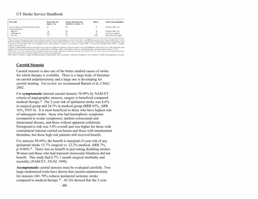
reduction. Secondary Prevention (most of our cases in neurology): Warfarin reduces stroke risk from 12% per year 4% per year * From Hart et al, Ann. Int. Med, 2003
-39-
UT Stroke Service Handbook
Carotid Stenosis Carotid stenosis is also one of the better studied causes of stroke for which therapy is available. There is a large body of literature on carotid endarterectomy and a large one is developing for carotid stenting. For review we recommend Barnett et al, CMAJ, 2002.
For symptomatic internal carotid stenosis 70-99% by NASCET criteria of angiographic stenosis, surgery is beneficial compared medical therapy.* The 2-year risk of ipsilateral stroke was 8.6% in surgical group and 24.5% in medical group (RRR 65%, ARR 16%, NNT 6). It is most beneficial to those who have highest risk of subsequent stroke: those who had hemispheric symptoms (compared to ocular symptoms), tandem extracranial and intracranial disease, and those without apparent collaterals. Perioperative risk was 5.8% overall and was higher for those with contralateral internal carotid occlusion and those with intraluminal thrombus, but those high risk patients still received benefit.
For stenosis 50-69%, the benefit is marginal (5-year risk of any ipsilateral stroke 15.7% surgical vs. 22.2% medical, ARR 7%, p=0.045) *. There was no benefit in preventing disabling strokes. Women and those who had transient monocular blindness did not benefit. This study had 6.7% 1 month surgical morbidity and mortality (NASCET, NEJM, 1998).
-40-
Asymptomatic carotid stenosis must be evaluated carefully. Two large randomized trials have shown that carotid endarterectomy for stenosis ≥60~70% reduces ipsilateral ischemic stroke compared to medical therapy *. ACAS showed that the 5-year

UT Stroke Service Handbook
risk was 5.1% for surgical vs. 11.0% for medical therapy (JAMA, 1995). The results were recently confirmed by ACST trial which showed 5 year-stroke risk of 6.4 % surgical vs. 11.8% medical therapy (Lancet, 2004). The overall subsequent risk is smaller than that for symptomatic carotid stenosis. The absolute risk reduction is 5-6%, with number needed to treat (NNT) to prevent one ipsilateral ischemic stroke of 17~19. The perioperative risk was 2.3% in ACAS and 3.1% in ACST. If one considers doing surgery on asymptomatic carotid stenosis, the surgeon must have volume and experience. Women for unclear reason have smaller benefit from endartectomy than men.
Carotid stenting, though performed for several years now, is still considered investigational. SAPPHIRE was a randomized trial designed to show carotid stenting with distal protection device is not inferior to endarterectomy in high risk patients with symptomatic and asymptomatic carotid stenosis. It showed that the stroke rates at 2 years are equivalent (stenting is not inferior) and there was a trend favoring stent in rate of adverse events (Yadav et al, NEJM, 2004). Several studies, including CREST, SPACE, EVA-3S, and CAVATAS-2, are ongoing to study the effectiveness of the procedure in stroke prevention. So far, safety appears similar except in patients >80 years old. Some unresolved issues are durability of the stent and restenosis rates. The eventual hope is that carotid stenting would be cheaper and safer, especially in those with multiple medical comorbidities.
When can you do surgery after stroke? The answer is not clear. Traditionally, it was recommended to wait 4-6 weeks after a large stroke to perform revascularization. The rationale is that reperfusing area of recent stroke might lead to hyperperfusion or even hemorrhage. Due to break down of the blood brain barrier, autoregulation may not work normally in the area of injury, leading to a hyperperfusion syndrome. An “unstable” neurological status is one of the strongest risk factors for peri-operative complications. Also, at the time of stroke, it may be unclear how well the patient will recover and how aggressive to be with various prevention strategies.
-41-

UT Stroke Service Handbook
It is probably safe to revascularize early, if the stroke is small clinically and radiographically. There have been several studies in this regard, but none conclusive. Can one risk stratify beyond degree of stenosis? It is logical that percent stenosis would correlate with level of cerebral perfusion and cerebrovascular reserve. The latter may be the critical determinant of stroke risk and can be measured using TCD (Cerebrovascular reactivity with TCD; Silvestrini et al, JAMA, 2000): Procedure: Mean flow velocity at rest is obtained by the continuous recording of a 1-minute period of normal breathing. Subjects were asked to hold their breath for 30 seconds. Breath-holding index (BHI): The BHI is obtained by dividing the percentage increase in mean flow velocity (MFV) occurring during breath holding by the length of time (seconds) subjects hold their breath after a normal inspiration BHI = [(MFVat the end of breath holding – MFVrest )] x 100 MFVrest x seconds of breath holding Asymptomatic carotid stenosis ≥70%: Normal BHI ≥ 0.69 4.1% ipsilateral stroke /year Impaired BHI <0.69 13.9 % ipsilateral stroke /year Acute Carotid Occlusion The traditional approach is to anticoagulate for several months after acute symptomatic carotid occlusion for presumed “stump emboli.” The thinking has been that the end of the occlusion intracranially has an unstable clot that can propagate or embolize. There is no evidence to support this. Acute carotid occlusion can also be revascularized surgically or endovascularly. The clot is usually at the internal carotid origin and the rest of the artery downstream might be open. Though this has been advocated by some, there are no prospective data to support that acute revascularization 1) improves the patient’s symptoms from current stroke 2) prevents recurrent stroke long term, and that this outweighs the risk of complications.
-42-

UT Stroke Service Handbook
-43-
Long term, these patients are at highest risk for recurrent stroke. Current evidence suggest that hemodynamic impairment (poor perfusion) is a large contributor to recurrent stroke rather than the traditional “stump emboli.” There are several measurement methods for risk stratification by hemodynamic reserve:
• Xenon-CT or Single Photon Emission CT (SPECT) with acetazolamide (Diamox) challenge
• PET with oxygen extraction fraction • TCD with breath-holding index (BHI)
Asymptomatic or Symptomatic carotid occlusion (Vernieri et al, Stroke, 1999).: Normal BHI ≥ 0.69 <10% ipsilateral stroke in 2 years Impaired BHI <0.69 40% ipsilateral stroke in 2 years. There is an age effect as well on this as well, with older patients having higher risk. What to do about the high-risk patients remains uncertain. In the U.S. there is an ongoing randomized NIH-sponsored trial looking at extracranial-intracranial carotid bypass surgery versus medical therapy (Carotid Occlusion Stroke Study). This surgical therapy would try to augment cerebral perfusion ipsilateral to carotid occlusion by connecting a temporal artery branch of the external carotid artery with the middle cerebral artery.
Lacunar Strokes Lacunar strokes, also known as small vessel disease or small vessel occlusion, comprise approximately 20-30% of strokes. They are defined as small ischemic stroke due to disease of end arteries off of major intracranial arteries (off of the MCA, basilar, PCA, ACA, and posterior communicating artery). These infarcts are <15mm diameter in size.
Are all small subcortical strokes <15mm lacunar strokes (i.e., due to small vessel disease)? No, 12% of small basal ganglia and 34% of centrum semiovale infarcts have cardioembolic source and 19% of small basal ganglia and 53% of centrum semiovale infarcts have large artery occlusive

UT Stroke Service Handbook
disease (Yonemura, et al, Stroke, 2002). Therefore, even a “lacunar-looking” stroke, especially if they do not fit into a classic syndrome, warrants careful work-up for large artery atherosclerosis and embolic source. What causes lacunar strokes? Lipohyalinosis is the classic pathology but atherosclerosis is also common for small vessel occlusion. Seen from an epidemiological standpoint, hypertension is the only consistent risk factor (as opposed to diabetes mellitus, smoking, or hyperlipidemia). Also, antihypertensive treatment is the only method that has clearly been shown to reduce lacunar strokes in particular (SHEP study, JAMA, 2000). Lacunar Syndromes
Pure Motor Hemiparesis (corona radiata, anterior or posterior limb of internal capsule, pons, and medullary pyramid)
Pure Sensory Stroke (ventral posterior thalamus)
Sensorimotor Stroke (thalamus, corona radiata)
Ataxic Hemiparesis (Not well localizing: pons, corona radiata, anterior or posterior limb of internal capsule, lentiform nucleus, cerebellum)
Dysarthria Clumsy Hand (anterior limb of internal capsule, genu, pons)
(Also read WM Landau. “Clinical neuromythology VI. Au clair de lacune: holy, wholly, holey logic.” Neurology. 1989; 39:725-730. It’s a rather sarcastic take on this point.) Arterial Dissection Arterial dissection is probably an under-recognized stroke mechanism. It occurs due to an intimal tear in the vessel
-44-

UT Stroke Service Handbook
wall and formation of intramural hematoma and, less frequently, a pseudoaneurysm. A history of neck, facial, or head pain in a patient without strong risk factors for vascular disease points toward the diagnosis. On examination carotid artery dissection might produce Horner's syndrome because of injury to sympathetic fibers lying on the outside of the carotid artery wall. Though ischemic stroke or TIA is the usual presenting symptom, subarachnoid hemorrhage can occur if the dissection occurs or extends intracranially because there are only two layers in the vessel wall compared to three extracranially. Neck or head trauma and chiropractic manipulation are known precipitating factors. Most people with cervical artery dissections do not have clear precipitating events. Fibromuscular dysplasia and heritable arteriopathies such as Ehlers-Danlos syndrome predispose to arterial dissections, but most individuals do not have a clear etiology. When suspected, diagnotic testing should go beyond routine carotid ultrasound and MR angiography. Carotid ultrasound testing tends to focus at the carotid bifurcation and thus may not detect dissection, which is often located more rostrally. MRA detects large dissections but not a subtle intimal tear and flap. Better diagnostic tests: MRI T1 sequence with fat suppression of the neck (talk to your radiologist)-hematoma in false lumen is bright. CT Angiogram-Good CTA can also give information similar to DSA (Chen et al, AJNR, 2004). Digital subtraction angiogram (DSA)-Find characteristic tapering lumen, rarely pseudoaneurysm, in locations usually not associated with atherosclerosis. Though considered a
-45-

UT Stroke Service Handbook
gold standard, sometimes it is not clear whether the abnormality is due to dissection or atherosclerosis. Several diagnostic methodologies might be necessary to conclude that an artery has a dissection. Anticoagulation for three to six months has been the traditional medical therapy. The mechanism of cerebral infarction is probably most often due to thromboembolism, though there are no randomized trials to support anticoagulation. The risk of stroke/TIA recurrence is low (~1.5%/year from Touze et al Neurology, 2003). Whether antiplatelet agents are sufficient remains uncertain. Most of the time, dissected arteries heal over time leaving variable degrees of residual stenosis. Sometimes dissection is treated by endovascular or surgical means, though these interventions are not needed in most cases. The reasons for interventions include expanding pseudoaneurysm and hemodynamically significant stenosis where the distal perfusion is compromised. Stents can expand the lumen and detachable coils can be placed in the pseudoaneurysm. Patent Foramen Ovale Though we routinely look for PFO, its role in pathophysiology and prevention of stroke remains controversial. PFO is detected in 20-30% of the general population. PFO is more commonly detected (30-50%) among stroke patients who are young and do not have other causes of stroke (cryptogenic, age <50-55), and is most likely causally related to the stroke when the PFO is large and associated with an atrial septal aneurysm. (Overell, Neurology, 2000). PFO is not a significant finding when it is found in a person who has known atherosclerosis, other significant risk factors, other known stroke mechanism, or is elderly (>60) (Messe et al, Neurology, 2004).
-46-

UT Stroke Service Handbook
The proposed mechanism relating PFO to ischemic stroke is “paradoxical embolism:” Venous thrombus in systemic venous circulation bypasses the pulmonary circulation and embolizes to the brain. Looking for deep venous thrombosis in the lower extremities (by ultrasound) or in the pelvic vein (by MRI), or venous hypercoagulability (Factor V Leiden, etc.) might give some hint of the mechanism. However, the bottom line is that in the patient that you are seeing it is difficult to know whether the PFO is an incidental finding or causally related to stroke. In regards to management, some find antiplatelet drugs safe and sufficient, others advocate long-term anticoagulation to prevent venous thrombosis, or endovascular closure of PFO. So far the data suggest that anticoagulation does not appear to offer additional benefit over aspirin (Homma et al, Circulation, 2002). Endovascular closure devices have improved over the past decade and are considered to carry a “low risk.” There are randomized trials ongoing in to answer the question whether endovascular PFO closure is better than medical therapy in stroke prevention (RESPECT, CLOSURE, and PC-Trial).
-47-

UT Stroke Service Handbook
Transient Ischemic Attack (TIA) Recent definition:
TIA is a brief episode of neurologic dysfunction caused by focal brain or retinal ischemia, with clinical symptoms typically lasting less than one hour, and without evidence of acute infarction on brain imaging. (Albers et al, NEJM, 2002)
TIA is a difficult entity to handle. Was it a transient ischemic attack or just some transient neurologic event? Most of the time one sees the patient after the event has resolved. The exam is by definition back to baseline. TIA is like angina, it may be a warning sign of an impending stroke. The purpose of urgent TIA evaluation is to prevent strokes! Evaluation of TIA. This is pretty much the same evaluation as for ischemic stroke since the pathophysiology of TIA and ischemic stroke are the same. TIA should be thought of as a briefer, smaller ischemic stroke, but with the same implications for recurrence. 1. CT is expected to be normal because
1. It was transient ischemia, 2. Ischemia continues to be present but it’s too small to see
on CT 3. It was NOT ischemia.
But CT can help if it shows you 1. A recent stroke or 2. Something that explains the event (e.g., seizure focus,
tumor). 2. MRI is more likely than CT to be helpful because:
1. It shows you a small stroke that you didn’t see on CT (ischemia improved to make the patient symptomatically back to baseline but tissue was damaged).
2. It shows you something else that makes you suspect that it was an ischemic event (small vessel disease, old stroke, arterial stenosis, etc.).
-48-

UT Stroke Service Handbook
3. It shows you some other explanation of the transient event.
3. Electrocardiogram (ECG) is helpful because if you see atrial fibrillation, you are likely to be looking at a TIA. 4. Measurement of blood sugar is helpful because hypoglycemia can explain the event. 5. Other electrolyte abnormalities may also explain the event. Clinical Approach to a patient with suspected TIA: 1. History and Physical Exam
• Make sure that the neurologic symptoms have resolved! o If you document a normal neurological exam and
later the patient develops recurrent neurological deficits, they can still be treated with t-PA because the clock starts over from the time of new symptoms, as long as they were back to normal in between.
• Get objective description as much as possible, perhaps from a witness:
o “Were you able to move your arm?” o “Was the speech slurred?” o “Were they able to walk normally?
2. Brain imaging. Consider skipping CT and go straight to MRI/MRA if possible.
3. Decide whether this is more likely a transient ischemic attack or something else. • DDX for TIA:
o Syncope—look for pre-syncopal symptoms, o Seizure—look for prior history, shaking, clouding
of consciousness, tongue biting, incontinence o Myelopathy o Peripheral nerve o Migraine o Anxiety
4. Decide how much observation and work-up you are going to do. (see Work-up and Management section).
-49-

UT Stroke Service Handbook
Prognosis after TIA
• After an ER visit for TIA (Johnston et al, JAMA, 2000): o 5.3% stroke risk within 2 days o 10.5% stroke in 90 days (21% fatal, 64%
disabling). • 1 in 9 patients will have a stroke within 3 months!! • The key problem is trying to predict who will have a
stroke. Five risk factors for stroke after TIA:
Number of Risk Factors
Estimated Risk of Stroke in 90 days
0 0%1 3%2 7%3 11%4 15%5 34%
-Duration of episode >10 minutes -Weakness with episode -Speech impairment with episode -Age >60 years -Diabetes mellitus
Note: 1. These are risk factors that make ischemic etiology more likely! 2. This prognostication score has NOT been prospectively validated. Work-up and Management: For persons you think are high risk for stroke, consider the following:
• Observe the patient 24 hours. • Start daily antiplatelets • MRI to evaluate for new and old stroke, arterial stenosis. • Carotid ultrasound or MRA of neck • ECG and consider ECG telemetry during observation • Cardiovascular risk factor evaluation of blood pressure,
hyperlipidemia, and diabetes • Consider Echocardiogram for evaluation of embolic source • Educate the patient about stroke risk factors, prevention,
symptoms, and calling 911 for acute stroke symptoms • Discharge with good follow-up
-50-

UT Stroke Service Handbook
Intracranial Hemorrhage (ICH) Types and common causes:
• Intracerebral Hemorrhage o Hypertension-- most common o Amyloid angiopathy o Vascular malformation (AVM, cavernous
angioma) o Cerebral vein thrombosis o Tumor o Drugs o Trauma
• Subarachnoid Hemorrhage o Aneurysm o Trauma
• Subdural Hemorrhage o Trauma
• Epidural Hemorrhage o Trauma
The discussion here will focus on intracerebral hemorrhage. Classic Locations for Hypertensive Intracerebral Hemorrhage
1. Basal Ganglia (Putamen most common) 2. Thalamus 3. Pons 4. Cerebellum
Figure: Location of Hemorrhages Penetrating cortical branches of the anterior, middle, or posterior cerebral arteries (A); basal ganglia, originating from ascending lenticulostriate branches of the middle cerebral artery (B); the thalamus, originating from ascending thalamogeniculate branches of the posterior cerebral artery (C); the pons, originating from paramedian branches of the basilar artery (D); and the cerebellum, originating from penetrating branches of the posterior inferior, anterior inferior, or superior cerebellar arteries (E). from NEJM 2001.
-51-

UT Stroke Service Handbook
nitialI assessment:
Physical Exam ), brainstem reflexes if
the bleed start? Is there significant mass
• eter C)/2 ss
• Talk
nit l
ed from outside hospital (the
alus.
kings and
h
• History and • Glasgow Coma Scale (GCS
comatose • Blood Pressure • CT: Where did
effect, intraventricular hemorrhage (IVH) or hydrocephalus? Measure the volume (A x diameter B x diam
o A= # of slices that show hemorrhage x thickneof the slices
o (See also Appendix: Numbers and Calculations) to family!
Neurosurgery consult (for possible • Consider getting evacuation or ventriculostomy).
• Look at platelet count, PT, and PTT among the labs.
I ia Management Considerations Repeat CT if patient was transferrbleed could have extended en route). Control blood pressure.
for hydroceph Consider ventriculostomy Consider intubation for airway protection.
oc Prevention of complications (compressive stsequential compression devices). Talk to family and start the process of coming to terms with the hemorrhage. This is a very important management consideration. Discuss “Do Not Resuscitate” (DNR) issues. Try not to withdraw in the ED. Give the family time with thepatient. Surgical evacuation of hematoma is to prevent death from mass effect. There is no evidence that routine surgical clotevacuation results in improved outcome (ISTICH trial)*. Surgical clot evacuation is usually reserved for patients witthe following*:
o Younger age—no absolute cutoff but almost
-52-
certainly < 75 yo o Cerebellar hemorrhages with:

UT Stroke Service Handbook
Displacement of 4th ventricle s (early
wait until the
o e to brain
comatose ent location
Activated Facto I at
s
Warfarin (Coumadin) Related Intracranial Hemorrhage
f FFP = 200-250
5.
Enlargement of temporal hornobstructive hydrocephalus)
Compression of brainstem Decreased LOC (but don’t
patient is comatose if above criteria are met)
Supratentorial hemorrhages with: • Superficial location—clossurface • Volume > 20 cc • Drowsy but not • More likely if not in eloqur V I (Novo Seven). Recent data suggest th
more than a third of ICH patients have substantial hematoma enlargement over the first hours after ICH, that this causes worse outcome, and that hemorrhage growth can be preventedby giving activated factor 7, 80-160 ug/kg. This drug (Novo-7) is expensive and can have dose related occlusive complications such as stroke, MI, pulmonary embolus, etc. Pending further data and recommendations, we are using thidrug if it can be started within 4 hours of symptom onset or if the ICH is associated with coagulopathy (see next section).
Goal: Normal INR using FFP 20 mg/kg and Vitamin K * 1. CT head STAT
Thrombin Time, D-dimers, Fibrinogen, 2. STAT PT, PTT,CBC
3. Type and Cross, order 4 units of fresh frozen plasma (FFP)
4. Give Vitamin K 10 mg IV over 10 min AND half of FFP (10 mg/kg). One unit oml. Give diuretics if needed. Repeat PT/INR and FFP 10 ml/kg q 20-30 min until PT/INR is normalized.
6. Activated factor 7 (Novo 7) (see above).
-53-

UT Stroke Service Handbook
Heparin Related Intracranial Hemorrhage
Platelets, CBC, Fibrinogen, Thrombin
se, check STAT aPTT
What is re after ICH?
r reduce the risk
s not a
ood
1. Stop Heparin 2. Head CT STAT
3. STAT PT, PTT, Time, D-dimers
4. Type and Cross for transfusion? 5. Give protamine: 25 mg initial do
10min later, if increased give 10mg additionally, repeat until aPTT normal *
the goal blood pressuDoes lowering blood pressure cause ischemia oof rebleeding? The simple answer is that we don’t know. There is a debate as to whether there is an ischemic region around the hematoma. Various studies using various techniques have resulted in conflicting data, but the general consensus is that ischemia imajor cause of damage except with very large hematomas, and that it is safe to lower a very high blood pressure. The risk of hematoma enlargement has been associated with increasing BP, with decreased risk associated with systolic blpressures (SBP) < 150 mm Hg, but whether lowering the blood pressure reduces the risk is unknown. The AHA/ASA guidelines recommend mean blood pressure(MAP) goal of 130 mm Hg * but it is of poevidence (level of evidence V, grade C recommendation). It possible that lower MAP (e.g., around 110 mm Hg) would resulin better outcome, but this remains to be tested. Until we have more data, we tend to be aggressive in lowering SBP to <
or quality is
t
150 and MAP to 100-120 in the first 12-24 hours post ICH. Neurologic Deterioration in ICH (the ranking is our impression): #1 cause: rebleeding:
32% hemorrhage growth rate observed in first few hours
#2 c ing)
after initial bleed. 46% have hemorrhage growth in the first 24 hours (Brott et al, Stroke, 1997). How to prevent rebleeding is uncertain.
ause: hydrocephalus (might be due to rebleed
-54-

UT Stroke Service Handbook
consider ventriculostomy #3 c on. ause: general medical conditi CH outcome correlates with initial GCS, size of hematoI ma, and
Also see he 1 month mortality
But also m ne treats
ICH Admission order (see appendix)
ressure ICP <20 mm Hg with CPP >70
ge:
s (infection, MI…) o T
: (Rehab consult,
CH Ge
rolling BP unless ICP issues king for
presence of IVH (Broderick et al, Stroke, 1993). • GCS <9 and ICH volume >60 cc 90% 1 month
mortality • GCS>=9 and ICH volume <30 cc 17% 1 month
mortality t ICH Score in the Appendix. • ICH Score ≥5 close to 100%• ICH Score ≥4 >90% 1 month mortality
y • ICH Score =2 20-30% 1 month mortalit• ICH Score ≤1 <15% 1 month mortality
If re ember, it can be a “self fulfilling prophecy:” owith the expectation that the patient will do badly, the patient will do badly.
ampleS CH Subsequent Care I
o Control blood po Ventriculostomy: Goal
mm Hg o Euvolemia, normothermia o Watch for neurologic chan
Rebleed edema Cerebral
Increased ICP Herniation
lnes Systemic ilalk to family
n DISPOSITION earlyo Start working ocase manager)
neral Timeline IIn ICU for 1-2 days contIn ICU/Stroke Unit total 2-4 days controlling BP, loostable neurological status
-55-

UT Stroke Service Handbook
Subarachnoid Hemorrhage (SAH) Trauma is the most common cause of SAH but we will not deal
ents are female). ulants.
l, and
Pro
edical attention and 1/3 died first month in a population based study. (Longstreth
o o
s (Kassel et
with it here. We will discuss spontaneous SAH. Epidemiology:
• 3% of all strokes but 5% of stroke deaths. 5/100,000 person years in the U.S. • Incidence 6-1
• Females have higher incidence (60% of pati• Risk factors: tobacco use, OCPs, EtOH, and stim• Other disease associated with aneurysms: Polycystic Kidney
Disease, Marfan’s Syndrome, Ehlers-Danlos Syndrome, Coarctation of the Aorta, Fibromuscular Dysplasia.
• Location: 30% Anterior Communicating, 25% Posterior Communicating, 20% MCAs, 10% Basilar, 5% Vertebra25% have multiple aneurysms.
gnosis: o Mortality: 3% died without m
in theet al. Neurology. 1993). A quarter of the deaths are attributable to initial bleed directly, another quarter to vasospasm, and another quarter to rebleeding. Morbidity: 1/3 had neurologic deficit. Rebleeding risk: With unclipped aneurysm, 6% rebleed
2 weekwithin first 3 days, and 12% in the firstal. 1990). Hypertension increases chance of rebleeding.
-56-

UT Stroke Service Handbook
One month outcome by clinical status at presentation, a opulation-based study (Longstreth, Neurology, 1993) p
Hunt & Hess Scale
Poor Outcome (Severe neuro deficits,
Relative Risk of poor outcome
vegetative or dead) Grade I 22% 1 Grade II 22% 1.0 (0.4-2.2) Grade III 2 (1.1-10.9) 50% 2.Grade IV 87% 3.9 (2.3-7.8) Grade V 100% ∞
GlasgowComa
me (Se Relative Risk of
Scale vegetative or dead) 13-15 24% 1 9-12 ) 84% 3.6 (2.4-5.23-8 1 (2.9-5.8) 97% 4. Unruptured Aneurys —Natural History:
.5-1% of the general population harbor unruptured intracranial
Most ru
dache of life. headache”
dache, sometimes associated with
T
high clinical mbar puncture
ms
Poor Outco vere neuro deficits, poor outcome
0aneurysms.
Controversy among experts what the natural course is: o USUIA study: bleed 0.05% per year if <10mm o Other observational studies 1-2% per year.
ptured aneurysms are <9 mm in size (90% of them).
Diagnosis: Clinical Picture:
• Worst hea• “Thunder-clap • Sudden onset, severe hea
neurologic symptoms. he evaluation should be done emergently.
Diagnosis of Subarachnoid Hemorrhage: CT of head without contrast.
If head CT is normal, but you have a u MUST do a lususpicion for SAH, yo-57-

UT Stroke Service Handbook
because CT can miss small or subtle SAHs especiamore than 72 hrs has passed since the ictus. puncture (LP) Don’t forget to personally examine the fluid for
. Measure RBCs in first a
lly if
Lumbar
nd last tube deliver the
Diagno
• action Angiography—the gold standard t depends on CT
sm >5
• Perim senphalic SAH—blood limited to anterior to midbrain eed is
• •
Ruptured Aneurysms: Management: G
iling ydrocephalus,
imal in the first 24 hours after SAH (4%). It carries a high mortality.
xanthochromiacollected. Also, it is often helpful to personally tubes of CSF to the lab to make sure that they are processed quickly.
sis of Intracranial Aneurysms: Digital Subtr
• CT Angiography—quite good, buequipment. Difficult to see aneurysms near bones.
y• MRA—fair test for screening for unruptured aneurmm.
Causes of SAH other than intracranial aneurysm: e
(or pons). Angiogram is normal. The cause of blunknown (venous?). It carries a good prognosis and benign course. Arteriovenous malformation (AVM) Arterial dissection (vertebral artery usually)
• Arteriovenous fistula • Pituitary apoplexy • Cocaine • Trauma
s • Vasculiti
oals: 1. Prevention of rebleeding clipping or co
ention of complications treating h2. Prevvasospasm, hyponatremia, infections, DVTs 3. Rehabilitation
bleeding Prevention of ReRebleeding is max
-58-

UT Stroke Service Handbook
The following measures are often performed, but without much
Tre mIt sh l ially in patients with mild prevent
y to prevent g term, BUT it carries morbidity (it is
es, and effectively takes the circulation. Subsequent rebleed rate is
lar coiling carries lower morbidity. multi-centered trial comparing the two
idates for both of pati ons grade 1
evidence. 1. Blood pressure control may be important before definitive
treatment to reduce rebleeding. 2. Bed Rest in ICU with monitoring.
at ent of the Aneurysm itself ou d be done as early as possible, espec-moderate clinical deficits, since the goal is to
rebleeding. 1. Surgical Clipping* Craniotomy and placement of metal clip takes the aneurysm out of the arterial circulation. It is believed to be the best waaneurysmal bleeding lonbrain surgery after all) and some aneurysms are not amenable to clipping due to location, shape, etc. 2. Endovascular Coiling* Coiling has become the alternative treatment. When one fills the neurysm with coils, it thrombosa
aneurysm out of the arterial not as low as surgical clipping but pretty close. This procedure may not be as durable as clipping, but long-term data are not available. Some aneurysms are not amenable to coiling due to distal location or shape. Clipping or Coiling? That is the big question. t appears that endovascuI
ISAT was a randomizedmethods (Lancet, 2002):
23.7% of coiled vs. 30.6% of clipped patients were dependent or dead at 1 year (absolute reduction in bad outcome by 6.9%).
For this trial, the patients were required to be good cand procedures (~60% were treated outside the trial). 88%
ents were World Federation of Neurological Surgeor 2 (see appendix). Locations: 51% were anterior cerebral or anterior communicating artery (ACA/AcomA) and 33% were ICA
-59-
UT Stroke Service Handbook
or posterior communicating artery (PcomA) aneurysms. Only 14% were MCA aneurysms and 2.7% were posterior circulation aneurysms. Decision to operate or coil an aneurysm depends on 5 main factors:
1. Previous history of bleeding –increases risk of either
s
3.
Based o h
. anterior circulation, and n/center –
Cerebr V--Onset usua
30 of SAH patients develop vasospasm and 15-20% of
morty ratio
--Cl dev
recurrence and weighs in favor of intervention, clipping or coiling
2. Aneurysm location –anterior circulation has lessurgical morbidity Aneurysm size –unruptured aneurysms larger than 7 mm more likely to bleed
4. Patient age –increased morbidity with any intervention with increased age
5. Surgical experience –peri-operative and peri-coiling morbidity is less in experienced hands
n t e above, the following are general caveats: 1. If aneurysm is cavernous or < 7mm -- leave alone
If aneurysm is > 7mm, and2patient is < 65 age, and experienced surgeosurgical clipping
3. If aneurysm is > 7mm, and posterior circulation, or patient is > 65 age, and experienced endovascular team --coiling
al asospasm and Delayed Ischemic Deficits
l 3-5 days p SAH, maximal at 5-10 days. %~
SAH patients go on to have ischemic strokes. Diagnosis: --TCD: Velocity trend with daily or sequential measurements is
e useful than one time snapshot of velocities. So get a and follow. Lindegaard ratio is the velocibaseline
MCA/ICA. --Angiography—can also lead to treatment
inical symptoms—you want to catch vasospasm before thiselops
-60-

UT Stroke Service Handbook
Treatment: o Hypertension-Hypervolemia-Hemodilution (HHH)—
commonly used. Combination of pressors and volume expanders such as albumin, synthetic starches, colloid,
mg d
Expecte
Neu u lt, admission to ICU. iogram or go directly to digital
hy
nt (coiling or clipping)
blood. o Calcium channel blockers—nimodipine (Nimotop) 60
PO q4h x3 weeks *, but adjust dose downwards if bloopressure falls so low that adequate CPP is endangered.
o Angioplasty
d Time Course Diagnosis of SAH with CT or LP
ros rgery Consu--Consider doing CT angsubtraction angiography --Consider ventriculostomy Diagnosis of ruptured Aneurysm with catheter angiograp
(hours-1day). Definitive aneurysm treatmeNimodipine per nasogastric (NG) tube Watch for vasospasm. (days)
-61-

UT Stroke Service Handbook
-62-
Organization of Stroke Care As stroke therapies develop, the context of providing stroke care is becoming more important. Creating and maintaining a good organization of stroke care within a region or even a hospital requires much commitment and effort. The European Stroke Initiative provides a good set of evidence-based recommendations (Cerebrovasc. Dis., 2003).
1. Timely care: Time is emerging as an important factor in improving outcome. IV t-PA must be provided within 3 hours, with better results the earlier it is administered. Most investigational stroke therapies for both ischemic and hemorrhagic strokes are focusing on early therapies. To do so important points arise: • Promotion of public awareness: Patients, family and
the general public must be educated regarding stroke symptom recognition, stroke therapy and the importance of emergent medical care.
• Education of prehospital providers: Ambulance drivers, emergency medical technicians, paramedics, and their medical supervisors must agree to prioritize acute stroke and train to increase stroke recognition.
• Coordination of speedy triage and evaluation: Acute stroke patients must be evaluated in a timely fashion. In Houston, prehospital providers notify the receiving facility or stroke team directly and shorten the time to evaluation. Emergency department physicians should evaluate immediately after patient arrival. Stroke team members should be notified at the earliest yet most practical time possible.
• Stroke Team: Establishment of a specialized stroke team may help in concentrating expertise and availability of acute stroke care.
2. Stroke Units: Specialized stroke units have been shown
to improve outcome (Stroke Unit Trialists’ Collaboration, Cochrane Database of Systematic Reviews, 2002).* Therefore, all acute stroke patients should ideally be

UT Stroke Service Handbook
admitted to a stroke unit. Some uncertainty exists regarding what features of the stroke unit are important. • Comprehensive stroke units should have trained
nurses, therapists (physical, occupational, speech) and physicians acting in a multidisciplinary approach. This type of stroke units have been shown to improve outcome.*
• Acute stroke care units in the North American model can take care of t-PA-treated patients. These include frequent monitoring of vitals signs, cardiac rhythm monitoring, and ability to handle some intravenous antihypertensives drugs.
3. Stroke Centers: Whenever possible, stroke patients
should be treated in hospitals with stroke units. This may involve the need to establish a regional organization of stroke care. • Primary Stroke Centers: There is a need for
establishment of facilities that can provide good basic acute stroke care with acute stroke teams, stroke units, and ability to administer IV t-PA. In the U.S. the Brain Attack Coalition has published criteria for primary stroke centers (JAMA, 2000). In the U.S. JCAHO has started accreditation of Primary Stroke Centers (www.jcaho.org).
• Comprehensive Stroke Centers would have further capability with availability of interventionalists and neurosurgeons.
• Quality assurance measures such as written protocols, and performance measurements should be part of stroke centers.
4. Stroke Team: Acute Stroke Teams help provide the
above care in expert fashion. The team might consist of neurologists, internists, emergency department physicians, rehabilitation physicians, endovascular interventionalists, ultrasonographers, nurses, therapists, dieticians, patient care managers, smoking cessation counselors, stroke
-63-
UT Stroke Service Handbook
educators, etc. Stroke care should be optimized to meet the needs of the local region and institution. • Acute stroke care might be provided by a mobile
stroke team going to different hospitals in a city/region.
• Care might be enhanced by providing streamlined access to a central stroke center.
• Providing consultations to a less expert facility (with or without new telecommunication methods) might increase safety of thrombolytic administration and might involve transfer to an expert facility.
-64-

UT Stroke Service Handbook
Appendix Numbers and Calculations: Weight: 1 pound = 0.4535924 kilograms 1 kilogram = 2.204622 pounds Pressure : 1 mm Hg = 1.359506 cm H20 1 cm H20 = 0.7355613 mm Hg Mean Arterial Pressure (MAP) = SBP+(DBP x 2)
3 Normal MAP 70-105 mm Hg
Intracranial Pressure (ICP) Normal <10-15 mm Hg Cerebral Perfusion Pressure (CPP) = MAP –ICP
Normal CPP 70-100 mm Hg Goal CPP >70-80 mm Hg
Hemorrhage volume calculation Lengths in cm Volume in mL (cc) ≈ A x B x C
2 A and B are perpendicular diameters at the level with the largest hematoma area C = thickness of hematoma in cm = (# of CT slices with visible hematoma ) x (thickness of slice) Do not include intraventricular blood.
-65-
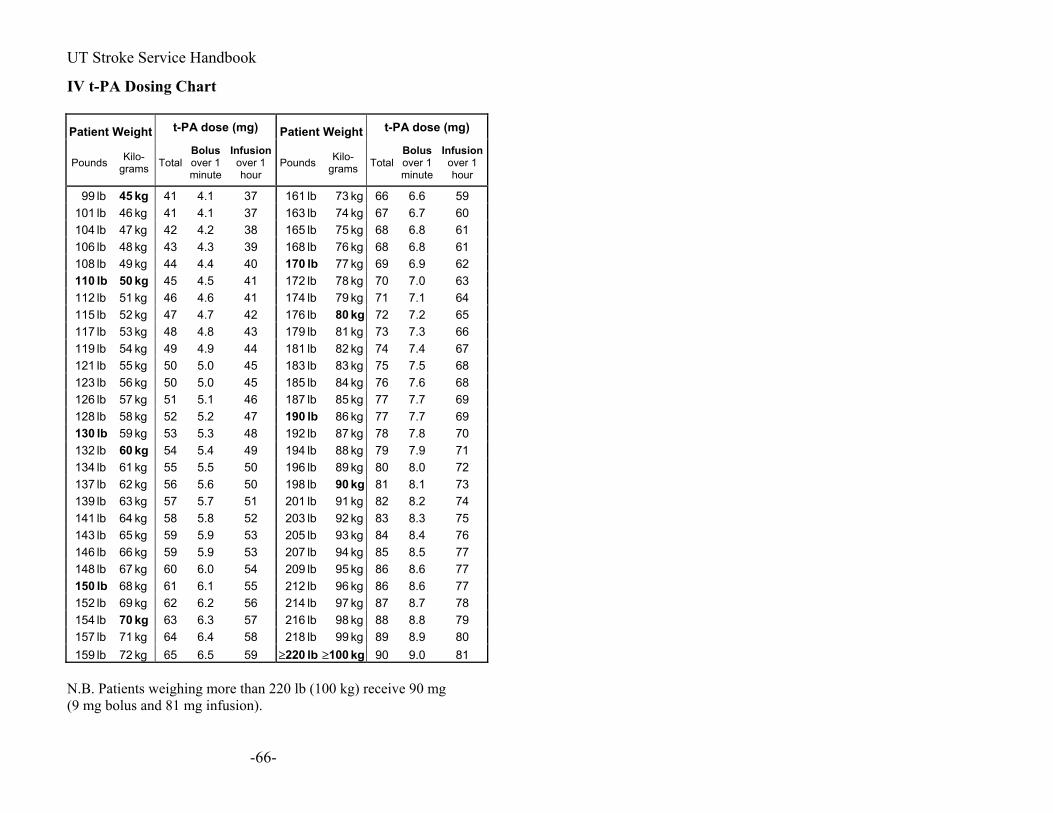
UT Stroke Service Handbook
IV t-PA Dosing Chart Patient Weight t-PA dose (mg) Patient Weight t-PA dose (mg)
Pounds Kilo-grams Total
Bolus over 1 minute
Infusion over 1 hour
Pounds Kilo-grams Total
Bolus over 1 minute
Infusion over 1 hour
99 lb 45 kg 41 4.1 37 161 lb 73 kg 66 6.6 59101 lb 46 kg 41 4.1 37 163 lb 74 kg 67 6.7 60 104 lb 47 kg 42 4.2 38 165 lb 75 kg 68 6.8 61 106 lb 48 kg 43 4.3 39 168 lb 76 kg 68 6.8 61 108 lb 49 kg 44 4.4 40 170 lb 77 kg 69 6.9 62110 lb 50 kg 45 4.5 41 172 lb 78 kg 70 7.0 63112 lb 51 kg 46 4.6 41 174 lb 79 kg 71 7.1 64 115 lb 52 kg 47 4.7 42 176 lb 80 kg 72 7.2 65117 lb 53 kg 48 4.8 43 179 lb 81 kg 73 7.3 66 119 lb 54 kg 49 4.9 44 181 lb 82 kg 74 7.4 67 121 lb 55 kg 50 5.0 45 183 lb 83 kg 75 7.5 68 123 lb 56 kg 50 5.0 45 185 lb 84 kg 76 7.6 68 126 lb 57 kg 51 5.1 46 187 lb 85 kg 77 7.7 69 128 lb 58 kg 52 5.2 47 190 lb 86 kg 77 7.7 69130 lb 59 kg 53 5.3 48 192 lb 87 kg 78 7.8 70132 lb 60 kg 54 5.4 49 194 lb 88 kg 79 7.9 71134 lb 61 kg 55 5.5 50 196 lb 89 kg 80 8.0 72 137 lb 62 kg 56 5.6 50 198 lb 90 kg 81 8.1 73139 lb 63 kg 57 5.7 51 201 lb 91 kg 82 8.2 74 141 lb 64 kg 58 5.8 52 203 lb 92 kg 83 8.3 75 143 lb 65 kg 59 5.9 53 205 lb 93 kg 84 8.4 76 146 lb 66 kg 59 5.9 53 207 lb 94 kg 85 8.5 77 148 lb 67 kg 60 6.0 54 209 lb 95 kg 86 8.6 77 150 lb 68 kg 61 6.1 55 212 lb 96 kg 86 8.6 77152 lb 69 kg 62 6.2 56 214 lb 97 kg 87 8.7 78 154 lb 70 kg 63 6.3 57 216 lb 98 kg 88 8.8 79157 lb 71 kg 64 6.4 58 218 lb 99 kg 89 8.9 80 159 lb 72 kg 65 6.5 59 ≥220 lb ≥100 kg 90 9.0 81
N.B. Patients weighing more than 220 lb (100 kg) receive 90 mg (9 mg bolus and 81 mg infusion).
-66-

UT Stroke Service Handbook
Sample Admission Orders Routine Ischemic Stroke: Admit to Stroke Unit (or NICU) on Stroke Service Attending X Diagnosis: _____ Resident Y Condition: guarded, critical,… Vitals & neuro checks q1h x 4 hours, then q2h x 8 hours, then routine Allergies: Activity: Bed rest overnight? , then out of bed with assistance Nursing: Head of Bed flat overnight? Call H.O. change in neuro status, for SBP>200 or <110, DBP >120 or <50, RR >24 or <8, T>101.4. (Note: BP parameter would be different post-t-PA!!) IVF: Normal saline with 20 mEq KCl at 80-125 cc/hr Diet: NPO until cleared by speech (or if patient is really looking good, AHA diet) Advance diet per speech therapy. Meds: Antiplatelets ?
ASA 325 mg PO/PR x1then 81 mg once daily, clopidogrel (Plavix) 375 mg PO x1 then 75 mg PO once daily
Decreased dose or stop antihypertensives Continue or institute statins in most cases Decrease dose of hypoglycemics Insulin sliding scale? Insulin drip? (see Appendix for protocol) Docusate sodium (Colace) 100 mg PO twice daily Acetaminophen (Tylenol) 650 mg q6h PRN T > 38 or pain Heparin 5000 U SC q12h or enoxaparin (Lovenox) 40 mg
SC once daily or compressive stockings, sequential compressive devices (SCDs)
Tests:
• MRI/MRA Brain and Neck with Diffusion Weighted Imaging (DWI) and Perfusion Weighted Imaging (PWI)
-67-
UT Stroke Service Handbook
Stroke Protocol (should be written among ER orders) Don’t forget to include an indication which is a symptom.
• 2D-Echocardiogram (TTE) with saline contrast for evaluation of embolic source
• CBC with differential and platelets, creatinine, blood urea nitrogen, glucose, electrolytes, INR, PTT if not done in ED
• Fasting lipid panel, Hemoglobin A1c, calcium, phosphorus, liver functions
• Urine toxicology screen for young stroke • Tests for stroke in the young/unusual stroke (see
Appendix) • Physical Therapy, Occupational Therapy, Speech
Pathology consultations* • Rehabilitation consultation*
Post t-PA orders (those that are different than previously outlined routine admission orders):
• Vitals and neuro checks: o Monitor blood pressure q 15 min x 2 hours, q30
min x 6 hours then q 1h x 16 hours. Goal SBP<180 and DBP<105
• Allergies: • Activity: Bed rest x 24 hours except for PT,OT, rehab
evaluation. • head of bed flat x 12 hours • Cardiac monitoring x 24 hours • Nursing: Monitor intake and output (I/O), measure
baseline weight, blood sugar checks QID (if needed), call HO if SBP>180 and DBP>105, signs of angioedema, change in neuro status, bleeding, etc.
• Diet: NPO x 24 h or until cleared by speech • MEDS:
-68-

UT Stroke Service Handbook
o IF SBP >180 or DBP 105 on two readings taken about 5 minutes apart, give labetalol (Trandate, Normodyne) 10-20 mg IV q 10-20 min PRN.
Alternatives: Nicardipine (Cardene) gtt. start at 5 mg/h,
increase by 2.5 mg/h q 15 min up to max 15 mg/h
o Add insulin sliding scales and glucose goals (<150)
o Famotidine (Pepcid) or equivalent 20 mg PO/IV q12 h
o No aspirin, heparin, ticlopidine (Ticlid), clopidogrel (Plavix), warfarin (Coumadin), NSAIDS for 24 h
• Avoid arterial sticks and other procedures that may predispose to bleeding for 24 hours.
• Compressive stockings and SCDs • Head CT without contrast (or MRI) at 24 hours
ICH Admission orders: --Admit to Neurological ICU (or Stroke Unit) on Stroke Service: Attending______, Resident_____ --Diagnosis: Intracerebral Hemorrhage --Condition: guarded, critical,… --Vitals & neuro checks q1h x 4 hours, then q2h x 8 hours, then routine --Allergies: --Activity: Bed rest x 24 hours --Nursing: Head of Bed up at 30 degrees --Call H.O. if change in neuro status, for SBP>150 or <110, DBP >100 or <50, RR >24 or <8, T>101.4. --IVF: NS with 20 mEq KCl at 80 cc/hr --Diet: NPO until cleared by speech (or if patient is really looking good, AHA diet) --Meds: • Antihypertensives options
IV
-69-

UT Stroke Service Handbook
o Labetalol (Trandate, Normodyne) 10-20 mg q10 min PRN BP…
o Nicardipine (Cardene) gtt. start at 5 mg/h, increase by 2.5 mg/h q 15 min up to max 15 mg/h
o Hydralazine (Apresoline) 10-20 mg q4-6h PRN o Enalapril (Vasotec) 0.625-1.25mg q 6h PRN Oral short-acting o Diltiazem (Cardizem) starting at 30 mg PO q6h o Captopril (Capoten) starting at 25-50 mg PO tid Oral long-term o ACE-I e.g., Perindopril (Aceon) 4 mg PO once
daily or ramipril (Altace) 5 mg PO once daily o Beta-blocker: e.g., Metoprolol (Lopressor, Toprol) o And many other antihypertensives
• Insulin sliding scale? • Docusate 250 mg PO twice daily • ulcer prophylaxis • Acetaminophen (Tylenol) 650 mg q6h PRN T > 38 or pain • Consider antiepileptic drugs or watch carefully for
seizures o No data on this but neurologists usually don’t load
with antiepileptic agents unless seizure activity is noted, but neurosurgeons usually load prophylactically.
--compressive stockings and SCDs --ICU consult --Physical Therapy, Occupational Therapy, Speech Pathology consultations --Rehabilitation consultation --Tests:
o Urine toxicology? for young stroke o Consider MRI ICH protocol (if young, no history of
hypertension, or hemorrhage in an unusual location) o Consider MR Venogram o Consider Digital Subtraction Angiogram
-70-

UT Stroke Service Handbook
Sample Discharge Summary Name MR# Admitting Date, Service, and Attending Physician: Discharge Date, Service, and Attending Physician: Admission Dx: Discharge Diagnosis: 1. Ischemic Stroke, Intracerebral
Hemorrhage, etc… 2. hypertension… History of Present Illness: Past Medical History: Past Surgical History: Medications: Social History: Family History: Review Of Systems: Physical Exam: Include general exam as well as neurologic exam. Include NIH Stroke Scale if possible. Labs: Radiology: Procedures: Consults: Hospital Course:
Include acute treatment, work-up, neurologic course, complications
Stroke Mechanism: For ischemia: cardioembolic, carotid stenosis, intracranial
stenosis, lacunar stroke, dissection, venous, cryptogenic, etc.
For ICH: hypertensive, AVM, amyloid angiopathy, unknown, etc.
Condition at discharge: stable, etc. Also describe remaining deficits, impairment. Discharge NIH stroke scale. Disposition: Home, Skilled Nursing Facility, Inpatient Rehabilitation, etc. Discharge Medications: Instructions to patients:
-71-
UT Stroke Service Handbook
Physical Activity: Diet:
Smoking cessation counseling, etc: Follow-up plan:
Send copy: to primary care provider
-72-

UT Stroke Service Handbook
Stroke Radiology CT Non-contrast head CT remains the standard procedure for evaluation of stroke initially. Acute Ischemia appearances (general guidelines).
<6 hours--- No change in appearance, or >1.5 hours--- Loss of grey-white differentiation >3 hours--- Hypodensity
>6 hours--- Swelling >weeks--- Ex vacuo changes
window width and level (WW/WL) for early CT: The standard brain view on CT is set at around 90/40. Setting of ~25/~30 may give high contrast of brain parenchyma to demonstrate the early ischemic signs more easily. Acute Hemorrhage (extravasated blood) appears hyperdense (bright) in 40-60 Hounsfield Units (HU). In the first few hours, the intensity may increase to 60-80 HUs. Intensity attenuates with time at~0.7-1.5 HU/day. The role of contrast head CT: CT Angiography (involves IV bolus of contrast, and imaging the arteries quickly during first pass of contrast). It allows visualization of vessels or lack thereof (occlusion, stenosis, AVM, aneurysms).
Requirements: --Adequate renal function because the contrast bolus is more than usual. --Good IV access (you don’t want contrast in soft tissues!!)
CT viewing window width/level at 800/100 may be best to visualize the arteries next to bones. Standard contrast head CT allows evaluation for stroke mimics by detecting blood brain barrier breakdown: Tumor, infection, inflammation, etc. How to try to prevent contrast nephropathy
-73-

UT Stroke Service Handbook
Acetylcysteine 600 mg PO q12h the day prior and the day of iodinated contrast administration for CT or angiography (Tepel et al, NEJM, 2000; Kay et al, JAMA, 2003).
Sodium Bicarbonate (Mertens et al, JAMA, 2004): o 154 mEq/L of sodium bicarbonate in dextrose and
H2O (adding 154 mL of 1000 mEq/L sodium bicarbonate to 846 mL of 5% dextrose).
o IV bolus at 3 mL/kg per hour x 1 hour immediately before radiocontrast injection (Maximum 330 ml/hr).
o Followed by 1 mL/kg per hour during the contrast exposure and for 6 hours after the procedure (Maximum 110 mL/hr).
Adequate hydration. Creatinine > 2.0 probably contraindicates use of IV
contrast in most cases. ASPECTS: scoring system of early CT changes in MCA territory infarction (Barber et al, Lancet, 2000). Level of basal ganglia (BG) and thalamus. Level just rostral to BG.
-74-

UT Stroke Service Handbook
ASPECTS study form 10 regions of interest: At the level of basal ganglia and thalamus:
At the level just rostral to deep nuclei:
C=caudate L=lentiform nuclei IC=internal capsule I=insular ribbon M1=anterior MCA cortex) M4 (superior to M1) M2=MCA cortex lateral to insula M5 (superior to M2) M3=posterior MCA cortex M6 (superior to M3) (Not included in scoring: A=ACA circulation; P=PCA circulation.) Subcortical structures are allotted 3 points (C, L, and IC). MCA cortex is allotted 7 points (insular cortex, M1, M2, M3, M4, M5, and M6). 1 point is subtracted for each defined area of early ischemic change, such as focal swelling, or parenchymal hypoattenuation. 10=normal and 0=abnormal in entire MCA distribution MRI You have to refer to a real MRI book to explain all physics of the sequences to you. Here is a brief simplification: T1-weighted sequence (usually in axial and sagittal sections): How to identify: Usually looks pale and bland. It looks the
“most similar to CT.” It appears exactly how you would expect the brain to look: CSF is black. Gray matter is gray. White matter is white.
Use: good for anatomy. Compare to T1 with contrast for leakage of BBB. T2-weighted sequence:
-75-
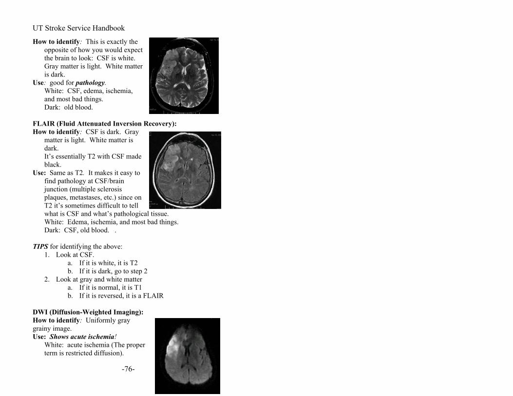
UT Stroke Service Handbook
How to identify: This is exactly the opposite of how you would expect the brain to look: CSF is white. Gray matter is light. White matter is dark.
Use: good for pathology. White: CSF, edema, ischemia,
and most bad things. Dark: old blood. FLAIR (Fluid Attenuated Inversion Recovery): How to identify: CSF is dark. Gray
matter is light. White matter is dark.
It’s essentially T2 with CSF made black.
Use: Same as T2. It makes it easy to find pathology at CSF/brain junction (multiple sclerosis plaques, metastases, etc.) since on T2 it’s sometimes difficult to tell what is CSF and what’s paWhite: Edema, ischem
thological tissue. ia, and most bad things.
s T2 p 2
2. Loo t
FLAIR DWI (Diffu
he proper .
Dark: CSF, old blood. .
IPS for identifying the above: T1. Look at CSF.
white, it ia. If it isb. If it is dark, go to stek a gray and white matter a. If it is normal, it is T1 b. If it is reversed, it is a
sion-Weighted Imaging): How to identify: Uniformly gray grainy image. Use: Shows acute ischemia! White: acute ischemia (T
term is restricted diffusion)
-76-
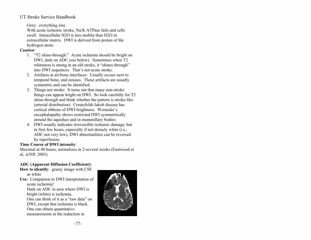
UT Stroke Service Handbook
Grey: everything else. With acute ischemic stroke, Na/K ATPase fails and cells
swell. Intracellular H2O is less mobile than H2O in
Caut on
ADC (see below). Sometimes when T2
2.
for T2 -like
4. but
Time CMaxima
: grainy image with CSF
n
extracellular matrix. DWI is derived from proton of the hydrogen atom. tion: 1. “T2 shine-through:” Acute ischemia should be brigh
I, dark onDWwhiteness is strong in an old stroke, it “shines through” into DWI sequences. That’s not acute stroke. Artifacts at air/bone interfaces: Usually occurs next to temporal bone, and sinuses. These artifacts are usually symmetric and can be identified.
3. Things not stroke: It turns out that many non-stroke things can appear bright on DWI. So look carefully shine-through and think whether the pattern is stroke(arterial distribution). Creutzfeldt-Jakob disease has cortical ribbons of DWI brightness. Wernicke’s encephalopathy shows restricted DWI symmetrically around the aqueduct and in mammillary bodies. DWI usually indicates irreversible ischemic damage, in first few hours, especially if not densely white (i.e.,ADC not very low), DWI abnormalities can be reversed by reperfusion. ourse of DWI intensity: l at 40 hours, normalizes in 2-several weeks (Eastwood et
al, AJNR, 2003). ADC (Apparent Diffusion Coefficient): How to identify
as white Use: Companion to DWI interpretation of
acute ischemia! Dark on ADC in area where DWI is
bright (white) is ischemia. One can think of it as a “raw data” o
DWI, except that ischemia is black. One can obtain quantitative measurements in the reduction in
-77-

UT Stroke Service Handbook
diffusion coefficient. e Course:
at 28 hoursTim
, pseudonormalizes at 10 days, then
Gradient-Echo sequence (MPGR): just
m us
the
gnt sequences used.
)
e
WI,
Maximal (dark) bright (Eastwood et al, AJNR, 2003).
How to identify: Grayish (It’s hard bylooking)
Use: Hemorrhages, whether new or old, appear dark.
--It’s useful to look for micro-hemorrhages such as those froamyloid angiopathy and cavernomalformations. --One cannot measure the size. Thesignal is amplified and is bigger thanamount of blood.
Perfusion Weighted Ima ing (PWI): How to identify: There are two differeMean Time to Enhancement (MTE) measures arrival time of bolus of dye. Areas of low cerebral perfusion look brighter (more light gray). Negative Enhancement Integral (NEImeasures cerebral blood volume (CBV). Areas of severe ischemia have reduced CBV and look dark. In mild ischemia, thvascular bed is dilated, CBV may be increased, and such regions will look bright. Use: PWI sequences measure cerebral blood flow. You look for a so-called “mismatch” between the changes on D
-78-
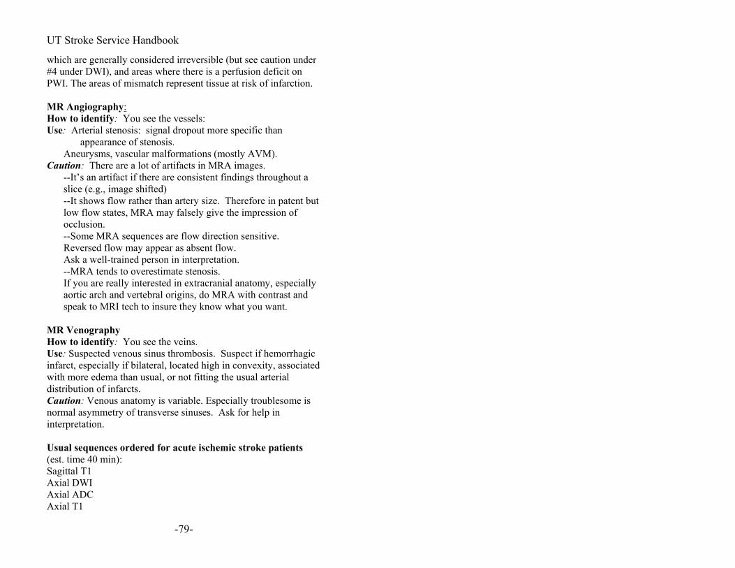
UT Stroke Service Handbook
which are generally considered irreversible (but see caution und#4 under DWI), and areas where there is a perfusion deficit on PWI. The areas of mismatch represent tissue at risk of infarction. MR Angiography:
er
How to identify: You see the vessels:
more specific than
Cau
out a
atomy, especially
MR
You see the veins. sis. Suspect if hemorrhagic
me is
nces ordered for acute ischemic stroke patients
Use: Arterial stenosis: signal dropout appearance of stenosis.
Aneurysms, vascular malformations (mostly AVM). tion: There are a lot of artifacts in MRA images.
gh--It’s an artifact if there are consistent findings throuslice (e.g., image shifted) --It shows flow rather than artery size. Therefore in patent but low flow states, MRA may falsely give the impression of occlusion. --Some MRA sequences are flow direction sensitive. Reversed flow may appear as absent flow. Ask a well-trained person in interpretation. --MRA tends to overestimate stenosis.
al anIf you are really interested in extracraniaortic arch and vertebral origins, do MRA with contrast and speak to MRI tech to insure they know what you want.
Venography How to identify: Use: Suspected venous sinus thromboinfarct, especially if bilateral, located high in convexity, associatedwith more edema than usual, or not fitting the usual arterial distribution of infarcts. Caution: Venous anatomy is variable. Especially troublesonormal asymmetry of transverse sinuses. Ask for help in interpretation. Usual seque(est. time 40 min): Sagittal T1 Axial DWI Axial ADC Axial T1
-79-

UT Stroke Service Handbook
Axial FLAIR
d NEI) (if PWI is done)
e)
uncooperative patients (est. time 10
Circle of Willis and Neck
nces ordered for acute ICH patients:
AIR
contrast st
Axial MPGR PWI (MTE anAxial T1 post contrastCoronal T1 post contrast (if PWI is donMRA Circle of Willis and Neck Abbreviated protocol for min): DWI
MRAT2 or FLAIR Usual sequeSagittal T1 Axial T1 Axial T2 Axial FLAxial MPGR Axial T1 postCoronal T1 post contraMRA Circle of Willis MRV ?
-80-

UT Stroke Service Handbook
MRI findings in hemorrhage: adapted from Parizel, et al, European Radiology, 2001:
Sequential signal intensity (SI) changes of intracranial hemorrhage on MRI (1.5 T). Hb hemoglobin; e- electrons; PEDD proton-electron dipole-dipole interaction; T2-PRE T2-proton relaxation enhancement; FeOOH ferric oxyhydroxide; isointense relative to normal gray matter; ↑ increased SI relative to normal gray matter; ↓ decreased SI relative to normal gray matter; ↓↓ markedly decreased SI relative to normal gray matter
Hyperacute hemorrhage
Acute hemorrhage
Early subacute hemorrhage
Late subacute hemorrhage
Chronic hemorrhage
What happens
Blood leaves the vascular system (extravasation)
Deoxygenation with formation of deoxy-Hb
Clot retraction and deoxy-Hb is oxidized to met-Hb
Cell lysis (membrane disruption)
Macrophages digest the clot
Time frame <12 h
Hours to days (weeks in center of hematoma)
A few days 4-7 days to 1 month
Weeks to years
Red blood cells
Intact erythrocytes
Intact, but hypoxic erythrocytes
Still intact, severely hypoxic
Lysis (solution of lysed cells)
Gone; encephalomalacia with proteinaceous fluid
State of Hb
Intracellular oxy-Hb
Intracellular deoxy-Hb
Intracellular met-Hb (first at periphery of clot)
Extracellular met-Hb
Hemosiderin (insoluble) and ferritin (water soluble)
Oxidation state
Ferrous (Fe2+), no unpaired e-
Ferrous (Fe2+), four unpaired e-
Ferric (Fe3+), five unpaired e-
Ferric (Fe3+), five unpaired e-
Ferric (Fe3+)
2000 5 unpaired e-
Magnetic properties
Diamagnetic
( )
Paramagnetic
( >0)
Paramagnetic
( >0)
Paramagnetic
( >0)
FeOOH is superparamagnetic
T1-weighted images
or↓ (or↓) (No
PEDD interaction)
↑↑ (PEDD interaction)
↑↑ (PEDD interaction)
or↓ (no PEDD interaction)
T2-weighted images
↑ (High water content)
↓T2 PRE (susceptibility effect)
↓↓T2 PRE (susceptibility effect)
↑↑No T2 PRE (loss of compart
↓↓T2 PRE (susceptibility effect)
-81-

UT Stroke Service Handbook
-82-
Hyperacute hemorrhage
Acute hemorrhage
Early subacute hemorrhage
Late subacute hemorrhage
Chronic hemorrhage
mentalization)
Transcranial Doppler Ultrasound (TCD) For various uses of TCD in stroke please read Cerebrovascular Ultrasound in Stroke Prevention and Treatment, edited by A.V. Alexandrov, 2004. There are many uses of TCD:
Diagnosis of intracranial stenosis Diagnosis of acute occlusion Monitoring of acute thrombolytic therapy Vasoreactivity (vascular reserve) with carotid disease Emboli monitoring Vascular monitoring during surgery Detection of Right-to-left shunt (patent foramen ovale)
Procedure for right-to-left shunt detection (you may be asked to help do this) European Society of Neurosonlogy and Cerebral Hemodynamics consensus, 1999 (Jauss and Zanette, Cerebrovasc. Dis, 2000; Angeli et al, Neurol. Sci., 2001) Equipment:
TCD Two normal saline bottles 15 cc Two 10cc syringe Flexible tubing Three-way stopcock (often found in ICU)
Preparation: The patient must be the supine position, with the arm
horizontal. An intravenous catheter (#18) is inserted into an antecubital vein (connected to a 250 ml bottle of physiologic solution by means of a flexible tube to maintain venous access).

UT Stroke Service Handbook
The right middle cerebral artery is traced by means of TCD (the examination is more sensitive if bilateral monitoring is used).
Procedure Two 10 ml (or 20 ml) syringes are prepared: one
containing 9 ml of physiologic solution and the other containing 1 ml of air. By means of a three-way stopcock, the contents of both syringes are rapidly mixed until a homogeneous solution is obtained.
The solution is rapidly injected in bolus form with the patient at rest.
The MCA is monitored for 40-60 seconds. The procedure is repeated with Valsalva maneuver.
The efficacy of Valsalva maneuver must be ascertained beforehand through the reduction by at least one third of the systolic flow velocity on MCA
Five seconds after injection of the contrast agent, the examiner orders the patient to begin Valsalva’s maneuver, which must last for at least 10 seconds.
Interpretation: The test is deemed positive if the appearance of at least one microbubble (MB) is recorded as high intensity transient signal (HITS) on the TCD trace within 40 seconds of terminating the injection; no agreement exists as to a cut-off interval between contrast injection and microbubble appearance. Although it takes about 11 seconds for the bubbles reach the MCA through an intracardiac shunt, and about 14 seconds through an intrapulmonary shunt, a time window cannot differentiate between RLS at the atrial level and RLS at different sites of the vascular system. It is in any case advisable to record the time of appearance of the first MB.
-83-

UT Stroke Service Handbook
The results of the two sessions (basal and with Valsalva) must be evaluated separately. Repeated testing may increase the sensitivity, and in the event of discrepancies the positive test must be considered
No HITS: test negative 1–10 HITS: low-grade shunt >10 HITS, but without “curtain” effect: medium-grade
shunt Curtain effect, seen when the microbubbles are so
numerous as to be no longer distinguishable separately: high-grade shunt.
With regard to the physiopathological features of the RLS, we define as:
Permanent, a shunt detected in basal conditions Latent, a shunt detected only with Valsalva
Drug Protocols Heparin drip
Check hematocrit, platelet count, PT/INR, aPTT at baseline (within the prior 72 hours).
Obtain or estimate patient’s weight (Use adjusted body weight for obese patients).
No bolus heparin.
-84-

UT Stroke Service Handbook
Initial Infusion
Weight Initial Infusion
<50 kg 500 Units/hr = 10 mL/hr
50-59 kg 600 Units/hr = 12 mL/hr
60-69 kg 700 Units/hr = 14 mL/hr
70-79 kg 800 Units/hr = 16 mL/hr
80-89 kg 900 Units/hr = 18 mL/hr
90-99 kg 1000 Units/hr = 20 mL/hr
100-109 kg 1100 Units/hr = 22 mL/hr
110-119 kg 1200 Units/hr = 24 mL/hr
>119 kg 1400 Units/hr = 28 mL/hr
Check aPTT every 6 hours after change in infusion or every 24 hours if within therapeutic range.
CBC q 3 days.
Goal aPTT 55-85 seconds
Adjustment every 6 hours:
aPTT (sec)
Stop infusion
Change rate of infusion
Check aPTT
<45 ↑ 200 Units/hr in 6 hours
45-54 ↑ 100 Units/hr in 6 hours
55-85 No change in 6 hours
86-90 ↓ 100 Units/hr Next a.m.
91-100 30 minutes ↓ 150 Units/hr in 6 hours
>100 60 minutes ↓ 250 Units/hr in 6 hours
Call H.O. if aPTT <45 or >100 seconds on 2 consecutive measurements or >125 seconds.
If significant bleeding occurs, stop heparin and assess.
-85-

UT Stroke Service Handbook
Protocol modified from Toth and Voll, Stroke, 2002. Insulin drip Make sure that the nursing unit is capable of using insulin gtt. with glucose measurement every 1 hour. Use insulin drip if the glucose in an acute stroke patient is over 250 mg/dL. Goal insulin level 80-110 mg/dL
Check capillary glucose q 1 hour.
Consider insulin bolus.
Start insulin gtt. at the calculated rate using the following formula:
(blood sugar – 60) X 0.03 ____Units/hr IV drip
Readjust glucose infusion every 1 hour using the above formula. If glucose is <200, then q 2 hour glucose check.
If glucose <60, give 1 ampule D50 and call physician. Regular Insulin Sliding Scale Recommended Indications: • As a supplement to a patient’s usual diabetes medications
(long-acting insulin or oral agents) to treat uncontrolled high blood sugars
• For short term use (24-48 hours) in a patient admitted with an unknown insulin requirement
Regimens: • Low Dose Scale: Suggested starting point for thin and elderly,
or those being initiated on total parenteral nutrition (TPN) • Moderate Dose Scale: Suggested starting point for average
patient • High Dose Scale: Suggested for patients with infections or
those receiving therapy with high dose corticosteroids
-86-

UT Stroke Service Handbook
Check blood sugars: AC & HS (6:30 AM, 11:30 AM, 4:30 PM, 9:30 PM) BID (6:30 AM, 4:30 PM) q6h (recommended for patients receiving continuous nutrition over 24h) q4h (recommended for patients requiring close monitoring) other __________________________
Blood Sugar (mg/dL)
Low Dose Scale
Moderate Dose Scale
High Dose Scale Patient-Specific Scale
<60
Initiate Hypoglycemia
Protocol
Initiate Hypoglycemia
Protocol
Initiate Hypoglycemia
Protocol
Initiate Hypoglycemia
Protocol
60-130 0 units 0 units ____ units
131-180 2 units 4 units 8 units ____ units
181-240 4 units 8 units 12 units ____ units
241-300 6 units 10 units 16 units ____ units
301-350 8 units 12 units 20 units ____ units
351-400 10 units 16 units 24 units ____ units
>400 12 units and call MD
20 units and call MD
28 units and call MD
____ units
0 units
Medical Complications: Prevention and Treatment Deep Venous Thrombosis (DVT)is not an uncommon complication. Prevention:
-87-

UT Stroke Service Handbook
• Enoxaparin (Lovenox) 40 mg SC once daily (Hillbom et al, Acta Neurol Scand, 2000)-probably the best choice
• Heparin 5000 U SC q12h * • Dalteparin (Fragmin) 5000 units once daily • Compressive stockings and sequential compression
devices (SCDs) Aspiration pneumonia Prevention:
1. NPO until speech path evaluation or bedside evaluation by specially trained nurses
2. Follow Speech pathology recommendations 3. Head of bed up 4. Sit upright when eating 5. Assistance with feeding
Diagnosis: constellation of symptoms and signs
Fever, hypoxia, CXR infiltrate, leukocytosis, clinical aspiration
Sputum culture not very reliable. Empiric Treatment: No need to cover for ANEROBES Cover for Gram + and Gram – (Pseudomonas, Enterobacteriaceae) Antibiotic Choices: (Be sure to check the patient’s allergies)
o Ceftriaxone (Rocephin) 1-2g IV q24h o Cefepime (Maxipime) 1g IV q12h o Gatifloxacin (Tequin) 400mg IV/PO once daily o For MRSA- Vancomycin (Vancocin) 1g IV q12 (check
trough levels before 3rd or 4th dose. Target 10-20 mcg/ml). o For MSSA- Nafcillin (Nallpen) 2 gm q 4 is better,
Cefepime has fair coverage Duration 10-14 days. Catheter Associated UTI Prevention:
-88-
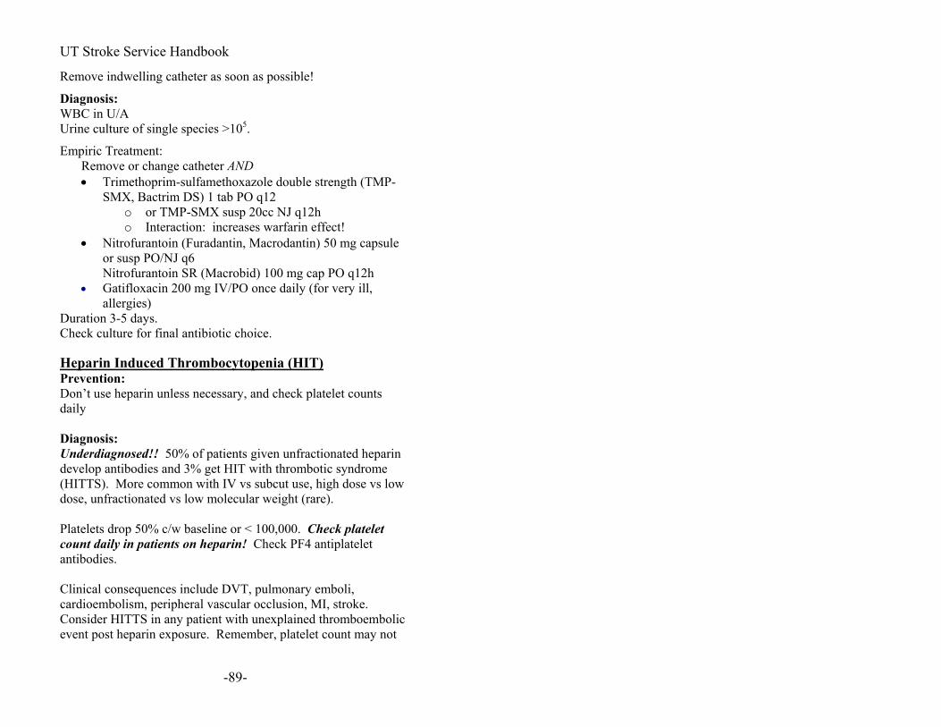
UT Stroke Service Handbook
Remove indwelling catheter as soon as possible!
Diagnosis: WBC in U/A Urine culture of single species >105.
Empiric Treatment: Remove or change catheter AND • Trimethoprim-sulfamethoxazole double strength (TMP-
SMX, Bactrim DS) 1 tab PO q12 o or TMP-SMX susp 20cc NJ q12h o Interaction: increases warfarin effect!
• Nitrofurantoin (Furadantin, Macrodantin) 50 mg capsule or susp PO/NJ q6 Nitrofurantoin SR (Macrobid) 100 mg cap PO q12h
• Gatifloxacin 200 mg IV/PO once daily (for very ill, allergies)
Duration 3-5 days. Check culture for final antibiotic choice. Heparin Induced Thrombocytopenia (HIT) Prevention: Don’t use heparin unless necessary, and check platelet counts daily Diagnosis: Underdiagnosed!! 50% of patients given unfractionated heparin develop antibodies and 3% get HIT with thrombotic syndrome (HITTS). More common with IV vs subcut use, high dose vs low dose, unfractionated vs low molecular weight (rare). Platelets drop 50% c/w baseline or < 100,000. Check platelet count daily in patients on heparin! Check PF4 antiplatelet antibodies. Clinical consequences include DVT, pulmonary emboli, cardioembolism, peripheral vascular occlusion, MI, stroke. Consider HITTS in any patient with unexplained thromboembolic event post heparin exposure. Remember, platelet count may not
-89-

UT Stroke Service Handbook
be low—just 50% drop compared with baseline, and baseline may be well above 200,000. Type 1: transient, mild, starts 4d post exposure but can start after a longer interval and after heparin stopped Type 2: two types—4-14 d post exposure or < 12 h post exposure Treatment: Stop heparin! Even so, 50% will still develop HITTS once platelet count starts to fall if you just stop heparin. Thrombin inhibitor even before platelet antibody test result is back.
• Argatroban—reversible, more potent, non-antigenic, hepatic clearance, T50= 40-50 min, adjust aPTT every 2-4 hrs to 1.5-3 X control. 2 micrograms/min- titrate up to < 10 micrograms/min. Lepirudin (Refludan) —irreversible, antigenic, renal clearance, bolus 0.4 mg/kg then infusion.
•
No warfarin until platelets normalize No platelets
-90-

UT Stroke Service Handbook
Brainstem Syndromes Look at cranial nerve (CN) abnormalities:
Lateral Medullary syndrome (aka Wallenberg syndrome)
o Vertigo, nausea, diplopia o Ipsilateral Headache (descending spinal tract of
the Vth nerve), facial or eye pain o Ataxia, hiccups o Contralateral body hemianalgesia
(pain+temperature) o Ipsilateral facial hemianalgesia
(pain+temperature) o Horner’s syndrome, Nystagmus o Ipsilateral palate, vocal cord weakness (nucleus
ambiguus) o Dysphagia o Cerebellar findings o KEY: Motor, tongue function, dorsal column
function spared because these structures lie medially in the medulla.
Millard-Gubler Syndrome--ventrocaudal pons with CN VI and VII involvement
o Contralateral hemiplegia (pyramidal tract), o Ipsilateral lateral rectus paresis (VI) o Ipsilateral LMN facial paresis (VII)
Foville Syndrome—dorsal caudal pontine lesion o Contralateral body hemiplegia o Ipsilateral LMN facial paresis (VII) o Inability to move eyes to ipsilateral to lesion
(Parapontine reticular formation, CN VI) Raymond-Cestan Syndrome—dorsal rostral pons
o Ataxia with coarse tremor o Contralateral hemisensory loss (face+body, all
modalities) o +/- contralateral hemiparesis
Marie Foix syndrome—lateral pons o Ipsilateral cerebellar ataxia o Contralateral hemiparesis
-91-

UT Stroke Service Handbook
o +/- contralateral hemisensory loss (pain & temp) due to spinothalamic tract
Weber syndrome --ventral midbrain o Contralateral hemiplegia (corticospinal and
corticobulbar tracts) o Ipsilateral oculomotor paresis, dilated pupil
Benedikt syndrome—midbrain tegmentum (Red Nucleus, CN III)
o Ipsilateral oculomotor paresis, dilated pupil o Contralateral intention tremor, hemichorea,
hemiathetosis Claude’s syndrome—midbrain tegmentum
o Ipsilateral oculomotor paresis o Contralateral cerebellar ataxia
Parinaud’s syndrome—dorsal midbrain (often with hydrocephalus, tumor)
o Upgaze paresis o Convergence-retraction nystagmus on upgaze o Large pupil with light-near dissociation, lid
retraction, lid lag
-92-

UT Stroke Service Handbook
-93-

UT Stroke Service Handbook
-94-

UT Stroke Service Handbook
Unusual Ischemic Stroke Tests to consider
• Urine drug screen • Go over home medications and supplements • Hypercoagulable testing. (There is no consensus on this).
o Arterial thromboses: Antiphospholipid antibody panel (should include Anticardiolipin IgM, Anticardiolipin IgG) Lupus-Anticoagulant (Testing can be both mixing and Russel viper venom test)
o Venous thromboses: Protein C Protein S Antithrombin III Activated Protein C (APC) resistance (biochemical test for Factor V Leiden, so you don’t need the DNA test if you order this) Factor V Leiden (DNA test) Factor II G20210A DNA test (not level of the protein!)
• Autoimmune labs o ESR, ANA, dsDNA, Complement3, Complement 4, SS-A,
SS-B (Sjögren’s syndrome is associated with vasculitis) • Fibrinogen level, highly sensitive CRP • Note: Protein C, S, and antithrombin III can be falsely
elevated acutely in stroke patients. Causes (Consult stroke texts for full discussion of these conditions) Extracranial causes
• Arterial dissection (carotid, vertebral, aortic) • Aortic atheroma • Paradoxical Embolus through PFO (venous clot) • Air embolus (central venous catheter?) • Fibromuscular dysplasia
Intracranial causes
-95-

UT Stroke Service Handbook
Cerebral venous thrombosis •
• •
• Vasculitis (Primary CNS angitis, Polyarteritis nodosa, Takayasu’s aortitis)
• Infection • Moyamoya disease • Intravascular lymphoma
Hematological causes
• Hypercoagulability Collagen vascular disease Drug abuse
Brain Death Criteria Nature of coma must be known Known structural disease or irreversible systemic metabolic cause that can explain the clinical picture. Some causes must be ruled out Body temperature must be above 32.2 C to rule out hypothermia No chance of drug intoxication or neuromuscular blockade Patient is not in shock Absence of cerebral and brain stem function 1) Unresponsive to stimuli (i.e., no flexor or extensor posturing). 2) Absent pupillary reflex. 3) Absent caloric vestibular-ocular reflex. 4) Absent corneal reflex. 5) Absent gag reflex. 6) Absent cough reflex. 7) Areflexic. The limbs are flaccid, and there is no movement, although primitive withdrawal movements in response to local painful stimuli, mediated at a spinal cord level, can occur (i.e., not decorticate or decerebrate). 8) Absent respiratory drive by apnea test.
-96-

UT Stroke Service Handbook
Apnea Test: --Preoxygenate with 100% FIO2. --Get baseline Arterial blood gas (pH and pCO2 should be normal). --Disconnect ventilator, give 100% by blow-by. Observe for spontaneous respirations. (If hypotension or arrhythmia occurs, immediately reconnect the ventilator.) --After 10 minutes or at earlier calculated interval, draw ABG, then reconnect the ventilator. --Patient is apneic if PCO2 >60 mm Hg and there is no respiratory effort. --Independent exam 6 hours apart by neurologist or neurosurgeon is required in some protocols. --12 hour observation is recommended in some protocols. Confirmatory tests (are not necessary to diagnose brain death) Cerebral blood flow studies:
TCD, Angiogram, Nuclear medicine cerebral blood flow study. Electroencephalogram (EEG) with no physiologic brain
activity
-97-

UT Stroke Service Handbook
Neurological Scales Glasgow Coma Scale (E+M+V)=3~15 Eye Opening
Response Score Characteristics
None 1 Eyes always closed; not attributable to ocular swelling
To pain 2 Eyes open in response to painful stimulus
To speech 3 Eyes open in response to speech or shout
Spontaneous 4 Eyes open; does not imply intact awareness
Best Motor Response No Response 1 No motor response to pain
Extension 2 Extension at elbow
Abnormal flexion 3
Includes preceding extension, stereotyped flexion posture, extreme wrist flexion, abduction of upper arm, flexion of fingers over thumb
Withdrawal 4 Normal flexor withdrawal; no localizing attempt to remove stimulus
Localizes pain 5 Attempt made to remove stimulus, e.g., hand
moves above chin toward supraocular stimulus
Obeys commands 6 Follows simple commands
-98-

UT Stroke Service Handbook
Best Verbal Response
No Response 1 No sounds
Incomprehensible 2 Moaning, groaning, grunting; incomprehensible
Inappropriate 3 Intelligible words, but not in a meaningful exchange; e.g., shouting, swearing.
Confused 4 Responds to questions in conversational manner, but responses indicate varying degrees of disorientation and confusion
Oriented 5 Normal orientation to time, place, person
The ICH Score (Hemphill et al, Stroke, 2001)
Component ICH Score Points
GCS score ____ 3–4 2
5–12 1
13–15 0
ICH volume, cm3 ____ 30
1
<30 0
IVH
____ Yes 1
No 0
-99-
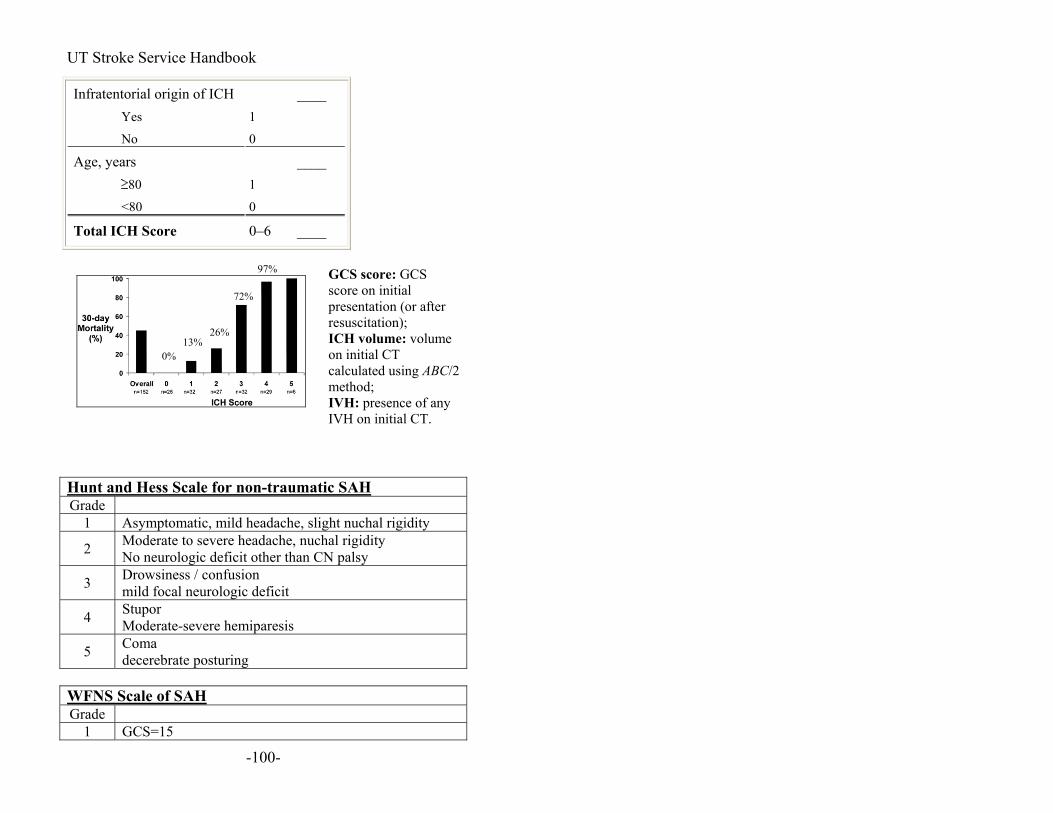
UT Stroke Service Handbook
Infratentorial origin of ICH ____ Yes 1
No 0
Age, years ____ 80
1
<80 0
Total ICH Score 0–6 ____
0%
72%
26% 13%
97% GCS score: GCS score on initial presentation (or after resuscitation); ICH volume: volume on initial CT calculated using ABC/2 method; IVH: presence of any IVH on initial CT.
Hunt and Hess Scale for non-traumatic SAH Grade
1 Asymptomatic, mild headache, slight nuchal rigidity
2 Moderate to severe headache, nuchal rigidity No neurologic deficit other than CN palsy
3 Drowsiness / confusion mild focal neurologic deficit
4 Stupor Moderate-severe hemiparesis
5 Coma decerebrate posturing
WFNS Scale of SAH Grade
1 GCS=15
-100-
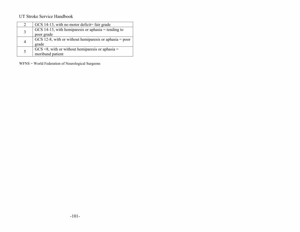
UT Stroke Service Handbook
2 GCS 14-13, with no motor deficit= fair grade
3 GCS 14-13, with hemiparesis or aphasia = tending to poor grade
4 GCS 12-8, with or without hemiparesis or aphasia = poor grade
5 GCS <8, with or without hemiparesis or aphasia = moribund patient
WFNS = World Federation of Neurological Surgeons
-101-

UT Stroke Service Handbook
-102-
National Institutes of Health Stroke Scale (NIHSS) NIH Stroke Scale Definitions:
1a. Level of Consciousness: The investigator must choose a response, even if a full evaluation is prevented by such obstacles as an endotracheal tube, language barrier, orotracheal trauma/bandages. A 3 is scored only if the patient makes no movement (other than reflexive posturing) in response to noxious stimulation.
0 = Alert; keenly responsive. 1 = Not alert, but arousable by minor stimulation to obey, answer, or respond. 2 = Not alert, requires repeated stimulation to attend, or is obtunded and requires strong or painful stimulation to make movements (not stereotyped). 3 = Responds only with reflex motor or autonomic effects or totally unresponsive, flaccid, areflexic.
1a: _____
1b. LOC Questions: The patient is asked the month and his/her age. The answer must be correct - there is no partial credit for being close. Aphasic and stuporous patients who do not comprehend the questions will score 2. Patients unable to speak because of endotracheal intubation, orotracheal trauma, severe dysarthria from any cause, language barrier or any other problem not secondary to aphasia are given a 1. It is important that only the initial answer be graded and that the examiner not "help" the patient with verbal or non-verbal cues.
0 = Answers both questions correctly.1 = Answers one question correctly. 2 = Answers neither question correctly.
1b: _____
1c. LOC Commands: The patient is asked to open and close the eyes and then to grip and release the non-paretic hand. Substitute another one step command if the hands cannot be used. Credit is given if an unequivocal attempt is made but not completed due to weakness. If the patient does not respond to command, the task should be demonstrated to them (pantomime) and score the result (i.e.,, follows none, one or two commands). Patients with trauma, amputation, or other physical impediments should be given suitable one-step commands. Only the first attempt is scored.
0 = Performs both tasks correctly 1 = Performs one task correctly 2 = Performs neither task correctly
1c: _____
2. Best Gaze: Only horizontal eye movements will be tested. Voluntary or reflexive (oculocephalic) eye movements will be scored but caloric testing is not
0 = Normal 1 = Partial gaze palsy. This score is given when gaze is abnormal in one or both eyes, but where forced
2: _____
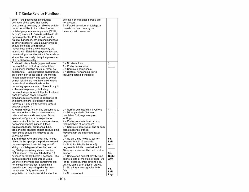
UT Stroke Service Handbook done. If the patient has a conjugate deviation of the eyes that can be overcome by voluntary or reflexive activity, the score will be 1. If a patient has an isolated peripheral nerve paresis (CN III, IV or VI) score a 1. Gaze is testable in all aphasic patients. Patients with ocular trauma, bandages, pre-existing blindness or other disorder of visual acuity or fields should be tested with reflexive movements and a choice made by the investigator. Establishing eye contact and then moving about the patient from side to side will occasionally clarify the presence of a partial gaze palsy.
deviation or total gaze paresis are not present. 2 = Forced deviation, or total gaze paresis not overcome by the oculocephalic maneuver.
3. Visual: Visual fields (upper and lower quadrants) are tested by confrontation, using finger counting or visual threat as appropriate. Patient must be encouraged, but if they look at the side of the moving fingers appropriately, this can be scored as normal. If there is unilateral blindness or enucleation, visual fields in the remaining eye are scored. Score 1 only if a clear-cut asymmetry, including quadrantanopia is found. If patient is blind from any cause score 3. Double simultaneous stimulation is performed at this point. If there is extinction patient receives a 1 and the results are used to answer question 11.
0 = No visual loss 1 = Partial hemianopia 2 = Complete hemianopia 3 = Bilateral hemianopia (blind including cortical blindness)
3: _____
4. Facial Palsy: Ask, or use pantomime to encourage the patient to show teeth or raise eyebrows and close eyes. Score symmetry of grimace in response to noxious stimuli in the poorly responsive or noncomprehending patient. If facial trauma/bandages, orotracheal tube, tape or other physical barrier obscures the face, these should be removed to the extent possible.
0 = Normal symmetrical movement 1 = Minor paralysis (flattened nasolabial fold, asymmetry on smiling) 2 = Partial paralysis (total or near total paralysis of lower face) 3 = Complete paralysis of one or both sides (absence of facial movement in the upper and lower face)
4: _____
5 & 6. Motor Arm and Leg: The limb is placed in the appropriate position: extend the arms (palms down) 90 degrees (if sitting) or 45 degrees (if supine) and the leg 30 degrees (always tested supine). Drift is scored if the arm falls before 10 seconds or the leg before 5 seconds. The aphasic patient is encouraged using urgency in the voice and pantomime but not noxious stimulation. Each limb is tested in turn, beginning with the non-paretic arm. Only in the case of amputation or joint fusion at the shoulder
0 = No drift, limb holds 90 (or 45) degrees for full 10 seconds. 1 = Drift, Limb holds 90 (or 45) degrees, but drifts down before full 10 seconds; does not hit bed or other support. 2 = Some effort against gravity, limb cannot get to or maintain (if cued) 90 (or 45) degrees, drifts down to bed, but has some effort against gravity. 3 = No effort against gravity, limb falls. 4 = No movement
5a. Left Arm _____ 5b. Right Arm _____ 6a. Left Leg
-103-

UT Stroke Service Handbook or hip may the score be "9" and the examiner must clearly write the explanation for scoring as a "9".
9 = Amputation, joint fusion explain: ______________________
_____ 6b. Right Leg
7. Limb Ataxia: This item is aimed at finding evidence of a unilateral cerebellar lesion. Test with eyes open. In case of visual defect, insure testing is done in intact visual field. The finger-nose-finger and heel shin tests are performed on both sides, and ataxia is scored only if present out of proportion to weakness. Ataxia is absent in the patient who cannot understand or is paralyzed. Only in the case of amputation or joint fusion may the item be scored "9", and the examiner must clearly write the explanation for not scoring. In case of blindness test by touching nose from extended arm position.
0 = Absent 1 = Present in one limb 2 = Present in two limbs If present, is ataxia in Right arm 1 = Yes 2 = No 9 = amputation or joint fusion, explain ___________________ Left arm 1 = Yes 2 = No 9 = amputation or joint fusion, explain ___________________ Right leg 1 = Yes 2 = No 9 = amputation or joint fusion, explain ___________________ Left leg 1 = Yes 2 = No 9 = amputation or joint fusion, explain ___________________
7: _____
8. Sensory: Sensation or grimace to pin prick when tested, or withdrawal from noxious stimulus in the obtunded or aphasic patient. Only sensory loss attributed to stroke is scored as abnormal and the examiner should test as many body areas [arms (not hands), legs, trunk, face] as needed to accurately check for hemisensory loss. A score of 2, "severe or total," should only be given when a severe or total loss of sensation can be clearly demonstrated. Stuporous and aphasic patients will therefore probably score 1 or 0. The patient with brain stem stroke who has bilateral loss of sensation is scored 2. If the patient does not respond and is quadriplegic score 2. Patients in coma
0 = Normal; no sensory loss. 1 = Mild to moderate sensory loss; patient feels pinprick is less sharp or is dull on the affected side; or there is a loss of superficial pain with pinprick but patient is aware he/she is being touched. 2 = Severe to total sensory loss; patient is not aware of being touched in the face, arm, and leg.
8: _____
9. Best Language: A great deal of information about comprehension will be obtained during the preceding sections of the examination. The patient is asked to describe what is happening in the attached picture, to name the items on the attached naming sheet, and to read from the attached list of sentences. Comprehension is judged from responses here as well as to all of the commands in the preceding general neurological exam. If visual loss interferes with the tests, ask the patient to identify objects placed in the hand, repeat, and produce speech. The intubated patient should be asked to write.
0 = No aphasia, normal 1 = Mild to moderate aphasia; some obvious loss of fluency or facility of comprehension, without significant limitation on ideas expressed or form of expression. Reduction of speech and/or comprehension, however, makes conversation about provided material difficult or impossible. For example in conversation about provided materials examiner can identify picture or naming card from patient's response. 2 = Severe aphasia; all
9: _____
-104-

UT Stroke Service Handbook The patient in coma (question 1a=3) will arbitrarily score 3 on this item. The examiner must choose a score in the patient with stupor or limited cooperation but a score of 3 should be used only if the patient is mute and follows no one step commands. (The patient's language will be tested by having the patient identify standard groups of objects and by reading a series of sentences. Comprehension of language should be judged as the physician performs the entire neurologic examination. The physician should give the patient adequate time to identify the objects on the sheet of paper. Only the first response is measured. If the patient misidentifies the object and later corrects himself, the response is still considered abnormal. The physician should then give the patient a sheet of paper with the series of sentences. The examiner should ask the patient to read at least three sentences. The first attempt to read the sentence is measured. If the patient misreads the sentence and later corrects himself, the response is still considered abnormal. If the patient's visual loss precludes visual identification of objects or reading, the examiner should ask the patient to identify objects placed in his/her hand and the examiner should judge the patient's spontaneous speech and ability to repeat sentences. If the examiner judges these responses as normal, the score should be 0. If the patient is intubated or is unable to speak, the examiner should check the patient's writing.)
communication is through fragmentary expression; great need for inference, questioning, and guessing by the listener. Range of information that can be exchanged is limited; listener carries burden of communication. Examiner cannot identify materials provided from patient response. 3 = Mute, global aphasia; no usable speech or auditory comprehension.
10. Dysarthria: If patient is thought to be normal an adequate sample of speech must be obtained by asking patient to read or repeat words from the attached list. If the patient has severe aphasia, the clarity of articulation of spontaneous speech can be rated. Only if the patient is intubated or has other physical barrier to producing speech, may the item be scored "9", and the examiner must clearly write an explanation for not scoring. Do not tell the patient why he/she is being tested. (The primary method of examination is to ask the patient to read and pronounce a
0 = Normal 1 = Mild to moderate; patient slurs at least some words and, at worst, can be understood with some difficulty. 2 = Severe; patient's speech is so slurred as to be unintelligible in the absence of or out of proportion to any dysphasia, or is mute/anarthric. 9 = Intubated or other physical barrier, explain_________________
10: _____
-105-

UT Stroke Service Handbook standard list of words from a sheet of paper. If the patient is unable to read the words because of visual loss, the physician may say the word and ask the patient to repeat it. If the patient has severe aphasia, the clarity of articulation of spontaneous speech should be rated. If the patient is mute or comatose (item 9, Best Language = 3 ) or has an endotracheal tube, this item can be rated as 9 - untestable.) 11. Extinction and Inattention (formerly Neglect): Sufficient information to identify neglect may be obtained during the prior testing. If the patient has a severe visual loss preventing visual double simultaneous stimulation, and the cutaneous stimuli are normal, the score is normal. If the patient has aphasia but does appear to attend to both sides, the score is normal. The presence of visual spatial neglect or anosognosia may also be taken as evidence of abnormality. Since the abnormality is scored only if present, the item is never untestable.
0 = No abnormality. 1 = Visual, tactile, auditory, spatial, or personal inattention or extinction to bilateral simultaneous stimulation in one of the sensory modalities. 2 = Profound hemi-inattention or hemi-inattention to more than one modality. Does not recognize own hand or orients to only one side of space. ______
11: _____
-106-

UT Stroke Service Handbook
-107-

UT Stroke Service Handbook
You know how.
Down to earth. I got home from work. Near the table in the dining room.
-108-

UT Stroke Service Handbook
They heard him speak on the radio last night.
MAMA
TIP-TOP
FIFTY-FIFTY
-109-

UT Stroke Service Handbook
THANKS
HUCKLEBERRY
BASEBALL PLAYER
CATERPILLAR
-110-

UT Stroke Service Handbook
-111-
UT Stroke Service Handbook
Recommended Reading: Textbooks: Mohr JP, Choi D, Grotta JC, Weir B, Wolf P. Stroke:
Pathophysiology, Diagnosis, & Management. 4th edition. Churchill Livingstone. 2004.
Warlow CP, Dennis MS, Van Gijn J, Hankey GJ. Stroke. 2nd edition. Blackwell Publishing, 2001.
Caplan, Louis. Caplan’s Stroke: A Clinical Approach. 3rd edition. Butterworth-Heineman Medical. April 2000.
Adams, Jr., Harold P., Hachinski, Vladimir, and Norris, John W. Ischemic Cerebrovascular Disease. Contemporary Neurology Series. Oxford University Press 2001.
Bogousslavsky J and Caplan L. Stroke Syndromes. 2nd edition. Cambridge University Press, 2001.
Bogousslavsky J and Capan L. Uncommon Causes of Stroke. 2nd edition. Cambridge University Press, 2001.
Guidelines: ASA Scientific Statement Guidelines for the Early Management of
Patients With Ischemic Stroke. Stroke. 2003;34:1056. AHA Scientific Statement: Guidelines for the Management of
Subarachnoid Hemorrhage. Circulation. 1994;90:2592-2605. ASA Scientific Statement: Guidelines for the Management of
Spontaneous Intracerebral Hemorrhage. Stroke. 1999;30:905-915.
AHA Scientific Statement: Recommendations for the Management of Patients with Unruptured Intracranial Aneurysms. Circulation. 2000;102:2300-2308.
European Stroke Initiative Recommendations for Stroke Management – Update 2003. Cerebrovasc. Dis. 2003;16:311–337.
European Stroke Initiative: http://www.eusi-stroke.com/recommendations/rc_overview.shtml
Stroke Prevention: Albers GW, Amarenco P, Easton JD, Sacco RL, Teal P.
Antithrombotics. Chest. 2001;119:300S-320S Gorelick PB. Stroke Prevention Therapy Beyond
Antithrombotics: Unifying Mechanisms in Ischemic Stroke
-112-

UT Stroke Service Handbook
-113-
Pathogenesis and Implications for Therapy. Stroke. 2002;33:862.
Thrombolysis: Tissue plasminogen activator for acute ischemic stroke. The
National Institute of Neurological Disorders and Stroke rt-PA Stroke Study Group. NEJM. 1995 Dec 14;333(24):1581-7.
Lyden, Patrick D. Thrombolytic Therapy for Stroke. Humana Press. Feb. 2001.
ICH: Qureshi AI, Tuhrim S, Broderick JP, Batjer HH, HondoH, Hanley
DF. Spontaneous Intracerebral Hemorrhage. NEJM. 2001; 344:1450-1460
Atrial Fibrillation: Atrial Fibrillation: Hart RG, Halperin JL, Pearce LA, Anderson
DC, Kronmal RA, McBride R, Nasco E, Sherman DG, Talbert RL, Marler JR. Lessons from the Stroke Prevention in Atrial Fibrillation Trials. Ann Intern Med. 2003;138:831-838.
Radiology: Parizel PM, Makkat S, Van Miert E, Van Goethem JW, van den
Hauwe L, and De Schepper AM. “Intracranial hemorrhage: principles of CT and MRI interpretation” European Radiolgy, 2001.
AVM: Fleetwood IG and Steinberg GK. Arteriovenous Malformations.
Lancet, 2002. SAH/Aneurysm: Van Gijn J, Rinkel GJE. Subarachnoid hemorrhage: diagnosis,
causes and management. Brain. 2001: 124; 249-278.