ARDS Acute Respiratory Distress Syndrome NURS 504 Liberty University Jenny Holloway
Acute Respiratory Distress Syndrome
(ARDS)
and Acute Lung Injury (ALI)
Revisited
Dr.Dhaher JS Al-habbo
FRCP London UK
Assistant Professor in Medicine
DEPARTMENT OF MEDICINE
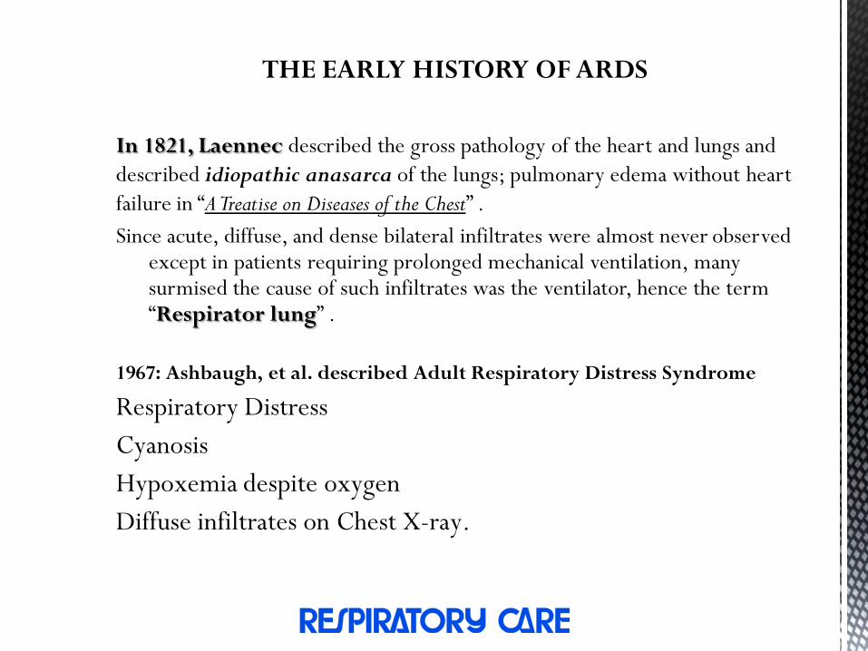
THE EARLY HISTORY OF ARDS
In 1821, Laennec described the gross pathology of the heart and lungs and
described idiopathic anasarca of the lungs; pulmonary edema without heart
failure in “A Treatise on Diseases of the Chest” .
Since acute, diffuse, and dense bilateral infiltrates were almost never observed except in patients requiring prolonged mechanical ventilation, many surmised the cause of such infiltrates was the ventilator, hence the term “Respirator lung” .
1967: Ashbaugh, et al. described Adult Respiratory Distress Syndrome
Respiratory Distress
Cyanosis
Hypoxemia despite oxygen
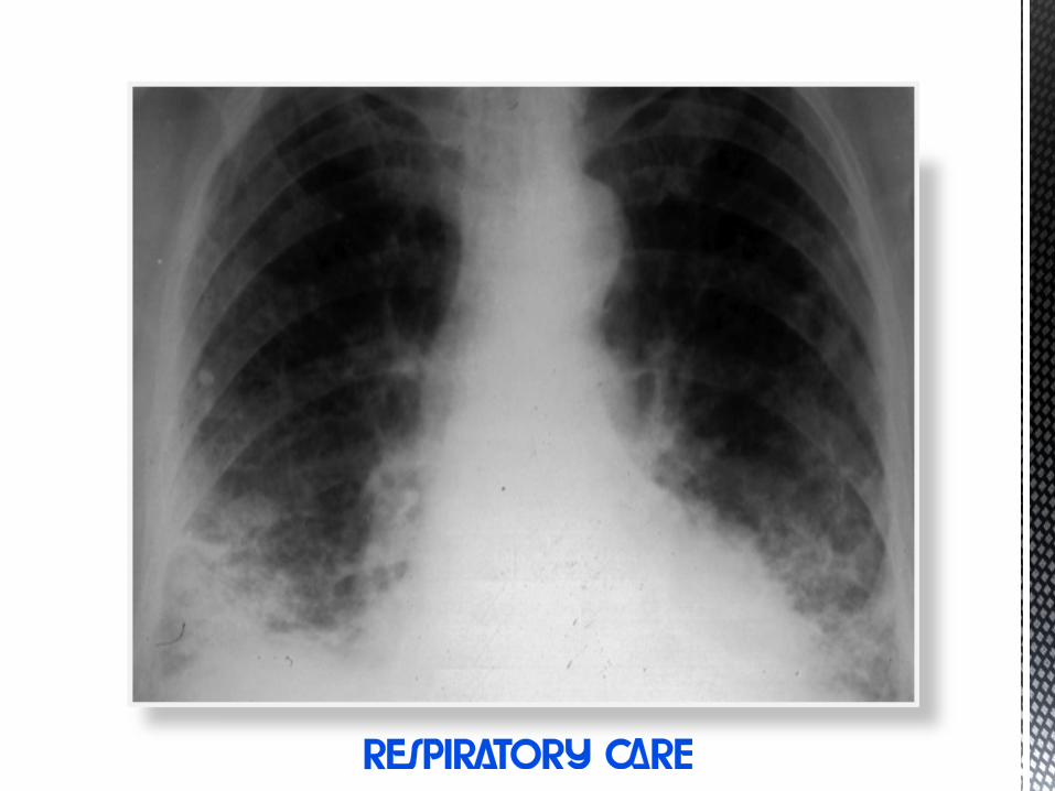
Diffuse infiltrates on Chest X-ray.
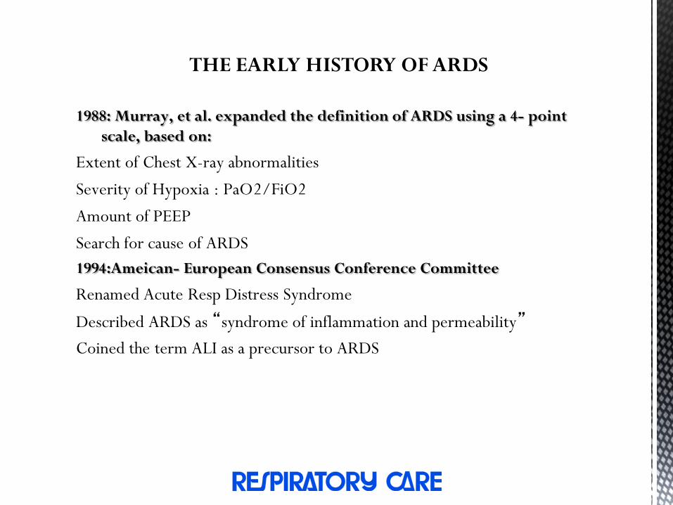
THE EARLY HISTORY OF ARDS
1988: Murray, et al. expanded the definition of ARDS using a 4- point
scale, based on:
Extent of Chest X-ray abnormalities
Severity of Hypoxia : PaO2/FiO2
Amount of PEEP
Search for cause of ARDS
1994:Ameican- European Consensus Conference Committee
Renamed Acute Resp Distress Syndrome
Described ARDS as “syndrome of inflammation and permeability”
Coined the term ALI as a precursor to ARDS

THE EARLY HISTORY OF ARDS
1994:Ameican- European Consensus Conference Committee
Criteria:
A-Acute onset of symptoms
B- Bilateral infiltrates on chest radiographs
C-PAWP≤ 18*
D-ALI: PaO2/FiO2 ≤ 300
E-ARDS: PaO2/FiO2 ≤ 200.**
* Pulmonary arterial wedge pressure of 18 mm Hg or less or no clinical signs of left
atrial hypertension
**The ratio of the alveolar partial pressure of oxygen (PaO2) to the fraction of
inspired oxygen (FIO2) of 200 mm Hg or less.

THE EARLY HISTORY OF ARDS
In 2012, the ARDS Definition Task Force met in Berlin and
decided on a new and improved definition of ARDS using 3
mutually exclusive categories of ARDS based on the degree
of hypoxemia:
Mild (200 mm Hg <pao2/FIO2 ≤300 mm Hg),
Moderate (100 mm Hg <pao2/FIO2 ≤200 mm Hg),
Severe (PaO2/FIO2 ≤100 mm Hg).
ARDS are clinical syndromes characterized by the acute
onset(<7 days)of hypoxaemia with bilateral pulmonary
infiltrates in the absence of clinical evidence of left atrial
hypertension
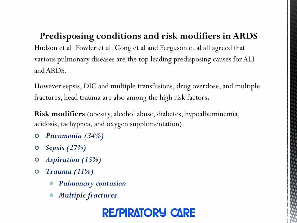
Predisposing conditions and risk modifiers in ARDS Hudson et al. Fowler et al. Gong et al and Ferguson et al all agreed that
various pulmonary diseases are the top leading predisposing causes for ALI
and ARDS.
However sepsis, DIC and multiple transfusions, drug overdose, and multiple
fractures, head trauma are also among the high risk factors.
Risk modifiers (obesity, alcohol abuse, diabetes, hypoalbuminemia,
acidosis, tachypnea, and oxygen supplementation).
Pneumonia (34%)
Sepsis (27%)
Aspiration (15%)
Trauma (11%)
Pulmonary contusion
Multiple fractures

NO Predisposition
Risks
EDLIPS
Points
NO
Predisposition
Risk Modifiers
EDLIPS
Points
1 Male gender 1 1 Diabetes Mellitus -0.5
2 Aspiration 2 2 Cirrhosis 1
3 Pneumonia 1 3 Chemotherapy 2
4 Sepsis 1 4 Obesity(BMI>30) 1.5
5 Shock 2 5 Acidosis(<7.35) 2
6 Lung contusion 1 6 FiO2>0.35(>4L/m) 2
7 Smoke inhalation 1.5 7 Albumin(<3.5) 1.5
8 Long bone fractures 2 8 SpO2 <95% 1.5
9 Brain injury 2
10 Cardiac Surgery 5
11 Aortic surgery 5
12 Spine surgery 5
13 Acute abdomen 2.5
Lung injury prediction score for the emergency department*
(EDLIPS)
*Trillo-Alvarez C, Cartin-Ceba R, Kor DJ, Kojicic M, Kashyap R, Thakur S, Thakur L, Herasevich V, Malinchoc M,
Gajic O: Acute lung injury prediction score: derivation and validation in a population-based sample. Eur Respir J 2011, 37:604-609
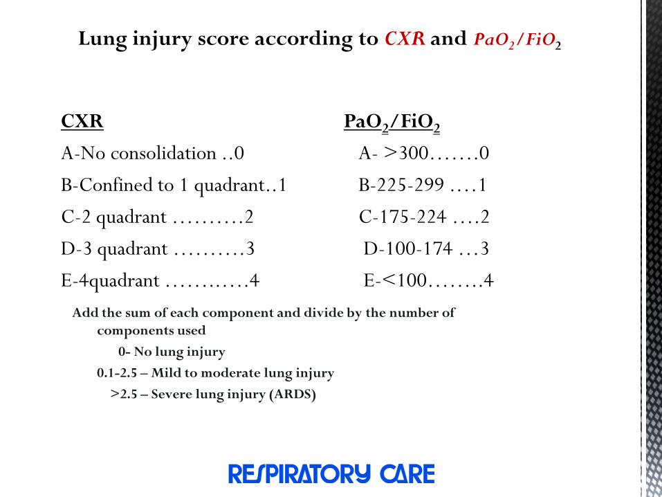
Lung injury score according to CXR and PaO2/FiO2
CXR
A-No consolidation ..0
B-Confined to 1 quadrant..1
C-2 quadrant ……….2
D-3 quadrant ……….3
E-4quadrant ……..….4
PaO2/FiO2
A- >300…….0
B-225-299 .…1
C-175-224 ….2
D-100-174 …3
E-<100……..4
Add the sum of each component and divide by the number of
components used
0- No lung injury
0.1-2.5 – Mild to moderate lung injury
>2.5 – Severe lung injury (ARDS)
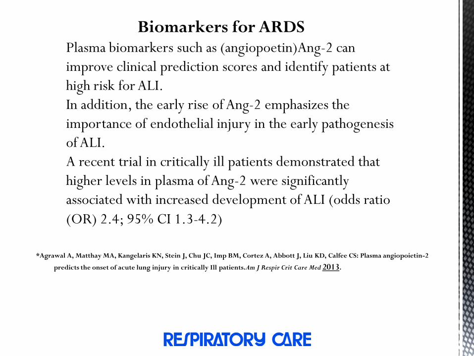
Biomarkers for ARDS Plasma biomarkers such as (angiopoetin)Ang-2 can
improve clinical prediction scores and identify patients at
high risk for ALI.
In addition, the early rise of Ang-2 emphasizes the
importance of endothelial injury in the early pathogenesis
of ALI.
A recent trial in critically ill patients demonstrated that
higher levels in plasma of Ang-2 were significantly
associated with increased development of ALI (odds ratio
(OR) 2.4; 95% CI 1.3-4.2)
*Agrawal A, Matthay MA, Kangelaris KN, Stein J, Chu JC, Imp BM, Cortez A, Abbott J, Liu KD, Calfee CS: Plasma angiopoietin-2
predicts the onset of acute lung injury in critically Ill patients.Am J Respir Crit Care Med 2013.
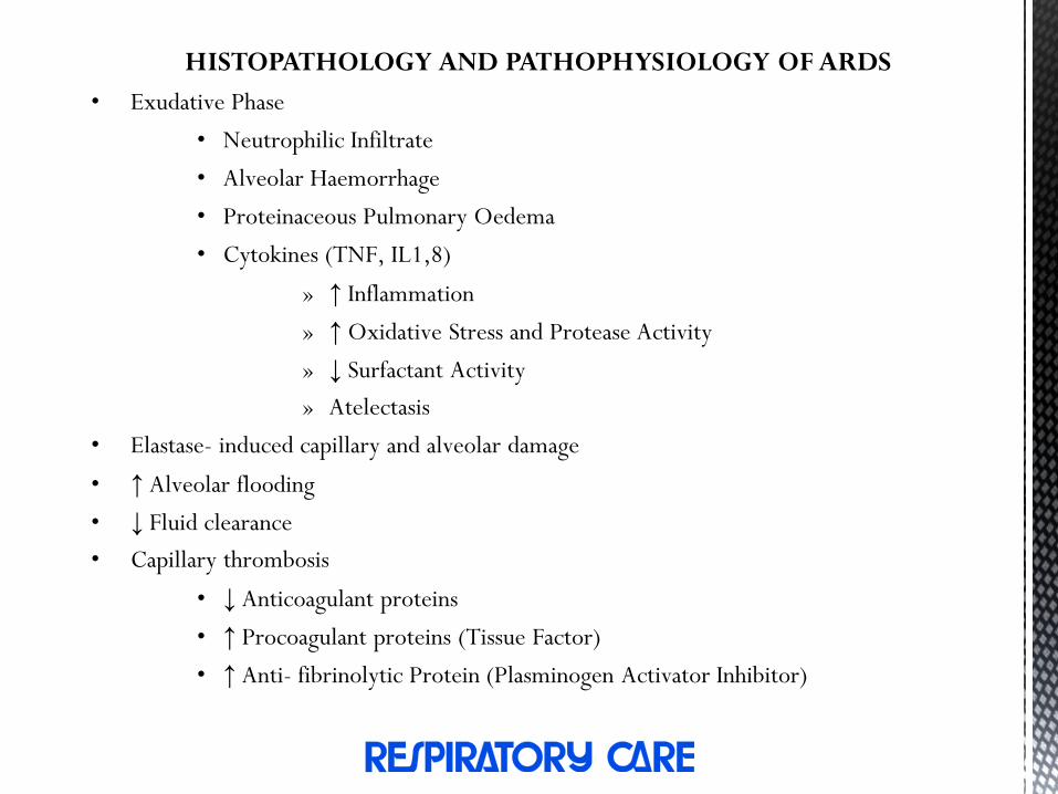
HISTOPATHOLOGY AND PATHOPHYSIOLOGY OF ARDS
• Exudative Phase
• Neutrophilic Infiltrate
• Alveolar Haemorrhage
• Proteinaceous Pulmonary Oedema
• Cytokines (TNF, IL1,8)
» ↑ Inflammation
» ↑ Oxidative Stress and Protease Activity
» ↓ Surfactant Activity
» Atelectasis
• Elastase- induced capillary and alveolar damage
• ↑ Alveolar flooding
• ↓ Fluid clearance
• Capillary thrombosis
• ↓ Anticoagulant proteins
• ↑ Procoagulant proteins (Tissue Factor)
• ↑ Anti- fibrinolytic Protein (Plasminogen Activator Inhibitor)
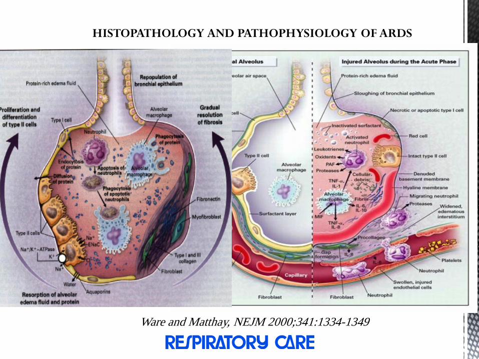
HISTOPATHOLOGY AND PATHOPHYSIOLOGY OF ARDS
Ware and Matthay, NEJM 2000;341:1334-1349
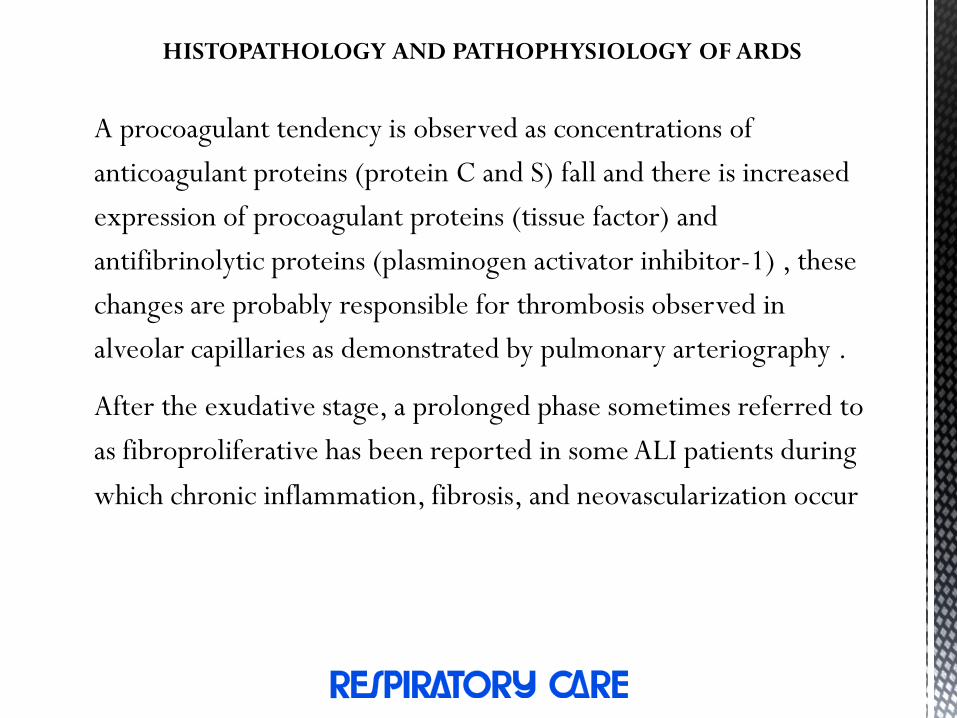
HISTOPATHOLOGY AND PATHOPHYSIOLOGY OF ARDS
A procoagulant tendency is observed as concentrations of
anticoagulant proteins (protein C and S) fall and there is increased
expression of procoagulant proteins (tissue factor) and
antifibrinolytic proteins (plasminogen activator inhibitor-1) , these
changes are probably responsible for thrombosis observed in
alveolar capillaries as demonstrated by pulmonary arteriography .
After the exudative stage, a prolonged phase sometimes referred to
as fibroproliferative has been reported in some ALI patients during
which chronic inflammation, fibrosis, and neovascularization occur
HISTOPATHOLOGY AND PATHOPHYSIOLOGY OF
ARDS (The recovery Phase)
Anti-inflammatory cytokines deactivate inciting neutrophils,
which then undergo apoptosis and phagocytosis.
Type II alveolar cells proliferate and differentiate into type I
cells, reestablishing the integrity of the epithelial lining and
creating an osmotic gradient that draws fluid out of the alveoli
and into the pulmonary microcirculation and lung lymphatics.
Simultaneously, alveolar cells and macrophages remove
protein compounds from the alveoli, allowing the lungs to
recover.
Clinical Course
Early findings on the chest radiograph include normal or diffuse alveolar
opacities (consolidation), which are often bilateral and which obscure the
pulmonary vascular markings.
Later, these opacities progress to more extensive consolidation that is diffuse,
and they are often asymmetrical.
Effusions and septal lines are not usually seen on chest radiographs
of patients affected by ARDS, although these findings are commonly seen
in patients with congestive heart failure (CHF).
Radiographic findings tend to stabilize (part of the clinical definition of
ARDS);
If further radiographic worsening occurs after 5-7 days, another disease
process should be considered.

Clinical Course
Rapid Onset
Exudates
Consolidations
Respiratory failure
Hypoxemia refractory to O2
Inflammation (even in non-edematous
lung)IL-1,6,8,10, Cytokines
Diminished Lung compliance
Patchy infiltrates Coalesce
Air Bronchograms
Pulmonary Hypertension
Intrapulmonary Shunting
Endogenous Vasoconstrictors
Hyperadrenergic State
Acute Phase
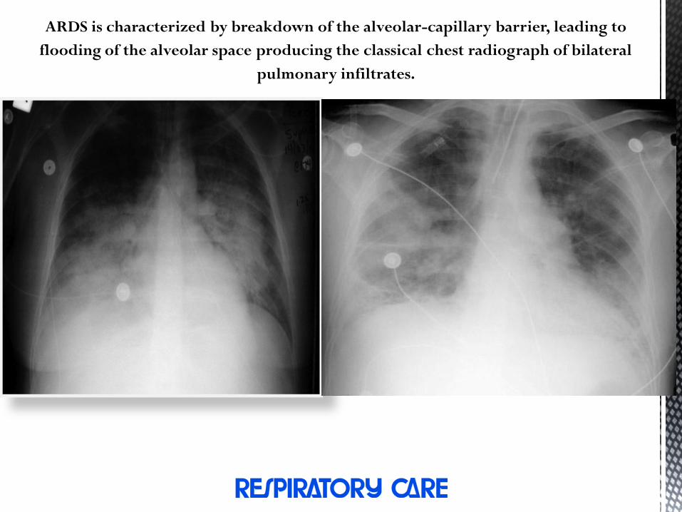
ARDS is characterized by breakdown of the alveolar-capillary barrier, leading to
flooding of the alveolar space producing the classical chest radiograph of bilateral
pulmonary infiltrates.
Clinical Course
Chest xray shows linear opacities consistent with evolving fibrosis.
Pneumothorax in 10-13% of patients.
CT: diffuse interstitial opacities and bullae.
Histologically, fibrosis, mesenchymal cells, vascular proliferation, collagen and fibronectin accumulation.
Can start 5-7 days after symptom onset.
Not present in every patient with ARDS, but does portend poorer prognosis
Persistent Hypoxia
Pulmonary Fibrosis
Worsening Compliance
Neovascularization
Pulmonary Hypertension
Macrophages clear neutrophils
Chronic Inflammation
Fibroproliferative phase
Management of ARDS
Ventilator Strategies
A-Lung recruitment maneuvers
B-Prone positioning
C-High-frequency oscillatory ventilation
(HFOV)
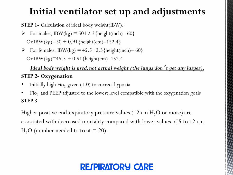
Initial ventilator set up and adjustments
STEP 1- Calculation of ideal body weight(IBW):
For males, IBW(kg) = 50+2.3{height(inch)– 60}
Or IBW(kg)=50 + 0.91{height(cm)–152.4}
For females, IBW(kg) = 45.5+2.3{height(inch)– 60}
Or IBW(kg)=45.5 + 0.91{height(cm)–152.4
Ideal body weight is used, not actual weight (the lungs don’t get any larger).
STEP 2- Oxygenation
• Initially high Fio2 given (1.0) to correct hypoxia
• Fio2 and PEEP adjusted to the lowest level compatible with the oxygenation goals
STEP 3
Higher positive end-expiratory pressure values (12 cm H2O or more) are
associated with decreased mortality compared with lower values of 5 to 12 cm
H2O (number needed to treat = 20).

Positive End-Expiratory Pressure (PEEP) Clinical practice guidelines recommend maintaining Oxygen saturation of 88 to 95
percent and a plateau pressure of 30 cm H2O or less to avoid barotrauma. Also to
maintain arterial pH of 7.30 to 7.45, although patients in some research trials have
tolerated permissive hypercapnia and a pH as low as 7.15.
PEEP is to decrease FiO2
Goal sat 88% with FiO2 <60%
• Minimize oxygen toxicity
PEEP can improve lung recruitment and decrease end-expiratory alveolar collapse (and therefore right-to-left shunt)
Can also decrease venous return, cause hemodynamic compromise, worsen pulmonary edema
ARDSnet PEEP trial of 549 patients show no difference in mortality or days on ventilator with high vs low PEEP

Positive End-Expiratory Pressure (PEEP) PEEP level separation at various FiO2 levels was in the range of 6
cm H2O (mean of 14 versus 8 cm H2O) .
The use of 6 ml/kg PBW tidal volume strategy and PEEP–
FiO2 scale as a starting point for ventilation is recommended but
routine use of recruitment maneuvers is not.
However, it would be reasonable to reserve higher levels of
PEEP and/or recruitment maneuvers for patients with
refractory hypoxemia in an attempt to improve oxygenation
when severity of the oxygenation defect is the most immediate
threat to survival.
FIO2 0.3 0.4 0.4 0.5 0.5 0.6 0.7 0.7 0.7 0.8 0.9 0.9 0.9 1.0
PEEP 5 5 8 8 10 10 10 12 14 14 14 16 18 20-24
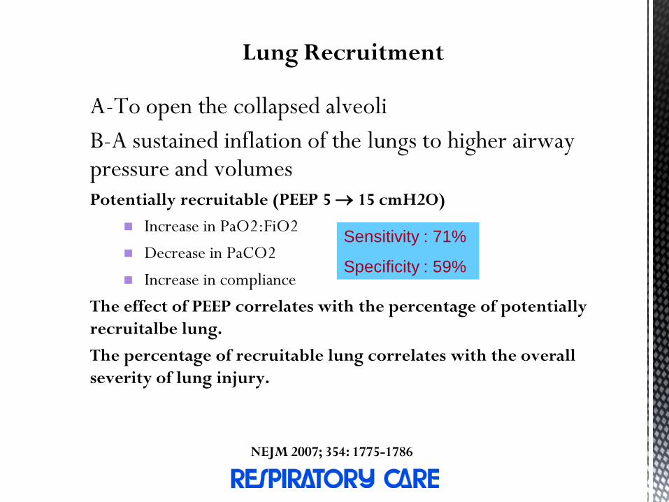
Lung Recruitment
A-To open the collapsed alveoli
B-A sustained inflation of the lungs to higher airway pressure and volumes Potentially recruitable (PEEP 5 15 cmH2O)
Increase in PaO2:FiO2
Decrease in PaCO2
Increase in compliance
The effect of PEEP correlates with the percentage of potentially
recruitalbe lung.
The percentage of recruitable lung correlates with the overall
severity of lung injury.
NEJM 2007; 354: 1775-1786
Sensitivity : 71%
Specificity : 59%
NEJM 2007; 354: 1775-1786
Strategy for Recruitment
Administer 40cm H2O of PEEP for 90s
Set the ventilator to an effective rate of zero (with no machine breaths)
and then immediately raise the PEEP to 40cm H2O for a carefully
timed period of one and a half minutes. Then re-institute ventilation as
before.
Wait and recheck the ABG
Wait for a period of five minutes, leaving the patient in the prone
position, and obtain a blood gas analysis. If the PaO2 is under
300mmHg, then consider repeating the maneuver at PEEPs of
45mmHg and (if this fails) 50mmHg, also for ninety seconds.
Prone Position advantages and disadvantages
Mechanisms to improve oxygenation:
a-Increase in end-expiratory lung volume
b-Better ventilation-perfusion matching
c-More efficient drainage of secretions
d-Improve oxygenation in about 2/3 of all treated patients
c-No improvement on survival, time on ventilation, or time in
ICU
f-Might be useful to treat refractory hypoxemia
g-Optimum timing or duration ?
h-Routine use is not recommended
Prone Position advantages and disadvantages
Investigators randomized 474 patients at 26 French ICUs (and one
in Spain) with severe ARDS (PaO2:FiO2 ratio of <150 mm Hg, PEEP
>5, FiO2 > 0.60) to receive standard care, or to also be “proned”
(turned face-down) for 16 hours a day, every day for up to 28 days
(or longer if their doctors so chose).
Far more prone-positioned patients survived their ARDS: 16%
mortality at 28 days (38 deaths in 237 patients) vs. 33% in the
supine group (75 of 229 patients), with a very low p-value, < 0.001.
That’s a staggering 17% absolute risk reduction. The benefit was
only slightly smaller at 90 days.
Claude Guérin et al’s PROVESA study (May 20,2013 New England Journal of
Medicine)
Prone Position advantages and disadvantages
Side effects of Prone Position
A-Facial edema, Airway obstruction Skin lesions
B-Difficulties with enteral feeding, Hypotension
C-Transitory decrease in oxygen saturation. Arrhythmias
D-Loss of venous accesses and probes,
E-Increase need for sedation.
F-Loss of dialysis drains and catheters, Accidental extubation.
G-Apical atelectasis due to incorrect positioning of the tracheal
tube
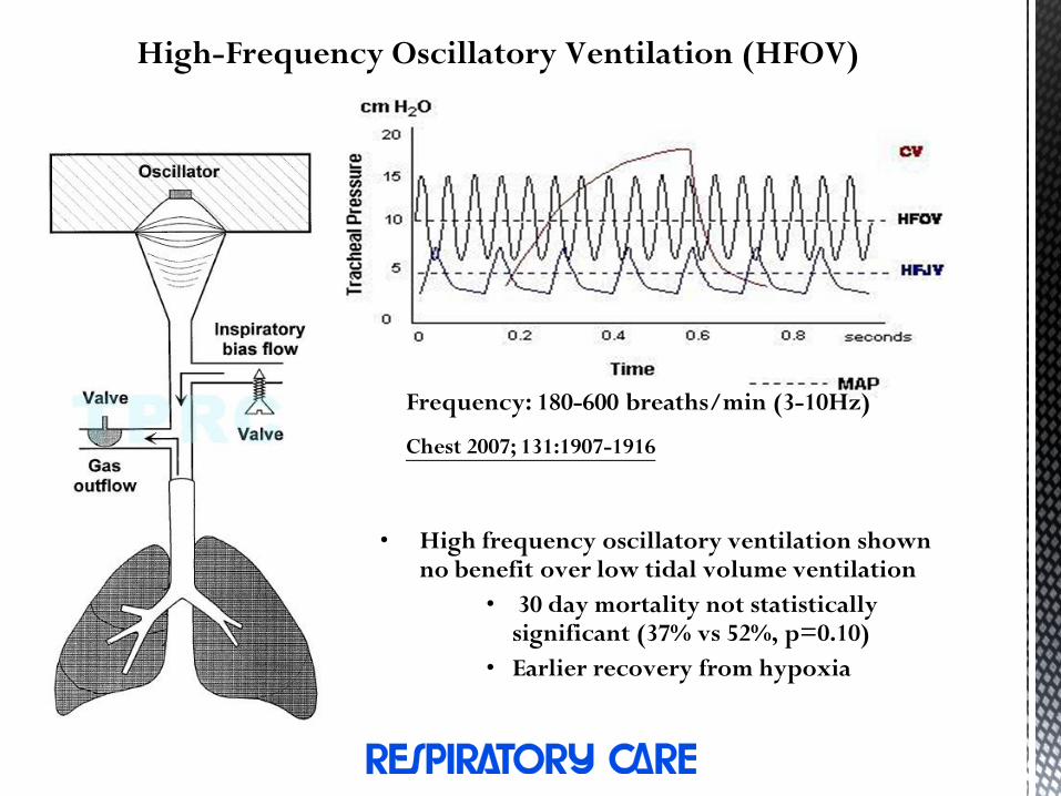
High-Frequency Oscillatory Ventilation (HFOV)
• High frequency oscillatory ventilation shown no benefit over low tidal volume ventilation
• 30 day mortality not statistically significant (37% vs 52%, p=0.10)
• Earlier recovery from hypoxia
Frequency: 180-600 breaths/min (3-10Hz)
Chest 2007; 131:1907-1916
Adjunctive Therapy
Steroid therapy The use of corticosteroids is controversial.
Randomized controlled trials and cohort studies tend
to support early use of corticosteroids (with dosages
of methylprednisolone [Solu-Medrol] ranging from 1
to 120 mg per kg per day) for decreasing the number
of days on a ventilator
MATTHEW FARGO, MD, MPH, Dwight D. Acute Respiratory Distress Syndrome:
Diagnosis and Management.Am Fam. Physician. 2012 Feb 15;85(4):352-358.

Adjunctive Therapy
Steroid therapy
A-Increase the number of ventilator-free and shock-free days during the first 28 day
B-Improve oxygenation, compliance and blood pressure
C-No increase in the rate of infectious complications
D-Higher rate of neuromuscular weakness
E-Routine use of steroid is not supported
F-Starting steroid more than 14 days after the onset of ARDS may increase mortality
Supportive treatments
Fluid Management
• Central venous pressure guided therapy – 10-14 mmHg
( ARDS Network Trial 2003)
• Study of conservative vs liberal fluid management5
• 60 day mortality: 25.5 vs 28.4% p=0.30
• 1st 28 days ventilator free: 14.6 vs 12.1 p<0.001
• 1st 28 days ICU free: 13.4 vs 11.2 p<0.001
• Difference in organ failure and need for dialysis not
statistically significant
• No specific mention of CVP/ PAOP levels which to
aim for
• Conservative = 4mmHg Liberal = 10-14mmHg CVP
Supportive treatments
A variety of coagulation inhibitors have been tested
including heparin, antithrombin, tissue factor pathway
inhibitor, factor VIIa, activated protein C, and
thrombomodulin in animal models and/or humans with
either sepsis or ALI .
To date only activated protein C has been proven
useful in severe sepsis, though it is not clear that it
directly improves lung function

Pharmacological treatments in ARDS
β-adrenoceptor agonists (β-agonists) are well established in the treatment of
airflow obstruction. In addition to actions as bronchodilators, they have anti-
inflammatory properties, promote the clearance of alveolar fluid, and
promote epithelial and endothelial repair((15 μg/kg/h) )
Pharmacological treatments in ARDS
Neuromuscular blockade(NMB )may permit lower-pressure, lower-tidal
volume ventilation with a consequent reduction in ventilator-induced lung
injury.
Neutrophil elastase inhibitors
Neutrophil elastase (NE) Excessive NE is capable of degrading endothelial
basement membrane, and has been implicated in the pathogenesis of ARDS.
Neutrophil elastase inhibitor, was investigated in an international randomized,
double-blind, placebo-controlled, multi-center phase III trial (STRIVE) .
The study was stopped prematurely due to an increase in 180-day all-cause
mortality.A more recent meta-analysis of eight clinical trials (including
STRIVE) investigating silvelestat has shown it to have no effect on short-term
mortality, and a worse outcome for 180-day mortality .
Andrew James Boyle, Rob Mac Sweeney and Daniel Francis McAuley. Pharmacological treatments in ARDS; a state of the art update.BMC
Medicine 2013, 11:166 doi:10.1186/1741-7015-11-166
Pharmacological treatments in ARDS
Statins, Heparin, Aspirin, Angiotensin converting enzyme
inhibitors/angiotensin receptor blockers. Stem cell therapy, Growth factors,
vitamin D and interferon-β (IFN-β).
Prone Position
A new strategy for Recruitment
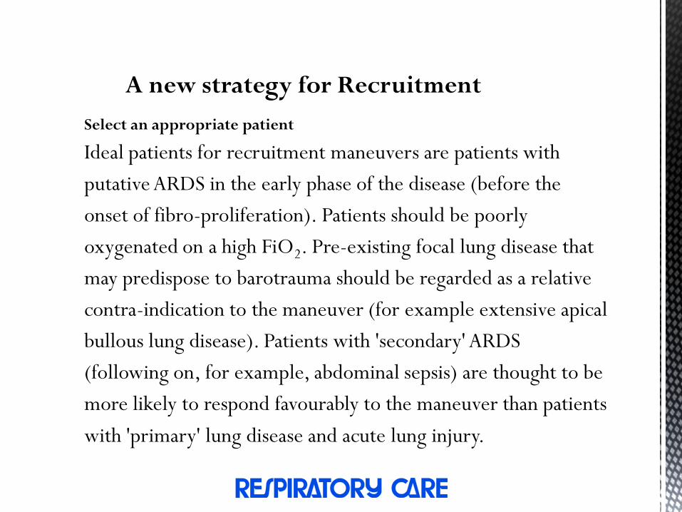
Select an appropriate patient
Ideal patients for recruitment maneuvers are patients with
putative ARDS in the early phase of the disease (before the
onset of fibro-proliferation). Patients should be poorly
oxygenated on a high FiO2. Pre-existing focal lung disease that
may predispose to barotrauma should be regarded as a relative
contra-indication to the maneuver (for example extensive apical
bullous lung disease). Patients with 'secondary' ARDS
(following on, for example, abdominal sepsis) are thought to be
more likely to respond favourably to the maneuver than patients
with 'primary' lung disease and acute lung injury.
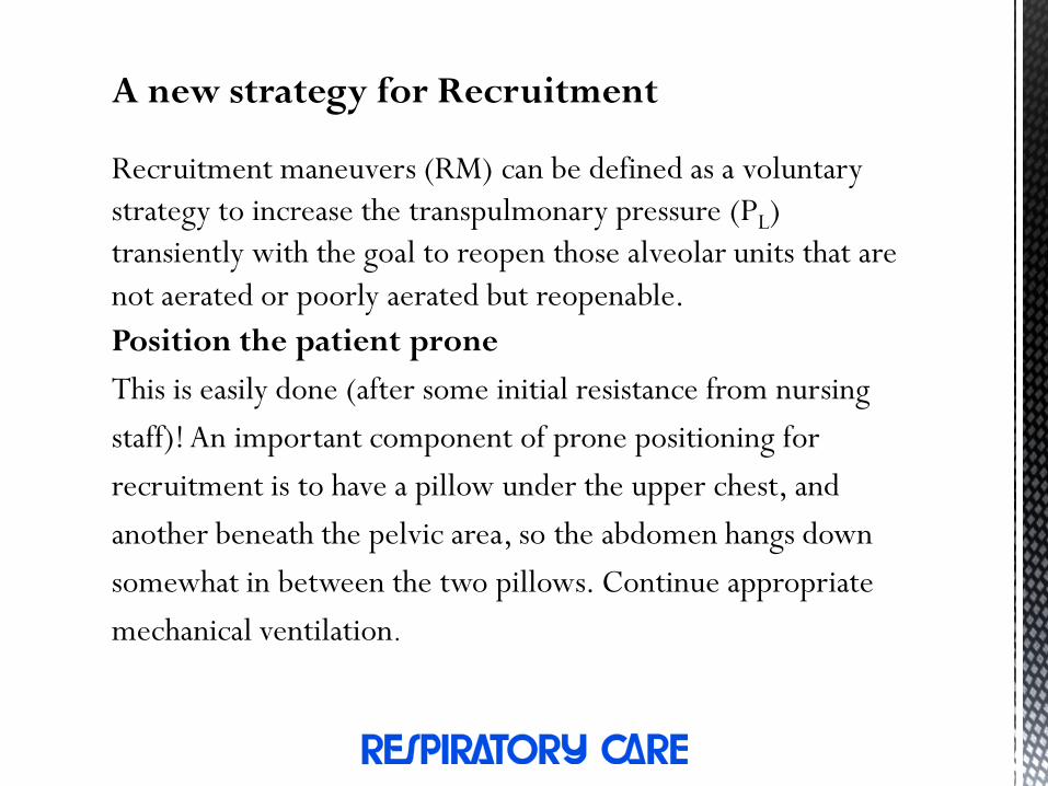
A new strategy for Recruitment
Recruitment maneuvers (RM) can be defined as a voluntary
strategy to increase the transpulmonary pressure (PL)
transiently with the goal to reopen those alveolar units that are
not aerated or poorly aerated but reopenable.
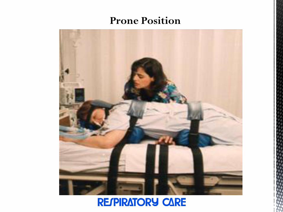
Position the patient prone
This is easily done (after some initial resistance from nursing
staff)! An important component of prone positioning for
recruitment is to have a pillow under the upper chest, and
another beneath the pelvic area, so the abdomen hangs down
somewhat in between the two pillows. Continue appropriate
mechanical ventilation.
A new strategy for Recruitment
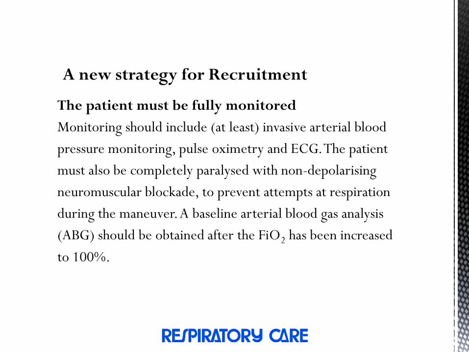
The patient must be fully monitored
Monitoring should include (at least) invasive arterial blood
pressure monitoring, pulse oximetry and ECG. The patient
must also be completely paralysed with non-depolarising
neuromuscular blockade, to prevent attempts at respiration
during the maneuver. A baseline arterial blood gas analysis
(ABG) should be obtained after the FiO2 has been increased
to 100%.
A new strategy for Recruitment
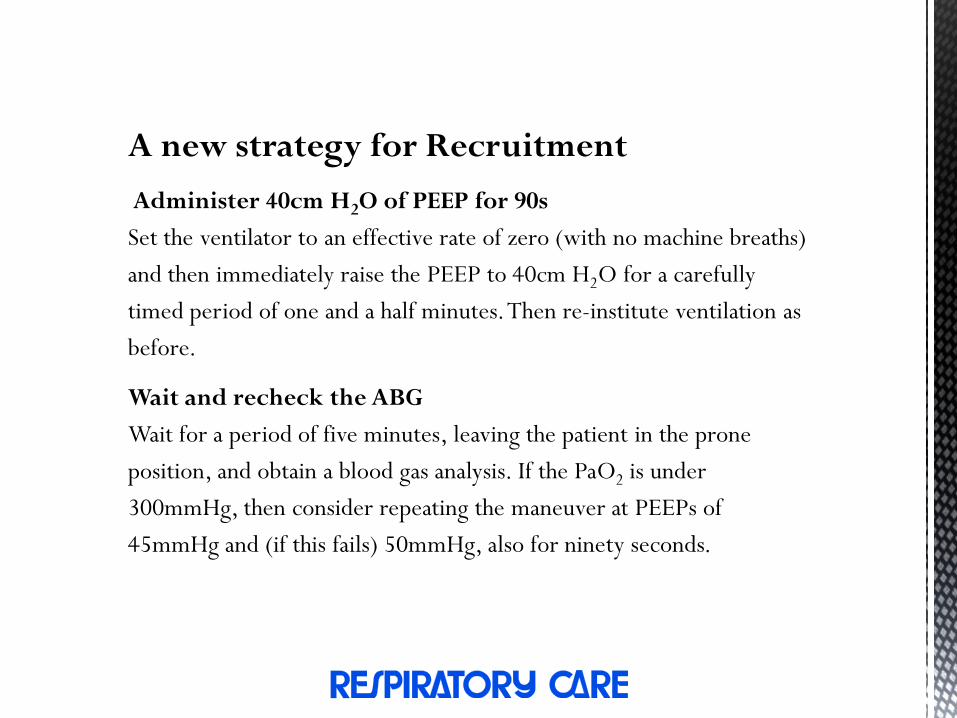
Administer 40cm H2O of PEEP for 90s
Set the ventilator to an effective rate of zero (with no machine breaths)
and then immediately raise the PEEP to 40cm H2O for a carefully
timed period of one and a half minutes. Then re-institute ventilation as
before.
Wait and recheck the ABG
Wait for a period of five minutes, leaving the patient in the prone
position, and obtain a blood gas analysis. If the PaO2 is under
300mmHg, then consider repeating the maneuver at PEEPs of
45mmHg and (if this fails) 50mmHg, also for ninety seconds.
A new strategy for Recruitment
Prevent 'de-recruitment'
The patient should now be maintained on a PEEP of 15
cmH2O. Often, the patient can be turned back to a supine
position without substantial worsening of oxygenation.
Ventilation should continue with a strategy that minimises
additional alveolar trauma (for example, inverse ratio
pressure-control ventilation, with every attempt to keep
trans-alveolar pressure to under 35cm H2O). Ventilator
tidal volumes should perhaps be limited to approximately 6
ml/kg.
A new strategy for Recruitment
Rationale
The rationale behind the above maneuver is that prone ventilation splints
the thoracic cage, especially the anterior portion and the area around the
upper lobes. If diaphragmatic excursion is promoted (by freeing up the
abdomen) then preferential ventilation of the lower lobes is encouraged,
and overdistension of the upper lobes is prevented.
Sustained pressures of 40 to 50 cm H2O are applied to the airway for a
sufficient time to distribute pressure to collapsed lung areas, and promote
recruitment.Once adequate recruitment has been achieved, high PEEP is
used to prevent recurrent airway collapse.
POINTES TO REMEMBER
{Ibsen in (1954)first used cuffed entdotracheal
tubes and positive pressure ventilation, which
clearly moved ARDS from a nearly universally
fatal form of “double pneumonia” to treatable
disease. Roentgen describes X-rays in 1896. Early
chest X-rays required exposure time of 20 min and
were not considered useful until the 1920s}