Acute Psychiatric Management - Ministry of Health · PDF filePsychosis in the Medically Ill 21...
92
Acute Psychiatric Management Edited by Dr Michael Robertson IMET | RESOURCE EXCELLENT MEDICAL TRAINING, EXCELLENT PATIENT CARE
Transcript of Acute Psychiatric Management - Ministry of Health · PDF filePsychosis in the Medically Ill 21...

Acute Psychiatric Management
Edited by Dr Michael Robertson
IMET | RESOURCE
ExcEllEnt mEdical training, ExcEllEnt patiEnt carE
Contributors
Dr Michael Robertson (Editor-in-Chief)
Dr Matthew Holton
Dr Kristin Barrett
Dr Amanda Bray
Dr Anne Wand
Dr Hung Tran

01
Contents
Foreword 02
Introduction 02
SECTION 1 – ASSESSMENT
Comprehensive approach to risk assessment 04
Psychiatric assessment after self-harm 12
The psychiatric assessment of Aboriginal and Torres Strait Islander peoples 16
Psychopathology in the general medical setting 18
Psychosis in the Medically Ill 21
SECTION 2 – TREATMENTS
Long acting injectable antipsychotics 24
Lithium therapy 26
Clozapine 28
Anticonvulsant medications in psychiatry 31
Psychotropic drugs in pregnancy and breastfeeding 34
Electroconvulsive Therapy (ECT) 38
Pro Re Nata (PRN) medication 41
SECTION 3 – MANAGEMENT CHALLENGES
Acute management of physical aggression 46
Acute management of alcohol and benzodiazepine withdrawal syndromes 50
Recognition and management of delirium 54
Neuroleptic Malignant Syndrome 60
The Serotonin Syndrome 62
SECTION 4 – COMPLICATIONS
Metabolic complications of antipsychotic treatment 66
QTc abnormalities and psychotropic treatment 69
Akathisia and tardive dyskinesia 72
Hyperprolactinaemia 75
SECTION 5 – IMPORTANT LEGISLATION
The Mental Health Act 2007 (NSW) 80
The Privacy Act 1998 (Cth) 83
The Guardianship Act 1987 (NSW) 84
IMET ACUTE PSYCHIATRIC MANAGEMENT

02
ForewordAll junior doctors encounter patients with acute psychiatric episodes. For many this is their first experience in the emergency department or in out-of-hours practice. This text is a straightforward guide to up-to-date management options for acute psychiatric conditions. It will certainly help junior doctors prepare for managing what is often a very stressful situation. It is not often that we have a text written specifically with the NSW public health system in mind. The Editorial Group who prepared this text are all prominent psychiatrists working within NSW mental health services, and they have created an excellent resource for all clinicians involved in psychiatry training. I am pleased that the NSW Institute of Medical Education and Training (IMET) and IMET’s Network Oversight Committee have been able to support the Editorial Group in this work. IMET is making this text available to all junior doctors and their supervisors via the IMET website, and I hope that it will help junior doctors manage the care of patients with acute psychiatric conditions confidently.
Dr Murray WrightPsychiatry Clinical ChairNSW Institute of Medical Education and TrainingDirector Clinical Operations, Mental Health, Drug & AlcoholGreater Southern Area Health Service
IntroductionThe commencement of psychiatric training is a daunting task for any medical officer. Whilst exposure to mental illness and the institutional systems which operate around it may occur during graduate medical training programs and some junior resident medical officer rotations, nothing prepares the new trainee in psychiatry for their many responsibilities in this early phase of their careers.
Didactic content is provided for psychiatric trainees by the NSW Institute of Psychiatry and local training networks, however information on how to provide safe and effective care to people with mental illnesses is invariably acquired in the course of working in acute mental health settings. With this in mind, the contributors to this resource have attempted to provide accessible overviews of the kind of information which might be needed in the course of working in acute adult mental health settings.
This resource is set out in a series of themes. It does not seek to provide a comprehensive reference, nor does it attempt to summarize text-books or the current literature in psychiatry. Each contributor has written a brief account of different topics of relevance to practice in acute adult psychiatry. The style of writing aims to provide the reader with a grasp of the necessary information, which can be absorbed rapidly by the inexperienced psychiatric trainee. Whilst not a manual of ‘how to be a registrar’, it aims to provide a ready reference to both common and classic challenges in the setting of acute adult mental health.
Michael Robertson MB BS (Hons), FRANZCP
Centre for Ethics, Values and the Law in MedicineUniversity of Sydney

Assessment
031

04
Comprehensive approach to risk assessmentIntroduction
The notion of “risk assessment” is usually considered the process of estimating the likelihood of dangerousness, such as completed suicide or harm to others. In the insurance industry, actuarial assessment is a mathematical discipline aimed at computing a probability of adversity, based upon a broad consideration of variables. Such an approach has been applied in criminology in the prediction of recidivism in sexual offences.1 Actuarial approaches to risk assessment in psychiatry attempt to integrate different situational and clinical factors in different populations at different times.2 Actuarial approaches have been challenged by their aims to predict, rather than anticipate and prevent dangerousness in psychiatry.3 The actuarial approach to risk assessment provides little more than passive prediction4 and is inferior to a standardised clinical assessment.5 The apparent superiority of clinical judgement appears to relate to its emphasis upon prevention, rather than prediction. The distinction between prevention and prediction is important, in that a recent UK review indicated that whilst around 28% of dangerousness was predictable, 65% was preventable.6 The clinical approach to risk assessment is also more appropriate in psychiatry, as it links the clinical tasks of gathering data, synthesising data and formulating a plan of action to alter the factors likely leading to a dangerous act on the part of a person suffering mental illness.
1Assessment
LEARNING OBJECTIVES
• Describe the components of a comprehensive risk assessment• Identify the variables associated with increase risk of adversity • Formulate a comprehensive management plan based on
assessment of risk
1. Silver E, Chow-Martin L. A multiple models approach to assessing recidivism: Implications for judicial decision making. Criminal Justice and Behavior 2002 29: 538–568
2. Monhan J. The prediction of violent behavior: Toward a second generation of theory and policy. American Journal of Psychiatry 1984 141, 10-15
3. Bauer A, Rosca P, Khawalled R et al. Dangerousness and risk assessment: the state of the art. Israel Journal of Psychiatry and Related Sciences 2003 40: 182-190
4. Hart S. The role of psychopathy in assessing risk for violence: conceptual and methodological issues. Legal and Criminological Psychology 1998 3: 121-137
5. Douglas K, Kropp P. A prevention-based paradigm for violence risk assessment: clinical and research applications. Criminal Justice and Behaviour 2002 29: 617-658

05
In this chapter, the approach to the assessment of risk moves this process beyond the short-term estimation of harm to a longer term and broader account of adversity facing the patient, considering many different individual, demographic and situational factors. In this approach, the term “risk assessment” refers to the propensity of an episode of mental illness to create adversity in the life of a patient in a broad array of domains.
The components of the comprehensive approach to risk assessment
The main domains of an actuarial approach to risk are outlined in Table 1. The short-term risk of physical or emotional harm is usually the main focus in the acute phase of care. Incomplete recovery, via the persistence of psychiatric disturbance or the development of co-morbid psychiatric or physical disorder, is the usual focus of the ‘post-acute’ phase of care. Chronic disability, the effect of stigma and social disadvantage and the impact of illness on long term social, interpersonal and vocational function, as well as the person’s experience of selfhood. Many components of the risk
Incomplete recovery
• Symptom persistence
• Treatment non-adherence
• Family or cultural resistance
• Co-morbid psychiatric disorder
• Post-illness impairment of personality
• Brain injury
Physical or emotional harm
• Harm to others
• Suicide or deliberate self harm
• Sexual assault or exploitation
• Iatrogenic insult
• Traumatic stress before and during episode of care
1
Table 1 – The main components to the actuarial approach to risk
Deterioration physical health
• Iatrogenic complications
• Neglect of self care
• Problems of access
Long-term impairment of psychosocial and interpersonal functioning
• Vocational impairment or job loss
• Relationship disruption
• Developmental disruption i.e. of Eriksonian tasks
• Existential aspects of illness experience
Chronicity of impairment
• Effect of illness process
• Stigma
• Incapacity to participate in comprehensive treatment
• Problems of access
• Effect of lifestyle
• Family and social determinants
IMET ACUTE PSYCHIATRIC MANAGEMENT
Assessment

06
assessment are addressed in more detail in other parts of this monograph. This chapter will focus upon assessment of risk of harm to others, risk of incomplete recovery and chronicity of impairment and longer term functioning.
Assessment of risk to others
Traditionally, there is an expectation of psychiatrists to accurately predict risk. Such an expectation is unrealistic in that the predictive capacity of psychiatrists in regards to future harm perpetrated by their patients has been shown to be low,6,7 with estimates of accuracy varying from 30-60%.8 The principle failing of psychiatric risk assessment is a tendency to overstate risk.9
Any assessment of the capacity for dangerousness to self or other integrates multiple dimensions of the patient’s situation including situational factors in the patient’s illness or immediate ecological setting, the pattern of previous dangerousness and the effectiveness of intervention. The strongest predictor of future ‘dangerousness’ is past dangerousness, but this in itself is a vacuous statement in the absence of consideration of the situational factors involved,10 e.g. a patient who becomes aggressive when the intensity of auditory hallucinations increases. Thus, statements of potential for future dangerousness, rather than a “crystal ball” prediction are more methodologically sound.
The MacArthur risk assessment study,11 a large scale study of the factors associated with violence (Table 2) identified a number of variables associated with heightened risk of dangerousness –
Variable Comment
Gender Men more likely than women to be violent, but the difference was not large. Violence by women more likely to be directed against family members and to occur at home.
Prior violence All measures of prior violence strongly related to future violence.
Childhood experiences
A history of child abuse or neglect and parental criminality was strongly associated with violent offending.
Neighborhood and race
Some trend of increased risk of aggression towards non-white members of the community, but this diminished when the neighborhood was considered ‘disadvantaged’.
Diagnosis A diagnosis of a major mental disorder – especially a diagnosis of schizophrenia – was associated with a lower rate of violence than a diagnosis of a personality or adjustment disorder. A co-morbid diagnosis of substance abuse was strongly predictive of violence.
Psychopathy Psychopathy was the strongest risk factor identified.
Delusions12 The presence of delusions – or the type of delusions or the content of delusions – was not associated with violence. A generally “suspicious” attitude toward others was related to later violence.
Hallucinations Hallucinations did not elevate the risk of violence. “Command” hallucinations specifically commanding a violent act increased risk, particularly when the voice is recognisable.13
Violent thoughts and Anger
Thinking or daydreaming about harming others was associated with violence, particularly if the thoughts or daydreams were persistent. High levels of anger correlated with violence.
Table 2 – Summary of the MacArthur study
6. Munro E, Rumgay J. Role of risk assessment in reducing homicides by people with mental illness. British Journal of Psychiatry 2000 176: 116-120
7. Steadman HJ, Cocozza JJ 1980. The prediction of dangerousness – Baxtrom: A case study. In: G. Cooke, ed. The Role of the Forensic Psychologist. Springfield, Il.: Thomas. Pp. 204-215
8. Appelbaum P, Guthiel T Clinical Handbook of Psychiatry & the Law. Philadelphia: Lippincott, Williams & Wilkins. 20079. Levinson R, Ramsay G. Dangerousness, Stress, and Mental Health Evaluations. Journal of Health and Social Behavior,
1979 20: 178-18710. Litwack TR. Assessments of dangerousness: Legal, research, and clinical developments. Administration and-Policy-
in-Mental-Health 1994 21, 361-37711. Monahan J, Steadman H, Silver E et al. Rethinking Risk Assessment: The MacArthur Study of Mental Disorder
and Violence. New York: Oxford University Press. 200112. Mullen P. Forensic Mental Health British Journal of Psychiatry 200013. Junginger J. Command hallucinations and the prediction of dangerousness. Psychiatric Services 1995 46: 911-914

07
Based upon the literature and clinical experience it is possible to accumulate a series of variables for risk of harm to others, with the expectation that the more modifiable variables will serve as the basis of a management plan.
The approach to the acute assessment of dangerousness requires consideration of both “static” and “dynamic” risk factors. Static risk factors are the components of a particular patient’s presentation, which are not amenable to intervention, such as age, gender or aspects of a patient’s previous history, such as a past history of violent offending. By contrast, dynamic risk factors are those which are potentially amenable to clinical intervention, such as active psychotic symptoms, problematic living circumstances or substance abuse. In formulating an assessment of the risk of particular patient poses to self or other, consideration of the dynamic factors of risk.
Dynamic risk factors may be quite changeable, such as the level of psychotic disturbance or acuity of a crisis in a person’s life, or they may be stable such as personality traits or problematic interpersonal relationships (Table 3).
Table 3 – Static and Dynamic Risk Factors
The instrumental value of such an approach is that certain factors amenable to clinical intervention can be identified and implemented, thus potentially reducing risk. Identifying factors historically associated with increased risk of dangerousness in a particular patient serves the basis of a credible risk management plan. For example, a male patient has been previously aggressive in response to command hallucinations, which tend to worsen when there are changes to his living situation and concomitant increase in alcohol use. In this circumstance, the assessing clinician applies the above algorithm and identifies the risk variables of previous aggression, and the relationship of this to increased intensity of psychotic symptoms, alcohol use and disturbed living situation. The risk management plan thus follows along the lines of closer monitoring of psychotic symptoms and further treatment, strategies to avoid or reduce alcohol use and strategies to stabilize the patient’s immediate living situation.
Psychometric Measurement of riskThe HCR 2014 is one of a number of psychometric measures, which assess for the severity of risk, which integrates historical, current clinical and management factors (Figure 2). The HCR-20 has the benefit of being relatively sensitive to change on the clinical and risk-management scales. Other psychometric scales such as the Hare Psychopathy Checklist measure components of potential dangerousness.
14. Webster C, Douglas K, Eaves D, Hart S. 1997. HCR-20: Assessing risk for violence (version 2). Burnaby, BC: Mental Health, Law, and Policy Institute, Simon Fraser University
• Age
• Gender
• Ethnicity
• Intellectual disability
• Social class
• Educational level
• Developmental trauma
• Criminal history
• History of previous dangerousness
• Treatment non-responsiveness
• Personality
• I nterpersonal support network
• Impulsivity
• Problem solving capacity
• Cognitive schema
• Attribution style
• Attachment style
• Immature or primitive psychological defences
• Psychotic symptoms
• Mood or anxiety symptoms
• Substance use
• Acute crisis
• Social isolation
• Unsatisfactory living situation
• Treatment non-adherence
IMET ACUTE PSYCHIATRIC MANAGEMENT
1
Static Dynamic Stable Dynamic Changable
Assessment

08
Figure 2 – The HCR-20
Risk of Incomplete Recovery and Chronic Disability
The strongest predictor of prognosis in mental illness is the adequacy of recovery from the index episode. While up to 70% of patients suffering a schizophreniform illness enjoy reduction to mild symptom levels to be classified as having their illness episode remitted, only 20-30% maintain this improvement for 6 months.15 Incomplete recovery can manifest as symptom persistence, cognitive impairment, the emergence of a comorbid psychiatric or physical disorder or non-specific persisting psychosocial disability. Figure 3 shows the variety of clinical and psychosocial domains in which incomplete recovery manifests. The most frequent instance of incomplete recovery is the incapacity of the person to return to their premorbid level of social, interpersonal and vocational level of function. The persistence of symptoms, albeit in a less severe manner often leads to ongoing psychiatric morbidity. Many patients develop comorbid illnesses, such as ‘post-psychotic depression’, anxiety or substance misuse disorders. A small percentage of patients will, despite adequate management, experience impairment in attentional or executive dysfunction, which presents an ongoing source of morbidity.
Items in the HCR-20 Risk Assessment Scheme
Sub-Scales Items
Horizontal Scale
H1 Previous violence
H2 Young age at first violent incident
H3 Relationship instability
H4 Employment problems
H5 Substance use problems
H6 Major mental illness
H7 Psychopathy
H8 Early maladjustment
H9 Personality disorder
H10 Prior supervision failure
Clinical Scale
C1 Lack of insight
C2 Negative attitudes
C3 Active symptoms of major mental illness
C4 Impulsivity
C5 Unresponsiveness to treatment
Risk Management Scale
R1 Plans lack feasibility
R2 Exposure to destabilizers
R3 Lack of personal support
R4 Noncompliance with remediation attempts
R5 Stress
15. Emsley R, Rabinowitz J, Medori R. Remission in early psychosis: Rates, predictors, and clinical and functional outcome correlates. Schizophrenia Research 2003 89: 129-139

09
Figure 3 – Clinical manifestation of incomplete recovery.
There are a number of variables associated with incomplete recovery from mental illness (Figure 4).
Intercurrent physical illness, ongoing use of drugs or alcohol and factors intrinsic to the illness process are frequently associated with incomplete recovery from an episode of psychiatric disorder. A patient’s incapacity to cope psychologically with the diagnosis and treatment of a severe mental illness, such as propensity for denial or other immature psychological defences can also complicate recovery. Insecure styles of attachment not only result in a greater vulnerability to psychopathology, but can also interfere with the development of a therapeutic alliance with the patient’s clinician. Poor social support has long been recognised as a vulnerability factor to psychological distress and psychiatric disorder. Inadequate access to treatment, such as clinical contact, availability of community supervision or even the ability to source appropriate pharmacological or psychosocial treatment are also risk factors for incomplete recovery.
Figure 4 – Risk factors associated with incomplete recovery from mental illness
Moreover, disincentives to recovery in the patient’s environment, such as relationship dynamics promoting illness behaviour or other sources of secondary gain must also be considered in formulating a patient’s risk of incomplete recovery. The actions of clinicians can also contribute significantly to the risk of incomplete recovery, such as delaying implementation of treatment through incorrect diagnosis, narrow application of legal enforcement of treatment or inappropriate treatment nihilism.
Cognitive Impairment
Comorbid Illness
Symptom Persistence
Non-specific Persisting Disability
Biological Factors
• Physical illness• Substance misuse
• Severity of illness e.g. early onset• Genetic factors
Social Factors
• Poor social support• Inadequate access to treatment• Social disincentives to recovery
Psychological Factors
• Poor coping strategies• Intellectual disability
• Insecure attachment style
Treatment Factors
• Delay in treatment• Poor tolerence of treatment
• Incorrect diagnosis• Inappropriate treatment nihilism
• Limited use of mental health legislation
INCOMPLETE RECOVERY
IMET ACUTE PSYCHIATRIC MANAGEMENT
1Assessment

10
Long-term impairment of psychosocial and interpersonal functioning
The prospect of chronic psychosocial disability relates to both factors in the illness and the experience of stigma by those suffering mental illness. Wing and Morris (1981) defined this phenomenon as “secondary disability” building upon the primary disability of the disorder itself. Such disability extends from the experience of the illness, in particular “adverse personal reactions” by those around the patient. Tertiary disabilities arise from the “social disablements” borne of broad community responses to people with mental illness.16
The notion of stigma is relevant to this process. Stigma, meaning ‘mark’ alludes to the process in which a person with a mental illness is ‘marked’ as different from others by that illness. Erving Goffman described stigma in relation to mental illness as a process of “spoiled identity”.17 One study found stigma as having multiple components – ‘social distance’, ‘dangerous/unpredictable’, ‘weak not sick’, ‘stigma perceived in others’ and ‘reluctance to disclose’.18 Despite recent efforts at educating the public about the stigma of mental illness, perceptions of the mentally remain in the realm of “psychokiller/maniac”, “indulgent”, “libidinous”, “pathetic and sad” and “dishonest hiding behind ‘psychobabble’ or doctors”.19 A survey by SANE Australia found that 76% of consumers and carers experienced stigma at least every few months. Moreover, virtually all people suffering from mental illness believe that negative portrayals of mental illness in the media had a negative effect, in particular, “self-stigma”.20 The portrayal of mental illness in the media often reflects and perpetuates the myths and misunderstandings associated with mental illness.21 So severe is the problem, that the World Health Organization and the World Psychiatric Association have identified stigma related to mental illness as the most significant challenge.22
The experience of stigma manifests as the propensity of a person with a mental illness to have lower expectations of themselves and their lives, this is particularly the case in seeking employment in an open market.23 Indeed, unemployment rates for people with serious and persistent psychiatric disabilities are typically 80-90%.24 Prospective employers are frequently reluctant to hire someone with past psychiatric history or currently undergoing treatment for depression, and approximately 70% are reluctant to hire someone with a history of substance abuse or someone currently taking antipsychotic medication.25 The experience of such discrimination leads many people with mental illness to view themselves as unemployable and stop seeking work altogether.26 Figure 5 provides an approach for formulating an assessment of a patient whose illness presents risk of psychosocial disability.
16. Wing JK, Morris B Handbook of Psychiatric Rehabilitation Practice Oxford: Oxford University Press, 198117. Goffman E. 1963 Stigma: Notes on the Management of Spoiled Identity. Engelwood Cliffs, NJ: Prentice-Hall18. Jorm A, Wright A. Influences on young people’s stigmatising attitudes towards peers with mental disorders: national survey
of young Australians and their parents. The British Journal of Psychiatry 2008 192: 144-14919. Byrne P. Stigma of mental illness and ways of diminishing it. Advances in Psychiatric Treatment 2000 6: 65-7220. SANE Australia. Make it Real 2005 www.mindframe-media.info/client_images/355550.pdf (accessed April 1 2008)21. Hyler SE, Gabbard GO, Schneider I. Homicidal maniacs and narcissistic parasites: Stigmatisation of mentally
ill persons in the movies. Hospital and Community Psychiatry 1991, 42, 1044-104822. Sartorius N. The World Psychiatric Association global programme against stigma and discrimination because of schizophrenia.
In: Crisp AH, editor. Every family in the land [revised edition]. London: Royal Society of Medicine Press; 2004. pp. 373-37523. Marrone JF, Follwy S, Selleck V. How mental health and welfare to work interact: the role of hope, sanctions, engagement,
and support. American Journal of Psychiatric Rehabilitation 2005; 8:81-10124. Crowther RE, Marchall M, Bond GR et al. Helping people with severe mental illness to obtain work: systematic review.
BMJ 2001; 322:204-20825. Scheid TL. Employment of individuals with mental disabilities: business response to the ADA’s challenge. Behavioral Sciences
and the Law 1999; 17:73-9126. Link B. Mental patient status, work, and income: an examination of the effects of a psychiatric label. American Sociological
Review 1982; 47:202-215

11
IMET ACUTE PSYCHIATRIC MANAGEMENT
Figure 5 – Approach to formulation of long-term psychosocial impairment arising from a mental illness
In this approach to a patient’s longer term-care, the clinical focus moves beyond identifying acute risk of physical or emotional harm, to identifying the risk of longer-term harm to the patient arising from factors other than their physical safety.
Risk of unemployment
Risk of homelessness
Risk of stigma
• Access to affordable accomodation• History of tenancy problems• Poor financial management skills• Significant functional impairment• Vulnerability to exploitation• Social isolation
• Prolonged periods of unemployment• Low educational attainment• Institutional barriers• Employer attitudes• Vulnerability of illness to stress
• Cultural setting• Weight gain other cosmetic
alteration from treatment• Prejudicial attitude within
social supports
1Assessment

12 Psychiatric assessment after self-harmIntroduction
All suicide attempts and expressions of suicidal intent should be taken seriously regardless of whether the individual has made multiple past attempts of low lethality, regardless of the presence of a suspected personality disorder and even if it has been suggested that the attempt was with the aim to manipulate others. At times a patient’s suicidal gesture will be described as ‘attention-seeking’. This term is often used in derogatory terms and is best avoided as it is likely to negatively influence an otherwise objective risk assessment.
Suicide and Deliberate Self Harm
There is a view that self-harm attempts can be categorized into ‘serious suicide attempts’ and more impulsive forms of deliberate self-harm (DSH). The former is typically associated with severe mental illness, high intended lethality and attempts to avoid rescue. The latter is considered a manifestation of personality disorder or acute crisis, where there are impulsive, poorly planned attempts at self harm. This rule of thumb may be a misleading dichotomy as, regardless of the potential for death or serious injury in the ‘DSH’ category, the rates of completed suicide years after a seemingly minor episode of so called ‘deliberate self harm’ are significant. An example is an Australian study, which followed patients from 1975 onwards. Of those who had made an attempt at deliberate self harm in the mid 1970’s, 4% had completed suicide at 4 years, 4.5% at ten years and 6-7% by 18 years.27
Risk Factors/Aetiology
Demographic/SocialThere are increased rates of completed suicide and DSH in the elderly and in young males. Men are more likely to complete suicide, whereas women make more attempts. There is an increased suicide risk if a male is widowed, divorced or separated. Other demographic factors that may increase suicide risk include living alone, social isolation, unemployment, financial difficulties and recent legal difficulties. Suicide risk is heightened when there is a family history of suicide or psychiatric illness.
PsychiatricDisorders associated with suicide include:
1. Affective disorders – 60% of completed suicides
2. Alcohol and drug abuse – 25% of completed suicides
3. Psychotic illness – 10% of completed suicides (particularly early in illness course)
4. Personality disorders – 5% of completed suicides.
LEARNING OBJECTIVES
• Understand the nature of suicide attempts and deliberate self harm• Develop an approach to the assessment of self-harming behavior• Formulate a management plan for a patient who has attempted
self harm
27. de Moore G, Robertson A. Suicide in the 18 Years After Deliberate Self-harm A Prospective Study. The British Journal of Psychiatry 1996 169: 489-49

13
IMET ACUTE PSYCHIATRIC MANAGEMENT
Up to 20% of people who complete suicide are intoxicated at the time of their death. Alcohol and drug intoxication affects judgment and impulsivity. The psychological factors associated with suicide include:
i. Hopelessness
ii. Low self-esteem
iii. Loss experiences
iv. Conflict
v. Bereavement
vi. Early life trauma
Most importantly, a history of a past suicide attempt is the strongest clinical predictor of a future attempt.
MedicalMedical illnesses that have been associated with an increased risk of suicide are shown in Table 4. Suicidal behavior has been described in association with numerous medical illnesses, some of which are associated with significant morbidity or disability, others which lead to varying states of dysphoria, disinhibiton or other neuropsychiatric sequelae. In some circumstances, sucidal behaviour may be induced by the neuropsychiatric adverse effects of treatments for conditions e.g. high dose corticosteroids or chemotherapeutic agents. Infection with HIV may be associated with stigma in some sections of the community, and thus lead to acute states of distress, which may increase the risk of suicide or self-harming behaviour. Frequently, suicidal behaviour emerges out of misunderstanding the illness, its treatment or prognosis.
HIV/AIDS Cardiopulmonary disease
Head trauma Peptic Ulcer Disease
Epilepsy Chronic Renal Failure
Multiple sclerosis Cushing’s Disease
Huntington’s Chorea Rheumatoid arthritis
Organic Brain Syndromes Cancer
Spinal cord injuries Chronic pain
Hypertension
Table 4 – Medical illnesses associated with suicide
Assessment of attempted suicide and DSH
1) Build RapportA patient who is being seen following self-harm or for the assessment of suicidal intent may be distressed, embarrassed or guarded and, therefore, maybe reluctant to engage or cooperate with history taking. However, patients are often relieved by the unburdening of their troubles rather than being annoyed or offended.
2) Psychiatric History:i. Information relating to the attempt or intent should be obtained in an open and direct manner
without ambiguity so that mistakes are not made
ii. It may be helpful to introduce questions regarding suicide in a sequential manner. For example, starting with “With all these problems that you are now facing, have you ever thought that you would rather be dead?” If an affirmative answer is given then it could be asked “Have you ever thought about deliberately ending your life?”, then “Have you thought about how you might do this?”
iii. It is often useful to run through a chronological description of the events leading up to, during and after the self-harm or suicide attempt to asses the level of risk.
1Assessment
14
Features of the history to consider:
a) Prior to self-harmi. Significant acute psychosocial stressors (possible precipitating factors) or medical problems
ii. Low mood or symptoms of a major mental illness
iii. Feelings of being better off dead
iv. Feelings of hopelessness
v. Drug or alcohol consumption
vi. Preparation for death; finalizing will or life insurance, giving away possessions, writing a suicide note
vii. Onset of suicidal ideation
viii. Degree of planning (versus impulsivity)
ix. The patient’s perception of the degree of lethality of the chosen means (a patient may strongly believe that five sleeping tablets would be lethal in overdose) and the patient’s intent (e.g. to die, to escape problems, to sleep).
b) Events at the time of the suicidal acti. The setting; were they at home with family around them or did they attempt suicide away from
home or at a time when they knew no one would be around
ii. Was the patient intoxicated?
iii. Acute stress present? For example, argument with partner.
c) Post self-harmi. Are they glad or disappointed that they are alive?
ii. Does the patient show remorse or regret about the attempt? Shame or regret may be a good or bad thing: some patients will regret the hurt that loved ones may have felt and be less inclined to attempt suicide in the future; a person with low self-esteem may feel even more of a burden or worthless and therefore more determined to carry out further attempts
iii. Actions or behaviour after self-harm or suicide attempt – e.g. immediately called someone for help or tried to hide the attempt from others
iv. Continued access to suicidal means e.g. does the security guard who presents after self-harm have access to a gun?
v. Willingness to engage with mental health services and accept treatment
vi. Ongoing or future suicidal intent or plans
vii. Plans for the future? Do they express plans to see friends, keep appointments or to try to obtain goals in the future?
viii. What supports are available in the community?
ix. Has the self-harm served a purpose or helped out the patient in someway e.g. release of frustration, mobilized the support of loved-ones that may result in a reduction of risk?
x. If the patient denies further suicidal intent or plan following an attempt, what has changed? For example, an acute stressor has passed, they have come to a realization that they are loved or that their death would be more significantly felt by others than they previously thought, social supports generated etc. If there doesn’t appear to be any change in the patient’s situation following a serious suicide attempt but the patient is denying further suicidal intent then consideration should be given to whether the patient is being deliberately deceptive
xi. Look for discrepancies in the recall of events that may indicate that the patient is being deceptive; e.g. a patient may present after an ‘accidental’ injury or overdose in the context of significant psychosocial stressors and have significant risk factors for suicide but denies suicidal ideation or intent; consider suicidal intent in a patient that has been involved in uncharacteristic risk-taking behaviour; a patient presents obtunded after taking a large amount of benzodiazepines but upon awakening say that their intent was just to get some sleep.

15
IMET ACUTE PSYCHIATRIC MANAGEMENT
The ability of the patient to guarantee their safety is not a reliable measure of risk.
d) Past Psychiatric History:i. Ask about previous suicide attempts; the psychosocial context in which they occurred,
the method used, degree of intent and lethality and treatment sought or provided
ii. It is useful to obtain history regarding the patient’s ability to engage with treating professionals or teams
iii. Presence or absence of diagnosed mental illness or personality disorder.
e) Collateral History:i. It is important to obtain collateral history from past medical files, family, friends, general
practitioner, treating psychiatrist or psychologist, or community mental health team
ii. Issues of privacy and confidentiality must be weighed against the level of risk; if the patient does not give consent to talk to third parties, then confidentiality may be broken if it is felt that the level of risk to the patient (or others) outweighs the patient’s right to privacy; a judgment may also need to be made about whether there is enough concern to risk jeopardizing the therapeutic alliance from contradicting the patient’s wishes.
3) Medical/Physical Assessment:i. This will be guided by emergency department or medical staff
ii. Consider a paracetamol level and other drug screen after self-harm as a routine (not all patients are reliable historians), if the medications ingested are unknown or if there has been a suspected polypharmacy overdose
iii. Consider a longer period of medical observation for medications with unusual metabolism or those that are slow release
iv. Patients who are intoxicated with alcohol or other substances may need to be detained in hospital and observed until they are sober so that a more thorough risk assessment can be undertaken
v. Assessment of cognitive function may be important as part of an assessment capacity if the patient is requesting to leave or to detect ongoing cognitive side effects of ingested substances.
Management/Modification of risk of suicide and DSH
Medical Management:i. This will be guided by the medical teams
ii. Sedative medication may be required to reduce distress or reduce risk of harmful behaviour
iii. It is important that a patient is medically stable prior to being transferred to a psychiatric ward.
Treatment Setting:i. Does the patient need to be admitted or could treatment be provided in the community?
a. This will depend on the patient’s need for medical management, their degree of risk, level of support in the community and their willingness to engage in treatment
ii. If the patient is to be admitted should this be as a voluntary patient or under the Mental Health Act?
a. This will also depend on the degree of risk and the patients level of cooperation with treatment
b. The least restrictive environment should be used
iii. When a patient is admitted consideration will need to be given to the type of ward and the level of nursing care
a. If the patient requires close nursing supervision or is at risk of absconding then it would be appropriate that patient is managed in a closed ward, or observation or high dependency unit
b. For patients considered to be of high immediate risk of self-harm consideration should be given to 1:1 nursing care
c. There may be a lower threshold for 1:1 nursing care on a medical or surgical ward as the expertise of the nursing staff to provide psychiatric care will be low.
1Assessment
16
Psychiatric Management/Modification of Risk Factors:i. Reduce psychological distress or symptoms
ii. Increase social support
iii. Offering alternatives to suicide (e.g. through problem solving techniques)
iv. Treat underlying psychiatric illness or substance abuse/dependence.
The psychiatric assessment of Aboriginal and Torres Strait Islander peoplesBackground
Aboriginal peoples endorse the broader, more holistic concept of social and emotional health and wellbeing rather than mental illness. The separation of mind and body often used in Western mental health is less relevant to Aboriginal peoples. Indigenous perceptions of mental health incorporate the mind, body, spirituality, environment (including relationships with family, land and culture) and socio-political factors, which have contributed to the development of disorder. When one or more of these elements of health is compromised the person may be predisposed to physical or mental problems.
The psychiatric interview
General principles• The centrality of relationships – establishing trust and a genuine connection.
• The consumer is the whole family – who is important? Support and engage them.
• Recognize diversity between and within Aboriginal cultures – avoid assumptions.
• Recognize that history affects current day relationships
• Adopt a strengths – based approach affirming Aboriginal cultural identity
• Cultural safety – establish respect, trust and a genuine partnership that values Aboriginal cultural identity, and an environment where people feel safe and empowered to express their cultural identity and may actively participate.
Start interviews by building rapport: be introduced to the patient by a familiar staff member, greet the patient with a loose handshake and brief eye contact, ensure adequate personal space, and give an explanation of who you are and of your role. Hunter advocates starting the interview with a genogram. This helps to quickly establish family relationships, losses, and living arrangements and conveys interest in the patient as a person. It also places the patient in the position of expert. Use non-threatening statements (such as commenting on events within a community or the person’s life) to put people at ease. Create a problem list with the patient. This focuses the interview on the patient’s priorities. Use humour, especially at your own expense. Keep language simple and clear. Make the patient a cup of tea, a gesture which symbolizes hospitality, humility, freeness with time.
Communication • Informed listening: demonstrate an understanding about salient background matters that put
the patient’s story in a context. Listen to both silences and what is said.
LEARNING OBJECTIVES
• Understand the holistic Aboriginal concept of social emotional health and wellbeing
• Recognise the importance of building rapport and good communication • Be aware of differences in the mental status examination between
Indigenous and non-Indigenous patients

17
IMET ACUTE PSYCHIATRIC MANAGEMENT
• Use open-ended questions. Indigenous people may feel confronted by direct questioning and give any answer, whether correct or not, in order to deflect attention. It is more important to make a connection with the patient, so talk about things other than the mental health issue first, talk around topics, and accept that not all information may be gathered in one sitting, but that investment in the relationship is most important.
• Talk slowly and wait patiently for a response (quick responses are seen as impolite), and be aware that anxiety about the interview may affect behaviour.
• Particular cultural considerations include not referring to a dead person by name, taboos associated with the use of personal names, recognizing that spiritual experiences are not necessarily psychotic, observing cultural norms (e.g. brief or intermittent eye contact, sit beside rather than opposite the patient), checking relationships and sense of belonging to country and family, awareness of the significance of spiritual issues and recognition of the effect of gender of the interviewer (if opposite to the patient, they may feel uncomfortable and unable to disclose information) and transference issues.
Obtain corroborative information from family, Aboriginal health workers and other involved clinicians and members of the community. Concerns about confidentiality must be weighed up, but obtaining accurate information is important. Clarify which family members are significant and address confidentiality and consent to give information early in the process.
Mental status examination
The recommendations of Sheldon, Hunter and others are briefly summarized:
Appearance, behaviour, rapport: Establishing the patient’s usual level of self care may help distinguish what is pathological (for example hygiene, the state of clothing) from normal grooming; recognizing that scars may be the result of traditional rituals and not self-harm; appreciating that shyness is common and so avoidance of eye contact may not represent illness.
Speech: Responses may be delayed, softly spoken and short. The clinician must also consider the patient’s familiarity with English.
Mood: If mood is not volunteered, then offering suggestions with words commonly understood in the local community e.g. “wild” for anger, “silly” for euphoric, “weak” for depressed and “strong” for good or well.
Affect: crying is uncommon as many Aboriginal children brought up traditionally are taught not to cry as it may cause sickness. Shyness and shame may also be mistaken for a flat or depressed affect. A patient may appear blank or expressionless with the clinician and yet be animated and reactive with relatives or familiar people.
Thought form: Disturbances of thought form may be more difficult to detect if the patient is not fluent in English. Seek the opinion of relatives, Aboriginal health workers and liaison officers.
Thought content: Interpreting the clinical significance of thought content requires awareness of accepted cultural beliefs. Check with the Aboriginal liaison officer or other clinician. For example, in some communities in far North Queensland black magic may be considered a cause of sickness or death and it may be accepted that the spirits of the deceased move around the living and are perceptible at times.
Perceptual abnormalities: Fleeting visual hallucinations such as spirits may be reported in the context of intense emotional experiences. However, auditory hallucinations are more likely to indicate mental illness.
Cognition: Be aware of biases which may adversely affect performance on Western psychological tests. Check knowledge of familiar material (e.g. sporting teams), observe behaviour in the community (assesses performance in everyday tasks), and check their cognitive reputation (talk to people close to the patient).
Time: Aboriginal people often place events in a circular, rather than linear, pattern of time. Events are placed in time according to their relative importance for the individual, with more important events located as ‘closer in time’. Assess event/time orientation using culturally and personally relevant events such as ‘memory milestones’ (e.g. seasons, deaths, and family gatherings). Assess cortical function by having the patient name common objects, copy a drawing of 2 intersecting boomerangs, Luria hand sequences and primitive reflexes.
1Assessment
18
Insight and judgement – Take into account cultural beliefs and norms, including traditional explanations of illness.
Key references
- Sheldon M. Psychiatric assessment in remote Aboriginal communities. Australian and New Zealand Journal of Psychiatry, 2001; 35: 435-442.
- Hunter E. Communication and Indigenous health. Australian Doctor, 2004; 4 June: 35-42.
- Central North Adelaide Health Service Mental Health Services: Working with Aboriginal and Torres Strait Islander People. Learning Guide. DRAFT. 2007.
- Vicary DA & Bishop BJ. Western psychotherapeutic practice: Engaging Aboriginal people in culturally appropriate and respectful ways. Australasian Psychologist, 2005; 40(1): 8-19.
- Armstrong T. Counselling interventions and indigenous mental health. Primary Mental Health Care update: Indigenous mental health issue, 2004; 14: 8-9.
Psychopathology in the general medical settingIntroduction
Psychiatric practice in the medical setting:
1) Referral – it is important to get a clear idea from the treating team what question is being asked or what is being requested of the psychiatric team; it is helpful to get accurate information regarding the patient’s medical illness, management and prognosis as the information will help shape the formulation and guide psychiatric treatment
2) Assessment – start by reading the patient’s current and past medical files; check recent medical investigations and the medication chart; when interviewing the patient, initially focus on their medical predicament so that the patient feels their physical complaints are being taken seriously (some patients will feel that a psychiatric referral has been organized as it felt that the patient’s problem is in their head); consider the interplay between the patient’s coping styles, the medical problem and the psychiatric problem generating the consultation; detailed cognitive testing and assessment of capacity are often required
3) Investigation – consider further investigations; e.g. TFT, syphilis serology, B12, folate, CT/MRI and gather collateral information
4) Management – be aware of drug interactions and how the medical problem(s) may alter the pharmacokinetic or pharmacodynamic properties of the psychotropic medication proposed; consider medications with alternate routes of administration if the patient is nil by mouth or refusing treatment; manage risk; determine whether detainment and treatment is necessary under ‘Duty of Care’, a Guardianship order or the Mental Health Act; communicate suggestions clearly to the treating team.
Interactions between medical and psychiatric problems:28
LEARNING OBJECTIVES
• Describe the features of depression, anxiety and psychosis in medically ill patients
• Outline investigation and management plans for such presentations
28. Modified from: Massachusetts General Hospital Handbook of General Hospital Psychiatry, fifth edition

19
IMET ACUTE PSYCHIATRIC MANAGEMENT
(1) Psychiatric presentation of a medical condition or treatment – e.g. delirium
(2) Psychiatric reactions to a medical condition – e.g. depression in the setting of cancer
(3) Medical/physical presentation of a psychiatric condition or treatment – e.g. conversion disorder, metabolic complications of antipsychotic use
(4) Comorbid medical and psychiatric conditions – e.g. a patient with schizophrenia is admitted with exacerbation of asthma.
Depression in the Medical Setting:
1) Epidemiology:• Prevalence of major depression in elderly inpatients is 10-30%29
• Prevalence of depression in inpatients with congestive heart failure 20-37%30
• Rate of major depression following myocardial infarction may be as high as 16-23%31
• 6 month mortality following MI 17% in depressed group compared to 3% in controls32
• 30% of patients depressed after stroke, associated with increased mortality.33
2) Diagnosis• Firstly consider whether the patient’s predominant symptom is depressed mood, if not the patient
may appear depressed or be psychomotor retarded secondary to delirium, dementia or a frontal lobe syndrome, or may have emotional lability secondary to central nervous system disease
• If the patient’s mood is depressed then consider whether the depressed mood is a psychological reaction to illness, secondary to a medical problem or a primary psychiatric illness
• Sadness or grief may be appropriate to the situation but a major depressive episode is never appropriate
• Medical conditions commonly associated with depression include: - pancreatic carcinoma- cerebrovascular disease- HIV/AIDS- Ischaemic heart disease- Hypothyroidism
• Neurovegetative symptoms are of limited use in depression in the medical setting as they may be the result of a medical illness. However, these symptoms may be useful for diagnostic purposes if they are out of proportion to what would be expected from the medical illness or if a temporal association between the illness and the symptoms is lacking- Anhedonia is another important symptom; if the patient does not derive pleasure from visits
from family or friends then a major depression may be present- Suicidal ideation may also indicate a major depression.
3) Management• If a major depression is thought to be secondary to a medical illness then consider whether
management will involve treating the underlying cause or whether the depression needs to be treated separately.
29. Cole MG, McCusker J, Ciampi A, Windholz S, Latimer E, Belzile E. The prognosis of major and minor depression in older medical inpatients. American Journal of Geriatric Psychiatry 2006 14(11): 966-975
30. Koenig HG. Recognition of depression in medical patients with heart failure. Psychosomatics 2007 48:338-34731. Schleiffer SJ, Macari-Hinson MM, Coyle DA et al. The nature and course of depression following myocardial infarction.
Archives of Internal Medicine 1989 149: 1785-178932. Roose SP, Glassman AH, Seidman SN. Relationship between depression and other medical illnesses. JAMA 2001 286(14):
1687-169033. Morris PLP, Robinson RG, Andrzejewski P, Samuels J, Price TR. Association of depression with 10-year poststroke mortality.
American Journal of Psychiatry 1993 150: 124-129
1Assessment

20
• Choice of antidepressant will depend on:- The most troubling target symptom (e.g. insomnia)- The side effect profile (e.g. avoid tricyclic antidepressants if cardiac abnormalities are present
as these may cause lengthening of PR and QT intervals)- Potential drug interactions (check the effect of the drug on the P450 microsomal enzyme
system).
Anxiety in the Medical Setting
1) Diagnosis:
• First consider whether the anxiety would be within normal limits for the situation or pathological
• The Massechusetts Handbook for Hospital Psychiatry makes this differentiation by focusing on the following features:- Autonomy – “has a life of its own”- Intensity – the level of distress- Duration – persistent rather than transient- Behaviour – avoidance or withdrawal.
• Pathological anxiety may result from:- The patient’s reaction to the meaning and implications of medical illness or to the medical
setting, based on personality, past individual experiences of the disease or experiences of a loved one, symbolic of early life experiences, conditioned responses
- A physical disorder- An underlying psychiatric disorder.
• Medical Illnesses mimicking an anxiety disorder:- Endocrine disorders – Cushing’s syndrome, Addison’s disease, carcinoid syndrome, diabetes,
hyperthyroidism, pheochromocytoma, testicular deficiency- Drug-related – intoxicated (analgesics, antidepressants, chemotherapy, thyroxine,
sympathomimetics), withdrawal ( alcohol, opiates, benzodiazepines)- Cardiovascular and circulatory – arrhythmia, mitral valve prolapse, myocardial infarction- Respiratory – asthma, pneumothorax, pulmonary embolism- Immunological/connective tissue disorder – SLE, PAN- Metabolic – acidosis, electrolyte abnormalities- Neurologic – tumours, syphilis, cerebrovascular disease, encephalopathy, epilepsy,
Huntington’s, multiple sclerosis, organic brain syndrome- Gastrointestinal – peptic ulcer, colitis- Infectious diseases – AIDS, malaria, tuberculsis, hepatitis- Miscellaneous – nephritis, nutritional disorders.
• Clues to a medical cause:- Illness and treatment with known association to symptoms of anxiety.- Presence of physical symptoms with lack of psychological symptoms.- Late onset of anxiety- A lack of personal or family history of anxiety- Absence of significant life events heralding or exacerbating anxiety symptoms- A lack of avoidance behaviour- A poor response to antianxiety agents.
3) Management:
• Anxiety managed with - Education – about anxiety and the medical illness, e.g. about misconceptions- Support- CBT- Medication – the short-term use of benzodiazepines (particularly if there is an immediate need
for a response while the patient is in hospital), anti-depressants, atypical antipsychotic for its anxiolytic effects.

21
IMET ACUTE PSYCHIATRIC MANAGEMENT
Psychosis in the Medically IllMedical conditions that can present with psychotic symptoms are shown in Table 5.
1) Epilepsy 2) Head trauma 3) Dementias 4) Cerebrovascular disease 5) Space-occupying lesions – tumours, abscesses 6) Hydrocephalus 7) MS 8) Neuropsychiatric disorders – Huntington’s disease, Wilson’s disease, Parkinson’s disease,
Friedreich’s ataxia 9) Autoimmune diseases – SLE, paraneoplastic syndrome, myasthenia gravis 10) Infections – encephalitis, neurosyphilis, HIV, toxoplasmosis, Cryptococcus infection 11) Endocrine disease – hypoglycaemia, Addison’s disease, Cushing’s syndrome, hypo and
hyperthyroidism, hyper and hypoparathyroidism 12) Narcolepsy 13) Nutritional deficiencies – Mg deficiency, vit A, D, B12 deficiency, zinc defiecncy 14) Metabolic disorders – porphyrias 15) Substances – intoxication; alcohol, anabolic steroids, amphetamine, cannabis, cocaine,
hallucinogens, inhalants; withdrawal; alcohol, benzo’s; medications – anticholinergic agents, antiparkinson meds, chemotherapy, corticosteroids, interferon
Table 5 – Medical illnesses associated with psychotic symptoms
• Assessment requires a medical history, review of systems, family history and physical examination. Cognitive testing should also be performed – deficits in attention, orientation and memory suggest delirium or dementia rather than a primary psychotic illness
• Consider temporal course – chronic, episodic or recent onset
• Consider drug-induced psychosis if the psychosis is of new onset, there is no family history or if the psychosis starts in hospital
• Perform investigations as indicated by clinical index of suspicion
• Consideration of competency – should the patient be managed under ‘duty of care’, Guardianship order or the Mental Health Act?
• Patients with primary psychotic illnesses can pose problems for nursing staff; e.g. they may be paranoid, disorganized and engage in inappropriate behaviour while on the ward, prominent negative symptoms will make patients seem apathetic or unappreciative and they may have poor hygiene. The psychiatrist may have to explain these difficulties and associate them with their illness rather than personality flaws.
Management of psychotic patients in a medical setting• Clarify the diagnosis
• If an antipsychotic is going to be used, important that this is communicated to the treating psychiatrist or other medical practitioner so that the medication is not continued indefinitely and risk of exposure to side effects in the case of an organic psychotic illness that may have a short duration
• Watch for dystonias in patients receiving high potency typical antipsychotics (haloperidol) especially in younger patients
• Be aware of drug interactions and monitor for side effects e.g. cardiac disturbance.
1Assessment

22
NOTES
Treatments
232

24
Long acting injectable antipsychoticsIntroduction
The advent of long acting injectable antipsychotic medications (LAI) or ‘depot antipsychotics’ represented a major advance in the ambulatory treatment of patients with chronic psychotic disorders. LAI enabled clinicians to ensure adherence to treatment and provide patients with greater capacity to live independently in the community. In recent years, newer forms of injectable antipsychotic medications have been developed for short-term use, and for the administration of second generation antipsychotics.
Pharmacokinetics of LAI
Traditional LAI’sThe LAIs are usually manufactured as organic salts or esthers dissolved in oil. In the case of estherified compounds, the active drug forms an esther bond with a long chain fatty acid and the resultant compound is then dissolved in a vegetable oil from which it slowly diffuses. After diffusion into tissue, the compounds are desesterified and the active compound is released. Some compounds are active and are released from the esther bond as injected, wheras others require conversion to an active metabolite, following administration.
The nature of the fatty acid esterification compound determines the rate of absorption. Most LAIs require 10-12 weeks to achieve a steady state, although fluphenazine decanoate acheives this between 4-6 weeks. One variant of this is the esterification of zuclopenthixol to acetate (Clopixol Acuphase©). This preparation is more rapidly absorbed over 24 hours, allowing acute psychotic disturbance to be better controlled by a daily injection of medication, rather than several injections in a 24-hour period. A Cochrane review did not find any suggestion that zuclopenthixol acetate is more or less effective in controlling aggressive acute psychosis, or in preventing adverse effects than intramuscular haloperidol, and neither seemed to have a rapid onset of action.34
The absorption rate constant is slower than the elimination rate constant and therefore, the depot antipsychotics exhibit ‘flip-flop’ kinetics where the time to steady-state is a function of the absorption
2Treatments
LEARNING OBJECTIVES
• Describe the various forms of injectable antipsychotics• Understand the different pharmacological profiles of injectable
antipsychotic agents
34. Gibson RC, Fenton M, da Silva Freire Coutinho E, Campbell C. Zuclopenthixol acetate for acute schizophrenia and similar serious mental illnesses. Cochrane Database of Systematic Reviews 2000, Issue 1. Art. No.: CD000525. DOI: 10.1002/14651858.CD000525.pub2
25
IMET ACUTE PSYCHIATRIC MANAGEMENT
rate, and the concentration at steady-state is a function of the elimination rate. 0ne additioanal advantage of all injectable medications is that they avoid first pass metabolism.
Risperdal Consta©
A long-acting form of risperidone has been marketed using a microsphere formulation in which risperidone is embedded in a matrix of glycolic acid-lactate copolymer and suspended in an aqueous solution (“buckyballs”). Gradual hydrolysis of the copolymer leads to release of the active drug over a period of several weeks. Because of this different preparation, Risperdal Consta© demonstrates different pharmacokinetics from traditional LAI’s. The active compound is not bioavailable for 14 days and steady state concentrations are not achieved until at least 4-5 injections have been administered.
Administration of esterified depot medications
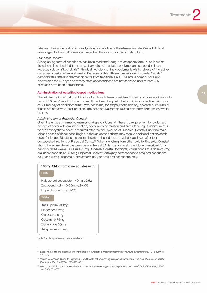
The administration of trational LAI’s has traditionally been considered in terms of dose equivalents to units of 100 mg/day of chlorpromazine. It has been long held, that a minimum effective daily dose of 300mg/day of chlorpromazine35 was necessary for antipsychotic efficacy, however such rules of thumb are not always best practice. The dose equivalents of 100mg chlorpromazine are shown in Table 6.
Administration of Risperdal Consta©
Given the unique pharmacodynamics of Risperdal Consta©, there is a requirement for prolonged periods of cover with oral medication, often involving titration and cross tapering. A minimum of 3 weeks antipsychotic cover is required after the first injection of Risperdal Consta© until the main release phase of risperidone begins, although some patients may require additional antipsychotic cover for longer. Steady state plasma levels of risperidone are typically achieved after four consecutive injections of Risperdal Consta©. When switching from other LAIs to Risperdal Consta© should be administered the week before the last LAI is due and oral risperidone prescribed for a period of three weeks. As a rule 25mg Risperdal Consta© fortnightly corresponds to a dose of 2mg oral risperidone daily; 37.5mg Risperdal Consta© fortnightly corresponds to 4mg oral risperidone daily; and 50mg Risperdal Consta© fortnightly to 6mg oral risperidone daily.36
Table 6 – Chlorpromazine dose equivalents
35. Lader M. Monitoring plasma concentrations of neuroleptics. Pharmakopsychiatr Neuropsychopharmakol 1976 Jul;9(4): 170–177
36. Wilson W. A Visual Guide to Expected Blood Levels of Long-Acting Injectable Risperidone in Clinical Practice. Journal of Psychiatric Practice 2004 10(6):393-401
37. Woods SW. Chlorpromazine equivalent doses for the newer atypical antipsychotics. Journal of Clinical Psychiatry 2003 Jun;64(6):663-667
LAIs
Haloperidol decanoate – 40mg q2/52
Zuclopenthixol – 10-20mg q2-4/52
Flupenthixol – 5mg q2/52
100mg Chlorpromazine equates with:
SGAs37
Amisulpiride 200mg
Risperidone 2mg
Olanzapine 5mg
Quetiapine 75mg
Ziprasidone 60mg
Aripiprazole 7.5 mg
2Treatments

26
Lithium therapyIntroduction
Lithium has well-established efficacy as an anti-manic agent and antidepressant in bipolar disorder, and for prophylaxis against mood episodes in bipolar disorder. It is probably more effective than the anticonvulsants in classical bipolar I disorder and in severe mania. It is one of the more effective augmentation strategies in major depression. 50% of antidepressant non-responders achieve remission with added lithium. 15% of people with bipolar disorder complete suicide. Only 10% of these deaths occur while the person is taking some form of mood stabilizer. Lithium is the most effective mood stabilizer at preventing suicide.38,39 If a patient is having some breakthrough episodes on an anticonvulsant, a change to lithium is warranted.
Each major mood episode worsens the person’s functional outcome, has a serious impact on the person’s ability to sustain a job, a marriage or other relationship, and increases the probability of developing dementia in old age. Of those who cease lithium after achieving a good response, about 15% become relatively lithium resistant when relapse forces them to recommence it. For these reasons, requests by a patient with established bipolar disorder to have their mood stabilizer reduced or ceased should be firmly resisted. Ceasing lithium quickly (over < 2 weeks) doubles the risk of relapse. Mixed or dysphoric mania, ultrarapid cycling bipolar disorder and personality disorder have a poor response to lithium, compared to anticonvulsants.
Some important side effects of lithium
Renal. There is a fall in GFR and rise in creatinine in around 15% of patients taking lithium in the long term, but this may be related to episodes of toxicity or cardiovascular problems than to lithium per se. It is more controversial whether lithium at a therapeutic blood level can cause permanent renal damage. Renal failure may certainly occur as a result of toxic levels of the drug, however, lithium also commonly causes a concentrating defect at therapeutic levels, resulting in polyuria, which may progress to diabetes insipidus. Warn patients to drink water for resulting thirst, rather than soft drinks or fruit juice, as these worsen weight gain. Abnormalities in renal function should be referred to a renal physician for investigation, as there are many possible causes.
Thyroid. Lithium suppresses the action of the thyroid, including the release of thyroid hormone from the gland. Clinical hypothyroidism occurs in up to 20% of people (especially women) taking lithium for ten years. A larger number have raised TSH with normal T4 (subclinical hypothyroidism).40 In the absence of pre-existing or familial thyroid disorder, thyroid function generally recovers on cessation of the drug. It is not necessary to cease lithium due to thyroid suppression. The patient should be warned at commencement that thyroid suppression is a possibility, and thyroxine replacement will be instituted if necessary. Endocrine referral may be of assistance. There is evidence that even subclinical hypothyroidism may destabilise bipolar disorder, and that thyroxine replacement helps in these cases.
Teratogenicity. Lithium is pregnancy category D. It causes serious malformations, especially cardiac anomalies such as Ebstein’s, in 4-12% of exposed foetuses. 1st trimester exposure is especially risky.
LEARNING OBJECTIVES
• To know the important effects and side effects of lithium• To be able successfully to commence and maintain lithium treatment
38. Goodwin et al. Suicide risk in bipolar disorder during treatment with lithium and divalproex. JAMA 2003; 290:146739. Tondo et al. Lithium maintenance treatment of depression and mania in bipolar I or bipolar II disorders. American Journal
of Psychiatry. 1998;155:63840. Perrild et al. Thyroid funciton and ultrasonically determined thyroid size in patients receiving long term lithium treatment.
American Journal of Psychiatry 147:1518-1521

27
IMET ACUTE PSYCHIATRIC MANAGEMENT
Other side effects
• Fine intention tremor, occasionally so severe as to require cessation of the drug. Propranolol may help in some cases.
• Significant weight gain (similar to valproate)
• Cognitive dulling and mild memory impairment in some patients. Beware of confounding with mild depression or hypothyroidism.
• Hair thinning
• Acne
• Benign T-wave flattening on ECG
• Benign neutrophilia (WCC around 10.0 X 10⁹ is common) due to increased mobilization from bone marrow stores.
Toxicity
Lithium is entirely excreted in the urine. Anything that impedes this excretion may cause blood levels to rise to toxic levels. Your patients will need to be warned to avoid:
• Excessive lithium intake, for example, some patients take extra tablets on “bad days”
• Missing blood tests. Regular tests are vital to detect gradually increasing lithium levels
• Dehydration, especially in summer. Some patients try to control the polyuria by reducing water intake, with disastrous results. Take extra fluids or reduce dose during severe diarrhoea or vomiting
• Medications that block excretion. All NSAIDS (now over-the-counter) can do this, with the exception of aspirin, and should be avoided. Warn the patient to check all prescription medications for interactions before commencing them.
Lithium toxicity can result in acute or chronic renal failure, seizures, coma, permanent neurotoxicity (especially cerebellar damage) or death. Dialysis is the treatment of choice at levels > 3.0mmol/L.
Symptoms include worsening tremor, worsening metallic taste in the mouth, nausea and fatigue, confusion, worsening polyuria and dehydration. Patients with mild symptoms should be advised to seek a trough lithium level within a day or so. Those with severe symptoms should cease taking lithium and present immediately at the emergency department for testing.
How to prescribe lithium
Before commencing, take baseline TFTs, EUC and, if relevant, ßHCG. Warn patients about potential side effects, including renal and thyroid, and teratogenicity. Warn a manic patient and their family that post-manic depression is common, and is not the result of the mood stabiliser. They should seek treatment for this, if it occurs, and not cease the medication.
Dosing. 250mg lithium carbonate and 450mg slow release forms are available. The slow release form can be helpful in offering once daily dosing if needed to promote compliance or reduce daytime side effects such as tremor, and in reducing the otherwise daunting number of tablets that the patient must take. Start at around 500mg per day and test after 5-7 days, then adjust dose accordingly. Reduce this for the elderly, who have a lower GFR.
Blood levels. Blood levels are taken 12 hours post dose. On single daily dosing, the level will be ~20% higher than with bd dosing. Levels should be done at least weekly until the correct level is attained, and continued every 1-3 months in the long term. TFTs and EUC should be done 6 monthly. With b.i.d dosing, aim for a serum lithium of 0.6 for augmentation in major depression, and 0.8-1.0 to treat acute mania. Bipolar prophylaxis is achieved for different patients with levels somewhere between 0.6 and 1.0. Titrate to response and side effects for the individual.
If insufficiently effective. Check the serum level, as noncompliance is common. If changing to an anticonvulsant, leave lithium in situ until a good level of anticonvulsant is achieved, then taper lithium. If neither class is completely effective, use lithium in combination with one of the anticonvulsants.
Treatments2
28Clozapine Introduction
Clozapine is still the only drug of proven efficacy in treatment-resistant schizophrenia.41 The significant response of neuroleptic-resistant schizophrenia patients to clozapine validates its efficacy in this group. Clozapine is of proven superiority over first generation antipsychotics42 and has a response rate of 50% among previously treatment-refractory patients and 76% among treatment-intolerant patients.43 The benefits of clozapine are seen in reduction of positive and negative symptoms of schizophrenia, as well as reduction in aggression and suicide.44
Clozapine is available under special access provisions of the pharmaceutical benefits scheme. It can only be prescribed by psychiatrists who have registered with the Clozapine Patient Monitoring Service (CPMS) whose remit is to monitor patients receiving clozapine for haematological abnormalities.
Intiation of Clozapine Therapy
The initiation of clozapine therapy requires the informed consent of the patient, or where appropriate, the Mental Health Review Tribunal. The risks of agranulocytosis, myocarditis and metabolic complications and the steps undertaken to minimize these must be explained to the patient. The patient must then be registered with the CPMS and baseline white cell count (WCC) and neutrophil count (NC) must also be provided. The patient’s blood type must also be identified.
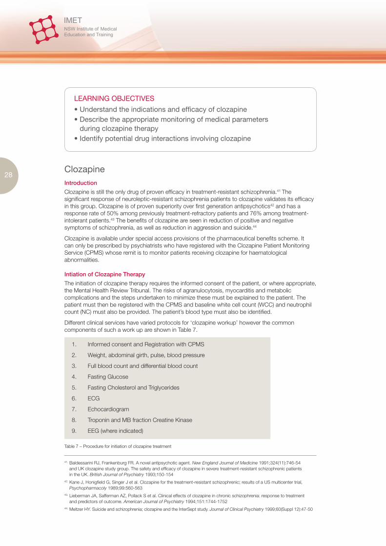
Different clinical services have varied protocols for ‘clozapine workup’ however the common components of such a work up are shown in Table 7.
1. Informed consent and Registration with CPMS
2. Weight, abdominal girth, pulse, blood pressure
3. Full blood count and differential blood count
4. Fasting Glucose
5. Fasting Cholesterol and Triglycerides
6. ECG
7. Echocardiogram
8. Troponin and MB fraction Creatine Kinase
9. EEG (where indicated)
Table 7 – Procedure for initiation of clozapine treatment
LEARNING OBJECTIVES
• Understand the indications and efficacy of clozapine• Describe the appropriate monitoring of medical parameters
during clozapine therapy• Identify potential drug interactions involving clozapine
41. Baldessarini RJ, Frankenburg FR. A novel antipsychotic agent. New England Journal of Medicine 1991;324(11):746-54 and UK clozapine study group. The safety and efficacy of clozapine in severe treatment-resistant schizophrenic patients in the UK. British Journal of Psychiatry 1993;150-154
42. Kane J, Honigfield G, Singer J et al. Clozapine for the treatment-resistant schizophrenic; results of a US multicenter trial, Psychopharmacoly 1989;99:560-563
43. Lieberman JA, Safferman AZ, Pollack S et al. Clinical effects of clozapine in chronic schizophrenia: response to treatment and predictors of outcome. American Journal of Psychiatry 1994;151:1744-1752
44. Meltzer HY. Suicide and schizophrenia; clozapine and the InterSept study. Journal of Clinical Psychiatry 1999;60(Suppl 12):47-50

29
IMET ACUTE PSYCHIATRIC MANAGEMENT
45. Gerson SL, Meltzer H Mechanisms of clozapine-induced agranulocytosis. Drug Saf 1992;7 Suppl 1:17-2545. Gerson SL, Arce C, Meltzer HY. N-desmethylclozapine: a clozapine metabolite that suppresses haemopoiesis.
British Journal of Haematology. 1994 86:555-561
The initiation of clozapine therarpy (“Day 1”) requires close monitoring of pulse, blood pressure, and temperature. There are rare instances of cardiovascular collapse described following the first dose of clozapine (6.25-12.5mg). This usually results from massive vasodilation. Isoprenaline infusion is the pressor of choice, as intravenous adrenaline may lead to further hypotension. Many services require inpatient admission for ‘Day 1’, however day-hospital admission is possible, assuming adequate resuscitation facilities are available. Some patients develop ‘flu-like’ symptoms in the initial phases of clozapine therapy, including pyrexia. This is not related to infection and, in the absence of abnormalities in neutrophil count, should not necessitate the cessation of clozapine.
Medical Review during dose titration
The patient is usually reviewed weekly during the first 18 weeks of treatment. During this period, clozapine therapy is titrated up towards the usual therapeutic doses of 300-600mg per day in divided doses. Dose increments vary from 12.5-50mg per week, depending upon the patient’s tolerance of treatment. Given the severity of illness usually associated with the need for clozapine therapy, there should be careful documentation of the patients progress (Table 8)
1. Recent progress/symptoms
2. Significant mental state features
3. Risk of harm to self or others
4. Current clozapine dose and tolerability/efficacy
5. Blood results
6. Weight (periodic measure of waist circumference)/BP/Pulse
7. Action plan
Table 8 – Standard Medical entry during first 18 weeks of clozapine therapy
Clinical monitoring during clozapine therapy
a. Haematological The most clinically significant adverse effect of clozapine therapy is the induction of agranulocytosis in up to 3% of patients. In order to prevent excess morbidity and mortality in clozapine therapy, the CPMS require regular testing of the patient’s white cell count and neutrophil count in order for clozapine therapy to continue. The CPMS requires weekly leukocyte and neutrophil counts for the first 18 weeks of clozapine therapy and monthly for the duration of therapy. A leukocyte count of > 3.0 x 10⁹/L or neutrophil count >1.5 x 10⁹/L represents a contraindication to initiation of clozapine therapy, or grounds for closer monitoring or discontinuation of clozapine therapy. A leukocyte count < 3.5 x 10⁹ or neutrophil count < 2.0 x 10⁹ places a patient in the at-risk category (the “amber zone”) and warrants closer haematological monitoring. There have been a variety of postulated mechanisms of agranulocytosis in clozapine therapy, although the most likely cause is a toxic metabolite of clozapine, N-desmethylclozapine.45 This compound is likely metabolized to an unstable compound which is toxic to haemopoietic precursors of both myeloid and erythroid lineages.46
Cardiac Monitoring
Clozapine therapy has been associated with a variety of potentially lethal cardiac abnormalities, acute myocarditis and cardiomyopathy. The risk of myocarditis is highest in the first two months of treatment. Cardiomyopathy is rare, but generally occurs later in treatment. Pericarditis and pericardial effusion have also been associated with clozapine treatment. Tachycardia occurs in about 25% of patients and is also a potential indicator of myocardial disease. Persisting tachycardia beyond two months of treatment, or in association with other symptoms of cardiac failure, palpitations or angina pectoris warrant urgent medical review. A minority of clozapine-treated patients show ECG changes
2Treatments

30
similar to those including S-T segment depression and flattening or inversion of T-waves. These may be benign abnormalities, but they also may be an indicator of myocarditis. The appearance of such anomalies warrants urgent cardiologist review. Patients with a history of cardiac illness or abnormal cardiac findings on physical examination should have a cardiology review prior to commencement of clozapine therapy. Cardiac monitoring during clozapine therapy should include:
a. Routine pulse and BP measurement
b. Pre-treatment ECG (serial measurements depending upon clinical indications)
c. Baseline cardiac enzymes – CK-MB, troponin I, troponin T (serial measurements every 6-12 months)
d. Echocardiogram (pre-treatment and annual)*
Clozapine Levels
Measuring serum clozapine levels is usually done when there is non-response or partial response to treatment, or where there are questions of treatment adherence. Clozapine levels should be checked in the following circumstances:
1. Where patients are on doses above 600mg daily or;
2. Concomitant administration of the following medications (Table 9):-
Neurological Monitoring
Drug induced movement disorders are uncommon with clozapine therapy, however there should be routine examination assessing for obvious abnormal involuntary movements, parkinsonism, and motor restlessness. Seizures occur at a frequency of 1.3% of patients taking clozapine. Seizures tend to occur at low doses (< 300 mg/d) during the titration phase of treatment, and at higher doses ( ≥ 600 mg/d) during the maintenance phase. Patients with a history of seizures or epilepsy are more likely to have seizures soon after initiation of therapy, on low doses. Such patients should have pre-treatment EEGs and routine monitoring as indicated by neurologist advice.
Psychotropics
Sertraline Phenytoin LithiumCitalopram St John’s Wort DiazepamCarbamazepine Phenobarbitone HaloperidolFluoxetine Risperidone ValproateFluvoxamine Modafinil LamotrigineParoxetine
Social/Recreational
Caffeine Nicotine Grapefruit juice
Anti-infectives
Ciprofloxacin Clarithromycin RifampicinErythromycin Ketoconazole Ritonavir
Cardiac
Lisinopril Quinidine
GI
Cimetidine
Other
Disulfiram Oral contraceptive pill
Table 9 – Potential Drug Interactions with clozapine

31
IMET ACUTE PSYCHIATRIC MANAGEMENT
Arguments exist as to whether patients taking clozapine doses of >600 mg/day should undergo routine EEG monitoring. This is best individualised until clearer data is available regarding the cost-benefit of such monitoring.
Other Side effects of clozapine therapy
There are isolated reports have been documented of clozapine-associated emergence of obsessive compulsive symptoms, priapism, allergic complications. pancreatitis, toxic hepatitis, elevation in creatinine kinase levels and diabetes-like symptoms.
Anticonvulsant medications in psychiatryIntroduction
In psychiatry, anticonvulsants are predominantly used as mood stabilizers in bipolar disorder. They may also be of benefit in controlling aggression, for example in brain damage or mental retardation. There is little evidence to support their use in unipolar depression. Patients poorly responsive to lithium should try an anticonvulsant, and vice versa. A combination of both classes is commonly required for maximum efficacy in preventing episodes.
Valproate
Sodium valproate is the anticonvulsant most commonly used in bipolar disorder. Some studies suggest equal efficacy with lithium; others note inferior long-term outcomes such its relative paucity of evidence for a reduction in suicide. It may be superior to lithium for mixed mood states, rapid cycling and other non-classical forms of bipolar disorder, but less effective for severe mania and classic bipolar I.47 The combination with lithium is more effective in preventing mood episodes than either agent alone.
Side effects• Usually better tolerated overall than lithium, with less noncompliance.
• Main serious side effect is hepatic failure. This is rare (1 in 20,000), but a benign elevation in transaminases (up to 3 X normal) is common. Mild elevations warrant regular monitoring, with drug cessation if elevation worsens.
• Less cognitive dulling than lithium
• Tremor (additive with lithium tremor in combination)
• Weight gain often significant.
• Hair thinning and deterioration in hair quality often unacceptable to patients.
• Causes polycystic ovarian syndrome (PCOS) in young women, resulting in weight gain, hirsutism, impaired fertility, impaired glucose metabolism, hyperandrogenism. The incidence of this disorder is controversial. Monitor for menstrual irregularity and weight in young women, and refer to gynaecologist if changes occur.
• Serious risk of teratogenicity. Increases risk of spina bifida to 1-4%.
LEARNING OBJECTIVES
• To understand the uses and side effects of anticonvulsants in psychiatry• To learn the practical steps involved in starting anticonvulsant treatment
47. Nasrallah et al. Carbamazepine and valproate for the treatment of bipolar disorder: a review of the literature. Journal of Affective Disorders. 2006, 95(1-3):69-78
* This has become routine but the cost-benefit data is unclear
2Treatments

32
Commencing and monitoring valproateBefore or at commencement, take baseline LFTs and a ßHCG if relevant. Warn women about teratogenicity and polycystic ovarian syndrome. Warn about rare hepatic failure and other side effects. Warn manic patients and their families that post-manic depression is common, and is not the result of the mood stabilizer. They should seek treatment for this, if it occurs, and not cease the medication.
Dosing• Gradual (starting with 500mg and increasing to therapeutic dose over 2 or more weeks) or loading
(20mg/kg, around 1500mg per day) regimes are safe and generally well tolerated. Loading may provide a faster response in acute mania
• Allow 5 days for steady state to be achieved, then take blood level. Stated therapeutic ranges are for anticonvulsant effect. Levels above 315 mmol/L are recommended, but the most effective range in bipolar disorder is unknown – it is best to titrate to clinical effect and side effects
• Dosing usually b.i.d, but can be once daily to improve compliance.
Drug interactions of particular note• Increases lamotrigine levels, which can result in Stevens-Johnson syndrome
• Carbamazepine reduces valproate level, while valproate increases carbamazepine level
• Mildly increases clozapine levels. Prevents clozapine seizures.
Carbamazepine
This drug is perhaps underused as a mood stabilizer. Studies suggest equal efficacy with valproate in bipolar disorder, and its long term side effects are often less problematic than those of valproate.48,49
Side effects• Rash (in 5-15%), which requires cessation, as rarely this can progress to dangerous rash such
as Stevens-Johnson syndrome. Re-challenge after a benign rash may be successful.
• Transient neutropenia is common, agranulocytosis rare.
• Serious risk of teratogenicity, namely spina bifida (1-3%), craniofacial abnormalities or developmental delay.
• Note less weight gain, hair loss and tremor than lithium or valproate.
• Sedation, dizziness or ataxia.
• Hyponatraemia is common. Monitor severity and cease if a significant reduction occurs.
• Can reduce T4 and T3, without changing TSH. Not clinically significant.
• Benign hepatic enzyme elevation may occur. If progressive, cease drug, as serious hepatic toxicity may rarely occur.
Use and interactions• Warn patients about side effects, especially rash and agranulocytosis, and teratogenicity. If
relevant, take a ß-HCG level to exclude pregnancy. Warn manic patients and their families that post-manic depression is common, and is not the result of the mood stabiliser. They should seek treatment for this, and not cease the medication.
• Start at 200mg bd and titrate to clinical effect and side effects. Again, stated therapeutic levels are for anticonvulsant action, and the therapeutic range in bipolar disorder is unknown.
• Once daily dosing of the slow release form can be used to increase compliance.
• Carbamazepine powerfully induces cytochrome p450 3A4. This results in several common, clinically significant interactions.
48. Post et al. Thirty years of clinical experience with carbamazepine in the treatment of bipolar illness: principles and practice. CNS Drugs 2007, 21(1):47-71
49. Nasrallah et al. Carbamazepine and valproate for the treatment of bipolar disorder: a review of the literature. Journal of Affective Disorders 2006, 95(1-3):69-78

33
IMET ACUTE PSYCHIATRIC MANAGEMENT
50. Bowden et al. A placebo controlled 18 month trial of lamotrigineand lithium maintenance treatment recently manic or hypomanic patients with bipolar I disorder. Archives of General Psychiatry 2003, 60:392-400
51. Homles et al. Increased frequency of isolated cleft palate in infants exposed to lamotrigine during pregnancy. Neurology 2008;70:2152-2158
52. Maidment ID. Gabapentin treatment for bipolar disorder. Annals of Pharmacotherapy 2001. 35(10):1264-1269.53. Chengappa et al. The evolving role of topiramate among other mood stabilisers in the management of bipolar disorder.
Bipolar Dis 2001, 3:215-232
- Autoinduction of its own metabolism and a drop in carbamazepine levels with time. This effect is maximal at one month after starting treatment.
- Reduction in blood levels of oestrogen from contraceptives, which can result in pregnancy. Use higher dose OCP or other contraceptive methods.
- Reduction in levels of many antidepressants, antipsychotics and anticonvulsants, among other drugs. Always check for interactions and adjust doses if necessary.
- Do not use with clozapine due to the risk of neutropenia.
Lamotrigine
Studies suggest effectiveness in bipolar depression, either as monotherapy or with an antidepressant, but a relative lack of efficacy in treating or preventing mania. It is therefore often combined with another mood stabilizer to target both mood poles.50 If rash does not occur, it is generally very well tolerated. Patients find the lack of weight gain or sedation particularly welcome. As is true for the other anticonvulsants, there is little evidence of efficacy in unipolar depression.
Some common interactionsValproate, often combined with lamotrigine, greatly decreases its metabolism. Conversly, lamotrigine increases valproate metabolism, causing valproate levels to fall by around 25%. Carbamazepine slightly increases lamotrigine metabolism, as does sertraline. There is no interaction with lithium.
Important side effects• 10% of patients develop rash with lamotrigine. 0.3-1% experience serious rash, including Stevens
Johnson syndrome, therefore, the drug must be ceased if there is any rash at all.
• A recent large study reports that taking lamotrigine during first trimester is associated with an increased incidence of cleft lip and palate.51
Gabapentin and topiramate
Despite initial hopes, evidence for the effectiveness of these anticonvulsants in mood disorders has unfortunately failed to materialize.52,53
Use and dosingRapid escalation of dose increases the incidence of rash, so adherence to a slow commencement protocol is very important. Start at 25mg per day and increase over 6 weeks to 200mg/day (see MIMS for details). A final dose range of 200-400 mg is generally used. If the patient is also taking valproate, the above doses must all be halved.
If a patient ceases the drug for more than 5 days, this dosing regime must be recommenced. There is no useful test for serum lamotrigine level, and no blood monitoring is recommended.
2Treatments
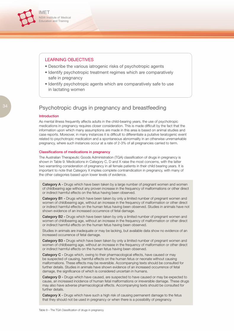
34 Psychotropic drugs in pregnancy and breastfeedingIntroduction
As mental illness frequently affects adults in the child-bearing years, the use of psychotropic medications in pregnancy requires closer consideration. This is made difficult by the fact that the information upon which many assumptions are made in this area is based on animal studies and case reports. Moreover, in many instances it is difficult to differentiate a putative teratogenic event related to psychotropic medication and a spontaneous abnormality in an otherwise unremarkable pregnancy, where such instances occur at a rate of 2-3% of all pregnancies carried to term.
Classifications of medications in pregnancy
The Australian Therapeutic Goods Administration (TGA) classification of drugs in pregnancy is shown in Table 9. Medications in Category C, D and X raise the most concerns, with the latter two warranting consideration of pregnancy in all female patients in their child bearing years. It is important to note that Category X implies complete contraindication in pregnancy, with many of the other categories based upon lower levels of evidence.
Category A - Drugs which have been taken by a large number of pregnant women and women of childbearing age without any proven increase in the frequency of malformations or other direct or indirect harmful effects on the fetus having been observed.
Category B1 - Drugs which have been taken by only a limited number of pregnant women and women of childbearing age, without an increase in the frequency of malformation or other direct or indirect harmful effects on the human fetus having been observed. Studies in animals have not shown evidence of an increased occurrence of fetal damage.
Category B2 - Drugs which have been taken by only a limited number of pregnant women and women of childbearing age, without an increase in the frequency of malformation or other direct or indirect harmful effects on the human fetus having been observed.
Studies in animals are inadequate or may be lacking, but available data show no evidence of an increased occurrence of fetal damage.
Category B3 - Drugs which have been taken by only a limited number of pregnant women and women of childbearing age, without an increase in the frequency of malformation or other direct or indirect harmful effects on the human fetus having been observed.
Category C - Drugs which, owing to their pharmacological effects, have caused or may be suspected of causing, harmful effects on the human fetus or neonate without causing malformations. These effects may be reversible. Accompanying texts should be consulted for further details. Studies in animals have shown evidence of an increased occurrence of fetal damage, the significance of which is considered uncertain in humans.
Category D - Drugs which have caused, are suspected to have caused or may be expected to cause, an increased incidence of human fetal malformations or irreversible damage. These drugs may also have adverse pharmacological effects. Accompanying texts should be consulted for further details.
Category X - Drugs which have such a high risk of causing permanent damage to the fetus that they should not be used in pregnancy or when there is a possibility of pregnancy.
Table 9 – The TGA Classification of drugs in pregnancy
LEARNING OBJECTIVES
• Describe the various iatrogenic risks of psychotropic agents• Identify psychotropic treatment regimes which are comparatively
safe in pregnancy• Identify psychotropic agents which are comparatively safe to use
in lactating women

35
IMET ACUTE PSYCHIATRIC MANAGEMENT
The current TGA classification for antipsychotic drugs is shown in Table 10.
Psychotropic Medications at high risk in pregnancy (Category D)
LithiumThe potential teratogenicity of lithium is well established. The risk ratio of cardiac anomalies following fetal exposure to lithium is estimated at between 1.2-7.7.54 The UK National Teratology Information Service have concluded that lithium increases the risk of all types of malformation of approximately three-fold and with a weighting towards cardiac malformations of around eight-fold. Whilst septal and valvular defects have been described following fetal exposure to lithium, the classic cardiac malformation is Ebstein anomaly. Ebstein anomaly is characterized by apical displacement of the septal and posterior tricuspid valve leaflets, leading to “atrialization” of the right ventricle with a variable degree of malformation and displacement of the anterior leaflet of the valve. The leaflet anomaly leads to tricuspid regurgitation. The severity of regurgitation depends on the extent of leaflet displacement, ranging from mild regurgitation with minimally displaced tricuspid leaflets to severe regurgitation with extreme displacement. Echocardiogram is the criterion standard for diagnosis. In M-mode echocardiography the finding is typically paradoxical septal motion and dilated right ventricle. There is delayed closure of tricuspid valve leaflets more than 65 milliseconds after mitral valve closure. On two-dimensional echocardiography there is apical displacement of the septal leaflet of tricuspid leaflet of greater than 8 mm/m2 – considered the most specific sign of the anomaly. Apart from cardiac malformations following first trimester exposure to lithium there are potential dangers towards the end of the third trimester, related to lithotoxicity in the fetus, with case reports of cardiac arrhythmias, cyanosis and hypertonicity. Some studies have also described congenital goitre and neonatal hypothyroidism.55 Moreover, the rapid shifts in fluid balance following parturition may predispose to lithium toxicity.
ParoxetineConcerns about the potential teratogenicity of paroxetine are based upon three recent studies. The first, a Danish population based cohort study1, found an association between maternal use of SSRIs during the first trimester and an increased risk of both congenital malformations overall (odds ratio 1.4 (95% CI 1.1-1.9)) and congenital cardiac malformations (odds ratio 1.6 (95% CI 1.0-2.6)).56 The second is a retrospective study conducted by the manufacturer, GlaxoSmithKline (GSK).57 The findings suggest that, compared with other antidepressants, paroxetine use during the first trimester is associated with an increased risk of both congenital malformations overall (odds ratio 2.2 (95% CI 1.34-3.63)) and congenital cardiac malformations (odds ratio 2.08 (95% CI 1.0-4.23). The most common abnormality was ventricular-septal defects, although others were described.
54. Cohen L, Freidman J, Jefferson J et al. A re-evaluation of risk of in-utero exposure to lithium. Journal of the American Psychiatric Association, 1994 271: 146-150
55. Williams K, Oke S. Lithium in Pregnancy. Psychiatric Bulletin 2000 24: 229-23156. Wogelius P, Nørgaard M, Munk EM, Mortensen PB, Lipworth L, Sørensen HT. Maternal use of selective serotonin reuptake
inhibitors and risk of adverse pregnancy outcome (Abstract) Pharmacoepidem Drug Safety 2005; 14: S72-S7357. http://ctr.gsk.co.uk/Summary/paroxetine/studylist.asp.
2Treatments

36
Most recently preliminary information about the results of a new study examining data from a Swedish Medical Birth Registry has been made available.58 This study suggests that babies born to mothers who have taken paroxetine in the first trimester of pregnancy are at an approximately 2 fold higher risk of congenital cardiac malformations compared with the equivalent frequency in the population (odds ratio 2.22 (95% CI 1.39-3.55)). This study also suggests that the other SSRIs examined (citalopram, fluoxetine and sertraline) are not associated with an increased risk of congenital malformations.
The results of these studies suggest that women should not take paroxetine in the first trimester, and that this does not appear to be an SSRI class effect.
Antipsychotics*
Category B1 – pimozide, thiothixene
Category B3 – quetiapine, ziprasidone, aripiprazole, olanzapine, risperidone
Category C – chlorpromazine, fuphenazine, pericyazine, perphenazine, promazine, thiopropazate, thioridazine, trifluoperazine, clozapine, flupenthixol, droperidol, haloperidol, zuclopenthixol
Palinperidone – limited data – recommend avoiding* When given in high doses during late pregnancy, related compounds have caused prolonged
neurological disturbances in the newborn infant.
Antidepressants*
Category B2 – venlafaxine, mianserin, tranylcypromineCategory B3 – Mirtazapine, moclobemide, nefazodone PhenelzineCategory C – citalopram, fluoxetine, fluvoxamine, sertraline, amitriptyline, clomipramine, desipramine, dothiepin, doxepin, imipramine, nortriptyline, protriptyline, trimipramineCategory D – Paroxetine* The use of SSRIs in the third trimester may result in a withdrawal state in the newborn infant.
Anticholinergics
Category A – Procyclidine
Category B1 – benzhexol
Category B2 – benztropine, biperiden
Anticonvulsants/Mood Stabilisers
Category B1 – gabapentin
Category B3 – tiagabine, topiramate
Category D – lithium, sodium valproate, carbamazepine, lamotrigine, phenytoin, methylphenobarbitone, phenobarbitone, primidone, ethosuximide, methsuximide, phensuximide, sulthiame, vigabatrin
Benzodiazepines
All Category C
Table 10. TGA Classification of common psychotropic drugs
58. www.janusinfo.org

37
IMET ACUTE PSYCHIATRIC MANAGEMENT
59. Kohen D. Psychotropic medication and breast-feeding. Advances in Psychiatric Treatment 2005 11: 371-379
Anticonvulsants
Sodium ValproateThere is a twenty fold increase in neural tube defects following fetal exposure to valproic acid compounds. A syndrome of specific craniofacial abnormalities and long, thin digits with hyperconvex nails has been described in infants exposed to valproic acid during pregnancy. Valproic acid appears to be associated with a higher risk for major congenital malformations as well as developmental delay and decreased verbal intelligence. These appear to be dose-related
CarbamazepinePrenatal exposure to carbamazepine increases the risk of neural tube defects ten-fold. A syndrome of craniofacial abnormalities, intellectual impairment and hypoplastic nails is described in infants exposed to carbamazepine in utero, Carbamazepine is also associated with a risk of cardiac anomalies.
Effects of untreated psychiatric disorder in pregnancy
Any treatment decision must be weighed against the deleterious impact of untreated or sub optimally treated psychiatric disorder. Untreated psychiatric disorder imperils the fetus or infant through a variety of risks including poor antenatal care, propensity to nutritional neglect or exposure to toxins or trauma. Moreover, infants under the care of a mentally ill parent are more likely to manifest non-organic failure to thrive or developmental delay, particular cognitive development.
Guidelines for breast feeding
Information comes from small case series and single case reports.59 This limited dataset indicates that all psychotropic drugs are excreted into breast milk and that the infant is therefore exposed to them. In recent decades sufficient data have accumulated to allow psychiatrists to confidently prescribe tricyclic antidepressants, selective serotonin reuptake inhibitors, conventional antipsychotics, carbamazepine and sodium valproate to breast-feeding mothers. There are not sufficient data on newer antipsychotic medications to allow women to breast-feed safely. Clozapine or lithium should not be used in breast-feeding women. Recommended practice is that breast-feeding mothers requiring psychotropic medication be on a low dose of one single drug.
2Treatments
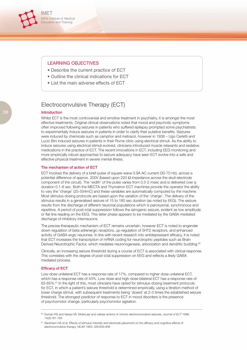
38
Electroconvulsive Therapy (ECT)Introduction
Whilst ECT is the most controversial and emotive treatment in psychiatry, it is amongst the most effective treatments. Original clinical observations noted that mood and psychotic symptoms often improved following seizures in patients who suffered epilepsy prompted some psychiatrists to experimentally induce seizures in patients in order to clarify their putative benefits. Seizures were induced by chemicals such as camphor and metrazol, however in 1938 – Ugo Cerletti and Lucio Bini induced seizures in patients in their Rome clinic using electrical stimuli. As the ability to induce seizures using electrical stimuli evolved, clinicians introduced muscle relaxants and sedative medications in the practice of ECT. The recent innovations in ECT, including EEG monitoring and more empirically robust approaches to seizure adequacy have seen ECT evolve into a safe and effective physical treatment in severe mental illness.
The mechanism of action of ECT
ECT involves the delivery of a brief-pulse of square-wave 0.9A AC current (30-70 Hz), across a potential difference of approx. 200V (based upon 220 Ω impedance across the skull-electrode component of the circuit). The ‘width’ of the pulse varies from 0.5-2 msec and is delivered over a duration 0.1-8 sec. Both the MECTA and Thymatron ECT machines provide the operator the ability to vary the ‘charge’ (25-504mC) and these variables are automatically computed by the machine. Most stimulus dosing protocols are based upon the variation of the ‘charge’. The delivery of the stimulus results in a generalised seizure of 15 to 180 sec duration (as noted by EEG). The seizure results from the discharge of different neuronal populations which is paroxysmal, synchronous and repetitive. A period of post-ictal suppression follows the iatrogenic seizure, evident as low amplitude or flat line reading on the EEG. This latter phase appears to be mediated by the GABA mediated discharge of inhibitory interneurons.
The precise therapeutic mechanism of ECT remains uncertain, however ECT is noted to engender down-regulation of beta adrenergic receptors, up-regulation of 5HT2 receptors, and enhanced activity of GABA-ergic neurones. In line with recent research into antidepressant efficacy, it is noted that ECT increases the transcription of mRNA coding for neurotrophic peptides such as Brain Derived Neurotrophic Factor, which mediates neuronogenesis, arborization and dendritic budding.60
Clinically, an increasing seizure threshold during a course of ECT is associated with clinical response. This correlates with the degree of post-ictal suppression on EEG and reflects a likely GABA mediated process.
Efficacy of ECT
Low-dose unilateral ECT has a response rate of 17%, compared to higher dose unilateral ECT, which has a response rate of 43%. Low dose and high-dose bilateral ECT has a response rate of 63-65%.61 In the light of this, most clinicians have opted for stimulus-dosing treatment protocols for ECT, in which a patient’s seizure threshold is determined empirically, using a titration method of lower charge stimuli, with subsequent treatments being ‘dosed’ at 2-3 times the established seizure threshold. The strongest predictor of response to ECT in mood disorders is the presence of psychomotor change, particularly psychomotor agitation.
LEARNING OBJECTIVES
• Describe the current practice of ECT• Outline the clinical indications for ECT • List the main adverse effects of ECT
60. Duman RS and Vaidya VA. Molecular and cellular actions of chronic electroconvulsive seizures. Journal of ECT 1998, 14(3):181-193
61. Sackheim HA et al. Effects of stimulus intensity and electrode placement on the efficacy and cognitive effects of electroconvulsive therapy. NEJM 1993; 328:839-846
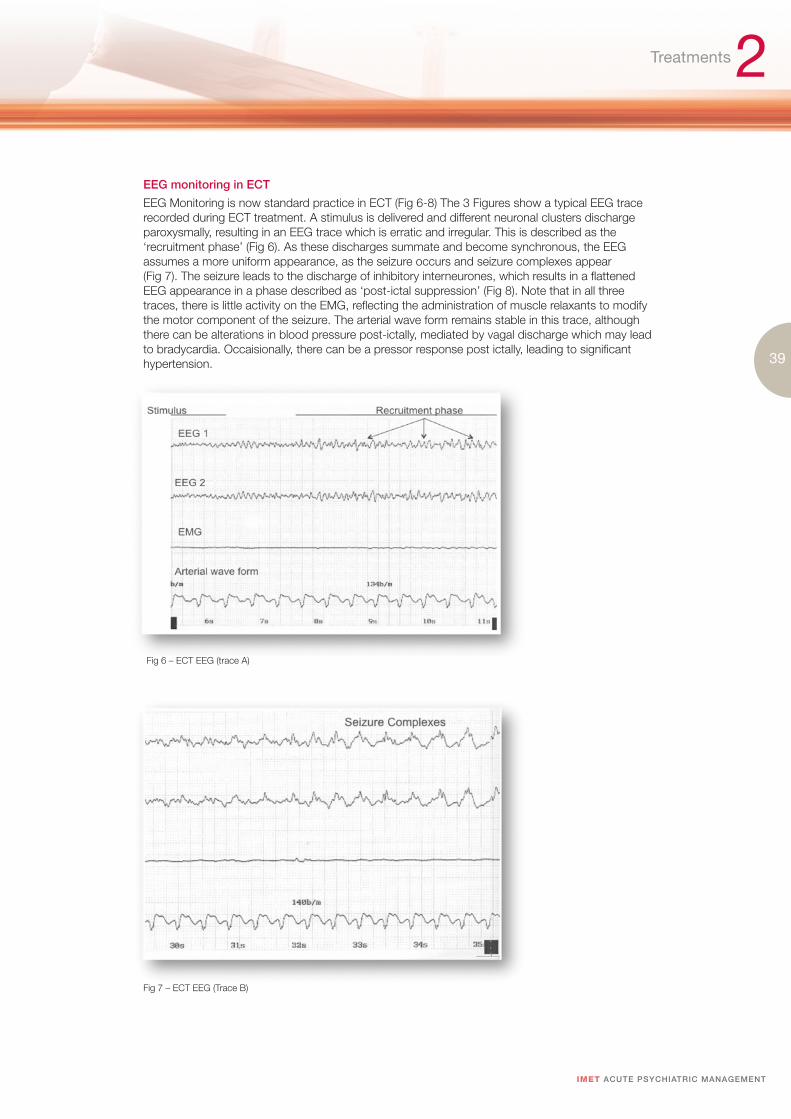
39
IMET ACUTE PSYCHIATRIC MANAGEMENT
EEG monitoring in ECT
EEG Monitoring is now standard practice in ECT (Fig 6-8) The 3 Figures show a typical EEG trace recorded during ECT treatment. A stimulus is delivered and different neuronal clusters discharge paroxysmally, resulting in an EEG trace which is erratic and irregular. This is described as the ‘recruitment phase’ (Fig 6). As these discharges summate and become synchronous, the EEG assumes a more uniform appearance, as the seizure occurs and seizure complexes appear (Fig 7). The seizure leads to the discharge of inhibitory interneurones, which results in a flattened EEG appearance in a phase described as ‘post-ictal suppression’ (Fig 8). Note that in all three traces, there is little activity on the EMG, reflecting the administration of muscle relaxants to modify the motor component of the seizure. The arterial wave form remains stable in this trace, although there can be alterations in blood pressure post-ictally, mediated by vagal discharge which may lead to bradycardia. Occaisionally, there can be a pressor response post ictally, leading to significant hypertension.
Fig 6 – ECT EEG (trace A)
Fig 7 – ECT EEG (Trace B)
2Treatments

40
Fig 8 – ECT EEG (Trace C)
Indications for ECT
ECT is most commonly administered for severe melancholic or psychotic depression, either when antidepressant treatment has been ineffective, or there are severe complications such as refusal to eat or catatonia. ECT is effective in terminating refractory episodes of severe mania and psychotic symptoms in schizophrenia. ECT rapidly reverses catatonic symptoms and may terminate an episode of neuroleptic malignant syndrome or protracted delirium. ECT appears to temporarily alleviate movement disorder in Parkinson’s disease and may be used in refractory cases of status epilepticus.
Adverse effects of ECT
The commonest adverse effects of ECT include transient confusion, headache, nausea, and myalgia. Memory impairment is common during a course of ECT, for both retrograde and anterograde modalities. This is a result of the impairment of working memory. There is no definitive evidence of loss of long-term semantic or episodic memory. Less commonly, ECT can lead to cardiac arrhythmias following the vagal discharge. Hypertension occasionally occurs and can rarely lead to cerebral haemorrhage or stroke. Inadequate muscle relaxation can lead to dental or orthopaedic injuries. Some patients may suffer complications of anaesthesia including aspiration, respiratory suppression or hypoxia. Status epilepticus has been described following administration of ECT. US data indicates a mortality rate of 1 in 80,000 treatments. In the light of this, intracranial lesions such as tumours or known vascular abnormalities represent a contraindication to ECT. Recent myocardial infarction is also a contraindication.
Treatment course for ECT
Typically, ECT is administered to consenting patients who are medically fit for the treatment. A typical course of ECT is between 6-12 treatments, administered three times per week. In rare instances of psychiatric emergency, treatments are administered daily. Acute treatment is administered until there is sustained clinical improvement and subsequent treatments may be administered less frequently. During a course of treatment, the patient’s seizure threshold will increase, necessitating upwards titration of the stimulus level. If ECT is to be administered under the Mental Health Act, consent can only be provided by the Mental Health Review Tribunal (MHRT). The MHRT typically require that a case be made as to why ECT is preferred over less invasive treatments. The MHRT also require reports of the patient’s clinical progress.
The MHRT rarely approve more than 12 sessions of ECT.
Once a patient has experienced sustained clinical benefit from ECT, progress is maintained using pharmacological treatments. In the case of severe depression, lithium appears to be the most efficacious agent for maintenance of ECT produced remissions. There are some instances, particularly in older patients, where pharmacological treatments are ineffective in maintaining a patient’s remission. In such circumstances, the patient may receive maintenance ECT varying from weekly to bimonthly treatments.

41
IMET ACUTE PSYCHIATRIC MANAGEMENT
Pro Re Nata (PRN) medicationIntroduction
Despite the provision of adequate regular psychotropic medication regimes, patients in acute treatment settings frequently experience distress arising from their illness which may increase the risk of harm to the patient, other patients or staff. In such circumstances additional medication regimes are necessary to alleviate the patient’s distress and reduce the risk of such harm.
Indications for prn medication
The administration of prn medication is either initiated by nursing staff or the patient. The common circumstances in which prn medication becomes necessary include:
• Distress arising from psychopathological symptoms
• Agitation arising in the course of an episode of severe mental illness
• Anxiety reactions to psychological phenomena or the ward environment
• Suicidal ideation or impulses to self-harm
• Insomnia
• Physical aggression
• Severe disorganization of behaviour engendering risk of misadventure
• Intoxication or withdrawal from substances.
Oral prn regimes for agitation or sedation
BenzodiazepinesOnly one type of benzodiazepine should be charted as prn medication:
Diazepam 5-10 mg orally in a healthy adult every 2-6 hours. Maximum 50 mg in 24 hours (prn and regular)
OR
Lorazepam 0.5-1 mg orally every 2-6 hours. Maximum 6 mg in 24 hours (prn and regular).
If underlying agitation is secondary to psychosis and benzodiazepines have not alleviated agitation adequately, antipsychotic medications either as a stat dose or as a prn regimen can be added. If an antipsychotic is the primary (regular) treatment, prn antipsychotic medication charted should match the regular antipsychotic.
Oral neurolepticsOlanzapine wafer or tablets (IM version should not be prescribed on the prn chart but as a statim dose when necessary) 5-10mg orally 2-4 hourly but the total of prn, stat dosing and regular regimen should not exceed 30 mg per day unless the treating psychiatrist has been consulted.
OR
Quetiapine 25-100 mg orally 2-4 hourly (total maximum of 800 mg/day for regular & prn combined).
LEARNING OBJECTIVES
• Describe the appropriate indications for prn medication• Outline appropriate administration of oral prn medication• Describe procedures for safe administration of parenteral prn regimes
2Treatments

42
Parenteral regimes for agitation
Intramuscular sedation is indicated when oral medication is not possible or safe for the patient or staff. The aim of parenteral prn medication is to engender a state of rousable sleep (i.e. the patient can be roused and cooperate with directions in response to voice or pain and sleepy if undisturbed) may have to be initiated on occasions.
EITHER
Lorazepam 0.5mg-2mg IMI 1-2 hourly, maximum 4mg per day (Peak plasma concentration at 60-90 minutes. )
OR (not both)
Midazolam 5-10 mg IMI 1-2 hourly, maximum 20 mg per day. Peak plasma concentration in healthy adults 30-60 minutes.
For psychotic/manic agitation not relieved by benzodiazepines.
Haloperidol 5-10 mg IMI 4-6 hourly, total of prn and regular medication is 20 mg a day
OR
Olanzapine 5-10mg IMI 2 hourly (prescribed as stat doses not as prn) for patients at risk of EPSE or in antipsychotic naïve patients. IM Olanzepine should not be given in combination with benzodiazepine or in those who are intoxicated with any substance. There should be a gap of 2 hours between IM benzodiazepines and IM olanzepine because of potential higher risk of respiratory depression. Maximum dose of regular, prn and stat dose of olanzepine should be 30 mg in 24 hours. Concentration peaks 15-45 minutes.
Zuclopenthixol acetate (Acuphase) 50-150 mg IM should not be given to any neuroleptic naïve patients or be charted as prn medications. It should only be ordered as a stat dose by the medical staff following assessment of the patient’s mental state. May be considered for cases where prolonged sedation (eg 2-3 days) is required and the patient has an established psychotic illness with a high risk of agitation/aggression not managed adequately by any of the above treatments. Peak plasma concentration 24-48 hours with a gradual decline to 1/3 at 72 hours.
The patient’s vital signs and the presence of drug induced movement disorder ought be monitored at baseline and every 10 minutes (as per local hospital protocol) following sedation for a minimum of 1 hour. The patient must be able to maintain own airway and should be nursed in a way that guarantees the airway remains patent.
If the patient is excessively sedated following prn medication, the patient should be placed in a coma position and continuous oxygen saturation monitor with an ambulatory oxymetry should be made. Blood sugar level should be obtained and thorough physical examination should be carried out. Once it has been determined that the underlying cause is sedation from medication, vital signs such as blood pressure, pulse, respiratory rate, oxygen saturations, temperature and Glasgow Coma Scale should be monitored every 10 minutes for 60 minutes, every 15 minutes for 30 minutes, every 30 minutes for 60 minutes then hourly for 4 hours or until awake. The Medical Officer should not leave the patient for at least 30 minutes and only if the patient could maintain their airway and whose vital signs are stable. Oxygen supplements 6L/minute for healthy individuals, 2L/min for patients with Chronic Obstructive Airway Disease should be provided.
For Acute Dystonia Benztropine 1-2mg orally, intramuscularly or intravenously 2-4 hourly maximum 6 mg in 24 hours.
Ethical prescribing practices
As most prn medication is initiated by nursing staff or patients, the ethical and clinically appropriate use of prn psychotropic medication requires the prescribing medical officer to specify on the patient’s medication chart the appropriate circumstances in which such treatment is prescribed and the limits of the dose and frequency of administration of such agents. As much of this occurs in the context of involuntary psychiatric treatment, the appropriate use of prn medication represents a significant ethical issue.
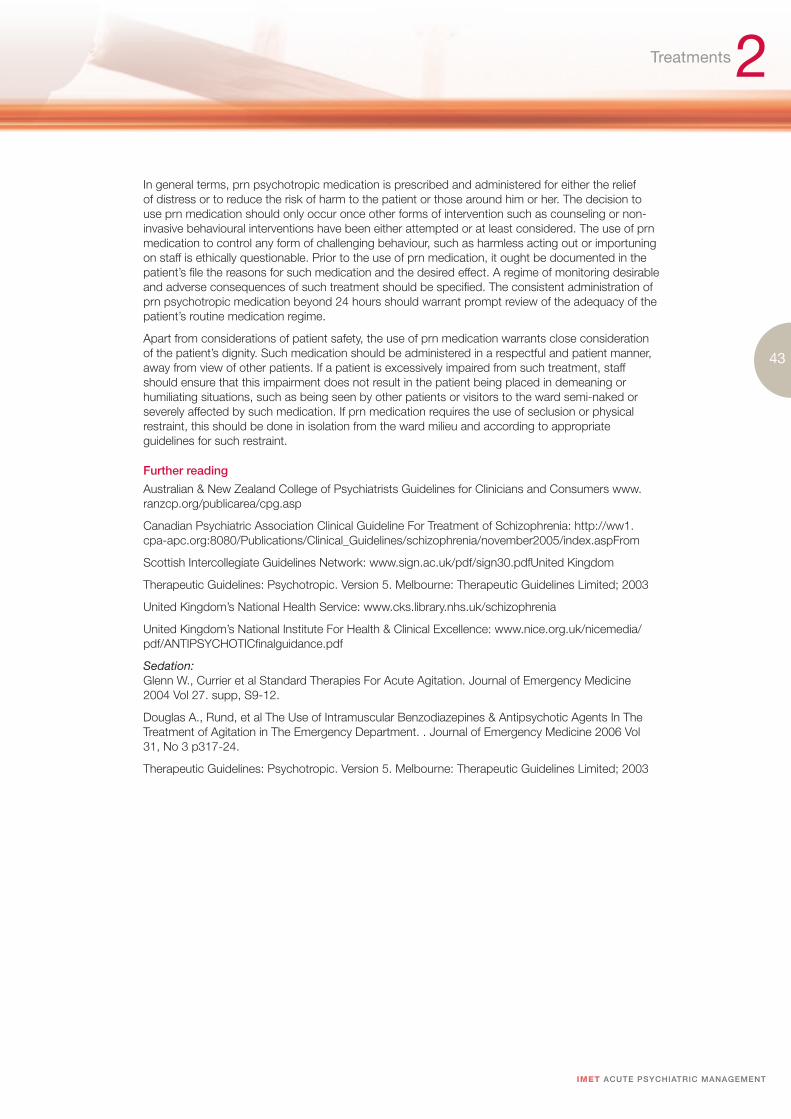
43
IMET ACUTE PSYCHIATRIC MANAGEMENT
In general terms, prn psychotropic medication is prescribed and administered for either the relief of distress or to reduce the risk of harm to the patient or those around him or her. The decision to use prn medication should only occur once other forms of intervention such as counseling or non-invasive behavioural interventions have been either attempted or at least considered. The use of prn medication to control any form of challenging behaviour, such as harmless acting out or importuning on staff is ethically questionable. Prior to the use of prn medication, it ought be documented in the patient’s file the reasons for such medication and the desired effect. A regime of monitoring desirable and adverse consequences of such treatment should be specified. The consistent administration of prn psychotropic medication beyond 24 hours should warrant prompt review of the adequacy of the patient’s routine medication regime.
Apart from considerations of patient safety, the use of prn medication warrants close consideration of the patient’s dignity. Such medication should be administered in a respectful and patient manner, away from view of other patients. If a patient is excessively impaired from such treatment, staff should ensure that this impairment does not result in the patient being placed in demeaning or humiliating situations, such as being seen by other patients or visitors to the ward semi-naked or severely affected by such medication. If prn medication requires the use of seclusion or physical restraint, this should be done in isolation from the ward milieu and according to appropriate guidelines for such restraint.
Further reading
Australian & New Zealand College of Psychiatrists Guidelines for Clinicians and Consumers www.ranzcp.org/publicarea/cpg.asp
Canadian Psychiatric Association Clinical Guideline For Treatment of Schizophrenia: http://ww1. cpa-apc.org:8080/Publications/Clinical_Guidelines/schizophrenia/november2005/index.aspFrom
Scottish Intercollegiate Guidelines Network: www.sign.ac.uk/pdf/sign30.pdfUnited Kingdom
Therapeutic Guidelines: Psychotropic. Version 5. Melbourne: Therapeutic Guidelines Limited; 2003
United Kingdom’s National Health Service: www.cks.library.nhs.uk/schizophrenia
United Kingdom’s National Institute For Health & Clinical Excellence: www.nice.org.uk/nicemedia/pdf/ANTIPSYCHOTICfinalguidance.pdf
Sedation:Glenn W., Currier et al Standard Therapies For Acute Agitation. Journal of Emergency Medicine 2004 Vol 27. supp, S9-12.
Douglas A., Rund, et al The Use of Intramuscular Benzodiazepines & Antipsychotic Agents In The Treatment of Agitation in The Emergency Department. . Journal of Emergency Medicine 2006 Vol 31, No 3 p317-24.
Therapeutic Guidelines: Psychotropic. Version 5. Melbourne: Therapeutic Guidelines Limited; 2003
2Treatments

NOTES
44

Management Challenges
453

46
Acute management of physical aggressionIntroduction
Physical aggression is an infrequent but problematic phenomenon in psychiatric practice. Such incidents can occur in a number of settings; however junior medical staff are usually required to manage them in Emergency Departments and in inpatient settings. Whilst this section outlines general principles of management, each clinical setting may have its own approach to the management of such incidents.
Determinants of Physical Aggression
Table 10 outlines the indicators of acute risk for physical aggression. As was described in the section dealing with formal risk assessment, there are historical or ‘static’ indicators and dynamic or ‘variable’ components. Whilst a history of previous aggression heightens the concern about a particular patient’s presentation, it is the observable, cross-sectional features of mental state, which determine the immediate likelihood of physical aggression.
General Principles of management
Different levels of physical or verbal aggression necessitate different responses. Levels of disturbance can be categorised from 1-3 with appropriate management approaches (Figs 9-11). In any process of intervention, the medical officer ought take responsibility for their own safety and the safety of other staff including:
• Wearing appropriate attire e.g. Breakable name tags
• Access to duress alarms
• Ensuring the patient and staff have a clear means of escape
• Ensuring there are adequate staff available for assistance.
3Management Challenges
LEARNING OBJECTIVES
• Describe the predictors of physical aggression in psychiatric patients• Outline the pharmacological management of physical aggression in
psychiatric patients • Outline the non-pharmacological management of physical aggression
in psychiatric patients
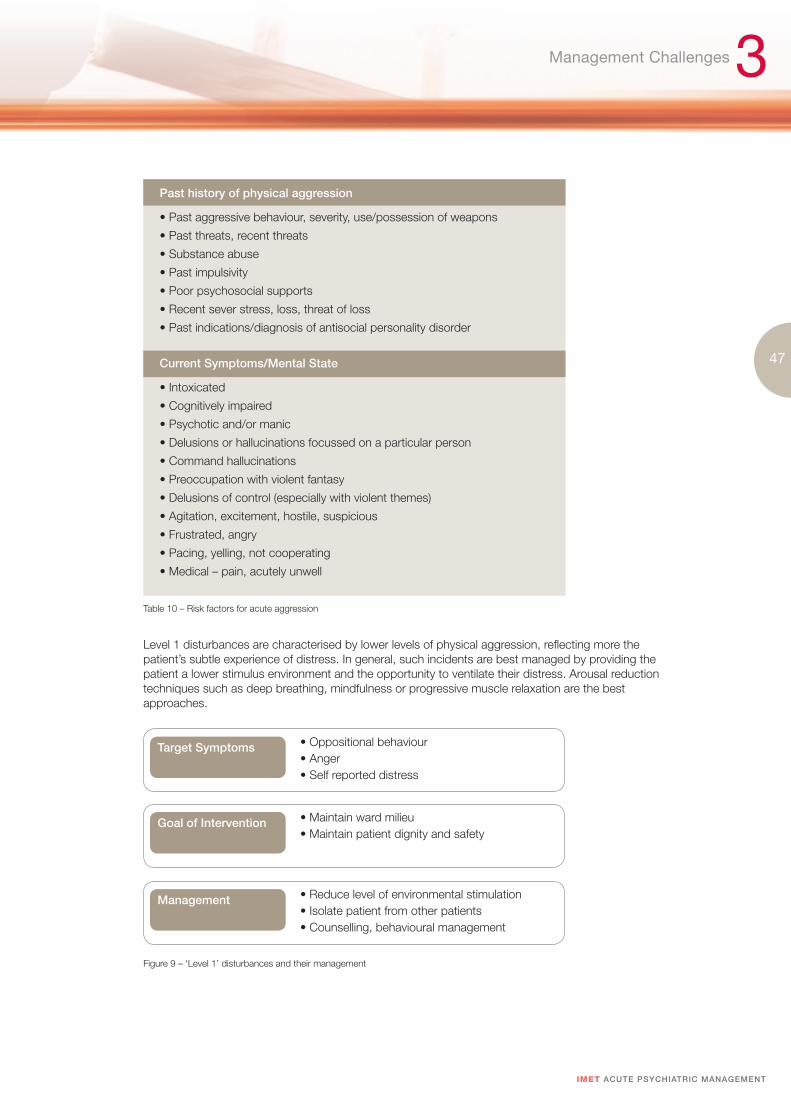
47
IMET ACUTE PSYCHIATRIC MANAGEMENT
Table 10 – Risk factors for acute aggression
Figure 9 – ‘Level 1’ disturbances and their management
Past history of physical aggression
• Past aggressive behaviour, severity, use/possession of weapons
• Past threats, recent threats
• Substance abuse
• Past impulsivity
• Poor psychosocial supports
• Recent sever stress, loss, threat of loss
• Past indications/diagnosis of antisocial personality disorder
• Oppositional behaviour• Anger• Self reported distress
• Maintain ward milieu• Maintain patient dignity and safety
• Reduce level of environmental stimulation• Isolate patient from other patients• Counselling, behavioural management
Current Symptoms/Mental State
• Intoxicated
• Cognitively impaired
• Psychotic and/or manic
• Delusions or hallucinations focussed on a particular person
• Command hallucinations
• Preoccupation with violent fantasy
• Delusions of control (especially with violent themes)
• Agitation, excitement, hostile, suspicious
• Frustrated, angry
• Pacing, yelling, not cooperating
• Medical – pain, acutely unwell
Target Symptoms
Goal of Intervention
Management
Level 1 disturbances are characterised by lower levels of physical aggression, reflecting more the patient’s subtle experience of distress. In general, such incidents are best managed by providing the patient a lower stimulus environment and the opportunity to ventilate their distress. Arousal reduction techniques such as deep breathing, mindfulness or progressive muscle relaxation are the best approaches.
3Management Challenges

48
Level 2 disturbances are characterised by more significant agitation, menacing behaviour on the part of the patient including verbal aggression and damage to property. The appropriate management of this level of disturbance includes a combination of recruiting appropriate numbers of staff via a so-called ‘duress’ or emergency call, isolation of the patient from the at risk environment and the appropriate use of oral or parenteral medication to relieve distress (Figure 10).
Figure 10 – Level 2 disturbances
Level 3 disturbances are the most severe, characterised by actual physical aggression or assaultive behaviour. Such patients are often severely distressed by psychotic symptoms or disinhibited by alcohol or other drugs. These instances represent psychiatric emergencies and warrant management including use of physical restraint, seclusion, the administration of parernteral psychotropic medication and the appropriate level of monitoring. The safe restraint of such patients, to protect both staff and the patient from injury, requires special training. Psychotropic medication administered parenterally, particularly when benzodiazepines are combined with antipsychotic medications, may be unpredictable in their effects (Figure 11). Seemingly small doses can lead to catastrophic complications such as upper airway collapse or respiratory arrest. As such, parenteral psychotropic treatment should only be used when adequate resuscitation facilities are available. The patient must be monitored for signs of cardiovascular instability, oxygen desaturation and airway protection. The circumstances of seclusion or administration of emergency parenteral antipsychotic medication require careful documentation in both the patient’s chart and a formal ‘seclusion register’ (a requirement of all Declared Mental Health Facilities).
Figure 11 – Level 3 disturbances
• Agitation• Menacing• Damage to property
• Physical aggression• Assaultive behaviour
• Patient safety• Limit property damage
• Patient safety• Staff safety
• Increase staff presence• Removal from main ward environment• Offer oral or intramuscular medication
• Increase staff presence• Appropriate restraint or seclusion• Oral or intramuscular medication
(rarely intravenous)• Appropriate post-sedation nursing and
medical management
Target Symptoms
Target Symptoms
Goal of Intervention
Goal of Intervention
Management
Management

49
IMET ACUTE PSYCHIATRIC MANAGEMENT
Psychotropic Drug Use in Managing Aggression
Oral Regimes – The management of some Level 1 and most Level 2 disturbances involves the use of oral psychotropic medication. In most circumstances, this will involve the use of a benzodiazepine with or without the co-admnistration of an antipsychotic medication (Table 11).
Table 11 – Oral psychotropic management of aggression
Parenteral Regimes: The management of Level 3 disturbances often involves the administration or parenteral psychotropic medication (Table 12). As this is largely done via intramuscular injection, only one psychotropic medication is used, to limit the number of injections. In circumstances where these are combined PARENTERAL OLANZAPINE SHOULD NOT BE COMBINED WITH BENZODIAZEPINES.
Table 12 – Parenteral psychotropic management of aggression
Antipsychotic medications
Lorazepam 0.5-2mg every 6-12 hours
Diazepam 2-10 mg every 12-24 hours
Antipsychotic Medications
Olanzapine 5-10mg
Quetiapine 50-200mg
Haloperidol 2.5-5mg
Risperidone 1-2mg
Chlorpromzine 50-200mg
Benzodiazepines
Lorazepam 0.5-2mg IMI
Midzolam 5-10mg IMI
Antipsychotic Medications
Olanzapine 5-10mg IMI
Haloperidol 2.5-5mg
Clopixol acuphase 50-100mg** Care must be taken with neuroleptic naïve patients. Repeated
administration of this agent must not exceed 300mg over 4 days
3Management Challenges
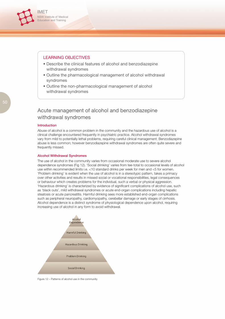
50
Acute management of alcohol and benzodiazepine withdrawal syndromesIntroduction
Abuse of alcohol is a common problem in the community and the hazardous use of alcohol is a clinical challenge encountered frequently in psychiatric practice. Alcohol withdrawal syndromes vary from mild to potentially lethal problems, requiring careful clinical management. Benzodiazepine abuse is less common; however benzodiazepine withdrawal syndromes are often quite severe and frequently missed.
Alcohol Withdrawal Syndromes
The use of alcohol in the community varies from occasional moderate use to severe alcohol dependence syndromes (Fig 12). ‘Social drinking’ varies from tee-total to occasional levels of alcohol use within recommended limits i.e. <10 standard drinks per week for men and <5 for women. ‘Problem drinking’ is evident when the use of alcohol is in a stereotypic pattern, takes a primacy over other activities and results in missed social or vocational responsibilities, legal consequences or behaviour which creates problems for the individual, such a verbal or physical aggression. ‘Hazardous drinking’ is characterized by evidence of significant complications of alcohol use, such as ‘black outs’, mild withdrawal syndromes or acute end-organ complications including hepatic steatosis or acute pancreatitis. Harmful drinking sees more established end-organ complications such as peripheral neuropathy, cardiomyopathy, cerebellar damage or early stages of cirrhosis. Alcohol dependence is a distinct syndrome of physiological dependence upon alcohol, requiring increasing use of alcohol in any form to avoid withdrawal.
Figure 12 – Patterns of alcohol use in the community
LEARNING OBJECTIVES
• Describe the clinical features of alcohol and benzodiazepine withdrawal syndromes
• Outline the pharmacological management of alcohol withdrawal syndromes
• Outline the non-pharmacological management of alcohol withdrawal syndromes

51
IMET ACUTE PSYCHIATRIC MANAGEMENT
The physical complications of prolonged severe alcohol use are shown in Table 13.
Table 13 – Physical complications of long-term alcohol abuse
Features of alcohol withdrawal
Alcohol withdrawal can vary in severity from mild discomfort to a severe agitated delirium which has 10% mortality. In most circumstances, patients will evidence signs of autonomic hyperarousal 12-24 hours after their last drink. This manifests as lability of pulse or blood pressure, tremulousness, diaphoresis or agitation. Untreated these may progress into more florid states of confusion. Some patients may suffer myoclonus or seizures (so-called ‘rum fits’). In less than 1-2% of cases, patients enter a phase of alcohol withdrawal delirium, previously termed ‘delirium tremens’. Such patients experience vivid and terrifying hallucinosis, psychomotor agitation, disorientation to day or night and formication (crawling skin). If the patient survives, the episode terminates after 36-48 hours and is followed by a prolonged period of somnolence. Death is usually caused by acute renal failure, hepato-renal syndrome, or complications of other medical conditions.
Management of alcohol withdrawal
The approach to alcohol withdrawal is shown in Figure 13.
Neurological
Cerebral atrophy (especially frontal lobes) Midline cerebellar atrophy
Korsakov’s syndrome Wernickes’ encephalopathy
Peripheral Neuropathy
Cardiovascular
Cardiomyopathy Hypertension
Accelerated dyslipidaemia and atheroma formation
Gastroenterological
Hepatic steatosis and steatohepatitis Malabsorption syndromes
Cirrhosis and portal hypertension Oropharyngeal cancers
Acute and chronic pancreatitis – malabsorption and diabetes
Gastritis and gastro-oesophageal reflux
Reproductive
Gonadal atrophy Feminization in males
Increased risk of breast cancer
3Management Challenges
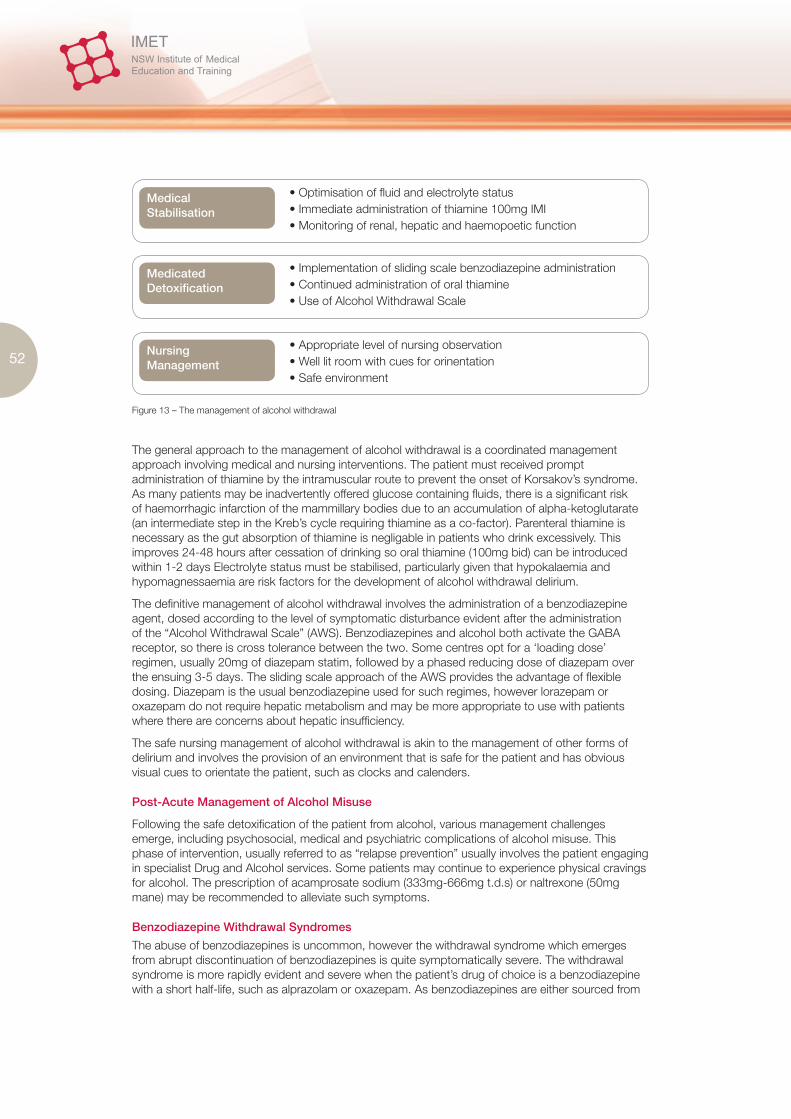
52
Figure 13 – The management of alcohol withdrawal
The general approach to the management of alcohol withdrawal is a coordinated management approach involving medical and nursing interventions. The patient must received prompt administration of thiamine by the intramuscular route to prevent the onset of Korsakov’s syndrome. As many patients may be inadvertently offered glucose containing fluids, there is a significant risk of haemorrhagic infarction of the mammillary bodies due to an accumulation of alpha-ketoglutarate (an intermediate step in the Kreb’s cycle requiring thiamine as a co-factor). Parenteral thiamine is necessary as the gut absorption of thiamine is negligable in patients who drink excessively. This improves 24-48 hours after cessation of drinking so oral thiamine (100mg bid) can be introduced within 1-2 days Electrolyte status must be stabilised, particularly given that hypokalaemia and hypomagnessaemia are risk factors for the development of alcohol withdrawal delirium.
The definitive management of alcohol withdrawal involves the administration of a benzodiazepine agent, dosed according to the level of symptomatic disturbance evident after the administration of the “Alcohol Withdrawal Scale” (AWS). Benzodiazepines and alcohol both activate the GABA receptor, so there is cross tolerance between the two. Some centres opt for a ‘loading dose’ regimen, usually 20mg of diazepam statim, followed by a phased reducing dose of diazepam over the ensuing 3-5 days. The sliding scale approach of the AWS provides the advantage of flexible dosing. Diazepam is the usual benzodiazepine used for such regimes, however lorazepam or oxazepam do not require hepatic metabolism and may be more appropriate to use with patients where there are concerns about hepatic insufficiency.
The safe nursing management of alcohol withdrawal is akin to the management of other forms of delirium and involves the provision of an environment that is safe for the patient and has obvious visual cues to orientate the patient, such as clocks and calenders.
Post-Acute Management of Alcohol Misuse
Following the safe detoxification of the patient from alcohol, various management challenges emerge, including psychosocial, medical and psychiatric complications of alcohol misuse. This phase of intervention, usually referred to as “relapse prevention” usually involves the patient engaging in specialist Drug and Alcohol services. Some patients may continue to experience physical cravings for alcohol. The prescription of acamprosate sodium (333mg-666mg t.d.s) or naltrexone (50mg mane) may be recommended to alleviate such symptoms.
Benzodiazepine Withdrawal Syndromes
The abuse of benzodiazepines is uncommon, however the withdrawal syndrome which emerges from abrupt discontinuation of benzodiazepines is quite symptomatically severe. The withdrawal syndrome is more rapidly evident and severe when the patient’s drug of choice is a benzodiazepine with a short half-life, such as alprazolam or oxazepam. As benzodiazepines are either sourced from
• Optimisation of fluid and electrolyte status• Immediate administration of thiamine 100mg IMI• Monitoring of renal, hepatic and haemopoetic function
• Implementation of sliding scale benzodiazepine administration• Continued administration of oral thiamine• Use of Alcohol Withdrawal Scale
• Appropriate level of nursing observation• Well lit room with cues for orinentation• Safe environment
Medical Stabilisation
Medicated Detoxification
Nursing Management
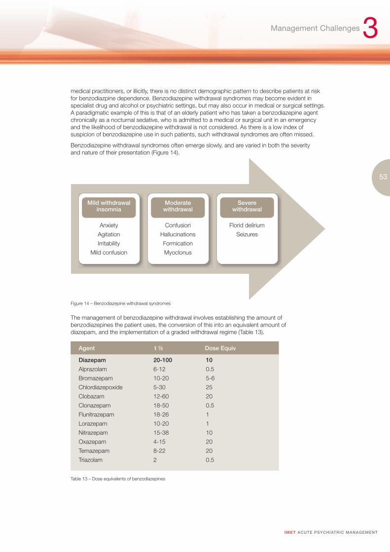
53
IMET ACUTE PSYCHIATRIC MANAGEMENT
medical practitioners, or illicitly, there is no distinct demographic pattern to describe patients at risk for benzodiazpine dependence. Benzodiazepine withdrawal syndromes may become evident in specialist drug and alcohol or psychiatric settings, but may also occur in medical or surgical settings. A paradigmatic example of this is that of an elderly patient who has taken a benzodiazepine agent chronically as a nocturnal sedative, who is admitted to a medical or surgical unit in an emergency and the likelihood of benzodiazepine withdrawal is not considered. As there is a low index of suspicion of benzodiazepine use in such patients, such withdrawal syndromes are often missed.
Benzodiazepine withdrawal syndromes often emerge slowly, and are varied in both the severity and nature of their presentation (Figure 14).
Figure 14 – Benzodiazepine withdrawal syndromes
The management of benzodiazepine withdrawal involves establishing the amount of benzodiazepines the patient uses, the conversion of this into an equivalent amount of diazepam, and the implementation of a graded withdrawal regime (Table 13).
Table 13 – Dose equivalents of benzodiazepines
Anxiety
Agitation
Irritability
Mild confusion
Confusion
Hallucinations
Formication
Myoclonus
Florid delirium
Seizures
Mild withdrawal insomnia
Moderate withdrawal
Severe withdrawal
Agent t ½ Dose Equiv
Diazepam 20-100 10
Alprazolam 6-12 0.5
Bromazepam 10-20 5-6
Chlordiazepoxide 5-30 25
Clobazam 12-60 20
Clonazepam 18-50 0.5
Flunitrazepam 18-26 1
Lorazepam 10-20 1
Nitrazepam 15-38 10
Oxazepam 4-15 20
Temazepam 8-22 20
Triazolam 2 0.5
3Management Challenges
54
Some centres recommend the patient be stabilized on a dose of diazepam equivalent to 50% of the patient’s habitual use of benzodiazepines, with a graded reduction following on from this. As some accounts of benzodiazepine use can be unreliable, monitoring the patient for signs of benzodiazepine withdrawal or overdosing is good practice. If a patient described using 2-3mg of alprazolm per day, this would translate into a dose of 40-60mg per day of diazepam. Such a patient would be stabilized on a dose of 20-30mg diazepam daily.
The rate of subsequent reduction of benzodiazepine dose is controversial. Some inpatient detoxification centres offer rapid detoxifications over periods of 7-10 days, whereas others advocate conservative reductions of 10% of the dose of benzodiazepine per week over the subsequent 2-3 months. The choice of method is more related to patient and clinician preference, rather than specific evidence either way.
Recognition and management of deliriumIntroduction
According to the Diagnostic and Statistical Manual of Mental Disorders, fourth edition (DSM-IV), the diagnostic criteria for delirium are:62
1) disturbance of consciousness with reduced ability to focus, sustain, or shift attention
2) change in cognition or development of a perceptual disturbance that is not better accounted for by dementia
3) acute onset with a tendency for the condition to fluctuate over the course of the day
4) evidence that the disturbance is caused by the direct physiological consequences of a general medical condition or a substance
Alternative terms include: acute confusional state, acute brain syndrome, toxic psychosis, acute brain failure, postoperative psychosis.
Epidemiology of delirium
Studies estimate that between 10-31% of medically ill patients in hospital have features of delirium.63 This rate may be higher in certain patient groups such as those in Intensive Care Units and in patients following cardiac surgery. In the elderly the rate may be as high as 40%.64 However, the condition is often missed. In one study delirium was missed in up to 67% of cases by physicians.65 The risk factors for delirium66 are shown in Table 14.
LEARNING OBJECTIVES
• Outline the features and epidemiology of delirium• Describe the causes and investigation of delirium• Implement a management plan for delirium
62. American Psychiatric Association: Diagnostic and statistical manual of mental disorders, (DSM-IV), 4th Ed, Washington, DC, 1994, American Psychiatric Association
63. Siddiqi N, House AO, Holmes JD. Occurrence and outcome of delirium in medical in-patients: a systematic literature review. Age and Ageing 2006 35: 350-364
64. Lipowski ZJ. Delirium (acute confusional states). JAMA 1987; 258: 1789-179265. Rockwood K, Cosway S, Stolee P et al. Increasing the recognition of delirium in elderly patients. Journal of the American
Geriatrics Society 1994 42:252-256 66. Weber JB, Coverdale JH, Kunik ME. Delirium: current trends in prevention and treatment. Internal Medicine Journal
2004 34(3): 115-121
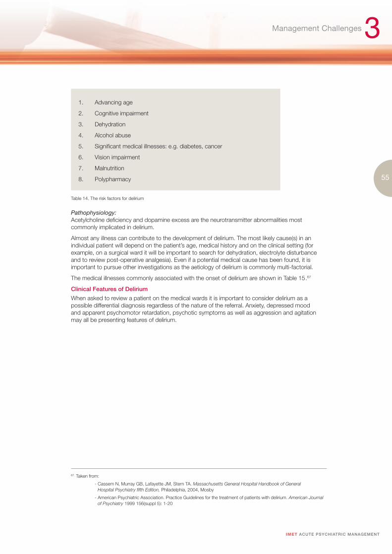
55
IMET ACUTE PSYCHIATRIC MANAGEMENT
67. Taken from:
- Cassem N, Murray GB, Lafayette JM, Stern TA. Massachusetts General Hospital Handbook of General Hospital Psychiatry fifth Edition, Philadelphia, 2004, Mosby
- American Psychiatric Association. Practice Guidelines for the treatment of patients with delirium. American Journal of Psychiatry 1999 156(suppl 5): 1-20
1. Advancing age
2. Cognitive impairment
3. Dehydration
4. Alcohol abuse
5. Significant medical illnesses: e.g. diabetes, cancer
6. Vision impairment
7. Malnutrition
8. Polypharmacy
Table 14. The risk factors for delirium
Pathophysiology:Acetylcholine deficiency and dopamine excess are the neurotransmitter abnormalities most commonly implicated in delirium.
Almost any illness can contribute to the development of delirium. The most likely cause(s) in an individual patient will depend on the patient’s age, medical history and on the clinical setting (for example, on a surgical ward it will be important to search for dehydration, electrolyte disturbance and to review post-operative analgesia). Even if a potential medical cause has been found, it is important to pursue other investigations as the aetiology of delirium is commonly multi-factorial.
The medical illnesses commonly associated with the onset of delirium are shown in Table 15.67
Clinical Features of Delirium
When asked to review a patient on the medical wards it is important to consider delirium as a possible differential diagnosis regardless of the nature of the referral. Anxiety, depressed mood and apparent psychomotor retardation, psychotic symptoms as well as aggression and agitation may all be presenting features of delirium.
3Management Challenges
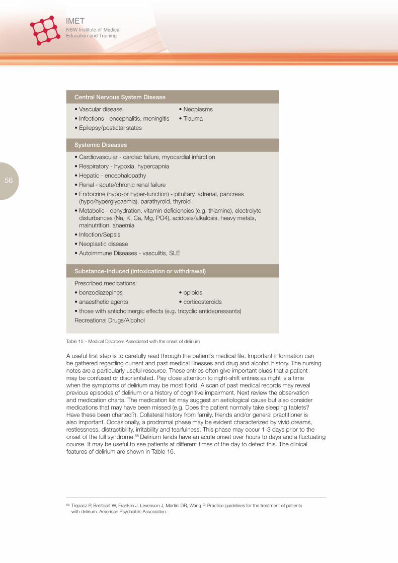
56
Table 15 – Medical Disorders Associated with the onset of delirium
A useful first step is to carefully read through the patient’s medical file. Important information can be gathered regarding current and past medical illnesses and drug and alcohol history. The nursing notes are a particularly useful resource. These entries often give important clues that a patient may be confused or disorientated. Pay close attention to night-shift entries as night is a time when the symptoms of delirium may be most florid. A scan of past medical records may reveal previous episodes of delirium or a history of cognitive impairment. Next review the observation and medication charts. The medication list may suggest an aetiological cause but also consider medications that may have been missed (e.g. Does the patient normally take sleeping tablets? Have these been charted?). Collateral history from family, friends and/or general practitioner is also important. Occasionally, a prodromal phase may be evident characterized by vivid dreams, restlessness, distractibility, irritability and tearfulness. This phase may occur 1-3 days prior to the onset of the full syndrome.68 Delirium tends have an acute onset over hours to days and a fluctuating course. It may be useful to see patients at different times of the day to detect this. The clinical features of delirium are shown in Table 16.
Systemic Diseases
• Cardiovascular - cardiac failure, myocardial infarction
• Respiratory - hypoxia, hypercapnia
• Hepatic - encephalopathy
• Renal - acute/chronic renal failure
• Endocrine (hypo-or hyper-function) - pituitary, adrenal, pancreas (hypo/hyperglycaemia), parathyroid, thyroid
• Metabolic - dehydration, vitamin deficiencies (e.g. thiamine), electrolyte disturbances (Na, K, Ca, Mg, PO4), acidosis/alkalosis, heavy metals, malnutrition, anaemia
• Infection/Sepsis
• Neoplastic disease
• Autoimmune Diseases - vasculitis, SLE
Central Nervous System Disease
• Vascular disease • Neoplasms
• Infections - encephalitis, meningitis • Trauma
• Epilepsy/postictal states
Substance-Induced (intoxication or withdrawal)
Prescribed medications:
• benzodiazepines • opioids
• anaesthetic agents • corticosteroids
• those with anticholinergic effects (e.g. tricyclic antidepressants)
Recreational Drugs/Alcohol
68. Trepacz P, Breitbart W, Franklin J, Levenson J, Martini DR, Wang P. Practice guidelines for the treatment of patients with delirium. American Psychiatric Association.

57
IMET ACUTE PSYCHIATRIC MANAGEMENT
(1) Consciousness: clouding (reduced awareness of surroundings)
(2) Cognition: generalised impairment that affects orientation (time>place>person), attention (poorly performed digit span), memory (recent memory most affected), planning and organisational skills
(3) Behaviour: disturbance of sleep-wake cycle, agitation or psychomotor retardation
(4) Mood/Affect: dysphoria, irritability, anxiety, euphoria, apathy
(5) Perceptual disturbance: misinterpretations, illusions or hallucinations (visual> auditory) often with secondary delusional beliefs (delusions often persecutory in nature and poorly systematised)
(6) Clinical features associated with the underlying aetiology: e.g. jaundice, hepatic fetor and asterixis from hepatic encephalopathy
Table 16 – Clinical Features of Delirium
Some authors divide delirium into two groups – hypoactive and hyperactive:69 (1, 2, 3)
Hypoactive Delirium – decreased activity, decreased alertness, somnolence, lethargy, apathy, EEG slowing, e.g. hepatic encephalopathy.
Hyperactive Delirium – increased activity and alertness, hypervigilance, fast or loud speech, irritability, wandering, EEG may be normal, possibly more likely to have hallucinations and delusions, e.g. alcohol withdrawal, anticholinergic toxicity .
There are rating scales available for screening (e.g. Clinical Assessment of Confusion-A70), diagnosis (e.g. Confusion Assessment Method71) and severity (e.g. Delirium Rating Scale72).
Differential Diagnoses of Delirium
Dementia – will have a more chronic, insidious course; fluctuations in cognition are less marked; attention less affected; consciousness is clear until the late stages; psychotic symptoms are less common. However, dementia will make individual patients more susceptible to delirium and delirium may be superimposed onto dementia – consider if acute deterioration and fluctuating symptoms. Collateral history may be needed to clarify the patient’s baseline cognition. Consider Lewy Body Dementia which may be associated with a fluctuating cognition and visual hallucinations.
Depression – hypoactive delirium may be misdiagnosed as depression. Depression will be associated with diurnal variation, a more subacute onset but with no clouding of consciousness or disorientation.
Manic episode – the agitated and irritable delirious patient may appear manic. In mania there will be more goal directed behaviour and less cognitive impairment.
Psychotic illness – hallucinations and delusions more constant and systematized than in delirium, and there is usually no clouding of consciousness or disorientation.
Substance intoxication or withdrawal – the patient who is affected by a substance may be somnolent or agitated but will not be considered delirious unless the cognitive impairment is out of proportion to what would be normally expected from the implicated substance.
69. a. Lipowski ZJ. Delirium (acute confusional states). JAMA 1987; 258:1789-1792 b. Ross CA, Peyser CE, Shapiro I. Delirium: phenomenologic and etiologic subtypes. International Psychogeriatrics
1991; 3:135-147 c. Liptzin B, Levkoff SE. An empirical study of delirium subtypes. British Journal of Psychiatry 1992 161:843-84570. Vermeersch PE. The clinical assessment of Confusion – A. Applied Nursing Research 1990; 3: 128-13371. Inouye SK, van Dyck CH, Alessi CA, Balkin S, Siegal AP, Horwitz RI. Clarifying confusion: the confusion assessment method,
a new method for the detection of delirium. Annals of Internal Medicine 1990; 113: 941-94872. Trezepacz P, Baker R, Greenhouse J. A symptom rating scale for delirium. Psychiatry Research 1988; 23: 89-97
3Management Challenges
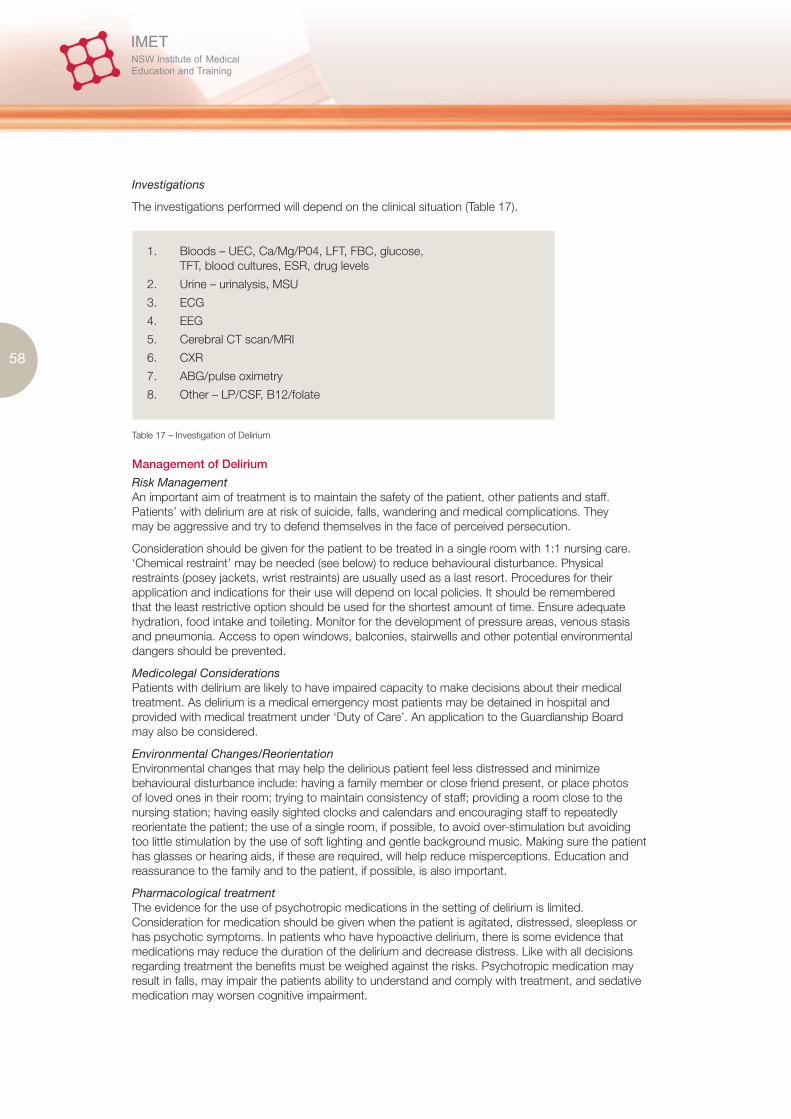
58
Investigations
The investigations performed will depend on the clinical situation (Table 17).
1. Bloods – UEC, Ca/Mg/P04, LFT, FBC, glucose, TFT, blood cultures, ESR, drug levels
2. Urine – urinalysis, MSU
3. ECG
4. EEG
5. Cerebral CT scan/MRI
6. CXR
7. ABG/pulse oximetry
8. Other – LP/CSF, B12/folate
Table 17 – Investigation of Delirium
Management of Delirium
Risk ManagementAn important aim of treatment is to maintain the safety of the patient, other patients and staff. Patients’ with delirium are at risk of suicide, falls, wandering and medical complications. They may be aggressive and try to defend themselves in the face of perceived persecution.
Consideration should be given for the patient to be treated in a single room with 1:1 nursing care. ‘Chemical restraint’ may be needed (see below) to reduce behavioural disturbance. Physical restraints (posey jackets, wrist restraints) are usually used as a last resort. Procedures for their application and indications for their use will depend on local policies. It should be remembered that the least restrictive option should be used for the shortest amount of time. Ensure adequate hydration, food intake and toileting. Monitor for the development of pressure areas, venous stasis and pneumonia. Access to open windows, balconies, stairwells and other potential environmental dangers should be prevented.
Medicolegal ConsiderationsPatients with delirium are likely to have impaired capacity to make decisions about their medical treatment. As delirium is a medical emergency most patients may be detained in hospital and provided with medical treatment under ‘Duty of Care’. An application to the Guardianship Board may also be considered.
Environmental Changes/ReorientationEnvironmental changes that may help the delirious patient feel less distressed and minimize behavioural disturbance include: having a family member or close friend present, or place photos of loved ones in their room; trying to maintain consistency of staff; providing a room close to the nursing station; having easily sighted clocks and calendars and encouraging staff to repeatedly reorientate the patient; the use of a single room, if possible, to avoid over-stimulation but avoiding too little stimulation by the use of soft lighting and gentle background music. Making sure the patient has glasses or hearing aids, if these are required, will help reduce misperceptions. Education and reassurance to the family and to the patient, if possible, is also important.
Pharmacological treatmentThe evidence for the use of psychotropic medications in the setting of delirium is limited. Consideration for medication should be given when the patient is agitated, distressed, sleepless or has psychotic symptoms. In patients who have hypoactive delirium, there is some evidence that medications may reduce the duration of the delirium and decrease distress. Like with all decisions regarding treatment the benefits must be weighed against the risks. Psychotropic medication may result in falls, may impair the patients ability to understand and comply with treatment, and sedative medication may worsen cognitive impairment.

59
IMET ACUTE PSYCHIATRIC MANAGEMENT
73. Kerr IB, Taylor D. Acute disturbed or violent behaviour: principles of treatment. Journal of Psychopharmacology 1997; 11: 271-279
74. Schwartz TL, Masand PS. The Role of Atypical Antipsychotics in the Treatment of Delirium. Psychosomatics 2002 43:171-174
75. Sirois F. Delirium: 100 cases. Canadian Journal of Psychiatry 1988; 33: 375-37876. Koponen H, Stenback U, Mattila E et al. Delirium among elderly persons admitted to a psychiatric hospital: Clinical
course during the acute stage and one-year follow-up. Acta Psychiatric Scandinavia 79: 579-58577. Meagher DJ. Delirium: optimising management. BMJ 2001 vol322 20; p144-14878. Liptzin B. Delirium. In J Sadavoy, LW Lazarus, LF Jarvik et al (Eds). Comprehensive Review of Geriatric Psychiatry
(second edition). Washington DC: American Psychiatric Press 199679. Cole G. Delirium in Elderly Patients. American Journal of Getiatric Psychiatry 2004 12:7-21
The most studied medication for the treatment of delirium is haloperidol. Haloperidol may be used in doses from 0.5mg to 10mg and has the benefit of oral and parental administration. The dose and frequency of administration will depend on the clinical situation. It has few anticholinergic or hypotensive effects. It may result in a prolonged QT interval on ECG and, therefore, potentially result in an increased risk of Torsades de Pointes. The patient must also be monitored for extrapyramidal side effects but, in the setting of delirium treated with haloperidol, the incidence of these effects is low.73 Olanzapine (up to 20mg/day), risperidone (up to 4mg/day), quetiapine (~200mg/day) and ziprasidone have also been used in the treatment of delirium. Olanzapine may be better tolerated than haloperidol and can be given intramuscularly. The evidence for these medications is limited. It has been suggested that antipsychotic medication be continued for a further 7-10 days following resolution of symptoms.74 Benzodiazepines may worsen confusion or cause disinhibition in the elderly, or in patients with pre-existing organic brain syndromes, and should be avoided in most cases. The exceptions to this rule are the setting of alcohol or benzodiazepine withdrawal and postictal delirium.
Identify and Treat the Underlying Cause(s)This will depend upon the clinical situation.
Prognosis of Delirium
There may be a rapid improvement in symptoms once the underlying cause has been found and adequately treated. Delirium often subsides over 10-12 days75 (1) but maybe more prolonged in the elderly (up to 81 days).76
Delirium has been associated with:77,78
• prolonged hospitalization
• high frequency of complications e.g. falls, infections, pressure sores
• increased need for care in institutions
• increased risk of death.
The mortality rate for patients who have had delirium is between 14-36% at 6 months.79
3Management Challenges

60
Neuroleptic Malignant SyndromeIntroduction
The neuroleptic malignant syndrome (NMS) is a rare but potentially lethal complication of antipsychotic treatment. NMS is estimated to have an incidence of between 0.02 to 3.23% of psychiatric inpatients receiving antipsychotic medication.80 NMS is twice as common in men and all D2 blocking agents, including antiemetics such as metoclopramide and prochlorperazine are implicated. It has been described with use of all classes of antidepressants, and with abrupt discontinuation of anti-parkinsonian medication. There have also been reports of NMS occurring spontaneously in neurological conditions and schizophrenic illnesses. The risks for NMS are shown in Table 18.
1. High doses of neuroleptics
2. Rapid escalation of dose
3. Dehydration
4. Past History of NMS
5. Affective disorder
6. Organic brain syndrome
Table 18 – Risk factors for development of NMS
The pathophysiology is largely unknown, although sudden disruption of dopaminergic activity in the striatum and hypothalamus appear to produce diffuse muscle rigidity leading to raised core body temperature, rhabdomyolsis and accompanying disturbances in physiology.81
Clinical Features of NMS
The clinical features of NMS are shown in Table 19. In essence, any patient taking antipsychotic medication who exhibits fever, rigidity or confusion should have NMS excluded.
1. Hyperthermia
2. Rigidity or other extrapyramidal symptoms
3. Autonomic dysregulation
4. Tachypnoea
5. Confusion or florid delirium
6. Rhabdomyolysis and myoglobinurea
7. Leukocytosis
8. Marked elevation of serum creatine kinase
Table 19 – Features of NMS
LEARNING OBJECTIVES
• Describe the clinical features of the neuroleptic malignant syndrome• Outline the process of diagnosis of the neuroleptic malignant syndrome• Describe the treatment of the neuroleptic malignant syndrome
80. Lazarus A, Mann SC, Caroff SN. The Neuroleptic Malignant Syndrome and Related Conditions. Washington, DC, American Psychiatric Press, 1989
81. Stoudemire A, Luther JS. Neuroleptic malignant syndrome and neuroleptic-induced catatonia: differential diagnosis and treatment. International Journal of Psychiatry in Medicine 1984 14:57–63

61
IMET ACUTE PSYCHIATRIC MANAGEMENT
82. Shalev A, Hermesh H, Munitz H. Mortality from neuroleptic malignant syndrome. Journal of Clinical Psychiatry 1989 50:18–2583. Lee S, Merriam A, Kim TS et al. Cerebellar degeneration in neuroleptic malignant syndrome: neuropathologic findings and
review of the literature concerning heat-related nervous system injury. Journal of Neurology, Neurosurgery, and Psychiatry 1989 52:387–391
Laboratory Investigations
The critical investigation is the serum levels MM fraction of creatine phosphokinase (CPK). Levels are often markedly elevated. CPK levels may be altered by IM injection and physical trauma during an episode of psychosis. In addition to raised CPK, the patient’s white cell count is often significantly raised. Given dehydration and sepsis may be comorbidly present, serum electrolytes and screening for infection is often required. Myoglobin may be present in the urine, warranting closer monitoring of renal function and fluid balance.
Differential Diagnosis of NMS
The differential diagnosis of NMS is shown in Table 20.
1. Serotonin Syndrome
2. Malignant Hyperthermia
3. Lethal Catatonia
4. Sepsis
5. Anticholinergic agent intoxication
6. Overdose of CNS stimulants including MDMA, Amphetamine, Cocaine
7. Delirium of other cause
Table 20 – Differential Diagnosis of NMS
Malignant hyperthermia is a hypermetabolic state of skeletal muscle most frequently associated with the administration of halogenated inhalation anesthetic agents and succinylcholine. It is heritable, and whilst originally thought to be transmitted via an autosomal dominant trait, it is now considered to have a multifactorial pattern of inheritance. The clinical presentation of malignant hyperthermia is identical NMS. The main clinical differentiation is the context of general anaesthesia via halogenated inhaled anaesthetics. The diagnosis of malignant hyperthermia (or of the latent trait) is reliably established by exposing biopsied muscle tissue to caffeine or halothane in vitro, which results in a hypercontractile response when compared with normal muscle. Treatment of malignant hyperthermia is through the use of dantrolene sodium. Muscle tissue from patients with neuroleptic malignant syndrome does not demonstrate a hypercontractile response to caffeine or halothane. Family histories of patients with malignant hyperthermia have not been documented in patients with neuroleptic malignant syndrome, and the conditions do not seem to be related. Lethal catatonia is a syndrome in which mutism, extreme motor excitement, clouding of consciousness, and fever may progress to severe autonomic disturbances, stupor and coma, and death. It may be a condition related to NMS.
Clinical Course
NMS is usually present for 2-14 days. Mortality from the condition is reducing with improved critical care. Prior to 1984 the mortality rate for NMS was 25%. Since 1984 it is 11.6%.82 NMS may be complicated by contractures, renal failure, and hypostatic complications such as deep venous thrombosis or pneumonia. There is evidence that prolonged hyperthermia may lead to cerebellar damage.83
3Management Challenges

62
84. Wells AJ, Sommi RW, Crismon ML. Neuroleptic rechallenge after neuroleptic malignant syndrome: case report and literature review. Drug Intelligence and Clinical Pharmacy 1988 22:475–480
Treatment of NMS
Treatment of NMS is based upon prompt recognition of the syndrome, the cessation of antipsychotic treatments agent and lithium (if co-aministered). Supportive care is usually provided in critical care setting, with monitoring of fluid balance, renal function and the use of antipyretic agents. ECT has proven efficacy in the treatment of NMS and may be used in severe or refractory cases, particularly where NMS may be related to the use of long acting injectable antipsychotic medication. Anitcholinergic agents, intravenous dantrolene and dopamine agonists such as bromocriptine, amantadine, and apomorphine have anecdotal benefits.If clinically indicated, patients who suffer NMS may be rechallenged with antipsychotic medication, ideally of a different class. The likelihood of recurrence of NMS is substantially lower if the antipsychotic rechallenge is >5 days.84 Patients should only be rechallenged with antipsychotic medications under close medical supervision with regular monitoring of vital signs, serum CPK and frequent neurological investigation.
The Serotonin SyndromeIntroduction
The Serotonin Syndrome is the result of overstimulation of 5-HT1A and 5-HT2 receptors in central grey nuclei and the medulla. The syndrome is potentially fatal and has increased in significance since the introduction of newer antidepressant and antipsychotic drugs which affect the serotonin neurotransmitter systems.
Pathogenesis of the Serotonin Syndrome
The circumstances in which the serotonin syndrome emerges are shown in Table 21.
Table 21 – Pathogenesis of the Serotonin Syndrome
LEARNING OBJECTIVES
• Describe the predictors of physical aggression in psychiatric patients• Outline the pharmacological management of physical aggression in
psychiatric patients • Outline the non-pharmacological management of physical aggression
in psychiatric patients
Mechanism
• Excess of precursors of serotonin or its agonists
• Increased release of serotonin
• Reduced reuptake of serotonin
• Slowing down of serotonin Metabolism
• Ectopic production of serotonin
Agents
Buspirone, L-dopa, lithium, LSD, Ltryptophan,Trazodone
Amphetamines, cocaine, MDMA (“ecstasy”), fenfluramine, reserpine
SSRI, TCA, trazodone, venlafaxine, meperidine
MAOI, e.g., isocarboxazid, selegiline
Carcinoid syndrome

63
IMET ACUTE PSYCHIATRIC MANAGEMENT
85. Radomski JW, Dursun SM, Revely MA, Kutcher SP. An exploratory approach to the Serotonin Syndrome; an update of clinical phenomenology and revised diagnostic criteria. Medical Hypotheses 2000;55:218-24
Diagnosis of the Serotonin Syndrome
The diagnostic criteria of the serotonin syndrome are shown in Table 22.85
Table 22 – Clinical features of the Serotonin Syndrome
Differential Diagnosis of Serotonin Syndrome
The differential diagnosis of the serotonin syndrome is shown in Table 23.
• Neuroleptic malignant syndrome
• CNS or systemic Infectious causes
• Other toxic encephalopathy
• Heat stroke
• Delirium tremens
• Anticholinergic delirium
Table 23 – The differential diagnosis of Serotonin Syndrome
Management
There is no definitive management of NMS other than supportive measures.
Outcome
60% of cases resolve within 24 hours. The length of the syndrome is usually dependant upon the t½ of the psychotropic drug involved.
Mental (cognitive and behavioural) symptoms
- Major symptoms: confusion, elevated mood, coma or semicoma
- Minor symptoms: agitation and nervousness, insomnia
Addition of a serotonergic agent to an already established treatment (or increase in dosage) and manifestation of at least 4 major symptoms or 3 major symptoms plus 2 minor ones:
Autonomic symptoms
- Major symptoms: fever, hyperhidrosis
- Minor symptoms: tachycardia, tachypnea and dyspnea,
diarrhea, low or high blood pressure
Neurological symptoms
- Major symptoms: myoclonus, tremors, chills, rigidity, hyperreflexia
- Minor symptoms: impaired co-ordination, mydriasis, akathisia
3Management Challenges

NOTES
64

Complications
654
66
Metabolic complications of antipsychotic treatmentSchizophrenia and Metabolic Complications
Up to 51% of males and 64% of females suffering from schizophrenia are obese (defined as BMI > 90th percentile). This is in contrast to 33% of individuals with other psychiatric diagnoses.86 Ischaemic Heart Disease (IHD) is a greater cause of mortality in psychiatric patients than suicide. Moreover, the IHD mortality rate amongst people suffering schizophrenia has not trended down as it has in the general population.87 People with mental illness had a higher prevalence of cardiovascular risk factors such as smoking, obesity, lack of exercise, alcohol consumption and salt intake when compared with control subjects from a community-based sample.88 It is a sad paradox that patients whose psychiatric symptoms respond best are also those most likely to gain weight as a consequence of treatment with antipsychotic medication.89 Being female, younger, and with a lower pre-treatment BMI and non-Anglo-Celtic ethnicity appear to elevate risk.90
Clozapine and olanzapine are associated with the greatest risk of clinically significant weight gain, with other agents producing relatively lower levels of risk. Risperidone, quetiapine, amisulpride and zotepine generally show low to moderate levels of mean weight gain and a modest risk of clinically significant increases in weight. Ziprasidone and aripiprazole treatment are generally associated with minimal mean weight gain.91 One study found 32% of olanzapine-treated patients possessed the ‘atherogenic’ metabolic triad comprising hyperinsulinaemia, increased apolipoprotein B concentration, and small, dense LDL, compared with a figure of 5% in risperidone-treated patients.92
4Complications
LEARNING OBJECTIVES
• Identify the metabolic complications of antipsychotic treatment• Describe the prevalence and features of the metabolic syndrome
in psychiatric populations• Outline the principles of management of the metabolic syndrome
in psychiatric populations
86. Dinan TG (ed.). Schizophrenia and diabetes 2003: an expert consensus meeting. British Journal of Psychiatry 2004; 184:(suppl 7):S53-S114
87. Lawrence DM, Holman CDJ, Jablensky AV et al. Death rate from ischaemic heart disease in Western Australian psychiatric patients 1980-1998. British Journal of Psychiatry 2003;182:31-36
88. Davidson S, Judd F, Jolley D et al. Cardiovascular risk factors for people with mental illness. Australia and New Zealand Journal of Psychiatry 2001;35:196-202
89. Czobor P, Volavka J, Sheitman B et al. Antipsychotic – induced weight gain and therapeutic response: a differential association. Journal of Clinical Psychopharmacology 2002;22:244-251
90. McIntyre RS, Mancini DA, Basile VS et al. Antipsychotic-induced weight gain: bipolar disorder and leptin. Journal of Clinical Psychopharmacology 2003;23:323-327
91. Newcomer JW. Second-generation (atypical) antipsychotics and metabolic effects: a comprehensive literature review. CNS Drugs 2005 19 Suppl 1: 1-93
92. Bouchard RH, Demers M, Simoneau I et al. Atypical antipsychotics and Cardiovascular Risk in Schizophrenic patients. Journal of Clinical Psychopharmacology 2001;21:110-111

67
IMET ACUTE PSYCHIATRIC MANAGEMENT
93. McEvoy JP, Meyer JM, Goff DC, Nasrallah HA, Davis SM, Sullivan L, Meltzer HY, Hsiao J, Scott Stroup T, Lieberman JA. Prevalence of the metabolic syndrome in patients with schizophrenia: baseline results from the Clinical Antipsychotic Trials of Intervention Effectiveness (CATIE) schizophrenia trial and comparison with national estimates from NHANES III. Schizophrenia Research 2005; 80:19–32
94. Filakovic P, Koic O et al. Second generation antipsychotics and risk of diabetes type II – comparison between olanzapine and risperidone. Coll Antropol 2007 31(4): 1105-1109
95. Hampson LJ, Mackin P et al. Stimulation of glycogen synthesis and inactivation of phosphorylase in hepatocytes by serotonergic mechanisms, and counter-regulation by atypical antipsychotic drugs. Diabetologia 2007 50(8): 1743-1751
96. Schwenkreis P, Assion HJ. Atypical antipsychotics and diabetes mellitus. World Journal of Biological Psychiatry 2004 5(2): 73-82
97. Hosojima H, Togo T et al. Early effects of olanzapine on serum levels of ghrelin, adiponectin and leptin in patients with schizophrenia. Journal of Psychopharmacology 2006 20(1): 75-79
98. Richards AA, Hickman IJ et al. Olanzapine treatment is associated with reduced high molecular weight adiponectin in serum: a potential mechanism for olanzapine-induced insulin resistance in patients with schizophrenia. Journal of Clinical Psychopharmacology 2006 26(3): 232-237
99. National Cholesterol Education Program (NCEP) Expert Panel on Detection, Evaluation, and Treatment of High Blood Cholesterol in Adults (Adult Treatment Panel III). Third Report of the National Cholesterol Education Program (NCEP) Expert Panel on Detection, Evaluation, and Treatment of High Blood Cholesterol in Adults (final report). Circulation 2002;106:3143-3421
Data from the National Institute of Mental Health-sponsored Clinical Antipsychotic Trials of Intervention Effectiveness (CATIE)93 found a prevalence of metabolic syndrome of 42.7%. The mean BMI of the subjects was 29.7 (SD=7.0). Among fasting subjects, 44.4% met criteria for the metabolic syndrome. The risk of metabolic syndrome in people suffering schizophrenia is 2-3 times that of the general population.
Putative mechanisms of metabolic consequences of antipsychotic treatment
There are a variety of theoretical models of the mechanism of weight gain related to antipsychotic therapy. Apart from the effects of illness on lifestyle there is the possibility of an intrinsic propensity to weight gain in schizophrenia. Stimulation of Histamine 1 receptors may trigger hunger or impair satiety in people taking medications such as clozapine or olanzapine. Insulin resistance and a propensity to glucose intolerance can occur as a consequence of multiple intercurrent effects of the antipsychotics including the effects of increased body mass and direct interference by of the antipsychotics in the glucose metabolism,94 in particular interference with hepatic glycogen synthesis through alteration of hepatocyte 5HT receptors.95 The induction of peripheral insulin resistance and the direct influence on pancreatic beta-cell function by 5-HT1A/2A/2C receptor antagonism, or by inhibitory effects via alpha 2-adrenergic receptor is a postulated mechanism.96 There has been recent interest in, three recently identified cytokines which play crucial roles in the regulation of energy balance and glucose metabolism – ghrelin, adiponectin and leptinm.97 Adipocyte expression or secretion of adiponectin an insulin-sensitizing cytokine is affected by olanzapine.98
The Metabolic Syndrome
The criteria proposed by the National Cholesterol Education Program (NCEP) Adult Treatment Panel III (ATP III), with minor modifications, are currently recommended and widely used.99 The American Heart Association and the National Heart, Lung, and Blood Institute recommend that the metabolic syndrome be identified as the presence of three or more of these components. The metabolic syndrome is characterized by a group of metabolic risk factors in one person (Table 24).
4Complications
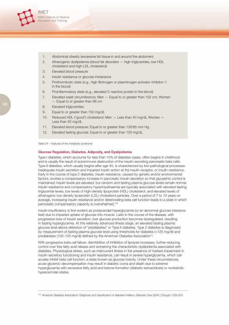
68
1. Abdominal obesity (excessive fat tissue in and around the abdomen)
2. Atherogenic dyslipidemia (blood fat disorders — high triglycerides, low HDL cholesterol and high LDL cholesterol)
3. Elevated blood pressure
4. Insulin resistance or glucose intolerance
5. Prothrombotic state (e.g., high fibrinogen or plasminogen activator inhibitor–1 in the blood)
6. Proinflammatory state (e.g., elevated C-reactive protein in the blood)
7. Elevated waist circumference: Men — Equal to or greater than 102 cm; Women — Equal to or greater than 88 cm
8. Elevated triglycerides:
9. Equal to or greater than 150 mg/dL
10. Reduced HDL (“good”) cholesterol: Men — Less than 40 mg/dL Women — Less than 50 mg/dL
11. Elevated blood pressure: Equal to or greater than 130/85 mm Hg
12. Elevated fasting glucose: Equal to or greater than 100 mg/dL
Table 24 – Features of the metabolic syndrome
Glucose Regulation, Diabetes, Adiposity, and Dyslipidemia
Type-I diabetes, which accounts for less than 10% of diabetes cases, often begins in childhood and is usually the result of autoimmune destruction of the insulin-secreting pancreatic beta cells. Type-II diabetes, which usually begins after age 45, is characterized by two pathological processes: inadequate insulin secretion and impaired insulin action at the insulin receptor, or insulin resistance. Early in the course of type 2 diabetes, insulin resistance, caused by genetic and/or environmental factors, evokes a compensatory increase in pancreatic insulin secretion so that glycaemic control is maintained; insulin levels are elevated, but random and fasting plasma glucose levels remain normal. Insulin resistance and compensatory hyperinsulinaemia are typically associated with elevated fasting triglyceride levels, low levels of high-density lipoprotein (HDL) cholesterol, and elevated levels of atherogenic low-density lipoprotein (LDL) cholesterol particles. Over a period of 7 to 10 years on average, increasing insulin resistance and/or deteriorating beta cell function leads to a state in which pancreatic compensatory capacity is overwhelmed.100
Insulin insufficiency is first evident as postprandial hyperglycemia (or an abnormal glucose tolerance test) due to impaired uptake of glucose into muscle. Later in the course of the disease, with progressive loss of insulin secretion, liver glucose production becomes dysregulated, resulting in fasting hyperglycemia. At this relatively advanced illness stage, an elevated fasting plasma glucose level allows detection of “prediabetes” or Type II diabetes. Type 2 diabetes is diagnosed by measurement of fasting plasma glucose level using thresholds for diabetes (>125 mg/dl) and prediabetes (100–125 mg/dl) defined by the American Diabetes Association15.
With progressive beta cell failure, disinhibition of inhibition of lipolysis increases, further reducing control over free fatty acid release and worsening the characteristic dyslipidemia associated with diabetes. Physiological stress, such as intercurrent illness in the presence of marked impairment in insulin secretory functioning and insulin resistance, can result in severe hyperglycemia, which can acutely inhibit beta cell function, a state known as glucose toxicity. Under these circumstances, acute glycemic decompensation may result in diabetic coma and death due to extreme hyperglycemia with excessive fatty acid and ketone formation (diabetic ketoacidosis) or nonketotic hyperosmolar states.
100. American Diabetes Association: Diagnosis and classification of diabetes mellitus. Diabetes Care 2004; 27(suppl 1):S5-S10

69
IMET ACUTE PSYCHIATRIC MANAGEMENT
101. Reaven G. The metabolic syndrome: is this diagnosis necessary? American Journal of Clinical Nutrition 2006; 83:1248-1251102. Expert Panel on Detection, Evaluation, and Treatment of High Blood Cholesterol in Adults: Executive Summary of the Third
Report of the National Cholesterol Education Program (NCEP) Expert Panel on Detection, Evaluation, and Treatment of High Blood Cholesterol in Adults (Adult Treatment Panel III). JAMA 2001; 285:2486–2497
Insulin resistance and type II diabetes occur most often in the context of overweight and obese patients, particularly those with excess abdominal adiposity. Adiposity and fitness are each thought to contribute about 30% of the inter-individual variance in insulin resistance, with genetic factors accounting for the remainder.101 Thus, while excessive abdominal adiposity is significantly related to risk of insulin resistance and diabetes, Type II diabetes can also occur in the absence of overweight or obesity.
Management of Metabolic Syndrome
The Adult Treatment Panel III treatment guidelines102 recommend therapeutic life style changes, including reduced intake of saturated fats and cholesterol, increased fiber intake, weight reduction, and increased physical activity as the first-line therapeutic approach to the risk of cardiovascular disease. LDL-lowering drugs, including HMG-CoA reductase inhibitors (statins), bile acid sequestrants, nicotinic acid, and fibric acids, are prescribed as needed to achieve target LDL levels.
The metabolic syndrome increases the risk of cardiovascular disease at any given level of LDL and is considered a secondary target of risk-reduction therapy after lowering LDL cholesterol. The Adult Treatment Panel III guidelines identify obesity as the primary target of treatment of the metabolic syndrome and weight loss and increased physical activity as the first-line treatment approaches. Weight loss lowers LDL cholesterol and triglycerides, increases HDL cholesterol, lowers blood pressure, and reduces insulin resistance. Metformin reduces insulin resistance, reduces new-onset coronary heart disease in obese patients with diabetes, and prevents or delays Type-II diabetes in patients with impaired glucose tolerance. Insulin sensitizers of the thiazolidinedione class also prevent or delay type 2 diabetes in at-risk patients. antipsychotics and meet criteria for the metabolic syndrome should be treated with therapeutic life style changes and medications in accordance with hypertension guidelines. Finally, low-dose aspirin may be indicated to mitigate the prothrombotic state in patients with metabolic syndrome at elevated risk of coronary heart disease.
QTc abnormalities and psychotropic treatmentIntroduction
The QT interval is an ECG measure that includes both depolarization and repolarization. It begins with the onset of ventricular depolarization (Q wave) and ends with completion of repolarization (T wave). Because the QT interval shortens with increasing heart rates, it is usually corrected for heart rate (QTc). QTc intervals are usually around 400 msec in duration, and values lower than 440 are considered normal (Fig 15). A QTc >500 msec has frequently been used as a cutoff because longer QTc interval measures are associated with substantially higher risk of cardiac arrhythmias.
LEARNING OBJECTIVES
• Understand the importance of abnormalities of QTc interval• List psychotropic treatments which can alter the QTc interval• Describe the risk of sudden death in patients with severe mental illness
4Complications

70Fig 15. The normal ECG complex from the P-T waves
A brief review of the physiology of cardiac conduction
Depolarization of ventricular myocardial cells is the result of a rapid influx of Na+ ions through selective Na+ channels. Repolarization occurs via cationic inflows through Ca++, Na+, and several K+ channels, including the hERG gene coded IKr potassium channel (a complex of 4 identical subunits each containing 6 transmembrane domains, numbered S1-S6, a pore helix and a slide helix). This process is reflected on the ECG by the QRS interval. The hERG/IKr channel is the most critical in drug-induced QTc prolongation syndromes. Drugs which blocking the IKr channel prolong the QTc interval and can induce life threatening cardiac arrhythmmias, the most significant of which is ‘torsade de pointes’. Sudden death in apparently healthy adults can occur as a result of drug induced QTc prolongation.103 Prolonged QTc intervals are associated with the risk of sudden death after myocardial infarction and the “long QT syndrome”. QTc interval prolongation is a flag that warns of the possibility of torsade de pointes and sudden death.
Torsade de Pointes
Torsade de pointes meaning “twisting around the point” (a reference to a ballet movement) refers to an uncommon variant of ventricular tachycardia (VT) in which the QRS complexes twist about the isoelectric axis of the ECG. The morphology of the QRS complexes varies from beat to beat. The ventricular rate can range from 150-250 beats per minute (Fig 16).
Figure 16 – Torsade de pointes
The QTc interval is a modest predictor of torsade de pointes. The underlying basis for the arrhythmia is delay in phase III of the myocardial action potential mediated by the IKr potassium channel. This prolonged period of repolarization and the irregularity of repolarization times among different myocardial fibers create re-entrant phenomena or ectopic electrical activity to occur, producing the arrhythmia. As the underlying etiology and management of torsade are, in general, quite different from VT, the management of torsade with group IA antiarrhythmic drugs can be lethal.
There are familial predispositons to torsade de points, mediated by 6 genetic variants of the hERG/IKr channel, producing the “long QT syndrome”.
103. Witchel HJ, Hancox JC. Familial and acquired long QT syndrome and the cardiac rapid delayed rectifier potassium current. Clinical and Experimental Pharmacology and Physiology 2000; 27:753-776

71
IMET ACUTE PSYCHIATRIC MANAGEMENT
104. Lipscomb PA. Cardiovascular side effects of phenothiazines and tricyclic antidepressants: a review. Postgraduate Medicine 1980; 67:189-92
Psychotropic Medications and abnormalities of the QTc
Not all drugs that prolong the QTc interval produce torsade de pointes and sudden death. Tricyclic antidepressants, for example, have a ‘quinidine like effect’104 and block the Na+ inflow channel, and, as a result, slow depolarization and widen both the QRS and the QTc intervals. Thioridazine blocks hERG channel, is more often associated with sudden death in otherwise healthy individuals through cases of torsade de pointes. Psychotropic medications associated with prolonged QTc and torsade de points are shown in Table 25.
Table 25 – Antipsychotic Drugs and the propensity for torsade de pointes
Other medication classes including antiarrhythmics, tricyclic antidepressants, antihistamines, and antibiotics are associated with QT prolongation syndromes and must be used with care in combination with antipsychotic medications implicated in QT prolongation.
Assessment for Risk of QTc Prolongation and torsade
The risk factors for prolonged QTc and torsade de pointes and suggested screening procedures (in addition to identification of risk factors in the clinical history) are shown in Figure 17. Assessment of a patient’s vulnerability to develop potentially fatal QT prolongation syndromes should include identification of these risk factors.
Prolonged QTc and torsade de pointes
• Thioridazine
• Ziprasidone
• Pimozide
• Amisulpiride
• Droperidol
• Sertindole
Prolonged QTc and possible torsade de pointes*
• Olanzapine
• Quetiapine
• Risperidone
• Chlorpromazine
• Clozapine
• Haloperidol
*high dose or drug interactions
4Complications

72
Previous QTabnormality(12-lead ECG)
Anorexia Nervosa(BMI)
Low serum K+, Mg++ or Ca++
(Serum Electrolytes)
Hypothyroidism(Thyroid function)
Alcohol Abuse(LFTs, MCV, CDT)
105. Ray W, Meredith S, Purushottam B et al. Antipsychotics and the Risk of Sudden Cardiac Death. Archives of General Psychiatry. 2001;58:1161-1167
106. Davidson M. Risk of cardiovascular disease and sudden death in schizophrenia. Journal of Clinical Psychiatry 2002; 63: 5-1107. Barnes TR, Braude WM. Akathisia variants and tardive dyskinesia. Archives of General Psychiatry 1985; 42:874-878108. Sachdev P. Research diagnostic criteria for drug-induced akathisia: conceptualization, rationale, and proposal.
Psychopharmacology 1994; 114:181-186
Figure 17 – Assessment of risk for drug induced QT prolongation and torsade
Sudden Death in Mentally Ill Populations
There is considerable evidence in the scientific literature that people suffering schizophrenia are at higher risk for sudden death. This is both related to antipsychotic treatment,105 and independent of this variable.106 Cardiovascular risk factors are covered in a separate section, however it is important to note that chronic mental illness is associated with the lifestyle risk factors for ischaemic heart disease. It is also evident that people with chronic mental illnesses have, for a variety of reasons, less access to early intervention in physical disease.
Akathisia and tardive dyskinesiaAkathisia
“Akathisia” is a drug-induced movement disorder, which presents as a syndrome of motor restlessness, usually in the lower extremities, often accompanied by a subjective sense of inner restlessness, and dysphoria.107 The term derives from the Greek , meaning “without sitting”. Akathisia exists in acute, chronic (duration >3 months) and tardive forms (with onset >3 months). A form of withdrawal akathisia may occur as a consequence of a reduction in the dose of antipsychotic medication.108 A syndrome resembling akathisia is seen following the initiation of treatment with aripiprazole, although this is an “activation” syndrome arising from partial D2 receptor agonism in the striatum.
LEARNING OBJECTIVES
• Describe the features of akathisia• Describe the treatment of akathisia• Understand the risks and features of tardive dyskinesia

73
IMET ACUTE PSYCHIATRIC MANAGEMENT
109. Sachdev P. Akathisia and Restless Legs. New York, Cambridge University Press, 1995110. Sachdev P. The epidemiology of drug-induced akathisia. II: chronic, tardive, and withdrawal akathisias. Schizophrenia Bulletin
1995:21:451-461111. Lieberman JA, Stroup TS. McEvoy et al Clinical Antipsychotic Trials of Intervention Effectiveness (CATIE) Investigators:
Effectiveness of antipsychotic drugs in patients with chronic schizophrenia. New England Journal of Medicine 2005; 353:1209-1223
112. Sachdev PS, Brune M. Animal models of acute drug-induced akathisia: a review. Neurosci Biobehav Rev 2000; 24:269-277113. Barries TR. A rating scale for drug-induced akathisia. British Journal of Psychiatry 1989; 154:672-676114. Kushida CA. Clinical presentation, diagnosis, and quality of life issues in restless legs syndrome. American Journal of Medicine
2007;120(suppl 1): S4-S12
The incidence of acute akathisia of amongst patients taking antipsychotic medication is 31%109 and the prevalence rate ranges up to 41%.110 In the Clinical Antipsychotic Trials of Intervention Effectiveness (CATIE) study, there were no significant differences between first and second generation antipsychotics in the incidence of extrapyramidal symptoms, although the rate of discontinuation of treatment was significantly higher for perphenazine, the only FGA studied.111 There is no parallel between the concurrent severity of akathisia and other extrapyrimadal side-effects of antipsychotic medication, implying that akathisia is a different phenomenon from the other recognized extrapyramidal side-effects of neuroleptic medication. Akathisia has been described in patients taking SSRI antidepressants. Moreover, the lower propensity or serotonin blocking SGA’s implies a serotonergic mechanism involved in the genesis of akathisia. Cholinergic and adrenergic pathways are also implicated.112
The risk Factors for akathisia are shown in Table 18.
Table 18 – Risk factors for akathisia
Assessment of akathisia
The diagnosis of akathisia is primarily clinical. Observations of motor restlessness in a patient taking antipsychotic or SSRI medication are the commonest clinical sign, although akathisia can present as a primarily subjective sense of restlessness, variably described by the patient as agitation, dysphoria or anxiety. The Barnes Akathisia Rating Scale can also be used to formally assess akathisia.113 Akathisia should be differentiated from psychotic agitation or psychomotor agitation in severe melancholic depression. Anxiety can be mistaken for akathisia. Some features of tardive dyskinesia, particularly truncal or limb movement, can be confounded for akathisia. Other neurological disorders producing choreoathetoid movements can resemble akathisia. There is a possible relationship between akathisia and the “Restless Legs Syndrome”. This condition is characterized by lower body movement similar to those seen in akathisia, although the symptoms usually occur only during rest, relaxation or sleep, and tend to follow a circadian pattern.114
• Rapid rate of increase of dosage
• Use of higher-potency FGA medication
Higher dosages of neuroleptic
medication
• Older age
• Female sex
Demographic
• Prominent negative symptoms
• Iron deficiency
• Cognitive dysfunction
• Affective disorder
Clinical Features
4Complications

74
Treatment of akathisia
Akathisia is a sinister symptom, and strongly correlates with medication noncompliance,115 worsening of psychotic symptoms, impulsive behaviour, increased liability for aggression, deliberate self-harm and completed suicide.116 Reduction of dose of the patient’s neuroleptic medication, or changing the patient’s treatment to a different agent with a lower propensity for such side effects is the first line measures for treatment. If this is not appropriate, benzodiazepines, propranolol, or anticholinergic medications may be of some help in treating acute akathisia (Fig 19).
Figure 19 – Management of akathisia
Tardive Dyskinesia
Tardive Dyskinesia (TD) is a potentially irreversible neurological syndrome characterised by choreo-athetoid movments of the orofacial muscles and, less commonly, truncal and limb muscles. Rare cases of TD involving the diaphragm and pectoral muscles have been described. The prevalence of TD varies from 15-20% in psychiatric patient populations and the risk is cumulative with ongoing exposure to neuroleptic medication. The aetiology of TD is still elusive. The two favoured theories are dopamine supersensitivity in the in the striatum, acquired during chronic neuroleptic exposure and the ‘free radical hypothesis’ in which accumulation of free radicals produce damage to the striatum.
The risk factors for TD are shown in Figure 20.
Figure 20 – Risk Factors for tardive dyskinesia
115. Van Putten T. Why do schizophrenic patients refuse to take their drugs? Archives of General Psychiatry 1975; 31:67-72116. Van Putten T, Mutalipassi LR, Malkin MD. Phenothiazine-induced decompensation. Archives of General Psychiatry 1974;
30:13-19
Patient Variable• Age• Female• Diabetes• Mood Disorder• Cognitive Impairment
Treatment Variable• Length of treatment• Previous EPSE
Risk Factor

75
IMET ACUTE PSYCHIATRIC MANAGEMENT
117. Gupta S, Mosnik D, Black D et al. Tardive Dyskinesia: Review of Treatments Past, Present, and Future. Annals of Clinical Psychiatry (1999) 11: 257-266
There is no definitive treatment for TD, and the aphorism “There are many treatments for TD and there are none” applies. A recent review117 considered various proposed treatments including – antioxidants (especially Vitamin E 1000 I.U. daily), Dopamine depleting agents such as tetrabenazine, medications acting on GABA receptors such as baclofen and the use of SGAs, in particular clozapine. There is no convincing data to support any one treatment modality in TD.
Suggested assessment for drug induced movement disorders
Given the clinical significance of all drug induced movement disorders, regular systematic review for the presence of signs of the various conditions should be routinely performed on all patients receiving neuroleptic medications. There are several rating scales available, such as the Abnormal Involuntary Movement Scale (AIMS), however a clinical algorithm for assessment of drug induced movement disorder is proposed below (Figure 21).
Figure 21 – Approach to the assessment of Drug Induced Movement Disorders
HyperprolactinaemiaIntroduction
Elevation of prolactin levels in patients is a frequent and problematic side-effect of the use of psychotropic medication. Hyperprolactinaemia produces undesirable side effects in the short term, affecting medication tolerability and treatment compliance. Longer-term morbidity chronic iatrogenic hyperprolactinaemia is also of concern.
The Physiology of Prolactin
The normal physiology of prolactin in humans is shown in Figure 22. Prolactin is released form the anterior pituitary gland in a pulsatile manner with up to 15 peaks per 24 hours. The normal physiological range of serum prolactin levels is between 10-25μg/L. Serum prolatin levels exhibit
• Bradykinesia
• Tremors
• Motor restlessness
• Choreo-athetoid movements
• Walking – reduced arm-swing, truncal movements
• Rigidity
• Truncal movements
• Dentures
• Tongue movements
• Mouth movements
• Facial movements
General observation of the
seated patient
General inspection of the patient
Inspection of oro-facial region
LEARNING OBJECTIVES
• Understand the normal physiology of prolactin• Describe the sequelae of raised levels of serum prolactin• Identify the propensity for different psychotropic agents
to raise serum prolactin
4Complications

76
a circadian pattern and usually peak after 4 hours of a sleep cycle. Prolactin has the primary function of lactogenesis via the stimulation of breast tissue and milk synthesis. It suppresses gonadotropin function and likely plays a role in mediating attachment behaviours in humans.
Fig 22. The normal physiology of prolactin in humans
The Effects of Hyperprolactinaemia
Cross sectional studies of patients administered neuroleptic medication indicate the prevalence of hyperprolactinaemia is 34% in men and 75% in women.118 Traditionally, hyperprolactinaemia is considered to be present at levels > 100μ/L, however clinically significant side-effects can emerge at lower levels. Women, particularly in the post-partum period and pubescent children are particularly vulnerable to iatrogenic hyperprolactinaemia. There is some evidence of menstrual irregularities in women with schizophrenia which predate treatment with antipsychotic medication.119
Short term120 Females – Reduced libido, amenorrhoea, galactorrhoea, mastodynia, anovulation, virilising effects, weight gain
Males – Gynaecomastia, reduced spermatogenesis, reduced libido, erectile dysfunction, weight gain
Longer termMany of the longer term consequences of iatrogenic hyperprolacinaemia are speculative and established with variable certainty. There is some evidence to suggest that chronic antipsychotic exposure may be associated with reduced bone density through suppression of sex-steroid function. A recent study found a 16% increase in the risk of breast cancer in women chronically exposed to D2 antagonists.121 Analysis of the FDA database indicates a possible risk of development of pituitary tumours, particularly with risperidone.122
118. Smith S, O’Keane V, Murray R et al. Sexual dysfunction in patients taking conventional antipsychotic medication. British Journal of Psychiatry 2002: 181 49-55
119. Wieck A, Haddad PM. Antipsychotic-induced hyperprolactinaemia in women: pathophysiology, severity and consequences: selective literature review. British Journal of Psychiatry 2003; 182: 199-204
120. Haddad P, Wieck A. Antipsychotic-Induced Hyperprolactinaemia: Mechanisms, Clinical Features and Management Drugs 2004; 64: 2291-2314
121. Wang PS, Walker AM, Tsuang MT et al. Dopamine antagonists and the development of breast cancer. Archives of General Psychiatry 2002; 59:1147–1154
122. Szarfman A, Tonning J, Levine G et al. Atypical antipsychotics and pituitary tumors: a pharmacovigilance study. Pharmotherapy 200 2006;26:748-758

77
IMET ACUTE PSYCHIATRIC MANAGEMENT
123. Kim KS, Pae CU, Chae JH et al. Effects of olanzapine on prolactin levels of female patients with schizophrenia treated with risperidone. Journal of Clinical Psychiatry 2002; 63: 408-413
Antipsychotic Medication and Hyperprolactinaemia
Any medication which affects some blockade at the D2 tuberoinfundibular neuronal cluster has the capacity to elevate serum prolactin. First generation antipsychotic medications, particularly high potency agents, have the greatest potential to increase serum prolactin, whereas some second generation antipsychotics have little propensity, or in the case of aripiprazole may reduce prolactin levels – Table 26.
Table 26 – Psychotropic Medication and Hyperprolactinaemia
Possible Treatment of Hyperprolactinaemia
Once it is established that the cause of hyperprolactinaemia is related to psychotropic use, the treatment of iatrogenic hyperprolactinaemia is based primarily upon clinical considerations. Patients who are concerned about reduced fertility or problematic endocrine related side-effects may warrant consideration of medication change to a prolactin sparing antipsychotic medication, although the strategy of changing agents may be associated with increased risk of relapse.123 Female patients may benefit from the oral contraceptive pill. The administration of dopaminergic agonists such as bromocriptine is ill-advised due to the potential to worsen psychotic symptoms.
Excessively elevated serum prolactin (>100μg/L) may require further investigations to exclude a general medical illness. See Table 27.
Medication
• FGAs - especially haloperidol, zuclopenthixol, flupenthixol, fluphenazine
• Risperidone
• Palinperidone
• Amisulpiride
• Ziprasidone
• SSRI antidepressants
• Olanzapine
• Quetiapine
• Clozapine
• Tricyclic antidepressants
• Aripiprazole
• Amantadine
• Bromocriptine
Moderate potential
Lower potential
(“prolactin sparing”)
May reverse
Hyperprolactinaemia
Significant potential
(“prolactin raising”)
4Complications
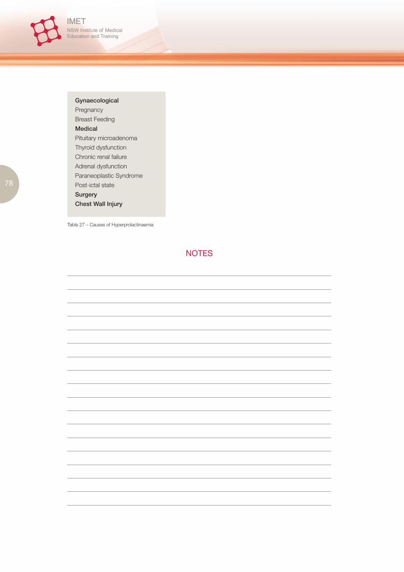
NOTES
78
Gynaecological
Pregnancy
Breast Feeding
Medical
Pituitary microadenoma
Thyroid dysfunction
Chronic renal failure
Adrenal dysfunction
Paraneoplastic Syndrome
Post-ictal state
Surgery
Chest Wall Injury
Table 27 – Causes of Hyperprolactinaemia

Important Legislation
795

80
The Mental Health Act 2007 (NSW)The Rationale for Mental Health Act
The NSW Mental Health Act 2007 (MHA or ‘Act’) governs “the care, treatment and control of mentally ill and mentally disordered persons and other matters relating to mental health”. The Act exists when a person’s reason o judgment is impaired by a mental illness – defined as a syndrome characterized by delusions, hallucinations, severe disturbance of thought, or severe disturbance of mood. The critical test for the Act is the presence of “risk” to the safety of others or the patient. The latter is expanded as a risk of physical harm or, in the case of severe mood disorder, the risk to a person’s reputation or financial interests.
Definitions in the Act
In the Act a “patient” is defined as a person who is admitted to a Declared Mental Health Facility (DMHF) in accordance with the Act. This term applies following the person’s admission and applies even if the person is absent (either agreed or unauthorised) from the ward. A patient who has been referred to a DMHF and is awaiting independent review by a magistrate is deemed an “Assessable patient”. If a magistrate finds that the person is “mentally ill” in terms of the Act and either a treatment order or a deferred discharge is decreed, then the patient is defined as an “Involuntary patient”. The Act defines an “authorised medical officer” as the medical superintendent of the DMHF or the medical officer, nominated by the medical superintendent.
Patient’s Rights under the Act
The patient is entitled to the best possible care in the least restrictive environment to enable their treatment to be effective. The impact of treatment on a patient’s dignity must be minimal. The patient and his or her family must be provided with information about their rights and the procedures of the Act. The treatment choices made under the Act must be for the benefit of the patient with the goal of enabling the person to live, work and participate in the community. Where possible, a patient and his or her family, must be notified of treatment options, the risks and likely benefits, and alternative treatment choices.
5Important Legislation
LEARNING OBJECTIVES
• Outline the rationale for the Mental Health Act• Describe the processes of referral to Declared Mental Health Facilities • Understand the provisions for involuntary psychiatric treatment in NSW

81
IMET ACUTE PSYCHIATRIC MANAGEMENT
Under Section 71 of the Act, patients nominate designated “Primary Carers”. These are usually the patient’s guardian, or a child’s parent or a person in a “close and continuing relationship” with a patient or a close relative or friend (defined as a close personal relationship, through frequent personal contact and interest in patient’s welfare). The Primary carer must be notified within 24 hours of person’s detention (s 75); of the proposed mental health inquiry by magistrate (s 76); regarding discharge planning and ongoing treatment (s 79); if the patient is absent without permission/fails to return from leave (s 78); if the patient is re-classification as a “voluntary patient” or is to be discharged from the DMHF.
Mentally Disordered Patients
A person may be admitted to a DMHF if the person’s immediate behaviour is “so irrational as to justify a conclusion on reasonable grounds that temporary care, treatment or control of the person is necessary for the person’s own protection from serious physical harm, or for the protection of others from serious physical harm”. Admission as a mentally disordered patient is limited to three working days, so theoretically could extend for periods of up to 5-6 days over weekends or public holidays. In general, prolonged admissions warrant reconsideration of diagnosis and the appropriate classification under the Act.
Pathways to Admission
The various pathways to admission and subsequent assessment process for the Act are shown in Figure 23.
Figure 23 – The pathways to treatment under the Mental Health Act (1997)
A person can be conveyed to a Declared Mental Health Facility (DMHF) by police, ambulance, medical officers, the courts or a primary carer in consultation with a mental health service. Once the patient is conveyed to a DMHF they become and ‘assessable person’. The person is assessed by a medical officer under S27 of the Act and is either deemed ‘mentally ill’ (S35) and detained in a DMHF or discharged or offered treatment as a voluntary patient. The patient is now deemed a “mentally ill patient”. The Area Mental Health Service is then obliged to present a case to a magistrate. At that hearing, a prima facie case must be made that the patient is ‘mentally ill’ in terms in of the Act and is a risk to either themselves or others. The patient has a legal representative present at the hearing, and the rules of evidence are akin to other court matters. The magistrate makes a legal determination as to whether there is sufficient evidence of the person being mentally ill and that they pose a risk to themself, others, their finances or their reputation. If these conditions are established the magistrate has the discretionary options of:
1. Deferring the patient’s discharge from the DMHF;
2. Making a treatment order for involuntary treatment in a DMHF for a specified period;
3. Making a Community Treatment Order.
S27 - Ambulance
S25 - Another MHF
S26 - Primary Carer
S19 - Medical Officer
S22 - Police
S23 - Court order
1. Assessable Person (S17)
2. Review by MO (S27) - Form 2 written
3. Deemed Mentally Ill (S35)
Then
MAGISTRATE’S ENQUIRY
1. Treatment Order for specified period or;
2. Adjournment of proceedings or;
3. Deferred Discharge or;
4. Community Treatment Order
Referral Assessment Treatment
5Important Legislation

82
In some instances, the treating clinician may not be in a position to have determined the nature of the patient’s problems, they may request the matter be adjourned for a period of up to two weeks.
There are two appellant processes for the patient – the patient may appeal to the Medical Superintendant of the DMHF or to the Mental Health Review Tribunal, an independent body comprising legal, medical and other social representatives.
As an involuntary patient, the patient is given opportunity to nominate a “primary carer”, who is legally entitled to be consulted on a number of matters relating to the care of the patient including
1. Changes to the patient’s legal status;
2. Substantial changes to the patient’s treatment plan;
3. Leave provisions;
4. Discharge planning.
There are circumstances where, if no suitable primary carer can be identified or if the nominated carer is not suitable, that this provision of the Act can be reasonably waived.
Community Treatment Orders
One of the main principles of the Act is the requirement to provide treatment under the ‘least restrictive option’. In most instances, the patient will be able to safely and effectively receive psychiatric treatment in a community setting. Involuntary treatment in the community is provided under the provisions of Community Treatment Orders (CTOs) can be sought by Authorised Medical Officers under the Act, a medical practitioner familiar with the person’s clinical history, a director of community mental health service familiar with the clinical history of the affected person, and the primary carer in consultation with the community mental health service. Under the 2007 revision of the Act, CTOs can be made whilst the patient is in the community, whereas previously this required an admission to a gazetted psychiatric hospital. Under the Act a CTO is made after application to either a magistrate or the Mental Health Review Tribunal for an inpatient. In the case of a patient in the community, the patient must receive a written notice of application for a CTO (s 52) including a ‘Treatment Plan’. The application must not be heard any earlier that 14 days after notice is given. A CTO can be made if person is absent from the hearing and proper notice has been given (s 55).
If a patient, subject to a CTO, is in breach of the conditions of the CTO e.g. consistent non-attendance to appointments, an application can be made for a breach of CTO (S58). This may result in the patient being taken to the community health centre or detained in a DMHF.
Forensic Patients
A ‘forensic patient’ refers to a patient who has been convicted of a serious criminal offence and is released conditionally from a Corrective Services facility to care in the community. Such patient’s care is provided by Area Mental Health Services and supervised closely by the Mental Health Review Tribunal. Based upon various clinical recommendations, the Mental Health Review Tribunal makes recommendations to the Minister for Health in regards to the ongoing care of such patients. Under the 2007 revision of the Act, such patient’s care is now provided under the Criminal Procedures Act. At the time of writing, this legislation was undergoing parliamentary review.

83
IMET ACUTE PSYCHIATRIC MANAGEMENT
The Commonwealth Privacy ActThe Commonwealth Privacy Act 1998 (Cth)
The Commonwealth Government framed privacy legislation in 1998. This law was to be applied across a large number of settings in society. In 2001 this law was adapted to apply to the health care sector. This modification involved the framing of 10 “National Privacy Principles” in relation to the delivery of health care. Whilst these apply to the private sector and do not alter the effect of state Mental Health Legislation, the principles are still relevant to public sector psychiatry.
These principles are listed in Figure 24.
Figure 24 – The National Privacy Principles applied to health-care
The 10 National Privacy Principles Applied to Health Care
NPP1: Collection of InformationAn organisation must not collect personal information unless the information is necessary health care. When the data is gathered, the person in question has to provided details of the organisation e.g. Health Service or medical practice, and contact details. It is preferred the information be collected, where practical, from the patient themselves. If information is obtained from a third party, the patient must be made aware of this.
NPP2: Use and Disclosure This principle outlines the use of information obtained about a patient. The primary purpose is for the benefit of the patient’s healthcare. Any secondary purpose eg. Research or epidemiological study must have the patient’s consent. Disclosure of information for marketing purposes must not include information of a sensitive nature. Breaches of this principle may occur in circumstances of public interest or the patient’s safety.
NPP3: Data Quality A health-care organization must take reasonable steps to make sure that the personal information it collects, uses or discloses is accurate, complete and up to date.
LEARNING OBJECTIVES
• Outline the basic principles of the Commonwealth Privacy Act • Describe the aspects of the Privacy Act in relation to healthcare
5Important Legislation

84
NPP4: Data Security Sets standards for keeping information up-to-date, accurate and complete, as well as for protecting and securing it from loss, misuse and unauthorized access. This includes de-identification of transmitted data and security for electronic medical records.
NPP5: Openness This principle requires providers to be open about how they handle health information. Each organization must develop a form of privacy policy to clearly explain to patients how it handles health information.
NPP6: Access & Correction This principle gives patients a general right of access to their own health records, and a right to have information corrected, if it is inaccurate, incomplete or out of date. Access to data can be reasonably refused if the request is frivolous, vexatious or may harm the patient’s health. Access can also be refused if accessing the data may compromise a criminal investigation of may harm public interest. Any charges for accessing information must not be “excessive”.
NPP7: Identifiers This principle limits the use of Commonwealth government identifiers, such as the Medicare number, Tax File Number or any Centrelink identifiers. Such data cannot be used outside of the institutional activities related to that identifier.
NPP8: Anonymity Where possible, patients must have the option of using health services without identifying themselves.
NPP9: Trans-border data flows A health care organization can only transmit information relating to a patient outside of Australian Commonwealth jurisdiction if it is in the patient’s best interest or, if related to a non-clinical purpose, it has the patient’s consent. The jurisdiction receiving the information must have similar provisions for handling that information to the Commonwealth Privacy Act.
NPP10: Sensitive InformationA healthcare organisation must not collect sensitive information about an individual unless the individual has consented; or the collection is required by law; or the collection is necessary to prevent or lessen a serious and imminent threat to the life or health of any individual. Any deviation from this principle relates to public interest or safety or the patient’s interest.
The Guardianship Act 1987 (NSW)Introduction
The Mental Health Act provides for involuntary detention and treatment of patients, where a disturbance of mental health creates acute risk to self or the community. By contrast the Guardianship Act focuses on the welfare, interests and rights of the person with the disability and not public interest or safety. The NSW Guardianship Act (1987) exists to protect the interests and legal rights of people over the age of 16 years, who have a disability, such as severe mental illness or dementia which affects their capacity to make decisions. The principles of the Guardianship Act are listed in Table 1.
LEARNING OBJECTIVES
• Describe the basic provisions of the Guardianship Act• Outline the role of the Office of the Protective Commissioner • Understand the role of the psychiatrist in Guardianship proceedings

85
IMET ACUTE PSYCHIATRIC MANAGEMENT
The Guardianship Act provides for the appointment of a guardian to make substitute decisions on behalf of an individual, where it is demonstrable that they have such impairment of judgment that they require additional support in making important personal decisions. The appointed guardian is either a private guardian, such as a family member, or the Public Guardian. The Public Guardian is usually appointed in situations where there is conflict, significant ethical considerations or where there is no other person able to take on the responsibility.
The appointed guardian takes on a role of making substitute decisions on personal issues such as accommodation, access to the patient, or consent to medical and dental treatment. Such decisions have the same legal status as if the patient had made the decision. In some circumstances, a guardian may give consent for the implementation of behavioural management strategies or restraint.
A guardian appointed by the Guardianship Board does not make decisions related to a patient’s financial affairs. This responsibility is assumed by the Office of the Protective Commissioner – affiliated with the Guardianship Board.
• The welfare of the person should be given paramount consideration
• The freedom of the person should be restricted as little as possible
• The person should as far as possible to live a normal life in the community
• The views of the person should be taken into consideration
• It is important to preserve family, cultural and linguistic environment of the person
• The person’s autonomy should be encouraged as far as possible
• The person should be protected from abuse, neglect and exploitation
• The community should be encouraged to promote these principles
Table 28 – The principles of the Guardianship Act 1987
The Office of the Protective Commissioner
The Office of the Protective Commissioner (OPC) is required by law to make decisions that are in the best interests of the person whose financial affairs are under management. If a patient’s financial affairs are to be placed under the OPC a Protective Estates Order (PEO) is made. The domains of interest in a PEO are listed in Figure 25.
Figure 25 – Scope of PEO
5Important Legislation

86
The role of psychiatrists in Guardianship or PEO
The primary legislative instrument in psychiatric practice is the Mental Health Act. In circumstances where the Mental Health Act applies, this in effect “trumps” the Guardianship Act. Psychiatrists do not usually make applications for PEO or guardianship on behalf of patients, however they are often requested to provide evidence to the Guardianship Board to inform its evaluation of applications. Such evidence usually addresses clinical issues such as the effect of a mental illness on a patient’s judgment or decision making capacity, any history of harm brought about by impaired judgment over affairs e.g. financial exploitation. Treatment decisions involving mental illness are usually considered in terms of the Mental Health Act.

NOTES
87
IMET ACUTE PSYCHIATRIC MANAGEMENT

NOTES
88


Building 12Gladesville HospitalVictoria RoadGladesville NSW 2111
Tel: (02) 9844 6551Fax: (02) 9844 6544email: [email protected]
www.imet.health.nsw.gov.au
IMET ACUTE PSYCHIATRIC MANAGEMENT