Retinoic Acid Receptor- Alpha and Its Role in Acute Promyelocytic Leukemia By Alex Sheng.

3341
□ CASE REPORT □
Acute Promyelocytic Leukemia with i(17)(q10)
Junki Inamura 1, Katsuya Ikuta 2, Nodoka Tsukada 1, Takaaki Hosoki 1,
Motohiro Shindo 2 and Kazuya Sato 1
Abstract
We herein report a rare chromosomal abnormality observed in an acute promyelocytic leukemia (APL) pa-
tient. She had several APL derivative clones including a clone with i(17)(q10) abnormality, which consists of
two kinds of structural abnormalities, a cryptic translocation of t(15;17) and an isochromosome of 17q. Al-
though an obvious microscopic t(15;17) change was not observed on either arms of the isochromosome,
PML/RARα fusion signals were detected on an interphase fluorescence in situ hybridization analysis. By sev-
eral cytogenetic analyses of her bone marrow cells, it was confirmed that the i(17)(q10) clone was derived
from the classic t(15;17) clone via another intervening clone, cryptic t(15;17).
Key words: acute promyelocytic leukemia, cryptic translocation, i(17)(q10)
(Intern Med 55: 3341-3345, 2016)(DOI: 10.2169/internalmedicine.55.7226)
Introduction
Acute promyelocytic leukemia (APL) is one of the most
widely known hematological neoplasms and is associated
with a typical chromosomal abnormality, the reciprocal
translocation of t(15;17)(q22;q21). According to Cervera et
al., additional chromosomal abnormalities are also seen in
28% of classic t(15;17)-positive APL patients (1). Although
trisomy 8 or partial monosomy, i.e., 7q deletion, were repre-
sentative in that study, an isochromosome change of 17q af-
ter the reciprocal translocation of t(15;17) was also occa-
sionally observed, which was described as ider(17)(q10)t(15;
17) (2). This chromosome does not have a short arm (17p),
but rather has two of the same arms derived from long arms
(17q after the reciprocal translocation) on both sides of the
centromere.
In addition, APL cases without classic t(15;17) abnormali-
ties have also been occasionally observed. According to
Grimwade et al., the incidence of this finding in patients
with APL is 9%, and the most common abnormality is cryp-
tic t(15;17) (3). Because this abnormality involves the
translocations of only submicroscopic fragments of chromo-
somes, the fusion gene PML/RARα exists on chromosome
15 or 17, without the detection of microscopic abnormalities
on a routine chromosomal analysis.
On the other hand, there are only few case reports of APL
with i(17)(q10) consisting of both the cryptic t(15;17) and
isochromosome of 17q. This condition does not have evi-
dence of microscopic t(15;17) changes, although it does in-
volve PML/RARα fusion genes (4-6).
We herein report a case of APL with i(17)(q10). Frequent
cytogenetic analyses performed at the time of diagnosis and
during treatment revealed that the patient had several APL
clones, including the i(17)(q10) clone as well as the classic
t(15;17) clone. These findings helped us to speculate the de-
velopmental mechanism of i(17)(q10) in APL patients.
Case Report
A 74-year-old woman was incidentally noted to have pan-
cytopenia on a regular blood examination during a follow-up
of reflux esophagitis and was introduced to our hospital. A
blood examination showed pancytopenia and abnormalities
in fibrinolysis: WBC 1,200/μL (neutrophils 64%, lympho-
cytes 29%, monocytes 5%, eosinophils 2%, no abnormal
cells were detected), Hb 10.0 g/dL, Plt 67×103/μL and fibrin
degradation products (FDP) 25.6 μg/mL (reference value:
0.0-5.0 μg/mL). However, neither the prothrombin time nor
activated partial thromboplastin time was prolonged. Bone
1Department of Hematology/Oncology, Asahikawa Kosei Hospital, Japan and 2Division of Gastroenterology and Hematology/Oncology, Depart-
ment of Medicine, Asahikawa Medical University, Japan
Received for publication February 2, 2016; Accepted for publication February 24, 2016
Correspondence to Dr. Katsuya Ikuta, [email protected]
Intern Med 55: 3341-3345, 2016 DOI: 10.2169/internalmedicine.55.7226
3342
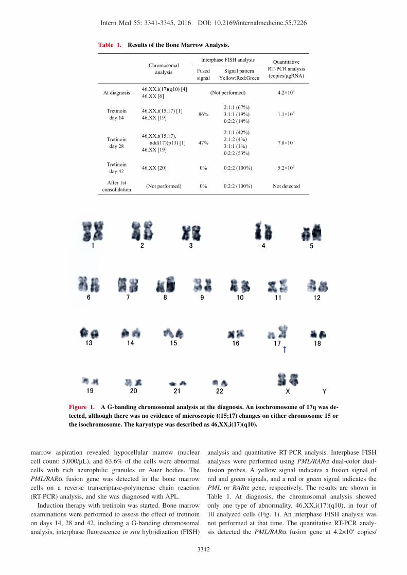
Figure 1. A G-banding chromosomal analysis at the diagnosis. An isochromosome of 17q was de-tected, although there was no evidence of microscopic t(15;17) changes on either chromosome 15 or the isochromosome. The karyotype was described as 46,XX,i(17)(q10).
Table 1. Results of the Bone Marrow Analysis.
Chromosomalanalysis
Interphase FISH analysis Quantitative RT-PCR analysis(copies/ gRNA)
Fusedsignal
Signal patternYellow:Red:Green
At diagnosis 46,XX,i(17)(q10) [4]46,XX [6] (Not performed) 4.2×10⁴
Tretinoinday 14
46,XX,t(15;17) [1]46,XX [19] 86%
2:1:1 (67%)3:1:1 (19%) 0:2:2 (14%)
1.1×10⁴
Tretinoinday 28
46,XX,t(15;17), add(17)(p13) [1]
46,XX [19]47%
2:1:1 (42%) 2:1:2 (4%) 3:1:1 (1%) 0:2:2 (53%)
7.8×10³
Tretinoinday 42 46,XX [20] 0% 0:2:2 (100%) 5.2×10²
After 1st consolidation (Not performed) 0% 0:2:2 (100%) Not detected
marrow aspiration revealed hypocellular marrow (nuclear
cell count: 5,000/μL), and 63.6% of the cells were abnormal
cells with rich azurophilic granules or Auer bodies. The
PML/RARα fusion gene was detected in the bone marrow
cells on a reverse transcriptase-polymerase chain reaction
(RT-PCR) analysis, and she was diagnosed with APL.
Induction therapy with tretinoin was started. Bone marrow
examinations were performed to assess the effect of tretinoin
on days 14, 28 and 42, including a G-banding chromosomal
analysis, interphase fluorescence in situ hybridization (FISH)
analysis and quantitative RT-PCR analysis. Interphase FISH
analyses were performed using PML/RARα dual-color dual-
fusion probes. A yellow signal indicates a fusion signal of
red and green signals, and a red or green signal indicates the
PML or RARα gene, respectively. The results are shown in
Table 1. At diagnosis, the chromosomal analysis showed
only one type of abnormality, 46,XX,i(17)(q10), in four of
10 analyzed cells (Fig. 1). An interphase FISH analysis was
not performed at that time. The quantitative RT-PCR analy-
sis detected the PML/RARα fusion gene at 4.2×10⁴ copies/

Intern Med 55: 3341-3345, 2016 DOI: 10.2169/internalmedicine.55.7226
3343
Table 2. Reported Cases of APL with i(17)(q10).
a The patient developed an acute monoblastic leukemia 10 months later with no evidence of relapse of APL.b The karyotype is 46,XX,del(7)(q31q33),i(17)(q10)
References AgeSex
PML-RARA fused gene detection
Completeremission Relapse
Detection ofclassic t(15;17)
clone
1 4 51FInterphase FISHMetaphase FISH
RT-PCRYesa No No
2 5 44F RT-PCR Yes Not described No
3
6
6
6 10F Metaphase FISHRT-PCR Yes No No
4 13F Metaphase FISHRT-PCR Yes Yes No
5 42Fb Metaphase FISHRT-PCR Yes No No
6 Ourcase 74F Interphase FISH
RT-PCR Yes No Yes
μg RNA. On day 14, the detected abnormal karyotype was
46,XX,t(15;17) in one of 20 analyzed cells, although the
clone with 46,XX,i(17)(q10) was not detected according to
the chromosomal analysis. According to the interphase FISH
analysis, the rate of PML/RARα fusion-positive cells was
86%; two patterns were observed: yellow:red:green=2:1:1
(67%) and 3:1:1 (19%). The amount of fusion gene de-
creased to 1.1×10⁴ copies/μg RNA according to a quantita-
tive RT-PCR analysis. On day 28, the detected abnormal
karyotype was 46,XX,t(15;17),add(17)(p13) in one of 20
analyzed cells on a chromosomal analysis. According to an
interphase FISH analysis, the positive rate decreased to
47%; three patterns were observed: yellow:red:green=2:1:1
(42%), 2:1:2 (4%), 3:1:1 (1%). The amount of fusion gene
also decreased to 7.8×10³ copies/μg RNA using a quantita-
tive RT-PCR analysis. On day 42, all of the abnormalities
had disappeared on both the chromosomal and interphase
FISH analyses, and the amount of fusion gene was de-
creased to 5.2×10² copies/μg RNA using a quantitative RT-
PCR analysis. Three courses of consolidation chemotherapy
were performed after 63 days of tretinoin treatment. Com-
plete molecular remission was achieved after the first course
of consolidation therapy. After consolidation therapy, main-
tenance therapy using tretinoin was performed, and the pa-
tient has maintained in remission for 54 months.
Throughout the study period, the patient showed three
types of abnormal karyotypes according to the chromosomal
analyses, 46,XX,i(17)(q10), 46,XX,t(15;17) and 46,XX,t(15;
17),add(17)(p13), and three types of PML/RARα fusion-
positive signal patterns on the interphase FISH analyses, yel-
low:red:green=2:1:1, 2:1:2 and 3:1:1. A clone with i(17)
(q10) was also detected at diagnosis, although it disap-
peared, despite the appearance of the classic t(15;17) clone
on day 14.
Discussion
It has been reported that acquired or constitutional iso-
chromosomes originate from aberrant mitosis or meio-
sis (7, 8). Not longitudinal, but rather the transverse, divi-
sion of the chromosome can yield a symmetrical chromo-
some with two identical arms in a daughter cell, which is
referred to as an isochromosome. It has also been shown
that the isochromosome of 17q, also described as i(17)(q10),
is occasionally observed as an additional abnormality among
various neoplasias, including hematological malignancies
and solid tumors (9). Additionally, in cases of APL, the iso-
chromosome of 17q after the translocation t(15;17), de-
scribed as ider(17)(q10)t(15;17), has been occasionally re-
ported.
However, APLs associated with the i(17)(q10) clone with
no evidence of microscopic t(15;17) are quite rare. As
shown in Table 2, only six cases, including ours, have been
described (4-6). The prognoses of these patients appear to
be generally good. In case 2, the PML/RARA oncogene was
detected using a PCR analysis; however, an interphase FISH
analysis did not detect any fusion signals, although the pre-
cise reason was not described (5). In four of six patients,
metaphase FISH analyses were performed, which can be
used to directly clarify the location of the oncogene at dis-
crete chromosomes. The oncogenes were located on chro-
mosome 15 in one case (case 3) and on both arms of i(17)
(q10) in three cases (cases 1, 4 and 5).
The process by which the APL clone with i(17)(q10) ap-
pears remains unknown because of its extreme rarity. It
might also be another reason that repetitive cytogenetic
analyses were not performed, such that only limited infor-
mation was available in past cases. In the present case, re-
petitive bone marrow tests revealed that the patient pos-
sessed several kinds of APL clones, including the clone with
i(17)(q10) and the classic t(15;17) clone. The changes in the
Intern Med 55: 3341-3345, 2016 DOI: 10.2169/internalmedicine.55.7226
3344
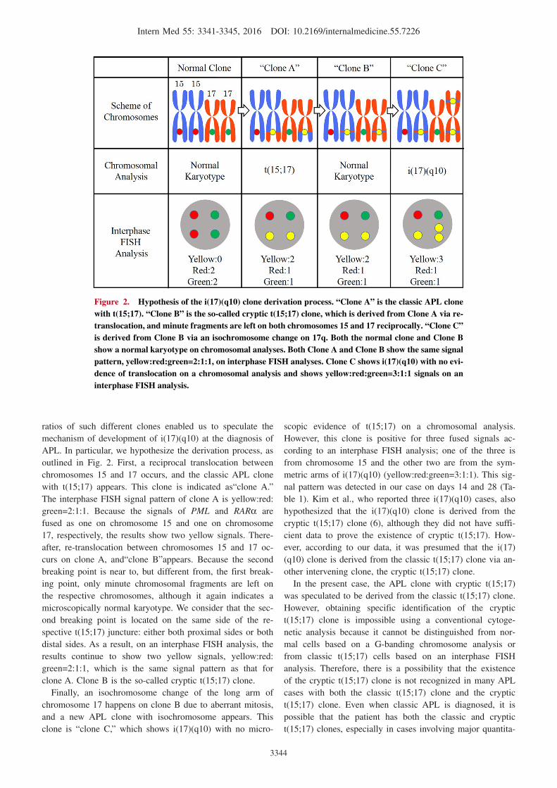
Figure 2. Hypothesis of the i(17)(q10) clone derivation process. “Clone A” is the classic APL clone with t(15;17). “Clone B” is the so-called cryptic t(15;17) clone, which is derived from Clone A via re-translocation, and minute fragments are left on both chromosomes 15 and 17 reciprocally. “Clone C” is derived from Clone B via an isochromosome change on 17q. Both the normal clone and Clone B show a normal karyotype on chromosomal analyses. Both Clone A and Clone B show the same signal pattern, yellow:red:green=2:1:1, on interphase FISH analyses. Clone C shows i(17)(q10) with no evi-dence of translocation on a chromosomal analysis and shows yellow:red:green=3:1:1 signals on an interphase FISH analysis.
ratios of such different clones enabled us to speculate the
mechanism of development of i(17)(q10) at the diagnosis of
APL. In particular, we hypothesize the derivation process, as
outlined in Fig. 2. First, a reciprocal translocation between
chromosomes 15 and 17 occurs, and the classic APL clone
with t(15;17) appears. This clone is indicated as“clone A.”
The interphase FISH signal pattern of clone A is yellow:red:
green=2:1:1. Because the signals of PML and RARα are
fused as one on chromosome 15 and one on chromosome
17, respectively, the results show two yellow signals. There-
after, re-translocation between chromosomes 15 and 17 oc-
curs on clone A, and“clone B”appears. Because the second
breaking point is near to, but different from, the first break-
ing point, only minute chromosomal fragments are left on
the respective chromosomes, although it again indicates a
microscopically normal karyotype. We consider that the sec-
ond breaking point is located on the same side of the re-
spective t(15;17) juncture: either both proximal sides or both
distal sides. As a result, on an interphase FISH analysis, the
results continue to show two yellow signals, yellow:red:
green=2:1:1, which is the same signal pattern as that for
clone A. Clone B is the so-called cryptic t(15;17) clone.
Finally, an isochromosome change of the long arm of
chromosome 17 happens on clone B due to aberrant mitosis,
and a new APL clone with isochromosome appears. This
clone is “clone C,” which shows i(17)(q10) with no micro-
scopic evidence of t(15;17) on a chromosomal analysis.
However, this clone is positive for three fused signals ac-
cording to an interphase FISH analysis; one of the three is
from chromosome 15 and the other two are from the sym-
metric arms of i(17)(q10) (yellow:red:green=3:1:1). This sig-
nal pattern was detected in our case on days 14 and 28 (Ta-
ble 1). Kim et al., who reported three i(17)(q10) cases, also
hypothesized that the i(17)(q10) clone is derived from the
cryptic t(15;17) clone (6), although they did not have suffi-
cient data to prove the existence of cryptic t(15;17). How-
ever, according to our data, it was presumed that the i(17)
(q10) clone is derived from the classic t(15;17) clone via an-
other intervening clone, the cryptic t(15;17) clone.
In the present case, the APL clone with cryptic t(15;17)
was speculated to be derived from the classic t(15;17) clone.
However, obtaining specific identification of the cryptic
t(15;17) clone is impossible using a conventional cytoge-
netic analysis because it cannot be distinguished from nor-
mal cells based on a G-banding chromosome analysis or
from classic t(15;17) cells based on an interphase FISH
analysis. Therefore, there is a possibility that the existence
of the cryptic t(15;17) clone is not recognized in many APL
cases with both the classic t(15;17) clone and the cryptic
t(15;17) clone. Even when classic APL is diagnosed, it is
possible that the patient has both the classic and cryptic
t(15;17) clones, especially in cases involving major quantita-

Intern Med 55: 3341-3345, 2016 DOI: 10.2169/internalmedicine.55.7226
3345
tive deviations between the chromosomal analysis and inter-
phase FISH analysis.
Interestingly, although only the i(17)(q10) clone, not the
classic t(15;17) clone, was detected at diagnosis in this case,
the classic t(15;17) clone, not the i(17)(q10) clone, was con-
versely detected on day 14. We hypothesize that the pa-
tient’s i(17)(q10) clone had a growth advantage and demon-
strated hyperresponsiveness to tretinoin compared to the
classic t(15;17) clone. If the second breaking point of both
chromosomes 15 and 17 in the clone A is on the proximal
side of the t(15;17) juncture, then the oncogene PML/RARαwill move from chromosome 15 to 17. Hence, the two
PML/RARA genes are on the i(17)(q10) chromosome. Due
to the presence of dual oncogenes, the patient’s i(17)(q10)
clone might have been expanded mainly at diagnosis,
whereas tretinoin possibly induced the i(17)(q10) clone to
differentiate more rapidly than the t(15;17) clone.
The cytogenetic analyses showed other abnormalities (Ta-
ble 1). The 46,XX,t(15;17),add(17)(p13) detected on day 28
is presumably another derivative of the APL clone. The
FISH signal pattern of yellow:red:green=2:1:2 noted on day
28 might also indicate the existence of another distinct APL
clone, while no explicable karyotypes were detected in our
analyses. The clinical course of our case appeared to be fa-
vorable, as in past reports of APLs with i(17)(q10), although
the patient has such various APL clones. Therefore, the i(17)
(q10) chromosome in cases of APL does not appear to be a
poor prognostic factor, although a further accumulation of
cases is necessary.
The authors state that they have no Conflict of Interest (COI).
References
1. Cervera J, Montesinos P, Hernández-Rivas JM, et al. Additional
chromosome abnormalities in patients with acute promyelocytic
leukemia treated with all-trans retinoic acid and chemotherapy.
Haematologica 95: 424-431, 2010.
2. Manola KN, Karakosta M, Sambani C, et al. Isochromosome of
der(17)(q10)t(15;17) in acute promyelocytic leukemia resulting in
an additional copy of the RARA-PML fusion gene: report of 4
cases and review of the literature. Acta Haematol 123: 162-170,
2010.
3. Grimwade D, Biondi A, Mozziconacci MJ, et al. Characterization
of acute promyelocytic leukemia cases lacking the classic t(15;17):
results of the European Working Party. Blood 96: 1297-1308,
2000.
4. Lee GY, Christina S, Tien SL, et al. Acute promyelocytic leukemia
with PML-RARA fusion on i(17q) and therapy-related acute
myeloid leukemia. Cancer Genet Cytogenet 159: 129-136, 2005.
5. Huh J, Moon H, Chi H, Chung W. Acute promyelocytic leukemia
with i(17)(q10) on G-banding and PML/RARA rearrangement by
RT-PCR without evidence of PML/RARA rearrangement on FISH.
Int J Lab Hematol 31: 372-374, 2009.
6. Kim M, Lim J, Kim Y, et al. The genetic characterization of acute
promyelocytic leukemia with cryptic t(15;17) including a new re-
current additional cytogenetic abnormality i(17)(q10). Leukemia
22: 881-883, 2008.
7. de la Chapelle A. How do human isochromosomes arise? Cancer
Genet Cytogenet 5: 173-179, 1982.
8. Darlington CD. Misdivision and the genetics of the centromere. J
Genet 37: 341-364, 1939.
9. Mertens F, Johansson B, Mitelman F. Isochromosomes in neopla-
sia. Genes Chromosomes Cancer 10: 221-230, 1994.
The Internal Medicine is an Open Access article distributed under the Creative
Commons Attribution-NonCommercial-NoDerivatives 4.0 International License. To
view the details of this license, please visit (https://creativecommons.org/licenses/
by-nc-nd/4.0/).
Ⓒ 2016 The Japanese Society of Internal Medicine
http://www.naika.or.jp/imonline/index.html