Acute pancreatitis mimicking myocardial ischemia: A...
6
Journal of Health and Social Sciences 2017; 2,2:209-214 209 Acute pancreatitis mimicking myocardial ischemia: A case report and a review of the literature Antonio Villa 1 , Francesca Campagna 1 , Giulia Gallotta 1 , Riccardo Bennicelli 2 , Paola Sartori 3 CASE REPORT IN INTERNAL MEDICINE KEY WORDS: Acute pancreatitis; electrocardiographic changes; myocardial ischemia. Abstract In literature, acute pancreatitis has been shown to be associated with a wide range of electrocardiographic changes. In the present report, we describe a case of a young woman who presented to our emergency room with chest pain and electrocardiographic alterations suggestive of myocardial ischemia; laboratory data showed elevated amylase and lipase levels. A computed tomography (CT) scan of the abdomen confirmed the diagnosis of acute pancreatitis. In the next day, she repeated the electrocardiogram, which showed a regression of the ST-segment depression. T-waves changes and ST-segment depression are common in acute pancreatitis and, therefore, emergency physicians should consider acute pancreatitis in the differential diagnosis of patients presenting with chest pain and electrocardiographic changes. Affiliations: 1 M.D., Department of Emergency, ASST Monza, PO Desio, Monza, Italy. 2 M.D., Department of Cardiology, ASST Monza, PO Desio, Monza, Italy. 3 M.D., Department of Surgery, ASST Monza, PO Desio, Monza, Italy. Corresponding author: Dr. Antonio Villa, ASST Monza, PO Desio, Via Fiuggi,56 20159 Milan, Italy. E-mail: [email protected]
Transcript of Acute pancreatitis mimicking myocardial ischemia: A...
Journal of Health and Social Sciences 2017; 2,2:209-214
209
Acute pancreatitis mimicking myocardial ischemia: A case report and a review of the
literature
Antonio Villa1, Francesca Campagna1, Giulia Gallotta1, Riccardo Bennicelli2, Paola Sartori3
CASE REPORT IN INTERNAL MEDICINE
KEY WORDS: Acute pancreatitis; electrocardiographic changes; myocardial ischemia.
Abstract
In literature, acute pancreatitis has been shown to be associated with a wide range of electrocardiographic changes. In the present report, we describe a case of a young woman who presented to our emergency room with chest pain and electrocardiographic alterations suggestive of myocardial ischemia; laboratory data showed elevated amylase and lipase levels. A computed tomography (CT) scan of the abdomen confi rmed the diagnosis of acute pancreatitis. In the next day, she repeated the electrocardiogram, which showed a regression of the ST-segment depression. T-waves changes and ST-segment depression are common in acute pancreatitis and, therefore, emergency physicians should consider acute pancreatitis in the diff erential diagnosis of patients presenting with chest pain and electrocardiographic changes.
Affi liations: 1 M.D., Department of Emergency, ASST Monza, PO Desio, Monza, Italy.2 M.D., Department of Cardiology, ASST Monza, PO Desio, Monza, Italy. 3 M.D., Department of Surgery, ASST Monza, PO Desio, Monza, Italy.
Corresponding author:
Dr. Antonio Villa, ASST Monza, PO Desio, Via Fiuggi,56 20159 Milan, Italy. E-mail: [email protected]
Journal of Health and Social Sciences 2017; 2,2:209-214
210
Riassunto
In letteratura, la pancreatite acuta è stata associata ad un’ampia gamma di alterazioni elettrocardio-grafiche. In questo lavoro presentiamo il caso di una giovane donna giunta alla nostra osservazione con dolore toracico ed alterazioni elettrocardiografiche suggestive di ischemia miocardica; i dati di laboratorio mostravano elevati livelli di amilasi e lipasi. Una TC dell’addome ha confermato la dia-gnosi di pancreatite acuta. Un ECG ripetuto il giorno successivo evidenziava una regressione del sottoslivellamento del tratto ST. Le modifiche dell’onda T ed il sottoslivellamento del segmento ST sono comuni nella pancreatite acuta e, pertanto, i medici che lavorano in emergenza dovrebbe-ro considerare la pancreatite acuta nella diagnosi differenziale dei pazienti con dolore toracico ed alterazioni elettrocardiografiche.
Competing interests - none declared.
Copyright © 2017 Antonio Villa et al. FS PublishersThis is an open access article distributed under the Creative Commons Attribution (CC BY 4.0) License, which per-mits unrestricted use, distribution, and reproduction in any medium, provided the original work is properly cited. See http:www.creativecommons.org/licenses/by/4.0/.
Cite this article as: Villa A, Campagna F, Gallotta G, Bennicelli R, Sartori P. Acute pancreatitis mimicking myocar-dial ischemia: A case report and a review of the literature. J Health Soc Sci. 2017;2(2):209-214
TAKE-HOME MESSAGEAcute pancreatitis has been shown to be associated with a wide range of electrocardiographic
alterations and symptoms mimicking myocardial infarction. Emergency physicians should consider acute pancreatitis in the differential diagnosis of patients presenting with chest pain and
electrocardiographic changes.
Received: 28/04/2017 Accepted: 18/06/2017 Published: 15/07/2017
DOI 10.19204/2017/ctpn7
Journal of Health and Social Sciences 2017; 2,2:209-214
211
INTRODUCTION Acute pancreatitis (AP) is an acute inflam-matory process of the pancreas that is asso-ciated with variable involvement of multiple organ system dysfunctions, in which cardio-vascular manifestations are frequent [1]. A wide range of electrocardiographic (EKG) changes including arrhythmias, bradycardia, T-wave changes, intraventricular conduction disturbances, and ST-segment elevation or depression have also been described [2–11].In the present report, we describe a case of a young woman who arrived at our department of emergency with chest pain and EKG alte-rations suggestive of myocardial ischemia.
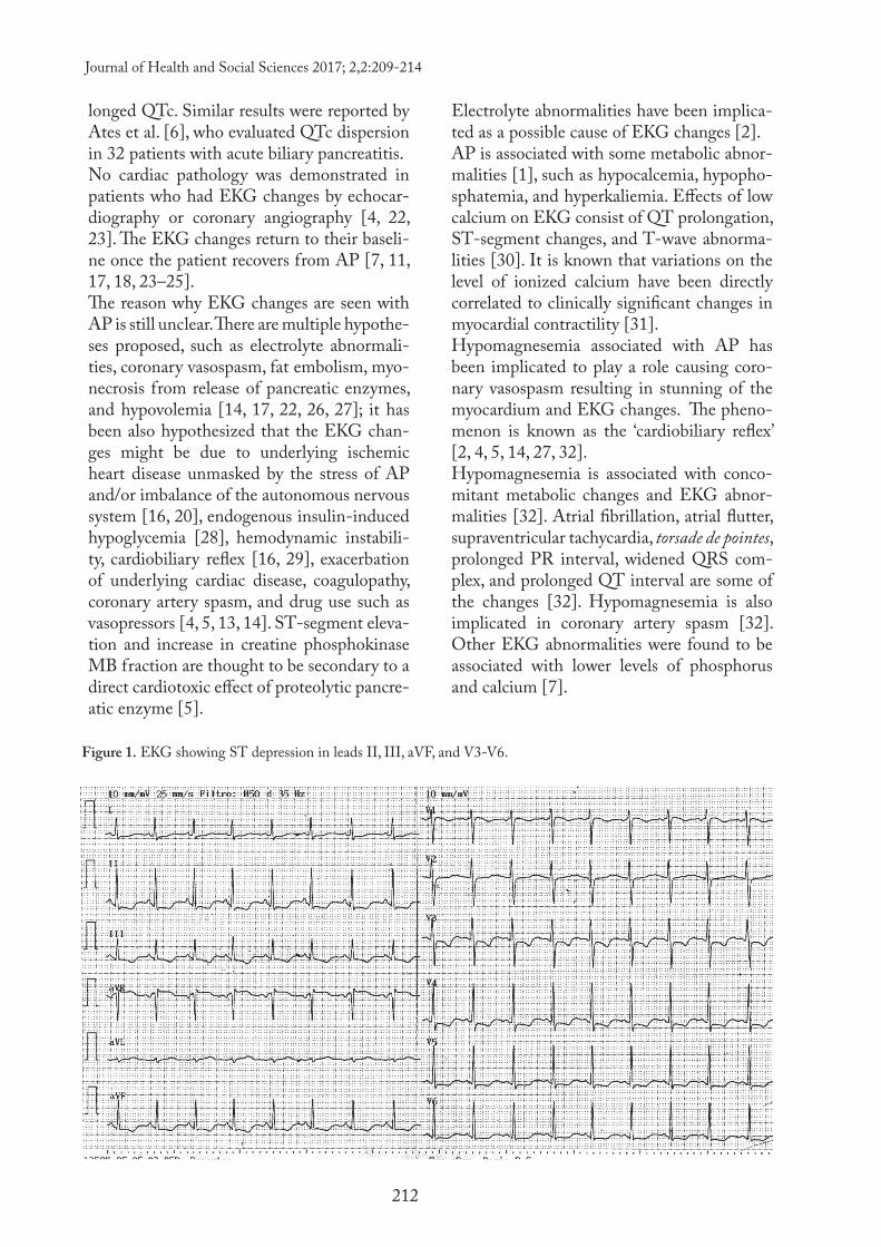
CASE REPORT A 21-year-old woman presented to our emer-gency room with lower mild sternal chest pain radiating posteriorly and associated with vo-miting and diarrhea. She denied any type of alcohol or drug usage; moreover, she did not have any cardiovascular risk factors, except for estroprogestinic anti-contraceptive therapy. Physical examination showed a blood pressu-re of 110/65 mmHg and a heart rate of 101 beats per minute (BPM). Her chest was clear to auscultation and heart sounds were normal. Her abdomen was soft and nontender with adequate bowel sounds. There were no guar-ding, rebound tenderness, or organomegaly.A 12-lead EKG showed diffuse ST-segment depression with T negative (Figure 1). Laboratory data showed: Potassium 3,4 mMol/L, calcium 2,12 mMol/L, aspartate aminotranspherase 44 U/L, amylase 1136 U/L, and lipase 1116 U/L; HS-troponin T was negative. Echocardiogram was normal. A computed tomography (CT) scan of the abdomen re-vealed a focal acute edematous pancreatic tail pancreatitis. She was treated with intrave-nous fluids and pain medication (nothing by mouth). In the next day, a repeat ECG showed regression of the ST-segment depression.
DISCUSSIONDrummond first reported EKG changes in AP in 1934 [1]. Tachycardia and fibrillation
were noted and suspected to be secondary to shock. Over the years, other authors also identified EKG abnormalities in patients with AP [4–7, 12–16]. Different types of ta-chyarrhythmias and bradyarrhythmias, such as atrial flutter and fibrillation, and supra-ventricular premature contractions, shorte-ned PR interval, QRS prolongation, various bundle-branch blocks, non-specific changes in repolarization, decrease T-wave voltage, T-wave changes, and ST-segment abnorma-lities were often seen, in approximately 50% of patients affected by AP. Most frequently associated changes with AP are T-wave in-version and ST-segment depression [1]. New onset ST-elevation in leads V2 and V4 with diffusely inverted T waves mimicking acu-te myocardial infarction have been reported with mild AP [4, 17, 18]. Although very rare, AP may even be complicated by myocardial infarction secondary to hypovolemia, coagu-lation abnormalities, or coronary artery spasm [19].Studies evaluating EKG changes in AP have yielded varying results. Buch et al. [3] repor-ted that 57% of patients with AP had tran-sient EKG abnormalities, which consisted mainly in non-specific T-wave changes and accelerated atrial or nodal rhythms. Mautner et al. [20] also reported that signifi-cant EKG alterations during AP usually oc-cur in patients with previous cardiac abnor-malities. Rubio-Tapia et al. [7] reported EKG abnor-malities in 55% of patients affected by AP, consisting in non-specific changes of repo-larization, sinus tachycardia, and left anterior hemiblock being the most frequent abnorma-lities.Pezzilli et al. [2] also reported that 51.8% of the patients with AP had EKG abnormali-ties, and electrolyte alterations were present in about one-half of these. Nadkarni et al. [21] found EKG abnormali-ties in more than 80% of patients with AP, and the 68% of patients with severe pancreatitis had prolonged QTc as compared to 35% of patients with mild pancreatitis. Furthermo-re, all the patients who succumbed had pro-
Journal of Health and Social Sciences 2017; 2,2:209-214
212
longed QTc. Similar results were reported by Ates et al. [6], who evaluated QTc dispersion in 32 patients with acute biliary pancreatitis. No cardiac pathology was demonstrated in patients who had EKG changes by echocar-diography or coronary angiography [4, 22, 23]. The EKG changes return to their baseli-ne once the patient recovers from AP [7, 11, 17, 18, 23–25].The reason why EKG changes are seen with AP is still unclear. There are multiple hypothe-ses proposed, such as electrolyte abnormali-ties, coronary vasospasm, fat embolism, myo-necrosis from release of pancreatic enzymes, and hypovolemia [14, 17, 22, 26, 27]; it has been also hypothesized that the EKG chan-ges might be due to underlying ischemic heart disease unmasked by the stress of AP and/or imbalance of the autonomous nervous system [16, 20], endogenous insulin-induced hypoglycemia [28], hemodynamic instabili-ty, cardiobiliary reflex [16, 29], exacerbation of underlying cardiac disease, coagulopathy, coronary artery spasm, and drug use such as vasopressors [4, 5, 13, 14]. ST-segment eleva-tion and increase in creatine phosphokinase MB fraction are thought to be secondary to a direct cardiotoxic effect of proteolytic pancre-atic enzyme [5].
Electrolyte abnormalities have been implica-ted as a possible cause of EKG changes [2].AP is associated with some metabolic abnor-malities [1], such as hypocalcemia, hypopho-sphatemia, and hyperkaliemia. Effects of low calcium on EKG consist of QT prolongation, ST-segment changes, and T-wave abnorma-lities [30]. It is known that variations on the level of ionized calcium have been directly correlated to clinically significant changes in myocardial contractility [31]. Hypomagnesemia associated with AP has been implicated to play a role causing coro-nary vasospasm resulting in stunning of the myocardium and EKG changes. The pheno-menon is known as the ‘cardiobiliary reflex’ [2, 4, 5, 14, 27, 32].Hypomagnesemia is associated with conco-mitant metabolic changes and EKG abnor-malities [32]. Atrial fibrillation, atrial flutter, supraventricular tachycardia, torsade de pointes, prolonged PR interval, widened QRS com-plex, and prolonged QT interval are some of the changes [32]. Hypomagnesemia is also implicated in coronary artery spasm [32]. Other EKG abnormalities were found to be associated with lower levels of phosphorus and calcium [7].
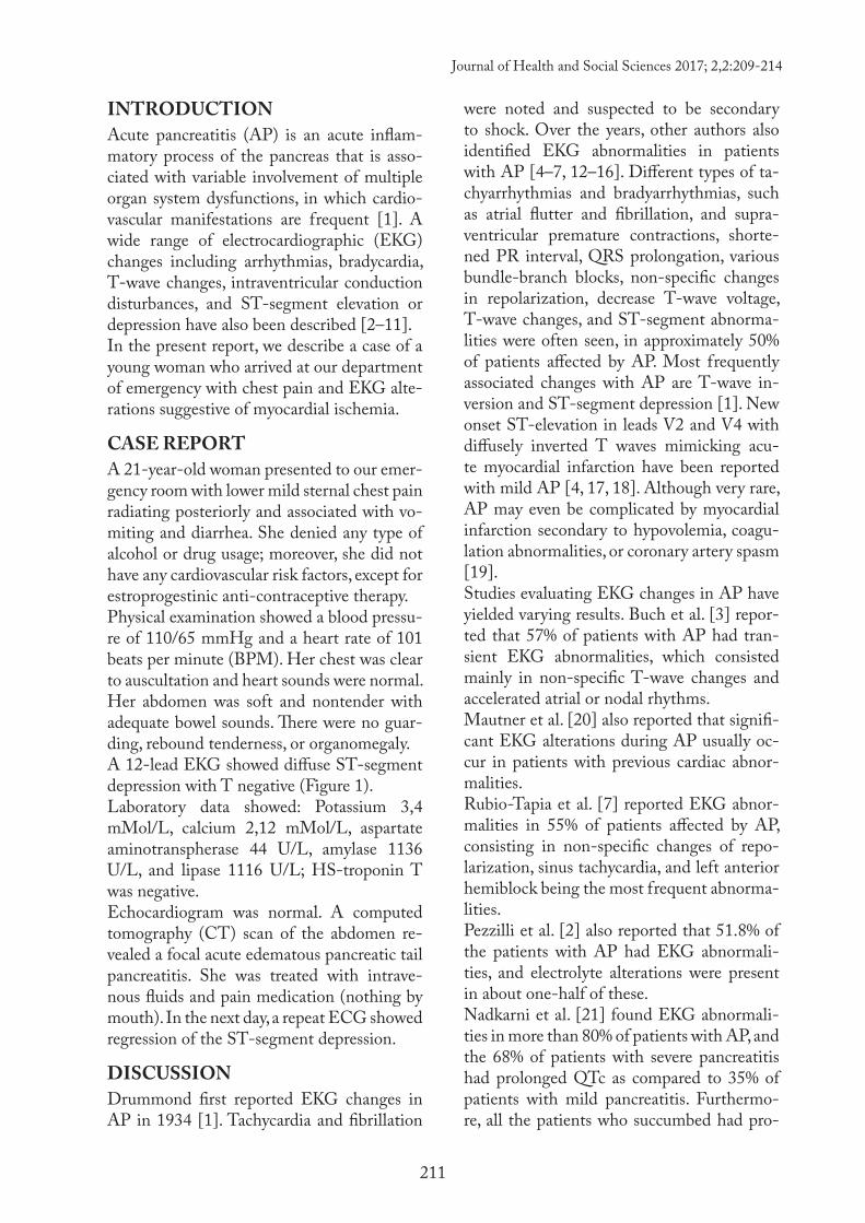
Figure 1. EKG showing ST depression in leads II, III, aVF, and V3-V6.
Journal of Health and Social Sciences 2017; 2,2:209-214
213
CONCLUSIONST-waves changes and ST-segment depression are common in AP and, therefore, AP should be considered by emergency physicians in the
differential diagnosis of patients presenting with chest pain and EKG changes mimicking myocardial ischemia.
References
1. Yegneswaran B, Kostis JB, Pitchumoni CS. Cardiovascular manifestations of acute pancreatitis. J Crit Care. 2011;26(2):225e11–18.
2. Pezzilli R, Barakat B, Billi P, Bertaccini B. Electrocardiographic abnormalities in acute pancreatitis. Eur J Emerg Med. 1999;6(1):27–29.
3. Buch J, Buch A, Schmidt A. Transient ECG changes during acute attacks of pancreatitis. Acta Cardiol. 1980;35(5):381–390.
4. Khairy P. Marsolais P. Pancreatitis with electrocardiographic changes mimicking acute myocardial infar-ction. Can J Gastroenterol. 2001;15(8):522–526.
5. Albrecht CA, Laws FA. ST segment elevation pattern of acute myocardial infarction induced by acute pancreatitis. Cardiol Rev. 2003;11(3):147–151.
6. Ates F, Kosar F, Aksoy Y, Yildirim B, Sahin I, Hilmioglu F. QT interval analysis in patients with acute biliary pancreatitis. Pancreas. 2005;31(3):238– 241.
7. Rubio-Tapia A, Garcia-Leiva J, Asensio-Lafuente E, Robles-Diaz G, Vargas-Vorackova F. Electrocardio-graphic abnormalities in patients with acute pancreatitis. J Clin Gastroenterol. 2005;39(9):815–818.
8. Makaryus AN, Adedeji O, Ali SK. Acute pancreatitis presenting as acute inferior wall ST-segment eleva-tions on electrocardiography. Am J Emerg Med. 2008;26(6):734.e1–4.
9. Meuleman VG, Schinkel AF, Vos J. Electrocardiographic abnormalities caused by acute pancreatitis. Neth Heart J. 2011;19(3):137–139.
10. Sokmen M, Bugdaci MS, Oztekin E. Electrocardiographic changes and importance of repolarization changes in cases with acute pancreatitis. Turk J Gastroenterol. 2011;22(3):315–320.
11. Sethi P, Murtaza G, Sharma A, Paul T. ST segment elevation with normal coronaries. Case Rep Med. 2016. Vol 2016; Article ID 3132654, 4 pages. doi: 10.1155/2016/3132654.
12. Faintuch JJ, Abrahao MM, Giacaglia LR, Junqueira PC, Salgado LF. Electrocardiographic changes in pancreatitis. Arq Bras Cardiol. 1989;52(5):259–260.
13. Wang K, Asinger RW, Mariott HJ. ST-segment elevation in conditions other than acute myocardial infar-ction. N Engl J Med. 2003;349(22):2128–2135.
14. Yu AC, Riegert-Johnson DL. A case of acute pancreatitis presenting with electrocardiographic signs of acute myocardial infarction. Pancreatology. 2003;3(6):515–517.
15. Sakagami J, Kataoka K, Sogame Y, Usui N, Kanemitsu D, Takada R, et al. Increased QT dispersion in patients with alcoholic pancreatitis. Pancreas. 2004;28(4):380–386.
16. Stimac D, Tomulic V, Hauser G, Jaklievic C, Radic M. Is there any connection between severity of acute pancreatitis and electrocardiographic changes? J Clin Gastroenterol. 2006;40(6):559–560.
17. Patel J, Movahed A, Reeves WC. Electrocardiographic and segmental wall motion abnormalities in pan-creatitis mimicking myocardial infarction. Clin Cardiol. 1994;17(9):505–509.
18. Tejada JG, Hernandez F, Chimeno J, Alonso MA, Martin R, Bastante T. Acute pancreatitis mimicking acute inferior myocardial infarction. Angiology. 2008;59(3):365–367.
Journal of Health and Social Sciences 2017; 2,2:209-214
214
19. Hsu PC, Lin TH, Su HM, Lin ZY, Lai WT, Sheu SH. Acute necrotizing pancreatitis complicated with ST elevation acute myocardial infarction: a case report and literature review. Kaohsiung J Med Sci. 2010;26(4):200–205.
20. Mautner RK, Siegel LA, Giles TD, Kayser J. Electrocardiographic changes in acute pancreatitis. South Med J. 1982;75(13):317–320.
21. Nadkarni N, Bhasin DK, Rana SS, Bahl A, Sinha SK, Rao C, Talwar KK. Diastolic dysfunction, prolon-ged QTc interval and pericardial effusion as predictors of mortality in acute pancreatitis. J Gastroenterol Hepatol. 2012;27(10):1576–1580.
22. Ro TK, Lang RM, Ward RP. Acute pancreatitis mimicking myocardial infarction: evaluation with myocar-dial contrast echocardiography. J Am Soc Echocardiogr. 2004;17(4):387–390.
23. Van de Walle SO; Gevaert SA, Gheeraert PJ, De Pau WM, Gillebert TC. Transient stress-induced car-diomyopathy with an “inverted Takotsubo” contractile pattern. Mayo Clin Proc. 2006; 81(11):1499–1502.
24. Altimari AF, Prinz RA, Leutz DW, Sandberg L, Kober PM, Raymond RM. Myocardial depression during acute pancreatitis: fact or fiction? Surgery. 1986;100(4):724–731.
25. Lee WK, Frasca M, Lee C, Haider B, Regan TJ. Depression of myocardial function during acute pancrea-titis. Circ Shock. 1981;8(3):369–374.
26. Kellner A, Robertson T. Selective necrosis of cardiac and skeletal muscle induced experimentally by means of proteolytic enzyme solutions given intravenously. J Exp Med. 1954;99(4):387–404.
27. Fulton MC, Mariott HJ. Acute pancreatitis simulating myocardial infarction in the electrocardiogram. Ann Intern Med. 1963;59:730–732.
28. Read RC, Doherty JE. Cardiovascular effects of induced insulin hypoglycemia in man during the Hollan-der test. Am J Surg. 1970;119(2):155–162.
29. Krasna MJ, Flancbaum L. Electrocardiographic changes in cardiac patients with acute gallbladder disease. Am Surg. 1986;52(10):541–543.
30. Lehmann G, Deisenhofer I, Ndrepepa G, Schmitt C. ECG changes in a 25-years-old woman with hypocalcemia due to hypoparathyroidism. Hypocalcemia mimicking acute myocardial infarction. Chest. 2000;118(1):260–262.
31. Lang RM, Fellner SK, Neumann A, Bushinsky DA, Borow KM. Left ventricular contractility varies di-rectly with blood ionized calcium. Ann Intern Med. 1988;108(4):524–529.
32. Fox C, Ramsoomair D, Carter C. Magnesium its proven and potential clinical significance. South Med J. 2001;94(12):1195–1201.