Acute myeloid leukemia fusion proteins deregulate genes...
11
The Journal of Clinical Investigation | December 2003 | Volume 112 | Number 11 1751 Introduction The pathogenesis of acute myeloid leukemias (AMLs) is linked to oncogenic fusion proteins, generated as a consequence of primary chromosome translocations or inversions (1). Many different types of translocations have been described in AMLs, the most frequent being the t(8;21), t(15;17), inv(16), and t(9;11), which, taken together with their variants, account for approximate- ly 40% of AML cases (2). Despite genetic heterogeneity, there is increasing evi- dence for common molecular and biological mecha- nisms in AMLs. In particular, one of the components of each fusion protein is invariably a transcription fac- tor, frequently involved in the regulation of differenti- ation (3). As a consequence, AML-associated fusion proteins function as aberrant transcriptional regula- tors with the potential to interfere with the processes of myeloid differentiation. Indeed, ectopic expression of different fusion proteins into hemopoietic precur- sors induces a state of partial refractoriness to terminal differentiation and increases cell survival (4–6). It has been suggested, therefore, that AML-associated fusion proteins contribute to the leukemic phenotype by inducing a differentiation block: a biological activity consistent with the main phenotypic trait of AMLs (i.e., the accumulation of hemopoietic precursors blocked at particular stages of myeloid development). Analysis of mice transgenic for various fusion proteins revealed, however, a more complex scenario. Transgenic mice develop leukemias after long latency, suggesting that while AML-associated fusion proteins induce a preleukemic state, other genetic events are necessary for progression to a frank leukemia (7–9). It is unclear whether fusion proteins also induce a mutator pheno- type, thereby favoring accumulation of further genetic alterations. In the preleukemic state, the myeloid com- partment of the transgenic animals appears morpholog- ically normal, and the only detectable abnormalities are an increase in the self-renewal capacity of hemopoietic progenitors and minor alterations of differentiation markers (8–10). This suggests that the effect of AML- associated fusion proteins on the differentiation program Received for publication December 11, 2002, and accepted in revised form August 5, 2003. Address correspondence to: Myriam Alcalay, IFOM, Via Adamello 16, 20139 Milano, Italy. Phone: 39-02-574303226; Fax: 39-02-574303231; E-mail: [email protected]. Conflict of interest: The authors have declared that no conflict of interest exists. Nonstandard abbreviations used: acute myeloid leukemias (AMLs); hemopoietic stem cell (HSC); U937 cells transfected with empty pSG-MtNEO vector (Mt); retinoic acid (RA); retinoic acid receptor α [RARα] Microarray Suite v.5 (MASv5); cycle threshold (CT); intracellular domain of Notch (ICD Notch); methyl methanesulfonate (MMS); base excision repair (BER). Acute myeloid leukemia fusion proteins deregulate genes involved in stem cell maintenance and DNA repair Myriam Alcalay, 1,2 Natalia Meani, 1,2 Vania Gelmetti, 1,2 Anna Fantozzi, 1,2 Marta Fagioli, 3 Annette Orleth, 3 Daniela Riganelli, 3 Carla Sebastiani, 3 Enrico Cappelli, 4 Cristina Casciari, 3 Maria Teresa Sciurpi, 3 Angela Rosa Mariano, 3 Simone Paolo Minardi, 2 Lucilla Luzi, 1,2 Heiko Muller, 1,2 Pier Paolo Di Fiore, 1,2 Guido Frosina, 4 and Pier Giuseppe Pelicci, 1,2 1 Department of Experimental Oncology, European Institute of Oncology, Milan, Italy 2 IFOM – Institute of Molecular Oncology of the Italian Foundation for Cancer Research, Milan, Italy 3 Department of Clinical and Experimental Medicine, Università degli Studi di Perugia, Policlinico Monteluce, Perugia, Italy 4 DNA Repair Unit, Mutagenesis Laboratory, Istituto Nazionale Ricerca Cancro, Genoa, Italy Acute myelogenous leukemias (AMLs) are genetically heterogeneous and characterized by chromo- somal rearrangements that produce fusion proteins with aberrant transcriptional regulatory activi- ties. Expression of AML fusion proteins in transgenic mice increases the risk of myeloid leukemias, suggesting that they induce a preleukemic state. The underlying molecular and biological mecha- nisms are, however, unknown. To address this issue, we performed a systematic analysis of fusion protein transcriptional targets. We expressed AML1/ETO, PML/RAR, and PLZF/RAR in U937 hemo- poietic precursor cells and measured global gene expression using oligonucleotide chips. We identi- fied 1,555 genes regulated concordantly by at least two fusion proteins that were further validated in patient samples and finally classified according to available functional information. Strikingly, we found that AML fusion proteins induce genes involved in the maintenance of the stem cell pheno- type and repress DNA repair genes, mainly of the base excision repair pathway. Functional studies confirmed that ectopic expression of fusion proteins constitutively activates pathways leading to increased stem cell renewal (e.g., the Jagged1/Notch pathway) and provokes accumulation of DNA damage. We propose that expansion of the stem cell compartment and induction of a mutator phe- notype are relevant features underlying the leukemic potential of AML-associated fusion proteins. J. Clin. Invest. 112:1751–1761 (2003). doi:10.1172/JCI200317595.
Transcript of Acute myeloid leukemia fusion proteins deregulate genes...

The Journal of Clinical Investigation | December 2003 | Volume 112 | Number 11 1751
IntroductionThe pathogenesis of acute myeloid leukemias (AMLs)is linked to oncogenic fusion proteins, generated as aconsequence of primary chromosome translocations orinversions (1). Many different types of translocationshave been described in AMLs, the most frequent beingthe t(8;21), t(15;17), inv(16), and t(9;11), which, takentogether with their variants, account for approximate-ly 40% of AML cases (2).
Despite genetic heterogeneity, there is increasing evi-dence for common molecular and biological mecha-nisms in AMLs. In particular, one of the componentsof each fusion protein is invariably a transcription fac-tor, frequently involved in the regulation of differenti-ation (3). As a consequence, AML-associated fusion
proteins function as aberrant transcriptional regula-tors with the potential to interfere with the processesof myeloid differentiation. Indeed, ectopic expressionof different fusion proteins into hemopoietic precur-sors induces a state of partial refractoriness to terminaldifferentiation and increases cell survival (4–6). It hasbeen suggested, therefore, that AML-associated fusionproteins contribute to the leukemic phenotype byinducing a differentiation block: a biological activityconsistent with the main phenotypic trait of AMLs (i.e.,the accumulation of hemopoietic precursors blockedat particular stages of myeloid development).
Analysis of mice transgenic for various fusion proteinsrevealed, however, a more complex scenario. Transgenicmice develop leukemias after long latency, suggestingthat while AML-associated fusion proteins induce apreleukemic state, other genetic events are necessary forprogression to a frank leukemia (7–9). It is unclearwhether fusion proteins also induce a mutator pheno-type, thereby favoring accumulation of further geneticalterations. In the preleukemic state, the myeloid com-partment of the transgenic animals appears morpholog-ically normal, and the only detectable abnormalities arean increase in the self-renewal capacity of hemopoieticprogenitors and minor alterations of differentiationmarkers (8–10). This suggests that the effect of AML-associated fusion proteins on the differentiation program
Received for publication December 11, 2002, and accepted in revised formAugust 5, 2003.
Address correspondence to: Myriam Alcalay, IFOM, ViaAdamello 16, 20139 Milano, Italy. Phone: 39-02-574303226; Fax: 39-02-574303231; E-mail: [email protected] of interest: The authors have declared that no conflict ofinterest exists.Nonstandard abbreviations used: acute myeloid leukemias(AMLs); hemopoietic stem cell (HSC); U937 cells transfected withempty pSG-MtNEO vector (Mt); retinoic acid (RA); retinoic acidreceptor α [RARα] Microarray Suite v.5 (MASv5); cycle threshold(CT); intracellular domain of Notch (ICD Notch); methylmethanesulfonate (MMS); base excision repair (BER).
Acute myeloid leukemia fusion proteins deregulate genesinvolved in stem cell maintenance and DNA repair
Myriam Alcalay,1,2 Natalia Meani,1,2 Vania Gelmetti,1,2 Anna Fantozzi,1,2 Marta Fagioli,3
Annette Orleth,3 Daniela Riganelli,3 Carla Sebastiani,3 Enrico Cappelli,4 Cristina Casciari,3
Maria Teresa Sciurpi,3 Angela Rosa Mariano,3 Simone Paolo Minardi,2 Lucilla Luzi,1,2
Heiko Muller,1,2 Pier Paolo Di Fiore,1,2 Guido Frosina,4 and Pier Giuseppe Pelicci,1,2
1Department of Experimental Oncology, European Institute of Oncology, Milan, Italy2IFOM – Institute of Molecular Oncology of the Italian Foundation for Cancer Research, Milan, Italy3Department of Clinical and Experimental Medicine, Università degli Studi di Perugia, Policlinico Monteluce, Perugia, Italy4DNA Repair Unit, Mutagenesis Laboratory, Istituto Nazionale Ricerca Cancro, Genoa, Italy
Acute myelogenous leukemias (AMLs) are genetically heterogeneous and characterized by chromo-somal rearrangements that produce fusion proteins with aberrant transcriptional regulatory activi-ties. Expression of AML fusion proteins in transgenic mice increases the risk of myeloid leukemias,suggesting that they induce a preleukemic state. The underlying molecular and biological mecha-nisms are, however, unknown. To address this issue, we performed a systematic analysis of fusionprotein transcriptional targets. We expressed AML1/ETO, PML/RAR, and PLZF/RAR in U937 hemo-poietic precursor cells and measured global gene expression using oligonucleotide chips. We identi-fied 1,555 genes regulated concordantly by at least two fusion proteins that were further validated inpatient samples and finally classified according to available functional information. Strikingly, wefound that AML fusion proteins induce genes involved in the maintenance of the stem cell pheno-type and repress DNA repair genes, mainly of the base excision repair pathway. Functional studiesconfirmed that ectopic expression of fusion proteins constitutively activates pathways leading toincreased stem cell renewal (e.g., the Jagged1/Notch pathway) and provokes accumulation of DNAdamage. We propose that expansion of the stem cell compartment and induction of a mutator phe-notype are relevant features underlying the leukemic potential of AML-associated fusion proteins.
J. Clin. Invest. 112:1751–1761 (2003). doi:10.1172/JCI200317595.

1752 The Journal of Clinical Investigation | December 2003 | Volume 112 | Number 11
cannot be explained solely by their ability to block differ-entiation. Thus, further investigations are needed to char-acterize the biological contribution of AML-associatedfusion proteins to the leukemogenic process in vivo.
A powerful approach to identify novel biological activ-ities of AML fusion proteins is the characterization oftheir transcriptional targets through microarray analy-ses of patient samples. Analysis of blast samples witht(8;21), t(15;17), and inv(16) has revealed a unique cor-relation between AML-specific cytogenetic aberrationsand gene expression profiles (11). Genes that are coreg-ulated by different AML fusion proteins would not beidentified by these studies, however, which are based oncluster analysis of different AML samples. To investigatecommon pathogenetic mechanisms in AMLs, weexpressed different fusion proteins (AML1/ETO, whichrepresents a fusion of the Acute Myeloid Leukemia 1 andthe Eight-Twenty One gene products and is generated bythe 8;21 translocation; PML/RAR, a fusion of thePromyelocytic Leukemia and the Retinoic Acid Receptorα [RARα] gene products, generated by the 15;17 translo-cation; and PLZF/RAR, a fusion of the PromyelocyticLeukemia Zinc Finger and the RARα gene products, gen-erated by the 11;17 translocation) in the same geneticbackground and measured global gene expression usingoligonucleotide chips. These experiments showed that arelevant number of regulated genes are targets of morethan one AML fusion protein. Functional clusteringrevealed that AML fusion proteins influence the activityof genes involved in the control of diverse functions,including differentiation, cell survival, DNA repair, sig-nal transduction, and several metabolic pathways. Wefurther demonstrated that expression of AML fusionproteins activates Notch signaling through overexpres-sion of the Jagged1 ligand, a function known to favorexpansion of the hemopoietic stem cell (HSC) compart-ment. We also show that AML fusion protein expressioncan inhibit DNA repair function, suggesting that anincrease in the rate of secondary mutations is a directconsequence of fusion protein expression.
MethodsCell lines and samples. The U937 PML/RAR#9, U937AML1/ETO-HA#9, and U937 PLZF/RAR#3 cloneswere generated by stable transfection of U937 cells withthe corresponding cDNAs cloned in the pSG-MtNEOplasmid vector; single clones were selected with 750 µMG418 for 15 days. A bulk population of U937 cellstransfected with the empty pSG-MtNEO vector (Mt)was used as the control. Unless specified otherwise,U937 cells lines were treated for 8 hours with 100 µMZnSO4 to allow expression of fusion proteins. Theseparticular clones were chosen because they presentedcomparable levels of protein expression after induction.AML blasts and all cell lines were maintained in RPMI-1640 supplemented with 50 U/ml penicillin, 50 µg/mlstreptomycin, and 10% FCS at 37°C in a humidifiedatmosphere containing 5% CO2. AML blasts and U937PML/RAR were treated with 10–6 M retinoic acid (RA)
(Sigma-Aldrich, St. Louis, Missouri, USA) for 4 and 12hours prior to RNA extraction, as indicated.
RNA extraction. Total RNA was extracted using TRIzolreagent (Invitrogen, Carlsbad, California, USA) method,followed by clean up on RNeasy Mini/Midi Kit (QIA-GEN, Valencia, California, USA). For each of the fourU937 cell lines (PML/RAR, AML1/ETO, PLZF/RAR,and Mt), three independent vials were thawed, and theZnSO4 inductions and RNA extractions were performedseparately. An equal quantity of each of the three RNApreparations was then mixed to generate an RNA poolfor each sample. Total RNA was extracted from CD34+
hemopoietic precursor cells (from healthy donors) puri-fied using standard procedures and from AML blastsbearing t(15;17) or t(8;21) derived from Ficoll-purifiedbone marrow samples with more than 80% of the blastscollected at disease onset (prior to any therapy).
Affymetrix GeneChip hybridization. Biotin-labeled cRNAtargets were obtained starting from 10 µg of total RNAderived from a pool of three different RNA extractionsfor each condition. cDNA synthesis was performed witha Gibco SuperScript Custom cDNA Synthesis Kit, andbiotin-labeled antisense RNA was transcribed in vitrousing the In Vitro Transcription System (Ambion Inc.,Austin, Texas, USA) and included Bio-16-UTP and Bio-11-CTP (Enzo Life Sciences Inc., Farmingdale, New York,USA) in the reaction. GeneChip hybridization, washing,staining, and scanning were performed according toAffymetrix (Santa Clara, California, USA) protocols. Twocopies of the entire HG-U95 chip set (HG-U95Av2, HG-U95B, HG-U95C, HG-U95D, and HG-U95E) werehybridized with each target. Absolute and comparativeanalyses were performed with Affymetrix MicroarraySuite version 5 (MASv5) software, scaling all images to avalue of 500. Results were further elaborated throughreplica analysis and statistical methods using softwareof our design (H. Muller, unpublished). Full details ofmicroarray methods are available (“SupplementaryMethods/Microarray Methods,” ref. 12).
Semiquantitative PCR. First-strand cDNA synthesiswas performed using the Superscript Microchip Kit(Gibco-BRL; Life Technologies Inc.), starting from 5 µgof total RNA. To analyze patient samples, RNAs fromfive different individuals were pooled prior to cDNAsynthesis. Each PCR reaction was performed using 1%of the cDNA sample. For each pair of primers, thenumber of cycles required to detect a clear signal in thelinear range was determined. The reactions were per-formed in triplicate and run on a 1.5% agarose gel; theGAPDH gene was used to normalize different samples.All sequences of primer pairs and specific PCR condi-tions for each gene can be found in “SupplementaryMethods/Semi-Quantitative PCR Protocols” (12).
Real-time RT-PCR. Each 25µl reaction contained the fol-lowing reagents: 0.5 µM of each primer (sequences select-ed with the use of Primer Express software; PE Biosys-tems, Foster City, CA), 12.5 µl of SYBR Green PCRMaster MIX, 100 ng of template. Thermal Cycling Para-meters were: 2 minutes at 50°C, followed by 10 minutes

The Journal of Clinical Investigation | December 2003 | Volume 112 | Number 11 1753
at 95°C, followed by 40 cycles of 15 seconds at 95°C, and1 minute at 60°C. Each sample was run in triplicate. Themean value of the replicates for each sample was calcu-lated and expressed as cycle threshold (CT, cycle numberat which each PCR reaction reaches a predetermined flu-orescence threshold, set within the linear range of allreactions). The amount of gene expression was then cal-culated as the difference (∆CT) between the CT value ofthe sample for the target gene and themean CT value of that sample for theendogenous control (GAPDH). Relativeexpression was calculated as the difference(∆∆CT) between the ∆CT values of the testsample and of the control sample, Mt, foreach target gene. The relative quantitationvalue was expressed and shown as 2–∆∆CT.All sequences of primer pairs can be foundin “Supplementary Data/SupplementaryMethods/Real Time RT PCR primers” (12).
Western blot analysis. U937 cell clones wereharvested and lysed after 8, 12, 24, 36, and48 hours of Zn+ induction. Western blotanalysis was performed using standardprocedures. PML/RAR expression was eval-uated using an anti-RARα Ab (C-20 sc-551;
Santa Cruz Biotechnology Inc., Santa Cruz, California,USA), AML1/ETO expression was detected using ananti-HA Ab (Covance Research Products, Princeton, NewJersey, USA), Jagged1 expression was detected using anti-Jagged1 Ab (C-20; Santa Cruz Biotechnology Inc.), andlamin expression was detected with an anti-lamin B Ab(C-20 sc-6216; Santa Cruz Biotechnology Inc.).
Transactivation assay. HeLa cells were transfected with200 ng of HES1 promoter-luciferase plasmid (pgl2-basicvector), 50 ng of pCMVβgal, and 200 ng of either CMVpromoter-driven PML/RAR (pcDNA3PML/RAR),AML1/ETO (pcDNA3AML1/ETO), or the constitutive-ly active mutant intracellular domain of Notch (ICDNotch; cyto hN1pcAMP) using the LipofectamineReagent (Invitrogen). Forty-eight hours after transfec-tion cells were lysed with lysis buffer (25 mM Tris-HCl,pH 7.8, 2 mM DTT, 2 mM trans-1,2-diaminocyclohexa-ne tetra-acetic acid (CDTA), 10% glycerol, 1% Triton X-100), and the luciferase activity was tested followingstandard protocols. Transfection efficiency was stan-dardized with β-gal activity.
Comet assay. An alkaline single-cell gel electrophoresis(comet) assay was performed using the Trevigen cometassay kit (Trevigen Inc., Gaithersburg, Maryland, USA)according to the manufacturer’s conditions. Control cellsand cells expressing AML fusion proteins were treatedwith 200 µM methyl methanesulfonate (MMS) for 2hours at 37°C, washed with ice-cold PBS, and finallyrinsed with RPMI/10% FBS. An alkaline comet assay wasperformed 2 and 6 hours after the end of MMS treat-ment. Samples were stained with supplied SYBR greendye, and the slides were viewed using a Leica DMRXA epi-fluorescence microscope. Images were analyzed with theNIH Image program (Bethesda, Maryland, USA). At least50 randomly chosen cells were analyzed per sample. Thepercentage of remaining MMS damage was calculated bycomparison with the total score (100%) of initial DNAdamage induced by MMS treatment (200 µM, 2 hours).
In vitro base excision repair assay. The procedures forpreparation of plasmids have been described elsewhere(13). Briefly, pGEM3Zf (+) single-stranded DNA(Promega, Madison, Wisconsin, USA) was annealed with
Figure 1(a) Western blot analysis of fusion-protein expression in the U937 clonesused in this study. Asterisks indicate bands corresponding to fusion pro-teins. The left panel shows PML/RAR levels in the NB4 cell line, whichare comparable to those of the U937 PML/RAR and U937 PLZF/RARcell lines after 8 hours of treatment with 100 µM ZnSO4. EndogenousRARα expression can be used to compare relative fusion-protein expres-sion levels among different samples. Mt was the control. (b) Venn dia-grams representing the overlaps among induced and repressed targetgenes. PR, PML/RAR; A1E, AML1/ETO; PZR, PLZF/RAR.
Table 1Microarray analysis of U937 cell lines expressing AML fusion proteins
Probe Sets Genes (UniGene Hs.159)GeneChip results GeneChip results
Total Induced Decreased Total Induced DecreasedPR 3707 1730 1977 3295 1569 1726A1E 2733 1373 1360 2308 1135 1173PZR 6939 3372 3567 5509 2604 2905
Comparisons Opposite regulation by third fusionCommon to: Total Induced Decreased Induced DecreasedALL 163 50 113 / /PR and A1E 276 94 182 28 (30%) 71 (39%)A1E and PZR 571 291 280 40 (14%) 34 (12%)PR and PZR 545 219 326 23 (11%) 49 (15%)
PR, PML/RAR; A1E, AML1/ETO; PZR, PLZF/RAR.

1754 The Journal of Clinical Investigation | December 2003 | Volume 112 | Number 11
a 22-mer oligonucleotide containing a single 8-oxoG inposition 12 (pGEM 8-oxoG; TIB MOLBIOL, Genoa,Italy), thus generating a single 8-oxoG/cytosine base pair.Closed, circular double-stranded DNA was obtained byincubating with T4 DNA polymerase, gene 32 protein,and DNA ligase. Control pGEM T plasmids were pre-pared with an oligonucleotide carrying the normal baseguanine in the same position. Plasmid substrate (300 ng)was incubated with 30 µg of extract protein for the indi-cated times at 30°C in the presence of 32PdGTP. After therepair reaction, the DNA reaction product was purified,treated with the SmaI and Hind III restriction endonu-cleases, and separated by PAGE in the presence of 7 Murea at 30 mA. The length of the fragments was deter-mined by 5′ end-labeled size markers.
ResultsIdentification of 1,555 genes deregulated by different AML fusionproteins and random validation of the microarray screening. Weanalyzed gene expression profiles of different U937 cell
clones conditionally expressing PML/RAR, AML1/ETO,or PLZF/RAR. In these cells, the corresponding cDNAsare under the transcriptional control of the Zn-induciblemouse metallothionein promoter. We used individualcell clones that showed comparable levels of fusion pro-tein expression after Zn induction (Figure 1a). A U937clone containing the empty cloning vector, Mt, was usedas control for each condition. For each clone we per-formed three independent experiments (100 µM Zn for8 hours) and then pooled the three RNAs. BiotinylatedcRNA targets were synthesized from each RNA pool andhybridized to Affymetrix oligonucleotide chips(GeneChip HG-U95 set). Results were analyzed usingMASv5 and further elaborated with proprietary software,as described in detail in “Microarray Methods” (12).GeneChip probe sets regulated by each fusion proteinwere clustered into nonredundant regulated genes fol-lowing the UniGene release Hs.159 and EMBL anddbEST release 73 (Table 1). Sets of 3,295, 2,308, and 5,509putative PML/RAR, AML1/ETO, and PLZF/RAR target
Figure 2(a) Semiquantitative PCR analysis of 14 predicted targets. For each gene, the number of cycles required to detect a signal in the linear range waspreviously determined (see “Supplementary Methods,” ref. 12). (b) Semiquantitative PCR analysis of AML blasts compared to CD34+ normalprecursors of 11 targets identified in the U937 system. Relative mRNA levels of induced target genes (c) and of repressed target genes (d) inU937 PML/RAR, U937 AML1/ETO or U937 PLZF/RAR cells assessed by real time RT-PCR. Mt was the control used in panels a, c, and d. (e)Expression levels of common target genes in U937 PML/RAR cells before and after 4 or 8 hours of treatment with 10–6 M RA. Values are calcu-lated relatively to expression levels in Mt cells receiving the same treatment. (f) Expression levels of common target genes in blasts derived formtwo APL patients (APL no. 1 and APL no. 2) after 4 hours of in vitro treatment with 10–6 M RA. All values are shown as compared to the control(C); i.e., expression levels in blasts from the same individual, prior to RA treatment. All experiments shown in this figure were performed in trip-licate. One representative experiment is shown for semiquantiative PCRs (a and b), whereas an average value of the three results was plotted forreal time RT-PCR data (c–f). GAPDH gene expression was used to normalize all experiments.

The Journal of Clinical Investigation | December 2003 | Volume 112 | Number 11 1755
genes were obtained, respectively (Table 1). Cross com-parisons of results identified 1,555 genes that are regu-lated in the same direction by at least two AML fusionproteins (AML common targets). Of these, 163 geneswere predicted to be targets of all three fusion proteins,276 were common to PML/RAR and AML1/ETO, 545were common to PML/RAR and PLZF/RAR, and 571were common to AML1/ETO and PLZF/RAR (Figure1b). Of the 1,555 AML common targets, 245 (15%) areregulated concordantly by two fusion proteins and in theopposite direction by the third fusion protein (Table 1).The complete results of the microarray study are available(12), including raw data, regulated probe sets, regulatedgenes, and common concordant targets.
It appears that some genes are regulated only by onefusion protein, while others are concordantly regulatedby two or all three fusion proteins. While the first groupof genes likely reflects specific biological properties ofeach fusion protein or different characteristics of the celllines, the common genes might represent common func-tions targeted by AML fusion proteins and universalpathways involved in myeloid leukemogenesis. There-fore, we considered for further analysis all genes concor-dantly deregulated by at least two fusion proteins.
To determine the reliability of microarray data, we val-idated 14 putative targets by semiquantitative PCR andfound full concordance between GeneChip predictionsand expression levels in the U937 system (Figure 2a,Table 2). For a sizable fraction of the tested genes (4 of14), more regulations were observed in the PCR assaythan were expected from chip data, suggesting that thecohort of common targets may be larger than predicted.
We further investigated whether the U937 model sys-tem is predictive of patterns of gene regulation in AMLs
by comparing levels of target gene expression in pools ofRNA samples derived from control CD34+ hemopoieticprecursor cells and t(15;17) or t(8;21) AML blasts, whichharbor the PML/RAR and AML1/ETO fusion proteins,respectively. We observed a good degree of concordanceof regulation in U937 PML/RAR cells and t(15;17) blastsand in U937 AML1/ETO cells and t(8;21) blasts (9 of 11and 7 of 11, respectively; Figure 2b, Table 2). The exis-tence of genes with discordant regulations may reflectdevelopmental rather than genetic differences betweenAML blasts (promyelocytes or M2 myeloblasts) andCD34+ control cells (hemopoietic precursors).
Functional classification of AML common target genes. Wenext attempted a functional classification of the 1,555AML common targets by collecting annotations andkeywords from various Web-based sources (NettAffx:http://www.affymetrix.com/analysis/index.affx; ESTannotation machine: http://bio.ifom-firc.it/EST_MACHINE; Stanford Online Universal Resource forClones and ESTs: http://genome-www5.stanford.edu/cgi-bin/SMD/source/sourceSearch; GeneCards: http://genome-www.stanford.edu/genecards/; and LocusLink:http://www.ncbi.nlm.nih.gov/LocusLink/). Function-al information was available for 876 genes. Initialclustering was then performed through the use of akeyword-clustering program (http://bio.ifom-firc.it/KW_CLUST) and available literature (http://www.ncbi.nlm.nih.gov/PubMed). Four hundred forty-ninegenes were classified within ten major functional cat-egories, whereas for the remaining 427 genes the avail-able information was insufficient (see Table 3 for cat-egories; complete results can be viewed in “FunctionalClassification,” ref. 12). We then analyzed individualmembers of each functional class with the aim ofidentifying functional pathways or transcriptionalregulatory networks of potential relevance for theleukemic phenotype. Next, we report the results ofthis investigation for members of two classes (hemo-poietic differentiation and DNA repair), for whichconcordance among physiological function, tran-scriptional regulation by AML fusion proteins, andcontribution to the leukemic phenotype was immedi-ately obvious and informative.
AML fusion proteins repress genes involved in commitment/dif-ferentiation control and induce genes involved in the mainte-nance of the stem cell phenotype. The group of AML commontargets involved in the regulation of hemopoietic differ-entiation includes 15 genes, 3 of which are induced and12 repressed by AML fusion proteins (Table 4). All areexpressed in normal hemopoietic cells, although with dif-ferent patterns: induced genes are predominantlyexpressed in HSC and/or early progenitors, whereasrepressed genes prevail in differentiating progenitorsand/or terminally differentiated hemopoietic cells.
Strikingly, induced genes (BCL11A, LMO1, and JAG1)have been implicated in the maintenance of the stem cellphenotype, mainly self-renewal of HSCs (14–16), whilerepressed genes control commitment decisions and mat-uration of HSCs toward the granulocytic (C/EBP family
Table 2 Validation of GeneChip results in the U937 model system and inAML blasts
GeneChip results Fold Change ConcordanceGene PR A1E PZR PR A1E PZR t(15;17) t(8;21)NFE2 D D D –4.3 –6.3 –3.4 Yes YesID2 D D D –3.7 –1.8 –2.1 YesRTP801 D D D –18.8 –5.4 –2.0 Yes YesTPS1 I I I 3.0 1.8 23.6 Yes YesJAG1 I I I 2.7 2.2 1.9 Yes YesMS4A7 I I I 4.3 7.7 5.9 YesBIN2 D (D) D –2.6 –0.1 –2.0HCK D D –1.7 –18.9 1.3 Yes NoMBTPS1 I (I) I 1.6 –1.3 2.1 Yes NoPIR D D I –14.2 –16.7 8.3 Yes YesCXX1 D D I –11.6 –21.6 2.3 Yes YesEST I (I) 5.5 1.4 –2.0ICAM1 D I (I) –1.8 3.1 1.8SLA D –0.8 –3.0 –0.6 No No
GeneChip predictions are shown in the first three columns (D, decreased; I,increased). Letters between brackets indicate regulations detected by PCRanalysis (Figure 2a) that were not predicted by GeneChip analysis. Foldchanges predicted by Affymetrix results are shown. Yes and No indicate con-cordance or discordance of results obtained in AML blasts (Figure 2b) withresults obtained in the U937 system. Mt was the control.

1756 The Journal of Clinical Investigation | December 2003 | Volume 112 | Number 11
members, JUN family members, GFI1, CSF3R, STAT5A,HCK, ZNF1A1) (17–21), erythroid/megakaryocytic(NFE2) (22), or lymphoid (STAT5A, TCF3) (23, 24) line-ages. Consistently, available genetic information indicatesthat induced genes contribute to leukemogenesis as over-expressed oncogenes, while the others have properties oftumor suppressors (Table 4). In the case of induced genes,null mutations in mice can lead to severe reduction of theHSC compartment (LMO1) (25), while enforced expres-sion induces leukemias (LMO1) (15), expansion of HSCs(JAG1) (16), or in vitro transformation (BCL11A) (26).Notably, gain-of-function mutations of these genes havebeen identified in human or mouse hemopoietic tumors,including activation by retroviral insertion, gene amplifi-cation, chromosome translocations, or aberrant expres-sion (15, 26). In the case of the repressed genes, instead,null mutations in mice lead to impaired differentiationof selected hemopoietic lineages (C/EBPs, JUNB, GFI1,CSF3R, STAT5A, NFE2, TCF3, and ZNF1A1), while theiroverexpression in HSCs or progenitors provokes termi-nal differentiation (C/EBPs and CSF3R) (17, 19, 20, 22,27–31). For some of these genes (C/EBPα, CSF3R, TCF3,and ZNF1A1), loss-of-function mutations have been iden-tified in spontaneously occurring leukemias (29, 32–34).Repression of CEBPα expression in AML1/ETO-express-ing cells has been described previously (35).
We then validated ten of the regulated genes by RT-PCR using RNA samples derived from U937 cellsexpressing the various AML fusion proteins (BCL11A,JAG1, LMO1, CEBPα, CEBPβ, CSF3R, ELF4, HCK, JUND,and NFE2). Results revealed concordance betweenGeneChip predictions and expression levels in the U937system (Figure 2, c and d). The seven downregulatedgenes (CEBPα, CEBPβ, CSF3R, ELF4, HCK, JUND, andNFE2) were repressed by PML/RAR and AML1/ETO, butnot by PLZF/RAR (Figure 2d), suggesting differenteffects of the latter on the commitment/differentiationprograms of hemopoietic precursors. Notably, thisobservation is in agreement with the results obtained intransgenic mice. In fact, while PML/RAR and AML1/ETOinduce a blastlike acute leukemia in mice, PLZF/RARtransgenic animals show leukocytosis, increased per-centage of mature myeloid cells, and increasedmyeloid/lymphoid ratio, and they develop a conditionthat resembles human chronic myeloid leukemia (7–10).
To test the relevance of these gene regulations for theleukemic phenotype, we measured their expression lev-els after 4 and 12 hours of RA treatment of U937-PML/RAR cells, as compared with U937 control cells.RA treatment, in fact, reverts the differentiation blockinduced by PML/RAR in vitro and induces diseaseremission in the patients (3). Strikingly, for all genestested, the deregulated expression caused by PML/RARwas counteracted by RA treatment, resulting either inexpression levels that are similar to those found in con-trol cells or to an inversion of the direction of the reg-ulation (Figure 2e). Expression levels for the same geneswere also measured in RNAs extracted from blasts oftwo APL patients before and after 4 hours of in vitro RA
treatment (Figure 2f). All genes, with the exception ofLMO1 and CSF3R, were regulated by RA treatment in adirection that is opposite that determined byPML/RAR expression in U937 cells. These data rein-force the hypothesis that commonly regulated genesare relevant to the leukemia phenotype and are direct-ly involved in the RA sensitivity mediated by PML/RAR.
AML fusion proteins activate the Notch-signaling pathway.The predicted induction of JAG1 expression, takentogether with the observation that other genes involvedin Notch signaling are also AML common targets(LFNG, ADAM10, PARL, TLE2, and TLE3), prompted usto analyze in greater detail the status of JAG1, for which
Table 3Functional classification of AML-fusion protein target genes
No. of genesRegulators of differentiation 21
Regulators of hemopoiesis 15Notch signaling 6
DNA repair 17BER pathway 8Other mechanisms 9
Chromatin regulators 17Cell cycle regulators 37
GO-G1 regulators 15S-phase regulators 13G2-M regulators 9
Apoptosis regulators 26Apoptosis inducers — mitochondrial 5pathway (intrinsic pathway)Apoptosis inducers — death 12receptor pathway (extrinsic pathway)Apoptosis inhibitors 9
Response to growth stimuli 160Growth factors 16Adaptor proteins 6G-proteins 13Kinases/phosphatses 35PI3K pathway 13Small GTP-binding proteins 47TOR pathway 30
Protein modification 12Cell structure and motility 54Modulators of immune response 37
Tumor-induced immune response 17MHCII processing 5Cell adhesion 15
Metabolism 113Energy metabolism 20ATPases 11Aminoacid metabolism 28Nucleotide metabolism 13Lipid metabolism 7Complex lipid metabolism 4Cofactors 14Carbohydrate metabolism 10Complex carbohydrate metabolism 6
Four hundred forty-nine common target genes were classified into 10 mainfunctional categories. 38 genes could be classified in more than one categoryand are represented in a redundant manner. Detailed classification is avail-able (“Functional Classification”, ref. 12).

The Journal of Clinical Investigation | December 2003 | Volume 112 | Number 11 1757
direct implication in leukemia is not available. Jagged1protein is expressed in bone marrow stromal cells andfunctions as a ligand for the Notch receptor, a criticalfactor in cell fate determination and maintenance ofprogenitor cells in many developmental systems (see ref.
36 for review). Notch-Jagged1 interactions cause anincrease in the number of primitive precursor cells innormal murine bone marrow, characterized by highproliferative and multilineage potential and high-replating efficiency (16). Notch signaling is modulated
Table 4Common targets genes involved in the control of hemopoietic differentiation
Gene Expression pattern Effects of null Effects of Involvement in PR A1E PZR Referencesmutations overexpression hemopoietic diseases
Repressed GenesC/EBP Granulocyte, monocyte, Impaired granulocytic Growth arrest and Dominant negative D D (17, 32)(α and β) and eosinophil lineages: (α) and adipocyte (β) neutrophilic mutations in AML
myeloid precursors (α); differentiation; differentiation of (α); rare mutations upregulated during Reduced B cell numbers; HSCs (α and β) CMLs (β)granulocytic differ- defects in macrophage entiation (β) activation (β)
CSF3R Granulocytic lineage Neutropenia and Induction of neutrophilic Mutated in severe D D (20, 29)impaired neutrophil differentiation upon congenital neutro- function G-CSF treatment penia and AML
ELF4 Normal adult NA Novel tumor suppressor NA D D (41)hemopoietic tissue gene; suppression of
tumorigenesis of lung adenocarcinoma A549 cells both in vitro and in vivo
GFI1 Mature granulocytes Impaired granulocytic NA Integration site for D D (19)differentiation Moloney murine
leukemia virusHCK Mature granulocytes, Modest myeloid NA NA D D (20, 21)
monocyte/macro- deficiency; double phages, B cell knockout with other progenitors Src-family kinases results
in extramedullary hemopoiesis and neutrophil defects
JunB, Induced during Myeloproliferative Growth suppression NA D D D (18, 28)JunD monocytic disorder (JunB) (JunB); slower growth
differentiation and increase in % of cells in G0/G1 (JunD)
NFE2 Differentiated erythroid, Thrombocytopenia, NA Mutated in the mouse D D D (22)megakaryocyte, and anemia hypochromic micro-mast cells cytic anemia (mk)
STAT5A NA Multilineage peripheral Eosinophil differ- Retroviral integration D D (30, 43)blood cytopenia and entiation of primary site in mouse pre-B reduced repopulating human hemopoietic lymphomasactivity cells
TCF3 Impaired B cell Ectopic expression Breakpoint site of D D (14, 31, 44)differentiation; of E47 or E12 promotes the t(1;19), t(17;19) Thymiclymphomas the death of E2A- in ALLs
deficient lymphomasZNFN1A1 Expressed by all Defects in the Leukemia and D D D (21, 34)
hemopoietic cells lymphoidlineage lymphoma
Induced GenesBCL11A HSCs repressed NA Transformation of Integration site in I I (14, 26)
during myeloid NIH3T3 cells retrovirally induced differentiation myeloid leukemias; \
involved in t(2;14) and 2p13 amplification in NHL
JAG1 HSCs; bone marrow Severe vascular defects Increase in the forma- NA I I I (16, 45, 46)stromal cells, mature tion of primitive mast cells, mega- precursor cell karyocytes populations
LMO1 Repressed during T Knockout of SCL/TAL1 Leukemias Aberrantly expressed I I (15, 47)cell development regulatory complex in lymphoid leukemias
results in absence of hemopoiesis in yolk sack
CML, chronic myeloid leukemia; ALL, acute lymphoblastic leukemia; NHL, non-Hodgkin lymphoma; NA, not available.

1758 The Journal of Clinical Investigation | December 2003 | Volume 112 | Number 11
at various levels, including (a) multiple ligands(Jagged/Serrate- or Delta-type ligands) and receptors(Notch1–Notch4); (b) glycosylation of the extracellulardomain of Notch by Fringe proteins (in particular,LFNG inhibits Jagged1-mediated signaling throughNotch1) (37); (c) cleavage of Notch receptors at the cellsurface through ADAM family metalloproteases andpresenilins; (d) processing of the intracellular domainand its translocation to the nucleus; and (e) interactionwith transcriptional regulatory proteins and activationof target genes, including HES family genes.
Expression of either PML/RAR or AML1/ETO fusionproteins in U937 cells results in a significant increaseof JAG1 mRNA (as opposed to a modest increase inJAG2 and no significant modification of DLL1) andprotein (Figure 3, a and b). LFNG mRNA expression(Figure 3c) is strongly repressed by both fusion pro-teins, suggesting that AML fusions activate the Notchpathway in AMLs preferentially through the Jagged1ligand. LFNG mRNA was strongly induced in APLblasts derived from two different patients (Figure 3d)after 4 hours of treatment in vitro with 10–6 M RA.
Coexpression in HeLa cells (Figure 3e) or Phoenixcells (data not shown) of AML1/ETO or PML/RARexpression vectors with a Notch-responsive luciferasereporter gene (HES-Luc, which carries the luciferasegene under the control of the Notch-responsive HES1promoter) resulted in a fivefold or tenfold increase ofluciferase activity, respectively, comparable to that
obtained by an activated ICD Notch 1 (eightfoldincrease). These results suggest that AML fusion pro-tein expression results in specific activation of Notchsignaling through the Jagged1 ligand.
We next performed RT-PCR to obtain a quantitativeestimate of JAG1 and of LFNG expression in blastsderived from AML patients. In four of seven samplesbearing t(8;21) and in six of seven samples bearingt(15;17), JAG1 mRNA levels were significantly higherthan in CD34+ precursor cells, with average levels ofinduction of 9.8-fold and 2.8-fold, respectively (Figure3f). LFNG expression in AML blasts was significantlydecreased in six of six samples with t(8;21) and in five offive with t(15;17) samples, with average levels of repres-sion of fivefold and 3.5-fold, respectively (Figure 3e).Finally, we investigated the level of expression of theNotch1 target gene HES1 in AML blasts and foundincreased HES1 mRNA levels in six of seven samples witht(8;21) and in seven of seven samples with t(15;17), withaverage levels of induction of 2.7-fold and 3.4-fold,respectively (Figure 3f). Interestingly, overexpression ofHES1 in vivo has been reported recently to induce lym-phoid and myeloid alterations (38). It appears, therefore,that Notch signaling is constitutively activated in AMLblasts. The activation of the Notch pathway by endoge-nously produced Jagged1 might provide a cell-auto-nomous mechanism of self-renewal, thus explaining thepropensity of leukemic blasts to leave the bone marrowand invade the peripheral blood and other organs.
Figure 3(a) mRNA levels of Notch ligands in Mt, U937-PML/RAR and U937-AML1/ETO cells, assessed by realtime RT-PCR. Relative expression levels are shown. (b)Western blot analysis of Jagged1 protein expression inU937-PML/RAR and U937-AML1/ETO cells treatedwith 100 µM ZnSO4 for the indicated number of hours.Jagged1 levels are shown in the top row, fusion proteinexpression in the middle row and lamin B expression inthe bottom row. Jagged1 expression in Mt cells was notmodified by ZnSO4 treatment (data not shown). (c)Relative mRNA levels of LFNG in U937 PML/RAR andU937 AML1/ETO cells. Mt was the control in panels aand c. (d) LFNG mRNA levels dramatically increaseafter RA treatment in vitro of blasts from two differentAPL patients, as compared to the control (C); i.e., thesame cells prior to treatment. (e) Expression ofPML/RAR or AML1/ETO in HeLa cells increases theactivity of a Notch 1-responsive promoter. All values areplotted as relative fold activation. The black bar showsHES-Luc transactivation by an activated, intracellulardomain of Notch 1 (ICD Notch1). (f) Relative expres-sion of JAG1, LFNG and HES1 genes in blasts bearingthe t(15;17) or the t(8;21) compared to CD34+ cells,assayed by real time RT-PCR. The results shown are theaverage values obtained from cells derived from 7 dif-ferent individuals for each condition. All RT-PCR datashown in this figure represents the average values ofthree independent measurements. GAPDH gene expres-sion was used for normalization. Fold mRNAs are cal-culated from real time curves.

The Journal of Clinical Investigation | December 2003 | Volume 112 | Number 11 1759
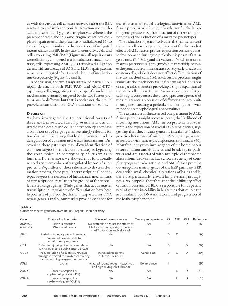
AML fusion proteins induce a mutator phenotype. The sec-ond class of AML common targets that we report hereare genes involved in diverse mechanisms of DNArepair. This class includes 17 genes that take part in var-ious pathways. Strikingly, eight genes are involved inbase excision repair (BER) (ADPRTL2, FEN1, OGG1,MPG, LIG3, POLB, POLD2, and POLD3). Null mutationsof these genes in mice are often incompatible with life,and when they are not, they generally result in anincreased sensitivity to mutagens and/or an accumula-tion of DNA damage (39, 40) (Table 5). All these genesare downregulated by AML fusion proteins, with theexception of POLB, which is induced (Table 5). Interest-ingly, overexpression of POLB in human cell lines resultsin increased spontaneous mutagenesis (39).
Validation of some of these regulations in the U937cell system (ADPRTL2, OGG1, MPG, and LIG3) or inAML patient blasts (OGG1 and LIG3) confirmed theGeneChip predictions (Figure 4, a and b). Therefore, wetested U937 cells expressing PML/RAR or AML1/ETOfor DNA repair efficiency using two approaches. First,
we analyzed by single-cell gel electrophoresis (COMETassay) the ability of U937 cells expressing PML/RAR orAML1/ETO to repair DNA damage induced by MMS(which causes single-strand DNA breaks). In this assay,damaged DNA is visualized by gel electrophoresisthrough the appearance of low-molecular-weight frag-ments or comets. A significant reduction in DNA repairwas observed in PML/RAR cells 6 hours after interrup-tion of MMS treatment, whereas AML1/ETO cellsshowed a more modest increase in comet tail momentwhen compared with control cells (Figure 4, c and d).
In a second approach, we measured the ability of lysatesfrom U937 cells to repair 8-oxoG, one of the most fre-quent endogenous DNA lesions, usually repaired by theBER pathway. Repair replication experiments were con-ducted using cell extracts and DNA plasmid substratesthat harbor a single 8-oxoG (pGEM 8-oxoG) located cen-trally (fifteenth nucleotide) within a SmaI/Hind III 33-mer restriction fragment (Figure 4e, bottom). DNAplasmid substrates harboring a single 8-oxoG within aSmaI/HindIII 33-mer restriction fragment were incubat-
Figure 4(a) The mRNA levels of OGG1, LIG3, ADPR2, andMPG in U937 PML/RAR, U937 AML1/ETO, orU937 PLZF/RAR cells, compared with Mt wereassessed by real-time RT-PCR. Shown are averagefold change values as calculated by real-time RT-PCR. (b) Relative expression of OGG1 andLIG3 genes in blasts bearing t(15;17) or t(8;21)compared with CD34+ cells, determined by realtime RT-PCR. The results shown are the averagevalues obtained from cells derived from seven dif-ferent individuals for each condition. Shown areaverage fold change values as calculated by real-time RT-PCR. (c) Representative images weretaken from alkaline comet assays of Mt, U937-PML/RAR and U937-AML1/ETO cells beforetreatment (Untreated) immediately after treat-ment with 200 µM MMS for two hours (T0), and2 and 6 hours after removal of MMS. (d) Medianrelative tail moment of more than 50 cells for eachdata point, calculated by comparison with thetotal score (100%) of initial DNA damage inducedby MMS treatment. (e) In vitro BER assay. See textfor detailed explanation. (f) Relative amount ofunligated fragments. Data are the means of fourindependent experiments. For all experiments,both fusion protein–expressing U937 cells andcontrol cells were treated with 100 µM ZnSO4 for8 hours to allow for maximal fusion proteinexpression. Mt was the control for panels a, c–f.
1760 The Journal of Clinical Investigation | December 2003 | Volume 112 | Number 11
ed with the various cell extracts recovered after the BERreaction, treated with appropriate restriction endonucle-ases, and separated by gel electrophoresis. Whereas thepresence of radiolabeled 33-mer fragments reflects com-pleted repair events, the presence of radiolabeled 15- to32-mer fragments indicates the persistence of unligatedintermediates of BER. In the case of control Mt cells andcells expressing PML/RAR (Figure 4e), all repair eventswere efficiently completed at all incubation times. In con-trast, cells expressing AML1/ETO displayed a ligationdefect, with an average of 5.5% and 12.5% repair patchesremaining unligated after 1.5 and 3 hours of incubationtime, respectively (Figure 4, e and f).
In conclusion, the two assays unraveled partial DNArepair defects in both PML/RAR- and AML1/ETO-expressing cells, suggesting that the specific molecularmechanisms primarily targeted by the two fusion pro-teins may be different, but that, in both cases, they couldprovoke accumulation of DNA mutations or lesions.
DiscussionWe have investigated the transcriptional targets ofthree AML-associated fusion proteins and demon-strated that, despite molecular heterogeneity, they havea common set of target genes seemingly relevant fortransformation, implying that leukemogenesis involvesderegulation of common molecular mechanisms. Dis-covering these pathways may allow identification ofcommon targets for antileukemic strategies, bypassingthe great molecular heterogeneity of leukemias inhumans. Furthermore, we showed that functionallyrelated genes are coherently regulated by AML fusionproteins. Regardless of their relevance to the transfor-mation process, these peculiar transcriptional pheno-types suggest the existence of hierarchical mechanismsof transcriptional regulation for groups of functional-ly related target genes. While genes that act as mastertranscriptional regulators of differentiation have beenhypothesized previously, this is unexpected for DNArepair genes. Finally, our results provide evidence for
the existence of novel biological activities of AMLfusion proteins, which might be relevant for the leuke-mogenic process (i.e., the induction of a stem cell phe-notype and the induction of a mutator phenotype).
The induction of genes involved in the maintenance ofthe stem cell phenotype might account for the modesteffects of AML-fusion protein expression on hemopoiet-ic development during the preleukemic phase of trans-genic mice (7–10). Ligand activation of Notch in murinemarrow precursors slightly (twofold to threefold) increas-es the generation or maintenance of very early precursorsor stem cells, while it does not affect differentiation ofmature myeloid cells (16). AML fusion proteins mightstimulate the machinery for self-renewing cell divisionsof target cells, therefore provoking a slight expansion ofthe stem cell compartment. An increased pool of stemcells might compensate the phenotypic consequences ofthe simultaneous repression of differentiation/commit-ment genes, creating a preleukemic hemopoiesis withminor or no morphological abnormalities.
The expansion of the stem cell compartment by AMLfusion proteins might increase, per se, the likelihood ofincoming mutations. AML fusion proteins, however,repress the expression of several DNA repair genes, sug-gesting that they induce genomic instability. Indeed,genetic alterations of various DNA repair genes areassociated with cancer predisposition in humans (40).Most frequently they involve genes of the homologousrecombination and double-strand break-repair path-ways and are associated with multiple chromosomeaberrations. Leukemias have a low frequency of com-plex cytogenetic aberrations, and AML fusion proteinsdownregulate mainly genes of the BER pathway. BERdeals with small chemical alterations of bases and is,therefore, particularly relevant for preventing mutage-nesis. We propose, therefore, that the inhibitory effectof fusion proteins on BER is responsible for a specifictype of genetic instability in leukemias that causes theaccumulation of DNA mutations and progression ofthe leukemic phenotype.
Table 5Common targets genes involved in DNA repair – BER pathway
Gene Effects of null mutations Effects of overexpression Cancer predisposition PR A1E PZR ReferencesADPRTL2 Delay in resealing No protection against the effects of NA D D (48)(PARP-2) DNA strand breaks DNA-damaging agents; can result
in ATP depletion and cell deathFEN1 Lethal in homozygous null animals; NA NA D D (49)
haploinsufficiency leads to rapid tumor progression
LIG3 Defect in rejoining of radiation-induced NA NA D D (50)DNA single- and double-strand breaks
OGG1 Accumulation of oxidative DNA base Increased repair rate Carcinomas D D D (39)damage restricted to slowly proliferating of 8-oxoG residues
tissues with high oxygen metabolismPOLB Lethal Increased spontaneous mutagenesis Breast cancer I I (39)
and high mutagenic tolerancePOLD2 Cancer susceptibility NA NA D D (51)
(by homology to POLD1)POLD3 Cancer susceptibility NA NA D D (51)
(by homology to POLD1)
The Journal of Clinical Investigation | December 2003 | Volume 112 | Number 11 1761
AcknowledgmentsThe authors are grateful to Clara Nervi, FrancescoLoCoco, and Antonio Tabilio for patient samples andCD34+ cells and to S. Minucci and F. McBlane for crit-ical reviewing of the manuscript. This work is support-ed by grants from Associazione Italiana per la Ricercasul Cancro to M. Alcalay, P.P. Di Fiore, G. Frosina, andP.G. Pelicci; from the European Community to P.G.Pelicci; and from Compagnia di S. Paolo to G. Frosina.N. Meani was supported by a fellowship from Fon-dazione Italiana per la Ricerca sul Cancro.
1. Mitelman, F. 1994. Catalogue of chromosome aberrations in Cancer. Wiley-Liss. New York, New York, USA. 4252 pp.
2. Look, A.T. 1997. Oncogenic transcription factors in the human acuteleukemias. Science. 278:1059–1064.
3. Alcalay, M., et al. 2001. Common themes in the pathogenesis of acutemyeloid leukemia. Oncogene. 20:5680–5694.
4. Grignani, F., et al. 1993. The acute promyelocytic leukemia-specificPML-RAR alpha fusion protein inhibits differentiation and promotessurvival of myeloid precursor cells. Cell. 74:423–431.
5. Gelmetti, V., et al. 1998. Aberrant recruitment of the nuclear receptorcorepressor-histone deacetylase complex by the acute myeloid leukemiafusion partner ETO. Mol. Cell Biol. 18:7185–7191.
6. Ruthardt, M., et al. 1997. Opposite effects of the acute promyelocyticleukemia PML-retinoic acid receptor alpha (RAR alpha) and PLZF-RARalpha fusion proteins on retinoic acid signalling. Mol. Cell Biol.17:4859–4869.
7. He, L.Z., et al. 1998. Distinct interactions of PML-RARalpha and PLZF-RARalpha with co-repressors determine differential responses to RA inAPL. Nat. Genet. 18:126–135.
8. Brown, D., et al. 1997. A PMLRARalpha transgene initiates murine acutepromyelocytic leukemia. Proc. Natl. Acad. Sci. U. S. A. 94:2551–2556.
9. Higuchi, M., et al. 2002. Expression of a conditional AML1-ETO onco-gene bypasses embryonic lethality and establishes a murine model ofhuman t(8;21) acute myeloid leukemia. Cancer Cell. 1:63–74.
10. Grisolano, J.L., Wesselschmidt, R.L., Pelicci, P.G., and Ley, T.J. 1997.Altered myeloid development and acute leukemia in transgenic miceexpressing PML-RAR alpha under control of cathepsin G regulatorysequences. Blood. 89:376–387.
11. Schoch, C., et al. 2002. Acute myeloid leukemias with reciprocalrearrangements can be distinguished by specific gene expression profiles.Proc. Natl. Acad. Sci. U. S. A. 99:10008–10013.
12. Alcalay, M., et al. 2003. Supplementary data. http://bio.ifom-firc.it/User/Suppl/.
13. Frosina, G., Cappelli, E., Fortini, P., and Dogliotti, E. 1999. In vitro baseexcision repair assay using mammalian cell extracts. Methods Mol. Biol.113:301–315.
14. Saiki, Y., Yamazaki, Y., Yoshida, M., Katoh, O., and Nakamura, T. 2000.Human EVI9, a homologue of the mouse myeloid leukemia gene, isexpressed in the hematopoietic progenitors and down-regulated duringmyeloid differentiation of HL60 cells. Genomics. 70:387–391.
15. Chervinsky, D.S., et al. 1999. Disordered T-cell development and T-cellmalignancies in SCL LMO1 double-transgenic mice: parallels with E2A-deficient mice. Mol. Cell Biol. 19:5025–5035.
16. Varnum-Finney, B., et al. 1998. The Notch ligand, Jagged-1, influencesthe development of primitive hematopoietic precursor cells. Blood.91:4084–4091.
17. Radomska, H.S., et al. 1998. CCAAT/enhancer binding protein alpha isa regulatory switch sufficient for induction of granulocytic developmentfrom bipotential myeloid progenitors. Mol. Cell Biol. 18:4301–4314.
18. Lord, K.A., Abdollahi, A., Hoffman-Liebermann, B., and Liebermann,D.A. 1993. Proto-oncogenes of the fos/jun family of transcription fac-tors are positive regulators of myeloid differentiation. Mol. Cell Biol.13:841–851.
19. Karsunky, H., et al. 2002. Inflammatory reactions and severe neutrope-nia in mice lacking the transcriptional repressor Gfi1. Nat. Genet.30:295–300.
20. Ward, A.C., Loeb, D.M., Soede-Bobok, A.A., Touw, I.P., and Friedman,A.D. 2000. Regulation of granulopoiesis by transcription factors andcytokine signals. Leukemia. 14:973–990.
21. Dumortier, A., Kirstetter, P., Kastner, P., and Chan, S. 2003. Ikaros regu-lates neutrophil differentiation. Blood. 101:2219–2226.
22. Li, Y.J., et al. 2001. p45(NFE2) is a negative regulator of erythroid prolif-eration which contributes to the progression of Friend virus-inducederythroleukemias. Mol. Cell Biol. 21:73–80.
23. Bain, G., et al. 1994. E2A proteins are required for proper B cell develop-ment and initiation of immunoglobulin gene rearrangements. Cell.79:885–892.
24. Sexl, V., et al. 2000. Stat5a/b contribute to interleukin 7-induced B-cellprecursor expansion, but abl- and bcr/abl-induced transformation areindependent of stat5. Blood. 96:2277–2283.
25. Zhu, J., and Emerson, S.G. 2002. Hematopoietic cytokines, transcriptionfactors and lineage commitment. Oncogene. 21:3295–3313.
26. Satterwhite, E., et al. 2001. The BCL11 gene family: involvement ofBCL11A in lymphoid malignancies. Blood. 98:3413–3420.
27. Zhang, D.E., et al. 1997. Absence of granulocyte colony-stimulating fac-tor signaling and neutrophil development in CCAAT enhancer bindingprotein alpha-deficient mice. Proc. Natl. Acad. Sci. U. S. A. 94:569–574.
28. Passegue, E., Jochum, W., Schorpp-Kistner, M., Mohle-Steinlein, U., andWagner, E.F. 2001. Chronic myeloid leukemia with increased granulo-cyte progenitors in mice lacking junB expression in the myeloid lineage.Cell. 104:21–32.
29. Hermans, M.H., et al. 1998. Perturbed granulopoiesis in mice with a tar-geted mutation in the granulocyte colony-stimulating factor receptorgene associated with severe chronic neutropenia. Blood. 92:32–39.
30. Buitenhuis, M., Baltus, B., Lammers, J.W., Coffer, P.J., and Koenderman,L. 2002. Signal transducer and activator of transcription 5a (STAT5a) isrequired for eosinophil differentiation of human cord blood derivedCD34+ cells. Blood. 101:134–142.
31. Engel, I., and Murre, C. 1999. Ectopic expression of E47 or E12 promotesthe death of E2A-deficient lymphomas. Proc. Natl. Acad. Sci. U. S. A.96:996–1001.
32. Pabst, T., et al. 2001. Dominant-negative mutations of CEBPA, encod-ing CCAAT/enhancer binding protein-alpha (C/EBPalpha), in acutemyeloid leukemia. Nat. Genet. 27:263–270.
33. Aspland, S.E., Bendall, H.H., and Murre, C. 2001. The role of E2A-PBX1in leukemogenesis. Oncogene. 20:5708–5717.
34. Winandy, S., Wu, P., and Georgopoulos, K. 1995. A dominant mutationin the Ikaros gene leads to rapid development of leukemia and lym-phoma. Cell. 83:289–299.
35. Pabst, T., et al. 2001. AML1-ETO downregulates the granulocytic differ-entiation factor C/EBPalpha in t(8;21) myeloid leukemia. Nat. Med.7:444–451.
36. Kopan, R. 2002. Notch: a membrane-bound transcription factor. J. CellSci. 115:1095–1097.
37. Moloney, D.J., et al. 2000. Fringe is a glycosyltransferase that modifiesNotch. Nature. 406:369–375.
38. Kawamata, S., Du, C., Li, K., and Lavau, C. 2002. Overexpression of theNotch target genes Hes in vivo induces lymphoid and myeloid alter-ations. Oncogene. 21:3855–3863.
39. Frosina, G. 2000. Overexpression of enzymes that repair endogenousdamage to DNA. Eur. J. Biochem. 267:2135–2149.
40. Hoeijmakers, J.H. 2001. Genome maintenance mechanisms for prevent-ing cancer. Nature. 411:366–374.
41. Seki, Y., et al. 2002. The ETS transcription factor MEF is a candidatetumor suppressor gene on the X chromosome. Cancer Res. 62:6579–6586.
42. English, B.K. 1996. Expression of the activated (Y501-F501) hck tyrosinekinase in 32Dcl3 myeloid cells prolongs survival in the absence of IL-3and blocks granulocytic differentiation in response to G-CSF. J. Leukoc.Biol. 60:667–673.
43. Ilaria, R.L., Jr., Hawley, R.G., and Van Etten, R.A. 1999. Dominant nega-tive mutants implicate STAT5 in myeloid cell proliferation and neu-trophil differentiation. Blood. 93:4154–4166.
44. Yan, W., et al. 1997. High incidence of T-cell tumors in E2A-null miceand E2A/Id1 double- knockout mice. Mol. Cell Biol. 17:7317–7327.
45. Tanaka, J., Iwata, M., Graf, L., Guest, I., and Torok-Storb, B. 1999. Stro-mal inhibition of myeloid differentiation. A possible role for hJagged1.Ann. N. Y. Acad. Sci. 872:171–172; discussion 172–175.
46. Karanu, F.N., et al. 2000. The notch ligand jagged-1 represents a novelgrowth factor of human hematopoietic stem cells. J. Exp. Med.192:1365–1372.
47. Valge-Archer, V., Forster, A., and Rabbitts, T.H. 1998. The LMO1 andLDB1 proteins interact in human T cell acute leukaemia with the chro-mosomal translocation t(11;14)(p15;q11). Oncogene. 17:3199–3202.
48. Schreiber, V., et al. 2002. Poly(ADP-ribose) polymerase-2 (PARP-2) isrequired for efficient base excision DNA repair in association with PARP-1 and XRCC1. J. Biol. Chem. 277:23028–23036.
49. Kucherlapati, M., et al. 2002. Haploinsufficiency of Flap endonuclease(Fen1) leads to rapid tumor progression. Proc. Natl. Acad. Sci. U. S. A.99:9924–9929.
50. Nocentini, S. 1999. Rejoining kinetics of DNA single- and double-strandbreaks in normal and DNA ligase-deficient cells after exposure to ultra-violet C and gamma radiation: an evaluation of ligating activitiesinvolved in different DNA repair processes. Radiat. Res. 151:423–432.
51. Goldsby, R.E., et al. 2001. Defective DNA polymerase-delta proofread-ing causes cancer susceptibility in mice. Nat. Med. 7:638–639.


![[XLS]doa.alaska.govdoa.alaska.gov/ogc/DigLog/CookInlet/CIindex.xls · Web viewILD SN SP DT GR MLS MLD CALI MGS ST 17595 20 SFL ILM ILD GR DRHO SP DT CNS CALI RHOB MGS ST 17595 23](https://static.fdocuments.us/doc/165x107/5b00bdd17f8b9a952f8d5544/xlsdoa-viewild-sn-sp-dt-gr-mls-mld-cali-mgs-st-17595-20-sfl-ilm-ild-gr-drho-sp.jpg)