
Acute liver failure - WordPress.com
13
Seminar www.thelancet.com Vol 394 September 7, 2019 869 Acute liver failure R Todd Stravitz, William M Lee Acute liver failure is a rare and severe consequence of abrupt hepatocyte injury, and can evolve over days or weeks to a lethal outcome. A variety of insults to liver cells result in a consistent pattern of rapid-onset elevation of aminotransferases, altered mentation, and disturbed coagulation. The absence of existing liver disease distinguishes acute liver failure from decompensated cirrhosis or acute-on-chronic liver failure. Causes of acute liver failure include paracetamol toxicity, hepatic ischaemia, viral and autoimmune hepatitis, and drug-induced liver injury from prescription drugs, and herbal and dietary supplements. Diagnosis requires careful review of medications taken, and serological testing for possible viral exposure. Because of its rarity, acute liver failure has not been studied in large, randomised trials, and most treatment recommendations represent expert opinion. Improvements in management have resulted in lower mortality, although liver transplantation, used in nearly 30% of patients with acute liver failure, still provides a life-saving alternative to medical management. Introduction The term acute liver failure applies to a unique and infrequently observed syndrome of severe injury to liver cells, which leads to altered coagulation and mentation in the absence of chronic liver disease. Acute liver failure is unexpected and has a rapid onset, affecting previously healthy individuals, with a frequently fatal outcome. Despite having diverse causes (figure 1), acute liver failure is characterised by remarkably similar clinical features. The principal manifestations of acute liver failure are prolonged international normalised ratio of prothrombin time and hepatic encephalopathy, which were first described in 1970 2 as fulminant hepatic failure, a term which has now been largely abandoned. Acute liver failure should not be confused with acute-on- chronic liver failure, 3 which is the acute decompensation of cirrhosis resulting in the failure of extrahepatic organ systems. Three causes of acute liver failure can present as a fulminating acute decompensation of chronic liver disease, and might still be considered cases of acute liver failure: Wilson disease, reactivation of chronic hepatitis B infection, and autoimmunity. Other common features of acute liver failure include cardiovascular instability, susceptibility to infection, acute kidney injury, and, most unique among liver diseases, the presence of cerebral oedema. 4 Despite its rarity, acute liver failure generates interest and study by multiple disciplines because it affects all organ systems, requires substantial resource use, and has a 30% mortality. Interest in studying acute liver failure is also driven by the insight the syndrome provides into the pathogenesis of liver injury and liver failure itself. Estimates of the incidence of acute liver failure are difficult to obtain, but range from 1 case per million people per year to about 2–3000 cases per year in the USA. 4 The overall incidence of acute liver failure might be declining, but as a result of its rarity and heterogeneity, few population-based studies have been published. Most data originate from large registries in the USA and Europe and are descriptive in nature. All- encompassing guidelines and position papers have been written about acute liver failure, reflecting expert opinion rather than evidence-based medicine. 6–8 Substances that lead to hepatocyte injury cause either direct toxic necrosis, or apoptosis and immune injury, which is a slower process. The time from the onset of symptoms to the onset of hepatic encephalopathy distinguishes the different forms of acute liver failure: a direct, very rapid injury (within hours), referred to as hyperacute liver failure; and a slower, immune-based injury (days to weeks), considered acute or subacute. Although previous reviews have categorised these dif- ferent forms into three timeframes (hyperacute, acute, and subacute), 9,10 it seems more logical to group acute and subacute into a single syndrome, because their speed of evolution overlaps, whereas patients with hyperacute liver failure have a distinct disease pattern. Hyperacute causes, principally paracetamol toxicity (referred to as acetaminophen in North America) and ischaemia, become clinically detectable within 6–36 h of insult, resolving within 3–5 days. By contrast, slower evolving injuries usually take 7 days to 24 weeks to become clinically evident. In this category of slowly progressing liver failure (caused by hepatitis B virus, autoimmunity, or drug-induced liver injury), evolution might vary, but more than 7 days are reliably required before there are signs of liver failure. Hyperacute causes feature very high aminotransferase concen- trations, and low bilirubin concentrations, whereas acute and subacute causes have lower aminotransferase Search strategy and selection criteria We searched PubMed and MEDLINE using the terms “acute liver failure” or “fulminant hepatic failure” to identify studies and publications not already familiar to the authors, with no restrictions on date or location of publication. The final search occurred on March 2, 2019. We reviewed our findings to identify work relevant to acute liver failure and chose references that provided the most insight and that were, where possible, evidence-based rather than expert opinion, so that readers could understand current knowledge of various aspects of both diagnosis and critical care of patients with acute liver failure. Lancet 2019; 394: 869–81 Hume-Lee Transplant Center of Virginia Commonwealth University, Richmond, VA, USA (R T Stravitz MD); and Digestive and Liver Diseases Division, University of Texas Southwestern Medical Center at Dallas, Dallas, TX, USA (W M Lee MD) Correspondence to: Dr William M Lee, Digestive and Liver Diseases Division, University of Texas Southwestern Medical Center at Dallas, 5959 Harry Hines Boulevard, Dallas, TX 75390–8887, USA william.lee@utsouthwestern. edu Downloaded for Anonymous User (n/a) at Stockholm County Council from ClinicalKey.com by Elsevier on April 15, 2021. For personal use only. No other uses without permission. Copyright ©2021. Elsevier Inc. All rights reserved.
Transcript of Acute liver failure - WordPress.com

Seminar
www.thelancet.com Vol 394 September 7, 2019 869
Acute liver failureR Todd Stravitz, William M Lee
Acute liver failure is a rare and severe consequence of abrupt hepatocyte injury, and can evolve over days or weeks to a lethal outcome. A variety of insults to liver cells result in a consistent pattern of rapid-onset elevation of aminotransferases, altered mentation, and disturbed coagulation. The absence of existing liver disease distinguishes acute liver failure from decompensated cirrhosis or acute-on-chronic liver failure. Causes of acute liver failure include paracetamol toxicity, hepatic ischaemia, viral and autoimmune hepatitis, and drug-induced liver injury from prescription drugs, and herbal and dietary supplements. Diagnosis requires careful review of medications taken, and serological testing for possible viral exposure. Because of its rarity, acute liver failure has not been studied in large, randomised trials, and most treatment recommendations represent expert opinion. Improvements in management have resulted in lower mortality, although liver transplantation, used in nearly 30% of patients with acute liver failure, still provides a life-saving alternative to medical management.
IntroductionThe term acute liver failure applies to a unique and infrequently observed syndrome of severe injury to liver cells, which leads to altered coagulation and mentation in the absence of chronic liver disease. Acute liver failure is unexpected and has a rapid onset, affecting previously healthy individuals, with a frequently fatal outcome. Despite having diverse causes (figure 1), acute liver failure is characterised by remarkably similar clinical features. The principal manifestations of acute liver failure are prolonged international normalised ratio of prothrombin time and hepatic encephalopathy, which were first described in 19702 as fulminant hepatic failure, a term which has now been largely abandoned. Acute liver failure should not be confused with acute-on-chronic liver failure,3 which is the acute decompensation of cirrhosis resulting in the failure of extrahepatic organ systems. Three causes of acute liver failure can present as a fulminating acute decompensation of chronic liver disease, and might still be considered cases of acute liver failure: Wilson disease, reactivation of chronic hepatitis B infection, and autoimmunity. Other common features of acute liver failure include cardiovascular instability, susceptibility to infection, acute kidney injury, and, most unique among liver diseases, the presence of cerebral oedema.4 Despite its rarity, acute liver failure generates interest and study by multiple disciplines because it affects all organ systems, requires substantial resource use, and has a 30% mortality. Interest in studying acute liver failure is also driven by the insight the syndrome provides into the pathogenesis of liver injury and liver failure itself. Estimates of the incidence of acute liver failure are difficult to obtain, but range from 1 case per million people per year to about 2–3000 cases per year in the USA.4 The overall incidence of acute liver failure might be declining, but as a result of its rarity and heterogeneity, few population-based studies have been published. Most data originate from large registries in the USA and Europe and are descriptive in nature. All-encompassing guidelines and position papers have been written about acute liver failure, reflecting expert opinion rather than evidence-based medicine.6–8
Substances that lead to hepatocyte injury cause either direct toxic necrosis, or apoptosis and immune injury, which is a slower process. The time from the onset of symptoms to the onset of hepatic encephalopathy distinguishes the different forms of acute liver failure: a direct, very rapid injury (within hours), referred to as hyperacute liver failure; and a slower, immune-based injury (days to weeks), considered acute or subacute. Although previous reviews have categorised these dif-ferent forms into three timeframes (hyperacute, acute, and subacute),9,10 it seems more logical to group acute and subacute into a single syndrome, because their speed of evolution overlaps, whereas patients with hyperacute liver failure have a distinct disease pattern. Hyperacute causes, principally paracetamol toxicity (referred to as acetaminophen in North America) and ischaemia, become clinically detectable within 6–36 h of insult, resolving within 3–5 days. By contrast, slower evolving injuries usually take 7 days to 24 weeks to become clinically evident. In this category of slowly progressing liver failure (caused by hepatitis B virus, autoimmunity, or drug-induced liver injury), evolution might vary, but more than 7 days are reliably required before there are signs of liver failure. Hyper acute causes feature very high aminotransferase concen-trations, and low bilirubin concentrations, whereas acute and subacute causes have lower aminotransferase
Search strategy and selection criteria
We searched PubMed and MEDLINE using the terms “acute liver failure” or “fulminant hepatic failure” to identify studies and publications not already familiar to the authors, with no restrictions on date or location of publication. The final search occurred on March 2, 2019. We reviewed our findings to identify work relevant to acute liver failure and chose references that provided the most insight and that were, where possible, evidence-based rather than expert opinion, so that readers could understand current knowledge of various aspects of both diagnosis and critical care of patients with acute liver failure.
Lancet 2019; 394: 869–81
Hume-Lee Transplant Center of Virginia Commonwealth University, Richmond, VA, USA (R T Stravitz MD); and Digestive and Liver Diseases Division, University of Texas Southwestern Medical Center at Dallas, Dallas, TX, USA (W M Lee MD)
Correspondence to: Dr William M Lee, Digestive and Liver Diseases Division, University of Texas Southwestern Medical Center at Dallas, 5959 Harry Hines Boulevard, Dallas, TX 75390–8887, USA [email protected]
Downloaded for Anonymous User (n/a) at Stockholm County Council from ClinicalKey.com by Elsevier on April 15, 2021. For personal use only. No other uses without permission. Copyright ©2021. Elsevier Inc. All rights reserved.
Seminar
870 www.thelancet.com Vol 394 September 7, 2019
concentrations and higher bilirubin concentrations. In general, patients with hyperacute liver injuries have better short-term survival than do patients with slowly progressing liver injuries, who do less well (table 1). Prognosis for acute liver failure has been linked to duration of illness, but the cause of the injury is a better predictor of outcome than the time of evolution. This Seminar highlights recent progress and future directions
for a better understanding and management of this unusual and intriguing condition.
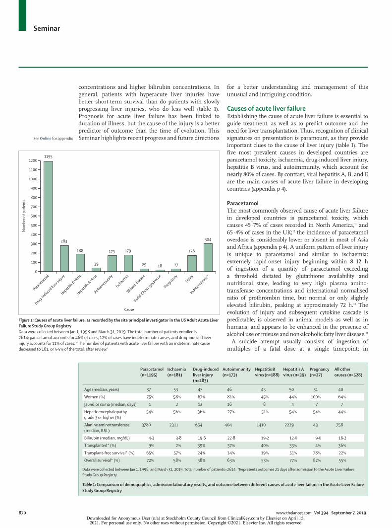
Causes of acute liver failureEstablishing the cause of acute liver failure is essential to guide treatment, as well as to predict outcome and the need for liver transplantation. Thus, recognition of clinical signatures on presentation is paramount, as they provide important clues to the cause of liver injury (table 1). The five most prevalent causes in developed countries are paracetamol toxicity, ischaemia, drug-induced liver injury, hepatitis B virus, and autoimmunity, which account for nearly 80% of cases. By contrast, viral hepatitis A, B, and E are the main causes of acute liver failure in developing countries (appendix p 4).
ParacetamolThe most commonly observed cause of acute liver failure in developed countries is paracetamol toxicity, which causes 45·7% of cases recorded in North America,11 and 65·4% of cases in the UK;12 the incidence of paracetamol overdose is considerably lower or absent in most of Asia and Africa (appendix p 4). A uniform pattern of liver injury is unique to paracetamol and similar to ischaemia: extremely rapid-onset injury beginning within 8–12 h of ingestion of a quantity of paracetamol exceeding a threshold dictated by glutathione availability and nutritional state, leading to very high plasma amino-transferase concentrations and international normalised ratio of prothrombin time, but normal or only slightly elevated bilirubin, peaking at approximately 72 h.13 The evolution of injury and subsequent cytokine cascade is predictable, is observed in animal models as well as in humans, and appears to be enhanced in the presence of alcohol use or misuse and non-alcoholic fatty liver disease.14
A suicide attempt usually consists of ingestion of multiples of a fatal dose at a single timepoint; in
Figure 1: Causes of acute liver failure, as recorded by the site principal investigator in the US Adult Acute Liver Failure Study Group RegistryData were collected between Jan 1, 1998 and March 31, 2019. The total number of patients enrolled is 2614; paracetamol accounts for 46% of cases, 12% of cases have indeterminate causes, and drug-induced liver injury accounts for 11% of cases. *The number of patients with acute liver failure with an indeterminate cause decreased to 161, or 5·5% of the total, after review.1
1195
283
188
39
173 179
29 18 27
176
304
Paracetamol
Drug-induced liv
er injury
Hepatitis A
virus
Hepatitis B
virus
Autoimmunity
Ischaemia
Wilson dise
ase
Budd-Chiari syndrome
PregnancyOther
Indeterminate*
0
100
200
300
400
500
600
700
800
900
1000
1100
1200
Num
ber o
f pat
ient
s
Cause
Paracetamol (n=1195)
Ischaemia (n=181)
Drug-induced liver injury (n=283)
Autoimmunity (n=173)
Hepatitis B virus (n=188)
Hepatitis A virus (n=39)
Pregnancy (n=27)
All other causes (n=528)
Age (median, years) 37 53 47 46 45 50 31 40
Women (%) 75% 58% 67% 81% 45% 44% 100% 64%
Jaundice coma (median, days) 1 2 12 16 8 4 7 7
Hepatic encephalopathy grade 3 or higher (%)
54% 56% 36% 27% 51% 54% 54% 44%
Alanine aminotransferase (median, IU/L)
3780 2311 654 404 1410 2229 43 758
Bilirubin (median, mg/dL) 4·3 3·8 19·6 22·8 19·2 12·0 9·0 16·2
Transplanted* (%) 9% 2% 39% 57% 40% 33% 4% 36%
Transplant-free survival* (%) 65% 57% 24% 14% 19% 51% 78% 22%
Overall survival* (%) 72% 58% 58% 63% 53% 77% 82% 55%
Data were collected between Jan 1, 1998, and March 31, 2019. Total number of patients=2614. *Represents outcomes 21 days after admission to the Acute Liver Failure Study Group Registry.
Table 1: Comparison of demographics, admission laboratory results, and outcome between different causes of acute liver failure in the Acute Liver Failure Study Group Registry
See Online for appendix
Downloaded for Anonymous User (n/a) at Stockholm County Council from ClinicalKey.com by Elsevier on April 15, 2021. For personal use only. No other uses without permission. Copyright ©2021. Elsevier Inc. All rights reserved.

Seminar
www.thelancet.com Vol 394 September 7, 2019 871
other instances, ongoing use of paracetamol at excessive daily doses, often with hydrocodone or other opioids, has a very similar result.15,16 Paracetamol is present and measurable in plasma shortly after ingestion, but has a short half-life; thus, only about half of patients with acute liver injury or acute liver failure will show any of the parent compound.17 Detection of paracetamol adducts in plasma, which are toxic byproducts of cell injury (paracetamol bound to cell proteins), have a longer plasma half-life than the parent compound, and might provide a valuable confirmatory test for paracetamol as the cause of acute liver failure, particularly for ingestions at multiple timepoints or more than 3 days before medical care.18,19 Acetylcysteine prevents liver injury if taken within 12 h of paracetamol ingestion and might be beneficial at later timepoints, although this is less certain.17,20 Despite efforts to reduce fatalities after para-cetamol overdose, whether intentional or unintentional, little progress has been made.11,21
Hepatic ischaemiaInadequate perfusion of oxygenated blood to the liver has various causes, and leads to a hyperacute but self-limited hepatic injury, characterised by very high aminotransferase concentrations and low bilirubin concentrations. With few exceptions (eg, heat stroke and cocaine toxicity), the differential diagnosis of aminotransferase concentrations of more than around 3000 IU/L and bilirubin less than 5·0 mg/dL includes mainly ischaemia and paracetamol (table 1). Ischaemia that is due to severe heart failure can be readily characterised by an echocardiogram showing depressed left-ventricular ejection fraction. Other causes of poor perfusion include hypotension due to depletion of blood volume (ie, shock), septic shock, hypoxia (such as respiratory depression due to opioids), pulmonary failure, and cocaine use. Acute kidney injury occurs in nearly equal numbers of patients as does acute liver injury in these settings. Although the short-term prognosis for acute liver failure caused by ischaemia is good because intravascular volume can be restored and cardiac haemodynamics improved quickly, the long-term outcome is uncertain if the underlying cause of haemodynamic failure is not reversible.22 Ischaemic liver injury rarely leads to a requirement for liver transplantation, unless the cause is a direct compromise of the hepatic circulation (eg, inadvertent surgical occlusion of the hepatic artery).23
Drug-induced liver injuryIdiosyncratic, drug-induced liver injury is the second most common cause of acute liver failure after paracetamol,11 and has a different pathogenesis with a worse prognosis.24 Only 10% of all patients with drug-induced liver injury develop acute liver failure, with the potential for liver transplantation or death.25 The pathogenesis of drug-induced liver injury is thought to be
immune based, with some individuals having a genetic predisposition, caused by unique HLA haplotypes;26 few unique polymorphisms responsible for liver injury have been identified.27 Antibiotics account for nearly 50% of all cases of drug-induced liver injury, partly because they are frequently prescribed, but also because they typically require high daily doses (ie, at least 500–1000 mg per day), which increases the risk of liver injury.28 By contrast with viral-induced acute liver failure, there are no specific diagnostic tests for drug-induced liver injury. Identification of a specific drug requires a careful history and exclusion of other causes.29 By contrast with ischaemia and paracetamol injuries, drug-induced liver injuries reaching the threshold of acute liver failure present with lower aminotransferase con-centrations, higher bilirubin concentrations, and worse overall outcomes (table 1), with fewer than 25% of patients recovering after any degree of encephalopathy has occurred.24,30
Autoimmune hepatitisAcute liver failure caused by autoimmune hepatitis presents as an acute-to-subacute liver injury that might defy initial diagnosis. This type of acute liver failure frequently begins insidiously and is suggested, but not definitively diagnosed, by the presence of hyper-globulinaemia, antinuclear antibodies, or anti-smooth-muscle antibodies, or any combination of these. 80% of patients with acute liver failure caused by autoimmune hepatitis are women. A liver biopsy could be helpful in diagnosing acute liver failure caused by autoimmune hepatitis.31 Encephalopathy might mean that it is challenging to take biopsy samples safely, although the transjugular approach is now widely used in tertiary centres.32 Many instances of indeterminate acute liver failure might actually be caused by autoimmune hepatitis, but specific diagnostic tests are absent. Similar to drug-induced liver injury, acute liver failure caused by auto-immune hepatitis has particularly poor outcomes and frequently requires liver transplantation (table 1).
Hepatitis B and hepatitis AIn the USA and Europe, hepatitis B virus causes around 8% of all cases of acute liver failure, considerably fewer than paracetamol, drug-induced liver injury, or autoimmune hepatitis, but this virus is still an important and readily diagnosed cause. Two-thirds of cases of hepatitis B are caused by new infections, and the remainder are caused by reactivation of often unrecognised chronic hepatitis B infection in the setting of chemotherapy or other immune modulation.33 Acute liver failure caused by reactivation of hepatitis B infection occurs when immunosuppression allows increased viral replication that had been at least partially contained by host immune surveillance and carries a worse prognosis than that caused by de-novo hepatitis B infection.34 Reactivation of hepatitis B infection might occur even in patients who
Downloaded for Anonymous User (n/a) at Stockholm County Council from ClinicalKey.com by Elsevier on April 15, 2021. For personal use only. No other uses without permission. Copyright ©2021. Elsevier Inc. All rights reserved.

Seminar
872 www.thelancet.com Vol 394 September 7, 2019
appeared to have cleared the infection—ie, those who are HBsAg negative when subjected to intense immune suppression.35 Early treatment after diagnosis once liver failure is present is of uncertain value.36,37
Hepatitis A virus infection generally has a mild course and seldom causes acute liver failure, causing fewer than 3% of cases of acute liver failure in developed countries. Outbreaks of hepatitis A infection among homeless people in the USA in the last 3 years have led to a spike in hospitalisations, highlighting the infrequent use of hepatitis A vaccines in the general population.38 Acute liver failure caused by hepatitis A has nearly 70% spontaneous or transplant-free survival (table 1), a better overall outcome than that caused by other viruses.39
Other causes of acute liver failureThe remaining causes of acute liver failure comprise fewer than 15% of the total and include heat stroke, pregnancy-associated injury (eg, acute fatty liver of preg-nancy and HELLP [haemolysis, elevated liver enzyme, and low platelet] syndrome), Budd-Chiari syndrome, non-hepatotrophic viral infections such as herpes simplex, and diffusely infiltrating malignancies.40–44 Heat stroke resembles ischaemia in its features of vascular collapse and hyperacute evolution, and occasionally requires liver transplantation.45 Fulminant Wilson disease presents with fibrosis or well established cirrhosis and a unique clinical signature, including intense intravascular haemo-lysis and acute kidney injury, that allows early diagnosis.46 Only rarely can a patient with this condition recover without liver transplantation, despite the best intensive care. Acute hepatitis C virus infection rarely progresses to acute liver failure; well documented cases that do not involve underlying non-alcoholic fatty liver disease or pre-existing cirrhosis are difficult to find in the literature. Hepatitis E virus is a major cause of acute liver failure in Asia, parts of Africa, and the Indian subcontinent; the virus occasionally causes infection and acute liver failure in Europe, but is rarely encountered in the USA.47,48
Indeterminate acute liver failureA small percentage of cases of acute liver failure might have an indeterminate cause—ie, assignment of a cause is impossible with the available data. Historically, it was suggested that the disease in these patients was brought about by unrecognised or occult viruses.49 However, it is more likely that patients with indeterminate acute liver failure have been incompletely evaluated—eg, by insuf-ficient history-taking (in patients with encephalopathy), or absence of full serological testing. Intensive screening using new methods, such as the paracetamol adduct test, deep sequencing for viruses, and adjudication by independent experts, reaches a diagnosis in at least 50% of patients with acute liver failure with an initially indeterminate cause;1 only around 5% of all diagnoses of acute liver failure therefore have indeterminate causes.
Pathogenesis and establishing a diagnosisThe pathology of acute liver failure on explant or autopsy most often shows widespread hepatic necrosis and apoptosis, with few remaining viable hepatocytes; regenerative nodules are often seen (figure 2). Liver weight can decrease from 1600 g to around 500 g, leaving only the hepatic stromal framework. Failure of regeneration is also an important feature of acute liver failure, at least for some causes. Human genetic factors probably have a role in confining liver injury to a small fraction of patients exposed to a drug or virus,26,50,51 and some HLA alleles have been associated with this effect.52,53
Although the striking presentation of most patients with acute liver failure seldom leaves the diagnosis in question, some patients present a conundrum, with a differential diagnosis that includes biliary sepsis, alcoholic hepatitis, and acute-on-chronic liver failure. Beyond a thorough history and physical examination, a standard battery of tests and considerations should be applied to all patients with acute liver failure to establish the correct cause (appendix p 1). There are some excep-tions: if the diagnosis is obvious, such as an admitted paracetamol overdose with detectable paracetamol in the blood, then searching for other causes is pointless.1 However, when no clear diagnosis is apparent after basic viral and autoimmune serologies, more intensive, second-tier testing is indicated (appendix p 1). Because the cause of acute liver failure influences the prognosis, and the time to correct diagnosis might be crucial, the most rapid and available methods of testing are required. For example, Wilson disease that is causing acute liver failure can be reliably discerned by measurements of haemo globin, alkaline phosphatase, and bilirubin, rendering copper measurements in plasma or urine con-firmatory.46 Likewise, herpes simplex virus IgM is reported more rapidly than nucleic acid assays, and is therefore preferred if infection is suspected.41 A smart-phone application for clinicians who treat acute liver failure is now available and includes a diagnostic and daily checklist to remind clinicians to review routine diagnostic and therapeutic measures.54
ManagementGeneral considerationsThe management of patients with acute liver failure varies between individual centres and physicians because of a dearth of randomised clinical trials. A liver trans plantation centre should be consulted regarding possible transfer shortly after stabilising the patient, because the complexity of care is beyond the scope of all but tertiary-care hospitals with the capability to do liver transplantations.
Cause-specific managementFew data support the efficacy of any cause-specific therapy for the primary liver injury (appendix p 2). Women with
Downloaded for Anonymous User (n/a) at Stockholm County Council from ClinicalKey.com by Elsevier on April 15, 2021. For personal use only. No other uses without permission. Copyright ©2021. Elsevier Inc. All rights reserved.

Seminar
www.thelancet.com Vol 394 September 7, 2019 873
acute liver failure who present with pre-eclampsia and liver injury in their third trimester of pregnancy should be promptly delivered, and thereafter generally have a favourable prognosis,43 although some women continue to deteriorate post partum.55,56 Patients with acute liver failure associated with hepatitis B virus should receive nucleos(t)ide hepatitis B virus DNA polymerase inhibitors to reduce the risk of recurrence after liver transplantation.37 Unfortunately, a randomised controlled trial did not show improvement in transplant-free survival in patients with severe, acute hepatitis B virus disease who received lamivudine compared with placebo.57 Any patient with acute liver failure that is suspected to be secondary to infection with herpes simplex virus on the basis of clinical features (ie, fever, skin or mucous membrane vesicles, or a patient who is immunosuppressed)58 should receive aciclovir while awaiting nucleic acid confirmation; however, favourable responses are uncommon.59 Amanita phalloides (and related species) mushroom poisoning, which causes acute liver failure, is rare in the USA, but occurs more frequently worldwide;60 anecdotal case reports suggest a potential benefit of silibinin, as well as acetylcysteine, penicillins, and nasobiliary drainage.61,62 Even in acute liver failure caused by autoimmune hepatitis, corticosteroids have not been convincingly shown to improve outcome.63 A trial of corticosteroids should be administered to patients with low-grade (grade 1 or 2) hepatic enceph alopathy due to autoimmune hepatitis, and only for a finite period of time (around 1 week) to avoid increasing the risk of peritransplantation infection.64
Acetylcysteine has been used as a treatment for paracetamol overdose for decades on the basis of a study using historical controls,65 and of small, randomised trials showing improved survival,66 with little toxicity and a thoroughly defined mechanism of action.67 However, the quality of the data on the optimal route of administration (ie, oral or intravenous), dose, and even the efficacy of acetylcysteine for paracetamol overdose is poor.68 Despite this poor data quality, authorities advise acetylcysteine administration when paracetamol overdose is first suspected, intravenously to ensure delivery, even for patients without an ingestion history or when paracetamol is undetected in the blood.17 Acetylcysteine might also be effective in treating patients with acute liver failure with causes other than paracetamol overdose; several uncon-trolled studies suggest improved outcome in these patients when they receive acetyl cysteine,69–71 but only one study in adults was randomised and placebo-controlled.72 Although this study did not meet its primary endpoint of improved overall survival, acetylcysteine was associated with improved transplant-free survival in patients with grade 1 or 2 hepatic encephalopathy, a secondary endpoint.
Management of liver failureDespite little change in the severity of illness during the past two decades, outcomes for patients with acute liver
failure have improved because of more widespread options for liver transplantation and improved intensive care. Improved outcomes have occurred despite lower use of resources in intensive care units (eg, vasopressors,
Figure 2: Examples of histopathology of acute liver failure(A) Normal liver at low power (100×). (B) Higher power (400×) view of a normal hepatic lobule showing a portal tract. (C) Higher power (400×) view of a normal hepatic lobule showing central hepatic vein. (D) Isoniazid toxicity with complete parenchymal loss, no hepatocytes remain and the normal parenchyma has been replaced by ductular reaction (400×). (E) Heterogeneous areas of necrosis and parenchymal regeneration observed with nitrofurantoin toxicity (40×). (F) Regenerative nodules are rimmed by chronic inflammation at higher power (100×) in the same case as in (E); the patient had taken nitrofurantoin intermittently for nearly 6 months before developing liver failure. Regenerative nodules are often seen in this setting and might be confused on imaging with cirrhosis. (G) Paracetamol toxicity with zone 3 (centrilobular) necrosis. Many hepatocytes show steatosis and in the necrotic zone, lipid drops have coalesced (200×). (H) Acute liver failure related to hepatitis B, with prominent plasma-cell infiltrate within a portal tract, a unique but common finding in fulminant hepatitis B (600×).50 Representative explants were drawn from the Acute Liver Failure Study Group pathology registry, courtesy of David E Kleiner, Laboratory of Pathology, National Cancer Institute, Bethseda, MD, USA.
A
C
E
G
B
D
F
H
Downloaded for Anonymous User (n/a) at Stockholm County Council from ClinicalKey.com by Elsevier on April 15, 2021. For personal use only. No other uses without permission. Copyright ©2021. Elsevier Inc. All rights reserved.

Seminar
874 www.thelancet.com Vol 394 September 7, 2019
blood products, ventilation, and intracranial pressure monitors) over the same time period.11 Innovative tech-nologies, such as albumin dialysis and bioartificial devices using hepatocyte cell lines or porcine hepatocyte bioreactors, have yielded poor results. Although liver-support devices might transiently improve biochemical indices of liver function, none has been shown to reliably bridge patients with acute liver failure to recovery with their native or a transplanted liver.73
In many regards, high-volume plasma exchange recapitulates liver support, because it removes metabolic end-products, toxins, and pro-inflammatory mediators, and replaces many beneficial, liver-derived proteins. In a multicentre, randomised study of standard medical treatment versus standard treatment plus high-volume plasma exchange,74 survival to hospital discharge stratified for liver transplantation was higher in patients who received high-volume plasma exchange. Thus, this treat-ment might be more readily available and better substantiated than other liver-support devices, but further study is required.
Management of systemic complications of acute liver failureThe most common cause of death in patients with acute liver failure is systemic complications, which follow the release of pro-inflammatory cytokines and damage-associated molecular patterns from necrotic hepatocytes, endothelial cells, and leucocytes.75,76 No clinical trial of specific inhibitors of any of these mediators has been done in humans. Consequently, the following discussion will outline the management of failure of individual organ systems.
In patients with acute liver failure, multisystem organ failure often appears as a battery of affected systems,77 including the cardiovascular system, heralded by systemic inflammatory response syndrome and haemodynamic instability. On presentation, patients with acute liver failure are often hypotensive because of volume depletion; therefore, the first intervention should include intra-venous normal saline. Persistent hypotension (mean arterial pressure <60 mm Hg) despite volume repletion should prompt vasopressor support; norepinephrine has been suggested by consensus as the preferred initial vasopressor.78 Cultures of blood and urine should be considered on admission for all patients with hypotension. Hypotension despite volume repletion and norepinephrine should prompt the addition of vasopressin. Persistent hypo tension despite norepinephrine and vasopressin or terlipressin warrants consideration of treatment with hydrocortisone (300 mg intravenously), because relative adrenal insufficiency has been documented in a sub-stantial proportion of patients with acute liver failure.79 Unfortunately, patients with acute liver failure who receive vasopressors might not reliably increase peripheral oxygen delivery because of persistent vasoconstriction of the microcirculation, and might thereby continue to have
end-organ dysfunction, tissue hypoxia, and lactic acidosis; vasodilators such as epoprostenol might be helpful.80
As well as cardiovascular collapse, patients with acute liver failure might have intracranial hypertension due to cerebral oedema. The pathogenesis of cerebral oedema in patients with acute liver failure is multifactorial and incompletely under stood.81 Hyperammonaemia is a key driver of astrocyte swelling. Severe hyperammonaemia appears to be associated with the presence of cerebral oedema.82,83 Because astrocytes comprise a large pro-portion of brain volume, even small increases of brain water within the rigid confines of the skull increase intracranial pressure, which can result in brain-stem herniation.82,83 Imaging by CT is not very sensitive,84 but might show poor differ entiation of grey and white matter, swollen gyri, and flattening of sulci (figure 3). Although the incidence of cerebral oedema has decreased in the past 20 years, intracranial hypertension remains a substantial cause of mortality.11,12
Prophylaxis against cerebral oedema is of paramount importance, because established intracranial hyper-tension can lead to permanent brain damage, and treatments delay the problem, but do not reverse the underlying pathogenesis of astrocyte swelling (appendix p 3). A patient with acute liver failure should be positioned to include elevation of the head of the bed to 30° and maintenance of the neck in neutral rotation to promote venous drainage of the brain. Hepatic encephalopathy drives central hyperventilation and respiratory alkalosis, which improves autoregulation of the cerebrovascular circulation; therefore, patients should be allowed to hyperventilate.85 Although standard drugs for hyper-ammonaemia in cirrhosis, such as lactulose and non-absorbable antibiotics (eg, rifaximin), have never been tested in patients with acute liver failure, they are intuitively appealing and reasonable; however, gaseous distension of the bowel caused by lactulose might hinder surgical access to the liver at transplantation.
Hyponatraemia promotes water entry into astrocytes and should be corrected. In a randomised, placebo-controlled study,86 patients who were managed at a serum sodium of 145–155 mEq/L after hypertonic saline boluses maintained lower intracranial pressure compared with patients managed at near-normal concentrations (137–142 mEq/L). Early initiation of renal replacement therapy by continuous veno-venous haemofiltration should also be considered in patients with oliguria, evidence of volume overload, or clinically significant hyperammonaemia (>150 µmol/L), because each of these have been associated with the development of cerebral oedema, and continuous renal replacement therapy can help to lower serum ammonia.87 Patients should not be warmed, as spontaneous hypothermia to 33–35°C might decrease the incidence of intracranial hyper-tension. Induction of moderate hypothermia has been shown to lower intracranial pressure as a bridge to liver trans plantation in patients with acute liver failure
Downloaded for Anonymous User (n/a) at Stockholm County Council from ClinicalKey.com by Elsevier on April 15, 2021. For personal use only. No other uses without permission. Copyright ©2021. Elsevier Inc. All rights reserved.

Seminar
www.thelancet.com Vol 394 September 7, 2019 875
with intracranial hypertension.88 However, as prophy-laxis against intracranial hypertension, management of patients with high grade hepatic encephalopathy under moderately hypothermic core temperature (34°C) was not more effective than management under normo thermic conditions (36°C).89 Once cerebral oedema has developed, therapeutic methods should be considered as a bridge to transplantation, because no treatment is curative. The decision to insert a monitor for intracranial pressure is centre-dependent and controversial. Although retro-spective studies90–92 have repeatedly shown that patients with intracranial pressure monitors receive more interventions to lower intracranial pressure than do patients without these monitors, none have suggested improvement in outcome and the use of these monitors appears to be declining. An intracranial pressure monitor might have value in identifying patients who should not have a transplantation because of prolonged intra-cranial hypertension or low cerebral perfusion pressure, by predicting poor or no neurological recovery.93
Standard ammonia-lowering drugs have not been systematically tested as treatment of established cerebral oedema in patients with acute liver failure. Although ammonia-scavenging agents such as ornithine might lower serum ammonia in patients with acute liver failure,94 these drugs have never been compared with placebo, and are not widely available. However, con tinuous veno-venous haemofiltration should be administered in all patients with established cerebral oedema for the reasons already cited.87 At the first signs of intracranial hypertension (intracranial pressure >20 mm Hg) or when the patient shows deterioration in neurological condition (often pupillary changes), a bolus of mannitol (0·5–1·0 g/kg bodyweight) should be administered intravenously.95,96 Alternatively, authorities in the field have used hypertonic saline boluses with an equivalent reduction in intracranial pressure.97,98 Relapse after initial therapy should prompt consideration of repeating mannitol or hypertonic saline boluses, and deeper sedation with propofol or barbiturates.99,100 If other supportive measures are absent, clinicians could consider bridging a patient to liver transplantation with therapeutic hypothermia (32–33°C).101
Acute kidney injury complicates acute liver failure in 70% of patients and requires renal replacement therapy in 30%.77 The risk of acute kidney injury depends on the cause of acute liver failure. In addition to pre-renal azotaemia on presentation of acute liver failure, acute kidney failure frequently follows, either because of acute tubular necrosis or functional renal failure. Renal tubular injury can follow exposure to a hepatotoxin with nephrotoxic potential; paracetamol is the most common. Other hepatotoxins that also cause tubular injury include sulfonamides, halothane, and amatoxin. Renal tubular injury usually develops shortly after the liver injury, and can be persistent, but usually resolves; treatment is supportive. By contrast, functional renal failure similar to the hepatorenal
syndrome of cirrhosis can occur in patients with acute liver failure from any cause. Here, vasoactive cytokines are thought to lower systemic vascular resistance, resulting in hypoperfusion of the kidneys.102 Treatment should include the restoration of mean arterial pressure as well as renal replacement therapy.
A crucial rule for managing patients with acute liver failure is to begin renal replacement therapy early. This therapy should be initiated if oliguria persists despite volume repletion and restoration of mean arterial pressure with vasopressors. Renal replacement therapy should be considered in any patient with a serum ammonia of more than 150–200 µmol/L, an increasing serum ammonia, or with established cerebral oedema, regardless of the serum creatinine. Continuous renal replacement therapy is preferred over intermittent haemodialysis because intermittent haemodialysis has been associated with wider swings in mean arterial pressure, with adverse effects on cerebral oedema and perfusion.103
Infection in patients with acute liver failure often triggers cerebral oedema and multisystem organ failure,
Figure 3: CT of the head in a patient with acute liver failure who developed cerebral oedema(A) Axial scan before the development of cerebral oedema. (B) Coronal scan before the development of cerebral oedema. (C) Axial scan after the development of cerebral oedema. (D) Coronal scan after development of cerebral oedema. All in a dying patient with brainstem herniation due to paracetamol-induced acute liver failure. (C) and (D) show the loss of grey–white demarcation and effacement of sulci.
A
C
B
D
Downloaded for Anonymous User (n/a) at Stockholm County Council from ClinicalKey.com by Elsevier on April 15, 2021. For personal use only. No other uses without permission. Copyright ©2021. Elsevier Inc. All rights reserved.

Seminar
876 www.thelancet.com Vol 394 September 7, 2019
and often renders the patient unfit for transplantation. Although systemic inflammatory response syndrome often heralds sepsis, this syndrome can occur in the absence of infection, and is associated with worsening hepatic encephalopathy and a poor outcome.104 Prophy-lactic antibiotics have not been shown to decrease the incidence of infection in patients with acute liver failure.105–107 However, a few practical guidelines can be extrapolated from these studies. First, the usual sites of infection associated with treatment in an intensive care unit apply to patients with acute liver failure, including pneumonia, urinary tract infections, catheter-associated blood stream infections, and spontaneous bacteraemia. These infec tions are caused by Gram-positive as well as Gram-negative organisms, with fungal (predominantly Candida) and anaerobic infections accounting for a small proportion.108 Therefore, antibiotics should be chosen to cover a wide spectrum of organisms. Second, surveillance cultures of blood, urine, and sputum should be sent for testing on presentation and regularly after admission to the intensive care unit. Third, prophylactic antibiotics should be strongly considered in patients awaiting liver transplantation because peritransplant infection has severe implications; patients with the highest risk of infection include individuals with renal failure or those fulfilling King’s College Criteria109 for poor outcome. Finally, empiric broad-spectrum antibiotics should be administered in the setting of systemic inflammatory response syndrome,104 worsening hepatic encepha-lopathy,110 or hypotension.
Patients with acute liver failure have elevated inter-national normalised ratio of prothrombin time and variable degrees of thrombocytopenia, the severity of which is proportional to that of systemic inflammatory response syndrome.111 Despite these clinical hallmarks of abnormal haemostasis, bleeding complications in patients with acute liver failure are uncommon (occurring in around 10% of patients) and are usually not clinically significant.112 Several groups have shown that apparent deficiencies in haemostasis in patients with acute liver failure are rebalanced by compensatory mechanisms fuelled by systemic inflammation and endothelial activation or injury; such mechanisms include hyper-secretion by the endothelium of clotting factor VIII, which might compensate for deficient liver-derived coagulation factors, and von Willebrand factor, which might compen-sate for thrombocytopenia.113,114 Procoagulant micro-particles, another result of systemic inflammation, also might compensate for deficiencies in haemostasis.115 Fibrinolysis can also be markedly impaired in patients with acute liver failure.116 Manage ment of coagulation abnormalities should focus on whether there is evidence of clinically significant bleeding. The use of blood products has steadily decreased during the past 17 years in the USA, whereas bleeding complications have remained stable at around 10% of patients with acute liver failure, suggesting that prophylactic transfusions might not be required.111
The failure of peripheral oxygenation in patients with acute liver failure culminates in progressive lactic acidosis, which is associated with poor prognosis.117 Respiratory failure is multifactorial, and can include ventilatory failure as well as defective delivery of oxygen to peripheral tissues. Respiratory failure from hypoventilation must be anticipated when patients develop high-grade hepatic encephalopathy; endotracheal intubation and mechanical ventilation should be a planned, highly controlled procedure when patients develop grade 3 hepatic encephalopathy. Failure of peripheral oxygen delivery is also multifactorial, and involves macrocir-culatory and microcirculatory dysfunction. Restoration of mean arterial pressure with vasopressors might restore systemic circulation haemodynamics, but microcirculatory arteriovenous shunting and defective oxygen dissociation from haemoglobin might continue to hamper peripheral tissue oxygenation.20,80
Liver transplantationBefore the widespread availability of liver transplantation for acute liver failure, overall mortality approached 80%. In studies from the USA and the UK in the past decade, around 25% of patients with acute liver failure received a liver transplant, contributing to the decrease in overall mortality to around 33%.11,12 Early post-transplantation mortality in patients with acute liver failure exceeds that of patients who receive a transplant for cirrhosis, reflecting the acuity and severity of acute liver failure. Thereafter, long-term survival stabilises. Patients with acute liver failure who have received a liver transplant have a lower mortality at 2 years than patients who survived the initial 21 days after liver injury but who were not transplanted (appendix p 5). In the USA and Europe, patients with acute liver failure receive status 1, or super-urgent status, on the transplant waiting list, ahead of patients who are awaiting liver transplantation for cirrhosis, because of the high risk of death within 7 days.
OutcomesThe cause of the liver injury is the most important determinant of transplant-free survival in patients with acute liver failure.11 Approximately 75% of patients with paracetamol-induced acute liver failure recover with their native liver compared with around 40% of patients with acute liver failure with other causes. Causes other than paracetamol with relatively favourable transplant-free survival include hepatitis A virus (transplant-free survival of 56%), ischaemic liver injury (74%), and pregnancy (83%). By contrast, causes that are associated with low transplant-free survival include hepatitis B virus (26%), drug-induced liver injury (41%), autoim-munity (25%), and indeterminate causes (33%). The development of high-grade hepatic encephalopathy is the second most important determinant of prognosis after the cause of the acute liver failure.
Downloaded for Anonymous User (n/a) at Stockholm County Council from ClinicalKey.com by Elsevier on April 15, 2021. For personal use only. No other uses without permission. Copyright ©2021. Elsevier Inc. All rights reserved.
Seminar
www.thelancet.com Vol 394 September 7, 2019 877
Establishing prognosisPredicting which individuals will recover with their native liver and which individuals will die without liver trans-plantation is imprecise. Death without trans plan tation, and transplantation in a patient who would have recovered without it, are both highly undesirable outcomes. Accurate prognostic indices have, therefore, been a focus of many investigations. Any prognostic score must take into account the diverse causes encountered as well as measures of disease severity, such as the grade of hepatic encephalopathy and various laboratory parameters. The two most important predictors of prognosis are the cause of the liver injury and the grade of hepatic encephalopathy on presentation (appendix p 6).118
Clinical characteristics that are associated with low transplant-free survival include single laboratory values (high ammonia, lactate, phosphate, international norma-lised ratio of prothrombin time, and clotting factor VIII; low platelet count, clotting factor V, and clotting factor VII), and several indices derived from retrospective analysis of large databases (table 2). The first prognostic indices for acute liver failure, one for paracetamol toxicity and one for the other causes, have become known as the King’s College Criteria.109 These indices sought to identify patients who were destined to die from acute liver failure, and were produced before liver transplantation became widely used for this condition. Validation of the King’s College Criteria using modern databases has shown them to be inaccurate, particularly for acute liver failure that is not induced by paracetamol.118 The model for end-stage liver disease (MELD), designed to predict death in patients with cirrhosis, might have better accuracy than the King’s College Criteria, but is inherently flawed because creatinine, a component of MELD, does not seem to be highly predictive of outcome in recent
studies.118 A meta-analysis127 has suggested that the King’s College Criteria perform better than MELD at identifying in-hospital mortality in acute liver failure associated with paracetamol toxicity, but MELD might out-perform the King’s College Criteria in acute liver failure with other causes. Liver stiffness by transient elastography increases in patients with acute liver failure and is associated with hepatocyte death, but has not yet been linked to prognosis.128 Liver volume, determined by CT, of less than 1000 cm³, is a measure of parenchymal collapse, and predicts death in patients with acute liver failure that is not caused by paracetamol.126
In contrast to the King’s College Criteria and MELD, which predict mortality, the Acute Liver Failure Study Group Prognostic Index was proposed in 2016, to identify 21-day transplant-free survival in patients with acute liver failure of all causes.118 The index, based on data from 1974 patients with test and validation cohorts, uses the five factors that were most associated with transplant-free survival to predict recovery without transplantation. These five factors are cause (favourable or unfavourable), grade of hepatic encephalopathy (mild or severe), requirement for vasopressors (yes or no), plus admission international normalised ratio of prothrombin time, and bilirubin concentrations, which are both continuous measurements (appendix p 6). The Acute Liver Failure Study Group Prognostic Index was found to be more accurate than the King’s College Criteria or MELD in predicting transplant-free survival (the area under the receiver operating characteristic curve=0·84) and is available as an application for smartphones (appendix pp 7–8). Another prognostic index, published in 2017, identified patients with acute liver failure induced by paracetamol at high risk of transplant futility (ie, death despite trans plantation) with very high accuracy.129
Cause of acute liver failure
Threshold for poor prognosis or need for liver transplantation
King’s College Criteria109 Paracetamol Arterial pH <7·30 or all of the following: prothrombin time >100 s (international normalised ratio >6·5), creatinine >3·4 mg/dL, and grade 3 or 4 encephalopathy
King’s College Criteria109 Non-paracetamol Prothrombin time >100 s (international normalised ratio >6·5) or any three of the following: non-A, non-B viral hepatitis, or drug or halothane cause; jaundice to encephalopathy >7 days; age between 10 and 40 years; prothrombin time >50 s; bilirubin >17·4 mg/dL
Factor V (Clichy criteria)119,120 Viral Age <30 years with clotting factor V <20% or any age with clotting factor V <30% and grade 3 or 4 encephalopathy
Liver biopsy121 Mixed causes Hepatocyte necrosis >70%
Arterial phosphate122 Paracetamol >1·2 mmol/L
Serum lactate117 Paracetamol >3·5 mmol/L
APACHE II score123 Paracetamol Score >15
MELD score124 Paracetamol Score >33
BiLE score125 Mixed causes Score >6·9
Volumetric CT126 Non-paracetamol Liver volume <1000 cm³
ALFSG Prognostic Index*118 Mixed causes Continuous
MELD=model for end-stage liver disease. BiLE=bilirubin, lactate, and etiology score. ALFSG=Acute Liver Failure Study Group. *The ALFSG Prognostic Index, in contrast to the other indices listed, is designed to predict transplant-free survival rather than death or need for liver transplantation.
Table 2: Tests and indices for predicting mortality and need for liver transplantation in patients with acute liver failure
Downloaded for Anonymous User (n/a) at Stockholm County Council from ClinicalKey.com by Elsevier on April 15, 2021. For personal use only. No other uses without permission. Copyright ©2021. Elsevier Inc. All rights reserved.

Seminar
878 www.thelancet.com Vol 394 September 7, 2019
Long-term outcomesMost patients who recover from acute liver failure bear no overtly detectable renal, neurological, or hepatic dysfunction, although some studies have questioned this.130,131 Some of the poor long-term functioning of patients after paracetamol overdose can be ascribed to poor function before acute liver failure, due to underlying psychiatric illness and substance abuse.131 Self-reported quality of life is lower in survivors of acute liver failure than in the general population. Transplant-free survival in patients with acute liver failure that was not caused by paracetamol 2 years after recovery is 76%, which is significantly lower than in transplant-free survivors of paracetamol overdose (90%), or in patients who are recovering from acute liver failure of any cause after receiving a liver transplant (92%), probably because of comorbid medical disorders (appendix p 5). At times, neuro psychological dysfunction after acute liver failure might persist despite recovery from the primary liver injury.132
Future research questions and needsRandomised, placebo-controlled studies using cause-specific treatments as well as targeting key pathogenic mediators of acute liver failure are urgently needed but are very difficult to complete. One requirement for such studies is an adequate number of patients with acute liver failure, a rare and very heterogeneous syndrome. Consequently, research consortia that are willing to adhere to uniform management protocols, rather than single-centre studies, are required. Overall, the suspected decline in the incidence of acute liver failure might be due to widespread vaccination for hepatitis A virus and hepatitis B virus, increased scrutiny of prescription drug approvals, and better patient education and product labelling about the risk of unintentional paracetamol overdoses.133 A decreasing incidence of acute liver failure is a desirable outcome. By contrast, a reduced number of new cases adds complexity to efforts to study this highly morbid and lethal condition.ContributorsBoth authors developed the literature search and reference list. WML wrote the introduction and sections on presentations and diagnosis of acute liver failure. RTS wrote the sections on management of acute liver failure, prognosis, outcomes, and future considerations.
Declaration of interestsWML has received research grant support from Merck, Conatus, Intercept, Bristol-Myers Squibb, Novo Nordisk, Synlogic, Eiger, Cumberland, Exalenz Bioscience Company, Instrumentation Laboratory, and Ocera Therapeutics (now Mallinckrodt Pharmaceuticals), in the past 36 months. He has received personal fees for consulting from Novartis, Sanofi, and Genentech. RTS has received grant support from Instrumentation Laboratory, Exalenz Bioscience Company, and Ocera Therapeutics (now Mallinckrodt Pharmaceuticals), in the past 36 months.
AcknowledgmentsMany of the insights gained from writing this Seminar derived from the experience the authors have had with the site investigators, coordinators, and patients and their families who participated in the Acute Liver
Failure Study Group (ALFSG) Registry over the past 22 years. We thank David Kleiner (National Cancer Institute, Bethesda, MD, USA) for providing the photomicrographs (figure 2), Michelle Gottfried (Medical University of South Carolina Data Coordinating Unit, Charleston, SC, USA) for managing tables and figures, and the entire ALFSG administrative team. The ALFSG has been supported since 1997 by the National Institute of Diabetes, Digestive and Kidney Diseases, the United States Food and Drug Administration, and the Southwestern Medical Foundation, Dallas, TX, USA.
References1 Ganger DR, Rule J, Rakela J, et al. Acute liver failure of
indeterminate etiology: a comprehensive systematic approach by an expert committee to establish causality. Am J Gastroenterol 2018; 113: 1319–28.
2 Trey C, Davidson CS. The management of fulminant hepatic failure. Prog Liver Dis 1970; 3: 282–98.
3 Bernal W, Jalan R, Quaglia A, Simpson K, Wendon J, Burroughs A. Acute-on-chronic liver failure. Lancet 2015; 386: 1576–87.
4 Wijdicks EFM. Hepatic encephalopathy. N Engl J Med 2017; 376: 186.5 Lee WM, Squires RH Jr, Nyberg SL, Doo E, Hoofnagle JH. Acute
liver failure: summary of a workshop. Hepatology 2008; 47: 1401–15.6 Wendon J, Cordoba J, Dhawan A, et al. EASL clinical practical
guidelines on the management of acute (fulminant) liver failure. J Hepatol 2017; 66: 1047–81.
7 Lee WM, Stravitz RT, Larson AM. Introduction to the revised American Association for the Study of Liver Diseases Position Paper on acute liver failure. Hepatology 2012; 55: 965–67.
8 Stravitz RT, Kramer AH, Davern T, et al. Intensive care of patients with acute liver failure: recommendations of the U.S. Acute Liver Failure Study Group. Crit Care Med 2007; 35: 2498–508.
9 O’Grady JG, Schalm SW, Williams R. Acute liver failure: redefining the syndromes. Lancet 1993; 342: 273–75.
10 Bernuau J, Benhamou JP. Classifying acute liver failure. Lancet 1993; 342: 252–53.
11 Reuben A, Tillman H, Fontana RJ, et al. Outcomes in adults with acute liver failure between 1998 and 2013: an observational cohort study. Ann Intern Med 2016; 164: 724–32.
12 Bernal W, Hyyrylainen A, Gera A, et al. Lessons from look-back in acute liver failure? A single centre experience of 3300 patients. J Hepatol 2013; 59: 74–80.
13 Nourjah P, Ahmad SR, Karwoski C, Willy M. Estimates of acetaminophen (paracetamol)—associated overdoses in the United States. Pharmacoepidemiol Drug Saf 2006; 15: 398–405.
14 Rumack BH. Acetaminophen hepatotoxicity: the first 35 years. J Toxicol Clin Toxicol 2002; 40: 3–20.
15 Schiodt FV, Rochling FA, Casey DL, Lee WM. Acetaminophen toxicity in an urban county hospital. N Engl J Med 1997; 337: 1112–17.
16 Khandelwal N, James LP, Sanders C, Larson AM, Lee WM. Unrecognized acetaminophen toxicity as a cause of indeterminate acute liver failure. Hepatology 2011; 53: 567–76.
17 Leventhal TM, Gottfried M, Olson JC, Subramanian RM, Hameed B, Lee WM. Acetaminophen is undetectable in plasma from more than half of patients believed to have acute liver failure due to overdose. Clin Gastroenterol Hepatol 2019; published online Feb 5. DOI:10.1016/j.cgh.2019.01.040.
18 Davern TJ, James LP, Hinson JA, et al. Measurement of serum acetaminophen-protein adducts in patients with acute liver failure. Gastroenterology 2006; 130: 687–94.
19 Roberts DW, Lee WM, Hinson JA, et al. An immunoassay to rapidly measure acetaminophen protein adducts accurately identifies patients with acute liver injury or failure. Clin Gastroenterol Hepatol 2017; 15: 555–62.
20 Harrison PM, Wendon JA, Gimson AE, Alexander GJ, Williams R. Improvement by acetylcysteine of hemodynamics and oxygen transport in fulminant hepatic failure. N Engl J Med 1991; 324: 1852–57.
21 Donnelly MC, Davidson JS, Martin K, Baird A, Hayes PC, Simpson KJ. Acute liver failure in Scotland: changes in aetiology and outcomes over time (the Scottish Look-Back Study). Aliment Pharmacol Ther 2017; 45: 833–43.
22 Taylor RM, Tujios S, Jinjuvadia K, et al. Short and long-term outcomes in patients with acute liver failure due to ischemic hepatitis. Dig Dis Sci 2012; 57: 777–85.
Downloaded for Anonymous User (n/a) at Stockholm County Council from ClinicalKey.com by Elsevier on April 15, 2021. For personal use only. No other uses without permission. Copyright ©2021. Elsevier Inc. All rights reserved.

Seminar
www.thelancet.com Vol 394 September 7, 2019 879
23 Tapper EB, Sengupta N, Bonder A. The incidence and outcomes of ischemic hepatitis: a systematic review with meta-analysis. Am J Med 2015; 128: 1314–21.
24 Reuben A, Koch DG, Lee WM. Drug-induced acute liver failure: results of a U.S. multicenter, prospective study. Hepatology 2010; 52: 2065–76.
25 Chalasani N, Bonkovsky HL, Fontana R, et al. Features and outcomes of 899 patients with drug-induced liver injury: the DILIN prospective study. Gastroenterology 2015; 148: 1340–52.
26 Nicoletti P, Aithal GP, Bjornsson ES, et al. Association of liver injury from specific drugs, or groups of drugs, with polymorphisms in HLA and other genes in a genome-wide association study. Gastroenterology 2017; 152: 1078–89.
27 Lammert C, Einarsson S, Saha C, Niklasson A, Bjornsson E, Chalasani N. Relationship between daily dose of oral medications and idiosyncratic drug-induced liver injury: search for signals. Hepatology 2008; 47: 2003–09.
28 Bjornsson E, Olsson R. Outcome and prognostic markers in severe drug-induced liver disease. Hepatology 2005; 42: 481–89.
29 Rockey DC, Seeff LB, Rochon J, et al. Causality assessment in drug-induced liver injury using a structured expert opinion process: comparison to the Roussel-Uclaf causality assessment method. Hepatology 2010; 51: 2117–26.
30 Reddy KR, Ellerbe C, Schilsky M, et al. Determinants of outcome among patients with acute liver failure listed for liver transplantation in the United States. Liver Transpl 2016; 22: 505–15.
31 Stravitz RT, Lefkowitch JH, Fontana RJ, et al. Autoimmune acute liver failure: proposed clinical and histological criteria. Hepatology 2011; 53: 517–26.
32 Singhal A, Vadlamudi S, Stokes K, et al. Liver histology as predictor of outcome in patients with acute liver failure. Transpl Int 2012; 25: 658–62.
33 Dao DY, Hynan LS, Yuan HJ, et al. Two distinct subtypes of hepatitis B virus-related acute liver failure are separable by quantitative serum immunoglobulin M anti-hepatitis B core antibody and hepatitis B virus DNA levels. Hepatology 2012; 55: 676–84.
34 Karvellas CJ, Cardoso FS, Gottfried M, et al. HBV-associated acute liver failure after immunosuppression and risk of death. Clin Gastroenterol Hepatol 2017; 15: 113–22.
35 Reddy KR, Beavers KL, Hammond SP, Lim JK, Falck-Ytter YT. AGA institute guideline on the prevention and treatment of hepatitis B virus reactivation during immunosuppressive drug therapy. Gastroenterology 2015; 148: 215–19.
36 Anastasiou OE, Widera M, Westhaus S, et al. Clinical outcome and viral genome variability of hepatitis B virus-induced acute liver failure. Hepatology 2019; 69: 993–1003.
37 Dao DY, Seremba E, Ajmera V, et al. Use of nucleoside (tide) analogues in patients with hepatitis B-related acute liver failure. Dig Dis Sci 2012; 57: 1349–57.
38 Centers for Disease Control and Prevention. Widespread outbreaks of hepatitis A across the United States. https://www.cdc.gov/hepatitis/outbreaks/2017March-HepatitisA.htm (accessed Aug 13, 2019).
39 Taylor RM, Davern T, Munoz S, et al. Fulminant hepatitis A virus infection in the United States: incidence, prognosis, and outcomes. Hepatology 2006; 44: 1589–97.
40 Davis BC, Tillman H, Chung RT, et al. Heat stroke leading to acute liver injury and failure: a case series from the Acute Liver Failure Study Group. Liver Int 2017; 37: 509–13.
41 Little L, Rule J, Peng L, Gottfried M, Lee WM. Herpes simplex virus-associated acute liver failure often goes unrecognized. Hepatology 2019; 69: 917–19.
42 Rich NE, Sanders C, Hughes RS, et al. Malignant infiltration of the liver presenting as acute liver failure. Clin Gastroenterol Hepatol 2015; 13: 1025–28.
43 Westbrook RH, Yeoman AD, Joshi D, et al. Outcomes of severe pregnancy-related liver disease: refining the role of transplantation. Am J Transplant 2010; 10: 2520–26.
44 Parekh J, Matei VM, Canas-Coto A, Friedman D, Lee WM. Budd-Chiari syndrome causing acute liver failure: a multicenter case series. Liver Transpl 2017; 23: 135–42.
45 Ichai P, Laurent-Bellue A, Camus C, et al. Liver transplantation in patients with liver failure related to exertional heatstroke. J Hepatol 2019; 70: 431–39.
46 Korman JD, Volenberg I, Balko J, et al. Screening for Wilson disease in acute liver failure: a comparison of currently available diagnostic tests. Hepatology 2008; 48: 1167–74.
47 Fontana RJ, Engle RE, Scaglione S, et al. The role of hepatitis E virus infection in adult Americans with acute liver failure. Hepatology 2016; 64: 1870–80.
48 Chauhan A, Webb E, Ferguson J. Clinical presentations of hepatitis E: a clinical review with representative case histories. Clin Res Hepatol Gastroenterol 2019; published online Feb 23. DOI:10.1016/j.clinre.2019.01.005.
49 Somasekar S, Lee D, Rule J, et al. Viral surveillance in serum samples from patients with acute liver failure by metagenomic next-generation sequencing. Clin Infect Dis 2017; 65: 1477–85.
50 Chen Z, Diaz G, Pollicino T, et al. Role of humoral immunity against hepatitis B virus core antigen in the pathogenesis of acute liver failure. Proc Natl Acad Sci USA 2018; 115: e11369–78.
51 Cirulli ET, Nicoletti P, Abramson K, et al. A missense variant in PTPN22 is a risk factor for drug-induced liver injury. Gastroenterology 2019; 156: 1707–17.
52 Lucena MI, Molokhia M, Shen Y, et al. Susceptibility to amoxicillin-clavulanate-induced liver injury is influenced by multiple HLA class I and II alleles. Gastroenterology 2011; 141: 338–47.
53 Casanova JL. Human genetic basis of interindividual variability in the course of infection. Proc Natl Acad Sci USA 2015; 112: e7118–27.
54 Fix OK, Liou I, Karvellas CJ, et al. Development and pilot of a checklist for management of acute liver failure in the intensive care unit. PLoS One 2016; 11: e0155500.
55 Zarrinpar A, Farmer DG, Ghobrial RM, et al. Liver transplantation for HELLP syndrome. Am Surg 2007; 73: 1013–16.
56 Nelson DB, Yost NP, Cunningham FG. Acute fatty liver of pregnancy: clinical outcomes and expected duration of recovery. Am J Obstet Gynecol 2013; 209: 456–57.
57 Kumar M, Satapathy S, Monga R, et al. A randomized controlled trial of lamivudine to treat acute hepatitis B. Hepatology 2007; 45: 97–101.
58 Ichai P, Afonso AM, Sebagh M, et al. Herpes simplex virus-associated acute liver failure: a difficult diagnosis with a poor prognosis. Liver Transpl 2005; 11: 1550–55.
59 Levitsky J, Duddempudi AT, Lakeman FD, et al. Detection and diagnosis of herpes simplex virus infection in adults with acute liver failure. Liver Transpl 2008; 14: 1498–504.
60 Karvellas CJ, Tillman H, Leung AA, et al. Acute liver injury and acute liver failure from mushroom poisoning in North America. Liver Int 2016; 36: 1043–50.
61 Montanini S, Sinardi D, Pratico C, Sinardi AU, Trimarchi G. Use of acetylcysteine as the life-saving antidote in Amanita phalloides (death cap) poisoning. Case report on 11 patients. Arzneimittelforschung 1999; 49: 1044–47.
62 Broussard CN, Aggarwal A, Lacey SR, et al. Mushroom poisoning—from diarrhea to liver transplantation. Am J Gastroenterol 2001; 96: 3195–98.
63 Karkhanis J, Verna EC, Chang MS, et al. Steroid use in acute liver failure. Hepatology 2014; 59: 612–21.
64 Ichai P, Duclos-Vallee JC, Guettier C, et al. Usefulness of corticosteroids for the treatment of severe and fulminant forms of autoimmune hepatitis. Liver Transpl 2007; 13: 996–1003.
65 Prescott LF, Illingworth RN, Critchley JA, Stewart MJ, Adam RD, Proudfoot AT. Intravenous N-acetylcystine: the treatment of choice for paracetamol poisoning. BMJ 1979; 2: 1097–100.
66 Keays R, Harrison PM, Wendon JA, et al. Intravenous acetylcysteine in paracetamol induced fulminant hepatic failure: a prospective controlled trial. BMJ 1991; 303: 1026–29.
67 Mitchell JR, Thorgeirsson SS, Potter WZ, Jollow DJ, Keiser H. Acetaminophen-induced hepatic injury: protective role of glutathione in man and rationale for therapy. Clin Pharmacol Ther 1974; 16: 676–84.
68 Chiew AL, Gluud C, Brok J, Buckley NA. Interventions for paracetamol (acetaminophen) overdose. Cochrane Database Syst Rev 2018; 2: CD003328.
69 Darweesh SK, Ibrahim MF, El-Tahawy MA. Effect of N-acetylcysteine on mortality and liver transplantation rate in non-acetaminophen-induced acute liver failure: a multicenter study. Clin Drug Investig 2017; 37: 473–82.
Downloaded for Anonymous User (n/a) at Stockholm County Council from ClinicalKey.com by Elsevier on April 15, 2021. For personal use only. No other uses without permission. Copyright ©2021. Elsevier Inc. All rights reserved.

Seminar
880 www.thelancet.com Vol 394 September 7, 2019
70 Nabi T, Nabi S, Rafiq N, Shah A. Role of N-acetylcysteine treatment in non-acetaminophen-induced acute liver failure: a prospective study. Saudi J Gastroenterol 2017; 23: 169–75.
71 Mumtaz K, Azam Z, Hamid S, et al. Role of N-acetylcysteine in adults with non-acetaminophen-induced acute liver failure in a center without the facility of liver transplantation. Hepatol Int 2009; 3: 563–70.
72 Lee WM, Hynan LS, Rossaro L, et al. Intravenous N-acetylcysteine improves transplant-free survival in early stage non-acetaminophen acute liver failure. Gastroenterology 2009; 137: 856–64.
73 Lee KC, Stadlbauer V, Jalan R. Extracorporeal liver support devices for listed patients. Liver Transpl 2016; 22: 839–48.
74 Larsen FS, Schmidt LE, Bernsmeier C, et al. High-volume plasma exchange in patients with acute liver failure: an open randomised controlled trial. J Hepatol 2016; 64: 69–78.
75 Jalan R, Pollok A, Shah SH, Madhavan K, Simpson KJ. Liver derived pro-inflammatory cytokines may be important in producing intracranial hypertension in acute liver failure. J Hepatol 2002; 37: 536–38.
76 Chung RT, Stravitz RT, Fontana RJ, et al. Pathogenesis of liver injury in acute liver failure. Gastroenterology 2012; 143: e1–7.
77 Tujios SR, Hynan LS, Vazquez MA, et al. Risk factors and outcomes of acute kidney injury in patients with acute liver failure. Clin Gastroenterol Hepatol 2015; 13: 352–59.
78 Eefsen M, Dethloff T, Frederiksen HJ, Hauerberg J, Hansen BA, Larsen FS. Comparison of terlipressin and noradrenalin on cerebral perfusion, intracranial pressure and cerebral extracellular concentrations of lactate and pyruvate in patients with acute liver failure in need of inotropic support. J Hepatol 2007; 47: 381–86.
79 Harry R, Auzinger G, Wendon J. The clinical importance of adrenal insufficiency in acute hepatic dysfunction. Hepatology 2002; 36: 395–402.
80 Wendon JA, Harrison PM, Keays R, Gimson AE, Alexander GJ, Williams R. Effects of vasopressor agents and epoprostenol on systemic hemodynamics and oxygen transport in fulminant hepatic failure. Hepatology 1992; 15: 1067–71.
81 Butterworth RF. Pathogenesis of hepatic encephalopathy and brain edema in acute liver failure. J Clin Exp Hepatol 2015; 5 (suppl 1): S96–103.
82 Clemmesen JO, Larsen FS, Kondrup J, Hansen BA, Ott P. Cerebral herniation in patients with acute liver failure is correlated with arterial ammonia concentration. Hepatology 1999; 29: 648–53.
83 Bernal W, Hall C, Karvellas CJ, Auzinger G, Sizer E, Wendon J. Arterial ammonia and clinical risk factors for encephalopathy and intracranial hypertension in acute liver failure. Hepatology 2007; 46: 1844–52.
84 Munoz SJ, Robinson M, Northrup B, et al. Elevated intracranial pressure and computed tomography of the brain in fulminant hepatocellular failure. Hepatology 1991; 13: 209–12.
85 Jalan R, Olde Damink SW, Deutz NE, Hayes PC, Lee A. Restoration of cerebral blood flow autoregulation and reactivity to carbon dioxide in acute liver failure by moderate hypothermia. Hepatology 2001; 34: 50–54.
86 Murphy N, Auzinger G, Bernel W, Wendon J. The effect of hypertonic sodium chloride on intracranial pressure in patients with acute liver failure. Hepatology 2004; 39: 464–70.
87 Cardoso FS, Gottfried M, Tujios S, Olson JC, Karvellas CJ, US Acute Liver Failure Study Group. Continuous renal replacement therapy is associated with reduced serum ammonia levels and mortality in acute liver failure. Hepatology 2018; 62: 711–20.
88 Jalan R, Olde Damink SW, Deutz NE, Hayes PC, Lee A. Moderate hypothermia in patients with acute liver failure and uncontrolled intracranial hypertension. Gastroenterology 2004; 127: 1338–46.
89 Bernal W, Murphy N, Brown S, et al. A multicentre randomized controlled trial of moderate hypothermia to prevent intracranial hypertension in acute liver failure. J Hepatol 2016; 65: 273–79.
90 Blei AT, Olafsson S, Webster S, Levy R. Complications of intracranial pressure monitoring in fulminant hepatic failure. Lancet 1993; 341: 157–58.
91 Vaquero J, Fontana RJ, Larson AM, et al. Complications and use of intracranial pressure monitoring in patients with acute liver failure and severe encephalopathy. Liver Transpl 2005; 11: 1581–89.
92 Karvellas CJ, Fix OK, Battenhouse H, Durkalski V, Sanders C, Lee WM. Outcomes and complications of intracranial pressure monitoring in acute liver failure: a retrospective cohort study. Crit Care Med 2014; 42: 1157–67.
93 Lidofsky SD, Bass NM, Prager MC, et al. Intracranial pressure monitoring and liver transplantation for fulminant hepatic failure. Hepatology 1992; 16: 1–7.
94 Stravitz RT, Gottfried M, Durkalski V, et al. Safety, tolerability, and pharmacokinetics of l-ornithine phenylacetate in patients with acute liver injury/failure and hyperammonemia. Hepatology 2018; 67: 1003–13.
95 Hanid MA, Davies M, Mellon PJ, et al. Clinical monitoring of intracranial pressure in fulminant hepatic failure. Gut 1980; 21: 866–69.
96 Canalese J, Gimson AE, Davis C, Mellon PJ, Davis M, Williams R. Controlled trial of dexamethasone and mannitol for the cerebral oedema of fulminant hepatic failure. Gut 1982; 23: 625–29.
97 Wendon J, Lee W. Encephalopathy and cerebral edema in the setting of acute liver failure: pathogenesis and management. Neurocrit Care 2008; 9: 97–102.
98 Bernal W, Wendon J. Acute liver failure. N Engl J Med 2013; 369: 2525–34.
99 Forbes A, Alexander GJ, O’Grady JG, et al. Thiopental infusion in the treatment of intracranial hypertension complicating fulminant hepatic failure. Hepatology 1989; 10: 306–10.
100 Wijdicks EF, Nyberg SL. Propofol to control intracranial pressure in fulminant hepatic failure. Transplant Proc 2002; 34: 1220–22.
101 Stravitz RT, Larsen FS. Therapeutic hypothermia for acute liver failure. Crit Care Med 2009; 37 (suppl 7): S258–64.
102 Leithead JA, Ferguson JW, Bates CM, et al. The systemic inflammatory response syndrome is predictive of renal dysfunction in patients with non-paracetamol-induced acute liver failure. Gut 2009; 58: 443–49.
103 Davenport A, Will EJ, Davidson AM. Improved cardiovascular stability during continuous modes of renal replacement therapy in critically ill patients with acute hepatic and renal failure. Crit Care Med 1993; 21: 328–38.
104 Rolando N, Wade J, Davalos M, Wendon J, Philpott-Howard J, Williams R. The systemic inflammatory response syndrome in acute liver failure. Hepatology 2000; 32: 734–39.
105 Rolando N, Gimson A, Wade J, Philpott-Howard J, Casewell M, Williams R. Prospective controlled trial of selective parenteral and enteral antimicrobial regimen in fulminant liver failure. Hepatology 1993; 17: 196–201.
106 Rolando N, Wade JJ, Stangou A, et al. Prospective study comparing the efficacy of prophylactic parenteral antimicrobials, with or without enteral decontamination, in patients with acute liver failure. Liver Transpl Surg 1996; 2: 8–13.
107 Karvellas CJ, Cavazos J, Battenhouse H, et al. Effects of antimicrobial prophylaxis and blood stream infections in patients with acute liver failure: a retrospective cohort study. Clin Gastroenterol Hepatol 2014; 12: 1942–49.
108 Rolando N, Harvey F, Brahm J, et al. Fungal infection: a common, unrecognised complication of acute liver failure. J Hepatol 1991; 12: 1–9.
109 O’Grady JG, Alexander GJ, Hayllar KM, Williams R. Early indicators of prognosis in fulminant hepatic failure. Gastroenterology 1989; 97: 439–45.
110 Vaquero J, Polson J, Chung C, et al. Infection and the progression of hepatic encephalopathy in acute liver failure. Gastroenterology 2003; 125: 755–64.
111 Stravitz RT, Ellerbe C, Durkalski V, Reuben A, Lisman T, Lee WM. Thrombocytopenia is associated with multi-organ system failure in patients with acute liver failure. Clin Gastroenterol Hepatol 2016; 14: 613–20.
112 Stravitz RT, Ellerbe C, Durkalski V, et al. Bleeding complications in acute liver failure. Hepatology 2018; 67: 1931–42.
113 Stravitz RT, Lisman T, Luketic VA, et al. Minimal effects of acute liver injury/acute liver failure on hemostasis as assessed by thromboelastography. J Hepatol 2012; 56: 129–36.
114 Agarwal B, Wright G, Gatt A, et al. Evaluation of coagulation abnormalities in acute liver failure. 2012; 57: 780–86.
Downloaded for Anonymous User (n/a) at Stockholm County Council from ClinicalKey.com by Elsevier on April 15, 2021. For personal use only. No other uses without permission. Copyright ©2021. Elsevier Inc. All rights reserved.

Seminar
www.thelancet.com Vol 394 September 7, 2019 881
115 Stravitz RT, Bowling R, Bradford RL, et al. Role of procoagulant microparticles in mediating complications and outcome of acute liver injury/acute liver failure. Hepatology 2013; 58: 304–13.
116 Lisman T, Bakhtiari K, Adelmeijer J, Meijers JC, Porte RJ, Stravitz RT. Intact thrombin generation and decreased fibrinolytic capacity in patients with acute liver injury or acute liver failure. J Thromb Haemost 2012; 10: 1312–19.
117 Bernal W, Donaldson N, Wyncoll D, Wendon J. Blood lactate as an early predictor of outcome in paracetamol-induced acute liver failure: a cohort study. Lancet 2002; 359: 558–63.
118 Koch DG, Tillman H, Durkalski V, Lee WM, Reuben A. Development of a model to predict transplant-free survival of patients with acute liver failure. Clin Gastroenterol Hepatol 2016; 14: 1199–206.
119 Bernuau J, Goudeau A, Poynard T, et al. Multivariate analysis of prognostic factors in fulminant hepatitis B. Hepatology 1986; 6: 648–51.
120 Bernuau J, Samuel D, Durand F. Criteria for emergency liver transplantation in patients with acute viral hepatitis and factor V below 50% of normal: a prospective study. Hepatology 1991; 14: 49A (abstr).
121 Donaldson BW, Gopinath R, Wanless IR, et al. The role of transjugular liver biopsy in fulminant liver failure: relation to other prognostic indicators. Hepatology 1993; 18: 1370–76.
122 Schmidt LE, Dalhoff K. Serum phosphate is an early predictor of outcome in severe acetaminophen-induced hepatotoxicity. Hepatology 2002; 36: 659–65.
123 Mitchell I, Bihari D, Chang R, Wendon J, Williams R. Earlier identification of patients at risk from acetaminophen-induced acute liver failure. Crit Care Med 1998; 26: 279–84.
124 Schmidt LE, Larsen FS. MELD score as a predictor of liver failure and death in patients with acetaminophen-induced liver injury. Hepatology 2007; 45: 789–96.
125 Hadem J, Stiefel P, Bahr MJ, et al. Prognostic implications of lactate, bilirubin, and etiology in German patients with acute liver failure. Clin Gastroenterol Hepatol 2008; 6: 339–45.
126 Zabron A, Quaglia A, Fatourou E, et al. Clinical and prognostic associations of liver volume determined by computed tomography in acute liver failure. Liver Int 2018; 38: 1592–601.
127 McPhail MJ, Farne H, Senvar N, Wendon JA, Bernal W. Ability of King’s College criteria and model for end-stage liver disease scores to predict mortality of patients with acute liver failure: a meta-analysis. Clin Gastroenterol Hepatol 2016; 14: 516–25.
128 Dechêne A, Sowa JP, Gieseler RK, et al. Acute liver failure is associated with elevated liver stiffness and hepatic stellate cell activation. Hepatology 2010; 52: 1008–16.
129 Figorilli F, Putignano A, Roux O, et al. Development of an organ failure score in acute liver failure for transplant selection and identification of patients at high risk of futility. PLoS One 2017; 12: e0188151.
130 Putignano A, Figorilli F, Alabsawy E, Agarwal B, Jalan R. Long-term outcome in patients with acute liver failure. Liver Int 2018; 38: 2228–38.
131 Fontana RJ, Ellerbe C, Durkalski VE, et al. Two-year outcomes in initial survivors with acute liver failure: results from a prospective, multicentre study. Liver Int 2015; 35: 370–80.
132 Jackson EW, Zacks S, Zinn S, et al. Delayed neuropsychologic dysfunction after liver transplantation for acute liver failure: a matched, case-controlled study. Liver Transpl 2002; 8: 932–36.
133 Bernal W, Lee WM, Wendon J, Larsen FS, Williams R. Acute liver failure: a curable disease by 2024? J Hepatol 2015; 62 (suppl 1): S112–20.
© 2019 Elsevier Ltd. All rights reserved.
Downloaded for Anonymous User (n/a) at Stockholm County Council from ClinicalKey.com by Elsevier on April 15, 2021. For personal use only. No other uses without permission. Copyright ©2021. Elsevier Inc. All rights reserved.


![[Lecture] Acute Liver Failure](https://static.fdocuments.us/doc/165x107/577cd6c11a28ab9e789d2879/lecture-acute-liver-failure.jpg)


