Acute Interventions for the Chronic Care Patient Ray Taylor Valencia Community College.
96
Acute Interventions for the Chronic Care Patient Ray Taylor Valencia Community College
-
Upload
paul-carter -
Category
Documents
-
view
215 -
download
0
Transcript of Acute Interventions for the Chronic Care Patient Ray Taylor Valencia Community College.
Acute Interventions for the Chronic Care Patient
Ray Taylor
Valencia Community College
Home Health Care
Home Care Providers
Home Care Pathologies
ALS Support for Home Care Patients
Hospice
Topics

Introduction
A major trend of health care involves the shifting of patients out of the hospital and back into their homes as soon as possible.
The result has been a huge increase in home health care services.
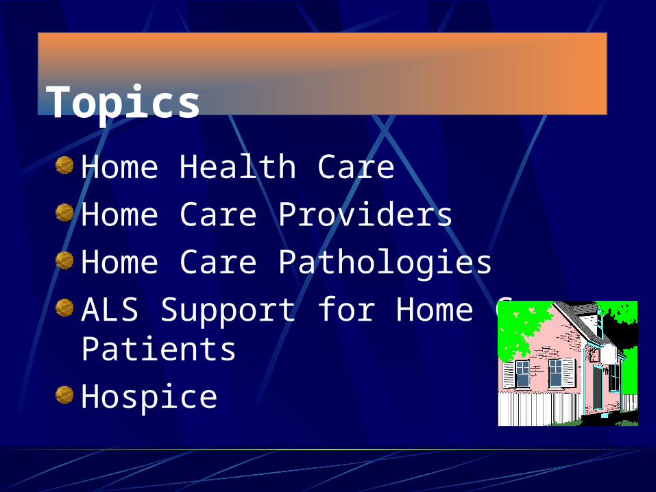
Epidemiology of Home Care
A number of factors have promoted the growth of home care in recent years. They include:
Enactment of Medicare in 1965The advent of HMOs Improved medical technologyChanges in the attitudes of doctors and
patients toward hospital care
In 1992…
Almost 75% of home-care patients were age 65 or older.
Of the elderly home-care patients, almost two-thirds are female.
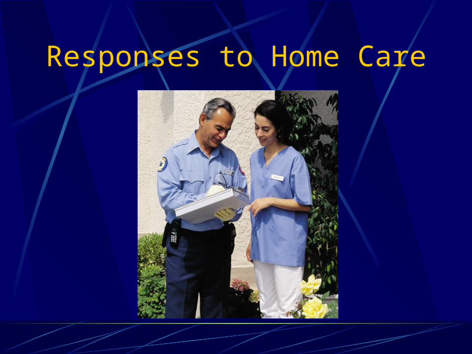
Responses to Home Care
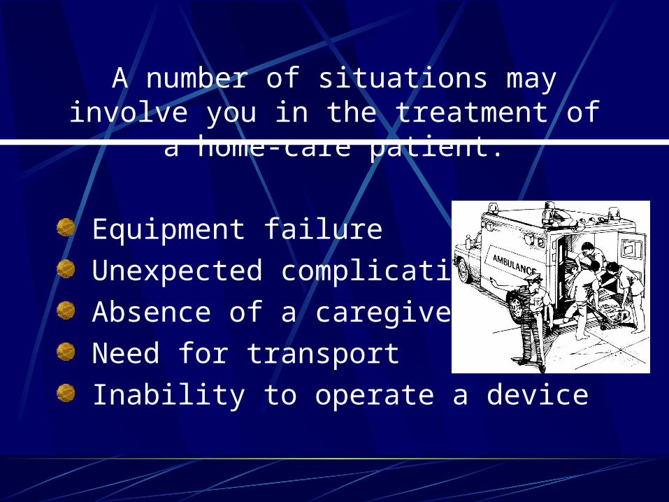
A number of situations may involve you in the treatment of a home-care patient:
Equipment failure
Unexpected complications
Absence of a caregiver
Need for transport
Inability to operate a device
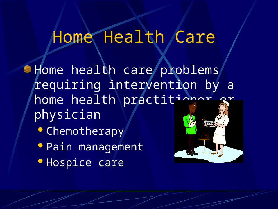
Home Health Care
Home health care problems requiring intervention by a home health practitioner or physicianChemotherapyPain managementHospice care

Figure 6-3

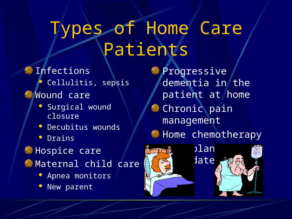
Types of Home Care Patients
Airway pathologies Inadequate pulmonary toilet Inadequate alveolar ventilation Inadequate alveolar oxygenation
Circulatory pathologies Alterations in peripheral circulation GI/GU pathologies Ostomies Catheters Home dialysis
Types of Home Care Patients
Infections Cellulitis, sepsis
Wound care Surgical wound closure Decubitus wounds Drains
Hospice care
Maternal child care Apnea monitors New parent
Progressive dementia in the patient at home
Chronic pain management
Home chemotherapy
Transplant candidate

Infection and Ulceration

Table 6-4
Home Health Preparation

Table 6-5
Prevention
General System Pathophysiology Assessment/Management

Assessment of the home-care patient follows the same basic steps as any other patient.
The one thing home-care calls have in common is their diversity.
Try to ascertain from the primary health care provider a baseline presentation for the patient.
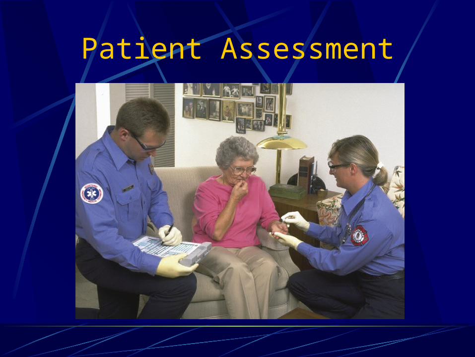
Patient Assessment
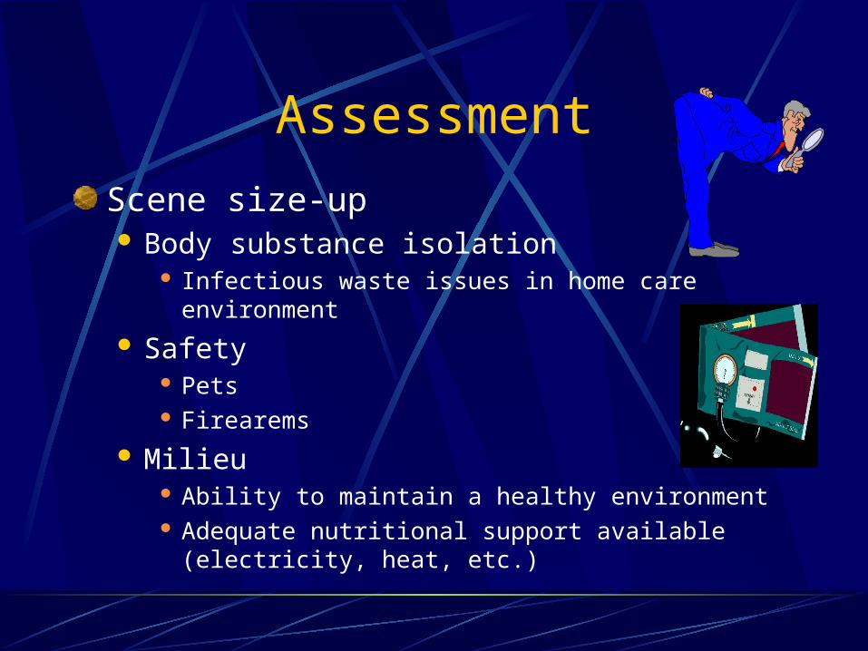
Assessment
Scene size-up Body substance isolation
Infectious waste issues in home care environment
Safety Pets Firearems
Milieu Ability to maintain a healthy environment Adequate nutritional support available (electricity, heat,
etc.)
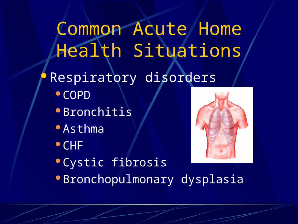
Common Acute Home Health Situations
Respiratory disordersCOPDBronchitisAsthmaCHFCystic fibrosisBronchopulmonary dysplasia

Common Acute Home Health Situations
Neuromuscular degenerative diseasesMuscular DystrophyPoliomyelitisGuillain-Barré SyndromeMyasthenia Gravis
Patients awaiting lung transplantsSleep apnea

Respiratory Disorders
Account for more than 630,000 of hospital patients discharged for home care annuallySimple pneumonia and pleurisy account for
37%COPD accounts for 50%
Respiratory Pathology
Increased risk of airway infections in the respiratory compromised patientProgression of chronic respiratory diseasesIncreased secretionsObstructed or malfunctioning airway devicesImproper application of medical device
Common Respiratory Equipment
Oxygen equipment
Portable suctioning machines
Aerosol equipment and nebulizers
Incentive spirometers
Home ventilators
Tracheostomy tubes and collars
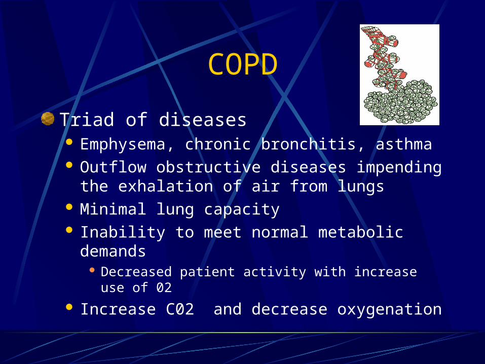
COPD
Triad of diseases Emphysema, chronic bronchitis, asthma Outflow obstructive diseases impending the
exhalation of air from lungs Minimal lung capacity Inability to meet normal metabolic demands
Decreased patient activity with increase use of 02
Increase C02 and decrease oxygenation
Bronchitis/EmphysemaBronchitis Chronic excessive production of mucus Narrowing bronchial passages restricting air flow
Large, obese patients (blue bloater)
Emphysema Enlargement and stiffening of alveoli and acenus Loss of elasticity and compliance requires a higher
pressure in lungs to facilitate gas exchange at alveolar level
Patients have increased A/P dimensions (increased air retention and decreased outflow
Thin patients due to increase caloric output

Acute ExacerbationPatients have difficult compensatory mechanisms Signs and symptoms
Wheezing, diminished breath sounds, use of accessory muscles, retractions, tripod positioning, inability to speak or form sentences
Home health care treatments Oxygen, nebulized aerosol treatments Ventilator: PEEP (via ETT), CPAP, BiPAP (mask
therapy)
Treatment Intervention
Oxygenation and ventilation
Nebulized beta-2 agonists
Nebulized anti-cholinergics
IV corticosteriods
AsthmaReactive and reversible airway disease seen at any age Characterized by bronchospasm and swelling of mucus
membranes
Signs and symptoms of acute attackHome therapy O2, oral medications, variety of nebulizers and/or
inhalants
Treatment Oxygenation and ventilation, beta agonists, anti-
cholinergics, corticosteriods Avoidance or elimination of reactants that trigger
problem
Cystic FibrosisGeneric disorder usually recognized in childhood Terminal disease
Characterized by chronic overproduction of mucus, inflammation of small airways and hyperinflation of alveoli, chronic infections, and erosion of the pulmonary blood vessels secondary to infectionExocrine disease causing other abnormalities GI disturbances, pancreatic disorders, glucose
intolerance

Home Health Treatment
Postural drainage of mucus
Chest physiotherapy
Mechanical vibrators to facilitate breakage of secretions
Medications aimed at mucus reduction and control of systemic bacterial infections

Bronchopulmonary Dysplasia
Primarily affects infants of low birth weightCharacterized by ongoing need for
mechanical ventilation in newborns Infants fail to wean from mandatory
ventilation or oxygenation Increased risk of respiratory infection
Management
EMSOxygenation and oximetric monitoringNeonatal transport to appropriate facility
Home health Intermittent mandatory ventilation (IMV)Pulmonary congestion and edemaLimit fluid intake
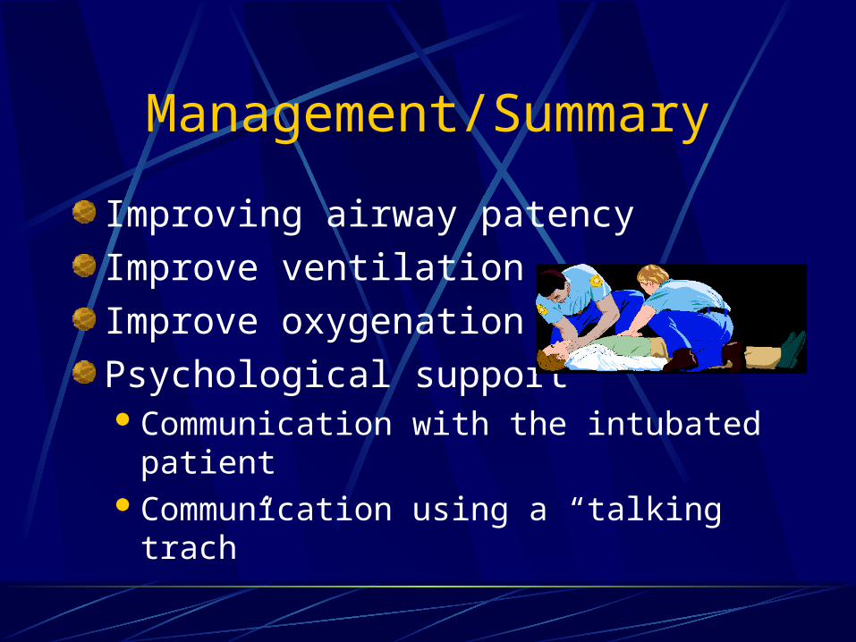
Management/Summary
Improving airway patency
Improve ventilation
Improve oxygenation
Psychological supportCommunication with the intubated patientCommunication using a “talking trach”
Neuromuscular Degenerative Diseases
Affect respiratory action through degeneration of muscles used for breathing
As disease progresses and involves more muscle groups, inability to ambulate increases infections and rapid decline of patients
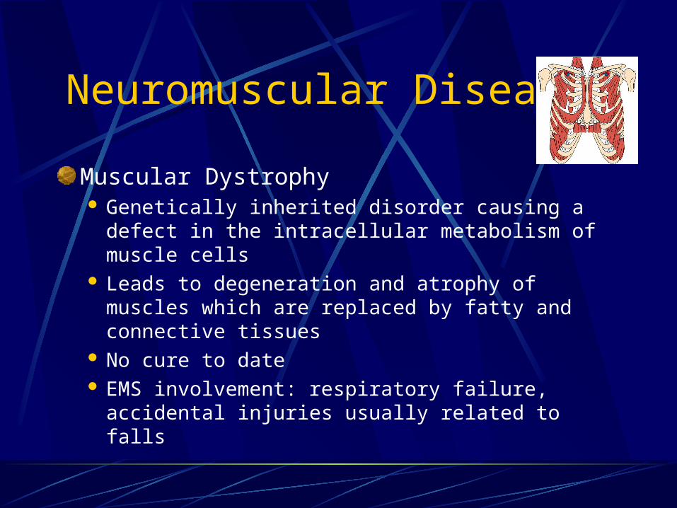
Neuromuscular Diseases
Muscular Dystrophy Genetically inherited disorder causing a defect in
the intracellular metabolism of muscle cells Leads to degeneration and atrophy of muscles
which are replaced by fatty and connective tissues No cure to date EMS involvement: respiratory failure, accidental
injuries usually related to falls
Neuromuscular Diseases
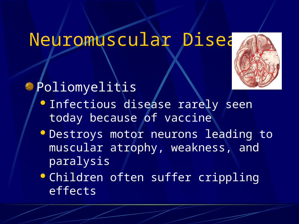
Poliomyelitis Infectious disease rarely seen today
because of vaccineDestroys motor neurons leading to
muscular atrophy, weakness, and paralysisChildren often suffer crippling effects
Neuromuscular Diseases
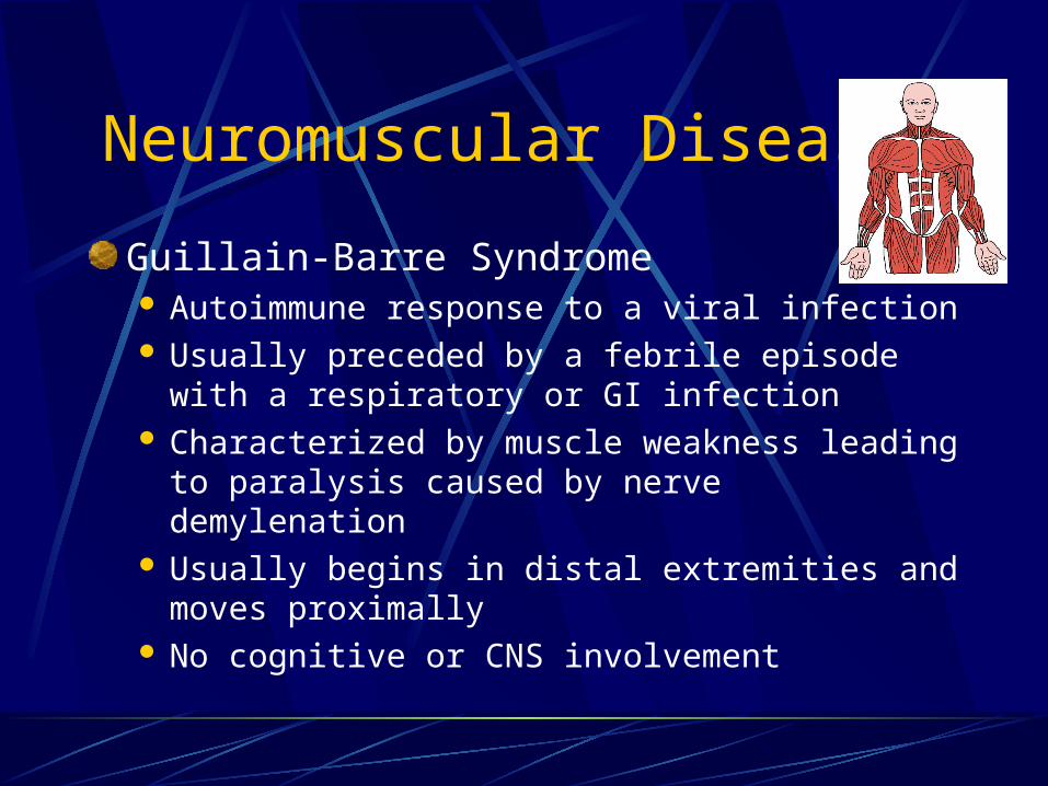
Guillain-Barre Syndrome Autoimmune response to a viral infection Usually preceded by a febrile episode with a
respiratory or GI infection Characterized by muscle weakness leading to
paralysis caused by nerve demylenation Usually begins in distal extremities and moves
proximally No cognitive or CNS involvement
Neuromuscular Diseases
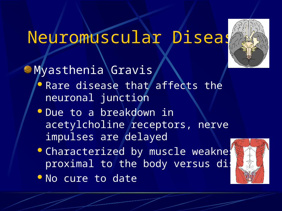
Myasthenia GravisRare disease that affects the neuronal
junctionDue to a breakdown in acetylcholine
receptors, nerve impulses are delayedCharacterized by muscle weakness
proximal to the body versus distalNo cure to date
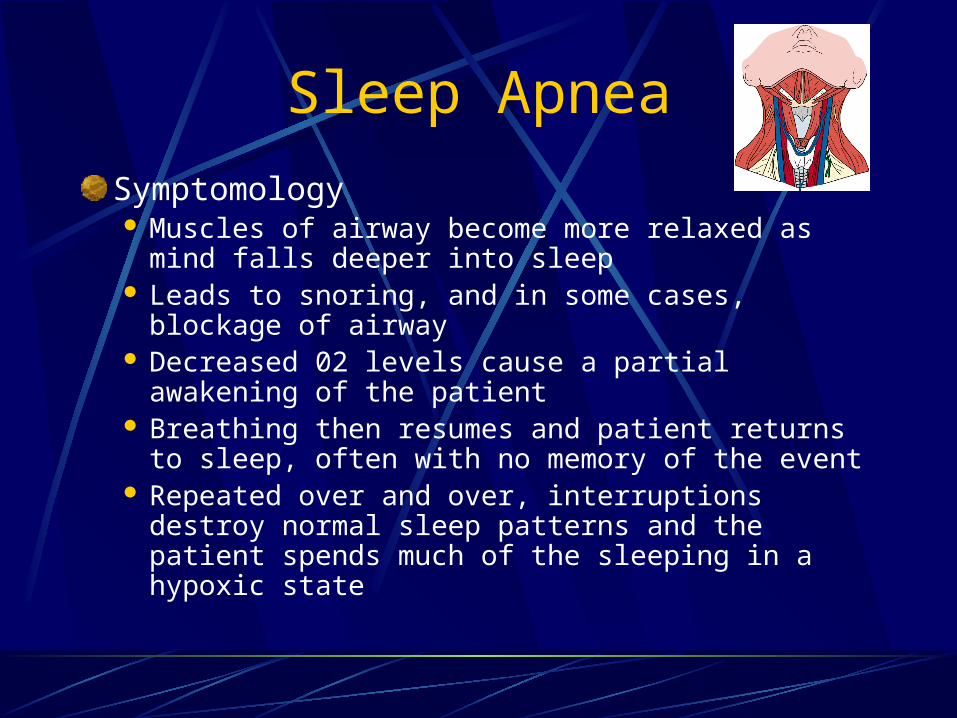
Sleep Apnea
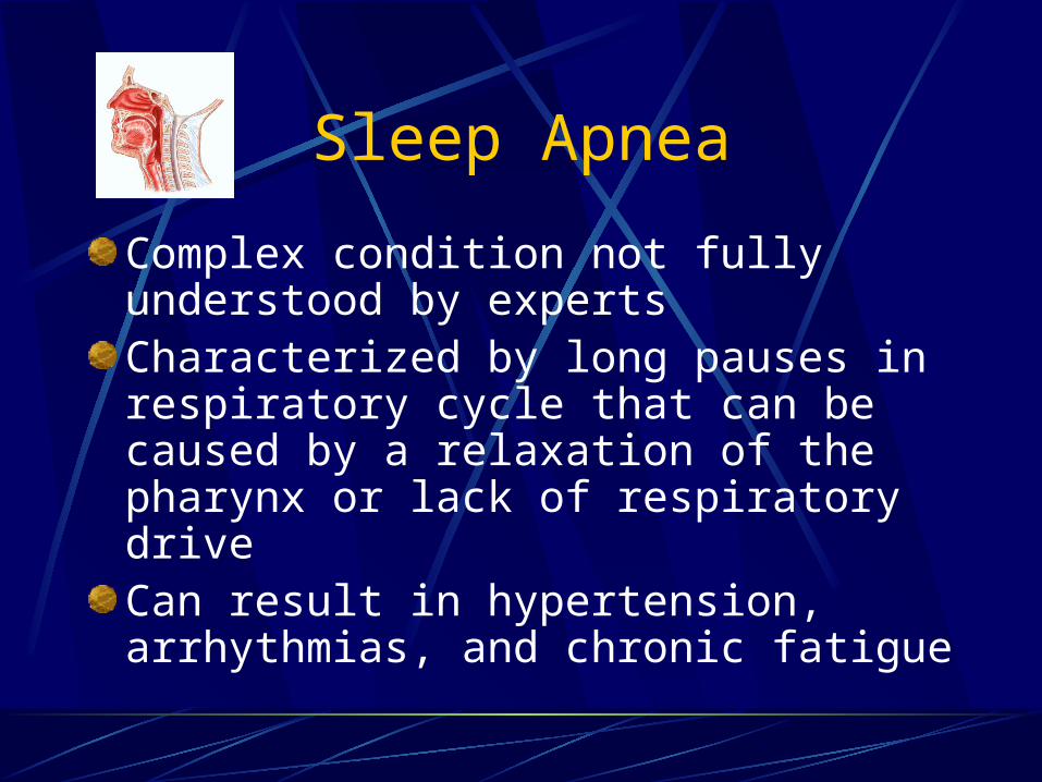
Complex condition not fully understood by expertsCharacterized by long pauses in respiratory cycle that can be caused by a relaxation of the pharynx or lack of respiratory driveCan result in hypertension, arrhythmias, and chronic fatigue
Sleep Apnea
Symptomology Muscles of airway become more relaxed as mind
falls deeper into sleep Leads to snoring, and in some cases, blockage of
airway Decreased 02 levels cause a partial awakening of
the patient Breathing then resumes and patient returns to
sleep, often with no memory of the event Repeated over and over, interruptions destroy
normal sleep patterns and the patient spends much of the sleeping in a hypoxic state

Sleep Apnea
General treatmentSurgical alteration of the airwayMedicationsPrescribed weight lossAvoidance of any CNS depressants
(alcohol)Use of CPAP ventilator
Medical Therapy Found in the Home Setting
Home oxygen therapy
Artificial airways/tracheostomies
Vascular access devices
Home Oxygen Therapy
Many advantages for home care patientsEasy to use Tolerated well by most patientsAdd to quality of a patient’s lifePrevents hypoxia that may result in
permanent cognitive damage or degeneration
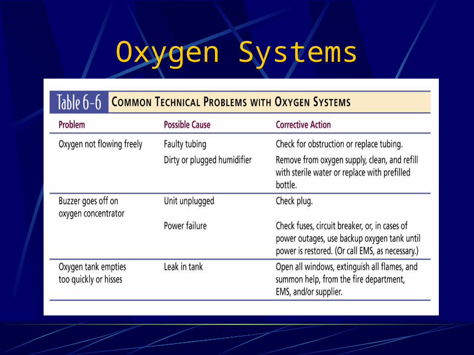
Oxygen Systems

Artificial Airways/TracheostomiesUsed for patients with long-term upper airway problemsTracheostomy may be temporary or permanentTechnique is used on patients who require
artificial ventilation for long periods of timePatients with damage to larynx, epiglottis,
or upper airway structures from surgery or trauma
Artificial Airway
Tracheostomy consists: Surgical opening (stoma) Outer cannula
Keeps stoma open Held in place by twill tape or velcro strap
Inner cannula Similar to a mini ET tube Slides down into trachea a few inches Distal low pressure cuff to hold in place and provide a
good seal

Tracheotomy tubes Top: Plastic tube
Bottom: metal tube with inner cannula
Artificial Airways
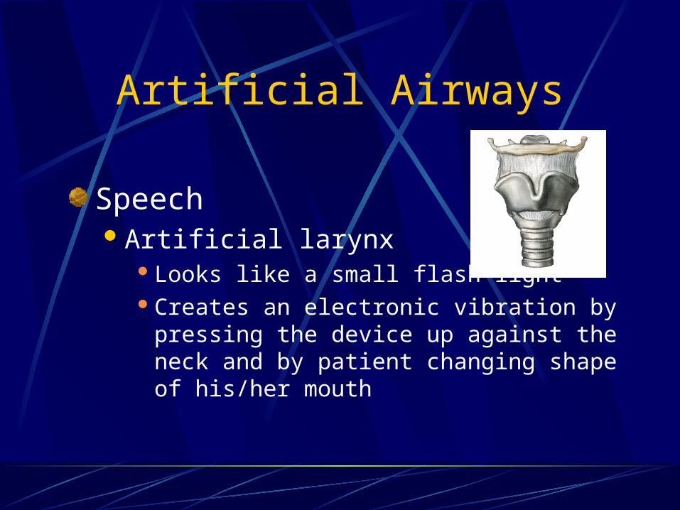
SpeechArtificial larynx
Looks like a small flash lightCreates an electronic vibration by pressing the
device up against the neck and by patient changing shape of his/her mouth
Routine Care of Tracheostomy
Keep stoma clean and dry Prevent pulmonary infections
Periodic changing of the outer cannulaChanging and cleaning the inner cannnula from every few weeks to every months, depending on the patientFor ventilator patients, routine changing of the ventilator hose connectionsFrequent suctioning, due to increased secretion
Common Complications of Tracheostomy Patients
Blockage of the airway by mucus and/or dislodged cannula Patient coughing to clear and suctioning Patient movement and child growth
Infection of the stomaDrying of tracheal mucus leading to crusting or bleedingTracheal erosion from an over-inflated cuff Tracheal necrosis

Management/Ventilation
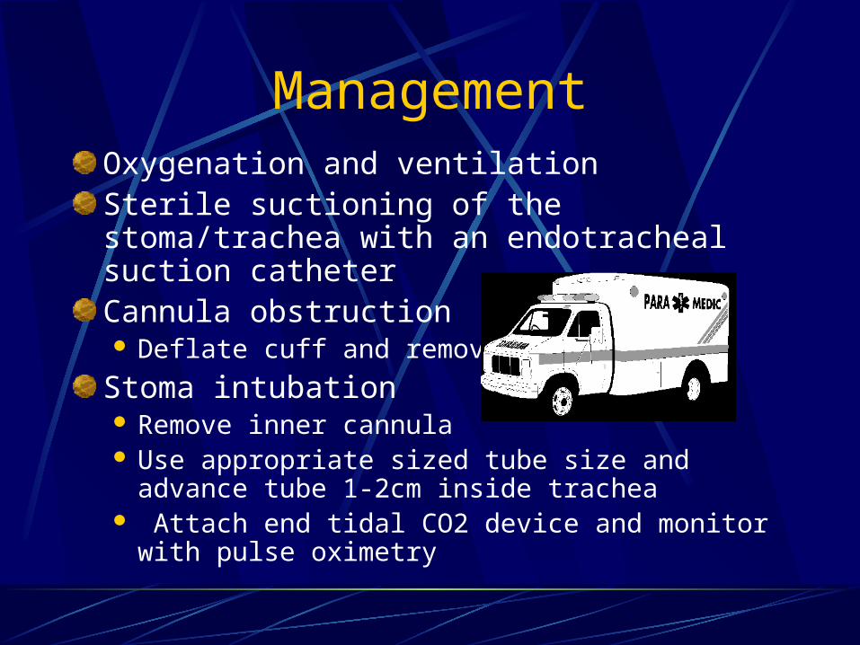
ManagementOxygenation and ventilationSterile suctioning of the stoma/trachea with an endotracheal suction catheterCannula obstruction Deflate cuff and remove
Stoma intubation Remove inner cannula Use appropriate sized tube size and advance tube
1-2cm inside trachea Attach end tidal CO2 device and monitor with
pulse oximetry
Home VentilationTypes of ventilatorsPositive pressureNegative pressure
Provision for ventilationVolume cycled ventilation
Historical standard for ventilatorsUsed to support multiple forms of respiratory
failure

Home VentilationPositive pressure ventilators (PPV) Recommended for acute respiratory failure Push air into lungs through a mask, nasal mask, or
tracheostomy Features
Variations: tidal volume respiratory rate flow rate pressure
Home Ventilation
Negative pressure ventilators Imitate normal breathing processApply negative pressure to the chest (pull
chest allowing it to expand)Allows air to flow into lungs
Patients usually use this form of device at night
Iron lung is an old example
PEEP/CPAP/BiPAP
Three ventilator options
Add pressures at various times during respiratory cycle
May be used on a full-time or part-time basis
Always possibility of pneumothorax due to increased pulmonary pressure
PEEPPositive end expiratory pressureUsed to maintain inflation of alveoliFunctions by providing a little back pressure at the end of expirationUses Premature infants with insufficient surfactant Adults with surfactant washout from PE, ARDS,
near drowning COPD
Emphysema patients require higher diffusion pressures for gas exchange
CPAPContinuous positive airway pressureUsed to keep pharyngeal structures from
collapsing at end of a breathOften prescribed for sleep apnea
Most patients use nasal CPAPPatients must keep mouth closed
Idea behind CPAP is the same a PEEPCPAP is delivered with a mask while PEEP
is delivered via ETT

Figure 6-7
Ventilation/CPAP-Nasal

BIPAP
Bilevel positive airway pressure
Provides two levels of pressure InspirationExpiration
Used for patients who require higher levels of pressure than CPAP
Assessment Findings
Work of breathing
Tidal volume
Peak flow
Oxygen saturation
Capnography
Breath sounds
Vascular Access Devices
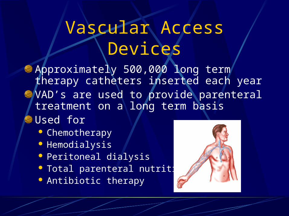
Approximately 500,000 long term therapy catheters inserted each yearVAD’s are used to provide parenteral treatment on a long term basisUsed for Chemotherapy Hemodialysis Peritoneal dialysis Total parenteral nutrition Antibiotic therapy
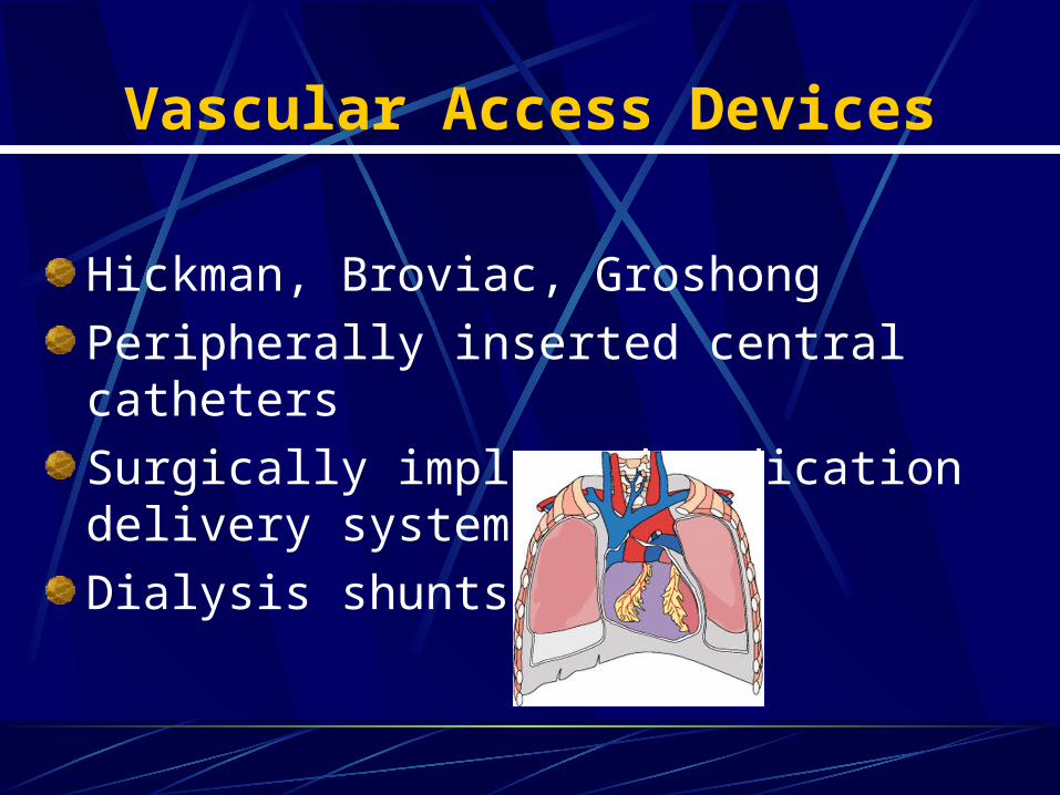
Vascular Access Devices
Hickman, Broviac, Groshong
Peripherally inserted central catheters
Surgically implanted medication delivery systems
Dialysis shunts
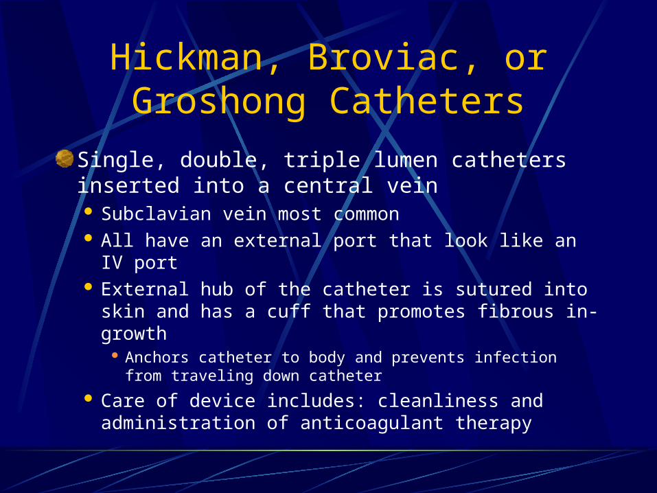
Hickman, Broviac, or Groshong Catheters
Single, double, triple lumen catheters inserted into a central vein Subclavian vein most common All have an external port that look like an IV port External hub of the catheter is sutured into skin
and has a cuff that promotes fibrous in-growth Anchors catheter to body and prevents infection from
traveling down catheter
Care of device includes: cleanliness and administration of anticoagulant therapy

Peripherally Inserted Central Catheters
PICC lines are most commonly inserted into median cubital vein in the ACF
Catheter is threaded from insertion site into central venous circulation
PICC lines are inserted under fluoroscopy
Surgically Implanted Medication Delivery Systems
Port-a-cath or Medi-portInfusion port is implanted completely below the skinDisc shaped devices that have a diaphragm that requires a specially shaped needle (Huber needle)Typically implanted in upper chest

Dialysis Shunt
Used for patients undergoing hemodialysis to filter their blood
Types of shunts AV shunt
Loop connecting an artery and vein, most common in distal arm where dialysis apparatus evacuates and returns blood
Fistula Connects artery and vein creating an artificially large
blood vessel for access
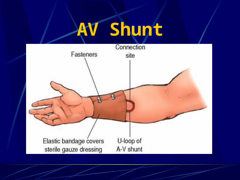
AV Shunt

VAD Complications
ObstructionThrombusEmbolus (air embolus)Catheter kinkingCatheter tip embolus Inactivity increases risk of clots
InfectionHemorrhage
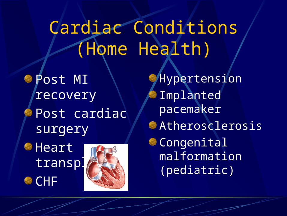
Cardiac Conditions (Home Health)
Post MI recovery
Post cardiac surgery
Heart transplant
CHF
Hypertension
Implanted pacemaker
Atherosclerosis
Congenital malformation (pediatric)
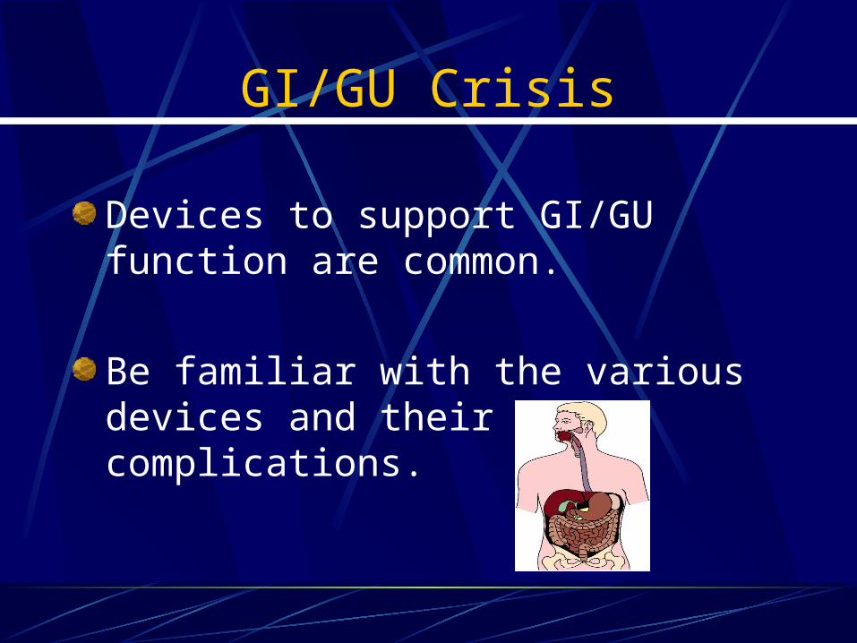
GI/GU Crisis
Devices to support GI/GU function are common.
Be familiar with the various devices and their complications.
GI/GU Devices
Urinary catheters or urostomiesSurgical diversion of the urinary tract to a
stoma, or hole in the abdominal wall
Indwelling nutritional support device (peg tube, G-tube)
Colostomies
NG tube

Figure 6-9
External Urinary Catheter (Texas Sheath)

Figure 6-10
Foley Catheter(Indwelling Catheter)

Urinary Device Complications
Infection or device malfunction Catheter device provides a pathway for infectious entry Foul smelling urine Discolored (cloudy) urine Blood tinged urine Systemic infection (fever) Redness, swelling of abdominal wall site with
urostomies
Device malfunction: accidental placement, obstruction, balloon ruptures
Gastrointestinal DevicesNasogastric tubes Decompress gastric contents Lavage GI system Short term use
Feeding tubes Rest on the duodenum or jejunum Weighted with steel filament to aid insertion and
passage through pyloric sphincter
Percutaneous endoscopic gastrostomy (PEG) Via abdominal wall Long term nutritional support

Nasogastric Feeding

Figure 6-12
Gastrostomy (PEG)
Colostomy
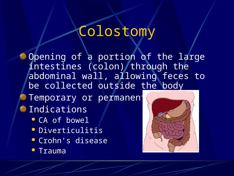
Opening of a portion of the large intestines (colon) through the abdominal wall, allowing feces to be collected outside the bodyTemporary or permanentIndications CA of bowel Diverticulitis Crohn’s disease Trauma
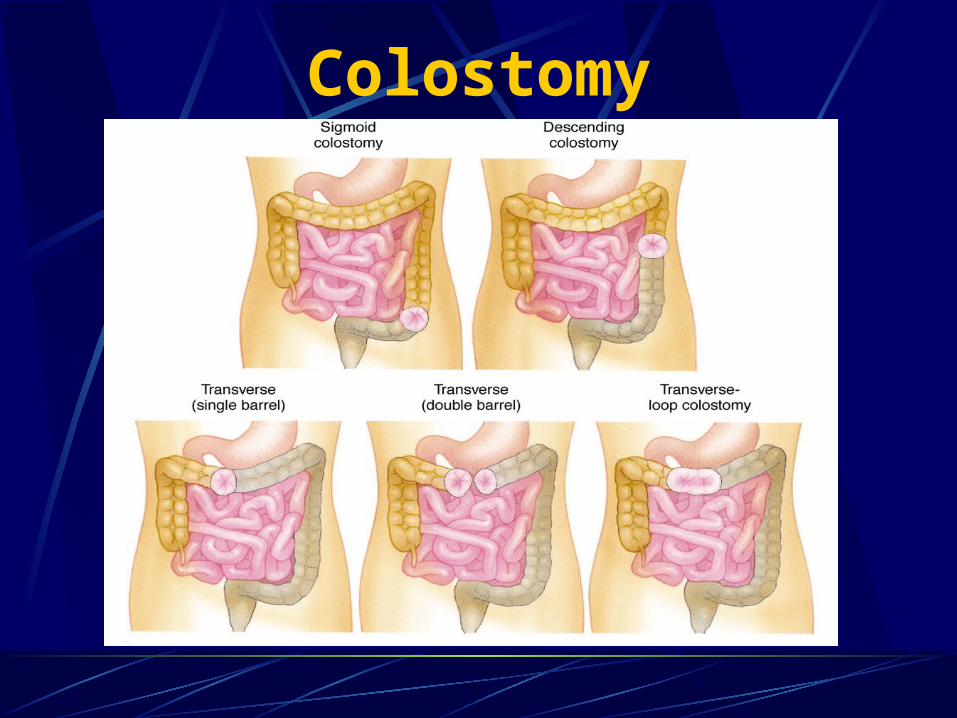
Figure 6-13
Colostomy

Assessment Findings
Abdominal pain and distention
Bowel sounds
Palpation of bladder
Color, character, amount of urine

Acute InfectionsIncreased rate of infections in the elderly, chronically ill and homebound
Decreased ability to perceive pain or perform self-care in many homebound populations
Pathophysiology
Increased risk of respiratory infection in the immunocompromised patient
Poor peripheral perfusion results in decreased healing and increased peripheral infections
Sedentary existence leads to skin breakdown and peripheral infections

Pathophysiology
Percutaneous and implanted medical devices increase risk for infections and sepsis
Patients discharged to home with open wounds and incisions
Chronic diseases may further impair healing
Poor nutrition, hygiene or ability to care for self impact infection rates
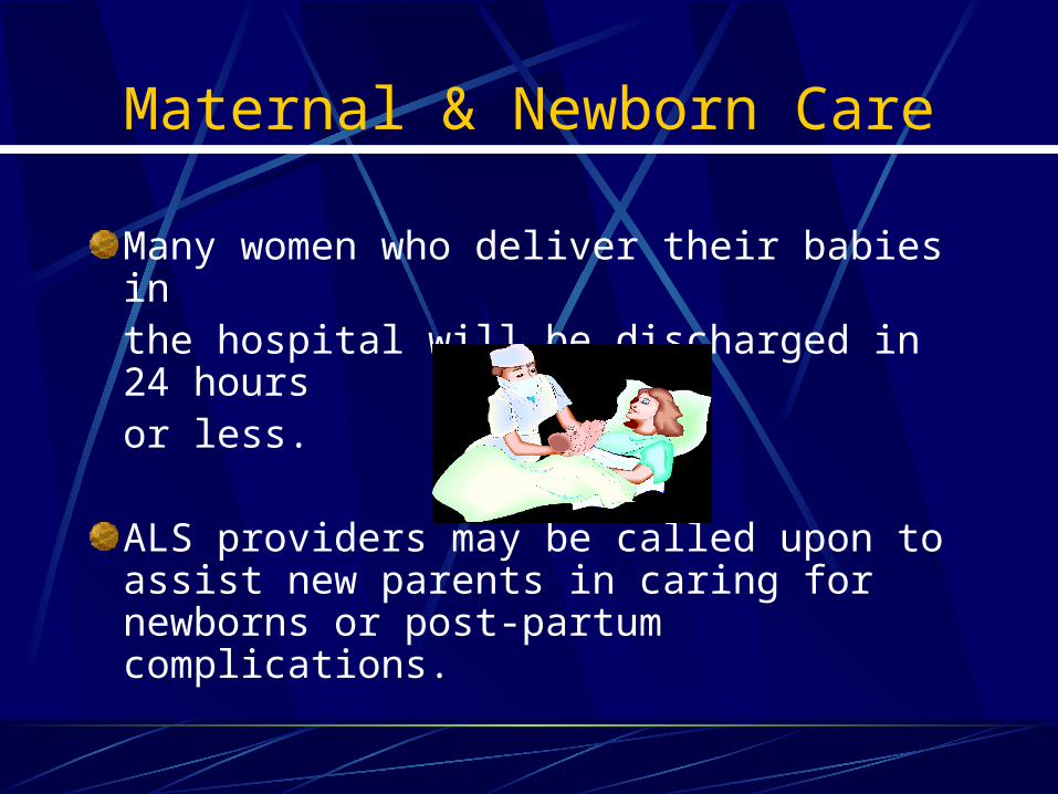
Maternal & Newborn Care
Many women who deliver their babies in the hospital will be discharged in 24 hours or less.
ALS providers may be called upon to assist new parents in caring for newborns or post-partum complications.
Post-partum bleeding and embolus are common complications
Management includes:
Massage of uterus
Administration of fluids
Administration of pitocin
Rapid transport, if necessary

Postpartum Depression
“Let down” feeling experienced during the period following birth
Occurs in 70%-80% of mothers
Women have difficulty caring for both themselves and newborn
Be sensitive to needs and non-urgent responses

Infants and Children with Special Needs
Many different types of childrenPremature babiesLung diseaseHeart diseasesNeurological diseasesChronic diseasesAltered functions from birth
Infants and Children with Special Needs
Common home-care devicesTracheostomy tubesApnea monitorsHome artificial ventilatorsCentral intravenous linesGastric feeding and gastrostomy tubes Shunts

Common Infant/Child Complications
Signs/symptoms of cardiorespiratory insufficiency include:
Cyanosis
Bradycardia (<100 BPM)
Rales
Respiratory Distress

Hospice
More than 2250 hospices provide support for the terminally ill and families.
The goal of hospice care is to provide palliative or comfort care rather than curative care.

Hospice
Hospice
Palliative care
Comfort care
Hospice care DNRMedical direction considerations
Pain control in the terminal patient
Common Hospice Diseases
Congestive heart failure
Cystic fibrosis
COPD
AIDS
Alzhemier’s
Cancer

Figure 6-14
HIV Children
Summary
Home Health Care
Home Care Providers
Home Care Pathologies
ALS Support for Home Care Patients
Hospice