Acute CNS infections
7
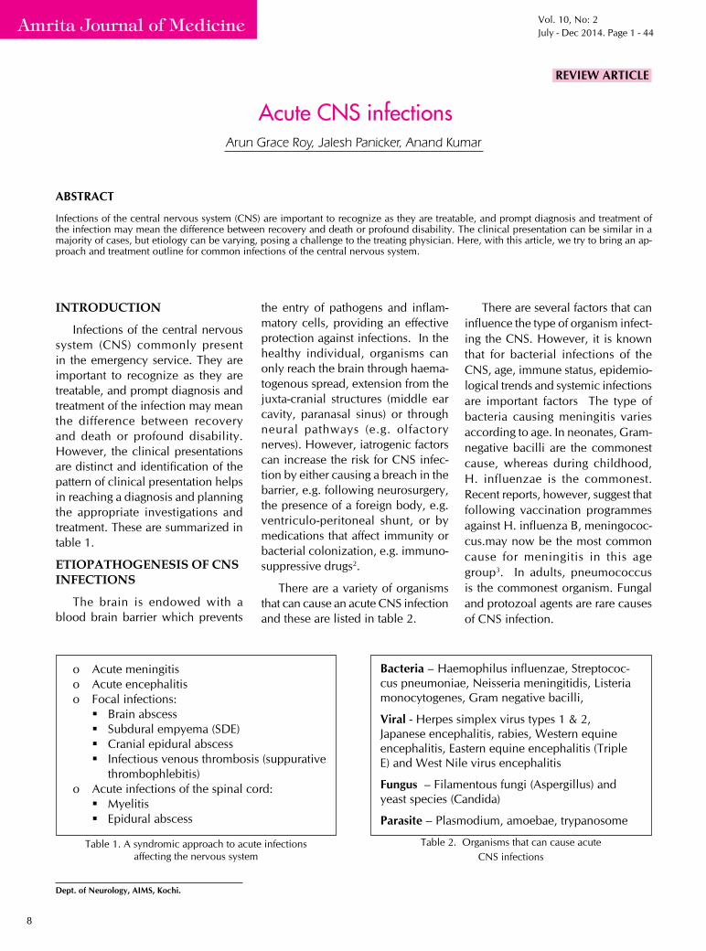
8 Amrita Journal of Medicine Acute CNS infections Arun Grace Roy, Jalesh Panicker, Anand Kumar Dept. of Neurology, AIMS, Kochi. INTRODUCTION Infections of the central nervous system (CNS) commonly present in the emergency service. They are important to recognize as they are treatable, and prompt diagnosis and treatment of the infection may mean the difference between recovery and death or profound disability. However, the clinical presentations are distinct and identification of the pattern of clinical presentation helps in reaching a diagnosis and planning the appropriate investigations and treatment. These are summarized in table 1. ETIOPATHOGENESIS OF CNS INFECTIONS The brain is endowed with a blood brain barrier which prevents ABSTRACT Infections of the central nervous system (CNS) are important to recognize as they are treatable, and prompt diagnosis and treatment of the infection may mean the difference between recovery and death or profound disability. The clinical presentation can be similar in a majority of cases, but etiology can be varying, posing a challenge to the treating physician. Here, with this article, we try to bring an ap- proach and treatment outline for common infections of the central nervous system. REVIEW ARTICLE There are several factors that can influence the type of organism infect- ing the CNS. However, it is known that for bacterial infections of the CNS, age, immune status, epidemio- logical trends and systemic infections are important factors The type of bacteria causing meningitis varies according to age. In neonates, Gram- negative bacilli are the commonest cause, whereas during childhood, H. influenzae is the commonest. Recent reports, however, suggest that following vaccination programmes against H. influenza B, meningococ- cus.may now be the most common cause for meningitis in this age group 3 . In adults, pneumococcus is the commonest organism. Fungal and protozoal agents are rare causes of CNS infection. Vol. 10, No: 2 July - Dec 2014. Page 1 - 44 Table 1. A syndromic approach to acute infections affecting the nervous system o Acute meningitis o Acute encephalitis o Focal infections: Brain abscess Subdural empyema (SDE) Cranial epidural abscess Infectious venous thrombosis (suppurative thrombophlebitis) o Acute infections of the spinal cord: Myelitis Epidural abscess Bacteria – Haemophilus influenzae, Streptococ- cus pneumoniae, Neisseria meningitidis, Listeria monocytogenes, Gram negative bacilli, Viral - Herpes simplex virus types 1 & 2, Japanese encephalitis, rabies, Western equine encephalitis, Eastern equine encephalitis (Triple E) and West Nile virus encephalitis Fungus – Filamentous fungi (Aspergillus) and yeast species (Candida) Parasite – Plasmodium, amoebae, trypanosome Table 2. Organisms that can cause acute CNS infections the entry of pathogens and inflam- matory cells, providing an effective protection against infections. In the healthy individual, organisms can only reach the brain through haema- togenous spread, extension from the juxta-cranial structures (middle ear cavity, paranasal sinus) or through neural pathways (e.g. olfactory nerves). However, iatrogenic factors can increase the risk for CNS infec- tion by either causing a breach in the barrier, e.g. following neurosurgery, the presence of a foreign body, e.g. ventriculo-peritoneal shunt, or by medications that affect immunity or bacterial colonization, e.g. immuno- suppressive drugs 2 . There are a variety of organisms that can cause an acute CNS infection and these are listed in table 2.
Transcript of Acute CNS infections
8
Amrita Journal of Medicine
Acute CNS infectionsArun Grace Roy, Jalesh Panicker, Anand Kumar
dept. of neurology, AiMS, Kochi.
IntroductIon
Infections of the central nervous system (CNS) commonly present in the emergency service. They are important to recognize as they are treatable, and prompt diagnosis and treatment of the infection may mean the difference between recovery and death or profound disability. However, the clinical presentations are distinct and identification of the pattern of clinical presentation helps in reaching a diagnosis and planning the appropriate investigations and treatment. These are summarized in table 1.
EtIopAthogEnEsIs of cns InfEctIons
The brain is endowed with a blood brain barrier which prevents
ABSTRACT
Infections of the central nervous system (CNS) are important to recognize as they are treatable, and prompt diagnosis and treatment of the infection may mean the difference between recovery and death or profound disability. The clinical presentation can be similar in a majority of cases, but etiology can be varying, posing a challenge to the treating physician. Here, with this article, we try to bring an ap-proach and treatment outline for common infections of the central nervous system.
REViEW ARTiClE
There are several factors that can influence the type of organism infect-ing the CNS. However, it is known that for bacterial infections of the CNS, age, immune status, epidemio-logical trends and systemic infections are important factors The type of bacteria causing meningitis varies according to age. In neonates, Gram-negative bacilli are the commonest cause, whereas during childhood, H. influenzae is the commonest. Recent reports, however, suggest that following vaccination programmes against H. influenza B, meningococ-cus.may now be the most common cause for meningitis in this age group3. In adults, pneumococcus is the commonest organism. Fungal and protozoal agents are rare causes of CNS infection.
Vol. 10, No: 2July - Dec 2014. Page 1 - 44
Table 1. A syndromic approach to acute infections affecting the nervous system
o Acute meningitiso Acute encephalitis o Focal infections: Brain abscess Subdural empyema (SDE) Cranial epidural abscess Infectious venous thrombosis (suppurative
thrombophlebitis) o Acute infections of the spinal cord: Myelitis Epidural abscess
Bacteria – Haemophilus influenzae, Streptococ-cus pneumoniae, Neisseria meningitidis, Listeria monocytogenes, Gram negative bacilli,
Viral - Herpes simplex virus types 1 & 2, Japanese encephalitis, rabies, Western equine encephalitis, Eastern equine encephalitis (Triple E) and West Nile virus encephalitis
Fungus – Filamentous fungi (Aspergillus) and yeast species (Candida)
parasite – Plasmodium, amoebae, trypanosome
Table 2. Organisms that can cause acute CNS infections
the entry of pathogens and inflam-matory cells, providing an effective protection against infections. In the healthy individual, organisms can only reach the brain through haema-togenous spread, extension from the juxta-cranial structures (middle ear cavity, paranasal sinus) or through neural pathways (e.g. olfactory nerves). However, iatrogenic factors can increase the risk for CNS infec-tion by either causing a breach in the barrier, e.g. following neurosurgery, the presence of a foreign body, e.g. ventriculo-peritoneal shunt, or by medications that affect immunity or bacterial colonization, e.g. immuno-suppressive drugs2.
There are a variety of organisms that can cause an acute CNS infection and these are listed in table 2.

9
Amrita Journal of Medicine
Most cases of bacterial meningitis begin with host acquisition of a new organism by nasopharyngeal colonization, followed by systemic invasion and devel-opment of a high-grade bacteraemia. Central nervous system invasion then occurs, although the exact site of bacterial entry into the central nervous system is un-known. Virulence factors are released by the bacteria. As part of the host immune response, inflammatory cytokines are released. This results in increased perme-ability of the blood-brain barrier, allowing proteins and neutrophils to enter into the subarachnoid space. There is, then, an intense subarachnoid space inflammatory response, which leads to many of the pathophysiological consequences of meningitis, including cerebral oedema and increased intracranial pressure4. The pathology of encephalitis includes viral cytopathology (viral destruc-tion of neurons), a para/postinfectious inflammation, or immune mediated response5. Although neurons are primarily affected, vasculitis, meningitis, myelitis and radiculitis can be present.
ApproAch to thE pAtIEnt prEsEntIng wIth An AcutE cns InfEctIon
When a patient presents with an acute CNS infection, it is crucial to commence appropriate treatment at the earliest possible time. As in other areas of emergency medicine, history taking is an essential part of the evaluation. History and examination help to establish the syndromic diagnosis. It can also help in establish-ing the temporal sequence of events, which facilitates the diagnosis. The examination should be focused on establishing the extent of neurological involvement, and signs of a systemic source of infection. Neck stiffness, resulting from irritation of the meninges, is a nonspecific sign and may occur not only in meningitis, but also if the intracranial pressure is raised as in encephalitis, or even part of a systemic infection (meningism).
routInE InvEstIgAtIons
Neutrophilic leucocytosis in the peripheral blood may be seen in pyogenic meningitis, and occasionally in acute tuberculous meningitis. ESR and C- reactive protein may be elevated in these conditions. Blood culture may be positive in upto 50% of patients with pyogenic meningitis. Chest x-ray may show pneumonia or changes suggesting tuberculosis.
nEuroImAgIng
Though lumbar puncture (LP) is the most essential test for the diagnosis and management of acute CNS infections, it is nowadays accepted practice to perform brain imaging before this invasive test, where available. CT imaging is often sufficient in assessing the risk of performing lumbar puncture in an acutely ill patient, al-
though this cannot exclude raised intracranial pressure. Presence of focal lesions with mass effect would indicate increased risk of brain herniation from performing the test. Established brain abscess can be identified in CT imaging. Complications of meningitis, such as infarcts, may be seen in CT as well. Meningeal enhancement in a contrast enhanced CT scan suggests the presence of exudates.
However, in hospitals which lack an acute neuro-imaging service, waiting for a CT scan may delay the lumbar puncture and thus the diagnosis. A brain scan is often unnecessary in uncomplicated meningitis and clinical features are helpful to determine the patient at risk who should have imaging before LP (table 3)6.
Table 3: Clinical features that would suggest the need for imaging before performing lumbar puncture6
Age greater than 60 years
Immunocompromised state
History of CNS lesion
History of seizures within one week before presentation
Altered level of consciousness
Gaze palsy
Abnormal visual fields
Facial palsy
Arm drift
Leg drift
Magnetic resonance imaging, though not often prac-tical in the acute setting, provides additional information about the diagnosis. Presence of signal changes in the medial temporal and orbito-frontal regions would sug-gest herpes simplex encephalitis
cErEbrospInAl fluId
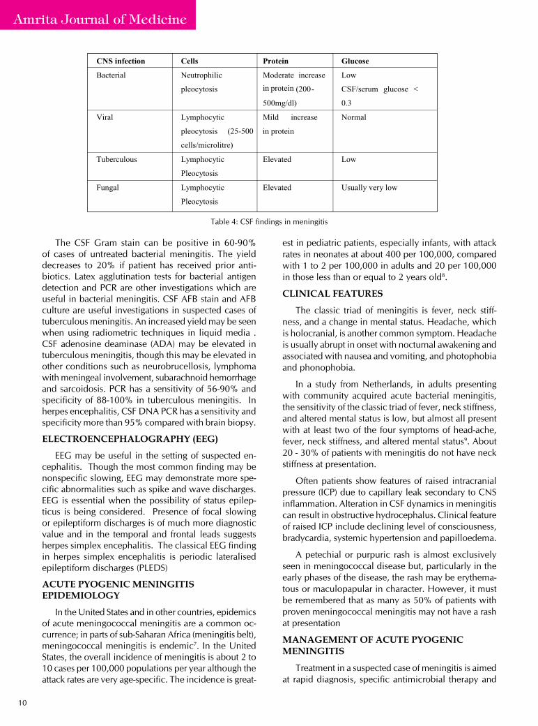
Lumbar puncture is the most important investiga-tion in acute CNS infections, and helps to confirm the diagnosis and identify the organism. CSF study should include cell count, protein, glucose and cultures (bacte-rial, fungal and mycobacterial), staining (gram, Giemsa, India Ink, acid fast bacilli), depending upon the sus-pected organism. CSF antigen detection, CSF antibody detection and DNA detection by PCR are various other useful investigations that could be seen in the appropri-ate setting. CSF abnormalities seen in various types of meningitis are given in table 4.
10
Amrita Journal of Medicine
The CSF Gram stain can be positive in 60-90% of cases of untreated bacterial meningitis. The yield decreases to 20% if patient has received prior anti-biotics. Latex agglutination tests for bacterial antigen detection and PCR are other investigations which are useful in bacterial meningitis. CSF AFB stain and AFB culture are useful investigations in suspected cases of tuberculous meningitis. An increased yield may be seen when using radiometric techniques in liquid media . CSF adenosine deaminase (ADA) may be elevated in tuberculous meningitis, though this may be elevated in other conditions such as neurobrucellosis, lymphoma with meningeal involvement, subarachnoid hemorrhage and sarcoidosis. PCR has a sensitivity of 56-90% and specificity of 88-100% in tuberculous meningitis. In herpes encephalitis, CSF DNA PCR has a sensitivity and specificity more than 95% compared with brain biopsy.
ElEctroEncEphAlogrAphy (EEg)
EEG may be useful in the setting of suspected en-cephalitis. Though the most common finding may be nonspecific slowing, EEG may demonstrate more spe-cific abnormalities such as spike and wave discharges. EEG is essential when the possibility of status epilep-ticus is being considered. Presence of focal slowing or epileptiform discharges is of much more diagnostic value and in the temporal and frontal leads suggests herpes simplex encephalitis. The classical EEG finding in herpes simplex encephalitis is periodic lateralised epileptiform discharges (PLEDS)
AcutE pyogEnIc mEnIngItIs EpIdEmIology
In the United States and in other countries, epidemics of acute meningococcal meningitis are a common oc-currence; in parts of sub-Saharan Africa (meningitis belt), meningococcal meningitis is endemic7. In the United States, the overall incidence of meningitis is about 2 to 10 cases per 100,000 populations per year although the attack rates are very age-specific. The incidence is great-
est in pediatric patients, especially infants, with attack rates in neonates at about 400 per 100,000, compared with 1 to 2 per 100,000 in adults and 20 per 100,000 in those less than or equal to 2 years old8.
clInIcAl fEAturEs
The classic triad of meningitis is fever, neck stiff-ness, and a change in mental status. Headache, which is holocranial, is another common symptom. Headache is usually abrupt in onset with nocturnal awakening and associated with nausea and vomiting, and photophobia and phonophobia.
In a study from Netherlands, in adults presenting with community acquired acute bacterial meningitis, the sensitivity of the classic triad of fever, neck stiffness, and altered mental status is low, but almost all present with at least two of the four symptoms of head-ache, fever, neck stiffness, and altered mental status9. About 20 - 30% of patients with meningitis do not have neck stiffness at presentation.
Often patients show features of raised intracranial pressure (ICP) due to capillary leak secondary to CNS inflammation. Alteration in CSF dynamics in meningitis can result in obstructive hydrocephalus. Clinical feature of raised ICP include declining level of consciousness, bradycardia, systemic hypertension and papilloedema.
A petechial or purpuric rash is almost exclusively seen in meningococcal disease but, particularly in the early phases of the disease, the rash may be erythema-tous or maculopapular in character. However, it must be remembered that as many as 50% of patients with proven meningococcal meningitis may not have a rash at presentation
mAnAgEmEnt of AcutE pyogEnIc mEnIngItIs
Treatment in a suspected case of meningitis is aimed at rapid diagnosis, specific antimicrobial therapy and
Table 4: CSF findings in meningitis

11
Amrita Journal of Medicine
adjunctive therapy. In a suspected case, the emergency physician should have samples collected for blood culture, urgent lumbar puncture and commence appro-priate antibiotics. If a physician requires imaging before LP, a blood culture should be sent and antibiotic and adjunctive therapy started, before sending patient for CT and then performing LP if required. Delay in treatment increases morbidity and mortality, so time should not be wasted for investigations. The choice of antibiotics depends on age and other associated factors like im-mune status, systemic diseases and epidemiological factors (tables 5, 6 and 7).
usE of stEroIds In pyogEnIc mEnIngItIs
On the basis of the available evidence, it is rec-ommended to use adjunctive steroids in adults with suspected or proven pneumococcal meningitis. Dexa-methasone is suggested (0.15 mg/kg q6h for 2–4 days with the first dose administered 10–20 minutes before, or at least concomitant with, the first dose of anti-micro-bial therapy). For infants and children six weeks of age and older, the Committee on Infectious Diseases of the American Academy of Pediatrics (2003) recommends that adjunctive therapy with dexamethasone may be considered after weighing the potential benefits and possible risks. There is insufficient data to recommend the use of steroids in neonates with pyogenic meningitis
Acute CNS infections

12
Amrita Journal of Medicine
Table 5. Antibiotic recommendations for meningitis
Table 6: Recommended dosages of antimicrobial therapy in patients with bacterial meningitis
Table 7: Duration of antimicrobial therapy for bacterial meningitis
Neisseria meningitidisHaemophilus influenzaeStreptococcus pneumoniaeStreptococcus agalactiaeAerobic gram-negative bacilliListeria monocytogenes
7days 7days10-14 days14-21 days21 days- / > 21 days
vIrAl mEnIngItIs And EncEphAlItIsEpIdEmIology
Epidemiologic studies estimate the incidence of viral encephalitis at 3.5-7.4 per 100,000 persons per year. Overall, viruses are the most common cause of encepha-litis. The Center for Disease Control and Prevention (CDC) estimates an annual incidence of approximately 20,000 new cases of encephalitis in the United States, mostly mild in nature. Seasonal variation can affect the incidence of viral encephalitis. Japanese encephalitis is more common in rainy season, while arboviral en-cephalitis is prevalent during warm and / or wet seasons. Japanese encephalitis virus is the most common cause of epidemic encephalitis worldwide, causing encephalitis
more often than meningitis. It is prevalent in Asia and Australia and affects children more than adults. Herpes simplex virus type 1 is the commonest cause of sporadic encephalitis. Enterovirus is the commonest cause of meningitis. Fever, neck stiffness and headache are the commonest symptoms in viral meningitis. Symptoms are less severe in comparison to bacterial meningitis.
In India, Japanese B Encephalitis is the commonest cause for epidemic encephalitis. Epidemiological stud-ies have been carried out which show that there are several arboviruses that are responsible for encephalitis in India, e.g. West Nile virus, dengue hemorrhagic fever.
Encephalitis should be suspected when a febrile patient presents with altered mentation or level of consciousness. Seizures, both focal and general, are common manifestations of encephalitis. HSV encepha-litis, which is the commonest, has a predilection for the temporal and orbitofrontal lobes resulting in memory impairment, behavioural change and altered sensorium.
Treatment is mainly supportive in viral meningitis, since it is a self-limiting disorder. Antipyretics and analgesics are the cornerstone of treatment. Oral or intravenous (IV) acy-clovir can be tried in HSV 1&2, varicella and Epstein-Barr virus infection. IV acyclovir for
13
Amrita Journal of Medicine
one week for severe HSV infection, and oral acyclovir 800mg five times a day, valacyclovir 1000mg TID and famcyclovir 500mg TID in mild cases of HSV meningitis can be used. The experimental drug Pleconaril has been proven to be effective in enteroviral meningitis.
hErpEs sImplEx EncEphAlItIs (hsE)
Although a wide range of viruses have been reported to cause encephalitis, specific antiviral therapy for vi-ral encephalitis is generally limited to disease caused by herpes simplex virus. Acyclovir is the treatment of choice for patients with herpes simplex encephalitis but morbidity and mortality remain high. In a retrospec-tive multicentre trial of 93 adult patients, multivariate analysis also identified a Simplified Acute Physiology Score greater than 27 at hospital admission and a delay of greater than two days between hospital admission and administration of acyclovir therapy as independent predictors of poor outcome8. The dosage of acyclovir in patients with normal renal function is 10 mg/kg intravenously every 8 h for 14–21 days. Recently, the use of higher dose acyclovir (20 mg/kg intravenously every 8 h for 21 days) in neonates with herpes simplex encephalitis has decreased mortality to 5%, with 40% of survivors developing normally9. A negative CSF PCR result at the end of therapy was associated with a better outcome, suggesting that another CSF specimen should be subjected to PCR for herpes simplex virus at the end of therapy in patients who have not had the appropriate clinical response if the result is positive, antiviral therapy should be continued10.
In patients with herpes simplex encephalitis, predic-tors of an adverse outcome include age of the patient (130 years), level of consciousness (Glasgow coma score, < 6), and duration of symptoms prior to start-ing acyclovir therapy (more than four days). The role of steroid in HSV encephalitis is controversial, since reports do not clearly document benefit in when steroid is added.
tubErculous mEnIngItIs
Tuberculous meningitis (TBM) is still a major cause of serious illness in many parts of the world. It usu-ally presents as subacute to chronic meningitis. But acute presentations in the form of acute meningitis, encephalopathy, raised intracranial pressure, seizures and acute focal deficits secondary to vasculitis are not uncommon. CSF examination shows normal to elevated CSF pressure, elevated protein (80-400mg/dl), low glucose (40mg/dl) and lymphocytic predominant (200-400 WBC/mm3) pleocytosis. Identifying the bacilli by smear and culture will establish the diagnosis. CSF TB PCR, which is been reportedly used, has sensitivity of 70-75%. Neuroimaging in TB meningitis will show basal exudates and hydrocephalus. In suspected case of TB meningitis, evidence of infection, active or chronic,
should be looked for in the chest and abdomen. The Mantoux test is another investigation that can help in the diagnosis.
Chemotherapy of TB meningitis include a regime of four drugs (rifampicin, INH, pyrazinamide, ethambutol/streptomycin) for two months or till the disease starts improving, followed by INH and rifampicin for at least ten months. The total duration of treatment should be a minimum of 1-1½ years. In multi-drug resistant TB and immunocompromised patients, five or seven drugs should be added, depending on the severity of illness. Steroids are indicated in TB meningitis in the presence of raised ICP, hydrocephalus, vasculitis, arachnoidiits, or markedly raised CSF protein.
fungAl mEnIngItIs
It is a less common cause of meningitis compared to bacterial and viral meningitis. CNS manifestations of fungal infection can vary from meningoencephalitis, focal lesions, vasculitis, and raised intracranial pres-sure. Yeast species (cryptococcus) have a predilection to cause meningeal infection, while filamentous fungi (aspergillus) will cause parenchymal infection. Fungal infections are more common in immunocompromised patients, including patients on chronic steroid therapy.
Meningitis can be acute, subacute, or chronic. These are often associated with focal deficits, encephalopathy and raised ICP.
CT or MRI is indicated in suspected cases of fungal meningitis to look for hydrocephalus and focal lesions. Another clue to the aetiological agent is the presence dilated Virchow Robin spaces filled with gelatinous exudates seen as T2 hyperintensity in the basal ganglia in 40 % of cryptococcal infections.
CSF in fungal infection shows high opening pressure with elevated cell count, low sugar and elevated pro-tein. CSF India ink preparation can show encapsulated yeasts in 50-75% of cryptococcal meningitis. Fungal culture, fungal antigen assay and antibody detection by radioimmunoassay are other useful investigations to detect the organisms. CSF pleocytosis is predominantly lymphocytic and rarely can be neutrophilic in aspergil-lus infections or eosinophilic in Coccidioides Immitis infection.
The successful treatment of central nervous system (CNS) fungal infections is highly dependent on the underlying immune status of the host, as well as on the prompt initiation of appropriate antifungal therapy. However, the diagnosis of these infections may be difficult, and proper therapy is often delayed. The stand-ard antifungal agent for the treatment of CNS fungal infections has been amphotericin B. However, poor CNS penetration, fungal resistance, and toxicity often eliminate the effectiveness of amphotericin B. Because
Acute CNS infections
14
Amrita Journal of Medicine
of the problems associated with use of amphotericin B, newer liposomal preparations, as well as azole antifun-gal agents, have been developed. A new azole that is undergoing clinical trials is Ravuconazole. Flucytosine is often used as an adjunct to amphotericin B in patients with invasive infections by cryptococcus, candida and aspergillus. The duration of treatment is weeks to months depending on the response.
cErEbrAl mAlArIA
Of all the four species of malaria that infect man, only plasmodium falciparum causes cerebral malaria. The transmission of plasmodium occurs through the bite of the female anopheles mosquito. In cerebral malaria, microvasculature of the brain is packed with red blood cells infested by the falciparum. The brain is swollen, with multiple micro-haemorrhages mainly in white matter. Microglial cells and macrophages surrounding the haemorrhagic foci are called Durcks granuloma11
who dEfInItIon for cErEbrAl mAlArIA
1. Unarousable coma / impairment of sensorium not attributable to any other cause in a patient with falciparum malaria. Coma should persist at least 30 minutes after a generalized convulsion to make the distinction from transient postictal coma.
2. Presence of asexual forms in peripheral blood smear or bone marrow smear
Fever is commonest clinical feature followed by en-cephalopathy. Focal deficits are rare. 50-60% of cases can have seizures. Fundus examination can show pap-illoedema, retinal ischaemia and hemorrhages. Severe cases may present with decerebrate or decorticate posturing.
Cerebral malaria is a neurological emergency re-quiring prompt diagnosis and treatment. It should be suspected in all cases of unexplained encephalopathy in malariaendemic regions. Diagnosis is by demonstration of malarial parasite in peripheral blood smears stained with Geimsa / Field stain. At least three peripheral smears repeated every 6-8 hours should be negative to exclude the diagnosis of malaria. The infestation rate of red cells is usually high (greater than 10%). Antibody testing and PCR detection of DNA is now available12
WHO guidelines recommend the following range of anti-malarial medications, there being insufficient evidence to recommend any one over the other for severe malaria:
– Artesunate 2.4 mg/kg bw i.v. or i.m given on ad-mission (time = 0), then at 12 h and 24 h, then once a day; artemether 3.2 mg/kg bw i.m given on admission
then 1.6 mg/kg bw per day; for total 5-7 days
– Quinine 20 mg salt/kg bw on admission (i.v. infu-sion or divided i.m. injection), then 10 mg/kg bw every 8 h; infusion rate should not exceed 5 mg salt/kg bw per hour. Once patient is conscious or afebrile then quinine is given orally (10mg/kg every 8 hours) for 5-7 days. When the patient is on quinine, ECG monitoring for QT interval prolongation is recommended.
Anti oedema measures for raised ICP, anticonvul-sants for seizures, and regular monitoring and treatment for hypoglycemia should be provided as required.
rEfErEncEs:1. Verma, A. Infections of the nervous system, bacterial infec-
tions. Chapter 57 1419-55. Neurology in clinical practice. Volume II. Fifth edition. Walter G Bradley, Robert B Daroff
2. Cohen J and Powderly W.G 2003 infectious disease 2nd edition Saunders Philadelphia.
3. Mark E Jones, Deborah C Draghi1, James A Karlowsky. Prevalence of anti-microbial resistance in bacteria isolated fromcentral nervous system specimens as reported by U.S. hospital laboratories from 2000 to 2002. Annals of Clinical Microbiology and Antimicrobials 2004, 3:3
4. A R Tunkel and W M Scheld Clin Microbiol Rev. 1993 April; 6(2): 118-136. Pathogenesis and pathophysiology of bacterial meningitis
5. Solomon T, Hart, IJ, Beeching NJ. Viral encephalitis: A clini-cians guide. Pract Neurol 2007;7(5): 288-305.
6. Hasbun R., Abrahams J., Jekel J., et al: Computed tomography of the head be-fore lumbar puncture in adults with suspected meningitis. N Engl J Med 2001; 345. (24): 1727-33.
7. Sharon E. Mace. Acute Bacterial Meningitis. Emergency medicine Clinics of North America. . 38 (2008) 281–317.
8. Tunkel AR, Hartman BJ, Kaplan SL, et al. Practice guidelines for the man-agement of bacterial meningitis. Clin Infect Dis. Nov 1 2004;39(9):1267-84.
9. Clinical features and prognostic factors in adults with bacterial meningitis. - Van de Beek D - N Engl J Med - 28-OCT-2004; 351(18): 849-59.
10. Raschilas F, Wolff M, Delatour F, et al. Outcome of and prognostic factors for herpes simplex encephalitis in adult patients: results of a multicenter study. Clin Infect Dis 2002; 35:254–60.
11. Isabelle M. Medana, Nicholas P. Day, Tran Tinh Hien. Axonal Injury in Cer-ebral Malaria. Am J Pathol 2002; 160: 655–66.
12. GalS, & Wainscoat J.S. 2006, Detection and quantification of circulating plasmodium falciparum DNA by polymerase chain reaction, MolBiol, Vol 336 155 –62
Acute CNS infections