Activity of Porophyllum ruderale leaf extract and 670-nm ... · Magalhães” Animal...
9
RESEARCH ARTICLE Open Access Activity of Porophyllum ruderale leaf extract and 670-nm InGaP laser during burns repair in rats Ana Cristina Justino Jácomo, Karina de Andrade Velozo, Raquel Gabilan Lotti, Lia Mara Grosso Neves, Fernanda Oliveira de Gaspari de Gaspi, Marcelo A. Marreto Esquisatto, Maria Esméria Corezola do Amaral, Fernanda A. Sampaio Mendonça and Gláucia Maria Tech dos Santos * Abstract Background: In this study, we investigated the effects of an extract of the leaves of Porophyllum ruderale and laser irradiation on the healing of burns. Methods: Seventy-two rats were divided in four groups: untreated controls, treated with laser irradiation, treated with P. ruderale and treated with both P. ruderale and laser irradiation. Burns were produced with a metal plate on the backs of the animals. Wound samples were collected for structural and morphometric analyses and to quantify the expression of TGF-β1 and VEGF. Results: Laser irradiation increased the number of fibroblasts, collagen fibers and newly formed vessels and decreased the number of granulocytes at the site of the wounds. Densitometric analysis revealed a significant increase in the expression of TGFβ-1 in the wounds treated with laser irradiation and with the P. ruderale extract at the beginning of the healing process and a decreased during the experimental period. The expression of VEGF was highlighted in the lesions irradiated with laser alone. Conclusion: Inspite of not showing a beneficial effect on the laser combination with the P. ruderale extract, when the laser was used separately, a positive effects to enhance the healing of second-degree burns was promoted. P. ruderale was effective in decreasing the granulocytes during the repair process indicating a possible anti-inflammatory action of this extract of native flora, widely used in folk medicine, but little studied experimentally. Keywords: Porophyllum ruderale, Laser, Burns, Tissue repair, Rats Background The healing of burn wounds has long been a research focus because of the problems associated with this type of injury, such as time of healing, infections, morbidity and mortality. In second-degree burns, the epidermis and superficial dermis are the main layers affected. In addition to destruction of the epithelial barrier, the pres- ence of degraded proteins and devitalized tissue provides an excellent environment for the growth and prolifera- tion of microorganisms [1] which are a serious public health problem. All phases of repair are mediated by molecular signals, including specific growth factors, which stimulate and organize multiple cellular activities [2]. Transforming growth factor (TGF-β1) attracts neutrophils, macro- phages and fibroblasts in the inflammatory focus [3]. Vascular endothelial growth factor (VEGF) is identified as a major regulator of vasculogenesis and angiogenesis [4], produced by fibroblasts, endothelial cells, platelets, neutrophils and macrophages [5]. Medicinal plants are widely used in folk medicine for the treatment of several injuries and demonstrate effi- cacy in repair for both normal and burn wounds [6–8]. Porophyllum ruderale (Jacq.) Cass. (Asteraceae), com- monly known as “arnica paulista”, possesses a variety of biological and pharmacological activities and tinctures or alcoholic leaf extracts of this plant are applied topically in folk medicine as healing or anti-inflammatory agents to treat cuts, wounds and abrasions [9]. Phytochemical studies of the aerial parts of the plant have revealed the presence of * Correspondence: [email protected] Graduate Program of Biomedical Sciences, Hermínio Ometto University Center, UNIARARAS, Av. Maximiliano Baruto, 500, CEP 13607-339 Araras, SP, Brazil © 2015 Jácomo et al. Open Access This article is distributed under the terms of the Creative Commons Attribution 4.0 International License (http://creativecommons.org/licenses/by/4.0/), which permits unrestricted use, distribution, and reproduction in any medium, provided you give appropriate credit to the original author(s) and the source, provide a link to the Creative Commons license, and indicate if changes were made. The Creative Commons Public Domain Dedication waiver (http://creativecommons.org/publicdomain/zero/1.0/) applies to the data made available in this article, unless otherwise stated. Jácomo et al. BMC Complementary and Alternative Medicine (2015) 15:274 DOI 10.1186/s12906-015-0805-2
Transcript of Activity of Porophyllum ruderale leaf extract and 670-nm ... · Magalhães” Animal...

RESEARCH ARTICLE Open Access
Activity of Porophyllum ruderale leaf extractand 670-nm InGaP laser during burns repair in ratsAna Cristina Justino Jácomo, Karina de Andrade Velozo, Raquel Gabilan Lotti, Lia Mara Grosso Neves,Fernanda Oliveira de Gaspari de Gaspi, Marcelo A. Marreto Esquisatto, Maria Esméria Corezola do Amaral,Fernanda A. Sampaio Mendonça and Gláucia Maria Tech dos Santos*
Abstract
Background: In this study, we investigated the effects of an extract of the leaves of Porophyllum ruderale and laserirradiation on the healing of burns.
Methods: Seventy-two rats were divided in four groups: untreated controls, treated with laser irradiation, treatedwith P. ruderale and treated with both P. ruderale and laser irradiation. Burns were produced with a metal plate onthe backs of the animals. Wound samples were collected for structural and morphometric analyses and to quantifythe expression of TGF-β1 and VEGF.
Results: Laser irradiation increased the number of fibroblasts, collagen fibers and newly formed vessels anddecreased the number of granulocytes at the site of the wounds. Densitometric analysis revealed a significantincrease in the expression of TGFβ-1 in the wounds treated with laser irradiation and with the P. ruderale extract atthe beginning of the healing process and a decreased during the experimental period. The expression of VEGF washighlighted in the lesions irradiated with laser alone.
Conclusion: Inspite of not showing a beneficial effect on the laser combination with the P. ruderale extract, when thelaser was used separately, a positive effects to enhance the healing of second-degree burns was promoted. P. ruderalewas effective in decreasing the granulocytes during the repair process indicating a possible anti-inflammatory action ofthis extract of native flora, widely used in folk medicine, but little studied experimentally.
Keywords: Porophyllum ruderale, Laser, Burns, Tissue repair, Rats
BackgroundThe healing of burn wounds has long been a researchfocus because of the problems associated with this typeof injury, such as time of healing, infections, morbidityand mortality. In second-degree burns, the epidermisand superficial dermis are the main layers affected. Inaddition to destruction of the epithelial barrier, the pres-ence of degraded proteins and devitalized tissue providesan excellent environment for the growth and prolifera-tion of microorganisms [1] which are a serious publichealth problem.All phases of repair are mediated by molecular signals,
including specific growth factors, which stimulate and
organize multiple cellular activities [2]. Transforminggrowth factor (TGF-β1) attracts neutrophils, macro-phages and fibroblasts in the inflammatory focus [3].Vascular endothelial growth factor (VEGF) is identifiedas a major regulator of vasculogenesis and angiogenesis[4], produced by fibroblasts, endothelial cells, platelets,neutrophils and macrophages [5].Medicinal plants are widely used in folk medicine for
the treatment of several injuries and demonstrate effi-cacy in repair for both normal and burn wounds [6–8].Porophyllum ruderale (Jacq.) Cass. (Asteraceae), com-monly known as “arnica paulista”, possesses a variety ofbiological and pharmacological activities and tinctures oralcoholic leaf extracts of this plant are applied topically infolk medicine as healing or anti-inflammatory agents totreat cuts, wounds and abrasions [9]. Phytochemical studiesof the aerial parts of the plant have revealed the presence of
* Correspondence: [email protected] Program of Biomedical Sciences, Hermínio Ometto UniversityCenter, UNIARARAS, Av. Maximiliano Baruto, 500, CEP 13607-339 Araras, SP,Brazil
© 2015 Jácomo et al. Open Access This article is distributed under the terms of the Creative Commons Attribution 4.0International License (http://creativecommons.org/licenses/by/4.0/), which permits unrestricted use, distribution, andreproduction in any medium, provided you give appropriate credit to the original author(s) and the source, provide a link tothe Creative Commons license, and indicate if changes were made. The Creative Commons Public Domain Dedication waiver(http://creativecommons.org/publicdomain/zero/1.0/) applies to the data made available in this article, unless otherwise stated.
Jácomo et al. BMC Complementary and Alternative Medicine (2015) 15:274 DOI 10.1186/s12906-015-0805-2

quercetin, a glycosidic flavonoid, in addition to tannins sa-ponins, resins and essential oils [10].The use of low-level laser irradiation (LLLI) to accel-
erate burn wound healing has been studied and thephotobiostimulation has been shown to stimulate ree-pithelization, collagen synthesis, fibroblast proliferation,angiogenesis and the formation of granulation tissue, andto reduce inflammatory infiltration [11]. However, the idealparameters of laser photobiostimulation for wound healingare conflicting due to the variety of treatment protocols,animal models and cell cultures used [12]. Nevertheless,laser therapy has been shown to be useful as a coadjuvant,favoring the healing process of different tissues [13].Considering the beneficial effects of herbal medicines
and laser therapy on the wound healing, the objective ofthe present study was to investigate the cellular responsesinduced by topical application of extract of P. ruderalealone or in combination with 670-nm Indium GalliumPhosphide (InGaP) laser irradiation on the healing ofburns produced on the back of rats.
MethodsPlant collection and extractionP. ruderale leaves were collected, early in the morningperiod in the month of in September (2011), in the Medi-cinal Plant Garden of Hermínio Ometto University Cen-ter, UNIARARAS, Araras, São Paulo, Brazil. A voucherspecimen was deposited at the herbarium of the Luiz deQueiroz College of Agriculture (Escola Superior de Agri-cultura Luiz de Queiroz—ESALQ-USP) (no ESA115686).After collection, the leaves (100 g) were selected and
properly cleaned under running water to remove im-purities. The hydroalcoholic extract of P. ruderale wasprepared by maceration of the leaves in a hydroalco-holic solution of 70 % (v/v solution of ethanol in water)for 7 days at room temperature, followed by vacuumfiltration using a qualitative paper filter and evaporationin a rotary evaporator (Fisatom, model 803) at 40 °C.The evaporation period was 1 h. The resulting sampleswere subjected to lyophilization [14] and the yield ofthe lyophilized extract was from 10 %, ie, after all pro-ceedings of extraction, the 100 g of leaves yield 10 g ofthe lyophilized. Obtaining this extract was based onpopular culture [15].
Phytochemical screening methodThe qualitative identification of chemical constituentswas carried out in the same extract as that used in thewound repair test using chemical methods [14] andthin-layer chromatography [16]. The chemical groupsanalysed were polyphenolic components, flavonoids, tan-nins, alkaloids, saponins, fatty acid, triterpenes, volatileoils, coumarins and anthraquinones.
The presence of polyphenol compounds was analyzedwith a solution of 1 % ferric chloride. Tannins have beenidentified using the dried extract dissolved in water, 2 mlof sodium chloride (2 %); filtered and mixed with 5 mlof 1 % gelatin. The presence of flavonoids was deter-mined using aluminum chloride solution 1 % in metha-nol, concentrated hydrochloric acid, magnesium andpotassium hydroxide. Dragendorff reagent was used toevaluate the presence of total alkaloids. Saponins wereanalyzed based on its ability to produce foam. For detec-tion of triterpenes extract was mixed with 5 ml ofchloroform was heated to 80 °C for 30 min and thentreated with a small volume of concentrated sulfuricacid. Moreover, the extract was analyzed by thin layerchromatography on silica gel using chloroform: metha-nol (98:2) and hexane:ethyl acetate (80:20) as eluent.The components were first visualized under UV lightand then by spraying the chromatographic plates andeach containing different specific solutions, followed byincubation at 100 °C for 5 min.The method of Chandra and Mejia Gonzalez [17] was
used for quantitative analysis of total polyphenols. Thehydroalcoholic extract (0.1 ml) was added to 1 ml Folin-Ciocalteau reagent and the mixture was left to stand for5 min. Next, 2 ml 20 % sodium carbonate was added.After incubation for 10 min at room temperature, ab-sorbance of the mixture was read in a spectrophotom-eter at 730 nm. The amount of total polyphenols isexpressed as catechin equivalents.For the analysis of flavonoids, the hydroalcoholic ex-
tract was incubated with 10 % aluminum chloride, 95 %ethanol, 1 mol/l sodium acetate, and distilled water for30 min at room temperature. Absorbance was read in aspectrophotometer at 425 nm and samples were ana-lyzed in triplicate. The amount of flavonols and flavonesis expressed as quercetin equivalents [18].
AnimalsSeventy-two (Rattus norvegicus albinus) male Wistarrats aged approximately 120 days and weighing on average250 g were obtained from the “Prof. Dr. Luiz Edmundo deMagalhães” Animal Experimentation Center, of HermínioOmetto University Center, UNIARARAS. The animalswere housed in individual polycarbonate cages at a con-stant temperature (23 ± 2 °C) and humidity (55 %) under a12-h light/dark cycle, with free access to standard chowand drinking water. All surgical and experimental proce-dures were approved by the Ethics Committee of Hermí-nio Ometto University Center, UNIARARAS (Protocolnumber 092/2011) and were conducted in accordancewith the ethical guidelines of the Brazilian College of Ani-mal Experimentation (COBEA) and of Guide for the Careand Use of Laboratory Animals (NIH).
Jácomo et al. BMC Complementary and Alternative Medicine (2015) 15:274 Page 2 of 9

Experimental design and burn wound creationThe animals were divided randomly into four groups of18 animals each: group C, untreated control; group L,treated with 670-nm InGaP laser; group P, treated withthe hydroalcoholic P. ruderale extract; group PL, treatedwith the hydroalcoholic P. ruderale extract and 670-nmInGaP laser (extract applied prior to laser therapy).For experimental wounding, the back of the animals
was shaved 48 h before the procedure under generalanesthesia induced by the intraperitoneal administrationof ketamine (1.0 ml/kg) and xylazine (0.2 mg/kg). Skinburns were produced on the backs of all animals, afterthe same anesthetic procedure, by applying an aluminumplate measuring 2 cm in diameter adapted to an appar-atus that maintained a constant temperature of 120 °C.The plate was pressed on the animal’s skin for 20 s forthe creation of a second-degree burn [19]. After induc-tion of burns, the animals received pain killers: sodiumdipyrone, one drop in the postoperative, after 12 and24 h and they were placed in individual cages. Besidesthe treatments for repair of burns was not carried outany specific care except individualize the animals through-out the experimental procedure. The treatments werestarted 24 h after injury and were applied daily at the sametime for 21 days. The animals were immobilized withoutsedation for treatment. The control (untreated) animalswere subjected to sham treatments to control for effectsof immobilization without treatment.For laser therapy, an InGaP laser (Phisiolux Dual Bio-
set®, Indústria de Tecnologia Eletrônica Ltda., Rio Claro,São Paulo, Brazil) operating at a wavelength of 670 nm(visible red) was applied in the continuous mode usingthe following parameters: power output of 30 mW, energydensity of 4.93 J/cm2 and total energy dose of 0.36 J, withthe laser beam covering an area of 0.073 cm2. Punctualirradiation was performed by non-contact energy deliveryat a distance of ± 2 mm and an angle of 90° in relation tothe wound surface. The time of application (12 s) wasdetermined by the specification of the equipment and thelaser was applied to four points within the burn area. Theapparatus was calibrated by the manufacturer.The hydroalcoholic extract of P. ruderale (1.0 ml/day)
was applied with a Pasteur pipette to the borders of thewound.
Structural and morphometric analysesWound samples were collected 7, 14 and 21 days afterinjury from three animals per group euthanized with anoverdose of the anesthetic. An area measuring 25 mm indiameter was delimited in the center of the wound toobtain standardized samples for structural and morpho-metric analysis.The tissue fragments were immersed in fixative solu-
tion containing 10 % formaldehyde in Millonig buffer,
pH 7.4, for 24 h at room temperature. Next, the speci-mens were washed in buffer and submitted to routine pro-cedures for embedding in Paraplast™ (Histosec®, Merck,Darmstadt, Germany). The blocks were cut into 6-μmlongitudinal sections.Longitudinal sections stained with Toluidine Blue and
by the Domini method [20] were used to determine thenumber of fibroblasts and granulocytes and newlyformed vessels in the repair area of the groups studied(n/104 μm2). Three samplings were performed from eachof the five sections obtained from whole sections of themiddle part of each surgical sample. The first 16 sectionsof the mid-section of the surgical specimen collectedfrom each animal per group were collected. The sectionswere mounted on eight slides and were chosen randomlyfor the methods described above. After staining, threesamplings of 104 μm2 (100 × 100 μm grid) were per-formed per section obtained in each test. Each samplewas photomicrographed and digitized bright-field imageswere obtained with a Leica DM2000 photomicroscope atthe Laboratory of Micromorphology, Hermínio OmettoUniversity Center, UNIARARAS. Masson’s trichromestaining was used to quantify collagen fiber content inthe repair area (percentage of total area). The fields wereseparated using the blue color distribution as a discrim-ination parameter. The intensity of blue represents thecollagen density. The color band was adjusted by trialand error until representative areas of collagen had beenseparated in the image. The same parameter was thenused to identify collagen fibers in all digitized fields. Next,the area occupied was calculated for each field [21].Samples measuring 104 μm2 (100 × 100 μm grid) ob-
tained from the area of the wound healing on days 7, 14and 21 of treatment were examined using the Leica ImageMeasure™ visual grid for morphometric analysis of the fol-lowing parameters: total number of fibroblasts, granulo-cytes, newly formed blood vessels (n/104 μm2), andcollagen fiber content (% area). The Sigma Scan Pro 6.0™program was used to evaluate the deposition of granula-tion tissue in the repair area. The results were entered intoExcel for Windows XP™ spreadsheets and compared byANOVA and the Tukey post-test (p < 0.05) [22].
Western blotting analysisFor analysis of protein expression by Western blotting,wound samples were collected from three animals pergroup after euthanasia with an overdose of the anesthetic,on days 7, 14 and 21 of treatment. For protein extraction,the samples obtained were chopped and homogenized ina Polytron homogenizer (PTA 20S model PT 10/35;Brinkmann Instruments, Westbury, NY, USA) operated atmaximum speed for 40 s in buffer (10 mM EDTA,100 mM Trizma base, 10 mM sodium pyrophosphate,100 mM sodium fluoride, 100 mM sodium orthovanadate,
Jácomo et al. BMC Complementary and Alternative Medicine (2015) 15:274 Page 3 of 9

2 mM PMSF, 0.1 mg/ml aprotinin, and deionized water;Sigma Chemical Co., USA). The extract was centrifuged at12,000 rpm for 20 min at 4 °C for removal of insolublematerial. The supernatant was collected for the measure-ment of protein concentration in the samples by theBiuret method (Protal colorimetric method, Laborlab, SãoPaulo, Brazil). Aliquots of the supernatant were treatedwith Laemmli buffer containing 100 mM DTT (SigmaChemical Co., St. Louis, MO, USA). Samples containing50 μg protein were boiled for 5 min and submitted to10 % (VEGF, 40 kDa) and 12 % (TGF-β1, 25 kDa) SDS-PAGE (Sodium dodecyl sulfate-polyacrylamide gel elec-trophoresis) in a mini-gel apparatus (Mini-Protean®,Bio-Rad-Richmond, CA, USA). Next, the protein bandswere transferred from the gel to a nitrocellulose mem-brane (Hybond ECL, 0.45 μm) [23]. The membraneswere washed in basal solution (1 M Trizma base, 5 MNaCl, Tween 20 a 0,005 %, and deionized water) andincubated in blocking solution (basal solution plus 5 %Molico® skim milk) for 2 h to reduce nonspecific pro-tein binding. After washing with basal solution, themembranes were incubated overnight at 4 °C with spe-cific antibodies (diluted 1:200) Anti-TGF-β1 (TB21,Santa Cruz Biotechnology, USA) and Anti-VEGF (VG-1, Santa Cruz Biotechnology, USA). Next, the mem-branes were incubated with the secondary goat anti-mouse IgG1:HRP antibody (diluted 1:1000, Santa CruzBiotechnology, USA) for 2 h at room temperature. Thereaction was developed using a chemoluminescence kit(SuperSignal® West Pico Chemiluminescent Substrate34080, Thermoscientific, Rockford, USA). The mem-branes were exposed in Syngene G: Box and the bandsintensity were evaluated by densitometry using theScion Image 4.0.3.2 software (Scion Co., USA). Thedensitometric values of VEGF and TGF signals areexpressed relative to proteins stained with Ponceau S,which were taken as 100 %. The results were analysedby ANOVA and the Tukey post-test (p < 0.05) 0.29using the GraphPad Prism® 3.0 program.
ResultsPhytochemical screeningThe following classes of chemical compounds were iden-tified in the extract: flavonoids, tannins, saponins, fattyacids. Alkaloids, volatile oils and anthranoids were alsodetected. The total polyphenols content was 2.29 μgcatechin equivalents/ml and the flavonols and flavonescontent was 0.625 μg quercetin equivalents/ml.
Structural and morphometric analyses of wound repairThe repair process after thermal injury, in both groups,was studied by comparing the inflammatory processes(leukocytosis, hemorrhage and exudate), proliferative (fibro-blast hyperplasia, angiogenesis and re-epithelialization)
besides the tissue reorganization in the samples ob-tained on the 7, 14 and 21 days of treatment.Qualitative analyses of samples collected from the le-
sion area of the different groups showed newly formedgranulation tissue on day 14 and at day 21. In thisperiod, the structural characteristics of the repair tissueindicate that the reorganization phase is already estab-lished in this period. There were no hemorrhages oredema on the different experimental periods. Depositionand reorganization of collagen fibers in the repair areashowed increasing compaction and reorganization levels.Proliferation of new epithelium can be observed startingfrom the 7th day. The new layers of cells originating fromthe lesion edges gradually occupied the entire surface ofthe lesion to re-cover it completely in 21 days (Fig. 1).Quantitative analysis of the data of all experimental
groups showed an increase in the number of fibroblastsduring the period studied with significant differencecompared to the groups treated with laser (L) and thiscombined with Porophyllum ruderale (PL) (Table 1).A significantly larger number of granulocytes were ob-
served in untreated wounds at the beginning of woundhealing (day 7). The number of these cells decreased grad-ually during the experimental period in all groups (Table 1).Significantly higher collagen fiber content was observed
in group L on day 21 and in group PL on days 14 and 21when compared to the other groups (Table 2).The total number of newly formed vessels in the tissue
repair area was significantly higher in samples of groupL in all experimental periods when the data of all groupswere compared (Table 3).
Western blotting analysisThe expression of TGF-β1 and VEGF (Fig. 2) proteinsevaluated by Western blotting showed some differencesdemonstrated by densitometry. A significant increase inthe expression of TGF-β1 was observed at the beginningof the repair process (day 7) in wounds submitted tolaser irradiation and application of the P. ruderale ex-tract when compared to the other groups. On days 14and 21, the expression of this growth factor was reducedat the samples of lesions in all experimental groups. Par-ticularly on samples irradiated with laser it is possible toobserve a decrease in the expression of TGFβ-1, as com-pared to other groups on day 21 (Fig. 2).Differences in the expression of VEGF were observed
throughout the experimental period in the treatedgroups. There was a significant increase in VEGF ex-pression in the laser-irradiated group (L) on day 14when compared to control, and was reduced on day 21.Treatment with extract of P. ruderale on day 7 increasedexpression of this protein compared to control, butdecreased in day 14 (Fig. 2).
Jácomo et al. BMC Complementary and Alternative Medicine (2015) 15:274 Page 4 of 9
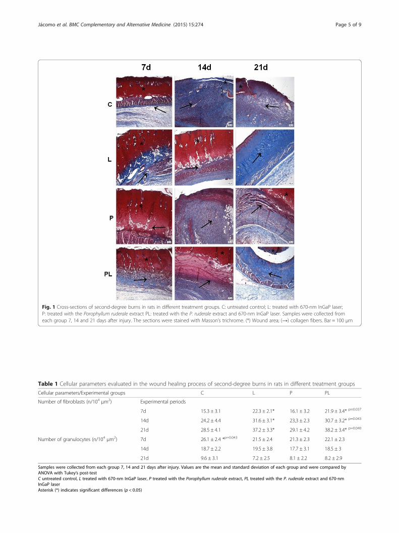
Fig. 1 Cross-sections of second-degree burns in rats in different treatment groups. C: untreated control; L: treated with 670-nm InGaP laser;P: treated with the Porophyllum ruderale extract PL: treated with the P. ruderale extract and 670-nm InGaP laser. Samples were collected fromeach group 7, 14 and 21 days after injury. The sections were stained with Masson’s trichrome. (*) Wound area; (→) collagen fibers. Bar = 100 μm
Table 1 Cellular parameters evaluated in the wound healing process of second-degree burns in rats in different treatment groups
Cellular parameters/Experimental groups C L P PL
Number of fibroblasts (n/104 μm2) Experimental periods
7d 15.3 ± 3.1 22.3 ± 2.1* 16.1 ± 3.2 21.9 ± 3.4* p=0.037
14d 24.2 ± 4.4 31.6 ± 3.1* 23,3 ± 2.3 30.7 ± 3.2* p=0.043
21d 28.5 ± 4.1 37.2 ± 3.3* 29.1 ± 4.2 38.2 ± 3.4* p=0.040
Number of granulocytes (n/104 μm2) 7d 26.1 ± 2.4 *p=0.043 21.5 ± 2.4 21.3 ± 2.3 22.1 ± 2.3
14d 18.7 ± 2.2 19.5 ± 3.8 17.7 ± 3.1 18.5 ± 3
21d 9.6 ± 3.1 7.2 ± 2.5 8.1 ± 2.2 8.2 ± 2.9
Samples were collected from each group 7, 14 and 21 days after injury. Values are the mean and standard deviation of each group and were compared byANOVA with Tukey’s post-testC untreated control, L treated with 670-nm InGaP laser, P treated with the Porophyllum ruderale extract, PL treated with the P. ruderale extract and 670-nmInGaP laserAsterisk (*) indicates significant differences (p < 0.05)
Jácomo et al. BMC Complementary and Alternative Medicine (2015) 15:274 Page 5 of 9

DiscussionExperimental studies have shown that some phytothera-peutic agents are effective in accelerating the healing ofburns [24, 25]. P. ruderale is used in the folk medicinefor cicatrization and its anti-inflammatory propertiesand cicatrizing activity [26]. Similarly, many experimen-tal studies have found beneficial results with the use oflow-level laser irradiation on burn wound healing [11].The aim of the combination of this medicinal plant withlaser exposure was to evaluate possible positive synergiceffect among these therapies on the healing of burns inrats as observed by Mendonça et al. [27] in the treat-ment of wound in sheep in which the authors obtained areduction in repair time of these lesions.In this study, the number of fibroblasts and collagen
fibers increased in the burns treated with 670-nm InGaPlaser alone and in those treated with both laser and P.ruderale extract. There was no increase in either fibro-blasts or collagen fibers in the wounds that were eitheruntreated or treated with the extract alone. This suggeststhat the laser treatment promoted a more effectiveresponse. Similarly, other studies have observed thatlow-intensity laser treatment of wounds in rats alsopromotes increased collagen production as a result of
enhancement of fibroblast proliferation [28]. During ree-pithelialization the cell proliferation is an essential eventso proliferating fibroblasts at the wound site ensure anadequate supply of cells that will migrate and cover thewound surface [29].Quantitative analysis revealed a significant increase in
the number of newly formed vessels in wound samplestreated only with the 670-nm InGaP laser. These resultsare in accordance with the literature that considers thelaser as an angiogenesis stimulator [30]. Neves et al. [31]studied the effect of laser irradiation 670-nm InGaP onexcisional wounds in Wistar rats and observed that thisapproach also promoted an increase in angiogenesis atthe injury site. Similarly, Sousa et al. [32] observed sig-nificant angiogenic effects promoted by low-level lasertherapy in skin burns and excisional wounds on the backof Wistar rats, respectively.The combination of laser with P. ruderale did not pro-
mote the same answers that L and P groups. In PLgroup was observed fibroblast proliferation and anti-inflammatory action. However, a synergistic effect wasnot observed between these two agents as expectedwhen the experimental design was conceived. A prob-able explanation for this fact may be related to the per-manence of the green pigment of leaves in P. ruderaleextract, even after lyophilization. This may have partiallycompromised the laser performance in the tissue, oncewhen the laser was applied alone it was possible to ob-serve its effectiveness in increasing the content of colla-gen fibers and the number of newly formed vessels inthe injured area. According to the considerations ofMelo et al. [33] despite experimental difficulties in evalu-ating the light transport in biological tissues, it is import-ant to know the absorption and light scattering coefficientin these, which are fundamental quantities that enable theunderstanding of the various effects of light-tissue inter-action. These authors have experimentally used differenttypes of lasers to evaluate the exponential attenuation ofthe laser light during its propagation in different rat tis-sues. High penetration of 630 nm laser red light in theabdominal fat layer of rat was observed and it was foundthat this was due to the absence of pigments in this tissuewhich facilitated its penetration [33]. Differently, Prateset al. [34] investigate the ability of malachite green com-bined with a low-power red laser (GaAlAs 660 nm) to killActinobacillus actinomycetemcomitans and showed thatan important periodontal bacterium can be photoinacti-vated by red light and malachite green. Malachite green(MG) shows strong absorption at the red end of the visiblespectrum [35].Different studies demonstrated that low-intensity laser,
used experimentally to repair wounds, promotes decreasein the number of inflammatory cells [36, 37] which agreeswith our results found through morphometric analyzes of
Table 2 Collagen content (% of area) evaluated in the woundhealing process of burns second degree in rats in differenttreatment groups
Experimentalgroups/periods
C L P PL
7d 20.5 ± 8.2 28.6 ± 7.2 22.6 ± 4.7 29.4 ± 5.6
14d 41.3 ± 8.1 39.6 ± 8.2 37.6 ± 5.7 56.9 ± 6.8* p=0.048
21d 53.4 ± 9.5 88.6 ± 10.4* p =0.046 48.8 ± 7.8 71.5 ± 7.2*
Samples were collected from each group 7, 14 and 21 days after injury. Valuesare the mean and standard deviation of each group and were compared byANOVA with Tukey’s post-testC untreated control, L treated with 670-nm InGaP laser, P treated with theP. ruderale extract, PL treated with the P. ruderale extract and 670-nm InGaP laserAsterisk (*) indicates significant differences (p < 0.05)
Table 3 Number of new vessels (n/104 μm2) evaluated in thewound healing process of burns second degree in rats indifferent treatment groups
Experimentalgroups/periods
C L P PL
7d 1.1 ± 0.2 1.8 ± 0.3* p=0.044 1.0 ± 0.4 1.2 ± 0.2
14d 1.7 ± 0.4 2.7 ± 0.3* p=0.042 1.4 ± 0.7 2.2 ± 0.1
21d 2.2 ± 0.4 3.1 ± 0.4* p=0.045 2.2 ± 0.3 2.0 ± 0.4
Samples were collected from each group 7, 14 and 21 days after injury. Valuesare the mean and standard deviation of each group and were compared byANOVA with Tukey’s post-testC untreated control, L treated with 670-nm InGaP laser, P treated with theP. ruderale extract, PL treated with the P. ruderale extract and 670-nmInGaP laserAsterisk (*) indicates significant differences (p < 0.05)
Jácomo et al. BMC Complementary and Alternative Medicine (2015) 15:274 Page 6 of 9

the samples from the lesions treated with laser irradiation.Lesions samples treated with the extract of P. ruderale(“arnica paulista”) results were similar to other samples ofthe wounds from groups submitted to treatment, havingreduced significantly the number of granulocytes cellswhen compared to untreated wounds during of the repairprocess. This result confirms other studies that have beenmade with other species of the Asteraceae family, forexample, Solidago microglossa (brazilian arnica) [38]and Arnica montana [39] showed anti-inflammatory ef-fects on skin wounds in Wistar rats. Probably due tothe presence of flavonoids in the composition of ar-nicas, indicating the anti-inflammatory properties ofthis plant. Flavonoids are known for their antioxidantand anti-inflammatory properties [40] and Park et al.[41] also described the high therapeutic potential ofthese compounds for thermal skin injuries in mice. Thephytochemical analysis in our study showed a largeamount of phenolic compounds in the leaves of P.ruderale, and Conde-Hernandez and Guerrero-Beltrán[10] considered that these coumpounds may be respon-sible for the antioxidant activity of this plant.Despite the large number of reports on the in vitro
and in vivo actions of TGF-β1, there is still controversyregarding the endogenous role of this growth factor whichseems to be involved in the onset of the inflammatoryphase during wound healing [42]. Neves et al. [31]
observed an increase in TGF-β1 expression at the be-ginning of the healing process (day 6) and a reductionon day 10 of treatment with 670-nm InGaP laser. Thisconfirms our results that showed expression of thiscytokine highlighted in the early phases of the healingof burn injuries in the samples treated with laser andalso with the extract of P. ruderale. The Asteraceaefamily (arnicas) favors wound healing because promotean anti-inflammatory response due to the presence offlavonoids in their constituents [43] and probably thesecompounds, as well as laser irradiation, favored the re-lease of this protein at this stage of the healing process.TGF-β1 has potent regulatory activity of inflammatory
processes since it exerts a chemoattractant role for neu-trophils and macrophages, which are important in theinitial processes of tissue repair [3, 44]. Wang et al. [45]investigated the healing of skin wounds in mice and sug-gested that the excessive and prolonged presence ofTGF-β1 at the site of injury does not benefit woundhealing. This is partially due to the proinflammatory ef-fect of this protein [45]. The decrease in TGF-β1 expres-sion from day 21 lesions treated with 670-nm InGaPlaser (L) may indicate that this treatment benefited therepair process by reducing the time of the inflamma-tory phase contributing to faster healing.Angiogenesis is modulated by hypoxia, nitric oxide
and growth factors such as VEGF [46] which is produced
Fig. 2 Immunoblot analysis of the expression of TGF-β1 and VEGF after 7, 14 and 21 days of the experiment in second-degree burns in rats indifferent treatment groups. C: untreated control; L: treated with 670-nm InGaP laser; P: treated with the Porophyllum ruderale extract PL: treatedwith the P. ruderale extract and 670-nm InGaP laser. Typical blots are shown above average densitometry results. Values are the mean andstandard deviation of each group and were compared by ANOVA with Tukey’s post-test (* p < 0.05)
Jácomo et al. BMC Complementary and Alternative Medicine (2015) 15:274 Page 7 of 9

by different types of cells that participate in wound heal-ing. Differences in the expression of VEGF were ob-served throughout the experimental period in thetreated groups. There was a significant increase in VEGFexpression in the laser-irradiated group (L) on day 14when compared to control, and was reduced on day 21.Treatment with extract of P. ruderale maintained the ex-pression of this protein throughout the experimentalperiod similarly to the control (Fig. 2).In our study we found that the expression of VEGF
has been modified in the irradiated samples with a sig-nificant increase at day 14 of treatment, indicating thatin this period the laser therapy promoted angiogenesis,since this growth factor is implicated in this process dur-ing skin development [47]. Neves et al. [31] comparedthe effects of microcurrent stimulation and LLLT onexperimental wound healing in healthy and diabeticWistar rats. The authors observed an increase in VEGFexpression in all diabetic groups on day 6 when com-pared to the respective groups of healthy animals. Thisfinding might be related to the tissue hypoxia commonlyseen in the skin of diabetic individuals as a consequenceof impaired microcirculation since tissue hypoxia signifi-cantly increases the expression of VEGF and this may beresponsible for improved tissue repair. Renno et al. [48]irradiated second-degree burns in rats with a low-levellaser (660 nm) and observed significant expression ofVEGF on day 14. The authors suggested that this treat-ment was beneficial since it reduced the necrotic area inthe wound through angiogenesis.
ConclusionsThe results of this study indicate that laser irradiation670-nm InGaP promoted beneficial responses in the re-pair process increasing collagen deposition and angio-genesis when was used separately. Porophyllum ruderalewas effective in decreasing the granulocytes during therepair process indicating a possible anti-inflammatoryaction of this native flora, widely used in folk medicine,but little studied experimentally.
Competing interestsThe authors declare that they have no competing interests.
Authors’ contributionsACJJ, KAV, RGL and LMGN carried out experimental work, data collection andevaluation, literature search and manuscript preparation. FOGG carried outtechnical procedures to produce the extract. MAME contributed to prepareand evaluation structural and morphometric analysis. MECA helped toprepare and evaluation Western Blotting analysis. FASM and GMTS raisedfunding, supervised research work and refined the manuscript forpublication. All authors read and approved the final manuscript forpublication.
AcknowledgmentsThe authors thank the Hermínio Omettto Foundation by financial support.
Received: 3 March 2015 Accepted: 6 August 2015
References1. Pruitt Jr BA, McManus AT. The changing epidemiology of infection in burn
patients. World J Surg. 1992;16:57–67.2. Singer AJ, Clarck RA. Cutaneous wound healing. New Engl J Med.
1999;341:738–46.3. Atkins S, Smith KG, Loescher AR, Boissonade FM, O’Kane S, Ferguson MW,
et al. The effect of antibodies to TGF-beta 1 and TGF-beta 2 at a site ofsciatic nerve repair. J Peripher Nerv Syst. 2006;11:286–93.
4. Schultz GS, Wysocki A. Interactions between extracellular matrix and growthfactors in wound healing. Wound Repair Regen. 2009;17:153–62.
5. Bao P, Kodra A, Tomic-Canic M, Golinko MS, Ehrlich HP, Brem H. The role ofvascular endothelial growth factor in wound healing. J Surg Res.2009;153:347–58.
6. Nayak BS, Ramlogan S, Chalapathi Rao A, Maharaj S. Neurolaena lobata L.promotes wound healing in Sprague Dawley rats. Int J Appl Basic Med Res.2014;4(2):106–10.
7. Nayak BS, Raju SS, Ramsubhag A. Investigation of wound healing activity ofLantana camara L. in Sprague dawley rats using a burn wound model. Int JAppl Res Nat Prod. 2008;1:15–9.
8. Kumarasamyraja D, Jeganathan NS, Manavalan R. A review on medicinalplants with potential wound healing activity. Int J Pharm Pharm Sci.2012;2:105–11.
9. Fonseca MCM, Meira RMSA, Casali VWD. Vegetative organ anatomy andhistolocalization of lipids and phenolics compounds in Porophyllum ruderale(Asteraceae). Planta Daninha. 2006;24:707–13.
10. Conde-Hernández LA, Guerrero-Beltrán JA. Total phenolics and antioxidantactivity of Piper auritum and Porophyllum ruderale. Food Chem.2014;142:455–60.
11. Andrade AG, Lima CF, Albuquerque AKB. Effects of the therapeutic laser onthe wound healing of burns: a bibliographic review. Rev Bras Queimaduras.2010;9:21–30.
12. Dall Agnol MA, Nicolau RA, Lima CJ, Munin E. Comparative analysis ofcoherent light action (laser) versus non-coherent light (light-emitting diode)for tissue repair in diabetic rats. Laser Med Sci. 2009;24:909–16.
13. Mendonça JS, Neves LMG, Esquisatto MAM, Mendonça FAS, Santos GMT.Comparative study of the application of microcurrent and AsGa 904 nmlaser radiation in the process of repair after calvaria bone excision in rats.Laser Phys. 2013;23:035605.
14. Harborne JB. Phytochemical methods. In: A guide to modern techniques ofplant analysis. London: Chapman and Hall; 1984. p. 302.
15. Costa VP, Mayworm MAS. Plantas medicinais utilizadas pela comunidade dobairro dos Tenentes - município de Extrema, MG, Brasil. Rev Bras PlantasMed. 2011;13:282–92.
16. Wagner H, Bladt S. Plant drug analysis: a thin layer chromatography atlas.Springer-Verlag: Berlin Heidelberg; 1996. p. 320.
17. Chandra S, Mejia-Gonzalez E. Polyphenolic compounds, antioxidant capacityand quinone reductase activity of an aqueous extract of Ardisia compressain comparision to mate (Ilex paraguaiensis) and green (Camellia sinensis)Teas. J Agr Food Chem. 2004;52:3583–9.
18. Kosalec I, Pepeljnjak S, Bakmaz M, Vladimir-Knezevic S. Flavonoid analysisand antimicrobial activity of commercially available propolis product. ActaPharm. 2005;55:423–30.
19. Chiarotto GB, Neves LMG, Esquisatto MAM, Amaral MEC, Santos GMT,Mendonça FAS. Effects of laser irradiation (670-nm InGaP and 830-nmGaAlAs) on burn of second-degree in rats. Laser Med Sci. 2014;29:1685–93.
20. Campos PP, Vasconcelos AC, Ferreira MA, Andrade SP. Alterations in thedynamics of inflammation, proliferation and apoptosis in subcutaneousimplants of lupus-prone mice. Histol Histopathol. 2011;26:433–42.
21. Ukong S, Ampawong S, Kengkoom K. Collagen measurement and stainingpattern of wound healing comparison with fixations and stains. J MicroscSoc Thail. 2008;22:37–41.
22. Miller RG. Simultaneous statistical inference. New York: Springer-Verlag:Berlin Heidelberg; 1981. p. 299.
23. Towbin H, Staehelin T, Gordon J. Electrophoretic transfer of proteins frompolyacrylamide gels to nitrocellulose sheets: procedure and someapplications. Proc Natl Acad Sci U S A. 1979;76:4350–4.
24. Ashani-Esfahani S, Imanieh MH, Khosneviszadeh M, Meshksar A, NoorafshanA, Geramizadeh B, et al. The healing effect of Arnebia euchroma in second
Jácomo et al. BMC Complementary and Alternative Medicine (2015) 15:274 Page 8 of 9

degree burn wounds in rat as an animal model. Iran Red Crescent Med J.2012;14:70–4.
25. Chen S, Sun MZ, Wang B, Hao L, Zhang C, Xin Y. Wound healing effects ofcactus extracts on second degree superficial burned mice. J Med Plants Res.2011;5:973–8.
26. Lima GM, Bonfim RR, Silva MR, Thomazzi SM, Santos MRV, Quintans-JúniorLJ, et al. Assessment of antinociceptive and anti-inflammatory activities ofPorophyllum ruderale (Jacq.) Cass., Asteraceae, aqueous extract. Rev. bras.Farmacogn. 2011;21:486–90.
27. Mendonça GBN, Moraes JM, Ferreira J, Lima FG, Bastos ER, Soares LK, et al.Laser As-Ga-Al de baixa potência associado com solução aquosa debarbatimão (Stryphynodendron barbatiman martius) na reparação tecidualde ferida cutânea séptica de ovino, Universidade Federal de Goiás. 2008.[Acesso em 06 de setembro de 2014]. Disponível em: http://www.sovergs.com.br/conbravet2008/anais/cd/resumos/R1190-3.pdf.
28. Nayak BS, Maiya A, Kumar P. Influence of Helium-Neon LaserPhotostimulation on excision wound healing in Wistar rats. J Biol Sci.2007;7:89–92.
29. Li J, Chen J, Kirsner R. Pathophysiology of acute wound healing. ClinDermatol. 2007;25:9–18.
30. Galiano RD, Tepper OM, Pelo CR, Bhatt KA, Callaghan M, Bastidas N, et al.Topical vascular endotelial growth factor accelerates diabetic woundhealing through increased angiogenesis and by mobilizing and recruitingbone marrow-derived cell. Am J Pathol. 2004;164:1935–47.
31. Neves LMG, Matheus RL, Santos GMT, Esquisatto MAM, Amaral MEC,Mendonça FAS. Effects of microcurrent application and 670 nm InGaP low-level laser irradiation on experimental wound healing in healthy anddiabetic Wistar rats. Laser Phys. 2013;23:035604.
32. Sousa AP, Paraguassú GM, Silveira NT, Souza J, Cangussú MC, Santos JN,et al. Laser and LED phototherapies on angiogenesis. Laser Med Sci.2013;28:981–7.
33. Melo CA, Lima AL, Brasil IR, Castro e Silva Jr O, Magalhães DV, Marcasssa LG,et al. Characterization of light penetration in rat tissues. J Clin Laser MedSurg. 2001;19:175–9.
34. Prates RA, Yamada Jr AM, Suzuki LC, Eiko Hashimoto MC, Cai S, Gouw-Soares S, et al. Bactericidal effect of malachite green and red laser onActinobacillus actinomycetemcomitans. J Photoch Photobio B Biol.2007;86:70–6.
35. Yamaoka K, Sasai R. Pulsed electric linear dichroism of triphenylmethanedyes adsorbed on Montmorillonite K10 in aqueous media. J ColloidInterface Sci. 2000;225:82–93.
36. Bjordal JM, Johnson MI, Iversen V, Aimbire F, Lopes-Martins RA.Photoradiation in acute pain: A systematic review of possible mechanismsof action and clinical effects in randomized placebo-controlled trials.Photomed Laser Surg. 2006;24:158–68.
37. de Moraes JM, Mendonça DEO, Moura VBL, Oliveira MAP, Afonso CL, VinaudMC, et al. Anti-inflammatory effect of low-intensity laser on the healing ofthird-degree burn wounds in rats. Laser Med Sci. 2013;28:1169–76.
38. Facury Neto MA, Fagundes DJ, Beletti ME, Novo NF, Juliano Y, Penha-SilvaN. Systematic use of Solidago microglossa DC in the cicatrization of opencutaneous wounds in rats. Braz J Morphol Sci. 2004;21:207–10.
39. Castro FCB, Magre A, Cherpinski R, Zelante PM, Neves LMG, Esquisatto MAM,et al. Effects of microcurrent application alone or in combination withtopical Hypericum perforatum L. and Arnica montana L. on surgicallyinduced wound healing in Wistar rats. Homeopathy. 2012;101:147–53.
40. Vieira AP, Santos NR, Borges JHS, Vincenzi MPA, Schmitz WO. Flavonoidaction in second intention healing in surgically-induced clean wounds inWistar rats. Semin Ciênc Biol Saúde. 2008;29:65–74.
41. Park BK, Lee S, Seo JN, Rhee JW, Park JB, Kim YS, et al. Protection of burn-induced skin injuries by the flavonoid kaempferol. J Biochem Mol Biol Rep.2010;43:46–51.
42. Chen W, Wahl SM. Manipulation of TGF-beta to control autoimmune andchronic inflammatory diseases. Microbes Infect. 1999;1:1367–80.
43. Klaas CA, Wagner G, Laufer S, Sosa S, Della Loggia R, Bomme U, et al.Studies on the anti-inflammatory activity of phytopharmaceuticals preparedfrom Arnica flowers. Planta Med. 2002;68:385–91.
44. Li MO, Flavell RA. Contextual regulation of inflammation: a duet bytransforming growth factor-beta and interleukin-10. Immunity. 2008;28:468–76.
45. Wang XJ, Han G, Owens P, Siddiqui Y, Li AG. Role of TGF beta-mediatedinflammation in cutaneous wound healing. J Investig Dermatol Symp Proc.2006;11:112–7.
46. Liekens S, De Clercq E, Neyts J. Angiogenesis: regulators and clinicalapplications. Biochem Pharmacol. 2001;61:253–70.
47. Colwell AS, Beanes SR, Soo C, Dang C, Ting K, Longaker MT, et al. Increasedangiogenesis and expression of vascular endothelial growth factor duringscarless repair. Plast Reconstr Surg. 2005;115:204–12.
48. Renno AC, Iwama AM, Shima P, Fernandes KR, Carvalho JG, de Oliveira P,et al. Effect of low-level laser therapy (660 nm) on the healing of second-degree skin burns in rats. J Cosmet Laser Ther. 2011;13:237–42.
Submit your next manuscript to BioMed Centraland take full advantage of:
• Convenient online submission
• Thorough peer review
• No space constraints or color figure charges
• Immediate publication on acceptance
• Inclusion in PubMed, CAS, Scopus and Google Scholar
• Research which is freely available for redistribution
Submit your manuscript at www.biomedcentral.com/submit
Jácomo et al. BMC Complementary and Alternative Medicine (2015) 15:274 Page 9 of 9