ACTIVE TRANSPORT BY THE CECROPIA MIDGUT II. Fine
28
ACTIVE TRANSPORT BY THE CECROPIA MIDGUT II. Fine Structure of the Midgut Epithelium EVERETT ANDERSON and WILLIAM R. HARVEY From the Department of Zoology, The University of Massachusetts, Amherst ABSTRACT A morphological basis for transcellular potassium transport in the midgut of the mature fifth instar larvae of Hyalophora cecropia has been established through studies with the light and electron microscopes. The single-layered epithelium consists of two distinct cell types, the columnar cell and the goblet cell. No regenerative cells are present. Both columnar and goblet cells rest on a well developed basement lamina. The basal portion of the columnar cell is incompletely divided into compartments by deep infoldings of the plasma membrane, whereas the apical end consists of numerous cytoplasmic projections, each of which is covered with a fine fuzzy or filamentous material. The cytoplasm of this cell contains large amounts of rough endoplasmic reticulum, microtubules, and mitochondria. In the basal region of the cell the mitochondria are oriented parallel to the long axes of the folded plasmaqemma, but in the intermediate and apical portions they are randomly scattered within the cytoplasmic matrix. Compared to the columnar cell, the goblet cell has relatively little endoplasmic reticulum. On the other hand, the plications of the plasma membrane of the goblet cell greatly exceed those of the columnar cell. One can distinguish at least four characteristic types of folding: (a) basal podocytelike extensions, (b) lateral evaginations, (c) apical microvilli, and (d) specialized cytoplasmic projections which line the goblet chamber. Apically, the projections are large and branch to form villus-like units, whereas in the major portion of the cavity each projection appears to contain an elongate mito- chondrion. Junctional complexes of similar kind and position appear between neighboring columnar cells and between adjacent columnar and goblet cells as follows : a zonula adherens is found near the luminal surface and is followed by one or more zonulae occludentes. The morphological data obtained in this study and the physiological information on ion trans- port through the midgut epithelium have encouraged us to suggest that the goblet cell may be the principal unit of active potassium transport from the hemolymph to the lumen of the midgut. We have postulated that ion accumulation by mitochondria in close association with plicated plasma membranes may play a role in the active movement of potassium across the midgut. Since the pioneering studies of Malpighi (47), and Swammerdam (72), the digestive system of insects has been a topic of interest and controversy among cytologists and physiologists. Although the alimentary canal consists of three basic regions, viz, foregut, midgut, and hindgut, attention has been centered on the midgut. The insect midgut is generally characterized by two cell types, columnar cells and regenerative cells. In lepidopterous and tricopterous insects a third type, the goblet cell, is present (67, 68). Regenera- tive cells allegedly give rise to columnar and goblet cells, although Shinoda (67) and Yung-Tai (83) reported that the latter cell types are found in newly hatched silkworm larvae in the absence of regenerative cells. Apparently, regenerative cells 107
Transcript of ACTIVE TRANSPORT BY THE CECROPIA MIDGUT II. Fine
ACTIVE TRANSPORT BY THE CECROPIA MIDGUT
II. Fine Structure of the Midgut Epithelium
E V E R E T T A N D E R S O N a n d W I L L I A M R. H A R V E Y
From the Department of Zoology, The University of Massachusetts, Amherst
A B S T R A C T
A morphological basis for transcellular potassium transport in the midgut of the mature fifth instar larvae of Hyalophora cecropia has been established through studies with the light and electron microscopes. The single-layered epithelium consists of two distinct cell types, the columnar cell and the goblet cell. No regenerative cells are present. Both columnar and goblet cells rest on a well developed basement lamina. The basal portion of the columnar cell is incompletely divided into compartments by deep infoldings of the plasma membrane, whereas the apical end consists of numerous cytoplasmic projections, each of which is covered with a fine fuzzy or filamentous material. The cytoplasm of this cell contains large amounts of rough endoplasmic reticulum, microtubules, and mitochondria. In the basal region of the cell the mitochondria are oriented parallel to the long axes of the folded plasmaqemma, but in the intermediate and apical portions they are randomly scattered within the cytoplasmic matrix. Compared to the columnar cell, the goblet cell has relatively little endoplasmic reticulum. On the other hand, the plications of the plasma membrane of the goblet cell greatly exceed those of the columnar cell. One can distinguish at least four characteristic types of folding: (a) basal podocytelike extensions, (b) lateral evaginations, (c) apical microvilli, and (d) specialized cytoplasmic projections which line the goblet chamber. Apically, the projections are large and branch to form villus-like units, whereas in the major portion of the cavity each projection appears to contain an elongate mito- chondrion. Junctional complexes of similar kind and position appear between neighboring columnar cells and between adjacent columnar and goblet cells as follows : a zonula adherens is found near the luminal surface and is followed by one or more zonulae occludentes. The morphological data obtained in this study and the physiological information on ion trans- port through the midgut epithelium have encouraged us to suggest that the goblet cell may be the principal unit of active potassium transport from the hemolymph to the lumen of the midgut. We have postulated that ion accumulation by mitochondria in close association with plicated plasma membranes may play a role in the active movement of potassium across the midgut.
Since the pioneering studies of Malpighi (47), and Swammerdam (72), the digestive system of insects has been a topic of interest and controversy among cytologists and physiologists. Although the alimentary canal consists of three basic regions, viz, foregut, midgut, and hindgut, attention has been centered on the midgut. The insect midgut is generally characterized by two cell types,
columnar cells and regenerative cells. In lepidopterous and tricopterous insects a third type, the goblet cell, is present (67, 68). Regenera- tive cells allegedly give rise to columnar and goblet cells, although Shinoda (67) and Yung-Tai (83) reported that the latter cell types are found in newly hatched silkworm larvae in the absence of regenerative cells. Apparently, regenerative cells
107

are absent from the midgut of mature Cecropia larvae. Therefore, the epithelium is composed of columnar and goblet cells. Two fundamental questions regarding these cells remain unan- swered: (a) Could one be a physiological phase of the other? and (b) Does one cell function prin- cipally in digestion and the other in absorption or does each cell type perform both functions?
In addit ion to its involvement in certain aspects of digestion and absorption (80), the midgut ap- parently engages in other activities such as the synthesis of blood proteins (40). Moreover, its intense endergonic role is reflected in the unus- ually high activity of respiratory enzymes. Indeed the important cytochrome b5 (m) was discovered in the Cecropia midgut by Sanborn and Williams (65). It now appears that, at least in plant-eating insects, the midgut is also a pr imary site of ion regulation. This regulation includes the active transport of potassium from the hemolymph to the lumen of the gut. The chemical details of the process are summarized by Harvey and Neder- gaard (26) and are to be described fnrther in other communicat ions of this series.
In our efforts to interpret and understand the chemical data on ion transport by the midgut, a detailed anatomical portrait of the cells compris- ing the epithelium is indispensable. Therefore, this communicat ion explores the cytoarchitecture of the midgut ceils of Cecropia with both the light and electron microscopes and attempts to relate their structure to certain aspects of ion transport.
M A T E R I A L S A N D M E T H O D S
Mature fifth instar larvae of Hyalophora cecropia (L) reared on the leaves of wild cherry trees (Prunus spp) or willow trees (Salix spp) were anesthetized with frozen carbon dioxide, and the midgut was isolated by the techniques of Harvey and Nedergaard (26). After the removal of the peritrophic membrane with its enclosed leaves, the midgut was cut into small pieces. Some pieces were fixed in Bouin's or Helly's solutions and stained with Mallory's triple stain or with Heidenhain's iron hematoxylin; others were fixed in a 2% solution of phosphate-buffered (pH 7.4) osmium tetroxide and embedded in celloidin.
For electron microscope observations, small pieces of the midgut were fixed for 2 hr in a 2~o solution of glutaraldehyde buffered to pH 7.4 with 0.1 M phos- phate buffer (64). After this initial fixation, the tissue was postfixed for 1 hr in a 0.1 M phosphate-buffered (pH 7.4) osmimn tetroxide solution and subsequently rapidly dehydrated, infiltrated, and embedded in
Epon (45). One-micron sections of Epon-embedded material were stained according to the recommenda- tions of Ito and Winchester (34). Thin sections were stained with uranyl acetate followed by lead citrate (78) and subsequently examined with both the RCA EMU-3E and Phillips 200 electron microscopes.
O B S E R V A T I O N S
Bennett ( l l ) pointed out that the term " m e m - brane" has been used to denote many things. He divided biological membranes into the follow- ing four categories: (a) tissue membranes, (b) cellular membranes, (c) basement membranes (basement laminae), and (d) cytoplasmic mem- branes. The " m e m b r a n e " described here consists of a single layer of epithelial cells.
A rigorous analysis of active ion transport across cellular membranes requires that the mem- brane be isolated so that the composition of the bathing solution, temperature, and pressure can be controlled while important parameters such as the electrical potential and the short-circuit currents are measured. The technique developed by Ussing and Zerahn (76) for studying the frog skin has been modified by Harvey and Neder- gaard (26) for the study of the midgut. As illus- trated in Fig. 2, the isolated midgut (M) is mounted in the gap of a section of glass tubing which forms the inner part of a perfusion chamber. Further details of techniques used in studying active ion transport across the midgut are de- scribed by Harvey et al. (27).
.Light Microscopy
HISTOLOGY OF THE MIDGUT
The cylindrical midgut of a mature 15-g larva is about 3.5 cm long and 1 cm in diameter (M, Fig. 1). The inner wall of the midgut is corru- gated. This corrugation is a manifestation of the major epithelial folding (EL, Fig. 3). The one- cell-thick epithelial layer consists of tall (65-~) columnar cells (CC, Fig. 4) and shorter (35-#) goblet cells (GCE, Fig. 4). Although the term goblet cell usually refers to a mucus-secreting ceil with a definite cytology, the goblet cell in the midgut of Cecropia neither secretes mucus nor appears cytologically similar to vertebrate goblet cells. However, its superficial resemblance to thc vertebrate goblet cell has prompted histologists to use this descriptive term which we will retain. The epithelium of the midgut rests on a basement lamina (BL, Fig. 4) with which are associated
108 TIIE JOURNAL OF CELL BIOLOGY • VOLUME 31, 1966
tracheae, tracheoles, an inner laycr of circular muscles (RM, Fig. 3) and an outer layer of lon- gitudinal muscles (LM, Fig. 3) The longitudinal muscle layer is clearly discontinuous, and there also appear to be spaces between the fiber bundles in the circular layer. Therefore, the epithelial layer and basement lamina appear to be the only continuous dittusion barriers between the hemo- lymph and the lumen of the gut. In order for an ion or molecule in the hemolymph to reach the midgut lumen, it must at least traverse the base- ment lamina. It then may take either an extra- cellular pathway or pass across the basal plasma membrane, cytoplasm, and apical plasma mem- brane of the epithelial cell layer. Finally, it must pass through perforations in the peritrophic "membrane" (48, 84). Since the peritrophic "membrane" is removed, this structure need not be considered in discussions of the transport path- way in vitro.
Each columnar cell (CC, Fig. 4) contains a large, centrally located, sometimes oval, nucleus (NC, Fig. 4). The apical cell border is clearly striated (BB, Fig. 4) and is refractile to staining with Heidenhain's iron hemtoxylin. The apical ends oI the columnar cells extend beyond the border of the goblet cells, thereby giving the epithelium a scalloped appearance. Pear-shaped buds of varied sizes are frequently seen on the luminal surfaces of the columnar cells, and in many sections spheres with a similar internal den- sity are seen within the lumen (SB, Fig. 4; see also PB in Fig. 9). These buds are associated with healthy cells and do not seem to be signs of de- generative changes such as those described by Yung-Tai (83). Rather, they appear to be mani- festations of an apocrine secretory process. The basal half of the columnar cell is densely stained with Heidenhain's iron hematoxylin and exhibits the longitudinal fibrillar appearance (BS, Fig. 4) noted by earlier cytologists.
The goblet cell is always surrounded by colum- nar cells, and its nucleus is consistently basally located (NG, Fig. 4). The infranuclear cytoplas- mic area is devoid of the longitudinally oriented fibrillar structures that are so characteristic of this area in the columnar cell. A long oval cavity (GC, Fig. 4), which communicates freely with the lumen of the gut, dominates the architecture of the goblet cell. At light microscope levels, the border of the goblet cavity resembles the striated border of the columnar cell (GMC, Fig. 4) but is
stainable with Heidenhain's iron hematoxylin. Apparently, the lining of the goblet cavity is sim- ilar to the apical surface of the columnar cell. We shall see presently that the cavity's border is lined with numerous, rather highly specialized cyto- plasmic projections.
Regenerative cells were not seen in any of the many preparations examined. However, we have observed tracheolar cells that are so closely as- sociated with the epithelium that it is possible, al light optical levels, to interpret such cells as being an integral part of the epithelium (for example, see TC, Fig. 18). One wonders if it is not this type of cell that investigators have called regenerative cells (cf. 13).
Electron Microscopy
COLUMNAR CELLS
Fig. 5 is a low magnification image of the basal portion of a columnar cell in which the plasma membrane is thrown into an extensive array of deep parallel folds (BIF) that are perpendicular to the basement lamina. The infoldings of the plasma membrane incompletely divide the basal cytoplasm into compartments of varying sizes. In addition to containing ribosomes, cisternae of the rough endoplasmic reticulum, and microtubules, the larger compartments are characterized by long, usually slender but sometimes thick, mito- chrondria with distinct cristae (MC). The mito- ' chondria are parallel to the folds of the membrane. The smaller compartments usually contain all of the organelles previously mentioned except mito- chondria.
Profiles obtained from the basal portion of the columnar cell, particularly where the plane of sectioning is somewhat oblique, give the impres- sion that there is at least as much extracellular space as there is cytoplasm (IC, Fig. 6). Effec- tively, the infoldings extend about two-thirds of the way to the apical border of the cell (IC, Fig. 7).
The conspicuous brush border seen with the light microscope is resolved into a regular array of long, thin microvilli (MVC, Fig. 8; see inset Fig. 9). Scattered within the apical cytoplasm and pro- jecting into each microvillus are fine (60- to 70-A) filaments (FMV, Fig. 8). The surface of each microvillus is covered with a fine filamentous ma- terial (MVC, inset Fig. 9). This material may be an extraneous covering; more likely, it is a "fuzzy
E. ANDERSON AND W. R. HARVEY Active Transport by Cecropia Midgut. II 109

coat" similar to that which has been reported by other investigators on the microvilli of certain epithelial cells (33). Occasionally, evidence of micropinocytotic activity is seen in the crypts between microvilli.
Microtubules, approximately 200 to 240 A in diameter, are found throughout the cytoplasmic matrix (MT, Figs. 8 and 10). Whereas some microtubules appear randomly dispersed within the cytoplasm, the majority are parallel to the long axis of the cell, and are most numerous in the intermediate and apical regions. The micro- tubules were never seen projecting into the micro- villi.
An abundance of both rough endoplasmic ret iculum and free ribosomes is found within the cytoplasmic matrix of the columnar cell (ER, Figs. 7 and 10). Many of the ribosomes are ag- gregated into polysomes (PR, Fig. 8), suggesting that the cell is involved in protein synthesis (60). Also scattered within the cytoplasm are some spherical homogeneous (SP, Fig. 6) and granular bodies (GB, Fig. 17) of unknown function. In- frequently, one sees in the cytoplasm of both the columnar and goblet cells dense granular bodies such as those labeled UB in Figs. 7 and 18. The identity and function of these bodies are unknown although they may be viruses. The relatively small Golgi complexes are usually found in a supranuclear region.
The perforated nuclear envelope encompasses a granular nucleoplasm in which are suspended many masses of chromat in material (CH, Fig. 7).
(~OBLET CELLS
The phenomenon of membrane-folding reaches a max imum in the goblet cell. At least four areas of extensive plications may be recognized: (a) basal, (b) lateral, (c) goblet chamber, and (d) apical. It will be helpful to describe in detail these four areas.
(a) At the base of the cell the plasma membrane is evaginated to form podocytelike extensions which appear to support the cell above the base- ment lamina (BOF, Figs. 11 and 18).
(b) More prominent evaginations of the plasma membrane are found along the lateral boundary of the cell and extend from the basal surface to the level of a zonula occludens (see below). These slender cvaginations create a laminar pattern be- tween the basal two-thirds of the goblet cell and adjacent columnar cells (LOF, Figs. 5, 7, and 12) and appear to communicate with each other by small cytoplasmic bridges. The extracellular space in the region of the lateral evaginations is very close to the goblet chamber and hence the midgut lumen via this chamber.
(c) Lining the entire goblet chamber (GC, Figs. 1 l, 12, 14, 16, and 17), but best developed on its basal (MVb Figs. II and 18) and lateral aspects
FIGURE 1 A photograph of a dissected mature fifth instar Cecropia larva showing the large midgut (M) X 3.
FIGURE e A diagrammatic representation of how the midgut is mounted when measure- ments are made of potential differences across an isolated preparation. Midgut (M); lu- men side (sparse stippling); blood side (close stippling); inlets for aerating and stirring gas (A and A'); calomel electrodes (C and C'); agar bridges (E and E'); potentiomete~ (mv); dry-cell battery (B); voltage divider (V); microammeter (uA); silver-silver chloride electrodes (S and S'). (After Harvey and Nedergaard, see reference ~6. Reprinted, by permission, from Proc. Nat. Acad. Sc., 1964, 51, 758.)
FIGURE 3 A section of a portion of the midgut fixed in a ~2% solution of buffered osnfimn tetroxide and embedded in celloidin, depicting major folding of the epithelium (EL), an outer layer of longitudinal nmscles (LM) and an inner layer of circular muscles (RM). X 100.
FIGURE 4 A section of the midgut illustrating columnar (CC) and goblet (GCE) cells resting on a basement lamina (BL). Nucleus of colmnnar cell (NC); brush horder of co- lumnar cell (BB); basal striations of columnar cell (BS); nucleus of goblet cell (NG); cavity of goblet cell (GC) limited by a relatively dense staining region (GMC); dense spherical bodies within the lumen of the midgut (SB). Heidenhain's iron henmtoxylin stained. × 700.
110 T h E JOURNAL OF CELL BIOLOGY - VOLUME 31, 1966

E. ANDEt~SON AND W. R. HARVEY Active Transport by Cecropla Midgut. 11 111

FIGUI/E 5 A section through tile basal region of tl cohmmar cell costing on a well defined basement lamina (BL). This protilc also depicts the nucleus (NC), a mitochond~'ion (MC) ~and basal infoldings (BIF). Note the lateral outfoldings of the plasma membrane of tlm adjacent goblet cell (I, OF) and the oblique section of a muscle fiber (MS).)< 10,000.
112
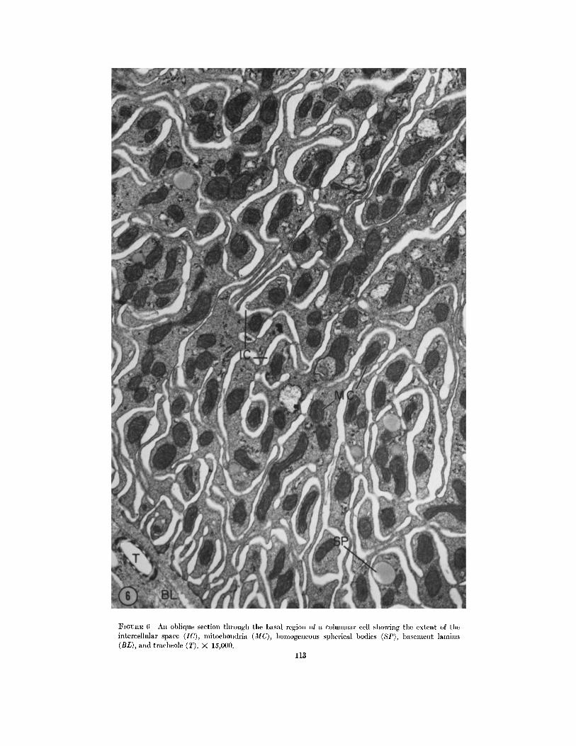
FIGURE 6 An ohlique section through the basal region of a columnar cell showing the extent of tile intercellular space (IC), mitoehondria (MC), homogeneous spherical bodies (SP), basement lamina (BL), and traeheole (T). X 15,000.
113

:FIGURE 7 An oblique section of a columnar cell at the level of the nucleus (NC), showing the il~ter- cellular spaces (IC), mitochondria (MC), endoplasmic reticulum (ER), and an unidentified granular body (UB). Note the lateral outfoldings of the adjacent goblet cell (LOF) and the mitochondria-filled projections (MV1) that line the lateral aspects of the goblet cavity. X 1~,000.

FIGLrRE 8 A small portion of the apical region of a columnar cell showing mierovilli (MVC), fine fila- ments within the cytoplasm many of which project into the microvilli (FMV), microtubules (MT), a mitochondrion (MC), and polyribosomes (PR).)< 30,000.
115

FIGURE 9 A portion of tile apical region of a columnar cell showing blebbing (PB). The inset illus- trates cross-sections of microvilli (MVC) whose surfaces are adorned with a fine fuzzy material. Fig. 9, X ~0,000; inset, X 80,000.
116

FIGURE 10 A section through the apical region of two adjacent columnar cells depicting junctional complexes in the form of a zonula adhaerens (ZA, inset), and a zonula occludens (ZO). The cytoplasmic matrix is filled with mitoehondria (MC), endoplasmic reticulum (ER), and mict'otubules (MT). Fig. 10, X '20,000; inset, X 40,000.
117

FIOURE 11 A section througlt tile basal portion of a goblet cell showing the cavity (GC) containing a flocculent materiM, cytoplasmic projections (MV1) each of which contains a mitochondrion, nucleus (NG), basal outfoldings (BOF), and basement lamina (BL). Note the fusion of the inner membranes of the mitochondria (MO; also see inset). Fig. 11, X 15,000; inset X 80,000.

FIo~nE 12 A section through a portion of a goblet cell and an adjacent columnar cell with its basal infoldings (B1F) resting on a basement lamina (BL). The goblet cavity (GC) is lined with a host of mitochondria-filled cytoplasmic projections (MV1). Many of the mitochondria show fusion of their inner limiting membranes (MO). Note the outfolding of the lateral plasma membrane (LOF) of tile goblet cell. X 18,000.

]7I~URE 13 A section through cytoplasmic projections (MV1) that line the cavity of a goblet cell. Note the spikelike units on the inner portion of the plasma meiabrane that forul these projections (*, Fig. 13 and insets). The inset at the lower right of the figure also shows portions of tile plasma membrane whose inner unit membrane is devoid of spikes (0). Fig. 18, X 30,000; upper right inset, X 60,000; lower right inset, X 70,000.

FIOURE 14 A cross-section through the upper portion of a goblet cell depicting its cavity (GC) and profiles of nonmitochondria-filled microvilli (ME2). This goblet cell is encompassed by four columnar cells (CC). Note the plasma membrane (CM) of a colmnnar ceil and the microtubules (MT) within the matrix of a columnar cell. X 15,000.
121

(MV1, Figs. 7, 12, and 17), are numerous cyto- plasmic projections. Each projection contains a single mitochondrion. In many profiles, the mito- chondria-containing projections extend into and virtually fill the chamber (MVj, Fig. 12). Oc- casionally a mitochondrion forks, sending branches into two adjacent projections. Some of the mitochondria are dumb-bell shaped. The in- ner mitochondrial membranes are often so closeiy apposed that it is difficult to resolve them as separate units (MO, Fig. l 1). Indeed, they appear to fuse (MO, inset Fig. 11) in much the same way that the outer leaflets of adjacent unit plasma membranes fuse when a zonula occludens is formed. Such profiles might represent stages in the dupli- cation of mitochondria or they might equally well represent a morphological change related to a presently unknown physiological condition. Oc- casionally, one finds similar mitochondrial con- figurations in columnar cells. These mitochondria are reminiscent of the "umbo-mitochondria" in the egg of the fern, Pteridium aquilinum, as described by Bell and Mtihlethaler (9). These authors in- terpreted such configurations as a stage in the development of this organelle.
Toward the apical end of the goblet chamber the cytoplasmic projections lining the cavity be- come smaller, less numerous, and contain no mi- tochondria (MV2, Figs. 14, 16, and 17). The core of these projections, like the microvilli of the co- lumnar cells, are filled with 60- to 70-A filaments.
The inner leaflet of the unit membrane of the cytoplasmic projections possesses a coat of fine spikelike units oriented perpendicular to the membrane (*, Fig. 13). Each spike is approxi- mately 50 to 60 A long and each is separated from adjacent ones by a space of about 30 A. In some images, the spikes appear to support a glob- ular structure 80 A in diameter (*, Fig. 13, insets). The organization of the entire unit reminds one of the construction of the "e lementary particles"
found on the cristae of mitochondria (24, 51). Of particular interest is the fact that this membrane specialization is only found here and is not a common feature of any other portion of the cell's membrane (O, Fig. 13, lower inset). Moreover, we have not observed this structural feature on any portion of the plasma membrane of the co- lumnar cell. Tha t it is a modification of the plasma membrane which may reflect altered permeabil- ity properties is made feasible both by the electrical measurements of Loewenstein and Kanno (43) and by the chemical studies of Ash- worth and Green (3) (see Discussion).
(d) In the apical region of the goblet cell the cytoplasm is drawn out into four or five large projections (P J, Fig. 15), each of which is oriented perpendicular to the long axis of the cell. As seen in Fig. 15, these large cytoplasmic re- gions are further subdivided into many villuslike units whose inner cores contain a filamentous component (VU, Figs. 15 and 16). In oblique sections the individual projections are organized so that they form a lacelike pattern (VU, Fig. 16). As the villuslike projections converge toward the middle of the goblet chamber they form a small canal of variable width (CA, Fig. 15). It is through this canal that the goblet cell chamber becomes confluent with the lumen of the gut. Because the canal is rather wide in some profiles, and more or less tightly closed in others, the structural evidence suggests that it may function as a valve to control the movement of fluids be- tween the goblet chamber and the midgut lumen.
The distribution and orientation of mitochon- dria within the goblet cells deserves further at- tention. The few mitochondria present in the cytoplasmic matr ix of the main cell body are randomly oriented, whereas the mi tochondr ia - cytoplasmic projection-associations are oriented about the circumference of the goblet cavity.
Scattered within the cytoplasm of the goblet
lq'IGUrCE 15 The inset is a photomicrograph of a Heidenhain's iron hematoxylin-stained preparation of a goblet cell, depicting its apical end (*) and cavity (GC). The cavity is bounded by a rather densely staining region (see line). To the right and left of this cell are columnar cells. The electron micrograph is a slightly oblique section made through the region marked by the asterisk in the inset. The large protoplasmic projection (P J) is further divided into numerous villuslike units (VU). These projections all converge at the midline to form a small channel (CA). Note the microtubules (MT) within the cyto- plasnaic matrix of the goblet cell. The cytoplasm of a columnar cell is labeled CC. Fig. 15, )< 16,000; inset, >( 300.
122 THE JOUIINAL OF CELL BIOLOGY • VOLUME 31, 1966

E. ANDERSON AND W. R, ~ARVEY Active Transport by Cecropia Midgut. I I 123

FIGURE 16 An oblique section through the apical portion of a goblet cell and two adjacent columnar cells (CC). Note the pattern made by the villuslike projections (VU) found at the apical tip of this cell, microtubulcs (MT), profiles of microvilli (MV2), and cavity of goblet cell (GC).)< ~0,000.
124

FIGURE 17 A section through a goblet cell and an adjacent columnar cell (CC). Note cytoplasmic pro- jections (MV1) and microvilli (MV2) extending into the cavity (GC) of the goblet cell, mitochoudria (MC), endoplasmic retieulum (ER), and microtubules (MT) within the cytoplasmic matrix of the columnar cell. Three zonulae occludentes are labeled ZO. X 18,000.
125

cell is a host of ribosomal units. However, the quant i ty of endoplasmic reticulum is scant in comparison to its abundance in the columnar cell. Microtubules are present throughout the cytoplasm, but are most numerous in the lateral and apical cytoplasm where they are randomly oriented (MT, Figs. 15 and 16). Occasionally, they are found within those cytoplasmic projec- tions which line the apical lateral portion of the goblet chamber.
JUNCTIONAL COMPLEXES OF THE
MIDGUT EPITttELIUM
The same kind and sequence of junct ional com- plexes are found both between adjacent columnar cells and between columnar and goblet cells. Fig. 10, a longitudinal section of the upper portion of two adjacent columnar cells, depicts the types of membrane specialization present. Beginning api- cally near the lumen of the midgut and proceeding basally, the first type of junct ional complex encountered is the zonula adhaerens (ZA, inset Fig. 10). The junct ion is rather large and is characterized by the presence of an intercellular gap and a conspicuous dense cytoplasmic area immediately adjacent to the plasma membrane. Proceeding basally, the next type of junct ion en- countered is the zonula occludens (ZO, Fig. ]0). This junct ion is recognized by the fusion of the outer lamellae of the unit membrane and the obliteration of the intercellular space. Below the first zonula occludens one may find other zonulae occludentes (ZO, Fig. 17). We have observed as many as five consecutive junctions of this type, particularly between adjacent goblet and colum- nar cells, As indicated above, evaginations from the lateral aspect of the goblet cell extend along the basal two-thirds of the lateral boundaries be- tween goblet and columnar cells. Junct ional com- plexes between these evaginations have never been observed.
BASEMENT LAMINA AND MUSCLE FIBERS
A thick basement lamina subtends the cells of the midgut epithelium and appears to be com- posed of two components (BL, Figs, 5, l l , 12, and 18), The component most closely associated with the basal aspect of the cell is filamentous; thc outer one is finely granular. The constituents of the basement lamina do not penetrate between either the infoldings of the columnar cells or the basal podocytelike extensions of the goblet cells.
The longitudinal and circular muscle fibers (MS, Fig. 5) contain the usual cytoplasmic con- stituents. A detailed description of them need not detain us here.
Our interpretat ion of the fine structure of the midgut of Cecropia is summarized in the schema- tic representation featured as Fig. 19.
DISCUSSION
This study has shown that the rnidgut and its epithelium is characterized by extensive plA- cations. These are: (a) the midgut wall including the muscle and epithelium, (b) apical microvilli and basal infoldings of the columnar cells, (c) the deep chamber of the goblet cell, and (d) the mod- ified (apical) mitochondria-filled cytoplasmic projections, the basal infoldings and the lateral evaginations of the goblet cell. Clearly, each order of folding represents a magnification of surface area available for the exchange of ions and mole- cules. This magnification of surface area is fur- ther increased by intracellular foldings such as mitochondrial cristae. In discussing the folding of the epithelium lining the gut of certain mammals, Fawcett (22) stated that, "The strategem of in- creasing the efficiency of organs by amplifying the areas of physiologically significant interfaces, appears to be one of nature 's commonest morpho- logical devices for 'miniatur izat ion ' of the meta- bolic machinery." The relationship of the greatly increased plasma membrane to its associated or- ganelles may explain one of the most striking activities of the Cecropia midgut- - i t s ability to transport potassium actively in massive amounts.
LOCALIZATION OF ACTIVE TRANSPORT SITE
The task of locating the cellular layer in which an active transport process occurs is a formidable one. Despite studies with the electron microscope (21), intracellular electrodes (16, 75) and clas- sical physiological techniques, the location of the cellular site of ion transport in the frog skin and toad bladder remains controversial (cf. 32). Al- though the toad bladder is a simpler organ it, nevertheless, contains at least four cell types (15, 52). By contrast, insect epithelia in general, are but one-cell layer thick; herein lies a tremendous advantage for the correlation of structure and function in solute transport. Assuming that the circular muscle layer is not involved in transport, one can conclude that active transport is localized in the epithelial layer and /o r its associated basal
126 THE JOURNAL OF CELL BIOLOGY - VOLUME 31, 1966
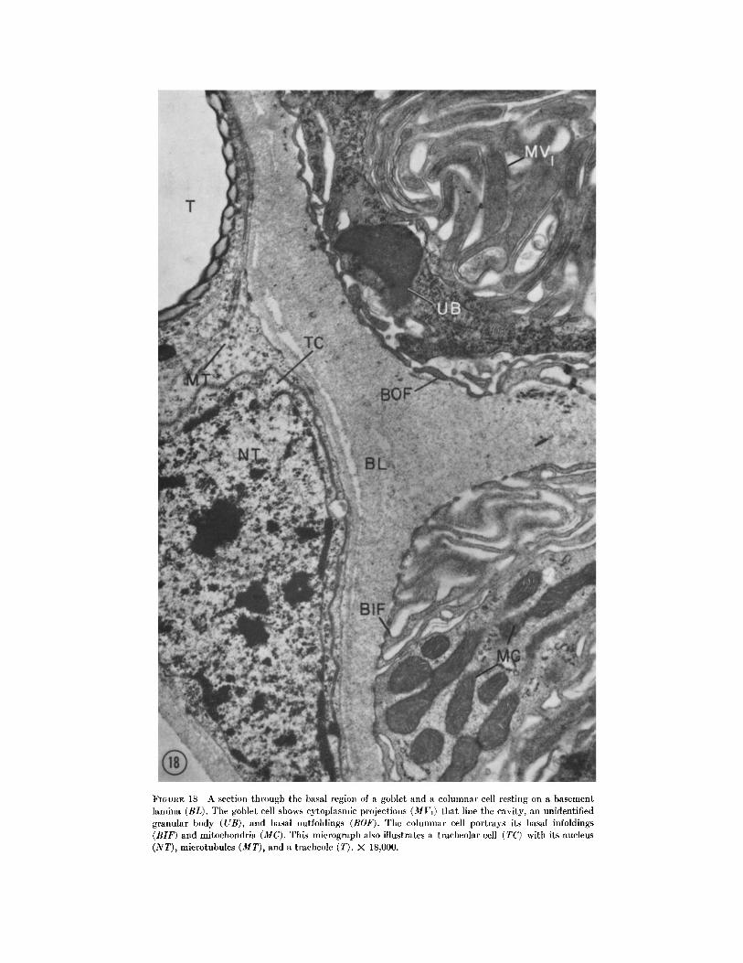
FIGURE 18 A section through the basal region of a goblet and a columnar cell resting on a basement lamina (BL). Ttle goblet cell shows cytoplasmic projections (MVj) that line tile cavity, an unidentified granular body (UB), and basal outfoldings (BOF). Tile columnar cell portrays its basal infoldings (BIF) and mitochondria (MC). This mierograph also illustrates a tracheolar cell (TC) with its nucleus (NT), microtubules (MT), and a traeheole (T) . )< 18,000.

FIGURE 19 A schematic representation of the cell types comprising the epithelium of the midgut of a mature fifth instar larva of Hyalophora cecropia. MVC, microvilli of the columnar cells; CA, canal formed by the villuslike units derived from the larger protoplasmic projections (P J) of the apical portion of the goblet cell; FMV, fine filaments within the microvilli of the columnar cell, ZA, zonula adhaerens; ZO, zonula occludens; MV2, mlcrovilli; MT, microtubules; ER, cudopl.asmic reticulum; GC, cavity of goblet cell; GC r, Golgi complex of columnar cell; NC, nucleus of col umuar c~ll; MV1, mitochon- dria-filled cytoplasmic projections that line the major portion of the cavity of the goblet cell; MC, mito- chondria of columnar cell; GC*, Golgi complex of goblet cell; NG, nucleus of goblet cell; BIF, basal infoldings of columnar cell; BL, basement lamina; MS, muscle fiber; BOF, basal podocytelike extensions of the goblet cell; LOF, lateral evaginations of the goblet cell; N T, nucleus of tracheolar cell; T, tracheole.
128 THE JOURNAL OF CELL BIOLOGY • VOLUME 31, 1966

lamina and proceed directly to the more interes- ting task of locating specific transport sites within the midgut cells. Because the physiological work to date has been performed on the midgut which contains both goblet and columnar cells, the prospect that either or both cell types contribute to ion transport must be considered. Indeed the close association of mitochondria with basal in- foldings in the columnar cells is very reminiscent of the modifications at the base of kidney cells thought to be involved in ion transport (see dis- cussion below). However, the dense mitochon- drial population associated with the plasma mem- brane of the goblet cell suggests a simple model for transcellular potassium ion transport. There- fore, principal attention in the following discus- sion will focus on the goblet cell.
THE CELL AS THE UNIT OF ACTIVE
ION TRANSPORT
Active transport is defined as the movement of a substance across a biological membrane which cannot be accounted for by known physical forces, i.e., transport of a substance against an electro- chemical gradient or with an electrochemical gradient but at a speed greater than that predicted from a knowledge of the physical forces involved (73). The energy to oppose the physical forces is provided by the metabolism of the living cell or by isolated organelles or fragments of living cells. This definition implies selectivity , ener- gization, and polarization of the solute movement . In the case of ion transport a charge separation is also implied.
According to Ussing (74), "Mos t of the cur- rent thinking about permeabil i ty problems is based on the acceptance of the membrane theory which presupposes that the main diffusion re- sistance in a cell is located at the surface mem- brane." Although this view does not necessarily presuppose that the active transport site is also located in the surface membrane, this assumption is implicit in most contemporary studies on ion transport (cf. 70). If active ion transport is to take place in the plasma membrane alone, a simple unit membrane must select, energize, and provide direction to the transported solute.
Attempts to locate the entire transport mechanism within the plasma membrane have been successful to some extent, and active ion transport in the absence of cytoplasmic consti- tuents has been demonstrated in red cell ghosts
(29) and in extruded squid axons (4). Moreover , all of the machinery necessary for oxidative me. tabolism is found in the cell wall of bacteria (49). Finally, phosphatidic acid and other proposed "carr ier" molecules (30) as well as "transport en- zymes," such as ouabain-sensitive, sodium- and potassium-activated ATPases, are localized in the plasma membrane (31). The theory that active transport mechanisms reside in the plasma mem- brane has been reviewed by Skou (70).
Since the accumulat ion of cations by isolated mitochondria is an active process, it is clear that active transport can and does occur in the ab- sence of the plasma membrane (14, 41, 53, 77). Large amounts of calcium and magesium and lesser amounts of potassium and sodium are taken up by isolated mitochondria through a process which is stoichiometrically linked to phosphate uptake and electron transport (61). In contrast to plasma membrane transport systems which are ouabain-sensitive (38), neither mitochondrial ion accumulation (cf. reference 41) nor potassium transport across the midgut (28) is inhibited by ouabain.
Since the pioneering studies of Sj6strand and Rhodin (69), Rhodin (59), Pease (54), and others, it is quite clear that many cells which engage in intensive transport activities have extensively folded plasma membranes intimately associated with mitochondria. Fawcett (22) has summarized several such well known cases (see also 55). In addition to the folding demonstrated for the mid- gut cells of Cecropia, an augmented but incom- plete list would also include: the apical canaliculi of the anal papillae of mosquito larvae (18) and cells of the branchiae of Artemia salenis (19), stri- ated ducts of the parotid gland (63), Malpighian tubules of insects (2, 8), the crayfish nephron (7), the epithelial cells of the gut of Malacosoma and Melanoplus (6), tracheal end cells of the light organ of the firefly, Photinus pyralis (5, 71), and the labial gland of adult Antheraea peryni (35). In other cases, such as the chloride cells of fish gills, very abundant but randomly oriented mitochon- dria are associated with that portion of the plasma membrane that forms an extensive system of tubu-
lar elements (56-58). Finally, there is al ignment of mitochondria in the absence of plasma mem- brane folding, as in the photocyte of the light or- gan of the firefly, Photinus (5, 71).
The magnitude of transport across the midgut cells is so great (short circuit currents of 2 to 4
E. ANDERSON AND W. R. HARVEY Active Transport by Cecropia Midgut. I I 129

ma for a segment of midgut 10 mm long and 5 to 10 mm in diameter are routinely measured by Harvey and associates) that it is difficult to be- lieve that oxidative machinery sufficient to en- ergize such massive flux could exist solely within the plasma membrane. Therefore, it is suggested that the mitochondria play an active role in this process. Their intimate membrane associations certainly shorten the diffusion distance to the plasma membrane for ATP. Equally important is the prospect that ion accumulat ion by mitochon- dria plays a central role in transcellular move- ment of ions (41). Either view implies that the entire cell, not just the plasma membrane, is the unit of active transport.
Ironically, neither the frog skin (21, 79) nor the toad bladder (15, 52) has cells that possess regular folds of the plasma membrane or close mi tochondr ia-membrane associations. Mitochon- dria are numerous in the cells of the stratum germinat ivum and stratum spinosum of the com- plex frog skin epithelium, and the so called mitochondria-rich cells of the bladder have dis- tinct vesicles and tubules packed in the apical region ( 15, 52). However, active sodium transport in the bladder and skin produces currents only about 1/~0 the magnitude of the potassium cur- rent of the midgut (66, 76; Harvey, Haskell, and Zerahn, unplublished data). It may be that the frog skin (el. 73) and toad bladder utilize a plas- ma membrane located system because the active ion movement in each case is ouabain-sensitive, as it is in isolated plasma membrane systems (cf. 66).
MITOCHONDRIAL PARTICIPATION IN ACTIVE
TRANSCELLULAR TRANSPORT
The results of an in vitro study of the potential and short-circuit current of the isolated Cecropia midgut (26 28) are far simpler to interpret by a model which invokes mitochondrial ion accumu- lation than by a model which interprets the trans- cellular potential as the sum of diffusion potentials and electrogenic pump potentials located in the plasma membrane. The presence of the goblet cavity may mean that potassium movement in vivo need not take place continuously against the entire lumen potassium concentration gradi- ent. The cavity may function in the same way that the apical cavity in the chloride cells of fish is alleged to aid salt movement (57, 58), i.e., by providing a space of relatively low potassium con-
centration into which potassium from the cell can diffuse.
Tha t the permeabil i ty properties of the cyto- plasmic projections and associated mitochondria of the goblet cell are different from those of the free plasma membrane is suggested by the struc- tural modification of the plasma membrane in the region of close mitochondrial association. It will be recalled that the inner leaflet of the unit membrane of the mitochondria-filled cytoplasmic projections of the goblet cell possesses recurring units, each of which appears to be composed of a stalk or spikelike portion that is capped by a globular structure. The units are not a feature of any other portion of the cell's plasma membrane, nor are they a characteristic of the plasma mem- brane of the columnar cells. In some respects, this coat of particles is indeed reminiscent of the fuzzy coat that is formed on the cytoplasmic side of the pits or invaginations that are formed by the plasma membrane during the initial stages of micropinocytosis. The end result of such a process is the formation of coated vesicles within the cytoplasm. The coated pits or invaginations ap- pear to be areas of the plasma membrane special- ized for the uptake of specific substances such as proteins (1, 12, 23, 62). Williamson (82) envisions that certain noncoated pits are also specialized areas of the plasma membrane; however, they are involved in the uptake of lipid.
The significance of the particles demonstrated on the inner portion of the unit membrane that forms the cytoplasmic projections of the goblet cell of Cecropia is unknown. As indicated previ- ously, we suspect that the ability of the midgut to transport massive amounts of potassium is a function of the goblet cell. Such a structural mod- ification as the particle-studded inner unit mem- brane might well play a role in the active transport of potassium in this system. A similar function has been suggested for particles on the inner leaflet of the highly folded area of the plas- ma membrane of cells comprising the rectal papillae of Calliphora erylhrocephala (25). Gupta and Berridge (25) have suggested that these parti- cles are the "seats of complex enzyme systems, generating energy for transport processes." Par- ticles similar in structure to the "elementary par- ticles" of mitochondria have also been observed on the outer plasma membrane of rat liver cells (10) and on the intestinal microvilli of rabbits (50).
Other plasma membrane specialization would
130 THE JOURNAL OF CELL BIOLOGY " VOLUME 31, 1966

include the surface coat of the intestinal microviUi as described by Ito (33), as well as junct ional complexes (see below). In addition to structural specialization, Berridge and Gupta (cf. 25) have shown that a magnesium and pH specific adeno- sine triphosphatase is localized on the leaflets of epithelial cells of the rectal papillae of Calliphora. Recently, Philpott (personal communicat ion) demonstrated that horseradish peroxidase is lo- calized on the extensive tubular system that is a modified portion of the plasma membrane of the chloride cells of Fundulus.
ION MOVEMENTS IN INTERCELLULAF$ SPACE
Farquhar and Palade (20) concluded that " the diffusion of water, ions, and small water-soluble molecules is impeded along the intercellular space of the epidermis by zonulae occludentes while it is facilitated from cell to cell within the epidermis by zonulae and maculae adhaerentes." This con- clusion is based on their finding that the zonulae occludentes are continuous barriers which leave the intercellular space open toward the dermis but closed to the external medium and on evidence that hemoglobin and ferritin penetrate the epi- dermis only to the zonula occludens. Their inter- pretation also considers the work of MacRobbie and Ussing (46) on the swelling of the epidermis in hypotonic solutions. A conclusive demonstration of the impermeabili ty of the zonulae occludentes to water is still lacking, especially since Ussing and Windhager (75) describe an extracellular shunt pathway for chloride in the isolated frog skin. At least one and as many as five zonulae occludentes are observed between columnar and goblet cells in the midgut. The finding of Harvey and Nedcrgaard (26) that potassium fluxes from lumen to blood-side are very small implies that these tight junctions (zonulae occludentes) or some other structure limits solute movement in the spaces between midgut cells.
In the salivary gland of Drosophila larvae the entire epi thel ium appears to be the unit of " ion environment ." Microclectrode measurements of the electrical resistance of epithelial cells by Loe- wenstein and Kanno (43, see also 36, 37, 44, 81) show a low value for contact surfaces between adjacent cells. This work demonstrates that ions can pass freely from cell to cell but not fi'om cell to exterior. These studies further indicate that the electrical properties of the plasma membrane of epithelial cells are not the same on all cell
surfaces because the specific resistance is lower at these contact surfaces, i. e., the lowered resist- ance is not the result of a simple increase in mem- brane area. This conclusion is supported by the analyses of phospholipid and sterol in plasma membranes and intercellular membranes by Ash- worth and Green (3) who found that the molar ratios of sterol to phospholipid in plasma mem- branes from different types of rat cells range from 0.24 to 132. Ashworth and Green state, " O u r results do not, however, support the idea of a common repeating chemical unit in all mem- branes of a cell (17) since the plasma membrane seems so different from the major intracellular membranes. It appears that there is both tissue specificity and functional specificity in the com- position and organization of cellular membranes."
Loewenstein and Kanno (43) postulate that the cell-to-cell ion movement occurs across sep- tate desmosomes which abound between adjacent Drosophila salivary gland cells. Such septate junc- tions are absent in the Cecropia midgut, so that if ions move freely between midgut cells other structures such as zonulae occludentes or zonulae adhaerentes must have special permeability prop- erties.
EVOLUTION OF TRANSPORT MECHANISMS
The morphological and physiological evidence suggests that active potassium transport by the Cecropia midgut differs radically from active sodium transport by the frog skin or toad bladder. We have suggested that the midgut employs an ouabain-insensitive mitochondrial ion accumula- tion device in which a close association of the plasma membrane with oriented and localized mitochondria is crucial; the skin and bladder may employ an ouabain-sensitive plasma mem- brane localized device. Recalling that the entire transport mechanism, as well as the machinery for oxidative metabolism, is present in the cell wall of bacteria, it is tempting to speculate that the primitive ion transport mechanism resides in the plasma membrane. When mitochondria evolved with the capacity to accumulate cations, another type of transport became possible. The ouabain-insensitive, electron transport-linked, ac-
tive potassium transport of yeast (39) may pro- vide a cellular example especially since aerobically grown yeast cells contain mitochondria (42). When epithelial cells evolved with mitochondria oriented along the basal-apical axis and in contact
E. ANDERSON AND W. R. HARVEY Active Transport by Cecropia Midgut. II 131

with plasma m e m b r a n e on ei ther the basal or apical surface, we postulate tha t the conditions for mi tochondr ia to part icipate in transcellular ion t ranspor t were fulfilled. Ions could ei ther be ac- cumulated actively from outside the cell and re- leased into the cytoplasm or accumulated from the cytoplasm and released outside the cell. I t is alto- gether possible tha t much of the controversy regard- ing mechanisms of active ion t ranspor t may stem from the presence of two fundamen ta l ly different t r anspor t mechanisms a p lasma m e m b r a n e
R E F E R E N C E S
1. ANDERSON, E., Oocyte differentiation and vitello- genesis in the roach, Periplaneta americana, J. Cell Biol., 1964, 211, 131.
2. ANDERSON, E., and BEAMS, H. W., The fine structure of the Malpighian tubules of 1)ytiscus sp. : A preliminary communication, Proc. Iowa Acad. Sc., 1957, 64,680.
3. ASHWORTH, L. A. E., and GREEN, C., Plasma membranes: Phospholipid and sterol content, Science, 1966, 151,210.
4. BAKER, P. F., HODGKIN, A. L., and SHAW, T. I., Replacement of the protoplasm of a giant nerve fiber with artificial solutions, Nature, 1961, 190, 885.
5. BEAMS, H. W., and ANDERSON, E., Light and electron microscope studies on the light organ of the firefly (Photinus pyralis), Biol. Bull., 1955, 109, 375.
6. BEAMS, H. W., and ANDERSON, E., Light and electron microscope studies on the striated border of the intestinal epithelial cells of in- sects, J. Morphol., 1957, 100, 601.
7. BEAMS, H. W., ANDERSON, E., and PRESS, N., Light and electron microscope studies on the cells of the distal portion of the crayfish nephron tubule, Cytologia, 1956, 21, 50.
~. BEAMS, H. W., TAHMISIAN, T. N., and DEVINE, R. L., Electron microscope studies on the cells of the Malpighian tubules of the grasshopper (Orthoptera, Acrididae), J. Biophysic. and Biochem. Cytol., 1955, 1, 197.
9. BELL, P., and M/]HLETHALER, K., The degenera- tion and reappearance of mitochondria in the egg cells of a plant, J. Cell Biol., 1964, 20, 235.
10. BENEDETTI, E. L., and EMMELO'r, P., Electron microscopic observations on negatively stained plasma membranes isolated from rat liver, J . Cell Biol., 1965, 26,299.
11. BENNETT, H. S., Introductory remarks in Intra- cellular Membranous Structure, (S. Seno and E. V. Cowdry, editors), Proceedings First International Symposium for Cellular Chemis- try, (suppl.) Symposium of the Society for
mechan i sm and a mi tochondr ia l m e c h a n i s m - - wi th the over-all character is t ics of t r anspor t de- pending on the degree to which each mechan i sm is developed or employed in a par t i cu la r tissue.
This investigation was supported by grants (GM- 08776 and AI-04291) from the National Institutes of Health, United States Public Health Service. The authors wish to thank Mr. and Mrs. L. Musante, Mr. J. Eversole, and Mr. J. Pallozola for their technical assistance.
Received for publication 9 ZVlarch 1966.
Cellular Chemistry, Okayama,J apan, Chugoku Press, Ltd., 1965, 14, 7.
12. BOWERS, B., Coated vesicles in the pericardial cells of the aphid (Myzus persicae Sulz), Proto- plasma, 1964, 59,351.
13. BOWERS, B., and WILLIAMS, C. M., Physiology of insect diapause. XII I . DNA synthesis during the metamorphosis of the Cecropia silkworm, Biol. Bull., 1964, 126, 205.
14. BRIERLEY, G. P., MURER, E., and GREEN, D. E., Participation of an intermediate of oxidative phosphorylation in ion accumulation by mito- chondria, Science, 1963, 140, 60.
15. CHOI, J. K., The fine structure of the urinary bladder of the toad, Bufo marinus, J. Cell Biol., 1963, 16, 53.
16. CHOWDlrIURY, T. K., and SNELL, F. M., A micro- electrode study of electrical potentials in frog skin and toad bladder, Biochem. et Bioph~sica Acta 1965, 94, 461.
17. CLAYTON, R. B., The utilization of sterols ill insects: A novel approach to the study of cell- membrane structure, Biochem. J., 1965, 95, 17P.
18. COPELAND, E., A mitochondrial pump in the cells of the anal papillae of mosquito larvae, J. Cell Biol., 1964, 23,253.
19. COPELAND, E., Salt transport organelle in Artemia salenis (Brine shrimp), Science, 1966, 151, 470.
20. FARQUnAR, M. G., and PALADE, G. E., Junc- tional complexes in various epithelia, J. Cell Biol., 1963, 17, 375.
21. FARqUHAR, M. G., and PALADE, G. E., Cell junctions in amphibian skin, J. Cell Biol., 1965, 26, 263.
22. FAWCETT, D. W., Physiologically significant specializations of the cell surface, Circulation, 1962, 26, 1105.
23. FAWCETT, D. W., Surface specializations of ab- sorbing cells, J. Histochem. and Cytochem., 1965, 13, 75.
24. FERNANDEz-MoRAN, H., ODA, T., BEAm, P. V.,
132 ThE JOURNAL OF CELL BIOLOOY • VOLUME 31, 1966

and GREEN, D. E., A macromolecular repeat- ing unit of mitochondrial structure and func- tion, d. Cell Biol., 1964, 22, 63.
25. GUPTA, B. L., and BERRIDGE, M. L., A coat of repeating subunits on the cytoplasmic surface of the plasma membrane in the rectal papillae of the blowfly, Calliphora erythrocephala (Meig.), studied in situ by electron microscopy, J. Cell Biol., 1966, 29, 376.
26. HARVEY, W. R., and NEDERGAARD, S., Sodium- independent active transport of potassium in the isolated midgut of the Cecropia silkworm, Proc. Nat. Acad. Sc., 1964, 51, 757.
27. HARVEY, W. R., HASKELL, J. A., and NEDER- OAARD, S., Active transport by the Cecropia midgut. II. Effects of changing ion concentra- tions on the midgut potential, in preparation.
28. HASKELL, J . A., CLEMONS, R. D., and HARVEY, W. R., Active transport in the Cecropia midgut. I. Inhibitors, stimulants, and potas- sium-transport, 3". Cell and Comp. Physiol., 1965, 65, 45.
29. HOFFMAN, J . F., Cation transport and structure of the red-cell plasma membrane, Circulation, 1962, 26, 1201.
30. HOKIN, L. E., and HOKIN, M. R., Studies on the carrier function of phosphatidic acid in sodium transport. I. The turnover of phosphatidic acid and phosphoinositide in the avian salt gland on stimulation of secretion, J. Gen. Physiol., 1960, 44, 61.
31. HOKIN, M. R., Studies on a Na + and K+-de - pendent ouabain-sensitive adenosine triphos- phatase in the avian salt gland, Biochem. et Biophysica Acta, 1963, 77, 108.
32. IMARURA, A., TAKEDA, H., and SASAKI, N., The accumulation of sodium and calcium in a specific layer of frog skin, J. Cell and Comp. Physiol., 1965, 66, 221.
33. ITO, S., The enteric coat on cat intestinal micro- villi, J. Cell Biol., 1965, 27, 475.
34. ITO, S., and WINCHESTER, R. J., The fine struc- ture of the gastric mucosa in the rat, J. Cell Biol., 1963, 16, 541.
35. KAFATOS, F. C., and WILLIAMS, C. M., En- zymatic mechanism for the escape of certain moths from their cocoon, Scienae, 1964, 146, 538.
36. KANNO, Y., and LOEWENSTEIN, W. R., Low- resistance coupling between gland cells. Some observations on intercellular contact mem- branes and intercellular space, Nature, 1964, 201, 194.
37. KANNO, Y., and LOEWENSTEIN, W. R., Inter- cellular diffusion, Science, 1964, 143, 959.
38. KAYE, G. I., and COLE, J. D., Electron micros- copy: Sodium localization in normal and
ouabain-treated transporting cells, Science, 1965, 150, 1167.
39. KERNAN, R. P., in Cell K, Washington, D.C., Butterworths, 1965.
40. LAUFER, H., Forms of enzymes in insect develop- ment, Ann. New York Acad. Sc., 1961, 94, 825.
41. LEHNINOER, A. L., The Mitochondrion, New York, W. R. Benjamin, Inc., 1964.
42. LINNANE, A. W., VITOLS, E., and NOWLAND, P. G., Studies on the origin of yeast mito- chondria, J. Cell Biol., 1962, 13, 345.
43. LOEWENSTEIN, W. R., and KANNO, Y., Studies on an epithelial (gland) cell junction. I. Modifications of surface membrane permea- bility, J. Cell Biol., 1964, 22, 565.
44. LOEWENSTEIN, W. R., SOCOLAR, S. J., HAOA- SmNO, S., KANNO, Y., and DAVlDSON, N., Intercellular communication: Renal, urinary bladder, sensory, and salivary gland cells, Science, 1965, 149, 295.
45. LUFT, J. H., Improvements in epoxy resin em- bedding methods, J. Biophysic. and Biochem. Cytol., 1961, 9,409.
46. MAcRonBIE, E. A. C., and USSING, H. H., Osmotic behavior of the epithelial cells of frog skin, Acta Physiol. Scand., 1961, 53, 348.
47. MALPIGHI, M., Observations of metamorphosis in Lepidoptera, m Dissertatio epistolica de Bombyce. Regiae societatis typogTaphos, Lon- don, (J. Martyn andJ . Allestry, editors), 1669.
48. MERCER, E. H., and DAY, M. F., The fine structure of the peritrophic membrane in Dixippus morosus, Biol. Bull., 1952, 103, 384.
49. MITCHELL, P., Biochemical cytology of micro- organisms, Ann. Rev. Microbiol., 1959, 13,407.
50. ODA, T., and SEKI, S., Molecular structure and biochemical function of the rnicrovilli mem- brane of intestinal epithelial cells with special emphasis on the elementary particles, J . Electron Micr., 1965, 14, 210.
51. PARSONS, D. F., CHANCE, B., and RACKER, E., Inner membrane subunits of mitochondria; possible function of oxidative phosphoryla- tion and mitochondrial contraction, J. Cell Biol., 1964, 23, 69A.
52. PEACHEY, L. O., and RASMUSSEN, H., Structure of the toad's urinary bladder as related to its physiology, J. Biophysic. and Biochem. Cytol., 1961, 10, 529.
53. PEACHEy, L. D., Electron microscopic observa- tions on the accumulation of divalent cations in intramitochondrial granules, J. Cell Biol., 1964, 20, 95.
54. PEASE, D. C., The fine structure of the kidney seen by electron microscopy, J. Histochem. and Cytochem., 1955, 3,295.
55. PEASE, D. C., Infolded basal plasma membranes found in epithelia noted for their water trans-
E. ANDEESON AND W. R. HARVEY Active Transport by Cecropia Midgut. 11 133

port, J. Biophysic. and Biochem. Cytol., 1956, 2, 203.
56. PHILPOTT, C. W., Electrolyte transport and acid nmcopolysaccharides of the cell surface, J. Cell Biol., 1964, 23, 74A.
57. PHILPOTT, C. W., Halide localization in the teleost chloride cell and its identification by selected area electron diffraction, Protoplasma, 1965, 60, 7.
58. PHILeOTT, C. W., and COPELAND, D. E., Fine structure of chloride cells from three species of Fundulus, J. Cell Biol., 1963, 18, 389.
59. RHODIN, J. , Correlation of ultrastruetural or- ganization and function in normal and ex- perimentally changed proxhnal convoluted tubule cells of the mouse kidney, Karolinska Institute, Stockhohn, Sweden, 1954, 1.
60. RICH, A., WARNER, J. R., and GOODMAN, H. M., The structure and function of polyribosomes, Syrup. Quant. Biol., 1963, 28, 269.
61. RossI, C. S., and LEHNINGER, A. L., Stoichio- metric relationships between accumulation of ions by mitochondria and the energy-coupling sites in the respiratory chain, Biochem. Z., 1963, 338, 698.
62. ROTH, T. F., and PORTER, K. R., Yolk protein uptake in the oocyte of the mosquito Aedes aegypti, J. Cell Biol., 1964, 20, 313.
63. RUTBER~, V., Ultrastructure and secretory mechanism of the parotid gland, Acta Odontol. Scan&, 1961, suppl. 30, 1.
64. SABATINI, D. D., BENSCH, K., and BARRNETT, R. J., Cytochemistry and electron microscopy. The preservation of cellular ultrastructure and enzymatic activity by aldehyde fixation, J. Cell Biol., 1963, 17, 19.
65. SANBORN, R. C., and WILLIAMS, C. M., The cytochrome system in Cecropia silkworin, with special reference to the properties of a new component, J. Gen. Physiol., 1950, 33, 579.
66. SHARP, G. W. G., and LEAF, A., Metabolic re- quirements for active sodium transport stimu- lated by aldosterone, d. Biol. Chem., 1965, 240, 4816.
67. SHINODA, O., Contributions to the knowledge of the intestinal secretion of insects. I. Mid- intestinal secretion of Lepidoptera, with an appendix: Behaviour of nfitochondria in the nfid-intestinal epithelium of the silkworm, Bombyx mori, Mere. College Sc. Kyoto Imp. Univ. Series B, 1926, 2, 93.
68. SHINODA, O., Contributions to the knowledge of insects. II. A comparative histo-cytology of the mid-intestine in various orders of insects. Z. Zellforsch. u. Mikr. Anat., 1927, 5, 278.
69. SJOSTRAND, F. S., and RHODIN, J., The ultra- structure of the proximal convoluted tubule
of the mouse kidney as revealed by high resolu- tion electron microscopy, Exp. Cell Research, 1953, 4,426.
70. SKOU, J. C., Enzymatic basis for active transport of Na + and K + across cell membranes, Physiol. Rev., 1965, 45, 596.
71. SMITH, D. S., The organization and innervation of the luminescent organ in a firefly, Photuris pennsylvanica (Coleoptera), J. Cell Biol., 1963, 16,323.
72. SWAM~aERDAM, J., The Book of Nature or, the History of Insects, London, C. G. Seyffert, 1758.
73. USSlNC, H. H., The alkali metal ions in isolated systerns and tissues, Handbuch Experiment. Pharmakol, 1960, 13, 1.
74. UssiNn, H. H., Passage of materials across bio- logical membranes, in Proceedings 1st Inter- national Pharmacology Meetings, The Mac- millan Company, New York, 1963, 4, 15.
75. Ussmc, H. H., and WINDHAGER, E. E., Active sodium transport at the cellular level, in Water and Electrolyte Metabolism (lI), (J. deGraeff and B. Leijnse, editors), West European Symposia on Clinical Chemistry, Arnsterdam, Elsevier Publishing Co., 1964, 3 ,3 .
76. USSING, H. H., and ZERAHN, K., Active transport of sodium as the source of electric current in the short-circuited isolated frog skin, Acta Physiol. Scand., 1951, 23, 110.
77, VASlN~TON, F. D., Some comments on the active uptake of divalent ions by mitoehondria, Bull. Inat. Cell Biol., Univ. Connecticut, 1965, 6: 1.
78. VENABLE, J. H., and COGGESHALL, R., A simpli- fied lead citrate stain for use in electron nficroscopy, J. Cell Biol., 1965, 25, 407.
79. VOUTE, C. L., An electron microscope study of the skin of the frog (Rana pipiens), J. Ultra- struct. Research, 1963, 9,497.
80. WATERHOUSE, D. R., Digestion in insects, Ann, Rev. Entomol., 1957, 2, 1.
81, \~?IENER, J., SI'IRO, D., and LOEWENSTEIN, W. R., Studies on an epithelial (gland) cell junction. II. Surface structure, J. Cell Biol., 1964, 22, 587.
82. WILLIAMSON, J. R., Adipose tissue : Morphological changes associated with lipid mobilization, J. Cell Biol., 1964, 2t), 57.
83. YUNG-TAI, T., L'histogenSse et l'histophysiologie de l'fipith61ium de l'intestin moyen chez un l~pidopt~re (Galleria mellonella), Bull. Biol. France et Belgique, 1929, suppl. 12, 1.
84. ZHUZHIKOV, I). P., Function of the peritrophic membrane in Musea domestica L. and Calliphora erythrocephala Meig., J. Insect Physiol., 1964, 10 273.
134 TnE JOURNAL OF C]~LL BIOLOGY " VOLUME 31, 1966