Activation of the mouse primary visual cortex by medial ... · Activation of the mouse primary...
11
SYSTEMS NEUROSCIENCE ORIGINAL RESEARCH ARTICLE published: 09 February 2015 doi: 10.3389/fnsys.2015.00001 Activation of the mouse primary visual cortex by medial prefrontal subregion stimulation is not mediated by cholinergic basalo-cortical projections Hoang Nam Nguyen, Frédéric Huppé-Gourgues and Elvire Vaucher * Laboratoire de Neurobiologie de la Cognition Visuelle, École D’optométrie, Université de Montréal, Montréal, QC, Canada Edited by: Chris John Tinsley, Nottingham Trent University, UK Reviewed by: Gregor Rainer, University of Fribourg, Switzerland Lutgarde Arckens, Catholic University, Belgium *Correspondence: Elvire Vaucher, Laboratoire de Neurobiologie de la Cognition Visuelle, École D’optométrie, Université de Montréal, CP 6128 Succursale Center-Ville, Montréal, QC H3C 3J7, Canada e-mail: [email protected] The medial prefrontal cortex (mPFC) exerts top-down control of primary visual cortex (V1) activity. As there is no direct neuronal projection from mPFC to V1, this functional connection may use an indirect route, i.e., via basalo-cortical cholinergic projections. The cholinergic projections to V1 originate from neurons in the horizontal limb of the diagonal band of Broca (HDB), which receive neuronal projections from the ventral part of the mPFC, composed of prelimbic (PrL) and infralimbic cortices (IL). Therefore, the objective of this study was to determine whether electrical stimulation of mice mPFC subregions activate (1) V1 neurons; and (2) HDB cholinergic neurons, suggesting that the HDB serves as a relay point in the mPFC-V1 interaction. Neuronal activation was quantified using c-Fos immunocytochemistry or thallium autometallography for each V1 layer using automated particle analysis tools and optical density measurement. Stimulation of IL and PrL induced significantly higher c-Fos expression or thallium labeling in layers II/III and V of V1 in the stimulated hemisphere only. A HDB cholinergic neuron-specific lesion by saporin administration reduced IL-induced c-Fos expression in layers II/III of V1 but not in layer V. However, there was no c-Fos expression or thallium labeling in the HDB neurons, suggesting that this area was not activated by IL stimulation. Stimulation of another mPFC subarea, the anterior cingulate cortex (AC), which is involved in attention and receives input from V1, activated neither V1 nor HDB. The present results indicate that IL and PrL, but not AC, stimulation activates V1 with the minor involvement of the HDB cholinergic projections. These results suggest a functional link between the ventral mPFC and V1, but this function is only marginally supported by HDB cholinergic neurons and may involve other brain regions. Keywords: prefrontal cortex, cholinergic neurons, basal forebrain, visual cortex, neuronal activity, autometallography, thallium, immunocytochemistry INTRODUCTION The medial prefrontal cortex (mPFC) plays a ubiquitous role in decision making, attention and other cognitive processes in humans, primates and rodents. However, the structure-function relationship of the mPFC within its different subregions and with other brain structures remains to be better defined. For example, there is a functional influence of the prefrontal modulation of primary visual cortex (V1) activity (Kuo et al., 2014), but the anatomical relationship between the two cortical areas is not known. The rat mPFC, as the homologs area in primates, the dorsolateral PFC, is involved in visual attention (Muir et al., 1996; Granon et al., 1998; Delatour and Gisquet-Verrier, 2000; Heidbreder and Groenewegen, 2003; Maddux and Holland, 2011) and cue guided behavior (Passetti et al., 2002; Kozak et al., 2006; Parikh et al., 2007). Both mechanisms may involve top- down control of the primary sensory areas, by modulating local responsiveness to afferent sensory stimuli. In this regard, mPFC has been shown to modulate V1 neuronal activity (Groenewegen and Uylings, 2000; Golmayo et al., 2003), but it does not directly project to V1 (Gabbott et al., 2005; Hoover and Vertes, 2007; Vogt and Paxinos, 2014). The mPFC in the rodent is composed of functionally and anatomically interacting subareas: the dorsal mPFC, which includes the medial agranular and anterior cingulate (AC) cortices, and the ventral mPFC (vmPFC), which includes the prelimbic (PrL) and infralimbic cortices (IL). The vmPFC is the major output region of the mPFC and shares anatomical connections with many parts of the brain, including the ventral tegmental area, the amygdala, several regions of the temporal lobe, the olfactory system, the hypothalamus, the hippocampal formation and the basal forebrain (BF), i.e., the diagonal band of Broca, substantia innominata and nucleus basalis magnocellularis (Vertes, 2004; Gabbott et al., 2005). In V1, attentional mechanisms are partly mediated by acetylcholine (ACh), as shown in primates (Disney et al., 2007; Frontiers in Systems Neuroscience www.frontiersin.org February 2015 | Volume 9 | Article 1 | 1
Transcript of Activation of the mouse primary visual cortex by medial ... · Activation of the mouse primary...
SYSTEMS NEUROSCIENCEORIGINAL RESEARCH ARTICLE
published: 09 February 2015doi: 10.3389/fnsys.2015.00001
Activation of the mouse primary visual cortex by medialprefrontal subregion stimulation is not mediated bycholinergic basalo-cortical projectionsHoang Nam Nguyen, Frédéric Huppé-Gourgues and Elvire Vaucher *
Laboratoire de Neurobiologie de la Cognition Visuelle, École D’optométrie, Université de Montréal, Montréal, QC, Canada
Edited by:Chris John Tinsley, NottinghamTrent University, UK
Reviewed by:Gregor Rainer, University ofFribourg, SwitzerlandLutgarde Arckens, CatholicUniversity, Belgium
*Correspondence:Elvire Vaucher, Laboratoire deNeurobiologie de la CognitionVisuelle, École D’optométrie,Université de Montréal, CP 6128Succursale Center-Ville, Montréal,QC H3C 3J7, Canadae-mail: [email protected]
The medial prefrontal cortex (mPFC) exerts top-down control of primary visual cortex(V1) activity. As there is no direct neuronal projection from mPFC to V1, this functionalconnection may use an indirect route, i.e., via basalo-cortical cholinergic projections. Thecholinergic projections to V1 originate from neurons in the horizontal limb of the diagonalband of Broca (HDB), which receive neuronal projections from the ventral part of the mPFC,composed of prelimbic (PrL) and infralimbic cortices (IL). Therefore, the objective of thisstudy was to determine whether electrical stimulation of mice mPFC subregions activate(1) V1 neurons; and (2) HDB cholinergic neurons, suggesting that the HDB serves as arelay point in the mPFC-V1 interaction. Neuronal activation was quantified using c-Fosimmunocytochemistry or thallium autometallography for each V1 layer using automatedparticle analysis tools and optical density measurement. Stimulation of IL and PrL inducedsignificantly higher c-Fos expression or thallium labeling in layers II/III and V of V1 inthe stimulated hemisphere only. A HDB cholinergic neuron-specific lesion by saporinadministration reduced IL-induced c-Fos expression in layers II/III of V1 but not in layerV. However, there was no c-Fos expression or thallium labeling in the HDB neurons,suggesting that this area was not activated by IL stimulation. Stimulation of another mPFCsubarea, the anterior cingulate cortex (AC), which is involved in attention and receivesinput from V1, activated neither V1 nor HDB. The present results indicate that IL and PrL,but not AC, stimulation activates V1 with the minor involvement of the HDB cholinergicprojections. These results suggest a functional link between the ventral mPFC and V1,but this function is only marginally supported by HDB cholinergic neurons and may involveother brain regions.
Keywords: prefrontal cortex, cholinergic neurons, basal forebrain, visual cortex, neuronal activity,autometallography, thallium, immunocytochemistry
INTRODUCTIONThe medial prefrontal cortex (mPFC) plays a ubiquitous rolein decision making, attention and other cognitive processes inhumans, primates and rodents. However, the structure-functionrelationship of the mPFC within its different subregions and withother brain structures remains to be better defined. For example,there is a functional influence of the prefrontal modulation ofprimary visual cortex (V1) activity (Kuo et al., 2014), but theanatomical relationship between the two cortical areas is notknown.
The rat mPFC, as the homologs area in primates, thedorsolateral PFC, is involved in visual attention (Muir et al.,1996; Granon et al., 1998; Delatour and Gisquet-Verrier, 2000;Heidbreder and Groenewegen, 2003; Maddux and Holland, 2011)and cue guided behavior (Passetti et al., 2002; Kozak et al.,2006; Parikh et al., 2007). Both mechanisms may involve top-down control of the primary sensory areas, by modulatinglocal responsiveness to afferent sensory stimuli. In this regard,
mPFC has been shown to modulate V1 neuronal activity(Groenewegen and Uylings, 2000; Golmayo et al., 2003), but itdoes not directly project to V1 (Gabbott et al., 2005; Hooverand Vertes, 2007; Vogt and Paxinos, 2014). The mPFC in therodent is composed of functionally and anatomically interactingsubareas: the dorsal mPFC, which includes the medial agranularand anterior cingulate (AC) cortices, and the ventral mPFC(vmPFC), which includes the prelimbic (PrL) and infralimbiccortices (IL). The vmPFC is the major output region of themPFC and shares anatomical connections with many parts ofthe brain, including the ventral tegmental area, the amygdala,several regions of the temporal lobe, the olfactory system,the hypothalamus, the hippocampal formation and the basalforebrain (BF), i.e., the diagonal band of Broca, substantiainnominata and nucleus basalis magnocellularis (Vertes, 2004;Gabbott et al., 2005).
In V1, attentional mechanisms are partly mediated byacetylcholine (ACh), as shown in primates (Disney et al., 2007;
Frontiers in Systems Neuroscience www.frontiersin.org February 2015 | Volume 9 | Article 1 | 1
Nguyen et al. mPFC activation of the primary visual cortex
Herrero et al., 2008) and rodents (Chudasama et al., 2004; Dalleyet al., 2004). Recent studies linking the anatomical projectionof the vmPFC to neurons of the BF (Zaborszky et al., 1997;Vertes, 2004) and the projection of those neurons to V1 (Gaykemaet al., 1990; Laplante et al., 2005) proposed that the BF couldbe an anatomical and functional relay between the vmPFC andV1 (Golmayo et al., 2003). The horizontal limb of the diagonalband of Broca (HDB) is the main basalo-cortical cholinergic inputto V1 (Mesulam et al., 1983; Gaykema et al., 1990; Laplanteet al., 2005; Newman et al., 2012) and is involved in theneuromodulation of V1 activity (Dotigny et al., 2008; Kang andVaucher, 2009; Kang et al., 2014). In addition to cholinergic cells,the BF contains GABAergic and glutamatergic cells with diverseprojection patterns (Freund and Gulyás, 1991; Gritti et al., 1993,2006; Zaborszky et al., 2012).
In the present study, the anatomo-functional interactionbetween the mPFC, HDB and V1 was examined in the mouse.Electrical stimulation of the two major mPFC subregions,IL/PrL and the AC, was performed, and the resulting neuronalactivation was examined in V1 and the HDB by c-Fosimmunocytochemistry and thallium (Tl) autometallography(TlAMG; Danscher, 1981; Goldschmidt et al., 2004; Stöberet al., 2014). The two complimentary techniques assess differentaspects of neuronal activity. The contribution of cholinergiccells and fibers to the neuronal activation in V1 was confirmedby specific lesion of the cholinergic fibers in mice expressinggreen fluorescent EGFP protein under the control of the cholineacetyltransferase (ChAT) promoter.
MATERIAL AND METHODSANIMALSAdult mice expressing green fluorescent EGFP protein underthe control of the ChAT promoter (B6g-tgRP23-EGFP, 30–40 g,founders from Jackson laboratory, Bar Harbor, ME, USA)and adult wild-type C57BL/6 mice (Charles rivers Canada, St-Constant, QC, Canada) were housed at a maximum of fiveper cage with food and water ad libitum in a temperature-and humidity- controlled room with a 12-h light/dark cycle.Experiments were conducted in accordance with the CanadianCouncil on Animal Care Guidelines and were approved by ourlocal ethics committee for animal experimentation (protocol #12–172). EGFP colocalization with ChAT, a marker of cholinergicneurons, was ≥90%, as confirmed by ChAT immunostaining.
SPECIFIC LESIONS OF THE CHOLINERGIC FIBERSIn the cholinergic deficit group, a specific lesion of thecholinergic fibers was performed by intraventricular injectionof the neurotoxin p75-Saporin (Berger-Sweeney et al., 2001;Dotigny et al., 2008; Kang et al., 2014) 2 weeks prior to electricalstimulation of the mPFC. Mice were anesthetized with isoflurane(5% for induction and 2.5% for maintenance) with an oxygenflow of 1 l/min and placed into a stereotaxic frame (Kopfinstruments). A hole in the skull was opened and a 33 gaugeneedle was inserted into the right lateral ventricle at: AP: −0.4;ML: 1.0 from Bregma; DV: −2.0 from dura matter (Berger-Sweeney et al., 2001). The neurotoxin p75-Saporin (mu p75-SAP,lot #94–69, Advanced Targeting Systems, San Diego, CA, USA)
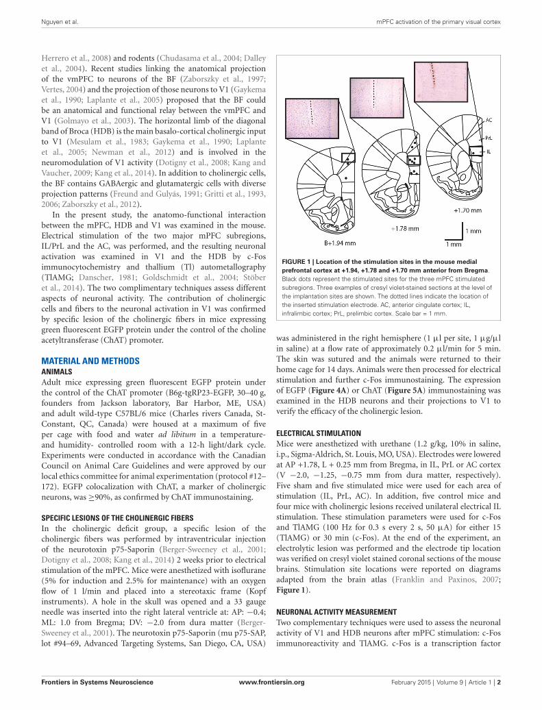
FIGURE 1 | Location of the stimulation sites in the mouse medialprefrontal cortex at +1.94, +1.78 and +1.70 mm anterior from Bregma.Black dots represent the stimulated sites for the three mPFC stimulatedsubregions. Three examples of cresyl violet-stained sections at the level ofthe implantation sites are shown. The dotted lines indicate the location ofthe inserted stimulation electrode. AC, anterior cingulate cortex; IL,infralimbic cortex; PrL, prelimbic cortex. Scale bar = 1 mm.
was administered in the right hemisphere (1 µl per site, 1 µg/µlin saline) at a flow rate of approximately 0.2 µl/min for 5 min.The skin was sutured and the animals were returned to theirhome cage for 14 days. Animals were then processed for electricalstimulation and further c-Fos immunostaining. The expressionof EGFP (Figure 4A) or ChAT (Figure 5A) immunostaining wasexamined in the HDB neurons and their projections to V1 toverify the efficacy of the cholinergic lesion.
ELECTRICAL STIMULATIONMice were anesthetized with urethane (1.2 g/kg, 10% in saline,i.p., Sigma-Aldrich, St. Louis, MO, USA). Electrodes were loweredat AP +1.78, L + 0.25 mm from Bregma, in IL, PrL or AC cortex(V −2.0, −1.25, −0.75 mm from dura matter, respectively).Five sham and five stimulated mice were used for each area ofstimulation (IL, PrL, AC). In addition, five control mice andfour mice with cholinergic lesions received unilateral electrical ILstimulation. These stimulation parameters were used for c-Fosand TlAMG (100 Hz for 0.3 s every 2 s, 50 µA) for either 15(TlAMG) or 30 min (c-Fos). At the end of the experiment, anelectrolytic lesion was performed and the electrode tip locationwas verified on cresyl violet stained coronal sections of the mousebrains. Stimulation site locations were reported on diagramsadapted from the brain atlas (Franklin and Paxinos, 2007;Figure 1).
NEURONAL ACTIVITY MEASUREMENTTwo complementary techniques were used to assess the neuronalactivity of V1 and HDB neurons after mPFC stimulation: c-Fosimmunoreactivity and TlAMG. c-Fos is a transcription factor
Frontiers in Systems Neuroscience www.frontiersin.org February 2015 | Volume 9 | Article 1 | 2

Nguyen et al. mPFC activation of the primary visual cortex
involved in the transcription of synaptic genes. Recently activatedcells overexpress the immediate-early gene c-Fos, the productof which can be visualized by immunocytochemistry. c-Fos hasbeen used as a powerful metabolic marker of functional activityacross brain areas, including in the visual system (Dragunowand Faull, 1989; Chaudhuri, 1997; Kaczmarek and Chaudhuri,1997; Arckens et al., 2000; Zangenehpour and Chaudhuri, 2002).However, some cells, such as the GABAergic cells, rarely expressc-Fos and the labeling is located only in the nucleus. TlAMGis based on the bioaccumulation of Tl+ ions that substitutepotassium ions. Tl accumulates in cells and neurites duringneuronal activation (Figure 2) through potassium channels.TlAMG Tl is then fixed by the perfusion of a sodium sulfidesolution and is developed with silver for visualization under amicroscope, as extensively described (Goldschmidt et al., 2004;Stöber et al., 2014). The use of TlAMG to visualize neuronalactivation is similar to the 2-deoxyglucose method from Sokoloff(Goldschmidt et al., 2004), but with a subcellular resolution. Thestaining is not restricted to neurons but also stains neurites andblood vessels. In addition, the nature of the activated neurons canbe assessed by their morphology. This technique has been usedhere to better visualize the possible activation of GABAergic BFneurons. A comparison of both stains is shown in Figure 2.
c-Fos ImmunocytochemistryTwenty-eight mice were used for c-Fos expression quantification.After electrical stimulation, urethane-anesthetized mice wererestrained in darkness for 1 h and then transcardially perfusedwith 60 ml of 4% paraformaldehyde (PFA, Sigma-Aldrich).The brains were then post-fixed for 24 h in 4% PFA. Coronalbrain sections (35 µm) covering the entire extent of the brainwere sliced with a vibratome (Leica VT1000S), serially collectedand stored in a phosphate buffer (PB)-glycerol based antifreezesolution for further use. Sections at 0.74 to 0.14 mm from Bregmawere chosen for immunostaining in HDB and at −2.30 to −4.04mm from Bregma for immunostaining in V1. Brain sectionswere incubated with rabbit anti-c-Fos (1:10000, OncogeneResearch Products, San Diego, CA, USA) primary antibody orgoat anti-ChAT (1:200, Millipore, Etobicoke, ON, Canada) in0.12 M PBS, 0.25% triton X-100 (Sigma-Aldrich) and 0.5%donkey serum (Jackson Laboratories). Secondary biotinylateddonkey anti-rabbit or anti-goat (Jackson Laboratories) antibodieswere coupled with the ABC complex for detection. Slides wereincubated for 1 h with the ABC complex and then visualizedusing the SG kit (Vector Laboratories).
Microphotographs of HDB and V1 were captured using aLeica DC500 camera. The counting of c-Fos-expressing cellswas completed with ImageJ (particle analysis, pixel size 60) afterthresholding on three slices at −2.54, −2.92 and −3.40 fromBregma for V1; at 0.74, 0.38 and 0.14 for HDB; at 1.34, 0.14and −0.94 for the primary somatosensory cortex (S1); at −1.58,−1.82 and −1.94 for the posterior parietal cortex (PPC) and at0.98, 0.26 and−0.22 for the midcingulate cortex (MC).
Thallium autometallographyAlternatively, the Tl-chelate solution was administered duringmPFC stimulation to perform TlAMG. Ten mice were
FIGURE 2 | Microphotographs representative of the neuronalactivation in V1 detected by c-Fos immunocytochemistry (A,C,E) orthallium autometallography (B,D,F). c-Fos is a transcription factorexpressed in the nucleus when neurons are activated. Thalliumautometallography alternatively relies on the proportional uptake of K+ ionswith neuronal activity. Note that c-Fos immunoreactivity is detectedprimarily in the layers II/III (A) and V (E) of V1 whereas TlAMG staining isobserved in all layers (B,D,F) with layer IV (D) showing communicatingfibers in TlAMG but not c-Fos (C). Scale bar = 10 µm.
anesthetized with freshly prepared urethane (1.2 mg/kg)and positioned on the stereotaxic frame. Immediately beforeinjection, equal volumes of 0.2% aqueous Tl (I) acetate (Sigma-Aldrich) solution and 0.2% sodium diethyldithiocarbamatetrihydrate (Sigma-Aldrich) dissolved in 1.8% NaCl were mixedtogether in a syringe, forming the Tl diethyldithiocarbamate(TlDDC) complex. Mice were injected i.p. with 300 µL of 0.1%TlDDC in 0.9% saline immediately prior to the beginning ofelectrical stimulation of the IL.
After electrical stimulation, the animals were perfused (3 min)with a freshly prepared 0.05% sodium sulfide (Sigma-Aldrich)solution containing 100 mM PB at pH 7.4. The brains were thenremoved and post-fixed for 24 h in a 5% acrolein-PB solution at4◦C. The brains were then washed three times in 100 mM PB andplaced in a 30% PB-sucrose solution for 48 h before freezing andstorage at −80◦C. Frozen brains were cut into 25 µm sectionsusing a Microm cryostat (HM500OM), directly mounted ongelatin-coated glass slides and air-dried for 2 h.
Frontiers in Systems Neuroscience www.frontiersin.org February 2015 | Volume 9 | Article 1 | 3
Nguyen et al. mPFC activation of the primary visual cortex
After drying, slides were placed in 0.1 N HCl for 30 minto remove zinc sulfide from the sections, washed three timesin distilled H2O, and bench air-dried again before staining. Adeveloper solution was prepared for staining in accordance withstandard autometallography methods as previously described byGoldschmidt et al. (2004, 2010). The sections were developedfor 120 min in the dark followed with 10 min of washing underrunning tap water to stop development. The sections were thendried, dehydrated and cover-slipped with Permount (FischerScientific, Fair Lawn, NJ, USA).
Gray-scaled microphotographs were captured with a 40XLeica Fluotar objective quantification and a Leica DC500camera. Neuron count and optical density quantification wereperformed using the mean gray values evaluated by ImageJ(Stöber et al., 2014). Three slices from each animal at −2.54,−2.92 and −3.40 mm from Bregma for V1 and at 0.74,0.38 and 0.14 mm from Bregma for HDB and substantiainnominata were selected for neuron count and/or meangray values analysis in layers II/III, IV and V of V1. Fiveequivalent sample areas from each animal/slice/layer werecompiled for analysis. High gray scale values correspondto low staining intensity in TlAMG, i.e., low neuronalactivity.
STATISTICAL ANALYSISMann-Whitney U tests were computed to compare neuronalactivity in the contralateral and ipsilateral hemispheres ofthe same animals and between groups for each layer usingSPSS 19 (SPSS Inc., Chicago, IL, USA) for c-Fos expression,TlAMG neuron count and TlAMG mean gray values.Statistical analyses were calculated for activity in the HDBand V1.
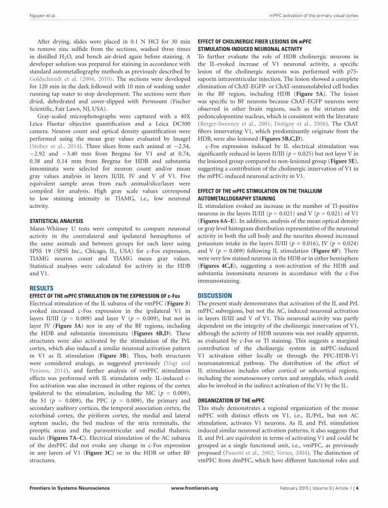
RESULTSEFFECT OF THE mPFC STIMULATION ON THE EXPRESSION OF c-FosElectrical stimulation of the IL subarea of the vmPFC (Figure 3)evoked increased c-Fos expression in the ipsilateral V1 inlayers II/III (p = 0.009) and layer V (p = 0.009), but not inlayer IV (Figure 3A) nor in any of the BF regions, includingthe HDB and substantia innominata (Figures 4B,D). Thesestructures were also activated by the stimulation of the PrLcortex, which also induced a similar neuronal activation patternin V1 as IL stimulation (Figure 3B). Thus, both structureswere considered analogs, as suggested previously (Vogt andPaxinos, 2014), and further analysis of vmPFC stimulationeffects was performed with IL stimulation only. IL-induced c-Fos activation was also increased in other regions of the cortexipsilateral to the stimulation, including the MC (p = 0.009),the S1 (p = 0.009), the PPC (p = 0.009), the primary andsecondary auditory cortices, the temporal association cortex, theectorhinal cortex, the piriform cortex, the medial and lateralseptum nuclei, the bed nucleus of the stria terminalis, thepreoptic areas and the paraventricular and medial thalamicnuclei (Figures 7A–C). Electrical stimulation of the AC subareaof the dmPFC did not evoke any change in c-Fos expressionin any layers of V1 (Figure 3C) or in the HDB or other BFstructures.
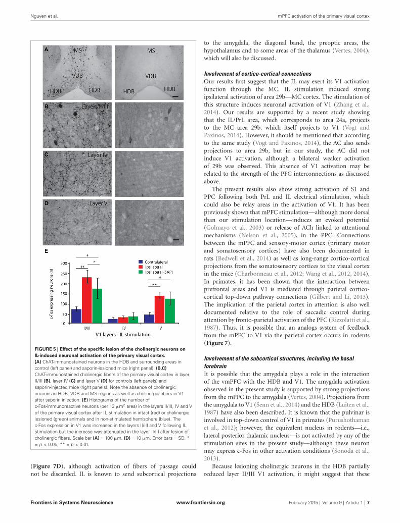
EFFECT OF CHOLINERGIC FIBER LESIONS ON mPFCSTIMULATION-INDUCED NEURONAL ACTIVITYTo further evaluate the role of HDB cholinergic neurons inthe IL-evoked increase of V1 neuronal activity, a specificlesion of the cholinergic neurons was performed with p75-saporin intraventricular injection. The lesion showed a completeelimination of ChAT-EGFP- or ChAT-immunolabeled cell bodiesin the BF region, including HDB (Figure 5A). The lesionwas specific to BF neurons because ChAT-EGFP neurons wereobserved in other brain regions, such as the striatum andpedonculopontine nucleus, which is consistent with the literature(Berger-Sweeney et al., 2001; Dotigny et al., 2008). The ChATfibers innervating V1, which predominantly originate from theHDB, were also lesioned (Figures 5B,C,D).
c-Fos expression induced by IL electrical stimulation wassignificantly reduced in layers II/III (p = 0.025) but not layer V inthe lesioned group compared to non-lesioned group (Figure 5E),suggesting a contribution of the cholinergic innervation of V1 inthe mPFC-induced neuronal activity in V1.
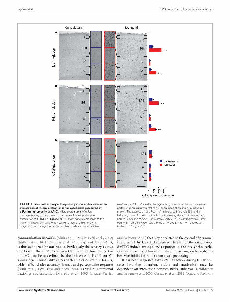
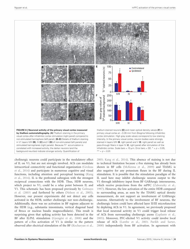
EFFECT OF THE mPFC STIMULATION ON THE THALLIUMAUTOMETALLOGRAPHY STAININGIL stimulation evoked an increase in the number of Tl-positiveneurons in the layers II/III (p = 0.021) and V (p = 0.021) of V1(Figures 6A–E). In addition, analysis of the mean optical densityor gray level histogram distribution representative of the neuronalactivity in both the cell body and the neurites showed increasedpotassium intake in the layers II/III (p = 0.016), IV (p = 0.024)and V (p = 0.009) following IL stimulation (Figure 6F). Therewere very few stained neurons in the HDB or in either hemisphere(Figures 4C,E), suggesting a non-activation of the HDB andsubstantia innominata neurons in accordance with the c-Fosimmunostaining.
DISCUSSIONThe present study demonstrates that activation of the IL and PrLmPFC subregions, but not the AC, induced neuronal activationin layers II/III and V of V1. This neuronal activity was partlydependent on the integrity of the cholinergic innervation of V1,although the activity of HDB neurons was not readily apparent,as evaluated by c-Fos or Tl staining. This suggests a marginalcontribution of the cholinergic system in mPFC-inducedV1 activation either locally or through the PFC-HDB-V1neuroanatomical pathway. The distribution of the effect ofIL stimulation includes other cortical or subcortical regions,including the somatosensory cortex and amygdala, which couldalso be involved in the indirect activation of the V1 by the IL.
ORGANIZATION OF THE mPFCThis study demonstrates a regional organization of the mousemPFC with distinct effects on V1, i.e., IL/PrL, but not ACstimulation, activates V1 neurons. As IL and PrL stimulationinduced similar neuronal activation patterns, it also suggests thatIL and PrL are equivalent in terms of activating V1 and could begrouped as a single functional unit, i.e., vmPFC, as previouslyproposed (Passetti et al., 2002; Vertes, 2004). The distinction ofvmPFC from dmPFC, which have different functional roles and
Frontiers in Systems Neuroscience www.frontiersin.org February 2015 | Volume 9 | Article 1 | 4
Nguyen et al. mPFC activation of the primary visual cortex
FIGURE 3 | Neuronal activity of the primary visual cortex induced bystimulation of medial prefrontal cortex subregions measured byc-Fos immunoreactivity. (A–C): Microphotographs of c-Fosimmunostaining in the primary visual cortex following electricalstimulation of IL (A), PrL (B) and AC (C) (right panels) compared to thenon-stimulated hemisphere (left panels) at low and high (indents)magnification. Histograms of the number of c-Fos immunoreactive
neurons (per 13 µm2 area) in the layers II/III, IV and V of the primary visualcortex after medial prefrontal cortex subregions stimulation (far right) areshown. The expression of c-Fos in V1 is increased in layers II/III and Vfollowing IL and PrL stimulation, but not following the AC stimulation. AC,anterior cingulate cortex; IL, infralimbic cortex; PrL, prelimbic cortex. Errorbars = Standard Deviation (SD). Scale bar = 500 µm (panels) and 50 µm(indents). ** = p < 0.01.
communication networks (Muir et al., 1996; Passetti et al., 2002;Guillem et al., 2011; Cassaday et al., 2014; Feja and Koch, 2014),is thus supported by our results. Particularly the sensory outputfunction of the vmPFC compared to the input function of thedmPFC may be underlined by the influence of IL/PrL on V1shown here. This duality agrees with studies of vmPFC lesions,which affect choice accuracy, latency and perseverative response(Muir et al., 1996; Feja and Koch, 2014) as well as attentionalflexibility and inhibition (Murphy et al., 2005; Gisquet-Verrier
and Delatour, 2006) that may be related to the control of neuronalfiring in V1 by IL/PrL. In contrast, lesions of the rat anteriordmPFC induce anticipatory responses in the five-choice serialreaction time task (Muir et al., 1996), suggesting a role related tobehavior inhibition rather than visual processing.
It has been suggested that mPFC function during behavioraltasks involving attention, vision and motivation may bedependent on interaction between mPFC subareas (Heidbrederand Groenewegen, 2003; Cassaday et al., 2014; Vogt and Paxinos,
Frontiers in Systems Neuroscience www.frontiersin.org February 2015 | Volume 9 | Article 1 | 5
Nguyen et al. mPFC activation of the primary visual cortex
FIGURE 4 | Basal forebrain cholinergic neurons do not express c-Fos orthallium staining induced by IL stimulation. (A–C) Microphotographs ofChAT:EGFP neurons (green) in the basal forebrain area with a highconcentration of EGFP cells in the HDB (A), c-Fos immunostaining (B) andthallium autometallography (C). There was no staining of ChAT positivecells either by c-Fos (B, right panel) or thallium autometallography (C, rightpanel) in the basal forebrain of coronal brain sections at Bregma +0.62 mmfollowing electrical stimulation of the IL (right panels) compared to thenon-stimulated hemisphere (left panels). (D,E) Histograms of the number of
c-Fos immunoreactive cells, note the near absence of c-Fos positive cells(D) or thallium stained cells (E) in the HDB and substantia innominatafollowing stimulation of the IL, which suggest HDB and substantiainnominata cholinergic neurons are not involved in V1 activation to vmPFCstimulation. HDB, Horizontal limb of the Diagonal band of Broca; LPO,Lateral Preoptic area; MPA, Medial Preoptic area; SI, Substantia Innominata;VP, Ventral Pallidum; Tu, Olfactory Tubercle. Delimitation of areas wasdesignated in accordance with the atlas. Error bars = SD. Scalebar = 250 µm.
2014). Our results do not confirm a possible interaction betweenIL/PrL and AC, although the AC is interconnected with PrLand IL in rats (Vogt and Miller, 1983; Bedwell et al., 2014; Vogtand Paxinos, 2014). There was no contralateral activation afterIL stimulation, although the IL projects to the contralateralmPFC (Vertes, 2004). These results may be due to experimentalconditions, i.e., passive and localized stimulation.
DISTRIBUTION OF THE vmPFC-INDUCED ACTIVITY IN V1Layers II/III and V of the V1 ipsilateral to the stimulated sidewere activated by unilateral IL/PrL electrical stimulation, asassessed by immunocytochemistry and autometallography. Theactivation of layer II/III neurons may indicate a cortico-corticalcomponent because feedback cortical projections predominantlyreach cortical layer II/III (Wang and Burkhalter, 2007; Gaoet al., 2010; Andermann et al., 2011; Wang et al., 2011). Theactivation of layer IV measured by densitometry (decreasedTlAMG mean optical density) only reflects the increased staining
of pass-through fibers between layers II/III and V becauseno neuron cell bodies were stained with TlAMG in layer IV.The pattern of activation in V1 thus does not appear to resultfrom a thalamo-cortical pathway. Layer V activation indicates acortico-thalamic output component induced by the stimulation.Thus, according to the pattern of V1 neuronal activation, whichis similar to an attentional top-down control (Gilbert and Li,2013), the vmPFC appears to exert feedback influence on layerII/III neuronal processing in V1, which is further transmittedto layer V. Given that the layer II/III neurons integrate visualinformation and transfer the visual information to higher visualareas, it is reasonable to suggest that our results correspond to anattention-related modulatory role of V1 processing by the IL.
POSSIBLE NEURAL PATHWAYS BETWEEN vmPFC AND V1The ipsilateral cerebral distribution of IL stimulation effects aswell as activation of S1, MC, and PPC areas suggests that IL-V1 interaction could be mediated by cortico-cortical pathways
Frontiers in Systems Neuroscience www.frontiersin.org February 2015 | Volume 9 | Article 1 | 6
Nguyen et al. mPFC activation of the primary visual cortex
FIGURE 5 | Effect of the specific lesion of the cholinergic neurons onIL-induced neuronal activation of the primary visual cortex.(A) ChAT-immunostained neurons in the HDB and surrounding areas incontrol (left panel) and saporin-lesioned mice (right panel). (B,C)ChAT-immunostained cholinergic fibers of the primary visual cortex in layerII/III (B), layer IV (C) and layer V (D) for controls (left panels) andsaporin-injected mice (right panels). Note the absence of cholinergicneurons in HDB, VDB and MS regions as well as cholinergic fibers in V1after saporin injection. (E) Histograms of the number ofc-Fos-immunoreactive neurons (per 13 µm2 area) in the layers II/III, IV and Vof the primary visual cortex after IL stimulation in intact (red) or cholinergiclesioned (green) animals and in non-stimulated hemisphere (blue). Thec-Fos expression in V1 was increased in the layers II/III and V following ILstimulation but the increase was attenuated in the layer II/III after lesion ofcholinergic fibers. Scale bar (A) = 100 µm, (D) = 10 µm. Error bars = SD. *= p < 0.05, ** = p < 0.01.
(Figure 7D), although activation of fibers of passage couldnot be discarded. IL is known to send subcortical projections
to the amygdala, the diagonal band, the preoptic areas, thehypothalamus and to some areas of the thalamus (Vertes, 2004),which will also be discussed.
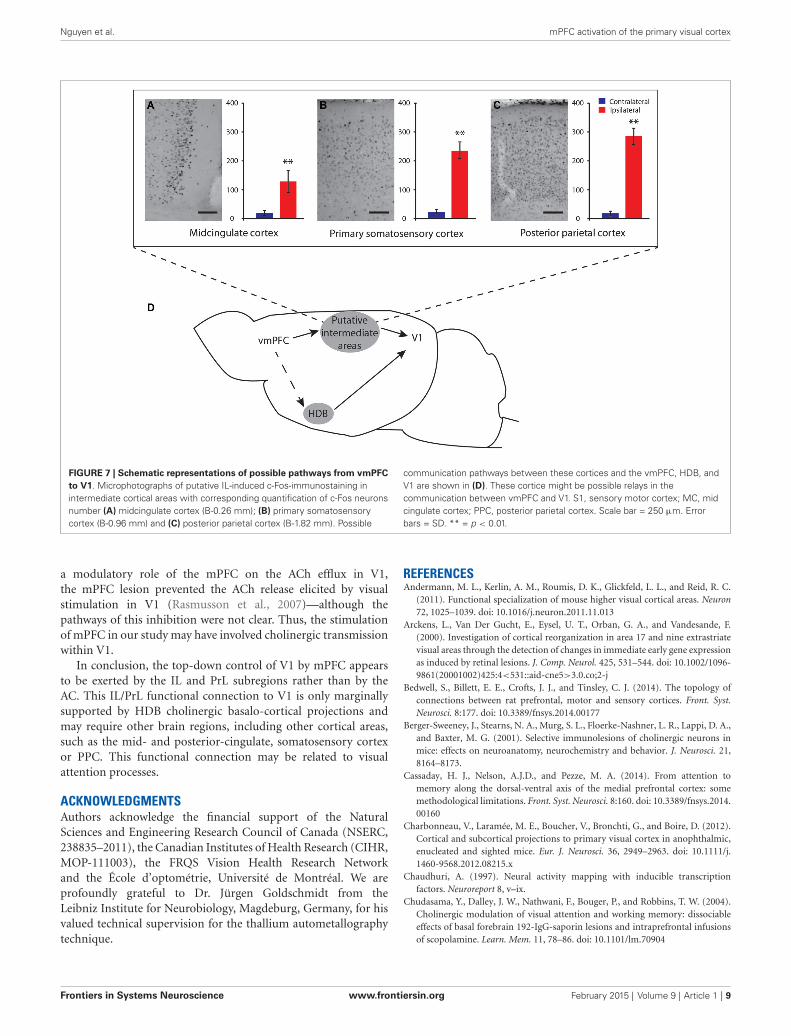
Involvement of cortico-cortical connectionsOur results first suggest that the IL may exert its V1 activationfunction through the MC. IL stimulation induced strongipsilateral activation of area 29b—MC cortex. The stimulation ofthis structure induces neuronal activation of V1 (Zhang et al.,2014). Our results are supported by a recent study showingthat the IL/PrL area, which corresponds to area 24a, projectsto the MC area 29b, which itself projects to V1 (Vogt andPaxinos, 2014). However, it should be mentioned that accordingto the same study (Vogt and Paxinos, 2014), the AC also sendsprojections to area 29b, but in our study, the AC did notinduce V1 activation, although a bilateral weaker activationof 29b was observed. This absence of V1 activation may berelated to the strength of the PFC interconnections as discussedabove.
The present results also show strong activation of S1 andPPC following both PrL and IL electrical stimulation, whichcould also be relay areas in the activation of V1. It has beenpreviously shown that mPFC stimulation—although more dorsalthan our stimulation location—induces an evoked potential(Golmayo et al., 2003) or release of ACh linked to attentionalmechanisms (Nelson et al., 2005), in the PPC. Connectionsbetween the mPFC and sensory-motor cortex (primary motorand somatosensory cortices) have also been documented inrats (Bedwell et al., 2014) as well as long-range cortico-corticalprojections from the somatosensory cortices to the visual cortexin the mice (Charbonneau et al., 2012; Wang et al., 2012, 2014).In primates, it has been shown that the interaction betweenprefrontal areas and V1 is mediated through parietal cortico-cortical top-down pathway connections (Gilbert and Li, 2013).The implication of the parietal cortex in attention is also welldocumented relative to the role of saccadic control duringattention by fronto-parietal activation of the PFC (Rizzolatti et al.,1987). Thus, it is possible that an analogs system of feedbackfrom the mPFC to V1 via the parietal cortex occurs in rodents(Figure 7).
Involvement of the subcortical structures, including the basalforebrainIt is possible that the amygdala plays a role in the interactionof the vmPFC with the HDB and V1. The amygdala activationobserved in the present study is supported by strong projectionsfrom the mPFC to the amygdala (Vertes, 2004). Projections fromthe amygdala to V1 (Senn et al., 2014) and the HDB (Luiten et al.,1987) have also been described. It is known that the pulvinar isinvolved in top-down control of V1 in primates (Purushothamanet al., 2012); however, the equivalent nucleus in rodents—i.e.,lateral posterior thalamic nucleus—is not activated by any of thestimulation sites in the present study—although these neuronmay express c-Fos in other activation conditions (Sonoda et al.,2013).
Because lesioning cholinergic neurons in the HDB partiallyreduced layer II/III V1 activation, it might suggest that these
Frontiers in Systems Neuroscience www.frontiersin.org February 2015 | Volume 9 | Article 1 | 7
Nguyen et al. mPFC activation of the primary visual cortex
FIGURE 6 | Neuronal activity of the primary visual cortex measuredby thallium autometallography. (A) Thallium staining in the primaryvisual cortex after infralimbic cortex stimulation (right panel) compared tonon-stimulated hemisphere (left panel). (B–D) Details of thallium stainingin V1 layers II/III (B), IV (C) and V (D) of non-stimulated (left panels) andstimulated hemispheres (right panels). Because Tl+ accumulation iscorrelated with increased activity, the darker neurons (and thebackground neurites) indicate stronger activity. Quantification of
thallium-stained neurons (E) and mean optical density values (F) inprimary visual cortex at −3.28 mm from Bregma following infralimbiccortex stimulation. High gray scale values correspond to low stainingintensity. In the primary visual cortex, neuron bodies were stronglystained in layers II/III (B, right panel) and V (D, right panel) as well aspass-through fibers in layer IV (C, right panel) after stimulation of theinfralimbic cortex. Scale bars = 10 µm. Error bars = SD. * = p < 0.05,** = p < 0.01.
cholinergic neurons could participate in the modulatory effectof IL on V1, but are not strongly involved. ACh can modulateintracortical connectivity and functional organization (Groleauet al., 2014) and participate in numerous cognitive and visualfunctions, including attention and perceptual learning (Kanget al., 2014). IL is the prefrontal subregion with the strongestreciprocal connections with the HDB. Thus, HDB neurons,which project to V1, could be a relay point between IL andV1. This schematic has been proposed previously by Golmayoet al. (2003) and furthered by others (Nelson et al., 2005).However, our present experiments did not detect any cellsactivated in the HDB, neither cholinergic nor non-cholinergic.Additionally, there was no activation in BF regions adjacent tothe HDB (e.g., substantia innominata, vertical diagonal bandof Broca or nucleus basalis magnocellularis). This result issurprising given that spiking activity has been detected in theBF after IL/PrL stimulation (Gyengési et al., 2008) and thepattern of c-Fos activation of V1 is similar to the patternobserved after electrical stimulation of the BF (Kocharyan et al.,
2005; Kang et al., 2014). This absence of staining is not dueto technical limitation because c-Fos staining has already beenshown in BF cells (McKenna et al., 2009) and TlAMG isalso negative for any potassium fluxes in the BF during ILstimulation. It is possible that the stimulation paradigm of theIL used here may inhibit cholinergic neuron output to theV1 through inhibitory input from BF GABAergic interneurons,which receive projections from the mPFC (Zaborszky et al.,1997). However, the low activation of the entire HDB comparedto surrounding areas, as seen by the TlAMG optical densitymeasurement, do not support an involvement of GABAergicneurons. Alternatively to the involvement of BF neurons, thecholinergic lesion could have affected layer II/III microfunctionby depleting ACh in V1. In agreement, we previously proposedthat local neuronal activity in V1 could trigger local releaseof ACh from surrounding cholinergic axons (Laplante et al.,2005). Moreover, PFC-elicited V1 activity could involve localrelease of ACh (Lambe et al., 2003; Parikh and Sarter,2008) independently from BF activation. In agreement with
Frontiers in Systems Neuroscience www.frontiersin.org February 2015 | Volume 9 | Article 1 | 8
Nguyen et al. mPFC activation of the primary visual cortex
FIGURE 7 | Schematic representations of possible pathways from vmPFCto V1. Microphotographs of putative IL-induced c-Fos-immunostaining inintermediate cortical areas with corresponding quantification of c-Fos neuronsnumber (A) midcingulate cortex (B-0.26 mm); (B) primary somatosensorycortex (B-0.96 mm) and (C) posterior parietal cortex (B-1.82 mm). Possible
communication pathways between these cortices and the vmPFC, HDB, andV1 are shown in (D). These cortice might be possible relays in thecommunication between vmPFC and V1. S1, sensory motor cortex; MC, midcingulate cortex; PPC, posterior parietal cortex. Scale bar = 250 µm. Errorbars = SD. ** = p < 0.01.
a modulatory role of the mPFC on the ACh efflux in V1,the mPFC lesion prevented the ACh release elicited by visualstimulation in V1 (Rasmusson et al., 2007)—although thepathways of this inhibition were not clear. Thus, the stimulationof mPFC in our study may have involved cholinergic transmissionwithin V1.
In conclusion, the top-down control of V1 by mPFC appearsto be exerted by the IL and PrL subregions rather than by theAC. This IL/PrL functional connection to V1 is only marginallysupported by HDB cholinergic basalo-cortical projections andmay require other brain regions, including other cortical areas,such as the mid- and posterior-cingulate, somatosensory cortexor PPC. This functional connection may be related to visualattention processes.
ACKNOWLEDGMENTSAuthors acknowledge the financial support of the NaturalSciences and Engineering Research Council of Canada (NSERC,238835–2011), the Canadian Institutes of Health Research (CIHR,MOP-111003), the FRQS Vision Health Research Networkand the École d’optométrie, Université de Montréal. We areprofoundly grateful to Dr. Jürgen Goldschmidt from theLeibniz Institute for Neurobiology, Magdeburg, Germany, for hisvalued technical supervision for the thallium autometallographytechnique.
REFERENCESAndermann, M. L., Kerlin, A. M., Roumis, D. K., Glickfeld, L. L., and Reid, R. C.
(2011). Functional specialization of mouse higher visual cortical areas. Neuron72, 1025–1039. doi: 10.1016/j.neuron.2011.11.013
Arckens, L., Van Der Gucht, E., Eysel, U. T., Orban, G. A., and Vandesande, F.(2000). Investigation of cortical reorganization in area 17 and nine extrastriatevisual areas through the detection of changes in immediate early gene expressionas induced by retinal lesions. J. Comp. Neurol. 425, 531–544. doi: 10.1002/1096-9861(20001002)425:4<531::aid-cne5>3.0.co;2-j
Bedwell, S., Billett, E. E., Crofts, J. J., and Tinsley, C. J. (2014). The topology ofconnections between rat prefrontal, motor and sensory cortices. Front. Syst.Neurosci. 8:177. doi: 10.3389/fnsys.2014.00177
Berger-Sweeney, J., Stearns, N. A., Murg, S. L., Floerke-Nashner, L. R., Lappi, D. A.,and Baxter, M. G. (2001). Selective immunolesions of cholinergic neurons inmice: effects on neuroanatomy, neurochemistry and behavior. J. Neurosci. 21,8164–8173.
Cassaday, H. J., Nelson, A.J.D., and Pezze, M. A. (2014). From attention tomemory along the dorsal-ventral axis of the medial prefrontal cortex: somemethodological limitations. Front. Syst. Neurosci. 8:160. doi: 10.3389/fnsys.2014.00160
Charbonneau, V., Laramée, M. E., Boucher, V., Bronchti, G., and Boire, D. (2012).Cortical and subcortical projections to primary visual cortex in anophthalmic,enucleated and sighted mice. Eur. J. Neurosci. 36, 2949–2963. doi: 10.1111/j.1460-9568.2012.08215.x
Chaudhuri, A. (1997). Neural activity mapping with inducible transcriptionfactors. Neuroreport 8, v–ix.
Chudasama, Y., Dalley, J. W., Nathwani, F., Bouger, P., and Robbins, T. W. (2004).Cholinergic modulation of visual attention and working memory: dissociableeffects of basal forebrain 192-IgG-saporin lesions and intraprefrontal infusionsof scopolamine. Learn. Mem. 11, 78–86. doi: 10.1101/lm.70904
Frontiers in Systems Neuroscience www.frontiersin.org February 2015 | Volume 9 | Article 1 | 9
Nguyen et al. mPFC activation of the primary visual cortex
Dalley, J. W., Theobald, D. E., Bouger, P., Chudasama, Y., Cardinal, R. N.,and Robbins, T. W. (2004). Cortical cholinergic function and deficits invisual attentional performance in rats following 192 IgG-saporin-inducedlesions of the medial prefrontal cortex. Cereb. Cortex 14, 922–932. doi: 10.1093/cercor/bhh052
Danscher, G. (1981). Histochemical demonstration of heavy metals. Arevised version of the sulphide silver method suitable for both light andelectronmicroscopy. Histochemistry 71, 1–16. doi: 10.1007/bf00592566
Delatour, B., and Gisquet-Verrier, P. (2000). Functional role of rat prelimbic-infralimbic cortices in spatial memory: evidence for their involvement inattention and behavioural flexibility. Behav. Brain Res. 109, 113–128. doi: 10.1016/s0166-4328(99)00168-0
Disney, A. A., Aoki, C., and Hawken, M. J. (2007). Gain modulation bynicotine in macaque v1. Neuron 56, 701–713. doi: 10.1016/j.neuron.2007.09.034
Dotigny, F., Ben Amor, A. Y., Burke, M., and Vaucher, E. (2008). Neuromodulatoryrole of acetylcholine in visually-induced cortical activation: behavioral andneuroanatomical correlates. Neuroscience 154, 1607–1618. doi: 10.1016/j.neuroscience.2008.04.030
Dragunow, M., and Faull, R. (1989). The use of c-fos as a metabolic marker inneuronal pathway tracing. J. Neurosci. Methods 29, 261–265. doi: 10.1016/0165-0270(89)90150-7
Feja, M., and Koch, M. (2014). Ventral medial prefrontal cortex inactivationimpairs impulse control but does not affect delay-discounting in rats. Behav.Brain Res. 264, 230–239. doi: 10.1016/j.bbr.2014.02.013
Franklin, K. B. J., and Paxinos, G. (2007). The Mouse Brain. Amsterdam: Elsevier.Freund, T. F., and Gulyás, A. I. (1991). GABAergic interneurons containing
calbindin D28K or somatostatin are major targets of GABAergic basal forebrainafferents in the rat neocortex. J. Comp. Neurol. 314, 187–199. doi: 10.1002/cne.903140117
Gabbott, P. L., Warner, T. A., Jays, P. R., Salway, P., and Busby, S. J. (2005).Prefrontal cortex in the rat: projections to subcortical autonomic, motor andlimbic centers. J. Comp. Neurol. 492, 145–177. doi: 10.1002/cne.20738
Gao, E., DeAngelis, G. C., and Burkhalter, A. (2010). Parallel input channels tomouse primary visual cortex. J. Neurosci. 30, 5912–5926. doi: 10.1523/jneurosci.6456-09.2010
Gaykema, R. P., Luiten, P. G., Nyakas, C., and Traber, J. (1990). Cortical projectionpatterns of the medial septum-diagonal band complex. J. Comp. Neurol. 293,103–124. doi: 10.1002/cne.902930109
Gilbert, C. D., and Li, W. (2013). Top-down influences on visual processing. Nat.Rev. Neurosci. 14, 350–363. doi: 10.1038/nrn3476
Gisquet-Verrier, P., and Delatour, B. (2006). The role of the ratprelimbic/infralimbic cortex in working memory: not involved in the short-term maintenance but in monitoring and processing functions. Neuroscience141, 585–596. doi: 10.1016/j.neuroscience.2006.04.009
Goldschmidt, J., Wanger, T., Engelhorn, A., Friedrich, H., Happel, M.,Ilango, A., et al. (2010). High-resolution mapping of neuronal activity usingthe lipophilic thallium chelate complex TlDDC: protocol and validationof the method. Neuroimage 49, 303–315. doi: 10.1016/j.neuroimage.2009.08.012
Goldschmidt, J., Zuschratter, W., and Scheich, H. (2004). High-resolution mappingof neuronal activity by thallium autometallography. Neuroimage 23, 638–647.doi: 10.1016/j.neuroimage.2004.05.023
Golmayo, L., Nuñez, A., and Zaborszky, L. (2003). Electrophysiological evidencefor the existence of a posterior cortical-prefrontal-basal forebrain circuitry inmodulating sensory responses in visual and somatosensory rat cortical areas.Neuroscience 119, 597–609. doi: 10.1016/s0306-4522(03)00031-9
Granon, S., Hardouin, J., Courtièr, A., and Poucet, B. (1998). Evidence for theinvolvement of the rat prefrontal cortex in sustained attention. Q. J. Exp. Psychol.B 51, 219–233.
Gritti, I., Henny, P., Galloni, F., Mainville, L., Mariotti, M., and Jones, B. E. (2006).Stereological estimates of the basal forebrain cell population in the rat, includingneurons containing choline acetyltransferase, glutamic acid decarboxylaseor phosphate-activated glutaminase and colocalizing vesicular glutamatetransporters. Neuroscience 143, 1051–1064. doi: 10.1016/j.neuroscience.2006.09.024
Gritti, I., Mainville, L., and Jones, B. E. (1993). Codistribution of GABA- withacetylcholine-synthesizing neurons in the basal forebrain of the rat. J. Comp.Neurol. 329, 438–457. doi: 10.1002/cne.903290403
Groenewegen, H. J., and Uylings, H. B. (2000). The prefrontal cortex and theintegration of sensory, limbic and autonomic information. Prog. Brain Res. 126,3–28. doi: 10.1016/s0079-6123(00)26003-2
Groleau, M., Nguyen, H. N., Vanni, M. P., Huppé-Gourgues, F., Casanova, C., andVaucher, E. (2014). Impaired functional organization in the visual cortex ofmuscarinic receptor knock-out mice. Neuroimage 98C, 233–242. doi: 10.1016/j.neuroimage.2014.05.016
Guillem, K., Bloem, B., Poorthuis, R. B., Loos, M., Smit, A. B., Maskos, U., et al.(2011). Nicotinic acetylcholine receptor beta2 subunits in the medial prefrontalcortex control attention. Science 333, 888–891. doi: 10.1126/science.1207079
Gyengési, E., Zaborszky, L., and Détári, L. (2008). The effect of prefrontalstimulation on the firing of basal forebrain neurons in urethane anesthetizedrat. Brain Res. Bull. 75, 570–580. doi: 10.1016/j.brainresbull.2007.09.008
Heidbreder, C. A., and Groenewegen, H. J. (2003). The medial prefrontal cortexin the rat: evidence for a dorso-ventral distinction based upon functional andanatomical characteristics. Neurosci. Biobehav. Rev. 27, 555–579. doi: 10.1016/j.neubiorev.2003.09.003
Herrero, J. L., Roberts, M. J., Delicato, L. S., Gieselmann, M. A., Dayan, P., andThiele, A. (2008). Acetylcholine contributes through muscarinic receptors toattentional modulation in V1. Nature 454, 1110–1114. doi: 10.1038/nature07141
Hoover, W. B., and Vertes, R. P. (2007). Anatomical analysis of afferent projectionsto the medial prefrontal cortex in the rat. Brain Struct. Funct. 212, 149–179.doi: 10.1007/s00429-007-0150-4
Kaczmarek, L., and Chaudhuri, A. (1997). Sensory regulation of immediate-early gene expression in mammalian visual cortex: implications for functionalmapping and neural plasticity. Brain Res. Brain Res. Rev. 23, 237–256. doi: 10.1016/s0165-0173(97)00005-2
Kang, J. I., Groleau, M., Dotigny, F., Giguère, H., and Vaucher, E. (2014). Visualtraining paired with electrical stimulation of the basal forebrain improvesorientation-selective visual acuity in the rat. Brain Struct. Funct. 219, 1493–1507.doi: 10.1007/s00429-013-0582-y
Kang, J. I., and Vaucher, E. (2009). Cholinergic pairing with visual activationresults in long-term enhancement of visual evoked potentials. PLoS One 4:e5995.doi: 10.1371/journal.pone.0005995
Kocharyan, A., Fernandes, P., Serluca, N., Vaucher, E., and Hamel, E. (2005).Chemical or electrical stimulation of basal forebrain neurons activates specificsubsets of cortical gaba-interneurons in parallel with increases in corticalcerebral blood flow. J. Cereb. Blood Flow Metab. 25:S159. doi: 10.1038/sj.jcbfm.9591524.0159
Kozak, R., Bruno, J. P., and Sarter, M. (2006). Augmented prefrontal acetylcholinerelease during challenged attentional performance. Cereb. Cortex 16, 9–17.doi: 10.1093/cercor/bhi079
Kuo, B. C., Stokes, M. G., Murray, A. M., and Nobre, A. C. (2014). Attention biasesvisual activity in visual short-term memory. J. Cogn. Neurosci. 26, 1377–1389.doi: 10.1162/jocn_a_00577
Lambe, E. K., Picciotto, M. R., and Aghajanian, G. K. (2003). Nicotineinduces glutamate release from thalamocortical terminals in prefrontal cortex.Neuropsychopharmacology 28, 216–225. doi: 10.1038/sj.npp.1300032
Laplante, F., Morin, Y., Quirion, R., and Vaucher, E. (2005). Acetylcholine release iselicited in the visual cortex, but not in the prefrontal cortex, by patterned visualstimulation: a dual in vivo microdialysis study with functional correlates in therat brain. Neuroscience 132, 501–510. doi: 10.1016/j.neuroscience.2004.11.059
Luiten, P. G., Gaykema, R. P., Traber, J., and Spencer, D. G. Jr. (1987). Corticalprojection patterns of magnocellular basal nucleus subdivisions as revealed byanterogradely transported phaseolus vulgaris leucoagglutinin. Brain Res. 413,229–250. doi: 10.1016/0006-8993(87)91014-6
Maddux, J. M., and Holland, P. C. (2011). Effects of dorsal or ventral medialprefrontal cortical lesions on five-choice serial reaction time performance inrats. Behav. Brain Res. 221, 63–74. doi: 10.1016/j.bbr.2011.02.031
McKenna, J. T., Cordeira, J. W., Jeffrey, B. A., Ward, C. P., Winston, S., McCarley,R. W., et al. (2009). c-Fos protein expression is increased in cholinergic neuronsof the rodent basal forebrain during spontaneous and induced wakefulness.Brain Res. Bull. 80, 382–388. doi: 10.1016/j.brainresbull.2009.08.015
Mesulam, M. M., Mufson, E. J., Wainer, B. H., and Levey, A. I. (1983).Central cholinergic pathways in the rat: an overview based on an alternativenomenclature (Ch1–Ch6). Neuroscience 10, 1185–1201. doi: 10.1016/0306-4522(83)90108-2
Frontiers in Systems Neuroscience www.frontiersin.org February 2015 | Volume 9 | Article 1 | 10
Nguyen et al. mPFC activation of the primary visual cortex
Muir, J. L., Everitt, B. J., and Robbins, T. W. (1996). The cerebral cortex ofthe rat and visual attentional function: dissociable effects of mediofrontal,cingulate, anterior dorsolateral and parietal cortex lesions on a five-choiceserial reaction time task. Cereb. Cortex 6, 470–481. doi: 10.1093/cercor/6.3.470
Murphy, E. R., Dalley, J. W., and Robbins, T. W. (2005). Local glutamate receptorantagonism in the rat prefrontal cortex disrupts response inhibition in avisuospatial attentional task. Psychopharmacology (Berl) 179, 99–107. doi: 10.1007/s00213-004-2068-3
Nelson, C. L., Sarter, M., and Bruno, J. P. (2005). Prefrontal cortical modulationof acetylcholine release in posterior parietal cortex. Neuroscience 132, 347–359.doi: 10.1016/j.neuroscience.2004.12.007
Newman, E. L., Gupta, K., Climer, J. R., Monaghan, C. K., and Hasselmo, M. E.(2012). Cholinergic modulation of cognitive processing: insights drawn fromcomputational models. Front. Behav. Neurosci. 6:24. doi: 10.3389/fnbeh.2012.00024
Parikh, V., Kozak, R., Martinez, V., and Sarter, M. (2007). Prefrontal acetylcholinerelease controls cue detection on multiple timescales. Neuron 56, 141–154.doi: 10.1016/j.neuron.2007.08.025
Parikh, V., and Sarter, M. (2008). Cholinergic mediation of attention: contributionsof phasic and tonic increases in prefrontal cholinergic activity. Ann. N Y Acad.Sci. 1129, 225–235. doi: 10.1196/annals.1417.021
Passetti, F., Chudasama, Y., and Robbins, T. W. (2002). The frontal cortex ofthe rat and visual attentional performance: dissociable functions of distinctmedial prefrontal subregions. Cereb. Cortex 12, 1254–1268. doi: 10.1093/cercor/12.12.1254
Purushothaman, G., Marion, R., Li, K., and Casagrande, V. A. (2012). Gating andcontrol of primary visual cortex by pulvinar. Nat. Neurosci. 15, 905–912. doi: 10.1038/nn.3106
Rasmusson, D. D., Smith, S. A., and Semba, K. (2007). Inactivation of prefrontalcortex abolishes cortical acetylcholine release evoked by sensory or sensorypathway stimulation in the rat. Neuroscience 149, 232–241. doi: 10.1016/j.neuroscience.2007.06.057
Rizzolatti, G., Riggio, L., Dascola, I., and Umiltá, C. (1987). Reorientingattention across the horizontal and vertical meridians: evidence in favor of apremotor theory of attention. Neuropsychologia 25, 31–40. doi: 10.1016/0028-3932(87)90041-8
Senn, V., Wolff, S. B., Herry, C., Grenier, F., Ehrlich, I., Gründemann, J., et al.(2014). Long-range connectivity defines behavioral specificity of amygdalaneurons. Neuron 81, 428–437. doi: 10.1016/j.neuron.2013.11.006
Sonoda, E. Y., Cysneiros, R. M., Arida, R. M., Cavalheiro, E. A., and Scorza,F. A. (2013). Activation and involvement of the lateral-posterior nucleus of thethalamus after a single generalized tonic-clonic seizure. Epilepsy Behav. 28, 104–107. doi: 10.1016/j.yebeh.2013.03.025
Stöber, F., Baldauf, K., Ziabreva, I., Harhausen, D., Zille, M., Neubert, J., et al.(2014). Single-cell resolution mapping of neuronal damage in acute focalcerebral ischemia using thallium autometallography. J. Cereb. Blood Flow Metab.34, 144–152. doi: 10.1038/jcbfm.2013.177
Vertes, R. P. (2004). Differential projections of the infralimbic and prelimbic cortexin the rat. Synapse 51, 32–58. doi: 10.1002/syn.10279
Vogt, B. A., and Miller, M. W. (1983). Cortical connections between rat cingulatecortex and visual, motor and postsubicular cortices. J. Comp. Neurol. 216, 192–210. doi: 10.1002/cne.902160207
Vogt, B. A., and Paxinos, G. (2014). Cytoarchitecture of mouse and rat cingulatecortex with human homologies. Brain Struct. Funct. 219, 185–192. doi: 10.1007/s00429-012-0493-3
Wang, Q., and Burkhalter, A. (2007). Area map of mouse visual cortex. J. Comp.Neurol. 502, 339–357. doi: 10.1002/cne.21286
Wang, Q., Gao, E., and Burkhalter, A. (2011). Gateways of ventral anddorsal streams in mouse visual cortex. J. Neurosci. 31, 1905–1918. doi: 10.1523/jneurosci.3488-10.2011
Wang, Q., Henry, A. M., Harris, J. A., Oh, S. W., Joines, K. M., Nyhus, J.,et al. (2014). Systematic comparison of adeno-associated virus and biotinylateddextran amine reveals equivalent sensitivity between tracers and novelprojection targets in the mouse brain. J. Comp. Neurol. 522, 1989–2012. doi: 10.1002/cne.23567
Wang, Q., Sporns, O., and Burkhalter, A. (2012). Network analysis of corticocorticalconnections reveals ventral and dorsal processing streams in mouse visualcortex. J. Neurosci. 32, 4386–4399. doi: 10.1523/jneurosci.6063-11.2012
Zaborszky, L., Gaykema, R. P., Swanson, D. J., and Cullinan, W. E. (1997). Corticalinput to the basal forebrain. Neuroscience 79, 1051–1078. doi: 10.1016/s0306-4522(97)00049-3
Zaborszky, L., Van Den Pol, A., and Gyengesi, E. (2012). “Chapter 28 - The basalforebrain cholinergic projection system in mice,” in The Mouse Nervous System,eds W. Charles, P. George and L. Puelles (San Diego: Academic Press), 684–718.
Zangenehpour, S., and Chaudhuri, A. (2002). Differential induction and decaycurves of c-fos and zif268 revealed through dual activity maps. Brain Res. Mol.Brain Res. 109, 221–225. doi: 10.1016/s0169-328x(02)00556-9
Zhang, S., Xu, M., Kamigaki, T., Hoang Do, J. P., Chang, W. C., Jenvay, S.,et al. (2014). Selective attention. Long-range and local circuits for top-down modulation of visual cortex processing. Science 345, 660–665. doi: 10.1126/science.1254126
Conflict of Interest Statement: The authors declare that the research was conductedin the absence of any commercial or financial relationships that could be construedas a potential conflict of interest.
Received: 11 October 2014; accepted: 06 January 2015; published online: 09 February2015.Citation: Nguyen HN, Huppé-Gourgues F and Vaucher E (2015) Activation ofthe mouse primary visual cortex by medial prefrontal subregion stimulation isnot mediated by cholinergic basalo-cortical projections. Front. Syst. Neurosci. 9:1.doi: 10.3389/fnsys.2015.00001This article was submitted to the journal Frontiers in Systems Neuroscience.Copyright © 2015 Nguyen, Huppé-Gourgues and Vaucher. This is an open-accessarticle distributed under the terms of the Creative Commons Attribution License (CCBY). The use, distribution and reproduction in other forums is permitted, providedthe original author(s) or licensor are credited and that the original publication in thisjournal is cited, in accordance with accepted academic practice. No use, distribution orreproduction is permitted which does not comply with these terms.
Frontiers in Systems Neuroscience www.frontiersin.org February 2015 | Volume 9 | Article 1 | 11