
Action execution and recognition: a neuropsychological...
179
ACTION EXECUTION AND RECOGNITION: A NEUROPSYCHOLOGICAL ANALYSIS By CAROLINA BONIVENTO A thesis submitted to The University of Birmingham For the degree of DOCTOR OF PHILOSOPY School of Psychology The University of Birmingham May 2011
-
Upload
dangkhuong -
Category
Documents
-
view
222 -
download
0
Transcript of Action execution and recognition: a neuropsychological...
ACTION EXECUTION AND RECOGNITION: A
NEUROPSYCHOLOGICAL ANALYSIS
By
CAROLINA BONIVENTO
A thesis submitted to
The University of Birmingham
For the degree of
DOCTOR OF PHILOSOPY
School of Psychology
The University of Birmingham
May 2011
University of Birmingham Research Archive
e-theses repository This unpublished thesis/dissertation is copyright of the author and/or third parties. The intellectual property rights of the author or third parties in respect of this work are as defined by The Copyright Designs and Patents Act 1988 or as modified by any successor legislation. Any use made of information contained in this thesis/dissertation must be in accordance with that legislation and must be properly acknowledged. Further distribution or reproduction in any format is prohibited without the permission of the copyright holder.
2
… To my sister.
3
Acknowledgements
I thank Prof. Glyn Humphreys for the big help he gave me for my PhD to be completed (almost) in
time, especially in the final rush.
Thanks to Dr Pia Rotshtein who was precious in teaching me all I needed for doing the VBM
analyses and in giving me support in all the phases of the work.
Thanks to Wai Ling Bickerton for her cooperation in the studies about the BCoS tasks.
Thanks to Prof. Raffaella Rumiati who gave me the opportunity to work in her lab where I run the
study on the Parkinson’s’ patient, and thanks to the doctors (Dr Emauele Biasutti, Dr Federica
Mondolo, Dr Roberto Capus, Dr Gilberto Pizzolato, Dr Antonietta Zadini, Dr Alberta Lunardelli)
who helped in that study.
Special thanks are for Dr Gioia Anna Lura Negri and Dr Anna Sverzut who did the scoring of the
huge amount of patients and controls I tested.
Also thanks a lot to my boss who was very tolerant in the past months and let me time to complete
this work.
I thank my colleagues at Birmingham University, and in particular Dr Alessia Correani. She had
been a lovely flatmate and will ever be a good friend.
And, of course, thanks to all the people who kindly put themselves out to do my tasks.
Finally “GRAZIE” to Giulio, Donatella, Caterina, Giorgia, Giuseppina and Max, my family, who
always supported me
4
TABLE OF CONTENTS
1. GENERAL INTRODUCTION 9
IMITATION, AN OVERVIEW 9
ACTION IMITATION AND RECOGNITION: NEURAL CORRELATES AND THE MIRROR NEURON SYSTEM 13
NEURAL CORRELATES OF ACTION IMITATION OF MF AND ML TRANSITIVE AND INTRANSITIVE
GESTURES 18
REASONS FOR THE PRESENT WORK 22
2. GENERAL METHODS 24
PATIENTS 25
CONTROL SUBJECTS 30
TASKS 32
NEUROIMAGING ASSESSMENT 41
3. IMITATING TRANSITIVE AND INTRANSITIVE GESTURES: AN ANALYSIS OF LESION
SIDE AND ASSOCIATED COGNITIVE IMPAIRMENTS. 44
ABSTRACT 44
INTRODUCTION 44
GENERAL METHODS 47
RESULTS 48
DISCUSSION 59
4. NEURAL CORRELATES OF TRANSITIVE AND INTRANSITIVE ACTION IMITATION: AN
INVESTIGATION USING VOXEL-BASED MORPHOMETRY (VBM) 67
ABSTRACT 67
INTRODUCTION 68
5
METHODS 71
BEHAVIOURAL RESULTS- TRANSITIVE (MF & ML) AND INTRANSITIVE (MF & ML) ACTIONS 72
VOXEL-BASED MORPHOMETRY 73
ANALYSES 73
VBM RESULTS 74
LESION OVERLAP MAPS 83
DISCUSSION 83
5. RECOGNIZING AND EXECUTING MOVEMENTS: AN INVESTIGATION USING VOXEL-
BASED MORPHOMETRY (VBM) 88
ABSTRACT 88
INTRODUCTION 88
METHODS 95
RESULTS 96
DISCUSSION 106
6. DISTINCT AND COMMON NEURAL CORRELATES OF APRAXIA FOR TRANSITIVE AND
INTRANSITIVE GESTURES: AN INVESTIGATION USING VOXEL-BASED MORPHOMETRY
(VBM) 112
ABSTRACT 112
INTRODUCTION 113
METHODS 116
DISCUSSION 129
7. THE ROLE OF THE BASAL GANGLIA IN ACTION IMITATION:
NEUROPSYCHOLOGICAL EVIDENCE FROM PARKINSON’S DISEASE PATIENTS 134
ABSTRACT 134
INTRODUCTION 134
6
METHODS 136
RESULTS 136
DISCUSSION 142
8. GENERAL DISCUSSION 144
9. SUMMARY 155
APPENDIX 1- CHAPTERS 3 AND 4 157
APPENDIX 2- CHAPTERS 5 AND 6 164
APPENDIX 3- CHAPTER 7 165
10. REFERENCES 167
7
ABSTRACT
Humans appear to show an innate tendency to imitate, and this may provide one of the foundations
of social communication. Several studies have been carried out in social and cognitive science in
order to understand how imitation works, which parts of the brain are involved, and what the role of
imitation might be in social behaviour. Previous brain imaging and neuropsychological studies
report data that favour a dual process account of imitation, according to which actions are imitated
through different mechanisms on the basis of whether they are meaningful and familiar (MF
actions) or meaningless/unfamiliar (ML actions). However many questions remain to be clarified –
such as which brain areas mediate these different actions. In addition to the distinction between MF
and ML gestures, there is considerable interest in the production of different types of known
gestures – particularly between actions involving tools (i.e. transitive actions) and those subserving
communicative (intransitive) gestures, and in how the production of these gestures relates to the
processes involved in recognizing the gestures as input. This thesis reports a neuropsychological
examination of the functional and neural bases of imitation using converging data from behavioural
studies with different patient groups (stroke patients, patients with Parkinson’s Disease, PD) and
structural brain imaging (particularly using voxel-based morphometric [VBM] analyses) to examine
lesion-symptom relations.
The first empirical chapter (Chapter 2) describes a neuropsychological study on the recognition and
production of MF actions and the imitation of ML gestures, in patients with unilateral left or right-
side brain damage (respectively: LBD and RBD patients). At a group level, LBD patient were
worse in imitation than RBD patients only when novel transitive actions had to be reproduced,
when both LBD and RBD differed from healthy participants, while intransitive gestures were
generally easier to be executed. Also both transitive and intransitive action imitation tasks were
correlated to action recognition. At a single subject level, however, there was evidence for some
8
dissociated symptoms, suggesting that at least partially different mechanisms mediate the imitation
of transitive and intransitive gestures and gesture production as opposed to recognition.
Chapter 3 presents a first attempt to use VBM to evaluate the relations between brain lesions and
the symptoms of apraxia, contrasting the imitation of meaningful (familiar) and meaningless
(unfamiliar) transitive and intransitive actions in a consecutive series of brain damaged patients.
Chapters 4 and 5 describe two investigations where VBM was again used in a large-scale lesion-
symptom analysis of deficits in i) the recognition and generation to command of MF actions and the
imitation of ML actions, and ii) the generation to command of different types of learned action
(transitive or intransitive gestures). All three investigations using VBM revealed common and
differential neural substrates involved in the execution of the tasks considered, and the data are
compatible with a model which posits that different processes are involved in MF and ML action
execution, as well as in action understanding. The results also suggest that the distinction between
transitive and intransitive actions may be included in an action reproduction system. In the final
empirical chapter (Chapter 6), I report a study on PD patients tested for imitation of transitive and
intransitive MF and ML actions, also relating their performance to the neurological/peripheral
symptoms of the disease. This study revealed that PD patients were impaired in imitation, and they
also had different pattern of deficit for transitive and intransitive actions. The correlation with
peripheral symptoms was not significant, though there were correlations with underlying cognitive
processes likely to support action production. Chapter 7 summarizes the different results and links
them back to functional and neural accounts of action recognition, production and imitation. The
relations between action production and recognition and other cognitive processes are discussed, as
are methodological issues concerning lesion-symptom mapping.
9
1. GENERAL INTRODUCTION
Imitation, an overview
Imitation is an innate tendency in humans, rather then an ability that is gradually learnt in the first
years of life. This was suggested first by Meltzoff & Moore (1977, 1983) who demonstrated that
newborn infants spontaneously imitate manual and facial gestures as well as head movements and
tongue-protrusion gestures, and that they do so even within the first hour of life. The same authors
also pointed out that babies not only imitate when gestures are displayed but also from memory
after an action has stopped (Metzoff & Moore 1989). The results are not confined to humans.
Spontaneous imitation has also been observed in newborn chimpanzees (Myowa et al. 1996; Bard
& Russell 1999; Myowa & Yamaloshi et al. 2005) and in infant macaques (Ferrari et al. 2006). In
chimpanzees and macaques, however, and unlike humans, imitation is observed only in the first
months of life. In contrast to this there is evidence for automatic imitation of action occurring in
human adults, and this may be critical both for social communication and for learning a wide range
of skills (e.g. effective tool use, or even language skills).
In recent years the existence of a class of visuo-motor neurons (the so-called mirror neurons, MNs)
has been reported in the ventral premotor cortex (area F5) of the monkey (Gallese et al. 1996;
Rizzolati et al. 1996), along also with a portion of the superior temporal sulcus (the STSa) and the
inferior parietal lobule (area PF) (see Rizzolati et al. 2001, 2004 for a review). These neurons are
termed ‘mirror neurons’ because they responded not only to the monkey acting but also when the
monkey saw the same action being performed. The discovery of MNs has prompted a wide series of
studies trying to demonstrate the existence and the location of neurons having mirror properties in
humans, which are reviewed in the next section. This has been supported by evidence from brain
imaging investigations showing areas that are active during action observation and
10
observation/execution tasks, and extending also to gestures (note that pantomimes do not activate
MN cells in monkeys; see Rizzolati et al. 2001 for a review).
The MN system will be discussed more extensively in the next section, but their existence was
introduced here because of the implications for interpreting clinical evidence from
neuropsychological patients with impairments in action (apraxia). The existence of the MN system
implies that people directly match sensory information into their motor systems in order to interpret
the world, and that seeing and reproducing a gesture involves the same multimodal representation in
the same brain areas. If that was true, then a brain lesion in areas where the MN system is located
would lead to deficits in both action execution and recognition, and, depending perhaps on whether
there is any differentiation within the MN system itself, the deficit would extend to any types of
gesture.
Brain lesions can impair the ability to imitate creating a set of symptoms that characterize the
syndrome known as ideomotor apraxia. Neuropsychological studies, as well as clinical experience
with brain damaged patients, indicates that ideomotor apraxia is not a unitary syndrome, however,
with symptom dissociations reported among patients in relation to the types of actions to be
executed and the body part involved in the task (see Rumiati et al. 2009 for a review). The
dissociations do not fit entirely with the MN hypothesis. For instance, neuropsychological
observations suggest a double dissociation between the production of meaningful and meaningless
gestures, with some studies reporting patients who are more impaired when imitating meaningful
(MF) compared to meaningless (ML) gestures (Goldenberg & Hagmann 1997; Peigneux et al. 2000;
Tessari et al 2007), while others show the opposite pattern (Bartolo et al. 2001; Tessari et al. 2007).
Given that all gestures should be enacted through the MN system, it is not clear why (e.g.) MF
gestures should be more difficult to perform that ML, given that MF gestures should have stronger
(learned) representations within the MN system.
11
A cognitive neuropsychological ‘dual route’ model of praxis was first proposed by Rothi et al.
(1991) and then modified by other authors (e.g. Goldenberg & Hagmann 1997; Cubelli et al. 2000;
Bauxbaum et al. 2001;Rumiati & Tessari 2002). This model postulates (i) a semantic route to
action, relying on long-term memory representations, which allow the reproduction of MF (known)
gestures, and (ii) a direct route, depending on a short-term memory/ innervatory pattern, which
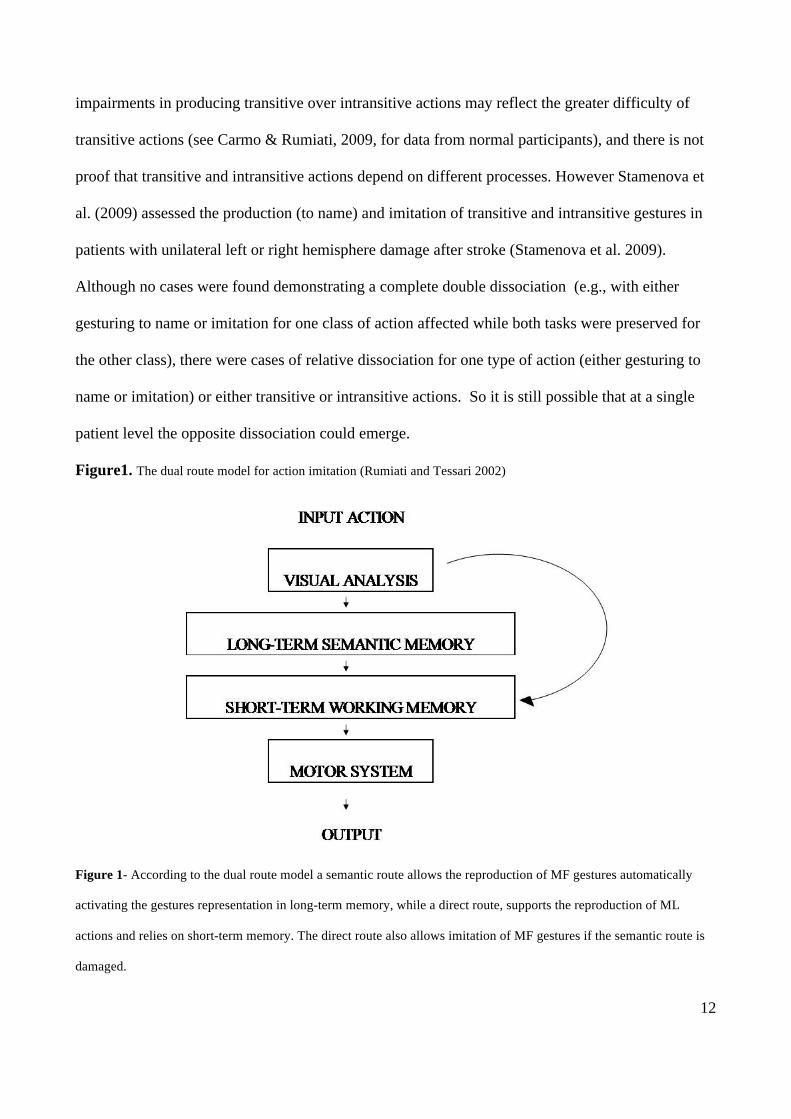
supports the reproduction of ML (new) actions (Figure 1). The starting point of both routes is a
visual analysis component, through which the visual properties of actions are processed. Also both
semantic and direct processes end at the level of the motor system involved in the actual
implementation of the action. In the present work I will consider the model proposed by Rumiati
and Tessari (2002) that is a simplified version of the original model by Rothi et al. (1991) and that
substitutes the ‘innervatory pattern’ of the original model with a short-term memory component,
stressing its role in the imitation of ML gestures.
Differently from the semantic route, which allows the reproduction only of those gestures that have
a representation in long-term memory, the direct route allows the execution of both ML and MF
gestures if the route is damaged or if the cognitive resources are limited and MF gestures are
presented intermingled with ML, thus avoiding the cognitive costs of switching from one route to
the other (Tessari et al. 2004). Besides neuropsychological observations, the existence of different
mechanisms for MF and ML actions has been suggested by several behavioural investigations in
healthy participants as well as in imaging studies (e.g. Rumiati et al. 2009; Tessari & Rumiati 2004;
Peigneux et al. 2004), and it holds for both transitive (too-related) and intransitive (not-tool related)
actions.
Within the class of MF actions, however, differences have been reported between transitive and
intransitive actions. For example, neuropsychological data show that apraxic patients can have more
difficulties in imitating transitive than intransitive gestures (Haaland et al. 2000; Bauxbaum et al.
2005, 2007), but the reverse dissociation has rarely been reported. The preponderance of
12
impairments in producing transitive over intransitive actions may reflect the greater difficulty of
transitive actions (see Carmo & Rumiati, 2009, for data from normal participants), and there is not
proof that transitive and intransitive actions depend on different processes. However Stamenova et
al. (2009) assessed the production (to name) and imitation of transitive and intransitive gestures in
patients with unilateral left or right hemisphere damage after stroke (Stamenova et al. 2009).
Although no cases were found demonstrating a complete double dissociation (e.g., with either
gesturing to name or imitation for one class of action affected while both tasks were preserved for
the other class), there were cases of relative dissociation for one type of action (either gesturing to
name or imitation) or either transitive or intransitive actions. So it is still possible that at a single
patient level the opposite dissociation could emerge.
Figure1. The dual route model for action imitation (Rumiati and Tessari 2002)
Figure 1- According to the dual route model a semantic route allows the reproduction of MF gestures automatically
activating the gestures representation in long-term memory, while a direct route, supports the reproduction of ML
actions and relies on short-term memory. The direct route also allows imitation of MF gestures if the semantic route is
damaged.
!
13
Action imitation and recognition: neural correlates and the mirror neuron
system
Mirror neurons are probably the most famous discovery in neuroscience over recent years. The key
initial evidence came from physiological studies in monkeys. These studies demonstrated the
existence of a class of visuo-motor neurons (the so-called mirror neurons, MNs), that responded
when the animals saw actions as well as when they had to produce them - some neurons responding
when the executed and perceived actions strictly coincided and others responding when the goals of
the enacted and perceived actions coincided (see Rizzolati et al. 2001, 2004 for a review). In
monkeys MNs have been documented to respond only for goal-directed actions, but not when those
actions were made using a tool. Also MNs appear not to respond to the mere presence of objects,
food, or to pantomimes or movements having an emotional meaning (see Hickok 2009, for a
review). It has also been pointed out that in monkeys MNs do not support imitation, as adult
monkeys do not imitate (Ferrari et al. 2006).
At first MNs were found in area F5 in the monkeys’ ventral premotor cortex (Gallese et al. 1996;
Rizzolati et al. 1996) but they have also been reported in a portion of the superior temporal sulcus
(the STSa) that responds also to biological motion and in the inferior parietal lobule (area PF) that
has been shown to respond strongly to the observation of action. The neurons in these three areas
also vary in the likelihood that they respond to action execution as well as observation, with almost
all neurons that respond to biological movements in F5 discharging also during action execution,
two thirds having similar properties in PF and only a minority of the neurons having both
characteristics in STMa (Rizzolati et al. 2001). Together these systems comprise the MN system,
which has been argued to play a critical role in a range of processes, from action understanding
through to intention encoding (Iacoboni et al. 2005; see also Rizzolati et al. 2001, 2010 for a
review).
14
Supportive evidence for a MN system in humans has come from various sources. Early suggestions
came in the form of electrophysiological evidence (e.g., EEG) showing activity in motor regions
when gestures were observed (Gastaut et al. 1954; Cohen-Seat et al. 1954; Cochin et al. 1998, 1999;
Altschuler et al. 1997, 2000; Salmelin et al. 1994; Hari et al. 1997; Salenius et al. 1997; Hari et al.
1998). This has been complemented by evidence from brain imaging investigations showing that
ventral premotor/inferior frontal areas (including Broca’s region), the STS and the inferior parietal
lobule are active during the observation of hand movements (see Rizzolati et al. 2001 for a review).
Furthermore, imaging studies using both action observation and observation/execution tasks have
shown that the left inferior frontal cortex and the right anterior parietal region are active both when
a gesture (lifting a finger) had to be executed following the observation of the same gesture and
when the gesture had just to be observed. The same area did not respond when the finger movement
had to be done after the observation of a control stimulus (a cross), or when participants just
attended to the cross (Iacoboni et al. 1999; see also Rizzolati at al. 2001 for review). The parieto-
frontal circuit was also found to activate in other fMRI studies when participants observed a human
or robot hand grasping an object (Gazzola et al. 2007), in answer to action related (Lewis et al.
2005) and hand and mouth related sounds (Gazzola et al. 2006), and when aplasic individuals (born
without arms and hands) both watched motor acts using hands and moved their feet (Gazzola et al.
2007) (see Rizzolati et al. 2010 for a review). It should be noted that, while in monkeys MN did not
respond to pantomimes and they could not be involved in imitation as imitation is not part of the
behavioural repertoire of adult monkeys, several experiments on MNs in humans have used
recognition and imitation of pantomimes (Kosky et al. 2002, 2003; Iacoboni et al. 1999; Grèzes et
al. 1998;) as well as the imitation of meaningless gestures (Iacoboni et al. 1999; Rizzolati &
Craighero 2004). These studies have often revealed activation in the inferior frontal gyrus and the
inferior precentral gyrus, which has been interpreted as proof of the existence of a MN system in
15
humans that has evolved to include both action recognition and production (see Hickok 2009 for a
review).
The MN system has been argued to have a main role in action understanding (Gallese et al. 1996;
Rizzolati et al. 2001). Rizzolati et al. (2001) defined “action understanding” as “the capacity to
achieve the internal description of an action and to use it to organize appropriate future behaviour”,
however other definitions exist (e.g. see Gallese et al. 1996). In the present work I will consider
action recognition and “action understanding” as synonymous, as the action recognition employed
in the thesis requires the perception and comprehension of learned motor actions. Apart from
questions over definitions, the assertion that mirror neurons are involved in action understanding
was based on observation that (i) those neurons in monkeys responded specifically on execution and
presentation of actions towards objects (e.g. lift an object/piece of food and put it in a container, or
take a piece of food and put it in the mouth), (ii) the neurons did not fire to pantomimes (without the
object present, and so having not a goal) but (iii) fired again to actions toward hidden objects, if the
monkey could see the object before it was hidden by a screen, as well as (iv) responding when the
stimulus was not visual, as when sounds related to purposeful actions were presented (e.g. cracking
a peanut shell or ripping paper). All this evidence has led to the claim that mirror neurons were not
simply visuo-motor association neurons, linking a visually presented object to its appropriate
action, but coded the meaning of the actions. However in a review Hickok (2009) has pointed out
that all the findings could be interpreted in more straightforward way, in terms of audiovisual
associative neurons, matching the representation of the object to its target action, with this
representation retrieved from the sound of the object itself or from working memory (as when
monkeys attended actions to hidden objects), rather then of neurons having ‘semantics’ (Hickok
2009).
Another argument put forward in favour of the MN system being related to recognizing the
meaning of an action comes from a study by Gallese et al. (1996). These authors simultaneously
16
recorded the activity in monkeys’ F5 and in the hand area of primary motor cortex (M1), as well as
recording EMG activity from several mouth and limb muscles. The results did not show any
activity from M1 and from limb and mouth muscles during action observation while mirror neurons
were responding (Gallese et al. 1996). This suggested that no covert movement were involved, and
cells in F5 were not mere motor-related neurons. However this result could be not replicated in
humans, in a transcranial magnetic stimulation (TMS) study that showed an increase in distal
muscle motor-evoked potentials (MEPs) during action observation (Fadiga et al. 1995). Inconsistent
results have come also from two recent studies using neural adaptation. Adaptation is a decrease in
the blood-oxygen-level dependent (BOLD) response from a brain area when a given sensory
stimulus is presented repeatedly several times, and adaptation across two separate stimuli is
considered to be evidence for the stimuli involving the same process and neural correlate. A first
fMRI study on healthy participants failed to show populations of neurons undergoing adaptation for
executed actions that were subsequently observed and vice versa (Dinstein et al. 2007) while a
second study pointed out the right inferior parietal lobe shows adaptation across action execution
and perception (Chong et al. 2008).
Neurophysiological evidence from the monkey points to action recognition and production sharing
neural and functional processes even though other possible, and more straightforward explanations
may be proposed (Hickok 2009, see above). Also the neuroimaging studies in humans fail to give
conclusive answers on the issue and it is clear that distinct component processes must also
contribute to each task. This is indicated perhaps most clearly by evidence of dissociations between
the tasks in neuropsychological patients.
For example, dissociations have been documented between patients who show spared recognition of
actions and gestures but who are impaired at producing the actions themselves (not concomitant on
a motor impairment, in cases of ideomotor apraxia; see Chainay & Humphreys, 2002). Also it
cannot be argued that the dissociation exists only because recognition tasks are easier than

17
producing gestures or imitating, as the reverse pattern of deficit, a syndrome labelled as
“pantomime agnosia” (Rothi et al. 1986), has also been reported (Bell 1994; Rothi et al. 1986;
Cubelli et al. 2000; see also Mahon et al. 2005 for a review). For instance Negri et al. (2007)
obtained case-level dissociations in a study where they tested an unselected group of 37 unilateral
brain damaged patients for object use, object recognition, pantomime imitation and pantomime
recognition. Although at group level they found significant correlations between each pair of task,
they also pointed out subsets of patients demonstrating dissociations between each test pairs (Negri
et al. 2007).
Gesture recognition has been linked to inferior frontal regions (Pazzaglia et al. 2008) as it has
pantomime imitation (Goldenbeg 2007). However the recognition of particular aspects of transitive
actions can be linked not to frontal regions but to inferior parietal (recognition of spatial aspects of
action) and posterior middle temporal cortex (recognition of semantic aspects of action). Parietal
regions have also been shown to be linked to ML action imitation (Rumiati et al. 2005; Goldenberg
et al. 2009).
Patients with lesions in the left parietal lobe have been reported to have poorer performance at
gesture to command than at imitation while patients with lesions in the left occipital and temporal
cortex have been documented with the opposite pattern, being almost normal when producing
actions on verbal command (Merians et al. 1997). Also, as described in the previous paragraph,
when gesture production is considered alone, there are dissociations in the production of different
types of action - for example, patients who can imitate familiar but not unfamiliar actions (see
Tessari et al. 2007 for a review). These dissociations point to there being separable components in
the system for processing actions as sensory stimuli for imitation and for learned gesture execution,
and they are compatible with a dual route account of action (see Tessari et al. 2007 and Rumiati et
al. 2010 for a review). The dual route model involves a semantic system where motor schemas of
already known gestures are stored and used for execution but not necessarily for action imitation (as
18
suggested by the double dissociations in patients who cannot recognize but can imitate and vice
versa). Also, a crucial point of the model is that access to the semantic system, needed to recognize
or understand the goal of an action, is not necessary for correctly imitating a gesture, as any gesture
can be processed through the direct route, and the representation of the action, before being
translated in motor terms is sustained by working memory.
Neural correlates of action imitation of MF and ML transitive and intransitive
gestures
Data on the neural correlates of praxis also come from neuropsychological observations on patients.
Traditionally the left hemisphere was considered dominant for controlling gesture production (see
Rothi et al. 1997) in right-handed people. However, even though there is a large consent on the
major involvement of the left hemisphere in action production, some authors suggest that this
depends upon the type of actions that have to be implemented. So, while the execution of transitive
(tool-related) actions seems to rely more on the left-hemisphere (Bartolo er al. 2001; Tessari et al.
2007), both right and left cortical areas are involved in the production of intransitive gestures
(Buxbaum et al. 2001, see also Tessari et al. 2007 and Rumiati et al. 2010 for a review). Also a role
for sub-cortical structures has been suggested by some studies (see Tessari et al. 2007; Leiguarda et
al. 1997, 2001; De Renzi et al. 1980), even though there is still not yet consent about which type of
gesture may be more impaired after lesions or deterioration in those structures.
One piece of evidence comes from the observation of patients suffering of ideomotor apraxia. In
right handed people ideomotor apraxia has classically been described in relation to lesions of the
left posterior parietal cortex (Liepmann, 1900, 1905)(see Rothi et al. 1991 for a review), though
other different studies point to the role of right brain areas as well as sub-cortical structures (see
Tessari et al. 2007; Leiguarda et al. 1997, 2001; De Renzi et al. 1980), especially when finger
configurations (Goldenberg & Strauss 2002; Della Sala et al. 2006) or movement sequences
19
(Canavan et al. 1989) have to be copied. While there is also neuropsychological data (e.g.
Goldenberg & Hagmann 1997; Peigneux et al. 2000; Cubelli et al. 2000; Bauxbaum et al. 2001;
Tessari et al. 2007) supporting the idea of two separate neural systems for imitating MF and ML
gestures, there is inconsistency with respect to the specific neuro-anatomical structures involved in
each (particularly concerning their hemispheric localisation). Lesions involving the parietal cortex,
especially the left angular gyrus, are reported to cause a deficit in the imitation of meaningless (ML)
as compared to meaningful (MF) actions in left-brain damaged patients (LBD) (Mehler 1987;
Goldenberg & Hagmann 1997; Peigneux et al. 2000; Tessari et al. 2007). In contrast Tessari et al.
(2007) reported two right brain damaged patients (RBD) who were more impaired in imitating ML
relative to MF gestures. For these two patients the lesions included the caudal portion of the
pallidum, the putamen and the posterior limb of the internal capsule.
Goldenberg (2008), in a review of apraxia, also argues for a role of parietal lesions in the imitation
of meaningless gestures, as well as on the use of tools and objects, and actually states that parietal
lesions have an impact on those functions and not on other aspects of praxis. Goldenberg further
points out that the role of the parietal lobes concerns the apprehension of spatial relationships
between body-parts, tools and objects rather than representations of motor acts (Goldenberg 2008).
Goldenberg et al. (2007) used lesion subtraction procedures to determine the locations specifically
associated with defective pantomime of tool use in patients with left-brain damage and
aphasia. Their results showed that damage to the left inferior frontal cortex was associated with a
deficit in pantomiming action, even though the area of lesion overlap further extended into the
underlying white matter (WM) and it is possible that damage of WM projections contributed to the
deficit (Liepmann 1905; Geschwind 1976; Catani and Ffytche 2005).
Observations of neuropsychological deficits also indicate a role of subcortical structures on
imitation that do not relate to peripheral neurological symptoms. For instance data from Leiguarda
et al. (1997, 2001) suggest that the basal ganglia are necessary for the imitation of transitive actions,

20
while intransitive actions may operate through cortical regions – with this holding even for
meaningless actions based on intransitive gestures. In patients with sub-cortical lesions then
imitation may be spared for meaningless actions, but the production of familiar transitive actions
may be disrupted (Hanna-Pladdy et al. 2001). In contrast, Tessari et al. (2007) described two right –
brain damaged patients whose lesions involved the basal ganglia. These individuals were more
impaired in imitating meaningless (ML) as compared to meaningful (MF) transitive (or tool-related)
pantomimes and their lesions overlapped on basal ganglia. The report from Leiguarda et al. (1997,
2001) gives the opposite picture to this, but in their case the MF transitive gestures could have been
more difficult than the ML gestures. There are data too suggesting that access to long-term semantic
memories for action may involve middle temporal regions, while deficits in access to (and possibly
planning for production) the spatial aspects of action link to inferior parietal damage (Buxbaum et
al. 2010). The implementation of learned transitive actions may operate through sub-cortical
routines (though see Tessari et al. 2007), while left inferior frontal regions are involved in aspects of
action production where lesions become most pronounced when there is a lack of convergent input
from holding the actual objects (see Chainay & Humphreys, 2002, about the use of convergent
sensory input into motor output and selection processes).
Consistent with the above argument, neuropsychological data show dissociations between transitive
and intransitive actions suggesting that the two classes of gestures can be functionally and neutrally
separable. In the majority of cases patients have been described with deficits in which transitive
actions are more impaired than intransitive actions (e.g., Buxbaum et al. 2007) and it has been
proposed that transitive actions relying on a repository of action engrams in the left inferior parietal
cortex (Buxbaum, 2001; Buxbaum et al. 2007; Liepmann, 1900-1905) and/or sub-cortical structures
(Leiguarda et al. 1997, 2001) whereas familiar intransitive actions may be supported by fronto-
parietal regions that respond to fine-grained dynamic properties of action (Buxbaum et al. 2007;
Haaland & Flaherty, 1984; Mozaz, 2002; Rapcsak et al. 1993).
21
Recent work by Stamenova, Roy and Black (2010) compared transitive and intransitive actions in
stroke patients. Selective deficits in transitive actions were associated with left hemisphere damage
and problems with intransitive action with right hemisphere damage, consistent with prior
arguments for the necessary involvement of the right hemisphere in intransitive action, perhaps
because these actions depend more on fine-grained spatio-temporal modulation of action. On the
other hand, the same investigators (Stamenova et al. 2009) have reported data on cortical-basal
degeneration patients showing selectively greater changes on the left or right side of the brain and
failed to find evidence for intransitive actions ever being more impaired than transitive. Hence the
case for a functional and neural distinction between these different classes of learned actions
remains inconclusive.
Behavioural and imaging studies on healthy participants (e.g. Rumiati et al. 2009; Tessari &
Rumiati 2004; Peigneux at al. 2004) show different brain areas to be active for MF and ML actions,
even though also in this case there is not unequivocal consent on the structures that are involved. In
studies using positron emission tomography (PET), imitation of MF actions has been observed to be
associated with activation in the left angular and middle frontal gyri, as well as the right
supramarginal gyrus and inferior parietal lobule (Peigneux et al. 2004), and also in the inferior
temporal, angular and parahyppocampal gyri in the left hemisphere (Rumiati et al. 2005). The
parieto-occipital and occipito-temporal junctions on the right, the superior temporal gyrus on the
left, and the superior parietal cortex bilaterally have also been shown to have increased activation
linked to imitation of ML actions (Rumiati et al. 2005). Other studies though suggest considerable
overlap in the brain areas used to make transitive and intransitive gestures, consistent with all
familiar gestures relying on a common neural network involving parietal and frontal cortices (e.g.,
Kroliczak & Fray, 2009).
22
Reasons for the present work
As the above review indicates, many studies have investigated action imitation and recognition, and
different fields of neuroscience have addressed the issue. However there is still not unanimous
consent on how imitation and recognition take place, especially in relation to the brain regions
involved. Neuropsychological and clinical observations can reveal symptom correlations and
dissociations that sometimes highlight different components involved in praxis, according to the
task (imitation or action recognition) and the type of action involved (MF or ML).
One caveat however is that neuropsychological studies using lesion-symptom mapping frequently
rely on categorical subdivisions of patients based on a given cut off as well as on observer-
dependent lesion demarcations. The use of a cut off, especially in tasks where each answer is scored
either 1 or 0, introduces the risk that patients having scores differing only for one point are treated
differently (e.g. scores that equal the cut off will be considered as defective, while scores higher
than the cut off for one point will be treated as borderline or not defective at all). Also manual
demarcations of lesions sites may lead to under- or over- estimation of differences in tissues
according to the strictness of the observer’s criteria. The present study aims to investigate the neural
substrates of action imitation, production to name and recognition using a complementary VBM
analysis based on segmented grey matter (GM). VBM uses the general linear model to statistically
assess the relations between brain tissue integrity and behavioural performance. Here, imitation,
action production and recognition scores were used as predictors of change in the signal intensity
coming from each voxel across the whole brain in groups of consecutively sampled patients, not
pre-selected on the basis of having apraxia, but using continuous scores on tests of gesture imitation
in order to characterise their abilities. This approach also allows us to control in the model
covariates of no interests such as age, gender, neglect, which may otherwise contaminate the results.
23
VBM analysis provides an instrument to test movement production offline and then to look for
neural correlates from brain images in unbiased samples of participants. Of course VBM works
only if brains differ in voxel intensity signal, and in adults that means that brains entered in the
analysis should have a lesion. Also, being based on correlation, VBM requires quite big samples to
give results. For this reason I have attempted to test large samples of participants on tasks that may
be considered to test praxis and then to use the performance of individual patients as a predictor of
the density of brain GM. I have also assessed patients at different stages of their disorder –
including patients scanned at a sub-acute stage (Chapter 5 and 6) and patients with chronic deficits
(Chapter 4), and I have analysed both MRI (Chapter 4) and CT (Chapter 5 and 6) data. Scanning at
different stages of recovery, and using different imaging procedures, both raise questions about the
comparability of the results, but they also allow conclusions to be drawn with greater confidence
when converging results emerge. The data from across the thesis point to both common patterns of
deficits across different tasks and action types, and also to some instances of dissociation – both in
behavioural and in imaging terms. The General Discussion chapter tries to draw the results together
to make general conclusions about the action production and recognition system.
24
2. GENERAL METHODS
This thesis presents five neuropsychological studies focusing on the performance of patients with
acquired brain lesions in gesture production and recognition Chapter 3 compares patients with
chronic acquired injury, involving either the left or the right hemisphere, and healthy volunteers in
the imitation and recognition of different types of action (familiar or unfamiliar transitive actions to
objects and familiar or unfamiliar intransitive, not-object-related, gestures).
Chapter 4 uses chronic brain injured patients’ scores in the imitation of familiar and unfamiliar
transitive and intransitive gestures to predict MRI signal changes from gray matter or white matter
segmented images. In this chapter voxel-based morphometry was performed on patients’ scores and
MRI signal values entered as continuous measures. However healthy controls’ data were also used
to describe the patient sample at a behavioral level (see the paragraph below and chapter 2 for more
details).
Chapter 5 analyzes the relation between GM and WM integrity and the skills to produce gestures on
verbal command, to imitate and to recognize actions, in a sample of acute stroke patients using
voxel-based morphometry on CT scans. In this chapter the data for the patients were not compared
to healthy controls’ scores.
Chapter 6 investigates the GM and WM correlates of production on verbal command of transitive
and intransitive familiar gestures, again on acute stroke patients using voxel-based morphometry on
CT scans. As in Chapter 5 the patients’ scores were not compared to controls.
Chapter 7 reports a neuropsychological investigation where we tested the ability of Parkinson’s
disease patients in imitation of familiar and unfamiliar transitive and intransitive gestures as
compared to healthy controls.
This section of the thesis reports a description of the samples of patients and healthy volunteers that
took part in the studies reported in this thesis, along with the materials and the tasks administered.
25
In some cases only some patients or controls from a main sample are included in a study. Also,
some tasks are included in more than one study while others are unique for one or two studies. For
this reason, in those cases where it may not be clear, tables are presented summarizing which
subjects or tasks are included in each chapter.
Also, the two final paragraphs describe the imaging data used in the present work for the analyses
using voxel-based morphometry (VBM), how they had been acquired and prepared for the analyses
and the tools used to run the VBM.
PATIENTS
Patients- Chapters 3 (comparing right and left brain damaged patients) and 4 (VBM on MRI
scans of patients in chronic phase)
Twenty-four patients (mean age= 67.9, SD= 10.9, age range= 36-82) were recruited at the
Behavioural Brain Science at the school of Psychology of the University of Birmingham. All the
patients had acquired brain lesions (twenty-three had a stroke, while one had a bilateral lesion
caused by encephalitis), and all were in the chronic stage (> 9 months after the occurrence of the
lesion). According to the Edinburgh Handedness Inventory (Oldfield, 1971) twenty-one patients
were right handed before the stroke and three were left-handed. Patients’ demographic data and
their Edinburgh Handedness Inventory scores are summarized in Appendix 1 (see Table 1 in
Appendix 1).
From this sample, the data of fifteen patients were used in the two studies presented in chapter 3
and chapter 4, three patients were included only in the study in chapter 4 and six patients were
included only in the study in chapter 4. Table 1 shows which patients were included in each chapter
(3 or 4 or both), the patients’ lesion side, handedness and the hand used to imitate (dominant or not
dominant).

26
In total, eighteen right-handed patients (sixteen male, two female) were included in the study
described in chapter 3. Nine patients had a lesion involving the right side of the brain (Right Brain
Damaged, RBD) (mean age= 69.22, SD= 8.07, age range= 55-78) and nine had a lesion on the left
(Left Brain Damaged, LBD) (mean age= 67.2, SD= 13.7, age range= 36-82), confirmed on either
MRI or CT scans. From the main sample of twenty-four patients, six were excluded from the first
study because of one of the following reasons: i) three patients resulted to be left-handed before the
stroke; ii) two patients had a bilateral brain lesion.
In a first version of the present work one patient (MMcD, highlighted in red in Table 1) was
included by mistake in the sample of LBD, and so his data were analysed also in chapter 3.
However, when we checked again the patient’s scan he resulted to have a bilateral lesion, which
was also likely to influence his behaviour. For this reason he was then excluded from the sample in
chapter 3. A final section of chapter 3 will describe briefly MMcD results and a picture of his lesion
will be reported.
Twenty-one patients (20 males and 1 female, 18 right-handed, 3 left-handed) (mean age: 68.18; SD:
11.13. Age range: 36-82) were included in the study reported in chapter 4. Seven patients had a
lesion involving the left side of the brain (Left Brain Damaged, LBD), eleven had a lesion on the
right (Right Brain Damaged, RBD) and three had bilateral brain lesions (BBL).
None of the patients who took part in the studies of chapter 3 and 4 were included in chapter 5, 6
and 7.
27
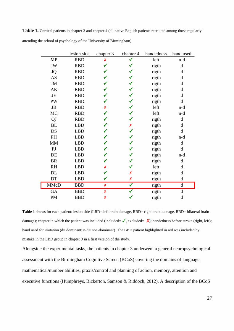
Table 1. Cortical patients in chapter 3 and chapter 4 (all native English patients recruited among those regularly
attending the school of psychology of the University of Birmingham)
Table 1 shows for each patient: lesion side (LBD= left brain damage, RBD= right brain damage, BBD= bilateral brain
damage); chapter in which the patient was included (included= ✓, excluded= ✗); handedness before stroke (right, left);
hand used for imitation (d= dominant; n-d= non-dominant). The BBD patient highlighted in red was included by
mistake in the LBD group in chapter 3 in a first version of the study.
Alongside the experimental tasks, the patients in chapter 3 underwent a general neuropsychological
assessment with the Birmingham Cognitive Screen (BCoS) covering the domains of language,
mathematical/number abilities, praxis/control and planning of action, memory, attention and
executive functions (Humphreys, Bickerton, Samson & Riddoch, 2012). A description of the BCoS
lesion side chapter 3 chapter 4 handedness hand usedMP RBD ✗ ✓ left n-dJW RBD ✓ ✓ rigth dJQ RBD ✓ ✓ rigth dAS RBD ✓ ✓ rigth dJM RBD ✓ ✓ rigth dAK RBD ✓ ✓ rigth dJE RBD ✓ ✓ rigth dPW RBD ✓ ✓ rigth dJB RBD ✗ ✓ left n-dMC RBD ✓ ✓ left n-dQJ RBD ✓ ✓ rigth dBL LBD ✓ ✗ rigth dDS LBD ✓ ✓ rigth dPH LBD ✓ ✓ rigth n-dMM LBD ✓ ✓ rigth dPJ LBD ✓ ✓ rigth dDE LBD ✓ ✓ rigth n-dBR LBD ✓ ✓ rigth dRH LBD ✗ ✓ left dDL LBD ✓ ✗ rigth dDT LBD ✓ ✗ rigth d
MMcD BBD ✗ ✓ rigth dGA BBD ✗ ✓ rigth dPM BBD ✗ ✓ rigth d

28
is reported in Appendix 1, along with the patients’ results (Table 2 in Appendix 1). The results on
the BCoS tests were used to perform correlations with the imitation scores.
Patients- chapters 5 and 6 (VBM on CT scans of acute patients)
CT scans and behavioural results from 233 stroke patients (110 male and 123 female; mean age=
70.64, SD= 14.40) were taken from the dataset collected by the Birmingham University Cognitive
Screen Trial group (http://www.bucs.bham.ac.uk) in 12 local stroke units, and were used for the
analyses. Each patient’s age, gender, education level, and the hand used to preform for gestures
production and imitation are reported in Appendix 2 (Table 1 in Appendix 2). All the patients had
ischemic stroke and were at a sub- acute stage (>2 days, ≤3 months after the stroke) at the time of
both behavioural testing and CT scan acquisition. At the time of testing all were alert and had
sufficient English comprehension to follow the instructions. As the data were taken from a very
large database, this allowed us to further filter the data, and to include only those patients who
reported to be pre-morbidly right-handed (based on self report or as reported by relatives). Patients
having bad quality CT scans as well as those who underwent CT on the same day of the stroke were
excluded from the sample. From the initial database of 539 patients 306 were discarded for the
following reasons: bad quality scan (N= 37); missing data on tasks used as covariates (N= 54); scan
acquisition was performed in the same day as the patient was admitted (N= 144); hemorrhagic
stroke (N= 38); the patient was left-handed (N= 33).
In chapters 5 and 6 patients’ scores were used as continuous measures, and data from controls were
not needed.
All patients gave informed consent.
The data of the same 233 acute patients were used in both chapters 5 and 6.
None of the patients in chapters 5 and 6 took part in the studies presented in the other chapters.
29
Parkinson’s patients- chapter 7 only
Nineteen native Italian speaking PD patients (9 male, 10 female; mean age= 66.37, SD= 6.95)
participated in the study. All the patients were right handed according to the Edinburgh Handedness
Inventory (Oldfield 1971). PD patients’ age, gender, the Edinburgh Handedness Inventory scores
and hand used for gestures imitation are reported in Appendix 3 (Table 1 in Appendix 3). The
patients were recruited according to the following criteria: diagnosis for idiopathic (unknown
aetiology) PD, absence of major cognitive decline, presence of asymmetric symptoms, having
normal or corrected to normal vision. The diagnosis was made by a neurologist on the basis of the
patients’ symptoms as well as of the results of anatomical scans of their brains (SPECT) proving the
presence and the asymmetry of the lesion. Twelve patients showed more pronounced symptoms on
the left and seven were more impaired on the right side of the body. PD patients’ neurological and
motor symptoms (rigidity or tremor) were assessed with scale III of the United Parkinson Disease
Rating Scale (see Table 1 in Appendix 3). All patients were under pharmacological treatment with
L-Dopa.
PD patients were first given the Mini Mental State Examination (MMSE) in order to exclude those
who could have shown a major cognitive decline. Also, PD patients were administered a battery of
neuropsychological tests, standardized for the normal Italian population, assessing general
intelligence, attention, language functions, visual perception, short-term memory for verbal material
and locations in space. A description of the tests is in Appendix 3 along with the summary of the
patients’ results (Table 2 in Appendix 3).
Only one PD patient obtained a MMSE score that was just below the cut-off for the normal Italian
population (cut-off= 24) when corrected for age and education (raw score = 26; score corrected for
age and education = 23.2); however as his performance on all the other tests (see Table 2 in
Appendix 3) was in the normal range, he was not excluded from the sample.
30
CONTROL SUBJECTS
Table 2 (see below) summarizes the composition of the control samples used in chapters 3, 4 and 7,
along with handedness and the hand used for imitation and tasks performed.
Controls- Chapters 3 and 4
Eighteen English-speaking healthy participants (mean age= 65.56, SD=10.18, age range= 37-78)
with no reported history of neurological and/or psychiatric disease served as controls for the studies
presented in chapters 3 and 4. Fifteen controls reported to be right-handed while three reported to
be left-handed. All had normal or corrected to normal vision. Only the fifteen right-handed (mean
age= 66.80, SD=7.72, age range= 50-78) were included in the study in chapter 3, while data for all
the eighteen healthy subjects were used in chapter 41.
The imitation scores (for transitive actions only) of seven of these eighteen English controls were
also used in chapter 7 (see next paragraph).
Controls- Chapter 7
Two different groups of healthy participants were used to compare the PD patients’ scores on the
intransitive and transitive action imitation tasks. The first group had been tested on the intransitive
actions prior to the present examination and these data were used again here. The second group was
tested on the transitive actions only.
For the intransitive actions we used data collected with 31 Italian native speaking controls, 10
female and 21 male (range = 53-75; mean age = 65.35; SD: 6.5). A t-test comparing the age of PD
and controls did not reveal any significant difference (t (48) = 0.52; p = 0.60, 2-tailed). None of the
controls had a history of neurological or psychiatric disorders and all had normal or corrected to
1 In chapter 4 the controls’ data were used to establish the cut-offs for the experimental tasks. The cut-offs were used
only to describe the patients’ sample imitation behaviour. In the VBM analyses patients’ imitation scores were used as
continuous measurements to predict voxel signal intensity.
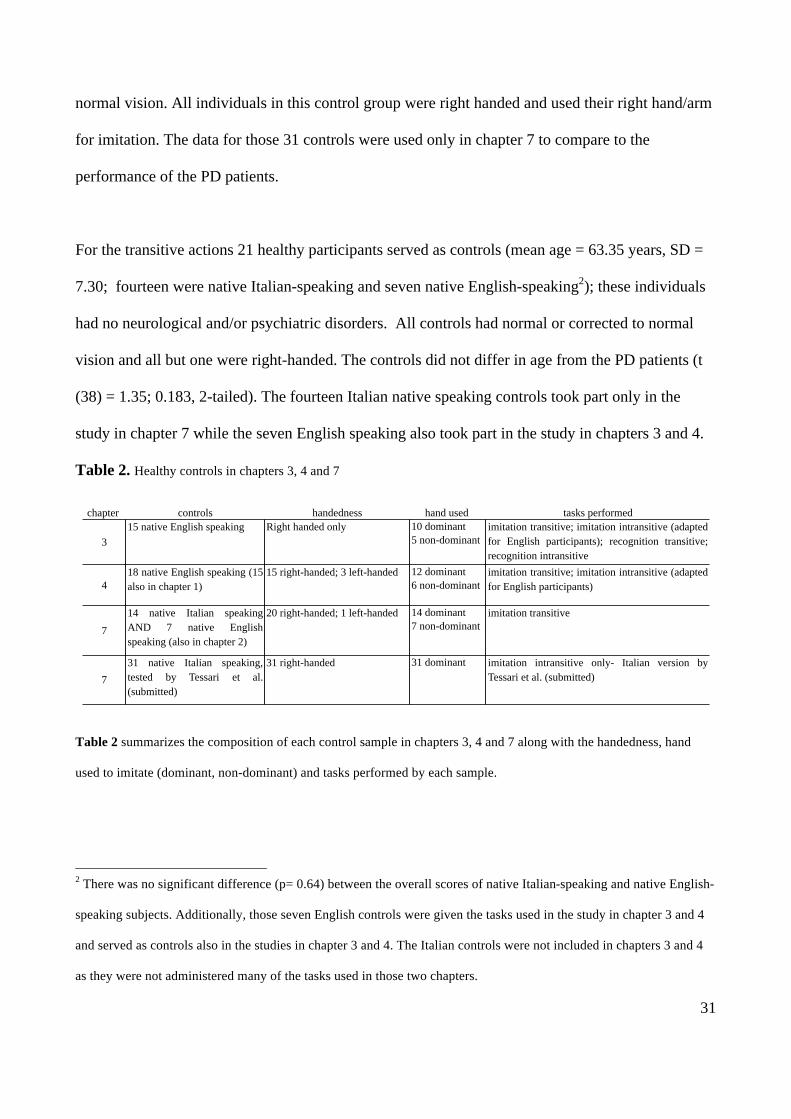
31
normal vision. All individuals in this control group were right handed and used their right hand/arm
for imitation. The data for those 31 controls were used only in chapter 7 to compare to the
performance of the PD patients.
For the transitive actions 21 healthy participants served as controls (mean age = 63.35 years, SD =
7.30; fourteen were native Italian-speaking and seven native English-speaking2); these individuals
had no neurological and/or psychiatric disorders. All controls had normal or corrected to normal
vision and all but one were right-handed. The controls did not differ in age from the PD patients (t
(38) = 1.35; 0.183, 2-tailed). The fourteen Italian native speaking controls took part only in the
study in chapter 7 while the seven English speaking also took part in the study in chapters 3 and 4.
Table 2. Healthy controls in chapters 3, 4 and 7
Table 2 summarizes the composition of each control sample in chapters 3, 4 and 7 along with the handedness, hand
used to imitate (dominant, non-dominant) and tasks performed by each sample.
2 There was no significant difference (p= 0.64) between the overall scores of native Italian-speaking and native English-
speaking subjects. Additionally, those seven English controls were given the tasks used in the study in chapter 3 and 4
and served as controls also in the studies in chapter 3 and 4. The Italian controls were not included in chapters 3 and 4
as they were not administered many of the tasks used in those two chapters.
chapter controls handedness hand used tasks performed10 dominant5 non-dominant
12 dominant6 non-dominant
14 dominant7 non-dominant
31 dominant7
31 native Italian speaking,tested by Tessari et al.(submitted)
31 right-handed imitation intransitive only- Italian version byTessari et al. (submitted)
7imitation transitive14 native Italian speaking
AND 7 native Englishspeaking (also in chapter 2)
20 right-handed; 1 left-handed
imitation transitive; imitation intransitive (adaptedfor English participants); recognition transitive;recognition intransitive imitation transitive; imitation intransitive (adaptedfor English participants)
15 native English speaking Right handed only3
418 native English speaking (15also in chapter 1)
15 right-handed; 3 left-handed
32
TASKS
Table 3 shows a summary of the tasks used in each of the five experimental chapters.
Imitation tasks in chapters 3, 4 and 7
Transitive actions: For the transitive actions we presented 20 meaningful (MF) pantomimes of
objects being used (e.g. hammering or drinking from a glass) and 20 unfamiliar meaningless (ML)
control actions derived from the MF actions (e.g. a action maintaining the grasp and arm
configuration for hammering but performed in an unusual direction; for details about the stimuli see
Tessari & Rumiati 2004). These stimuli were employed because they were already used in
previously published studies on the topic, with both patients and controls, and they have been
utilized in studies across different nationalities (e.g., in Italy (Tessari et al. 2004) and Germany
(Rumiati et al. 2005)). The list of the 20 MF and 20 ML actions is reported in Appendix 4. Pictures
showing a sample of MF and ML transitive stimuli are in Figure 1.
Each transitive action was presented once to each participant and scored as 1 if performed correctly
or 0 if it was performed incorrectly (maximum MF score= 20, ML= 20; total= 40).
The same list of actions was administered to patients and controls in chapters 3, 4 and 7.
33
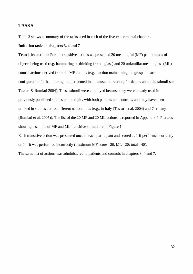
Figure 1. Imitation MF and ML transitive gestures- stimuli- chapters 3, 4 and 7
Figure 1 shows an example of an MF pantomime (left), in this case “drinking from a glass”), and its ML version
(right).
Intransitive actions: The stimuli were taken from the original set of 18 MF and 18 ML intransitive
gestures by Tessari et al. (submitted). The intransitive MF actions were a sample of those gestures
people commonly use for communication (e.g. waving “hallo”) and were selected as 10 Italian
independent raters easily recognized them. The intransitive ML actions were created in order to
match the MF for complexity of execution and were judged as unrecognizable by the 10 Italian
independent raters (Tessari et al. submitted). The same list of MF and ML actions had already been
used in previous studies on healthy Italian-speaking participants (Carmo & Rumiati 2009; Rumiati
et al. 2009). One half of the MF and ML actions involved movement of the hand (i.e. distal) while
the other half involved the use of an arm (i.e. proximal). In chapter 7 (studies on Italian Parkinson’s
patients) the whole set of 18 MF and 18 ML actions was used. In chapters 3 and 4, to adapt the task
to the English patients and controls, 3 MF gestures were removed from the original MF list
(Tessari, in prep.) as eighteen healthy English controls consistently failed to indicate that they had a
meaning. The remaining 15 MF stimuli were easily recognized and named correctly by the healthy
English native raters. Three actions were also discarded from the original ML list in order for this
list to equal the MF list length. The discarded ML actions matched for complexity the gestures
eliminated from the MF set according to the data from ten native English independent raters. The 18
34
MF and 18 ML stimuli are listed in Appendix 4. The 3 MF and 3 ML stimuli that were removed to
adapt the task for the English participants are reported in bold (see Appendix 4). Two examples of
the stimuli are in Figure 2 in this chapter.
In chapters 3 and 4, using the task adapted to test English participants, each intransitive action was
shown only once and performance was scored 1 if the action was correctly executed and 0 if it was
done incorrectly (maximum MF score= 15, ML= 15; total= 30).
In chapter 7, using the Italian version of the task to test Italian PD participants, the intransitive
actions could be presented twice, if the patient failed to reproduce an action at the first attempt. In
this chapter the intransitive actions were administered following the same procedure to the one
previously used by Tessari et al. (submitted.) to test the Italian controls whose data also served to
calculate cut-offs for MF, ML and total performance (cut-offs are reported in Appendix 4 below the
list of intransitive gestures). This allowed us to identify those PD patients scoring below the cut-off
for the normal population, beside the group analyses. Each action was rated 2 if correct at the first
presentation, 1 if correct after the second presentation and 0 if incorrect both at the first and at the
second attempt (maximum MF score= 36, ML= 36; total= 72).
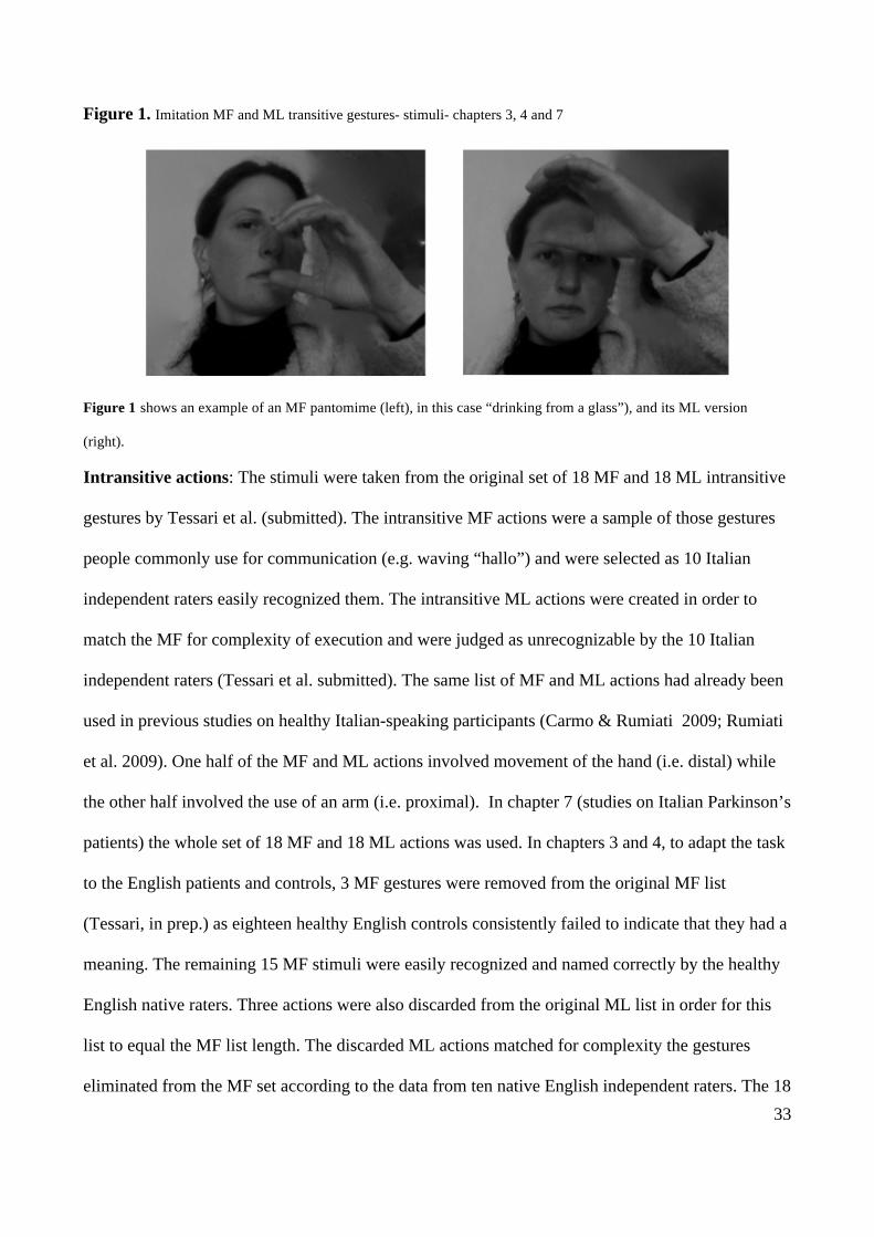
Figure 2. Imitation MF and ML intransitive gestures- stimuli- chapters 3, 4 and 7
Figure 2 shows one of the MF intransitive gestures (i.e. “mad”) used as stimuli (left) and one of the ML (right).
Procedure, common to transitive and intransitive gestures in chapters 3, 4 and 7: Each type of
action was presented in a separate block to maximize the use of differential imitation processes,
35
with the order of presentation randomized within each list. There were four blocks of stimuli: i) MF
transitive; ii) ML transitive; iii) MF intransitive; iv) ML intransitive. The block of MF pantomimes
was administered before the ML pantomimes. After a short break, the intransitive actions were
presented, again with the MF followed by the ML block. The MF actions were administered before
the ML stimuli in order to reduce the likelihood of selecting the common ‘direct’ route for imitation
of MF as well as ML actions, given that MF actions could be reproduced using the same, direct
imitative route as ML actions (Tessari & Rumiati, 2004).
The experimenter demonstrated each action using always the right (dominant) hand. This was done
to maximize the consistency of stimulus presentation across participants. We avoided using video
stimulus presentations as it could have jeopardized patients’ performances because of reasons
unrelated to a praxis deficit (e.g. if patients could not clearly see the stimuli because of the relative
small size of the computer screen). Patients and controls were instructed to reproduce the action as
similarly as possible to the model. The cortical patients in chapters 3 and 4 performed the
pantomimes using either their dominant hand or the ipsilesional hand if having a paresis of the
contralesional limb. Nineteen of the twenty-four cortical patients who participated in the studies in
chapters 3 or 4 (or both) used their dominant limb while five used their non-dominant limb (see
Table 1 above in this method section for details).
The PD patients in chapter 7 used the hand that was less affected by the disease. Twelve PD
patients performed the imitation task using their right arm/hand while seven imitated using their left
(non dominant) arm/hand.
In all three chapters (i.e. chapters 3, 4 and 7), to control for the hand used, some controls were asked
to use their dominant hand while other used their non-dominant hand when responding3 (see Table
2 in this method section for details on hand used by controls to imitate).
3 On average there was not an effect of the hand used (p > 0.1) on both patients and controls’ performance.
36
The performance of each participant was video-recorded and later scored by two independent raters
blind to the experimental conditions4. For both transitive and intransitive actions, an action was
scored as incorrect if the participant performed a spatial error of the hand or the arm; a visual error
(i.e. the action was: i) a combination of two items included in the list; ii) a action that was visually
similar to the target; iii) a meaningful action, visually similar to the meaningless target), or an
omission (for a detailed description of the errors see Tessari and Rumiati, 2004).
Chapter 3 only- Recognition of transitive and intransitive gestures
In addition to the imitation tasks patients and controls were tested for their ability to recognize the
MF transitive and intransitive actions they imitated. The MF action recognition tasks were given at
the end of the examination protocol. This was done in order to minimize the possibility that the
performance in the imitation and recognition tasks influenced each other.
Recognizing MF transitive and intransitive actions
Stimuli: The same 20 MF transitive and 15 MF intransitive gestures previously imitated were
presented for recognition. The stimuli in each list were given in a pseudo-random order.
Procedure: Actions were showed one at a time by the experimenter using her right hand.
Participants had to provide either the action name or any information that demonstrated
unequivocally that they knew its meaning (e.g. description of circumstances where the gesture was
usually performed). Correct answers were scored 1 otherwise 0. Each gesture was shown only once
(transitive maximum score= 20, intransitive maximum score= 15).
4 The Cohen’s k agreement coefficient was calculated on the scores provided by the two independent raters for all the
three chapters. The coefficient was computed for MF and ML actions taken separately, and for the total action scores.
The analysis was performed separately for PD, cortical patients and controls. As the coefficient was ≥ 0.80 in all the
cases considered, the scores of only one rater (the same for patients and controls) were used.
37
Chapter 7 only- MF transitive and MF intransitive gestures naming
After being tested for imitation, PD patients were showed again the same MF transitive and MF
intransitive actions they just imitated. As the PD performance was at ceiling (i.e. all the PD easily
recognized and correctly named all the actions) the results at this task were not further considered in
the analyses.
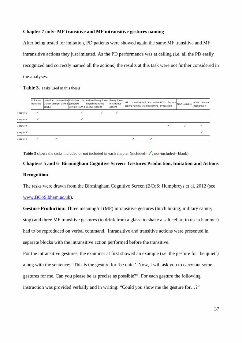
Table 3. Tasks used in this thesis
Table 3 shows the tasks included or not included in each chapter (included= ✓; not-included= blank).
Chapters 5 and 6- Birmingham Cognitive Screen- Gestures Production, Imitation and Actions
Recognition
The tasks were drawn from the Birmingham Cognitive Screen (BCoS; Humphreys et al. 2012 (see
www.BCoS.bham.ac.uk).
Gesture Production: Three meaningful (MF) intransitive gestures (hitch hiking; military salute;
stop) and three MF transitive gestures (to drink from a glass; to shake a salt cellar; to use a hammer)
had to be reproduced on verbal command. Intransitive and transitive actions were presented in
separate blocks with the intransitive action performed before the transitive.
For the intransitive gestures, the examiner at first showed an example (i.e. the gesture for ´be quiet´)
along with the sentence: “This is the gesture for ´be quiet′. Now, I will ask you to carry out some
gestures for me. Can you please be as precise as possible?”. For each gesture the following
instruction was provided verbally and in writing: “Could you show me the gesture for…?”
chapter(3 ✓ ✓ ✓ ✓
chapter(4 ✓ ✓
chapter(5 ✓ ✓ ✓
chapter(6 ✓
chapter(7 ✓ ✓ ✓ ✓
MF intransitiveactions(naming
BCoS GestureProduction
BCoS(Imitation BCoS ActionsRecognition
Imitation(transitive
Imitation intransitive(Italian versionC 18M &18ML)
Imitation intransitive(adapted EnglishversionC(15M(&(15ML)
RecognitionCtransitive(actions
RecognitionCintransitive(actions
MF transitiveactions(naming
38
Performance was given a score of 0 if no response was given after 15 seconds, or if the patient
produced an unrecognizable gesture (e.g. for hitch-hiking, shaking open palm forwards) or a
perseveration from the previous action. A score of 1 point was given if a gesture was recognizable
but contained spatial errors (e.g. for the salute, the hand touches the cheek instead of the forehead)
or movement errors (e.g. for hitch-hiking, correct hand gesture but with wrist rotation instead of
forearm oscillation), while 2 points were given to correct and accurate gestures.
For the transitive pantomimes the following instruction was provided: “I will give you the name of
an object and ask you to pretend that you have the object in your hand. I will then ask you to show
me how to use it. For example, if you have to show how you would use a toothbrush, you could
make a gesture like this (show gesture)”. Each pantomime was preceded by the question: “how
would you use …?”
As for the intransitive gestures, answers delayed more than 15 seconds after the question;
unrecognizable actions and perseverations were scored 0. A gesture was scored 1 if recognizable
but containing errors [spatial errors (e.g. for glass, pouring gesture towards the chest instead of the
mouth); incorrect grip (e.g. for hammer, the grip indicates that the hammer is held perpendicular to
forearm); movement errors (e.g. for hammer, the oscillation is too small to be effective for a
hammer); incomplete sequence of action (e.g. for salt cellar, correct grip but no shaking of salt); or
concretization (i.e. use of body part as object)]. Recognizable and correct performances were scored
2 (maximum intransitive score= 6, transitive= 6, total= 12).
Patients had to reproduce both type of action using their most effective hand.
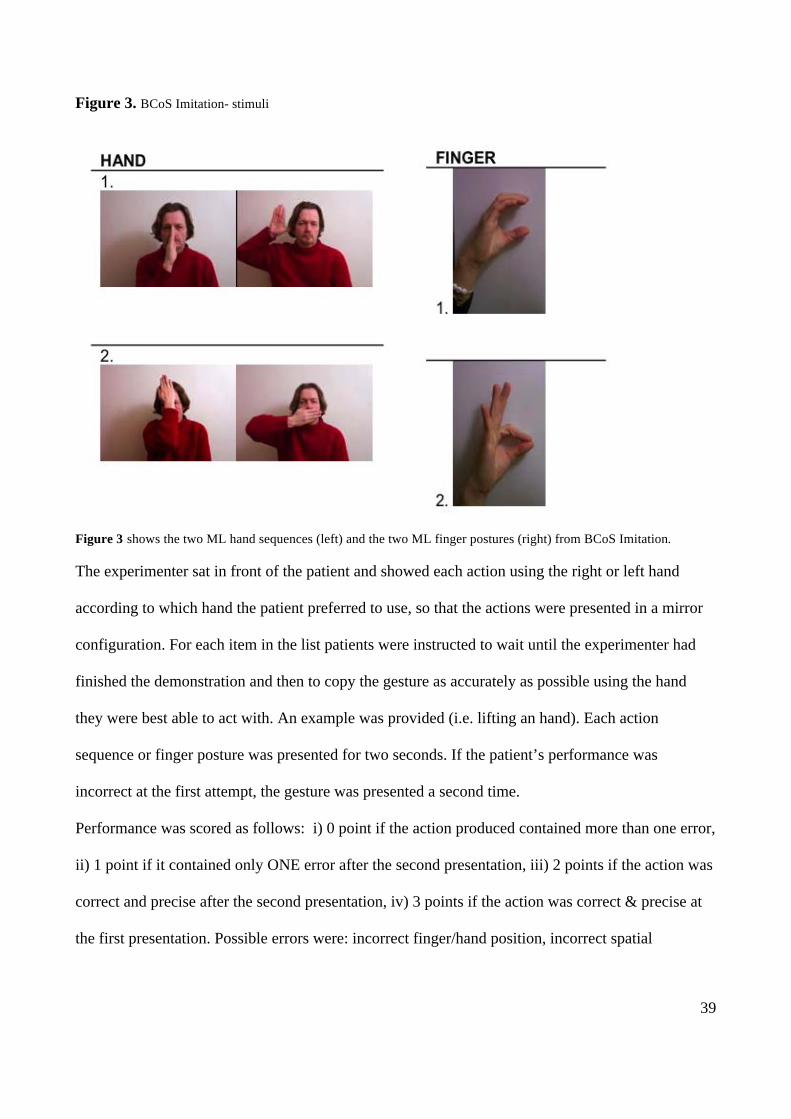
Imitation: Two ML hand sequences and two ML finger postures were presented for imitation.
Patients were informed that they were to imitate gestures that had no particular meaning. The two
hand sequences were presented before the two finger postures. The stimuli used for Imitation are
displayed in Figure 2.
39
Figure 3. BCoS Imitation- stimuli
Figure 3 shows the two ML hand sequences (left) and the two ML finger postures (right) from BCoS Imitation.
The experimenter sat in front of the patient and showed each action using the right or left hand
according to which hand the patient preferred to use, so that the actions were presented in a mirror
configuration. For each item in the list patients were instructed to wait until the experimenter had
finished the demonstration and then to copy the gesture as accurately as possible using the hand
they were best able to act with. An example was provided (i.e. lifting an hand). Each action
sequence or finger posture was presented for two seconds. If the patient’s performance was
incorrect at the first attempt, the gesture was presented a second time.
Performance was scored as follows: i) 0 point if the action produced contained more than one error,
ii) 1 point if it contained only ONE error after the second presentation, iii) 2 points if the action was
correct and precise after the second presentation, iv) 3 points if the action was correct & precise at
the first presentation. Possible errors were: incorrect finger/hand position, incorrect spatial
40
relationship between hand and head, incomplete movement sequence. The maximum possible score
was 12.
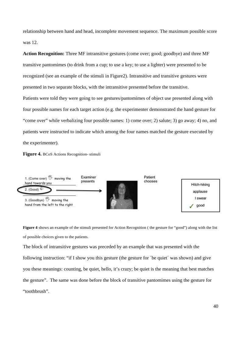
Action Recognition: Three MF intransitive gestures (come over; good; goodbye) and three MF
transitive pantomimes (to drink from a cup; to use a key; to use a lighter) were presented to be
recognized (see an example of the stimuli in Figure2). Intransitive and transitive gestures were
presented in two separate blocks, with the intransitive presented before the transitive.
Patients were told they were going to see gestures/pantomimes of object use presented along with
four possible names for each target action (e.g. the experimenter demonstrated the hand gesture for
“come over” while verbalizing four possible names: 1) come over; 2) salute; 3) go away; 4) no, and
patients were instructed to indicate which among the four names matched the gesture executed by
the experimenter).
Figure 4. BCoS Actions Recognition- stimuli
Figure 4 shows an example of the stimuli presented for Action Recognition ( the gesture for “good”) along with the list
of possible choices given to the patients.
The block of intransitive gestures was preceded by an example that was presented with the
following instruction: “if I show you this gesture (the gesture for ´be quiet´ was shown) and give
you these meanings: counting, be quiet, hello, it’s crazy; be quiet is the meaning that best matches
the gesture”. The same was done before the block of transitive pantomimes using the gesture for
“toothbrush”.

41
The experimenter demonstrated the actions using the dominant hand.
For each item a maximum time of 15 seconds was allowed for the patient to produce the answer.
Answers received a score of 1 point if correct and 0 if incorrect or omitted, so the maximum
possible score was 6.
NEUROIMAGING ASSESSMENT
Chapter 4- MRI scans
The MRI scans of patients were taken at the Birmingham University Imaging Center (BUIC) on a
3T Philips Achieva MRI system with 8 channels phased array SENSE head coil. A sagittal T1-
weighted sequence (sagittal orientation, echo time/time to repetition, TE/TR = 3.8/8.4 ms, voxel
size 1 x 1 x 1 mm3) was used to acquire the anatomical scans.
Pre processing of the T1 data
All images were pre-processed as part of the neural Birmingham University Cognitive Screen
project following the below steps. TheT1 scans were converted and reoriented with MRICro (Chris
Rorden, University of South Carolina, Columbia, SC, USA) and then preprocessed using SPM5
(Statistical Parametric Mapping; Friston, Ashburner, Kiebel, Nichols, & Penny 2007, Welcome
Department of Cognitive Neurobiology, London, UK). The scans were transformed into the
standard MNI space (Montreal Neurological Institute) using a modified unified-segmentation
procedure (Ashburner & Friston 2005; Seghier et al. 2008), designed to be optimised for patients
with brain lesions by including a fourth tissue type that depicts abnormal tissue. The procedure
output four classified tissues maps for grey matter (GM), white matter (WM), cerebrospinal fluid
(CSF) and abnormal tissue, on the basis of the intensity of the signal in each voxel using a priori
knowledge of the expected location of that tissue, with each map representing the probability that a
given voxel belonged to GM, WM, CSF or, with low probability, to an abnormal class. A brain
lesion causes a change in the signal intensity from the damaged tissue, so this tissue is mapped as a
42
region of reduced likelihood of representing either GM or WM (notice that, as we tested patients at
a chronic stage, the damaged brain regions could have been replaced by CSF if not classified as
abnormal). After segmentation, the resulting images were smoothed using a 12-mm FWHM
Gaussian filter.
Chapters 5 and 6- CT scans
Computed Tomography (CT) images were acquired for each patient as part of their clinical
assessment. The CTs were taken on average 3.4 days after the stroke, with 94% of cases within a
week from the stroke. We obtained the image in the digital DICOM format. The CTs were acquired
using the following scanners: Siemens sensation 16; GE medical system LightSpeed 16 and
LightSpeed plus. The images covered the whole brain with an in-plane resolution of 0.5 x 0.5 mm2
and a slice thickness varying between 4-5 mm. CT images depict the density of the tissue and as
such have a clear biological interpretation and provide an undistorted image of the brain tissue
density.
Pre processing of the CT data
The quality of the CT scans was assessed by eye; bad quality data due to head movement, enlarged
ventricles or other image artefacts were removed from the analysis. The CT images were pre-
processed using SPM5 (www.fil.ion.ucl.ac.uk/~spm). Images were first converted to Nifti format
then normalized to an in-house CT template (Ashburner and Friston, 2003). This initial
normalization stage was mostly based on skull shape and aimed to linearly transform the images
into MNI space to optimize the following procedures. Next we used the unified-segmentation
algorithm as implemented in SPM5 (i.e. Seghier, Ashburner and Friston, 2005). In the unified
model, the priors of the tissue class, from which intensities are drawn, are encoded by deformable
tissue probability maps. The a-priori tissue class maps indicate the probability of finding expected
signal sources: grey matter (GM), white matter (WM), cerebrospinal fluid (CSF), bone, fat and air
at each voxel. To account for the presence of an abnormal tissue due to the stroke condition we
43
adapted a similar approach to Seghier et al. (2008) and added a seventh tissue class. In creating the
extra abnormal tissue we assumed that in each grey or white matter voxel there is 10% chance of it
being an abnormal tissue class. Furthermore, for the grey and white matter tissue classification, we
assumed a single Gaussian distribution for the underlying intensities. To account for potential
inhomogeneity of the abnormal tissue two different Gaussian distributions were used to model the
intensities in this tissue class. Finally CT images as opposed to MRI do not suffer field bias due to
field strength inhomogeneity, therefore we did not correct for that in the model. The segmented
white and grey matter images were then smoothed using a 12 mm3 FHWM Gaussian kernel to
accommodate random field theory assumptions of continuity (Worsley and Friston, 1995).
VBM analyses- tools- common to chapters 4, 5 and 6
In chapters 2, 3 and 4 voxel-based morphometry was carried out analyzing the data with SPM8
(Statistical Parametric Mapping, Welcome Trust Centre for Neuroimaging, London, UK) using
parametric statistics in the framework of general linear model (Ashburner & Friston, 2000).
The anatomical localization of the lesions was done using AAL3 toolbox and confirmed using the
Duvernoy human brain atlas (Duvernoy, Cabanis, & Vannson, 1991). The brain coordinates are
presented in the standardized MNI space.
44
3. Imitating transitive and intransitive gestures: An analysis of
lesion side and associated cognitive impairments.
ABSTRACT
The ability to imitate gestures has traditionally been considered to be supported by brain structures
in the left hemisphere, even though neuropsychological studies, as well as behavioural and imaging
studies in healthy participants, suggest a role for right hemisphere and sub-cortical structures too.
The present study tested a group of patients with unilateral brain damage on the imitation of
meaningful (MF) and meaningless (ML) transitive and intransitive gestures, and on recognition of
the same gestures. The results showed that right brain damaged (RBD) patients generally
performed better than left brain damaged (LBD) patients on transitive actions, while the two groups
did not differ when imitating intransitive actions. Also, at group level, the relative ease of
intransitive actions was demonstrated when compared to transitive, and of action recognition
compared to imitation. However, at single subject level, there were dissociations with sometimes
better performance for transitive relative to intransitive action imitation, as well as good
performance in imitating MF intransitive actions that could not be recognized. The implications for
understanding action recognition and production are discussed.
INTRODUCTION
Ideomotor apraxia is syndrome that impairs the ability to voluntarily reproduce gestures in imitation
and/or on verbal command. Traditionally the left hemisphere of the human brain has been
considered dominant for controlling gesture production (see Rothi et al. 1997) in right-handed
people, depending perhaps upon the type of action that has to be implemented. So, while the
execution of transitive (tool-related) actions seems to rely more on the left-hemisphere (Bartolo et
al. 2001; Tessari et al. 2007), both right and left cortical areas have been linked to the production of
45
intransitive gestures (Bauxbaum et al. 2001, see also Tessari et al. 2007, and Rumiati et al. 2010 for
a review). Also a role for sub-cortical structures has been suggested by some studies (see Tessari et
al. 2007; Leiguarda et al. 1997, 2001; De Renzi et al. 1980), even though there is still not consensus
yet about which type of gestures are more impaired after lesions to those structures.
Several neuropsychological studies have shown that ideomotor apraxia is not a unitary syndrome –
there are symptom dissociations between patients in relation to the type of actions to be executed
and the body part required for the task (see Rumiati et al. 2009 for a review). For instance, some
studies report patients who are more impaired when imitating meaningful (MF) compared to
meaningless (ML) gestures (Goldenberg & Hagmann 1997; Peigneux et al. 2000; Tessari et al
2007), while others report the opposite result (Bartolo et al. 2001; Tessari et al. 2007). Such double
dissociations suggest that the imitation of MF and ML actions may depend on distinct neural
structures.
A dual route model of action production has been proposed to account for the double dissociation
between abilities to reproduce MF and ML gestures (Rothi et al. 1997; Rumiati and Tessari, 2002).
This model involves a semantic route, relying on Long Term Memory, which supports the
reproduction of MF (known) gestures, and a direct route, depending on Short Term Memory, which
allows ML (new) actions to be imitated (see Figure1 in the general introduction). Input into both
routes is provided by a visual analysis component, through which the visual properties of actions
are processed. Output from both routes operates through a common motor system supporting the
actual implementation of the action.
Alongside the neuropsychological observations, the argument for different mechanisms for
transitive and intransitive actions has been confirmed by several behavioural investigations in
healthy participants as well as by imaging studies (e.g. Rumiati et al. 2009; Tessari & Rumiati
2004; Peigneux at al. 2004). For instance, Peigneux et al (2004) reported that the imitation of MF
actions was associated with activation in the left angular and middle frontal gyri, as well as the right
46
supramarginal gyrus and inferior parietal lobule. Activity in the inferior temporal, angular and
parahyppocampal gyri in the left hemisphere has also been noted for MF actions (Rumiati et al.
2005). In contrast, observation of ML actions has been associated with the parieto-occipital and
occipito-temporal junctions on the right, the superior temporal gyrus on the left, and the superior
parietal cortex bilaterally (Rumiati et al. 2005).
On the other hand, arguments can also be made against the different actions being fully dissociated.
For example, neuropsychological studies have reported that apraxic patients have more difficulties
in imitating transitive than intransitive gestures (Haaland et al. 2000; Bauxbaum et al. 2005, 2007),
but the reverse dissociation has not been convincingly reported. This suggests that intransitive
actions are merely easier to perform, and indeed these actions are typically easier for normal
participants too (Carmo & Rumiati, 2009). If arguments are to be made about the functional
dissociation between transitive and intransitive actions, then evidence is needed that more clearly
shows that intransitive actions can be impaired when transitive are not.
A crucial point for the model is the suggestion that access to the semantic system in order to
recognize or understand the goal of an action is not necessary for correctly executing a gesture, as
any gesture can be processed through the direct route, where representation of the action before
being translated into motor terms is sustained by working memory. This happens for novel gestures
as well as for MF gestures if the semantic route is unavailable. On the other hand, the consequences
of impairment of the direct route are less clear. For example, if damage affects visual analysis
processes then there may be problems in imitating MF as well as ML gestures, along also with
impairments to MF gesture recognition. In contrast, if the deficits are more on the output side then
problems may affect MF action production as well as imitation, though action recognition should be
spared. The problems may also affect ML more than MF gestures, if the representations of learned
gestures (on the input or output side) are more robust to brain lesion. Thus a variety of different
dissociations may be expected. Double dissociations have been shown between imitation and
47
recognition tasks, with patients being impaired in imitation but not in recognition and vice versa
(Negri et al. 2007, see also Mahon et al. 2005 and 2008), which does support the general
architecture of the dual route model, but the neural underpinnings of these effects has still be
exactly established. For example, are production deficits more apparent after left hemisphere
damage? Are recognition deficits contingent on the presence of right as well as left hemisphere
lesions? To examine these questions in more detail, and to assess how imitation and recognition
operate for transitive and intransitive gestures, and imitation for MF and ML actions, a group study
was conducted with patients with chronic deficits following brain lesion. The aim was to look at the
pattern of deficits of patients according to the side of their lesion, for action recognition as well as
for imitation, and for MF and ML transitive and intransitive actions (here the ML actions were
derived from transitive and intransitive real actions). The deficits in action recognition and
production in the patients were also related to the cognitive profile of the patients derived from the
Birmingham Cognitive Screen (BCoS; Humphreys et al. 2012).
GENERAL METHODS
This study compares right-brain damaged (RBD) and left-brain damaged (LBD) patients in the
imitation and recognition of transitive and intransitive MF and ML gestures. For details on the
patients and control samples and on the tasks employed in this study see the method chapter (pgs.
21- 38).
As already mentioned in the method chapter, in a previous version of this chapter one bilateral brain
damaged (BBD) patient was included by mistake among the LBD (patent MMcD, see Table 1 in the
method chapter). In the present version MMcD scores were excluded from the group analyses
however, as a matter of honesty, his results will be briefly presented and discussed at the end of the
chapter and a picture reporting his lesion will be reported. MMcD scores are shown in Table 1 of
the present chapter below the main table showing the results of the unilateral patients.
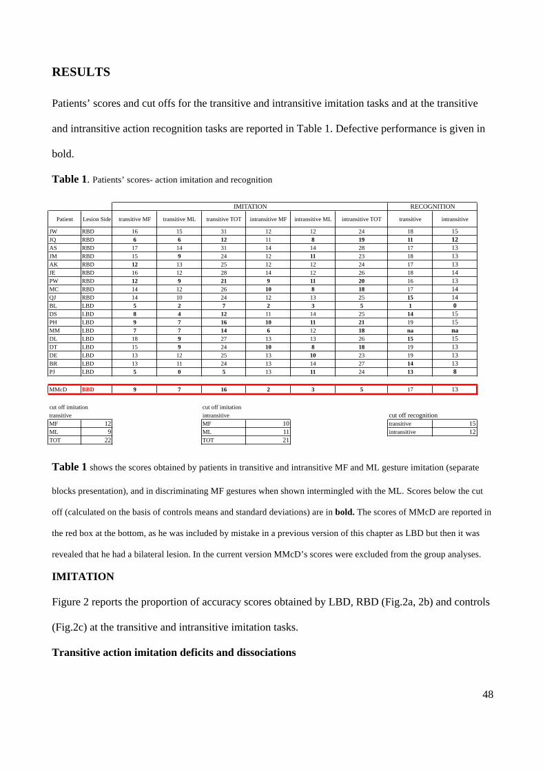
48
RESULTS
Patients’ scores and cut offs for the transitive and intransitive imitation tasks and at the transitive
and intransitive action recognition tasks are reported in Table 1. Defective performance is given in
bold.
Table 1. Patients’ scores- action imitation and recognition
Table 1 shows the scores obtained by patients in transitive and intransitive MF and ML gesture imitation (separate
blocks presentation), and in discriminating MF gestures when shown intermingled with the ML. Scores below the cut
off (calculated on the basis of controls means and standard deviations) are in bold. The scores of MMcD are reported in
the red box at the bottom, as he was included by mistake in a previous version of this chapter as LBD but then it was
revealed that he had a bilateral lesion. In the current version MMcD’s scores were excluded from the group analyses.
IMITATION
Figure 2 reports the proportion of accuracy scores obtained by LBD, RBD (Fig.2a, 2b) and controls
(Fig.2c) at the transitive and intransitive imitation tasks.
Transitive action imitation deficits and dissociations
JW RBD 16 15 31 12 12 24 18 15JQ RBD 6 6 12 11 8 19 11 12AS RBD 17 14 31 14 14 28 17 13JM RBD 15 9 24 12 11 23 18 13AK RBD 12 13 25 12 12 24 17 13JE RBD 16 12 28 14 12 26 18 14PW RBD 12 9 21 9 11 20 16 13MC RBD 14 12 26 10 8 18 17 14QJ RBD 14 10 24 12 13 25 15 14BL LBD 5 2 7 2 3 5 1 0DS LBD 8 4 12 11 14 25 14 15PH LBD 9 7 16 10 11 21 19 15MM LBD 7 7 14 6 12 18 na naDL LBD 18 9 27 13 13 26 15 15DT LBD 15 9 24 10 8 18 19 13DE LBD 13 12 25 13 10 23 19 13BR LBD 13 11 24 13 14 27 14 13PJ LBD 5 0 5 13 11 24 13 8
MMcD BBD 9 7 16 2 3 5 17 13
cut off imitation cut off imitation transitive intransitive cut off recognitionMF 12 MF 10 transitive 15ML 9 ML 11 intransitive 12TOT 22 TOT 21
transitive intransitiveLesion Side
IMITATION RECOGNITION
intransitive MLPatient transitive MF transitive ML transitive TOT intransitive MF intransitive TOT
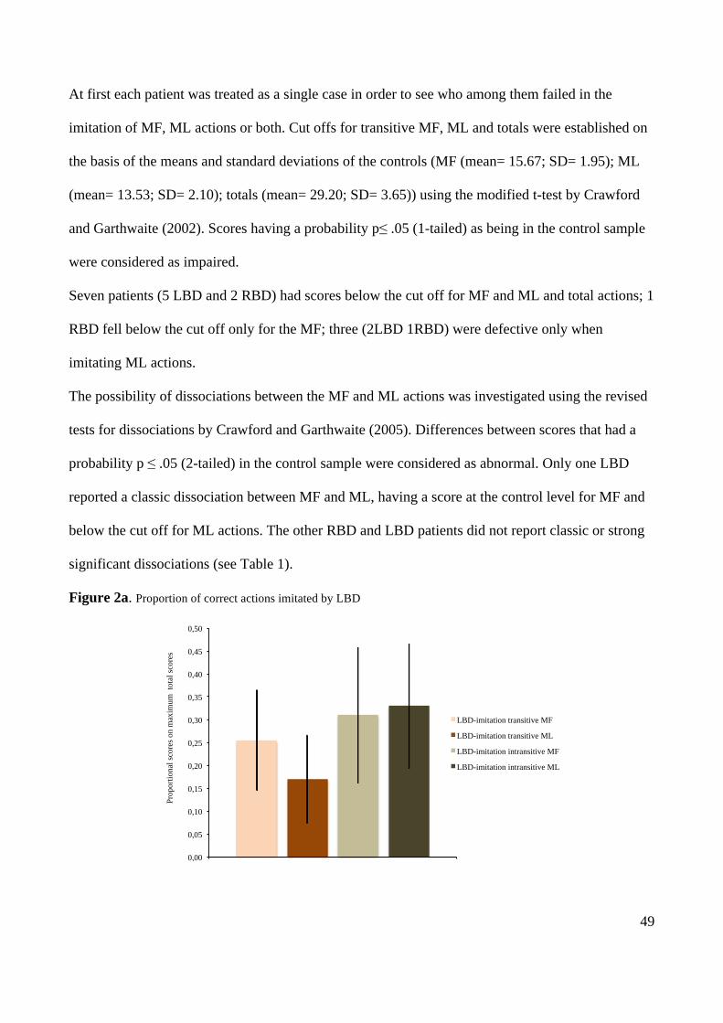
49
At first each patient was treated as a single case in order to see who among them failed in the
imitation of MF, ML actions or both. Cut offs for transitive MF, ML and totals were established on
the basis of the means and standard deviations of the controls (MF (mean= 15.67; SD= 1.95); ML
(mean= 13.53; SD= 2.10); totals (mean= 29.20; SD= 3.65)) using the modified t-test by Crawford
and Garthwaite (2002). Scores having a probability p≤ .05 (1-tailed) as being in the control sample
were considered as impaired.
Seven patients (5 LBD and 2 RBD) had scores below the cut off for MF and ML and total actions; 1
RBD fell below the cut off only for the MF; three (2LBD 1RBD) were defective only when
imitating ML actions.
The possibility of dissociations between the MF and ML actions was investigated using the revised
tests for dissociations by Crawford and Garthwaite (2005). Differences between scores that had a
probability p ≤ .05 (2-tailed) in the control sample were considered as abnormal. Only one LBD
reported a classic dissociation between MF and ML, having a score at the control level for MF and
below the cut off for ML actions. The other RBD and LBD patients did not report classic or strong
significant dissociations (see Table 1).
Figure 2a. Proportion of correct actions imitated by LBD
0,00
0,05
0,10
0,15
0,20
0,25
0,30
0,35
0,40
0,45
0,50
Prop
ortio
nal s
core
s on
max
imum
tot
al sc
ores
LBD-imitation transitive MF
LBD-imitation transitive ML
LBD-imitation intransitive MF
LBD-imitation intransitive ML
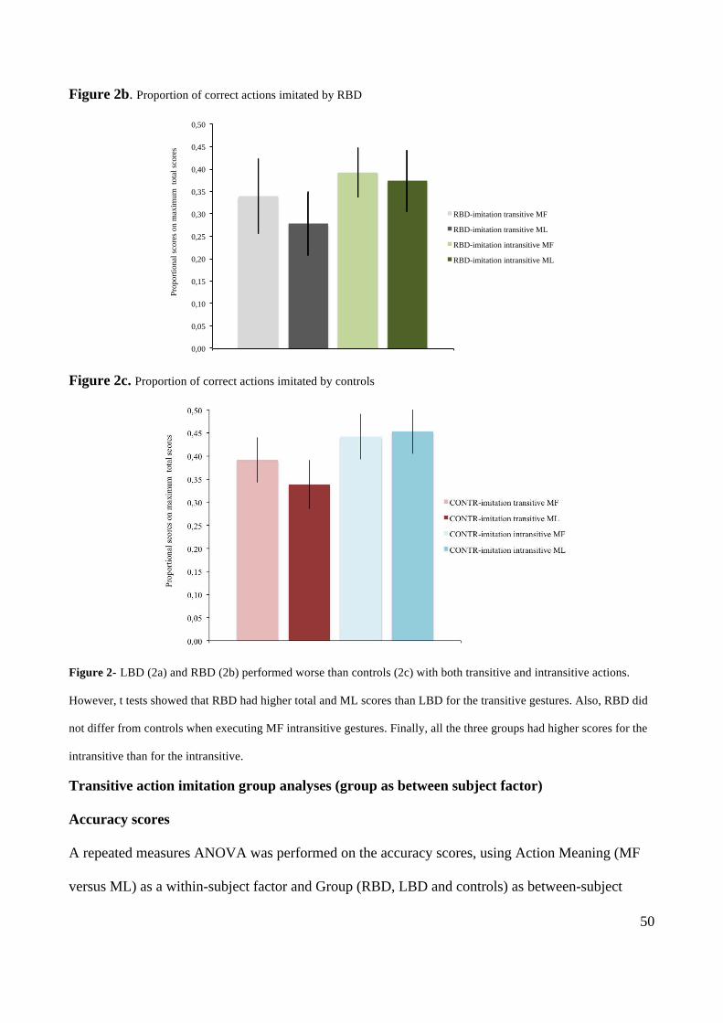
50
Figure 2b. Proportion of correct actions imitated by RBD
Figure 2c. Proportion of correct actions imitated by controls
Figure 2- LBD (2a) and RBD (2b) performed worse than controls (2c) with both transitive and intransitive actions.
However, t tests showed that RBD had higher total and ML scores than LBD for the transitive gestures. Also, RBD did
not differ from controls when executing MF intransitive gestures. Finally, all the three groups had higher scores for the
intransitive than for the intransitive.
Transitive action imitation group analyses (group as between subject factor)
Accuracy scores
A repeated measures ANOVA was performed on the accuracy scores, using Action Meaning (MF
versus ML) as a within-subject factor and Group (RBD, LBD and controls) as between-subject
0,00
0,05
0,10
0,15
0,20
0,25
0,30
0,35
0,40
0,45
0,50
Prop
ortio
nal s
core
s on
max
imum
tot
al sc
ores
RBD-imitation transitive MF
RBD-imitation transitive ML
RBD-imitation intransitive MF
RBD-imitation intransitive ML
51
factor. The analysis revealed a significant main effect of Group (F (2, 30) = 12.39; p≤ 0.0001) and
Action Meaning (F (1, 30) = 47.65; p≤ 0.0001). Generally controls had higher total accuracy scores
(mean= 29.20; SD= 3.65) than both LBD (mean= 17.1; SD= 8.23) and RBD patients (mean= 24.67;
SD= 5.79), and all the three groups, when considered together, imitated MF actions (mean= 13.50;
SD= 3.86) better than ML actions (mean= 10.91; SD= 4.01). The interaction between Action
Meaning and Group was not significant (F (2, 30) = 1.05; p= 0.36).
Independent samples t-tests showed significant differences between controls and LBD patients for
MF (t (corr. df. = 11.43) = 3.72; p = 0.003, 2-tailed), ML (t (22) = 5.69; p≤ 0.0001, 2-tailed) and
total action performance (t (corr. df. = 11.69) = 4.64; p= 0.001, 2-tailed). RBD patients differed
significantly from controls for ML (t (22) = 2.4; p = 0.026, 2-tailed) and total action scores (t (22) =
2.37; p = 0.027, 2-tailed), while the difference for the MF was close to the full significant threshold
(t (22) = 1.97; p = 0.061, 2-tailed). Also significant differences emerged between RBD and LBD for
ML (t (16) = 2.76; p= 0.013, 2-tailed) and total action scores (t (16) = 2.42; p= 0.027, 2-tailed), but
not for MF (t (16) = 1.87; p= 0.1, 2-tailed).
Paired-samples t-tests were assessed if MF and ML actions differed within RBD, LBD and control
groups. MF and ML differed significantly for all the three groups (LBD (t (8) = 4.02; p= 0.003);
RBD (t (8) = 3.36; p= 0.01) and controls (t (14) = 4.68; p≤ 0.0001)), with MF pantomimes
performed more accurately by all the three groups.
Intransitive action imitation deficits and dissociations
Each patient was treated as a single case in order to see who among them failed in imitating
intransitive actions. Cut offs for intransitive MF, ML and totals were established on the basis of the
means and standard deviations for the controls (MF (mean= 13.27; SD= 1.49); ML (mean= 13.60;
SD= 1.45); totals (mean= 26.87; SD= 2.83)) using the modified t-test by Crawford and Garthwaite
(2002). Scores having a probability p≤ .05 (1-tailed) of being found in the control sample were
considered as defective.
52
Five patients (3 LBD and 2 RBD) had scores below the cut-off for MF and ML and total action
scores; one RBD patient had normal performance with MF but felt below the cut off for the ML
stimuli and total actions; two LBD5 and one RBD patient were defective only when imitating ML
actions while having normal total action scores; one LBD patient had MF and total action scores
below the cut off.
Also, the presence of dissociations between MF and ML action scores were investigated using the
revised test for dissociations by Crawford and Garthwaite (2005). Differences between scores that
had a probability p ≤ .05 (2-tailed) relative to the control sample were considered as outside the
normal limits. Three LBD (one of them reported a strong dissociation) and two RBD (one of them
reported a strong dissociation) patients imitated MF better than ML actions, while one LBD did
better on ML then MF actions (classic dissociation). All the other patients did not report any classic
or strong dissociation (see Table 1).
Intransitive action imitation group analyses (group as between subject factor)
RBD and LBD patient’s scores were compared to those of the controls, as well as relative to each
other.
Accuracy scores 6
An ANOVA was run on accuracy scores for RBD, LBD and controls with Group (RBD, LBD,
controls) as a between-subject factor and Action Meaning (MF Vs ML) as a within-subject factor.
The analysis showed a significant main effect of Group (F (2, 30) = 7.01; p= 0.003) but no effect of
Action Meaning (F (1, 30) = 0.173; p= 0.68). Also the interaction between the two main factors was
5 One of the two LBD having a deficit only for ML intransitive actions had a score= 11 that had a probability of p= .052
to be found in a normal sample, based on our controls data distribution. This was the only patient having this score.
6 The Cohen’s k agreement coefficient was calculated on the participants’ scores provided by the two independent
raters. The coefficient was computed for Totals and separately for MF and ML. As the k was ≥ 0.80 in all the cases
considered, the scores of only one rater were used.
53
not significant (F (2, 30) = 1.18; p= 0.32). Both LBD (mean= 19.20; SD= 8.08) and RBD (mean=
23.00; DS= 3.35) patients had generally poorer performances than controls (mean= 26.87; SD=
2.83), even though RBD did slightly better than LBD. Overall MF (mean= 11.71; SD= 3.13) actions
were imitated as accurately as ML (mean= 11.88; SD= 3.01).
Independent samples t-tests revealed differences between LBD patients and controls for MF (t (corr.
df. = 10.37) = 2.74; p= 0.02, 2-tailed), ML (t (corr. df. = 10.55) = 2.76; p= 0.019, 2-tailed) and total
action scores (t (corr. df. = 10.48) = 2.89; p= 0.016, 2-tailed). Also RBD patients had significantly
lower performance than controls for MF (t (22) = 2.29; p= 0.032, 2-tailed), ML (t (22) = 3.33; p=
0.003, 2-tailed) and total actions (t (22) = 3.03; p= 0.006, 2-tailed). A comparison between RBD
and LBD did not reveal any significant differences for the MF (t (corr. df. = 1.66) = 1.65; p= 0.126,
2-tailed), ML (t (16) = 0.88; p= 0.39; 2-tailed), totals (t (16) = 1.31; p= 0.21; 2-tailed))7.
Finally, paired samples t-tests did not reveal significant differences between MF and ML within the
LBD (t (8) = -0.72; p= 0.49), RBD (t (8) = 1.05; p= 0.33) and control groups (t (14) = -1.58; p=
0.14)8.
Comparison between imitation of transitive and intransitive gestures
At first the presence of dissociations between transitive and intransitive action imitation scores was
investigated using the revised tests for dissociation of Crawford and Garthwaite (2005). Differences
between scores that had a probability of p ≤ .05 (2-tailed) as belonging to the control sample were
considered as being outside the normal range. See Table 1 for the scores at the imitation tasks.
Two LBD patients showed a classic dissociation where they generally performed better on
intransitive than on transitive gestures (respectively inside and outside the normal range). For one of
7 Independent samples t-tests and non-parametrical tests (Mann-Withney for controls and Kruskal-Wallis for LBD) did
not show any difference between participants using the dominant or non-dominant hand within controls and LBD
(p>0.1). RBD all imitated using the dominant hand.
8 The results of the t-tests were all confirmed by non-parametrical tests (all p< 0.05).
54
these LBD patients the deficit was clearer for MF stimuli for the other it was most apparent with
ML stimuli. One RBD patient had dissociations between MF transitive and ML intransitive actions,
as well as between ML transitive and ML intransitive, with scores in the normal range when
imitating both transitive MF and transitive ML actions, but he did not showed dissociation between
MF or ML transitive and MF intransitive even though his score with the MF intransitive were just
below the cut off.
Also, two LBD had a classic dissociation when MF transitive and ML intransitive actions were
compared. One performed at control level with the MF transitive pantomimes while being defective
with the ML intransitive gestures, while one had the opposite pattern. It is to be noted that this last
patients had scores within the normal range only for the ML intransitive actions, and that he was the
only patient in the sample whose aphasia was so severe that it was not possible to test him for the
action recognition task.
Finally, one RBD had a dissociation between MF transitive and MF intransitive, being impaired in
execution of MF transitive while doing at control level with the MF intransitive.
Proportional scores
As the scores for the transitive and intransitive action tasks were based on different scales,
proportional scores were calculated based on accuracy relative to the total maximum score for
transitive and intransitive gestures, summed over the MF and ML stimuli (maximum scores= 40 and
30 respectively). After being transformed in this way, the patients’ results were subjected to an
ANOVA, with Group (RBD and LBD) as a between subject factor, Type of Action (Transitive and
Intransitive) and Action Meaning (MF and ML) as within-subjects factors. The analysis showed a
significant between-subject effect of Group (F (1, 16) = 4.53; p= 0.05), significant main effects of
Action Type (F (1, 16) = 14.12; p= 0.002) and Action Meaning (F (1, 16) = 7.91; p= 0.012) and a
significant interaction between Action Type and Action Meaning (F (1, 16) = 17.73; p= 0.001). All
the other interactions were not significant (Action Type x Group (F (1, 16) = 0.39; p= 0.54); Action
55
Meaning x Group (F (1, 16) = 0.08; p= 0.79); Action Type x Action Meaning x Group (F (1, 16) =
3.4; p= 0.08)).
The interaction arose because the drop from MF to ML actions was greater for transitive than for
intransitive actions. For intransitive actions there was no difference between the MF and ML
actions and for transitive actions there was a reliable advantage for MF over ML actions (t tests
reported in above section).
The data for the controls were also transformed as for the patients and then entered into a 2x2
ANOVA with Type of Action (Transitive and Intransitive) and Action Meaning (MF and ML) as
within-subjects factors. The analysis showed significant main effects of Action Type (F (1, 14) =
30.82; p≤ 0.0001) and Action Meaning (F (1, 14) = 12.38; p= 0.003) and a significant interaction
between Type of Action and Action Meaning (F (1, 14) = 18.21; p= 0.001). Generally intransitive
gestures (mean= 0.9, SD= 0.09) were reproduced more accurately than transitive (mean= 0.73; SD=
0.09), and both MF (mean= 0.44; SD= 0.05) and ML (mean= 0.45; 0.05) intransitive gestures were
executed better than transitive MF (mean= 0.39; SD= 0.05) and ML (mean= 0.34; SD= 0.05)
(Fig2b). As for the patients, there was an effect of meaningfulness for transitive actions (MF>ML)
but not for intransitive actions (MF=ML) (t tests reported above).
RECOGNITION
Patients’ scores and cut offs for the transitive and intransitive action recognition tasks are reported
in Table 1. Defective scores are highlighted in bold. Figure 3 reports the proportion of accuracy
scores obtained by RBD, LBD and controls for transitive and intransitive actions. One LBD patient
could not be given the tasks because of expressive aphasia that made his answers for the most part
incomprehensible.
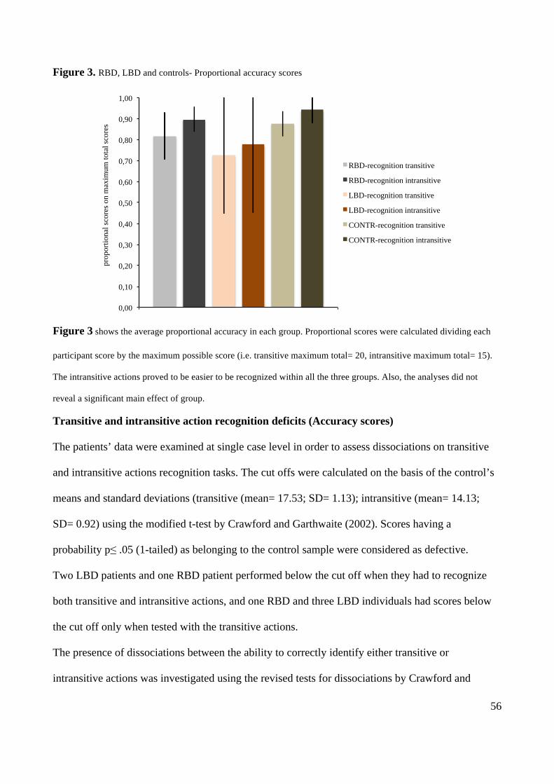
56
Figure 3. RBD, LBD and controls- Proportional accuracy scores
Figure 3 shows the average proportional accuracy in each group. Proportional scores were calculated dividing each
participant score by the maximum possible score (i.e. transitive maximum total= 20, intransitive maximum total= 15).
The intransitive actions proved to be easier to be recognized within all the three groups. Also, the analyses did not
reveal a significant main effect of group.
Transitive and intransitive action recognition deficits (Accuracy scores)
The patients’ data were examined at single case level in order to assess dissociations on transitive
and intransitive actions recognition tasks. The cut offs were calculated on the basis of the control’s
means and standard deviations (transitive (mean= 17.53; SD= 1.13); intransitive (mean= 14.13;
SD= 0.92) using the modified t-test by Crawford and Garthwaite (2002). Scores having a
probability p≤ .05 (1-tailed) as belonging to the control sample were considered as defective.
Two LBD patients and one RBD patient performed below the cut off when they had to recognize
both transitive and intransitive actions, and one RBD and three LBD individuals had scores below
the cut off only when tested with the transitive actions.
The presence of dissociations between the ability to correctly identify either transitive or
intransitive actions was investigated using the revised tests for dissociations by Crawford and
0,00
0,10
0,20
0,30
0,40
0,50
0,60
0,70
0,80
0,90
1,00 pr
opor
tiona
l sco
res o
n m
axim
um to
tal s
core
s
RBD-recognition transitive
RBD-recognition intransitive
LBD-recognition transitive
LBD-recognition intransitive
CONTR-recognition transitive
CONTR-recognition intransitive
57
Garthwaite (2005). Differences between scores that had a probability p ≤ .05 (2-tailed) in to be
found in the control sample were considered as suggesting dissociating performances. Three LBD
revealed a classic dissociation, two of them having scores below the cut off for the transitive but
performing at control level with the intransitive. One LBD showed a strong dissociation, having
scores below the cut off in both tasks but did better when he had to recognize transitive
pantomimes.
In addition to examining problems in recognition only, dissociations between imitation and
recognition of MF transitive pantomimes were tested. Two LBD patients showed a classic
dissociation between recognition and imitation of MF transitive pantomimes. One of them had
scores in the normal range in imitation and defective scores in recognition, one showed the opposite
pattern (defective imitation and normal recognition imitation). This last LBD patient also had a
classic dissociation, with normal scores in recognition but defective imitation for intransitive MF
actions. Finally, another LBD had normal scores in intransitive MF action imitation and defective
score in MF intransitive action recognition (the same patient did not have a dissociation between
imitation and recognition of MF transitive gestures, and was defective in both).
Proportional scores for gesture recognition
As the scores for the transitive and intransitive action recognition tasks were based on different
scales, a proportional score was calculated based on accuracy relative to the total maximum score
for transitive and intransitive gestures (maximum scores= 20 and 15 respectively). An ANOVA was
conducted with Group (LBD, RBD, controls) as between-subject factor and Action Type (transitive
and intransitive) as a within -subject factor. The analysis revealed a significant main effect of
Action Type (F (1, 30) = 12.37; p= 0.001), with intransitive actions (mean= 0.88; SD= 0.18)
identified correctly more frequently than transitive (mean= 0.82; SD= 0.17). There was not a main
effect of Group (F (2, 30) = 2.66; p= 0.09) or a significant interaction between the two factors (F (2,
30) = 0.18; p= 0.84).
58
Imitation and recognition tasks (correlations)
To provide a finer-grained analysis of performance in the different conditions, correlations were
conducted including all the participants but one (who could not be given the recognition task
because his production aphasia was too severe to obtain any understandable answer). Significant
correlations emerged between transitive action recognition and imitation of transitive MF actions
(Pearson Correlation (N= 32) = 0.66; p≤0.0001, 2-tailed) and of transitive ML actions (Pearson
Correlation (N= 32) = 0.64; p≤0.0001, 2-tailed). This was also the case for intransitive action
recognition and the imitation of both MF intransitive actions (N= 32) = 0.56; p≤0.0001, 2-tailed)
and ML intransitive actions (N= 32) = 0.60; p≤0.0001, 2-tailed).
When only the data for patients were analyzed, the correlations remained. For transitive gesture
recognition there were correlations with the imitation of MF transitive actions (Pearson Correlation
(N=17) = 0.62; p = 0.006, 2-tailed) and ML transitive actions (Pearson Correlation (N=17) = 0.62; p
= 0.006, 2-tailed). For intransitive action recognition there were correlations with MF intransitive
actions (Pearson Correlation (N=17) = 0.54; p= 0.02, 2-tailed) and ML intransitive actions (Pearson
Correlation (N=17) = 0.58; p= 0.01, 2-tailed).
For the control group alone, there was a reliable correlation between transitive action recognition
and transitive action imitation for MF stimuli (Pearson Correlation (N=15) = 0.67; p= 0.006, 2-
tailed) but not for ML stimuli (Pearson Correlation (N= 15) = 0.45; p= 0.1, 2-tailed). The
correlations with intransitive action recognition were not reliable (Pearson Correlation (N=15) =
0.23; p= 0.4, 2-tailed relative to MF intransitive imitation, and Pearson Correlation (N= 15) = 0.31;
p= 0.26, 2-tailed relative to ML intransitive imitation.
Birmingham Cognitive Screen (BCoS) and imitation
The relations between imitation performance and the scores on sub-tests of the BCoS assessing
personal orientation, control functions, naming and praxis were explored. A description of the
normative samples, tasks and the results of the patients (scores below the cut offs are in bold), along
59
with the cut offs, are reported in Appendix 1. The scores at the BCoS tasks of five patients were not
available, so those patients could not be included in this analysis.
Imitation of transitive MF actions correlated significantly with Picture Naming (Pearson Correlation
(N=13) = 0.67; p= 0.01, 2-tailed) and Action Recognition (Pearson Correlation (N=13) = 0.6; p=
0.03). Also, there was a reliable correlation between the imitation of ML transitive actions and
BCoS Gesture Production (Pearson Correlation (N=13)= 0.57; p= 0.04). BCoS Picture Naming also
significantly correlated with imitation of MF intransitive actions (Pearson Correlation (N=13) =
0.55; p= 0.05). Moreover, there was a significant correlation between MF and ML intransitive
action imitation and BCoS Figure Copy (Pearson Correlation (N=13) = 0.65; p= 0.02, 2-tailed and
Pearson Correlation (N=13)=0.74; p=0.004 respectively), Imitation (MF (Pearson Correlation
(N=13) = 0.73; p< 0.001, 2-tailed), Gesture Production (Pearson Correlation (N= 13)= 0.6; p= 0.03)
and Personal Information (Pearson Correlation (N= 13)= 0.56; p= 0.04). Intransitive ML did not
relate significantly with any of the BCoS tasks.
DISCUSSION
The patients overall performed worse than the controls in transitive and intransitive action imitation
but not in transitive and intransitive action recognition. This might arise because the recognition
task was easier than the production task (imitation). The recognition tasks tended to correlate with
the production tasks, suggesting that they did tap some common underlying processes. But in
addition at a single case level there were two patients whose dissociations were in an opposite
direction. Indeed one LBD patient (patient DL, see Table 1) had scores within the normal range
when imitating MF transitive actions but failed in the MF transitive action recognition task. This
patient also performed at a control level when she had to execute MF transitive and intransitive, as
well as ML intransitive gestures, but was impaired in reproducing ML transitive actions. During the
testing session patient DL failed with the ML transitive gestures because as soon as the presentation
60
of the gesture terminated she was no longer able to remember it and asked for more presentations,
overtly admitting that she did not remember them. According to the dual route model, ML gestures
require short-term memory to sustain the representation of the action and the ML transitive actions
we used were more demanding than the ML intransitive, as proved by the fact that they were
systematically performed worse than all the other actions. So it is possible that the patient had
difficulties in remembering gestures only when the task was more demanding. It is interesting that
deficits in visual short-term memory have previously been reported in this patient (Gillebert &
Humphreys, 2009). In our investigation we did not include tasks tapping short-term and long-term
memory for actions so we cannot draw more detailed conclusions on patient DL’s specific memory
skills nor about the relationship between her apparent deficit in remembering the actions in the short
term and her failure in the action recognition task. The main point here is that this patient showed
accurate performance in imitating gestures that later she could not name or give information about
(not due to speech problems, see Table 2 in Appendix 1, Picture Naming, patient DL). However it is
worth noting that this same patient was not impaired in the BCoS Gesture Recognition. This last
task is a multiple-choice task (four names of actions are provided for the patent to chose the correct
answer between them), and this could have facilitated her to access the information about the
actions presented. Another patient (patient PJ) showed a similar dissociation with scores in the
normal range for imitation of MF intransitive gestures and defective scores in MF intransitive
gesture recognition. This patient was impaired in all tasks apart from the imitation of MF
intransitive actions. Unfortunately the BCoS scores for patient PJ were not available. Nevertheless,
his performance suggests a certain degree of independence between imitation and action
recognition.
For action imitation, there were no reliable differences between the two patient groups when asked
to imitate intransitive gestures. Instead RBD patients had better total scores than LBD individuals
with the transitive gestures, even though RBD showed an actual advantage only for the transitive
61
ML but not for the MF. This suggests that a general impairment in reproducing actions can be
present after damage confined to the right hemisphere, especially for the intransitive gestures. At a
single case level more LBD patients fell below the control norms, but in almost all cases there were
instances where at least one RBD patient performed the same way. Failures in imitation may occur
for different reasons that should be analyzed more deeply at single case level. Possibly selecting
patients with very focal lesions in a specific part of the right or left hemisphere may be helpful here
in order to give a better insight into the involvement of specific structures in the two hemispheres in
praxis. However the fact that some RBD patients showed an impairment in copying visually
presented actions fits with previously reported data (Peigneux at al. 2004; Rumiati et al. 2005; see
Tessari et al. 2007, for a review). As pointed out in the introduction, RBD patients have been shown
to be defective in imitating intransitive gestures (Buxbaum et al. 2001; Tessari et al. 2007). Also
studies using positron PET reveal that imitation of MF actions are associated with activation in the
right supramarginal gyrus and inferior parietal lobule (Peigneux et al. 2004), and in the parieto-
occipital and occipito-temporal junctions on the right (Rumiati et al. 2005).
In general intransitive actions were performed and recognized better than transitive actions, even
though dissociations in the opposite directions were revealed, both for imitation and recognition.
Two LBD patients (DS and PJ) showed a general advantage for intransitive actions, being impaired
in transitive but not in the intransitive imitation task. Opposite to this, one RBD (patient MC, see
Table 1) had scores at control level for MF and ML transitive actions while being below the cut off
for both MF and ML intransitive gestures. Moreover MC performance with MF and ML transitive
gestures significantly dissociated from imitation of the ML intransitive. Together, the pattern of
dissociations in the LBDs and RBD suggests that different processes may underlie imitation of
transitive and intransitive gestures. Also the results are compatible with previous studies indicating
a main role of left-brain areas in imitation of transitive gestures (Stamenova et al. 2010; Buxbaum,
2007; Buxbaum et al. 2001; Liepmann, 1900-1905) and of right-brain areas in imitation of the
62
intransitive (Stamenova et al. 2010; Peigneux at al. 2004; Rumiati et al. 2005; see Tessari et al.
2007, for a review).
Beside the dissociations in overall performance with the transitive and intransitive actions, more
specific dissociations also emerged. For instance, one LBD cases (patients DT) performed better
with MF transitive than ML intransitive actions. Further investigations are required to understand
why this dissociation emerged; however it indicates that the difference between transitive and
intransitive actions may be not relate only to the difficulty of the tasks. There is strong evidence that
intransitive actions used in imitation tasks to date were easier than the transitive tasks, but this does
not exclude the possibility that stimuli more balanced in term of difficulty may help to point out
differential mechanisms involved in the imitation of transitive and intransitive gestures.
Intransitive actions were also recognized more frequently than transitive by all but one LDB patient
(patient PJ) who, despite being impaired with in both transitive and intransitive recognition tasks,
did significantly better with the transitive. It is possible that transitive actions were more robust in
this patient because: i) transitive gestures were supported by an additional mechanism, independent
of the mechanisms supporting intransitive action, and this additional mechanism was relatively
spared in this patient; ii) his lesion involved areas relating to social communication (notice that MF
intransitive actions were all communicative gestures, e.g. waving “hallo”), or iii) for some personal
reason (may be he was particularly keen for tool use, e.g. DIY). Nonetheless this demonstrates that
transitive and intransitive actions are not necessarily impaired equally and it is not always the case
that intransitive are more robust than transitive.
The issue of tasks tapping different processes or being simply more or less difficult to perform
emerges also for the distinction between MF and ML actions. At group level, transitive MF
pantomimes were generally executed better than ML gestures while there was no difference
between MF and ML intransitive. At the level of single patients, only one LBD (DL, already
mentioned above) had a classic dissociation between the two types of transitive gestures, with better
63
performance for the MF, while for the intransitive two RBD (JQ, classic dissociation, and MC,
strong dissociation) and three LBD (PJ and DE, classic dissociation; DT, strong dissociation) were
significantly advantaged when imitated the MF than the ML. Opposite to this however, one LBD
patient (patient MM) had a significant dissociation where there was normal performance with the
ML gestures along with a deficitwith MF intransitive gestures. MM also had poor performance
with both MF and ML transitive actions. This was the only dissociation found in favour of ML
gestures, when gestures of the same type (i.e. MF intransitive versus ML intransitive or MF
transitive versus ML transitive) were directly compared at single subject level. So, in the present
study the advantage for the ML gestures was less frequent than for the MF. Similarly, previous
studies reported less frequent dissociations in favour of ML gestures (Bartolo et al. 2001; Tessari et
al. 2007) compared with MF gestures (see Tessari et al. 2007 and Rumiati et al. 2009 for a review).
Nonetheless, this evidence does not fit well with an explanation of MF and ML gestures differing
only in terms of difficulty, instead suggesting that they may involve at least some different
processes. It is true that for the transitive gestures MM did not show a significant dissociation,
performing the ML only slightly better than the MF gestures (ML 20% correct and MF 18%
correct). This result may be due to several reasons. For instance MM performed below the cut-off
on many of the BCoS test administered (see Table 2 in Appendix 1), with his results suggesting a
general loss of cognitive resources. So, it may be possible that transitive gestures were generally too
demanding for MM regardless from them having a meaning or not, ad so regardless from the
cognitive processes MM could activate to accomplish the task.
It should be pointed out that, although there were dissociations between the tasks, the tasks
generally correlated across the participants (even for control subjects). This favours the hypothesis
that there is a strong relation between the different actions and between recognition and imitation,
and also a single rather than a dual route account (where the same knowledge representations are
called upon irrespective of whether the action is familiar or unfamiliar). It also remains possible
64
though that the correlations emerged from the contribution of some co-varying factor such as the
processing resources the patient could bring to the task. Correlations with the tasks from the BCoS
across the whole group of patients showed that both MF transitive and intransitive action imitation
correlated with BCoS picture naming while ML transitive and ML intransitive actions did not. The
correlations could come about due to the anatomical overlap of both processes. For example,
damage to the inferior frontal cortex in the left hemisphere will disrupt picture naming and possible
also action coding, because this brain region supports both functions rather than because there are
common processes involved. Other two interesting correlations emerged between: i) imitation of
MF transitive gestures and BCoS Action Recognition and ii) imitation of MF and ML intransitive
actions and BCoS Figure Copy. Those correlations suggest that the transitive actions may rely more
on a semantic representation of the target action while the intransitive may rely more on the correct
processing of spatial configuration of the limb.
Patient MMcD
As stated previously, a patient (MMcD) was included by mistake in the LBD group in a previous
version of this chapter, reflecting an earlier error in data entry into a patient database. Because the
imitation behaviour of MMcD was unusual for a patient with unilateral left brain damage, we
checked his MRI scan9 using the binary lesion map (see chapter 4 for details of on how we
proceeded) and noticed that MMcD actually had damage involving both hemispheres (see Figure
4).
9 The scans of the other patients were checked as well with the same procedure. For those patients who did not have
MRI scans available we could only visually check their CT scans. Even though visual checking, being observer
dependent, may be prone to errors, here we did not look for fine-grained lesions of specific brain structures. Also, those
patients did not show behaviors that clearly contrasted with the observed lesions.
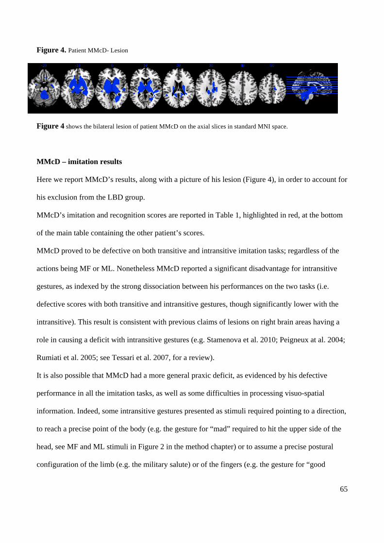
65
Figure 4. Patient MMcD- Lesion
Figure 4 shows the bilateral lesion of patient MMcD on the axial slices in standard MNI space.
MMcD – imitation results
Here we report MMcD’s results, along with a picture of his lesion (Figure 4), in order to account for
his exclusion from the LBD group.
MMcD’s imitation and recognition scores are reported in Table 1, highlighted in red, at the bottom
of the main table containing the other patient’s scores.
MMcD proved to be defective on both transitive and intransitive imitation tasks; regardless of the
actions being MF or ML. Nonetheless MMcD reported a significant disadvantage for intransitive
gestures, as indexed by the strong dissociation between his performances on the two tasks (i.e.
defective scores with both transitive and intransitive gestures, though significantly lower with the
intransitive). This result is consistent with previous claims of lesions on right brain areas having a
role in causing a deficit with intransitive gestures (e.g. Stamenova et al. 2010; Peigneux at al. 2004;
Rumiati et al. 2005; see Tessari et al. 2007, for a review).
It is also possible that MMcD had a more general praxic deficit, as evidenced by his defective
performance in all the imitation tasks, as well as some difficulties in processing visuo-spatial
information. Indeed, some intransitive gestures presented as stimuli required pointing to a direction,
to reach a precise point of the body (e.g. the gesture for “mad” required to hit the upper side of the
head, see MF and ML stimuli in Figure 2 in the method chapter) or to assume a precise postural
configuration of the limb (e.g. the military salute) or of the fingers (e.g. the gesture for “good
66
luck”/”finger crossed”). In the present work we did not formally analyzed the patients’ errors, as in
general they were too sparse to make meaningful analyses. However, at single patient level, we did
notice that MMcD errors were compatible with a difficulty in processing of the visuo-spatial
properties of the actions. For instance, for the gestures for “mad”, which require hitting the temple
with the tip of the index finger, MMcD started the gesture correctly but then he failed do hit his
head, moving instead toward a different point in the space. Also, in the distal gestures (hand
gestures), he constantly failed to move his fingers in order to reach the correct configuration. For
instance he was not able to move his middle finger above the index finger when he had to reproduce
the gesture for “good luck”.
Despite the possible visuo-spatial problems MMcD was not categorized as having a deficit in the
BCoS Figure Copy. However, it is to be noticed that MMcD performance was at the limit for being
considered as normal, as he reported a score of 37 that is the cut-off below which a performance
must be considered as pathological. More testing, possibly administering more tasks assessing
visuo-spatial abilities are needed to draw conclusions on MMcD’s deficits.
67
4. Neural correlates of transitive and intransitive action imitation:
An investigation using voxel-based morphometry (VBM)
ABSTRACT
The ability to reproduce visually presented actions has been studied through neuropsychological
observations of patients with ideomotor apraxia while attempts to understand the neural basis of
action reproduction have been made through lesion-symptom mapping with discrete patients groups
(classified according to the presence of different forms of apraxia). Lesion-symptom mapping
studies have reported that areas in the parietal and frontal lobes in the left hemisphere are involved
respectively in the imitation of meaningless actions and meaningful pantomimes of object use.
However there is still not consent on this issue, and there is also a question if the results can be
generalized to all meaningful and meaningless actions, regardless of whether actions are related or
not to tool use, and whether they have a strong grasping component. In the present study I report a
first attempt to use voxel –based morphometry (VBM) to evaluate the relations between brain
lesions and the symptoms of apraxia. The imitation of meaningful (familiar) and meaningless
(unfamiliar) tool-related (transitive) and intransitive (not-tool related) actions were examined in a
consecutive series of brain-lesioned patients. The analysis showed that the left parietal cortex was
involved in the imitation of novel gestures, regardless of whether they were transitive or
intransitive. Also, familiar transitive actions and both types of unfamiliar actions were associated
with damage to the right frontal cortex and right supplementary motor areas. Although surprising,
the result in the right hemisphere may be explained by possible attention deficits of some of our
patients. There were no reliable correlations between the lesions and performance on MF
intransitive actions.
68
INTRODUCTION
Imitation is an innate tendency in humans (Meltzoff & Moore, 1977, 1983, 1989) as well as in
newborn chimpanzees (Myova 1996; Bard & Russel 1999; Myowa-Yamakoshi et al. 2005) and
macaques (Ferrari et al. 2006). The ability to reproduce gestures may provide one of the
foundations of social communication and it may have an important role in learning effective tool
use (e.g. how to use a hammer etc.).
Following a brain lesion people can suffer from a deficit in their ability to imitate visually presented
actions, characterized neuropsychologically in the syndrome of ideomotor apraxia. In right handed
people this syndrome has classically been described in relations to lesions of the left posterior
parietal cortex (Liepmann, 1900-1905)(see Rothi et al. 1991 for a review), though other different
studies point to the role of right brain areas as well as sub-cortical structures (see Tessari et al.
2007; Leiguarda et al. 1997, 2001; De Renzi et al. 1980), especially when finger configurations
(Goldenberg & Strauss 2002; Della Sala et al. 2006) or movement sequences (Canavan et al. 1989)
have to be copied.
Prior neuropsychological evidence indicates that ideomotor apraxia is not a unitary phenomenon,
with symptom dissociations reported among patients (see Rumiati et al. 2009 for a review). For
instance, neuropsychological observations suggest a double dissociation between the production of
meaningful and meaningless gestures, with some studies reporting patients who are more impaired
when imitating meaningful (MF) compared to meaningless (ML) gestures (Goldenberg & Hagmann
1997; Peigneux et al. 2000; Tessari et al 2007), and others showing the opposite pattern (Bartolo et
al. 2001; Tessari et al. 2007). Double dissociations have also been observed between finger and
hand gestures, with patients able to imitate hand postures but not finger configurations and other
with the opposite pattern (Goldenbeg & Karnat 2006).
69
A cognitive neuropsychological model of praxis was first proposed by Rothi et al. (1991) and then
modified by other authors (e.g. Goldenberg & Hagmann 1997; Cubelli et al. 2000; Bauxbaum et al.
2001;Tessari et al. 2007). This model postulates (i) a semantic route to action, relying on long-term
memory representations which allow the reproduction of MF (known) gestures, and (ii) a direct
route, depending on short-term memory (i.e. the innervatory pattern in the original model from
Rothi et al. (1991)), which supports the reproduction of ML (new) actions (see Figure 1 in the
general introduction). The starting point of both routes is a visual analysis component, through
which the visual properties of actions are processed. Also both semantic and direct processes end at
the level of the motor system involved in the actual implementation of the action.
Neuropsychological observations on unilateral brain damaged patients (e.g. Goldenberg &
Hagmann 1997; Peigneux et al. 2000; Cubelli et al. 2000; Bauxbaum et al. 2001; Tessari et al.
2007), as well as behavioural and imaging studies on healthy participants (e.g. Rumiati et al. 2009;
Tessari & Rumiati 2004; Peigneux at al. 2004), support the idea of two separate neural systems for
imitating MF and ML gestures but are inconsistent with respect to the neuro-anatomical structures
involved in each (particularly concerning their hemispheric localisation). Lesions involving the
parietal cortex, especially the angular gyrus, are reported to cause a deficit in the imitation of
meaningless (ML) as compared to meaningful (MF) actions in left-brain damaged patients (LBD)
(Mehler 1987; Goldenberg & Hagmann 1997; Peigneux et al. 2000; Tessari et al. 2007). In contrast
Tessari et al. (2007) report two right brain damaged patients (RBD)(with lesions including the
caudal portion of the pallidum, the putamen and the posterior limb of the internal capsule) who
were more impaired in imitating ML vs. MF gestures.
In studies using positron emission tomography (PET), imitation of MF actions has been observed to
be associated with activation in the left angular and middle frontal gyri, as well as the right
supramarginal gyrus and inferior parietal lobule (Peigneux et al. 2004), and also in the inferior
temporal, angular and parahyppocampal gyri in the left hemisphere (Rumiati et al. 2005). The
70
parieto-occipital and occipito-temporal junctions on the right, the superior temporal gyrus on the
left, and the superior parietal cortex bilaterally have also been shown to have increased activation
linked to imitation of ML actions (Rumiati et al. 2005).
Goldenberg et al. (2007) used lesion subtraction analysis to determine the locations specifically
associated with defective pantomime of tool use in patients with left-brain damage and aphasia.
Their results showed left inferior frontal cortex to be associated with a deficit in pantomime, even
though the area of lesion overlap further extended into the underlying white matter - it is possible
that damage of white matter projections contributed to the deficit (Liepmann 1905; Geschwind
1976; Catani & Fytche 2005).
One caveat with the neuropsychological studies described above is that they rely on categorical
subdivisions of patients based on a given cut off as well as on observer-dependent lesion
demarcations. The use of a cut off, especially in tasks where each answer is scored either 1 or 0,
introduces the risk that patients having scores differing only for one point are treated differently
(e.g. scores that equal the cut off will be considered as defective, while scores higher than the cut
off for one point will be treated as borderline or not defective at all). Also manual demarcations of
lesions sites can lead to under- or over- estimation of differences in tissues according to the
strictness of the observer’s criteria. The present study aims to investigate the neural substrates of
action imitation using a complementary VBM analysis based on segmented grey matter (GM) and
white matter (WM) tissue. VBM uses the general linear model to statistically assess the relations
between brain tissue integrity and behavioural performance. Here, MF and ML imitation scores
were used as predictors of change in signal intensity coming from each voxel across the whole brain
in a group of consecutively sampled patients, not pre-selected on the basis of having apraxia, but
using continuous scores on tests of gesture imitation in order to characterise their imitation abilities.
This approach also allows us to control in the model covariates of no interests such as age, gender,
neglect, which may otherwise contaminate the results.
71
The dual route model can be applied to both symbolic and non-symbolic intransitive (not-tool
related) gestures (Rumiati et al. 2009), however the neural substrates of MF and ML intransitive
actions, relative to MF and matched ML transitive actions, have not been tested together in the same
study. Here we used the scores patients obtained in imitation of MF transitive and intransitive
pantomimes, along with matched ML transitive and intransitive actions, as predictors of voxel
signal intensity.
METHODS
The sample of 21 patients, the imitation tasks, the MRI scans acquisition and preprocessing as well
as the tools used for the analyses and the anatomical localization of the regions highlighted by the
analyses are described in the method section of this thesis (pgs. 21-38).
A final section of the chapter will present the lesion overlap maps in the GM and WM showing the
number of patients having a lesion in the regions highlighted by the analyses. Patients’ lesions were
identified using the automated lesion identification procedure described by Seghier at al. (2008).
The 21 patients’ MRI structural images were segmented, normalized and smoothed (8mm). Then
outlier voxels were detected by comparing the segmented GM and WM of the patient to the ones of
150 controls using fuzzy clustering (Seghier et al. 2008) and the outlier voxels in each tissue class
were assigned to the lesion. The image showing the number of patients whose lesion overlap in the
GM and WM were created using the “image calculator” function of SPM5 (Statistical Parametric
Mapping; Friston, Ashburner, Kiebel, Nichols, & Penny, 2007, Welcome Department of Cognitive
Neurology, London, UK) using the expression: ‘i1 + i2 + i3 + i4 + … + i21”.
The images for GM and WM were displayed with MRICRON (Chris Rorden, University of South
Carolina, Columbia, SC, USA) and saved as bitmap files.
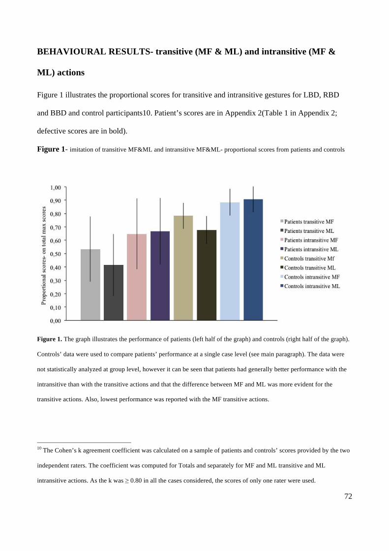
72
BEHAVIOURAL RESULTS- transitive (MF & ML) and intransitive (MF &
ML) actions
Figure 1 illustrates the proportional scores for transitive and intransitive gestures for LBD, RBD
and BBD and control participants10. Patient’s scores are in Appendix 2(Table 1 in Appendix 2;
defective scores are in bold).
Figure 1- imitation of transitive MF&ML and intransitive MF&ML- proportional scores from patients and controls
Figure 1. The graph illustrates the performance of patients (left half of the graph) and controls (right half of the graph).
Controls’ data were used to compare patients’ performance at a single case level (see main paragraph). The data were
not statistically analyzed at group level, however it can be seen that patients had generally better performance with the
intransitive than with the transitive actions and that the difference between MF and ML was more evident for the
transitive actions. Also, lowest performance was reported with the MF transitive actions.
10 The Cohen’s k agreement coefficient was calculated on a sample of patients and controls’ scores provided by the two
independent raters. The coefficient was computed for Totals and separately for MF and ML transitive and ML
intransitive actions. As the k was ≥ 0.80 in all the cases considered, the scores of only one rater were used.
73
To provide an initial descriptive analysis of the behavioural data, cut offs were established on the
basis of the control’s means and standard deviations for the transitive (MF (mean= 15.78; SD=
1.93;ML (mean= 13.89; SD=2.30); totals (mean= 29.67; SD=3.50)) and intransitive (MF (mean=
13.44; SD= 1.42) ML (mean= 13.72; SD= 1.36); totals= 27.17; SD= 2.66) actions using the
modified t-test by Crawford and Garthwaite (2002). For the transitive action imitation task eleven
patients (6 LBD, 4 RBD and 1 BBD) performed below the cut off for MF (≤ 12); ML (≤ 9) and total
action performance (≤ 23); one LBD had normal performance with MF but fell below the cut off for
the ML and total action scores; one RBD was defective only when imitating MF actions and one
RBD was below the cut off only for the ML actions. At the intransitive action imitation task four
LBD, three RBD and one BBD performed below the cut off for MF (≤10) and ML (≤ 11) and total
actions (≤22); one LBD and one RBD were below the cut-off for MF and totals; one RBD was
defective for ML and total action scores and one RBD and one BBD were defective only for the ML
actions.
VOXEL-BASED MORPHOMETRY
The MRI scans of 21 patients were used to investigate the relationship between GM and WM
integrity and scores for the transitive MF and ML and intransitive MF and ML imitation tasks on a
voxel-by-voxel basis.
ANALYSES
The patients’ raw scores at imitation of transitive MF and ML and intransitive MF and ML gestures
were used as the covariates of interest. Two models were estimated including either GM or WM
segmented images along with the four covariates of interest: i.e. i) transitive MF, ii) transitive ML,
iii) intransitive MF, iv) intransitive ML. Also, eight further models were estimated, each including
only one covariate of interest at a time and either GM or WM: 1) transitive MF and GM; 2)
74
transitive ML and GM; 3) intransitive MF and GM; 4) intransitive ML and GM; 5) transitive MF
and WM; 6) transitive ML and WM; 7) intransitive MF and WM; 8) intransitive ML and WM.
Age, handedness11, education and gender were used covariates of no interest in all the models.
T- contrasts were run on each model using a mixed peak and cluster threshold with at least 50
voxels showing a Z > 2.6 (i.e. p ≤ 0.005, uncorrected). Here we decided to use a p- value a bit less
restrictive than a p≤ 0.001 (uncorrected) because of the size of the patients sample, as well as to
have the same threshold as the in the next two chapter where CT scans, instead of MRI scans, were
used for the VBM analyses (see chapter 5 and 6 for more details).
VBM RESULTS
GREY MATTER
Table 1 (below) shows the MNI coordinates and the statistical values of the results obtained from
the contrasts run on all the models including GM scans.
GM- transitive MF&ML and intransitive MF&ML
The model including all the four covariate of interest along with GM scans was analyzed and
revealed a significant cluster (p ≤ 0.05 FWE-corr, cluster level) within the parietal cortex, including
the precuneus and extended to the cuneus in the left hemisphere.
A plot of the results on the main peak on this cluster showed that the imitation of intransitive
gestures had the stronger relation with the intensity of the signal (Figure 2).
11 Patients were divided in right-handed and left-handed according to the scores they received at the Edinburgh
Handedness Inventory (Oldfield, 1971).
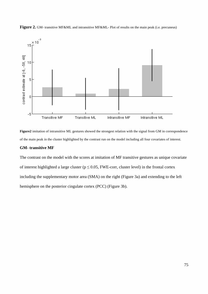
75
Figure 2. GM- transitive MF&ML and intransitive MF&ML- Plot of results on the main peak (i.e. precuneus)
Figure2 imitation of intransitive ML gestures showed the strongest relation with the signal from GM in correspondence
of the main peak in the cluster highlighted by the contrast run on the model including all four covariates of interest.
GM- transitive MF
The contrast on the model with the scores at imitation of MF transitive gestures as unique covariate
of interest highlighted a large cluster (p ≤ 0.05, FWE-corr, cluster level) in the frontal cortex
including the supplementary motor area (SMA) on the right (Figure 3a) and extending to the left
hemisphere on the posterior cingulate cortex (PCC) (Figure 3b).
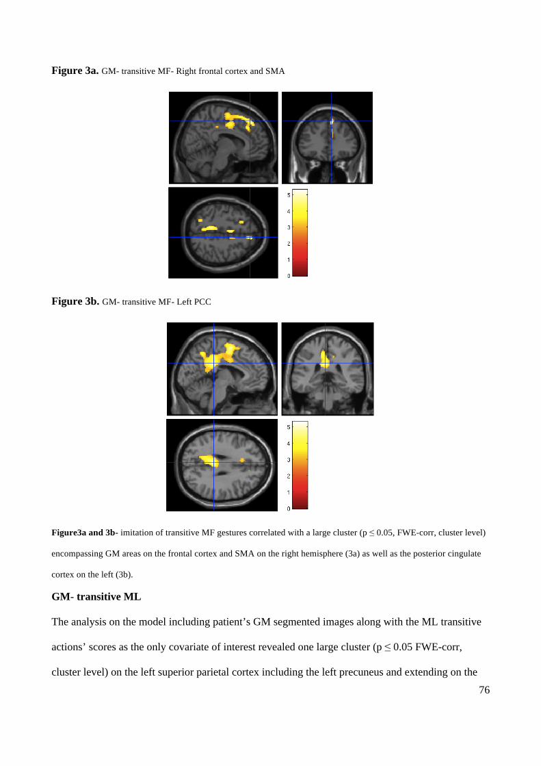
76
Figure 3a. GM- transitive MF- Right frontal cortex and SMA
Figure 3b. GM- transitive MF- Left PCC
Figure3a and 3b- imitation of transitive MF gestures correlated with a large cluster (p ≤ 0.05, FWE-corr, cluster level)
encompassing GM areas on the frontal cortex and SMA on the right hemisphere (3a) as well as the posterior cingulate
cortex on the left (3b).
GM- transitive ML
The analysis on the model including patient’s GM segmented images along with the ML transitive
actions’ scores as the only covariate of interest revealed one large cluster (p ≤ 0.05 FWE-corr,
cluster level) on the left superior parietal cortex including the left precuneus and extending on the
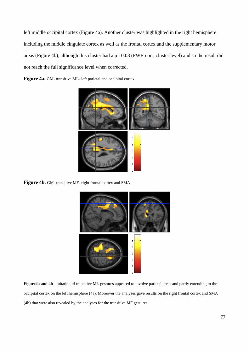
77
left middle occipital cortex (Figure 4a). Another cluster was highlighted in the right hemisphere
including the middle cingulate cortex as well as the frontal cortex and the supplementary motor
areas (Figure 4b), although this cluster had a p= 0.08 (FWE-corr, cluster level) and so the result did
not reach the full significance level when corrected.
Figure 4a. GM- transitive ML- left parietal and occipital cortex
Figure 4b. GM- transitive MF- right frontal cortex and SMA
Figure4a and 4b- imitation of transitive ML gestures appeared to involve parietal areas and partly extending to the
occipital cortex on the left hemisphere (4a). Moreover the analyses gave results on the right frontal cortex and SMA
(4b) that were also revealed by the analyses for the transitive MF gestures.
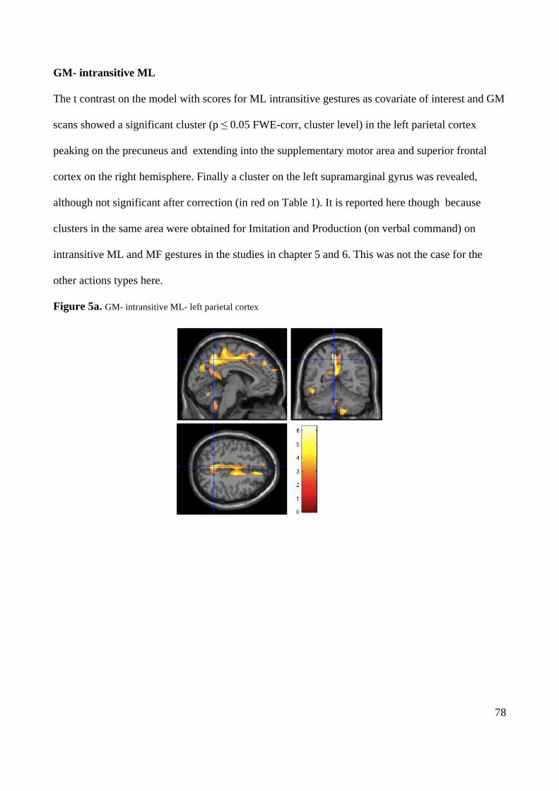
78
GM- intransitive ML
The t contrast on the model with scores for ML intransitive gestures as covariate of interest and GM
scans showed a significant cluster (p ≤ 0.05 FWE-corr, cluster level) in the left parietal cortex
peaking on the precuneus and extending into the supplementary motor area and superior frontal
cortex on the right hemisphere. Finally a cluster on the left supramarginal gyrus was revealed,
although not significant after correction (in red on Table 1). It is reported here though because
clusters in the same area were obtained for Imitation and Production (on verbal command) on
intransitive ML and MF gestures in the studies in chapter 5 and 6. This was not the case for the
other actions types here.
Figure 5a. GM- intransitive ML- left parietal cortex
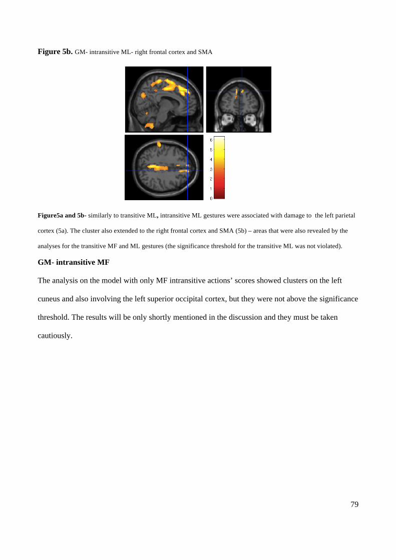
79
Figure 5b. GM- intransitive ML- right frontal cortex and SMA
Figure5a and 5b- similarly to transitive ML, intransitive ML gestures were associated with damage to the left parietal
cortex (5a). The cluster also extended to the right frontal cortex and SMA (5b) – areas that were also revealed by the
analyses for the transitive MF and ML gestures (the significance threshold for the transitive ML was not violated).
GM- intransitive MF
The analysis on the model with only MF intransitive actions’ scores showed clusters on the left
cuneus and also involving the left superior occipital cortex, but they were not above the significance
threshold. The results will be only shortly mentioned in the discussion and they must be taken
cautiously.
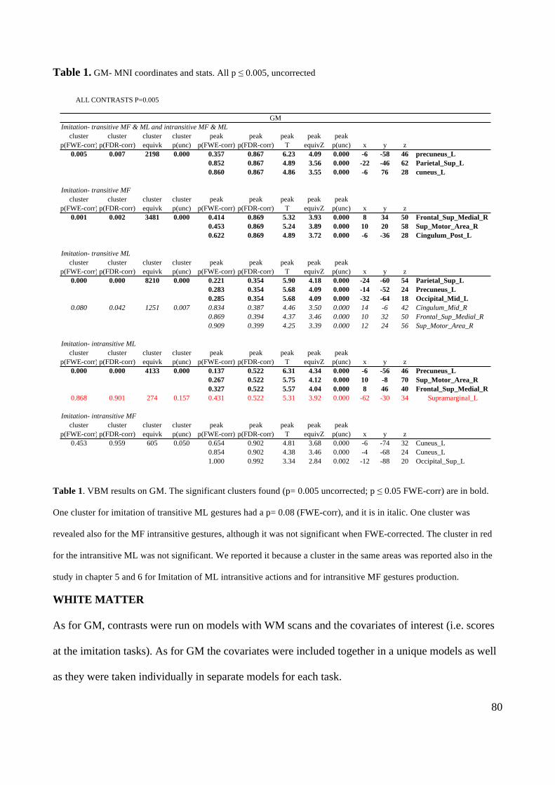
80
Table 1. GM- MNI coordinates and stats. All p ≤ 0.005, uncorrected
Table 1. VBM results on GM. The significant clusters found (p= 0.005 uncorrected; p ≤ 0.05 FWE-corr) are in bold.
One cluster for imitation of transitive ML gestures had a p= 0.08 (FWE-corr), and it is in italic. One cluster was
revealed also for the MF intransitive gestures, although it was not significant when FWE-corrected. The cluster in red
for the intransitive ML was not significant. We reported it because a cluster in the same areas was reported also in the
study in chapter 5 and 6 for Imitation of ML intransitive actions and for intransitive MF gestures production.
WHITE MATTER
As for GM, contrasts were run on models with WM scans and the covariates of interest (i.e. scores
at the imitation tasks). As for GM the covariates were included together in a unique models as well
as they were taken individually in separate models for each task.
ALL CONTRASTS P=0.005
Imitation- transitive MF & ML and intransitive MF & MLcluster cluster cluster cluster peak peak peak peak peak
p(FWE-corr) p(FDR-corr) equivk p(unc) p(FWE-corr) p(FDR-corr) T equivZ p(unc) x y z0.005 0.007 2198 0.000 0.357 0.867 6.23 4.09 0.000 -6 -58 46 precuneus_L
0.852 0.867 4.89 3.56 0.000 -22 -46 62 Parietal_Sup_L0.860 0.867 4.86 3.55 0.000 -6 76 28 cuneus_L
Imitation- transitive MF cluster cluster cluster cluster peak peak peak peak peak
p(FWE-corr) p(FDR-corr) equivk p(unc) p(FWE-corr) p(FDR-corr) T equivZ p(unc) x y z0.001 0.002 3481 0.000 0.414 0.869 5.32 3.93 0.000 8 34 50 Frontal_Sup_Medial_R
0.453 0.869 5.24 3.89 0.000 10 20 58 Sup_Motor_Area_R0.622 0.869 4.89 3.72 0.000 -6 -36 28 Cingulum_Post_L
Imitation- transitive MLcluster cluster cluster cluster peak peak peak peak peak
p(FWE-corr) p(FDR-corr) equivk p(unc) p(FWE-corr) p(FDR-corr) T equivZ p(unc) x y z0.000 0.000 8210 0.000 0.221 0.354 5.90 4.18 0.000 -24 -60 54 Parietal_Sup_L
0.283 0.354 5.68 4.09 0.000 -14 -52 24 Precuneus_L0.285 0.354 5.68 4.09 0.000 -32 -64 18 Occipital_Mid_L
0.080 0.042 1251 0.007 0.834 0.387 4.46 3.50 0.000 14 -6 42 Cingulum_Mid_R0.869 0.394 4.37 3.46 0.000 10 32 50 Frontal_Sup_Medial_R0.909 0.399 4.25 3.39 0.000 12 24 56 Sup_Motor_Area_R
Imitation- intransitive MLcluster cluster cluster cluster peak peak peak peak peak
p(FWE-corr) p(FDR-corr) equivk p(unc) p(FWE-corr) p(FDR-corr) T equivZ p(unc) x y z0.000 0.000 4133 0.000 0.137 0.522 6.31 4.34 0.000 -6 -56 46 Precuneus_L
0.267 0.522 5.75 4.12 0.000 10 -8 70 Sup_Motor_Area_R0.327 0.522 5.57 4.04 0.000 8 46 40 Frontal_Sup_Medial_R
0.868 0.901 274 0.157 0.431 0.522 5.31 3.92 0.000 -62 -30 34 Supramarginal_L
Imitation- intransitive MF cluster cluster cluster cluster peak peak peak peak peak
p(FWE-corr) p(FDR-corr) equivk p(unc) p(FWE-corr) p(FDR-corr) T equivZ p(unc) x y z0.453 0.959 605 0.050 0.654 0.902 4.81 3.68 0.000 -6 -74 32 Cuneus_L
0.854 0.902 4.38 3.46 0.000 -4 -68 24 Cuneus_L1.000 0.992 3.34 2.84 0.002 -12 -88 20 Occipital_Sup_L
GM
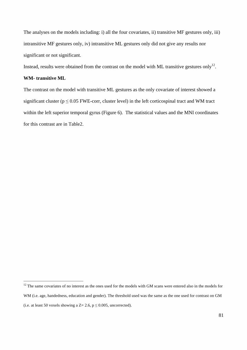
81
The analyses on the models including: i) all the four covariates, ii) transitive MF gestures only, iii)
intransitive MF gestures only, iv) intransitive ML gestures only did not give any results nor
significant or not significant.
Instead, results were obtained from the contrast on the model with ML transitive gestures only12.
WM- transitive ML
The contrast on the model with transitive ML gestures as the only covariate of interest showed a
significant cluster (p ≤ 0.05 FWE-corr, cluster level) in the left corticospinal tract and WM tract
within the left superior temporal gyrus (Figure 6). The statistical values and the MNI coordinates
for this contrast are in Table2.
12 The same covariates of no interest as the ones used for the models with GM scans were entered also in the models for
WM (i.e. age, handedness, education and gender). The threshold used was the same as the one used for contrast on GM
(i.e. at least 50 voxels showing a Z= 2.6, p ≤ 0.005, uncorrected).
82
Figure 6. WM- transitive ML- left corticospinal tract.
Figure 6- only the contrast on the model with WM and scores in imitation of ML transitive gestures gave results and
highlighted a significant cluster into the corticospinal tract on the left hemisphere.
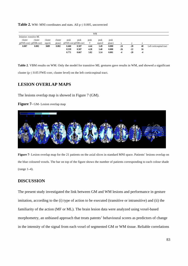
83
Table 2. WM- MNI coordinates and stats. All p ≤ 0.005, uncorrected
Table 2. VBM results on WM. Only the model for transitive ML gestures gave results in WM, and showed a significant
cluster (p ≤ 0.05 FWE-corr, cluster level) on the left corticospinal tract.
LESION OVERLAP MAPS
The lesions overlap map is showed in Figure 7 (GM).
Figure 7- GM- Lesion overlap map
Figure 7- Lesion overlap map for the 21 patients on the axial slices in standard MNI space. Patients’ lesions overlap on
the blue coloured voxels. The bar on top of the figure shows the number of patients corresponding to each colour shade
(range 1–4).
DISCUSSION
The present study investigated the link between GM and WM lesions and performance in gesture
imitation, according to the (i) type of action to be executed (transitive or intransitive) and (ii) the
familiarity of the action (MF or ML). The brain lesion data were analyzed using voxel-based
morphometry, an unbiased approach that treats patents’ behavioural scores as predictors of change
in the intensity of the signal from each voxel of segmented GM or WM tissue. Reliable correlations
WM
Page 1
Imitation- transitive MLcluster cluster cluster cluster peak peak peak peak peak
p(FWE-corr) p(FDR-corr) equivk p(unc) p(FWE-corr)p(FDR-corr) T equivZ p(unc) x y z0.007 0.002 3689 0.002 0.460 0.587 4.44 3.49 0.000 -24 -28 40 Left corticospinal tract
0.539 0.587 4.28 3.40 0.000 -26 -22 160.771 0.667 3.82 3.14 0.001 -4 -20 -4
WM
84
were found between impairments in gesturing and neural changes on GM for the contrasts run on
the model including all the four imitation tasks, as well as on three of the models with individual
tasks as unique covariate of interest (i.e. imitation of i) MF transitive, ii) ML transitive, iii) ML
intransitive gestures).
A cluster in the left superior parietal cortex involving the precuneus, and extending also to the
cuneus in the occipital cortex, appeared to relate on average to all the four tasks, even though the
plot of the results showed a stronger relation between signal change in this cluster and the imitation
of ML intransitive gestures. However the contrasts run on models with single covariate of interest
showed that both ML transitive and ML intransitive actions related to the reduced signal from the
left parietal cortex. Lesions involving the left parietal cortex were already indicated as causing the
symptoms of ideomotor apraxia (see Rothy et al. 1991 for a review), and observations on unilateral
brain damaged patients showed that LDB patients with lesions on superior parietal structures had
poorer performance in the imitation of ML as compared to MF gestures (Mehler 1987; Goldenberg
& Hagmann 1997; Peigneux et al. 2000; Tessari et al. 2007). Also brain imaging studies indicate
that the left superior parietal cortex shows increased activation when ML actions were imitated
(Rumiati et al. 2005). Moreover, the cluster we found in the left parietal cortex for both types of ML
gestures involved the left precuneus, a structure that was reported to be involved in shifting
attention between different locations in space (see Cavanna et al. 2006 for a review). Those data
suggest that the parietal areas highlighted by our analyses may play a role in processing visuo-
spatial information that can be necessary for imitating ML unfamiliar actions. Moreover, the
extension of the cluster in the occipital cortex for the ML transitive gestures suggests that imitation
of those actions may rely also on basic visual processes.
No cluster in parietal and occipital cortex was found for the transitive MF gestures. So, it may be
possible that imitation of MF gestures is based on information available from other components in
the system (e.g. representations in long term memory), which are activated instead of visuo-spatial
85
attention and basic visual processes. However, in the present study we could not find areas involved
for MF gestures only, so it is not possible to draw strong conclusions about the neural substrates
and the processes that are unique to this type of action. Also, the cluster in the occipital cortex was
revealed for MF intransitive gestures that also can rely on information stored in memory. However,
this last cluster was not significant when corrected and it is possible that it was mainly due to the
presence of few patients who also had a deficit specific to recognition of communicative intransitive
actions, and hence processed these gestures as ML.
Imitation of MF transitive gestures appeared to correlate only with the integrity of the frontal cortex
and SMA in the right hemisphere. Also, the same areas were revealed by the contrasts run on the
two models including either ML intransitive or ML transitive action imitation scores, although the
cluster was not fully significant for the ML transitive. Frontal areas and SMA have previously been
found to be involved in praxis. For instance Peigneux et al. (2004) included SMA together with the
superior parietal lobule in a set of brain areas serving innervatory motor patterns common to both
MF actions, implemented through the lexical route, and ML actions, processed through the direct
route. Also, SMA has been reported to be crucial for implementing organized and smooth action
sequences (Goldenberg 1985) and Grèzes et al. (1998) reported selective activation of the SMA
when participants saw actions and had the intention to imitate them as compared to mere
observation. Moreover, in the study by Gèzes et al. (1999) SMA was activated for MF gestures only
and not for ML movements. Those previous studies indicate that the SMA may have a role in
imitation, particularly for MF gestures, however our results differ from the previous investigations
in that the clusters found by our analyses were on the right and not on the left hemisphere. For our
data we propose that the cluster on the right frontal cortex and SMA was less related to imitation
and more to attention and control processes that are necessary to correctly perform the task. It may
be possible that, when an actor displays an action, there is automatic activation of a set of
representations that are the most similar to the action displayed, in order to retrieve the target action
86
meaning (if MF) or to categorize it as ML. If an action has to be imitated the viewer must modulate
the representation of the MF action, in order to perform it as similarly as possible to the model,
avoiding a “prototypicalization” error (i.e. a gesture that is the prototypical version of the target
gesture), or, in the case of a MF gesture, inhibit any representation and switch to a direct imitation
process, avoiding the production of a “lexicalization” error (i.e. execution of a MF gesture
somehow similar to the ML target). So, it may be that our patients who had a lesion to right frontal
areas and SMA were not able to deal with the conflict between their mental representation and the
actual action displayed. Here the patients’ errors were quite sparse (i.e. patients did not report
consistently a specific type of errors) and were not further analyzed. Nonetheless, it is still possible
that their problem in dealing with the conflict generally distracted them from the task leading them
to produce wrong actions.
Results from the WM analysis indicated a large cluster on the corticospinal tract running within the
left hemisphere. At the best of our knowledge little is still known about the role of the different WM
tracts in cognitive functions, however this finding is coherent with the role of this WM tract in
connecting the cortical motor areas to the spinal cord and so allowing the impulses to pass from the
brain to the peripheral motor system. The role of WM in praxis, as well as in other cognitive
functions, still needs more investigation, possibly using Diffusion Tensor Imaging on larger groups
of patients and controls.
Due to time limits the present study included a relatively small sample of patients (three more
patients were tested, however it was not possible to have their MRI scans in time to meet the
deadline for thesis submission and so they had to be excluded from this study). The reduced patient
sample size led to the decision to use a p≤ 0.005 instead of a p≤ 0.001, as a stricter threshold could
have caused a loss of information from our data. This decision was made also in order to have the
same p value used in the next two chapters where CT scans of acute patients, instead of MRI scans
of chronic patients, were used for VBM.
87
Finally, the lesion overlap map showed that up to 4 patients had overlapping lesions. Nonetheless,
here continuous behavioural scores and continuous values associated to grey or white matter were
analyzed. This made possible to account also for those areas of minor volume loss, due for instance
to dendritic loss on areas connected to damaged regions, and that could not be identified by
methods that assign binary values to damaged or intact tissues.
88
5. Recognizing and executing movements: An investigation using
Voxel-Based Morphometry (VBM)
ABSTRACT
The skills to execute and understand actions are crucial for everyday life and likely depend on both
overlapping and distinct processes (Rothi et al. 1991; Rumiati et al. 2002). Action execution and
recognition was investigated in the present study by carrying out a large-scale lesion-symptom
analysis of deficits in action recognition, generation to command and imitation in stroke patients.
Voxel-based morphometric analysis revealed distinct regions of neural change associated with each
key symptom, along with some overlapping regions in parietal and temporal cortex, and the
underlying white mater, for gesture recognition production on command and imitation. Also, as the
three praxis tasks used here are part of a new battery of test aimed at acute stroke patients, a further
aim of the present study is to look at the areas that are likely to be involved in those three tasks, as
well as to have a baseline for a large number of stroke patients in the acute phase that will be re-
tested in a follow up when in the chronic phase. The implications are discussed for understanding
the neural control of action recognition and production in acute stroke patients.
INTRODUCTION
In the past decade several studies have advocated the existence of a common neural system
involved in representing actions, both when actions are merely observed and when they are
executed. The key initial evidence came from physiological studies in monkeys. These studies
demonstrated the existence of a class of visuo-motor neurons (the so-called mirror neurons, MNs),
that responded when the animals saw actions as well as when they had to produce them (see
Rizzolati et al. 2001 for a review). At first MNs were found in area F5 in the monkeys’ ventral
premotor cortex (Gallese et al. 1996; Rizzolati et al. 1996) but they have also been reported in a

89
variety of regions (e.g., in a portion of the superior temporal sulcus (the STSa) that responds also to
biological motion and the inferior parietal lobule (area PF) that has been shown to respond strongly
to the observation of action). Together these regions comprise the MN system which has been
argued to play a critical role in a range of processes, from action understanding through to intention
encoding a (Iacoboni et al. 2005; see also Rizzolati et al. 2001, 2010 for a review).
Supportive evidence for a MN system in humans has been come from various sources.
Electrophysiological evidence (e.g., EEG) has shown activity in motor regions when gestures are
observed (Gastaut et al. 1954; Cohen-Seat et al. 1954; see also Cochin et al. 1998, 1999; Altschuler
et al. 1997, 2000; Salmelin et al. 1994; Hari et al. 1997; Salenius et al. 1997; Hari et al. 1998). This
is been supported by evidence from brain imaging investigations showing that ventral
premotor/inferior frontal areas (including Broca’s region), the STS and the inferior parietal lobule
are active when hand movements are observed (see Rizzolati et al. 2001 for review). Furthermore
imaging studies using both action observation and execution tasks have shown that the left inferior
frontal cortex and the right anterior parietal region are active both when a gesture (lifting a finger)
has to be executed following the observation of the same action and when the gesture has just to be
observed. The same area does not respond when the finger movement has to be enacted after the
observation of a control stimulus (a cross), or when participants just attend to a cross (Iacoboni et
al. 1999; see also Rizzolati at al. 2001 for review). The same parieto-frontal circuit also activates in
other fMRI studies when participants view a human or robot hand grasping an object (Gazzola et al.
2007) and even when aplasic individuals (born without arms and hands) watch hand actions and
moved their feet (Gazzola et al. 2007) (see Rizzolati et al. 2010 for a review).
It has been argued that the MN system has an important role in action understanding (Gallese et al.
1996; Rizzolati et al. 2001). In the present work we will consider action recognition and “action
understanding” as synonymous, and an action recognition task is examined which required the
perception and comprehension of motor gestures. The assertion that MNs are involved in action
90
understanding is based on the finding that MNs in monkeys respond both on execution and
presentation of actions toward objects (e.g. lift an object/piece of food and put it in a container, or
take a piece of food and put it in the mouth). However, these same neurons have not been found to
fire to pantomimed action (without the object present, and so having not a goal) but do fire again to
actions toward hidden objects, if the monkey could see the object before it was hidden by a screen.
This evidence has led to the claim that MNs are not simply visuo-motor association neurons,
linking a visually presented object to its appropriate action, but rather MNs code the meaning of
actions. Other interpretations (e.g., Hickok 2009) suggest that MNs are cross-modal associative
neurons, reflecting the presence of an object, and an action, irrespective of whether the object is
present or represented in working memory (as when monkeys attend to actions to hidden objects).
Another argument put forward in favour of the MN system being related to recognizing the
meaning of an action comes from a study by Gallese et al. (1996). These authors simultaneously
recorded activity in region F5 in the monkey and in the hand area of primary motor cortex (M1), as
well as recording EMG activity from several mouth and limb muscles. The results showed that there
was activity in MNs but not in cells in primary motor cortex (Gallese et al. 1996). This suggests that
no covert movement were involved, and hence that MNs in F5 are not ‘merely’ motor -related.
However in humans there us evidence for an increase in activity in distal muscle motor-evoked
potentials (MEPs) evoked by trans-cranial magnetic stimulation during action observation (Fadiga
et al. 1995). Inconsistent results have also come from two recent studies using neural adaptation and
fMRI. fMRI adaptation is the decrease in the blood-oxygen-level dependent (BOLD) response from
a brain area when stimuli or responses are repeatedly presented. Adaptation across two separate
stimuli is considered to be evidence for the same processes and neural correlates being recruited. It
follows that, if action execution and recognition involve the same processes, then the neural
response to action execution should ‘adapt’ after action observation and vice versa. Dinstein et al.
(2007) failed to find this and concluded that action recognition and execution do not involve
91
common neural populations. In contrast, Chong et al. (2008) reported that there was adaptation in
the right inferior parietal lobe across action execution and perception (Chong et al. 2008). .
This review suggests that (i) there is neurophysiological evidence from the monkey suggesting that
action recognition and production share neural and functional processes - though the exact account
of the role of these processes remains in debate (Hickok 2009, see above), but (ii) there is
inconclusive neuroimaging evidence on the issue while (iii) it is clear that distinct component
processes must also contribute to each task. This latter argument is indicated perhaps most clearly
by evidence of dissociations between the tasks in neuropsychological patients. For example,
dissociations have been documented between patients who show spared recognition of actions and
gestures but who are impaired at producing the actions themselves (not concomitant on a motor
impairment, in cases of ideomotor apraxia; see Chainay & Humphreys, 2002). Also it cannot be
argued that the dissociation exists only because recognition tasks are easier than action production
or imitation as the reverse pattern of deficit has also been documented (in the syndrome
“pantomime agnosia”, Bell 1994; Rothi et al. 1986; Cubelli et al. 2000; see also Mahon et al. 2005
for a review). In one study Negri et al. (2007) obtained case-level dissociations within an unselected
group of 37 unilateral brain damaged patients who undertook object use, object recognition,
pantomime imitation and pantomime recognition tasks. Although at group level there were
significant correlations between each pair of tasks, the authors also pointed out subsets of patients
demonstrating dissociations between each test pairing (Negri et al. 2007).
Patients with lesions in the left parietal lobe are reported to have particularly poor performance at
gesturing to command compared to imitation with recognition spared, while those with lesions in
the left occipital and temporal cortex present with the opposite pattern, being almost normal when
producing actions on verbal command but impaired at action recognition (Merians et al. 1997).
Also, even when gestures production is considered alone, there are dissociations in the production
of different types of action for example, patients who can imitate familiar but not unfamiliar actions
92
(see Tessari et al. 2007 for a review). These dissociations point to there being separable components
of the action recognition and production systems.
The ‘dual route’ cognitive neuropsychological model of praxis (e.g. Rothi et al. 1991; Goldenberg
& Hagmann 1997; Cubelli et al. 2000; Buxbaum et al. 2001;Rumiati and Tessari 2002)
distinguishes between a semantic route, relying on Long Term Memory (LTM), which supports the
reproduction of MF (known) gestures, and a direct route, dependent on a Short Term Memory
(STM) system (i.e. the innervatory pattern in the original model from Rothi et al. (1991)), which
supports the reproduction of ML (new) actions (see Figure 1 in the general introduction). The
starting point of both routes is a visual analysis component, through which the visual properties of
actions are processed. Both the semantic and direct processes end at the level of the motor system
involved in the generation of the motor pattern for action. This framework explains impaired action
recognition in terms of poor access to, or impaired representation of, LTM for action. Poor LTM for
action should also result in problems in producing actions to names, a problem that could also
reflect impairment in output from the LTM system. Deficits in imitation should stem from damage
to the indirect route.
Although the neuropsychological literature indicates functional dissociations between patients, there
is much less consensus about the underlying lesion pathologies. Classically, ideomotor apraxia
(reflecting poor action production specifically) has been associated with lesions involving the left
inferior parietal lobe (Liepmann, 1900-1905, Heliman et al. 1982). However, the subsequent
literature has pointed to the involvement of a variety of other cortical structures (inferior frontal,
posterior middle temporal, and also sub-cortical structures) that can be linked to different apraxic
deficits. For example, it has been argued that the left inferior cortex is particularly implicated in
pantomime (acting without an object) whereas deficits in actual use of objects is linked to inferior
parietal damage (Weiss & Fink, 2010; see also Goldenberg et al. 2007). Gesture recognition too has
been linked to inferior frontal regions (Pazzaglia et al. 2008) while recognition of particular aspects
93
of transitive (object-oriented) actions have been linked not to frontal regions but to inferior parietal
(recognition of spatial aspects of action) and posterior middle temporal regions (recognition of
semantic aspects of action). In patients with sub-cortical lesions imitation may be spared for
meaningless actions, but the production of familiar transitive actions may be disrupted (Hanna-
Pladdy et al. 2001). Taken together these neuropsychological results suggest that access to long-
term semantic memories for object-related actions may involve middle temporal regions. Deficits in
access to (and possible planning for production) spatial aspects of action link to inferior parietal
damage. The implementation of learned transitive actions may operate through sub-cortical
routines, while left inferior frontal regions are involved in aspects of action production, with lesions
to this region producing their most pronounced effects when patients pantomime action (when they
lack convergent input from holding the actual objects; see Chainay & Humphreys, 2002, about the
use of convergent sensory input into motor output and selection processes).
The present study used a voxel-based morphometric (VBM) analysis to assess effects of brain
lesion on action recognition and production in a large, unbiased sample of stroke patients. Three
tasks were assessed:
(1) Gesture Production (the generation of meaningful, learned actions to names),
(2) Imitation (the replication by the patient of meaningless gestures made by the examiner), and
(3) Action Recognition (the identification by the patient of meaningful, learned actions by the
examiner).
The Gesture Production task was chose in order to require access to LTM prior to production
(stressing output section of the semantic route to production). The Imitation task was used in order
to assess the indirect route (which must be used for meaningless actions, according to the model in
Figure 1). The Action Recognition task was chosen to assess input from observed action into the
LTM system.
94
Regions where damage leads to problems in both Action Recognition and Imitation tasks are likely
to support these input processes. Action Recognition and Gesture Production differ in the stimulus
input (hand movements vs. name) and in their output demands (output only required for the
production task), but they both call on long-term memories for actions (with familiar actions being
tested in each case). Regions of damage linked to both problems can be linked to the common
LTM-based processes. Imitation and Gesture Production both required motor output but differ in
input processing (arm movements vs. names) and in their demands on LTM (not required for
imitation but required for production). For this it can be argued that areas that lead to problems on
both tasks are those involved in access to motor output.
Alongside the analyses of the regions that on average relate to all the three tasks, we also assessed
deficits linked to single tasks, to evaluate lesion sites linked to ‘classical’ task impairments in
apraxia.
The VBM analysis was performed on segmented grey matter (GM), to focus on the grey-level
correlates of impaired action recognition and execution. Differently from VBM, other approaches to
analyzing lesion-symptoms associations use techniques that could be biased, as they require
subdivisions of patients based on a given cut off or according to observer-dependent lesion
demarcations. The use of a cut off, especially in tasks where each answer is scored categorically,
raises the possibility that patients having scores differing only for one point are treated differently
(e.g. scores that equal the cut off will be considered as defective, while scores higher than but still
close to the cut off will be treated not defective). Also making a priori demarcations of lesions
sites can lead either to critical areas being ignored or overestimated according to the strictness of the
used criteria. VBM uses the covariates of interest, in this case the three BCoS tests’ raw scores, as
predictors of changes in the signal intensity coming from each voxel across the whole brain in the
patient group and it does not require to make subdivisions based on performances or lesions.
95
METHODS
The patients’ sample, the tasks, the neuroimaging assessment and the preprocessing of the CT scans
are described in the methods section of this thesis (see pgs. 21-38).
VOXEL-BASED MORPHOMETRY
The segmented GM and WM maps of the 233 patients were used to look at the relation between the
intensity of the signal from GM or WM across the whole brain and abilities in three domain of
praxis: i.e. producing MF actions on verbal command (Gesture Production); imitating ML hand
sequences and ML finger postures (Imitation); recognizing MF pantomimes and MF
communicative gestures (Action Recognition).
ANALYSES
Analyses were carried out separately on GM and WM segmented images. At first two models were
estimated including all the three covariates of interest, corresponding to the raw scores at the three
tasks: i) Gesture Production; ii) Imitation; iii) Action Recognition. Analyses were run on the models
in order to look at the average contribution of the three covariates in predicting changes in the
signal intensity in either GM or WM.
Further models were estimated for each covariate of interest taken individually (i.e. i) Action
Recognition only; ii) Gesture Production only; iii) Imitation only), both for GM and WM. Age,
gender, years of education and hand (used for imitation/gestures production, dominant or non
dominant) were introduced as covariates of non-interest13 in all the models analysed.
After models estimation the t contrasts were run using a mixed peak and cluster threshold with at
least 50 voxels showing a Z ≥ 2.6 (i.e. P ≤ 0.005, uncorrected). The same threshold was used for all
the contrasts reported below. The p-value threshold was set at p≤ 0.005, instead of a p≤ 0.00, for
two reasons: 1) here we used CT scans that are a bit more noisy; 2) the tasks could have proven
13 Handedness was not included as only right-handed patients were included in the sample (see also above).
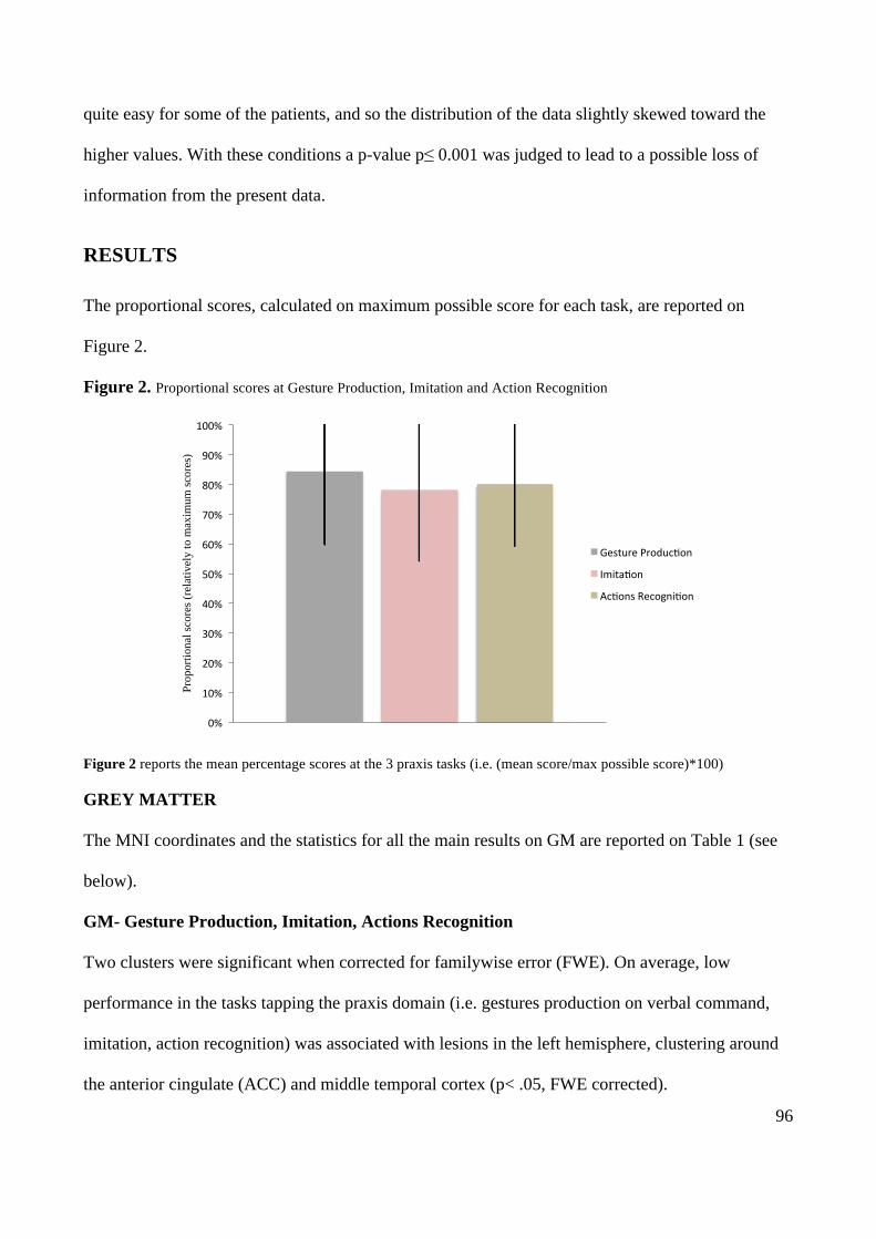
96
quite easy for some of the patients, and so the distribution of the data slightly skewed toward the
higher values. With these conditions a p-value p≤ 0.001 was judged to lead to a possible loss of
information from the present data.
RESULTS
The proportional scores, calculated on maximum possible score for each task, are reported on
Figure 2.
Figure 2. Proportional scores at Gesture Production, Imitation and Action Recognition
Figure 2 reports the mean percentage scores at the 3 praxis tasks (i.e. (mean score/max possible score)*100)
GREY MATTER
The MNI coordinates and the statistics for all the main results on GM are reported on Table 1 (see
below).
GM- Gesture Production, Imitation, Actions Recognition
Two clusters were significant when corrected for familywise error (FWE). On average, low
performance in the tasks tapping the praxis domain (i.e. gestures production on verbal command,
imitation, action recognition) was associated with lesions in the left hemisphere, clustering around
the anterior cingulate (ACC) and middle temporal cortex (p< .05, FWE corrected).
0%#
10%#
20%#
30%#
40%#
50%#
60%#
70%#
80%#
90%#
100%#
Prop
ortio
nal s
core
s (re
lativ
ely
to m
axim
um sc
ores
)
Gesture#Produc7on#
Imita7on#
Ac7ons#Recogni7on#
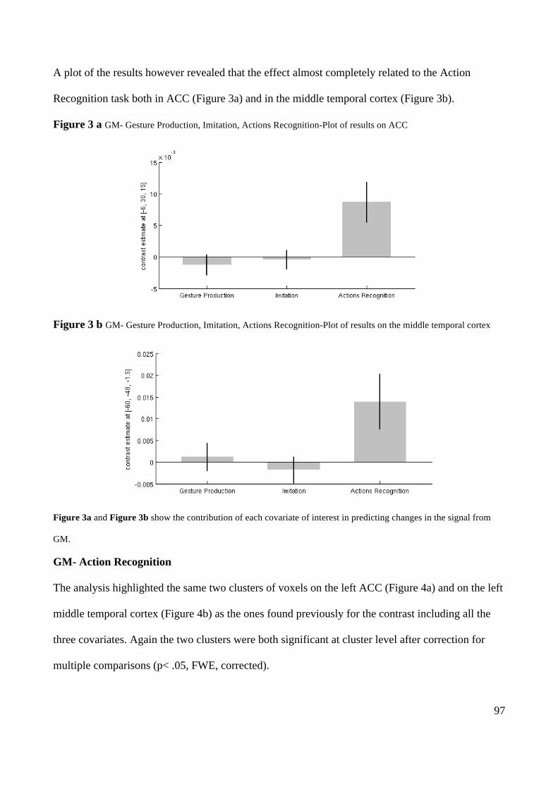
97
A plot of the results however revealed that the effect almost completely related to the Action
Recognition task both in ACC (Figure 3a) and in the middle temporal cortex (Figure 3b).
Figure 3 a GM- Gesture Production, Imitation, Actions Recognition-Plot of results on ACC
Figure 3 b GM- Gesture Production, Imitation, Actions Recognition-Plot of results on the middle temporal cortex
Figure 3a and Figure 3b show the contribution of each covariate of interest in predicting changes in the signal from
GM.
GM- Action Recognition
The analysis highlighted the same two clusters of voxels on the left ACC (Figure 4a) and on the left
middle temporal cortex (Figure 4b) as the ones found previously for the contrast including all the
three covariates. Again the two clusters were both significant at cluster level after correction for
multiple comparisons (p< .05, FWE, corrected).
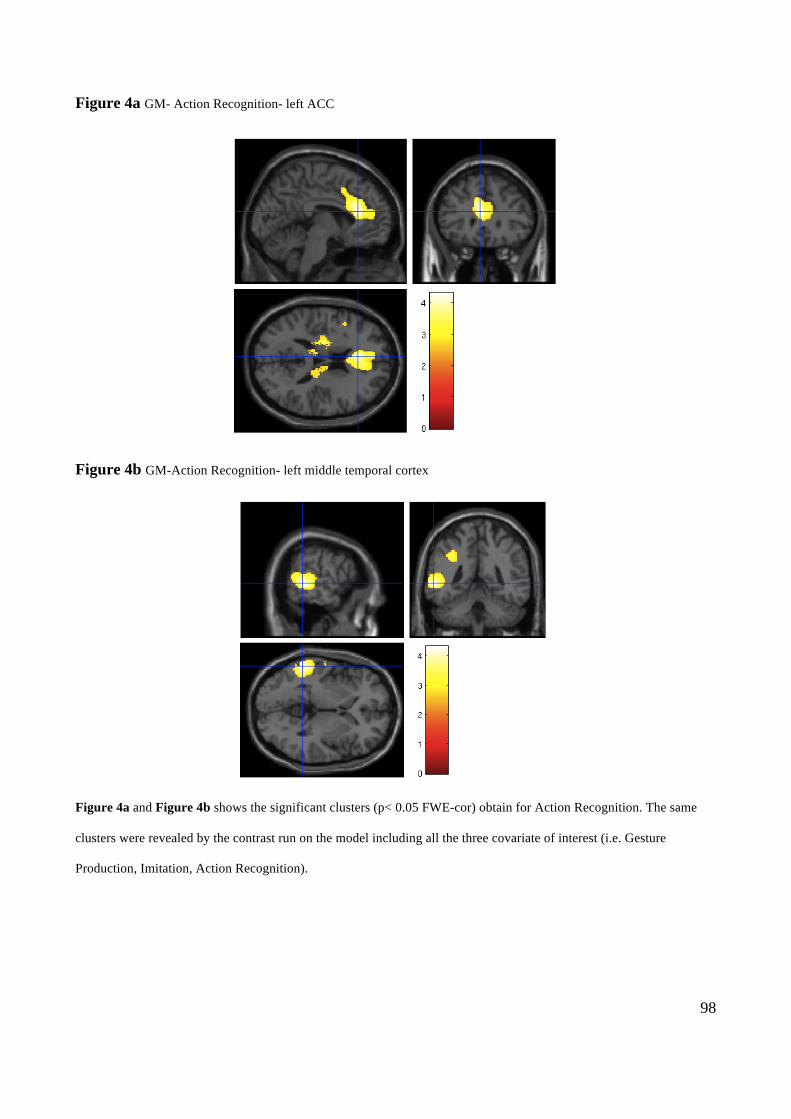
98
Figure 4a GM- Action Recognition- left ACC
Figure 4b GM-Action Recognition- left middle temporal cortex
Figure 4a and Figure 4b shows the significant clusters (p< 0.05 FWE-cor) obtain for Action Recognition. The same
clusters were revealed by the contrast run on the model including all the three covariate of interest (i.e. Gesture
Production, Imitation, Action Recognition).
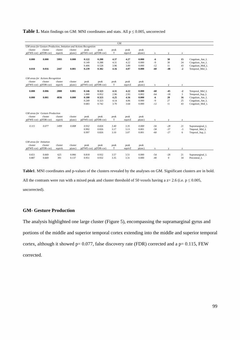
99
Table 1. Main findings on GM: MNI coordinates and stats. All p ≤ 0.005, uncorrected
Table1. MNI coordinates and p-values of the clusters revealed by the analyses on GM. Significant clusters are in bold.
All the contrasts were run with a mixed peak and cluster threshold of 50 voxels having a z= 2.6 (i.e. p ≤ 0.005,
uncorrected).
GM- Gesture Production
The analysis highlighted one large cluster (Figure 5), encompassing the supramarginal gyrus and
portions of the middle and superior temporal cortex extending into the middle and superior temporal
cortex, although it showed p= 0.077, false discovery rate (FDR) corrected and a p= 0.115, FEW
corrected.
cluster cluster cluster cluster peak peak peak peak peakp(FWE-cor) p(FDR-cor) equivk p(unc) p(FWE-cor) p(FDR-cor) T equivZ p(unc) x y z
0.000 0.000 5993 0.000 0.122 0.288 4.37 4.27 0.000 -6 30 15 Cingulum_Ant_L0.149 0.288 4.31 4.22 0.000 -9 30 24 Cingulum_Ant_L0.406 0.528 3.96 3.89 0.000 -12 11 43 Cingulum_Mid_L
0.018 0.016 2447 0.001 0.239 0.382 4.16 4.07 0.000 -60 -48 -2 Temporal_Mid_L
cluster cluster cluster cluster peak peak peak peak peakp(FWE-cor) p(FDR-cor) equivk p(unc) p(FWE-cor) p(FDR-cor) T equivZ p(unc) x y z
0.008 0.006 2888 0.001 0.146 0.323 4.31 4.22 0.000 -60 -45 -2 Temporal_Mid_L1.000 0.952 2.96 2.93 0.002 -64 -18 3 Temporal_Sup_L
0.000 0.001 4836 0.000 0.180 0.323 4.25 4.16 0.000 -6 28 16 Cingulum_Ant_L0.249 0.323 4.14 4.06 0.000 -9 27 25 Cingulum_Ant_L0.683 0.742 3.70 3.64 0.000 -12 11 43 Cingulum_Mid_L
cluster cluster cluster cluster peak peak peak peak peakp(FWE-cor) p(FDR-cor) equivk p(unc) p(FWE-cor) p(FDR-cor) T equivZ p(unc) x y z
0.115 0.077 1499 0.008 0.932 0.826 3.40 3.35 0.000 -56 -28 21 Supramarginal_L0.992 0.826 3.17 3.13 0.001 -58 -37 -3 Tmporal_Mid_L0.997 0.826 3.10 3.07 0.001 -60 -27 6 Tmporal_Sup_L
cluster cluster cluster cluster peak peak peak peak peakp(FWE-cor) p(FDR-cor) equivk p(unc) p(FWE-cor) p(FDR-cor) T equivZ p(unc) x y z
0.651 0.669 625 0.066 0.818 0.932 3.57 3.51 0.000 -54 -28 21 Supramarginal_L0.887 0.669 391 0.137 0.951 0.932 3.35 3.31 0.000 -40 0 34 Precentral_L
GM areas for Gesture Production, Imitation and Actions Recognition
GM areas for Actions Recognition
GM areas for Gesture Production
GM areas for Imitation
GM
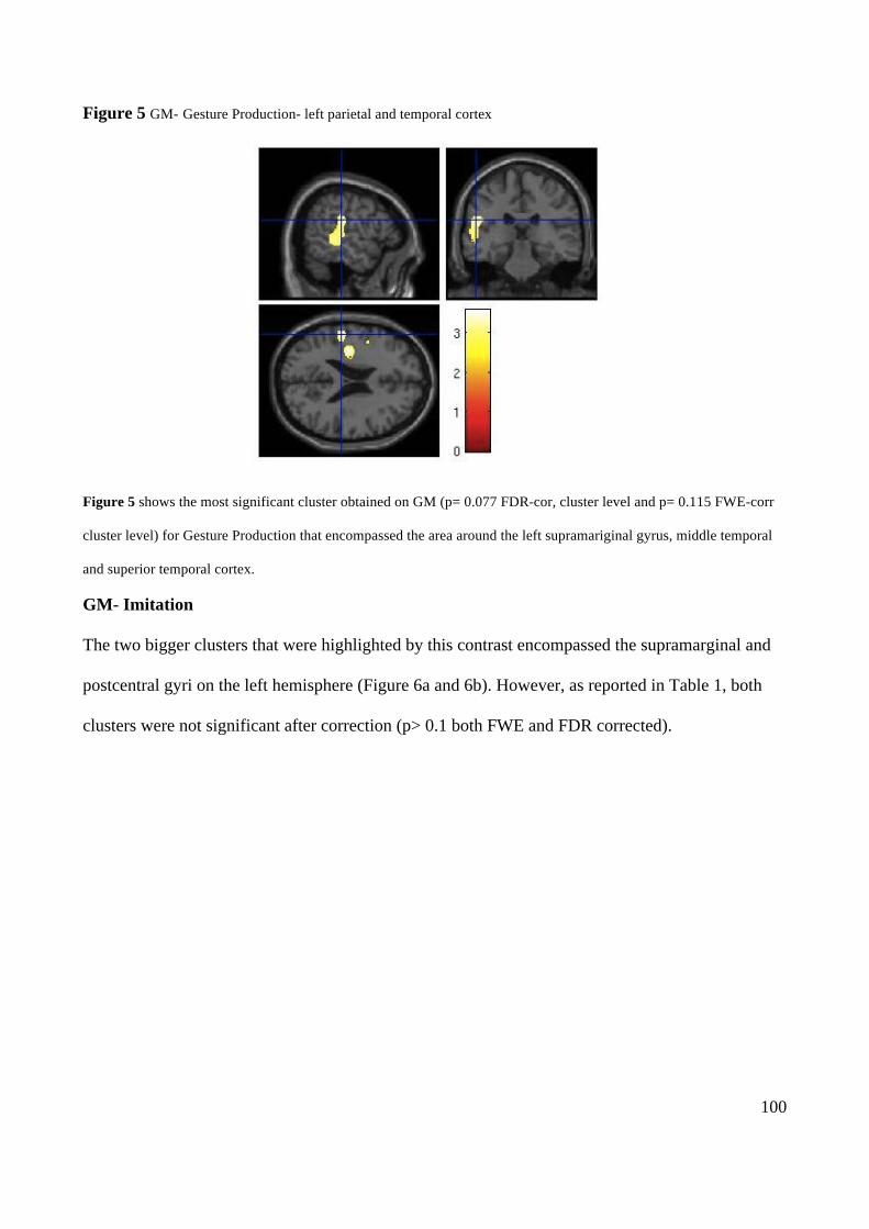
100
Figure 5 GM- Gesture Production- left parietal and temporal cortex
Figure 5 shows the most significant cluster obtained on GM (p= 0.077 FDR-cor, cluster level and p= 0.115 FWE-corr
cluster level) for Gesture Production that encompassed the area around the left supramariginal gyrus, middle temporal
and superior temporal cortex.
GM- Imitation
The two bigger clusters that were highlighted by this contrast encompassed the supramarginal and
postcentral gyri on the left hemisphere (Figure 6a and 6b). However, as reported in Table 1, both
clusters were not significant after correction (p> 0.1 both FWE and FDR corrected).
!
Figure 7
Figure 7 shows the most significant cluster (p= 0.077 FDR-cor, cluster level) obtained for the contrast run on the model with Gesture Production as the only covariate of interest
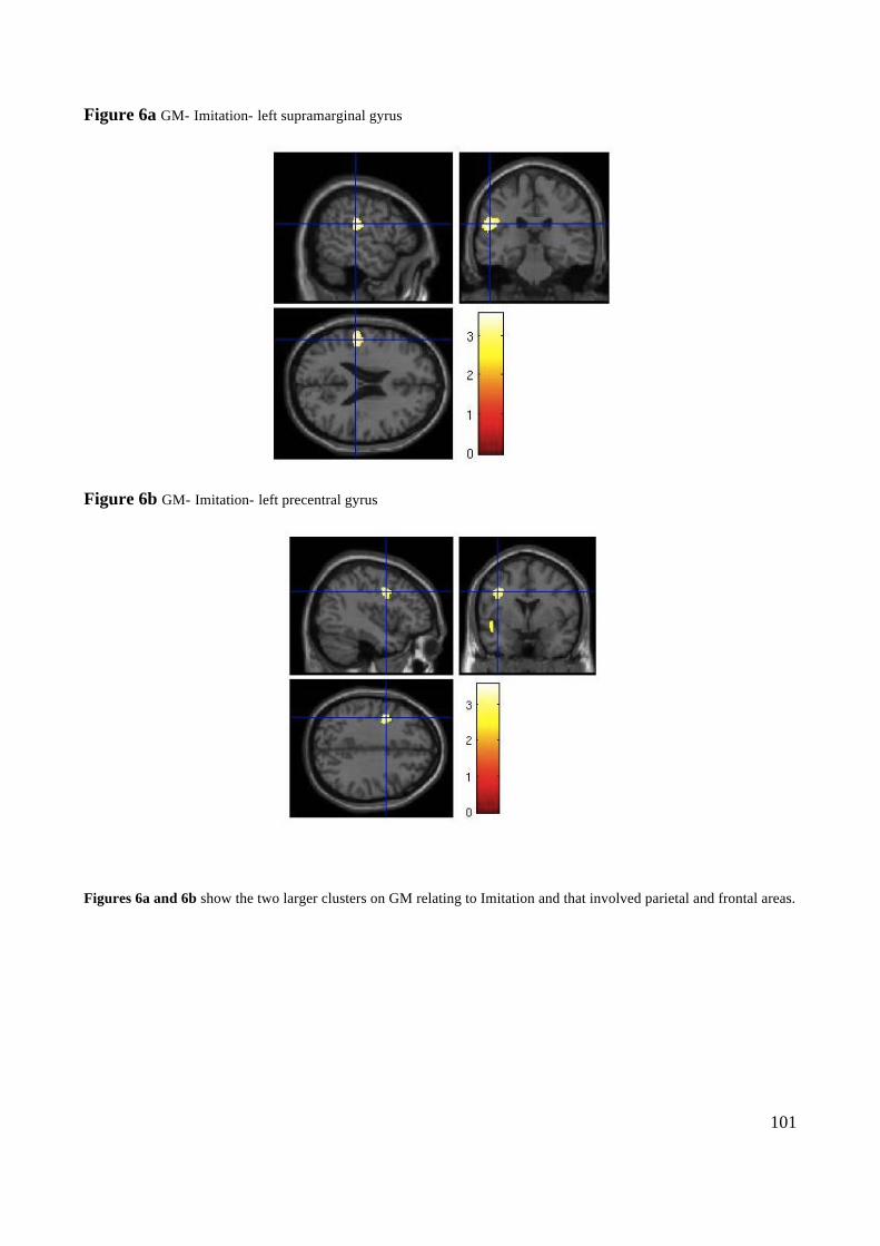
101
Figure 6a GM- Imitation- left supramarginal gyrus
Figure 6b GM- Imitation- left precentral gyrus
Figures 6a and 6b show the two larger clusters on GM relating to Imitation and that involved parietal and frontal areas.
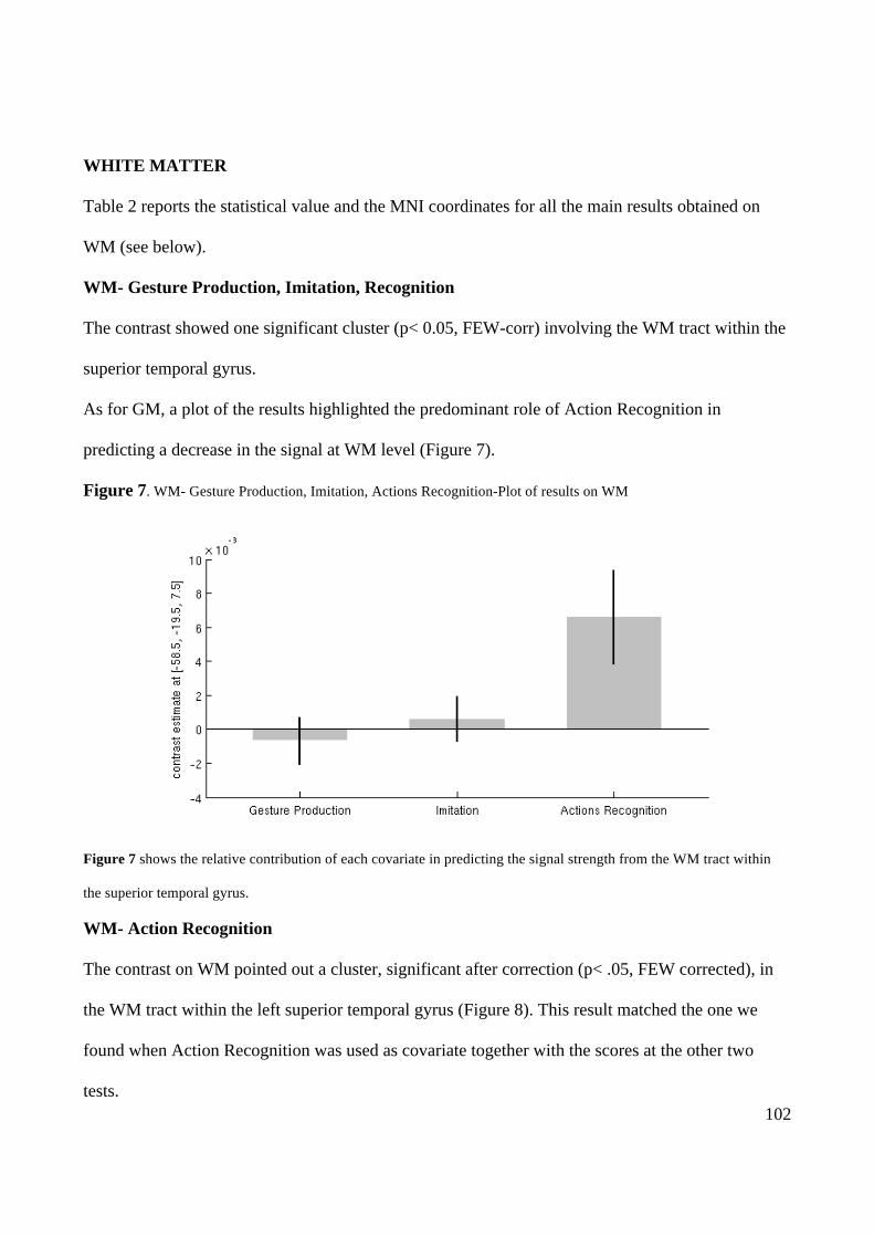
102
WHITE MATTER
Table 2 reports the statistical value and the MNI coordinates for all the main results obtained on
WM (see below).
WM- Gesture Production, Imitation, Recognition
The contrast showed one significant cluster (p< 0.05, FEW-corr) involving the WM tract within the
superior temporal gyrus.
As for GM, a plot of the results highlighted the predominant role of Action Recognition in
predicting a decrease in the signal at WM level (Figure 7).
Figure 7. WM- Gesture Production, Imitation, Actions Recognition-Plot of results on WM
Figure 7 shows the relative contribution of each covariate in predicting the signal strength from the WM tract within
the superior temporal gyrus.
WM- Action Recognition
The contrast on WM pointed out a cluster, significant after correction (p< .05, FEW corrected), in
the WM tract within the left superior temporal gyrus (Figure 8). This result matched the one we
found when Action Recognition was used as covariate together with the scores at the other two
tests.
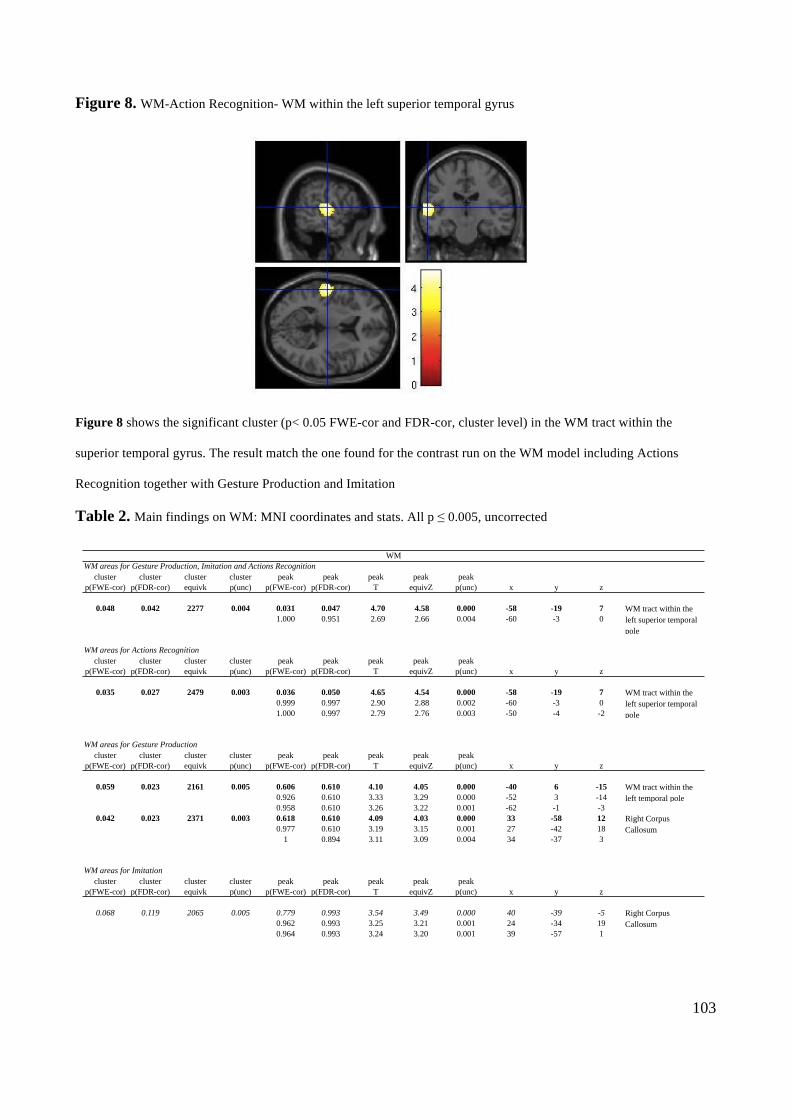
103
Figure 8. WM-Action Recognition- WM within the left superior temporal gyrus
Figure 8 shows the significant cluster (p< 0.05 FWE-cor and FDR-cor, cluster level) in the WM tract within the
superior temporal gyrus. The result match the one found for the contrast run on the WM model including Actions
Recognition together with Gesture Production and Imitation
Table 2. Main findings on WM: MNI coordinates and stats. All p ≤ 0.005, uncorrected
!
Figure 3
Figure 3 shows the significant cluster (p< 0.05 FWE-cor and FDR-cor, cluster level) in the WM tract running below the superior temporal lobe (???).
cluster cluster cluster cluster peak peak peak peak peakp(FWE-cor) p(FDR-cor) equivk p(unc) p(FWE-cor) p(FDR-cor) T equivZ p(unc) x y z
0.048 0.042 2277 0.004 0.031 0.047 4.70 4.58 0.000 -58 -19 71.000 0.951 2.69 2.66 0.004 -60 -3 0
cluster cluster cluster cluster peak peak peak peak peakp(FWE-cor) p(FDR-cor) equivk p(unc) p(FWE-cor) p(FDR-cor) T equivZ p(unc) x y z
0.035 0.027 2479 0.003 0.036 0.050 4.65 4.54 0.000 -58 -19 70.999 0.997 2.90 2.88 0.002 -60 -3 01.000 0.997 2.79 2.76 0.003 -50 -4 -2
cluster cluster cluster cluster peak peak peak peak peakp(FWE-cor) p(FDR-cor) equivk p(unc) p(FWE-cor) p(FDR-cor) T equivZ p(unc) x y z
0.059 0.023 2161 0.005 0.606 0.610 4.10 4.05 0.000 -40 6 -150.926 0.610 3.33 3.29 0.000 -52 3 -140.958 0.610 3.26 3.22 0.001 -62 -1 -3
0.042 0.023 2371 0.003 0.618 0.610 4.09 4.03 0.000 33 -58 120.977 0.610 3.19 3.15 0.001 27 -42 18
1 0.894 3.11 3.09 0.004 34 -37 3
cluster cluster cluster cluster peak peak peak peak peakp(FWE-cor) p(FDR-cor) equivk p(unc) p(FWE-cor) p(FDR-cor) T equivZ p(unc) x y z
0.068 0.119 2065 0.005 0.779 0.993 3.54 3.49 0.000 40 -39 -50.962 0.993 3.25 3.21 0.001 24 -34 190.964 0.993 3.24 3.20 0.001 39 -57 1
WM
Right Corpus Callosum
WM tract within the left superior temporal pole
WM tract within the left superior temporal pole
WM areas for Gesture Production, Imitation and Actions Recognition
WM areas for Actions Recognition
WM areas for Gesture Production
WM areas for Imitation
WM tract within the left temporal pole
Right Corpus Callosum
104
Table2. MNI coordinates and p-values of the clusters revealed by the analyses on WM. Significant clusters are in bold.
All the contrasts were run with a mixed peak and cluster threshold of 50 voxels having a z= 2.6 (i.e. p ≤ 0.005,
uncorrected).
WM- Gesture Production
The analysis on WM revealed a significant cluster (p< 0.05, FWE corrected) involving the caudal
part of the corpus callosum on the right hemisphere (Figure 9a), as well as a cluster having a p=
0.059, FEW corrected and a p≤ 0.05, FDR corrected, overlapping the WM tracts within the left
superior temporal cortex (Figure 9b).
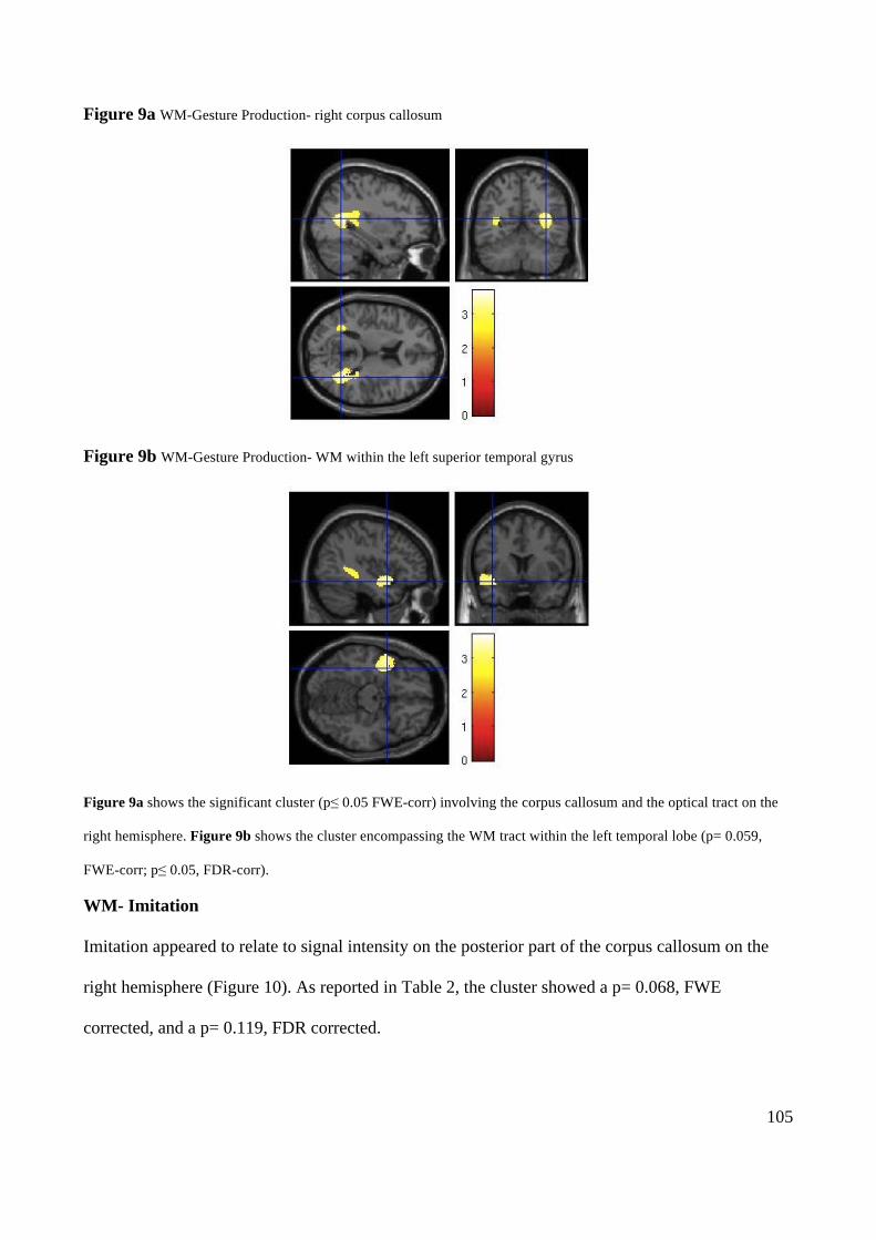
105
Figure 9a WM-Gesture Production- right corpus callosum
Figure 9b WM-Gesture Production- WM within the left superior temporal gyrus
Figure 9a shows the significant cluster (p≤ 0.05 FWE-corr) involving the corpus callosum and the optical tract on the
right hemisphere. Figure 9b shows the cluster encompassing the WM tract within the left temporal lobe (p= 0.059,
FWE-corr; p≤ 0.05, FDR-corr).
WM- Imitation
Imitation appeared to relate to signal intensity on the posterior part of the corpus callosum on the
right hemisphere (Figure 10). As reported in Table 2, the cluster showed a p= 0.068, FWE
corrected, and a p= 0.119, FDR corrected.
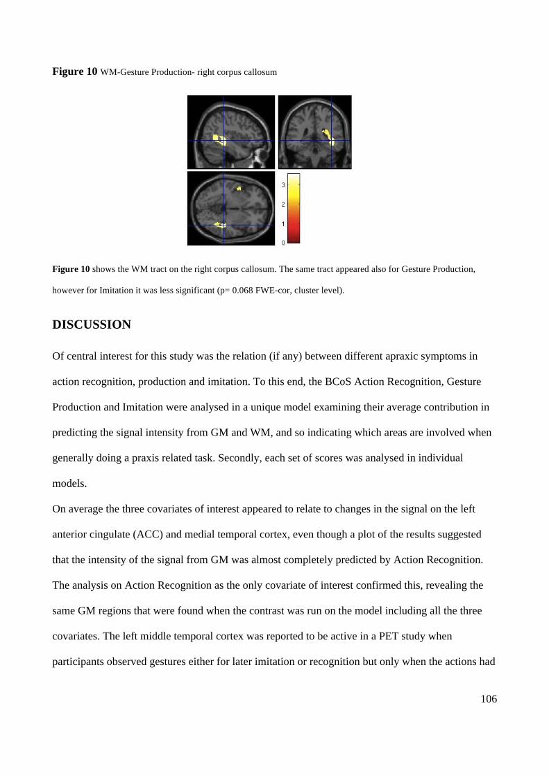
106
Figure 10 WM-Gesture Production- right corpus callosum
Figure 10 shows the WM tract on the right corpus callosum. The same tract appeared also for Gesture Production,
however for Imitation it was less significant (p= 0.068 FWE-cor, cluster level).
DISCUSSION
Of central interest for this study was the relation (if any) between different apraxic symptoms in
action recognition, production and imitation. To this end, the BCoS Action Recognition, Gesture
Production and Imitation were analysed in a unique model examining their average contribution in
predicting the signal intensity from GM and WM, and so indicating which areas are involved when
generally doing a praxis related task. Secondly, each set of scores was analysed in individual
models.
On average the three covariates of interest appeared to relate to changes in the signal on the left
anterior cingulate (ACC) and medial temporal cortex, even though a plot of the results suggested
that the intensity of the signal from GM was almost completely predicted by Action Recognition.
The analysis on Action Recognition as the only covariate of interest confirmed this, revealing the
same GM regions that were found when the contrast was run on the model including all the three
covariates. The left middle temporal cortex was reported to be active in a PET study when
participants observed gestures either for later imitation or recognition but only when the actions had
!
Figure 10
Figure 10 shows the WM tract that appear to be involved when Imitation was considered alone as covariate of interest. The cluster that had a only a trend for significance (p= 0.068 FWE-cor, cluster level).
107
a meaning (Decety et al. 1997). Also in other research, the same region has been found to be
damaged inn two patients who were not able to produce actions to objects, while still retaining the
knowledge of the objects they could not use (Price et al. 2010). Moreover, Price et al. (2010)
reported higher incidence of impairment in gesture production among patients whose lesion
extended into left temporal (and middle temporal) cortex. This also fits with our finding of a cluster
extending to the left middle temporal cortex for Gesture Production, where the patients had to
reproduce meaningful gestures on verbal command, although this cluster showed only a trend for
significance when FDR-corrected, and an even higher p-value when FWE- corrected. As both
successful recognition and production of meaningful gestures may require retrieving information
from LTM, those results point to a role of the medial temporal cortex in the action retrieval process.
This interpretation is supported by previous investigations that indicated the medial temporal cortex
as having a role in encoding the right sequence of events within episodes stored in LTM (Tubridy et
al. 2011). To activate a representation of the right movement sequence may be crucial not only to
execute a movement correctly but also to recognize a gesture, especially in the case of pantomimes
where the actual object is not present to help gesture identification.
A cluster in the left ACC was revealed only for Action Recognition. This result was unexpected and
does not clearly relate to lesions reported in studies of apraxia. The ACC has most typically been
linked to action monitoring and response selection (e.g. Dehanne et al. 1994; Pardo et al. 1990).
However, the ACC keeps a great number of interconnections between different cortical areas.
Moreover, it has been proposed that ACC could be divided into two parts: i) a more posterior part
primarily connected to areas appointed to monitoring behaviour, response selection and motor
control (e.g. prefrontal, parietal, premotor and supplementary motor areas); ii) a more anterior part
connected to the hippocampus, and to other structures belonging to the limbic system, that is
involved in assessing the salience of motivational and emotional/affective information (see Bush et
al. 2000 for a review). It should also be noted that the hippocampus was previously indicated as a
108
structure involved in the retrieval of semantic information about pantomimes of object use (e.g.
Tessari et al. 2007). The present results do not enable us to identify fine-grained subdivisions within
the ACC, however it is possible that a lesion on the more anterior part impaired action recognition
in our patients because the ACC could not pass information to the hippocampus. Alternatively the
patients may have failed in the task because of a deficit in identifying the motivation or the
intention of the actions. The Action Recognition task administered in the present study was
composed of both transitive pantomimes of object use and intransitive communicational gestures.
Here the total scores were considered, so it is not possible to distinguish which between the two
types of gestures is more responsible for the results. For instance, if the relation between ACC and
performance holds true only for the transitive pantomimes, this may support the hypothesis of a
disconnection from the hippocampus, which can be involved in the retrieval of information about
those actions (e.g. Rumiati et al. 2005, Tessari et al. 2007). Conversely, if the relation is due only to
the intransitive communicative gestures, it is more likely that a lesion on ACC led to a failure in
recognizing the intentions of the actor. More analyses may address this issue.
The strongest results on the GM analysis emerged for Action Recognition, however also Gesture
Production and Imitation gave some results, although they are not as strong and must be taken
cautiously. The results on GM for Gesture Production, besides partially overlapping with the results
for Action Recognition in the middle temporal cortex, also extended more superiorly on the left
supramarginal gyrus, an area that was also related to performance in Imitation and that was reported
to be damaged in patients with ideomotor apraxia (e.g. Heliman et al. 1982). Moreover a small
cluster on the frontal cortex, although not significant, appeared to be linked to Imitation only.
For the WM analysis the contrast revealed an average contribution across the tasks predicting signal
changes in tracts within the superior temporal gyrus in the left hemisphere. Similarly to the GM
analysis, a major contribution of Action Recognition emerged. The analysis of the three covariates
taken separately confirmed a relation between the integrity of the WM tract within the left superior

109
temporal gyrus and performance in Action Recognition, and revealed also a relation between this
same WM tract and Gesture Production, even though it was less significant than for Action
Recognition. Gesture Production appeared to relate also to the signal changes from a WM tract
overlapping the posterior part of the corpus callosum in the right hemisphere and the same tract
appeared also for Imitation. At the best of our knowledge there are no studies in literature
discussing the role of white matter tracts in processing and producing action. The mapping of the
different WM tracts is quite novel, and more studies are needed to understand the role of WM in
praxis, as well as in the other cognitive functions. Moreover, other techniques would be more
appropriate than VBM for investigating white matter damage (e.g. Diffusion Tensor Imaging
(DTI)). Nonetheless the interesting point that emerges from our data is the overlap between areas
involved in the three tasks has a similar pattern in WM and GM. Indeed, common WM tracts were
revealed for Action Recognition and Gesture Production and for Gesture Production and Imitation.
Also, Action Recognition and Imitation, in this case imitation of meaningless intransitive gestures,
did not overlap, for either the GM or the WM analysis. Future studies could be performed relating
DTI signal to the performance of patients in tasks tapping different abilities involved in praxis.
In recent years, investigations on praxis have been boosted by the discovery of the MN in
monkeys. Some studies have claimed proof of the existence of the MN in humans, though often
from indirect evidences of common neurophysiological phenomena (e.g. EEG signal or activations
in fMRI studies) relating both to imitation and observation or recognition of different kinds of
gesture (see Rizzolati et al. 2001, 2004 for a revew). However, to imitate a meaningful gesture is
alsolikely to require recognition of the gesture itself and a common activation, for instance, can
possibly be due to common memory processes. In the present study Action Recognition and
Gesture Production showed at least some partially overlap both in GM and WM areas that could
have a role in retrieving semantic representations of meaningful gestures. Also the overlapping on
GM and WM between Gesture Production and Imitation seems to be better explained by the fact
110
that both require the actual production of a movement that is accomplished through a common
motor-output process. Together, those results seem to fit more with a dual-route model of praxis
(Rothi et al. 1991; Rumiati et al. 2002) than with the hypothesis of a unique population of neurons
serving both gesture production and recognition or understanding.
Finally, consideration must be given as to why we choose a statistical threshold with a p-value
≤0.005 (instead of p≤0.001). As already mentioned, this was done because the analyses were run on
CT scans that could have some noise. Also the tasks used to test the patients could have been easy
for some of them, thus generating ceiling effects. Given these conditions, the use of a very strict
threshold (as p p≤0.001) may have caused a loss of information. However, there are two main
reasons for which CT scans and BCoS tasks, instead of MRI and longer or more difficult tasks,
were employed. First, this investigation used the scores at the praxis tasks taken from the
Birmingham Cognitive Screening (BCoS; Humphreys et al. 2012), a new battery of tests aimed at
stroke patients in the acute phase. Beside the wish to understand praxis functions and to have data to
compare with previous findings, our further aim was to explore the brain areas correlating with
performance at three of the praxis BCoS sub-tests in patients who just suffered from a stroke (i.e.
when other compensation mechanisms, at behavioural or neural level, were not established). The
BCoS was built to be an efficient screening tool for a population of patients who may not be able to
endure long and hard testing sessions and for this reason only a few items were included in each
task. It should be noted that the BCoS praxis tasks correlate highly with other praxis assessments
more frequently used and also relate to measures of patients’ general functioning in everyday life,
both in a sub-acute phase and in a follow up done nine month later (see Bickerton et al. 2012). In
addition, the present investigation was carried out on sub-acute patients (i.e. the population of
patients for which the BCoS was created), and for this kind of patient only CT scans were available,
as they were taken routinely when patients were admitted into the hospital, and collecting MRI
scans for all our patients was not possible.
111
As already done for the behavioural data (Bickerton et al. 2012), it would be useful to follow up on
the same patients as it could help to obtain a clearer picture of the neural substrates tapped by the
BCoS praxis tasks, and to understand how these substrates reorganize over time or in relation to the
rehabilitation programs (if any) underwent by the patients. .
112
6. Distinct and Common Neural Correlates of Apraxia for
Transitive and Intransitive Gestures: An investigation using
Voxel-Based Morphometry (VBM)
ABSTRACT
Previous neuropsychological investigations suggest that intransitive gestures are typically
maintained better than transitive gestures after brain lesion. However it is not clear which are the
common and distinct neural mechanisms that underlie the production of those two types of gestures.
The present study tried to clarify this issue using a lesion-symptom approach (voxel-based
morphometry, VBM) based on transitive and intransitive gestures performed by a large group of
subacute stroke patients and clinical computed tomography scans. At a behavioural level, the
analyses of the patients’ scores revealed significantly higher performance for transitive than
intransitive gesture production. This contrast with prior results may reflect a difference between
gesture production to name (used here) and imitation (used previously). At a neural level, VBM
analyses revealed that, on average, producing a gesture on verbal command involved the insula and
inferior parietal areas on the left. The results indicating damage within the insula were driven by the
production of transitive gestures, while both types of gesture seemed to involve, at least to some
extent, the parietal cortex. Analyses run independently for transitive and intransitive gestures
showed that, at gray matter level (GM), distinct regions of neural change were associated with each
key symptom. Impairments in intransitive gestures were associated with damage to left frontal and
parietal cortex, while lesions within deeper brain regions including the left insular cortex were
related to transitive gestures to objects. Distinct regions for intransitive and transitive gestures were
also revealed in the white matter (WM), in the corpus callosum and in the WM within the temporal
113
cortex respectively. Also, transitive actions had at least some relation with damage to the corpus
callosum.
All together, the behavioural scores and their correlations with the neuroimaging data suggest that
both common and discrete neural mechanisms are involved when transitive and intransitive gestures
have to be performed.
INTRODUCTION
Within the neuropsychological syndrome of apraxia, people can suffer from impairments in the
ability to execute actions that are not caused by a primary motor deficit (Liepmann, 1900-1905).
Apraxia is not a unitary phenomenon and there are dissociations reported between patients in
relation to the type of action to be executed and the body part involved in the task (see Rumiati et
al. 2009 for a review). For instance, neuropsychological observations have been made of patients
who are more impaired when imitating meaningful (familiar) compared to meaningless (unfamiliar)
gestures (Goldenberg & Hagmann 1997; Peigneux et al. 2000; Tessari et al 2007), as well as
patients showing the opposite pattern (Bartolo et al. 2001; Tessari et al. 2007), and also between
impairments in imitation as opposed to actual use of the same objects (e.g., Chainay & Humphreys,
2002). These differences (e.g., between the production of familiar and unfamiliar gestures) also
appear to reflect the involvement of different brain structures (e.g. Goldenberg & Hagmann 1997;
Peigneux et al. 2000; Cubelli et al. 2000; Buxbaum et al. 2001; Tessari et al. 2007), Whether
differences occur in the production of different kinds of familiar gestures, and whether different
familiar gestures depend on contrasting neural structures, remains a moot point however. The most
studied contrast is between transitive pantomimes of object use and intransitive (communicative)
gestures (e.g., miming how to use a hammer vs. waving to greet someone).
Functional imaging studies in normal participants suggest considerable overlap in the brain activity
used to make transitive and intransitive gestures, consistent with all familiar gestures relying on a
114
common neural network involving parietal and frontal cortices (e.g., Kroliczak & Fray, 2009). On
the other hand, neuropsychological data showing dissociations between the two classes of patient
indicate that the actions may be functionally and neutrally separable. In the majority of cases
patients have been described with deficits in which transitive actions are more impaired than
intransitive actions (e.g., Buxbaum et al. 2007) and it has been proposed that transitive actions
relying on a repository of action engrams in the left inferior parietal cortex (Buxbaum, 2001;
Buxbaum et al. 2007; Liepmann, 1900-1905). In contrast, familiar intransitive actions may be
supported by bilateral fronto-parietal regions that respond to fine-grained dynamic properties of
action (Buxbaum et al. 2007; Haaland & Flaherty, 1984; Mozaz, 2002; Rapcsak et al. 1993). The
preponderance of impairments in producing transitive over intransitive actions, however, may
reflect the greater difficulty of transitive actions, as better performance in imitating intransitive over
transitive actions has been reported (Carmo & Rumiati, 2009).
A stronger argument for a distinction between transitive and intransitive actions comes from recent
work by Stamenova, Roy and Black (2010). These investigators assessed the production (to name)
and imitation of transitive and intransitive gestures in patients with unilateral left or right
hemisphere damage after stroke (Stamenova et al. 2010). Although no cases were found
demonstrating a complete double dissociation between transitive and intransitive gestures (e.g., with
both gesturing to name and imitation for one class of action affected while both tasks were
preserved for the other class), there were instances of selective dissociation for one type of action
(either gesturing to name or imitation) or for either transitive or intransitive actions. Selective
deficits in transitive actions were associated with left hemisphere damage and problems with
intransitive action with right hemisphere damage, consistent with prior arguments for the necessary
involvement of the right hemisphere in intransitive action, perhaps because these actions depend
more on fine-grained spatio-temporal modulation of action. On the other hand, the same
investigators (Stamenova et al. 2009) have reported data on cortical-basal degeneration patients

115
showing selectively greater changes in the left or right hemisphere and failed to find evidence for
intransitive actions ever being more impaired than transitive. Hence the case for a functional and
neural distinction between these different classes of learned actions remains inconclusive.
In the present paper, a different approach is taken in which a non-selected sample of stroke patients
was examined for the presence of deficits in transitive or intransitive action production to name, and
the underlying lesions were examined using voxel-based morphometry. Voxel-based morphometry
(VBM) is an unbiased means of assessing the correlation between neural change after brain lesion
and behavioural symptoms of interest. The procedure requires data from patients who do and also
patients who do not show the cardinal symptoms being assessed so that the latter can serve as
‘lesion controls’ – enabling the analysis to extract out damaged areas in the critical patients which
might reflect anatomical constraints on the brain lesion (e.g., after occlusion of the middle cerebral
artery) rather than the necessary role of the brain structure in behaviour (see Rorden & Karnath,
2004). The analysis used VBM based on continuous measures of performance in patients within a
set time period post lesion to provide a fine-grained measure of brain-behaviour relations (see
Chechlacz et al. 2010, for a similar approach). Given prior reports on the relations between deficits
in transitive relative to intransitive actions then it can be expected that selective impairments in
transitive actions will be associated with unilateral damage in the left inferior parietal cortex
(Buxbaum et al. 2007). On the other hand, selective deficits in familiar intransitive actions may be
result from more bilateral (and particularly more right hemisphere damage) in patients. Lesions
linked to both classes of action may reflect common processes in programming for motor output,
for instance involving pre-motor cortex and sub-cortical systems (e.g., Stamenova et al. 2010,
2011).
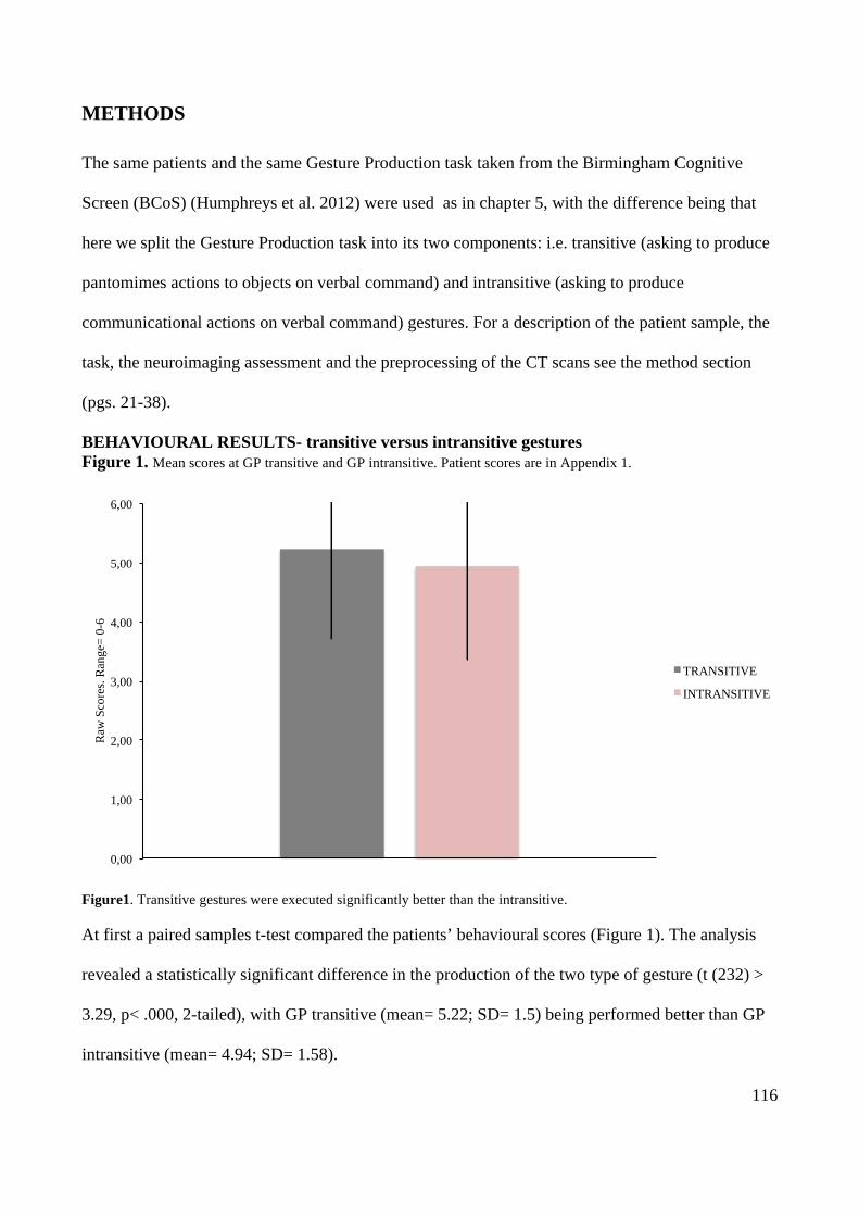
116
METHODS
The same patients and the same Gesture Production task taken from the Birmingham Cognitive
Screen (BCoS) (Humphreys et al. 2012) were used as in chapter 5, with the difference being that
here we split the Gesture Production task into its two components: i.e. transitive (asking to produce
pantomimes actions to objects on verbal command) and intransitive (asking to produce
communicational actions on verbal command) gestures. For a description of the patient sample, the
task, the neuroimaging assessment and the preprocessing of the CT scans see the method section
(pgs. 21-38).
BEHAVIOURAL RESULTS- transitive versus intransitive gestures Figure 1. Mean scores at GP transitive and GP intransitive. Patient scores are in Appendix 1.
Figure1. Transitive gestures were executed significantly better than the intransitive.
At first a paired samples t-test compared the patients’ behavioural scores (Figure 1). The analysis
revealed a statistically significant difference in the production of the two type of gesture (t (232) >
3.29, p< .000, 2-tailed), with GP transitive (mean= 5.22; SD= 1.5) being performed better than GP
intransitive (mean= 4.94; SD= 1.58).
0,00
1,00
2,00
3,00
4,00
5,00
6,00
Raw
Sco
res.
Ran
ge=
0-6
TRANSITIVE
INTRANSITIVE

117
Although the two types of actions differed in difficulty, they also correlated with each other
(Pearson correlation= .93, p< .0000, 2-tailed).
VOXEL-BASED MORPHOMETRY
As in the study presented in chapter 5, the CT scans available for the 233 acute stroke patients were
used for the analyses. The aim of the present study was to explore the relations between the changes
in the signal intensity from either gray matter (GM) or white matter (WM) across the whole brain
and performance at production of meaningful intransitive and transitive gestures on verbal
command.
ANALYSES
The raw scores for producing intransitive and transitive gestures were included together in two
models as the two covariates of interest, along with GM and WM respectively. In addition, four
models were including one covariate of interest at a time (i.e. i) scores for the production of
intransitive gestures and GM; ii) scores for the production of transitive gestures and GM; iii) scores
for the production of intransitive gestures and WM; iv) scores for the production of transitive
gestures and WM) were estimated in order to analyse the discrete effect of each covariate on GM
and WM. Age, gender, years of education and hand (used for imitation/gestures production,
dominant or non dominant) were included as covariates of non-interest in all the models14. T-
contrasts were run on the models estimated. As in the study presented in chapter 5, CT scans of
acute patients were used, along with the same BCoS Gesture Production task that was designed for
screening cognitive functions in patients who just survived a stroke. As already stated in chapter 5,
CT scans can be noisy and the performance of some patients was at ceiling. Accordingly we used a
mixed peak and cluster threshold with at least 50 voxels showing a Z ≥2.6 (i.e. P≤ 0.005,
14 Handedness was not included as only right-handed patients were included in the sample (see also above).
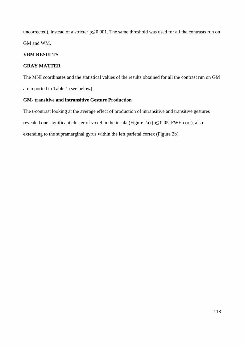
118
uncorrected), instead of a stricter p≤ 0.001. The same threshold was used for all the contrasts run on
GM and WM.
VBM RESULTS
GRAY MATTER
The MNI coordinates and the statistical values of the results obtained for all the contrast run on GM
are reported in Table 1 (see below).
GM- transitive and intransitive Gesture Production
The t-contrast looking at the average effect of production of intransitive and transitive gestures
revealed one significant cluster of voxel in the insula (Figure 2a) (p≤ 0.05, FWE-corr), also
extending to the supramarginal gyrus within the left parietal cortex (Figure 2b).
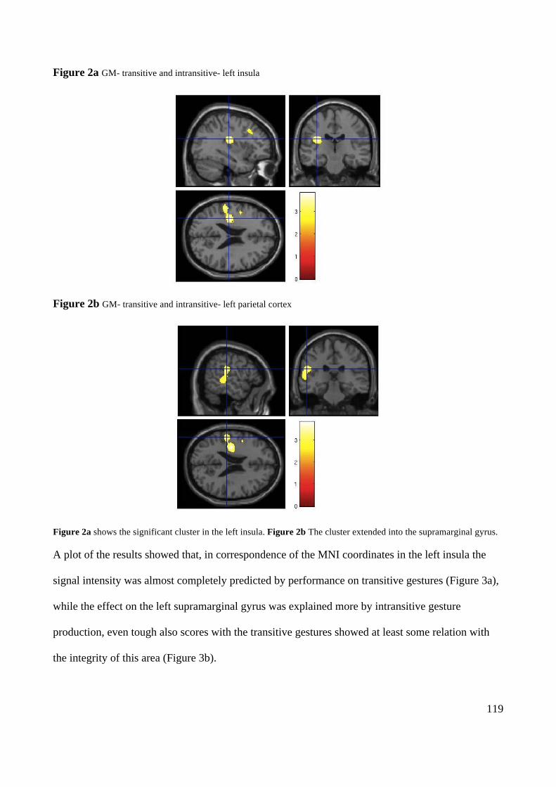
119
Figure 2a GM- transitive and intransitive- left insula
Figure 2b GM- transitive and intransitive- left parietal cortex
Figure 2a shows the significant cluster in the left insula. Figure 2b The cluster extended into the supramarginal gyrus.
A plot of the results showed that, in correspondence of the MNI coordinates in the left insula the
signal intensity was almost completely predicted by performance on transitive gestures (Figure 3a),
while the effect on the left supramarginal gyrus was explained more by intransitive gesture
production, even tough also scores with the transitive gestures showed at least some relation with
the integrity of this area (Figure 3b).
120
Figure 3a GM- transitive and intransitive- Plot of results on the left insula
Figure 3b GM- transitive and intransitive- Plot of results on the left supramarginal gyrus
Figure 3a and Figure 3b illustrate the contribution of each covariate to determine the effect in the insula (Figure 3a)
and on the supramarginal gyrus (Figure 3b).
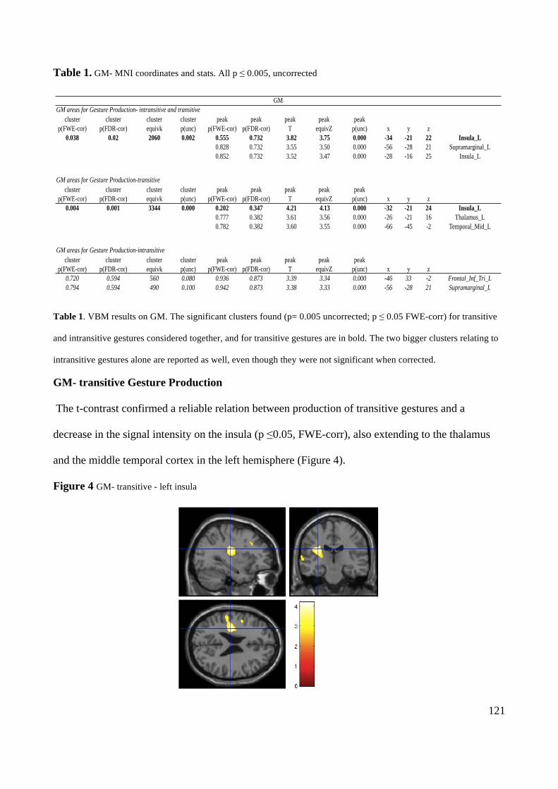
121
Table 1. GM- MNI coordinates and stats. All p ≤ 0.005, uncorrected
Table 1. VBM results on GM. The significant clusters found (p= 0.005 uncorrected; p ≤ 0.05 FWE-corr) for transitive
and intransitive gestures considered together, and for transitive gestures are in bold. The two bigger clusters relating to
intransitive gestures alone are reported as well, even though they were not significant when corrected.
GM- transitive Gesture Production
The t-contrast confirmed a reliable relation between production of transitive gestures and a
decrease in the signal intensity on the insula (p ≤0.05, FWE-corr), also extending to the thalamus
and the middle temporal cortex in the left hemisphere (Figure 4).
Figure 4 GM- transitive - left insula
SIGNIFICANT
Page 1
cluster cluster cluster cluster peak peak peak peak peakp(FWE-cor) p(FDR-cor) equivk p(unc) p(FWE-cor) p(FDR-cor) T equivZ p(unc) x y z
0.038 0.02 2060 0.002 0.555 0.732 3.82 3.75 0.000 -34 -21 22 Insula_L0.828 0.732 3.55 3.50 0.000 -56 -28 21 Supramarginal_L0.852 0.732 3.52 3.47 0.000 -28 -16 25 Insula_L
cluster cluster cluster cluster peak peak peak peak peakp(FWE-cor) p(FDR-cor) equivk p(unc) p(FWE-cor) p(FDR-cor) T equivZ p(unc) x y z
0.004 0.001 3344 0.000 0.202 0.347 4.21 4.13 0.000 -32 -21 24 Insula_L0.777 0.382 3.61 3.56 0.000 -26 -21 16 Thalamus_L0.782 0.382 3.60 3.55 0.000 -66 -45 -2 Temporal_Mid_L
cluster cluster cluster cluster peak peak peak peak peakp(FWE-cor) p(FDR-cor) equivk p(unc) p(FWE-cor) p(FDR-cor) T equivZ p(unc) x y z
0.720 0.594 560 0.080 0.936 0.873 3.39 3.34 0.000 -46 33 -2 Frontal_Inf_Tri_L0.794 0.594 490 0.100 0.942 0.873 3.38 3.33 0.000 -56 -28 21 Supramarginal_L
GMGM areas for Gesture Production- intransitive and transitive
GM areas for Gesture Production-transitive
GM areas for Gesture Production-intransitive
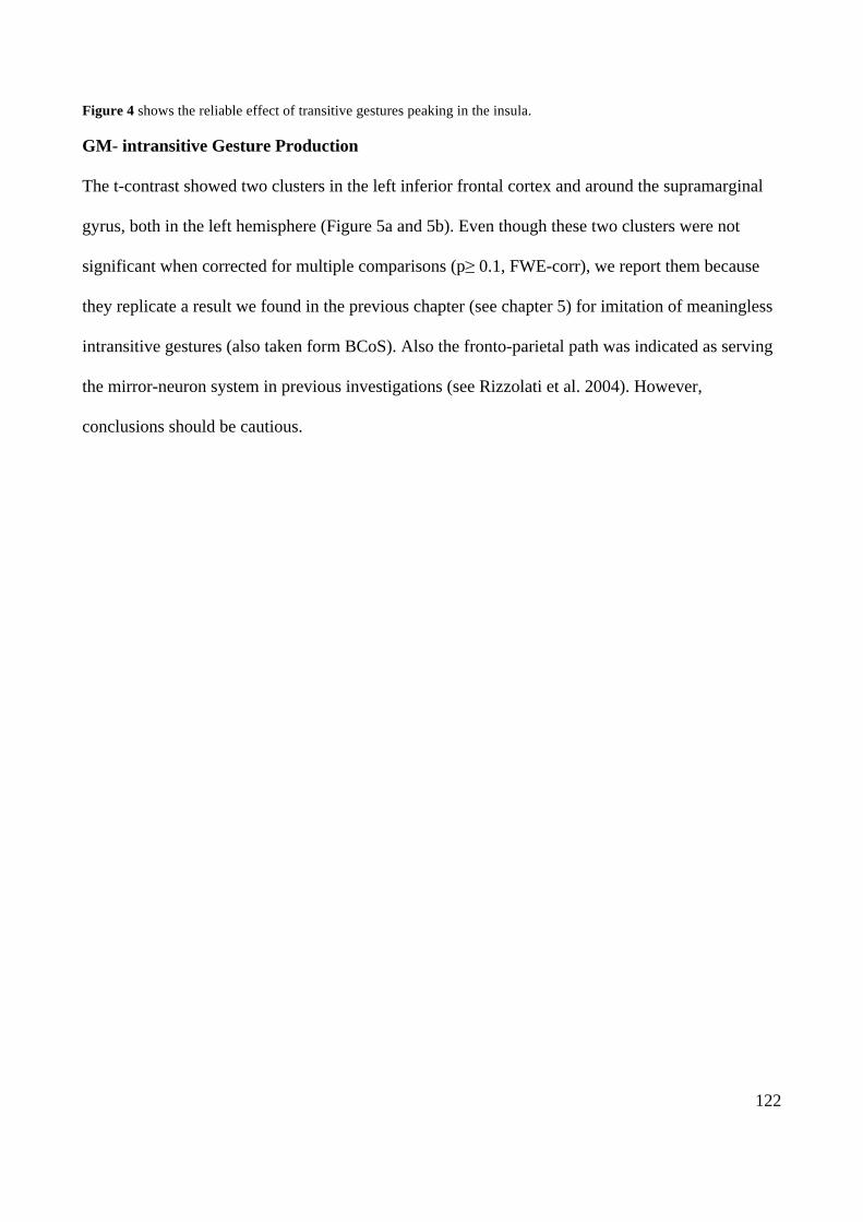
122
Figure 4 shows the reliable effect of transitive gestures peaking in the insula.
GM- intransitive Gesture Production
The t-contrast showed two clusters in the left inferior frontal cortex and around the supramarginal
gyrus, both in the left hemisphere (Figure 5a and 5b). Even though these two clusters were not
significant when corrected for multiple comparisons (p≥ 0.1, FWE-corr), we report them because
they replicate a result we found in the previous chapter (see chapter 5) for imitation of meaningless
intransitive gestures (also taken form BCoS). Also the fronto-parietal path was indicated as serving
the mirror-neuron system in previous investigations (see Rizzolati et al. 2004). However,
conclusions should be cautious.
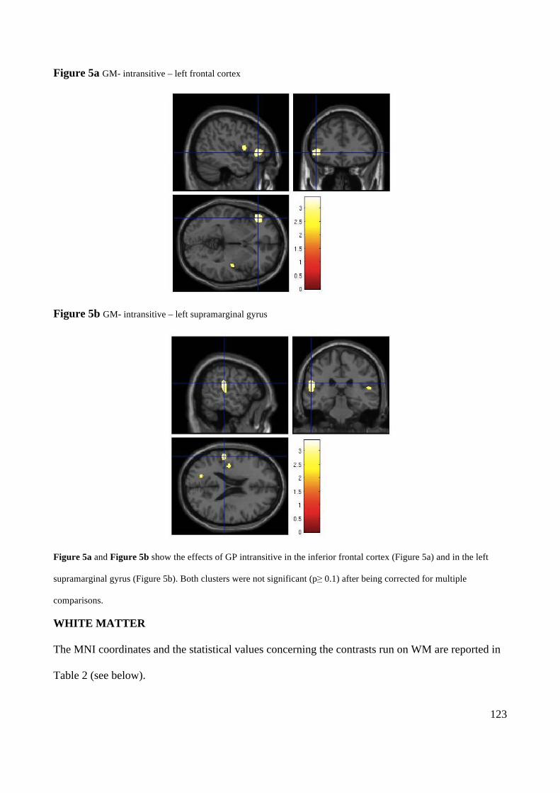
123
Figure 5a GM- intransitive – left frontal cortex
Figure 5b GM- intransitive – left supramarginal gyrus
Figure 5a and Figure 5b show the effects of GP intransitive in the inferior frontal cortex (Figure 5a) and in the left
supramarginal gyrus (Figure 5b). Both clusters were not significant (p≥ 0.1) after being corrected for multiple
comparisons.
WHITE MATTER
The MNI coordinates and the statistical values concerning the contrasts run on WM are reported in
Table 2 (see below).
124
WM- transitive and intransitive Gesture Production
The t-contrast showed an average significant effect of production of transitive and intransitive
gestures on the corpus callosum on the right hemisphere (p≤ 0.05, FWE-corr) (Figure 6a). Also, a
cluster was revealed on the WM tract within the left temporal pole (p= 0.058, FWE-corr) (Figure
6b).
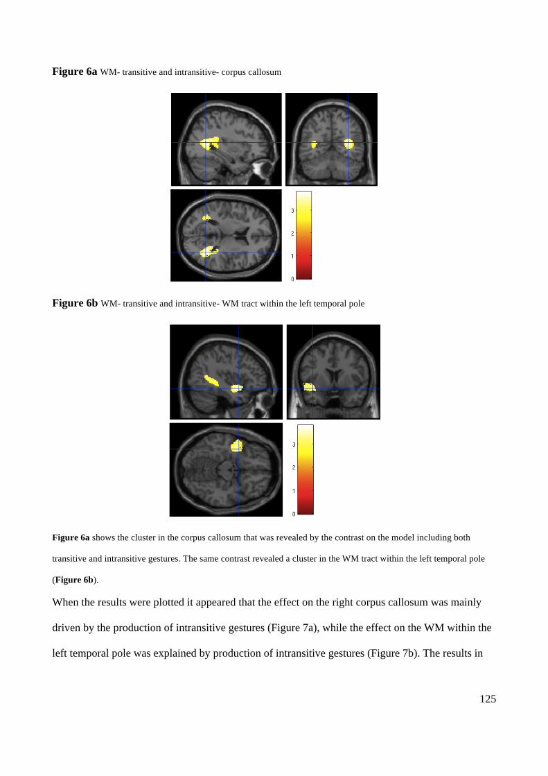
125
Figure 6a WM- transitive and intransitive- corpus callosum
Figure 6b WM- transitive and intransitive- WM tract within the left temporal pole
Figure 6a shows the cluster in the corpus callosum that was revealed by the contrast on the model including both
transitive and intransitive gestures. The same contrast revealed a cluster in the WM tract within the left temporal pole
(Figure 6b).
When the results were plotted it appeared that the effect on the right corpus callosum was mainly
driven by the production of intransitive gestures (Figure 7a), while the effect on the WM within the
left temporal pole was explained by production of intransitive gestures (Figure 7b). The results in
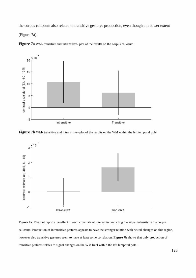
126
the corpus callosum also related to transitive gestures production, even though at a lower extent
(Figure 7a).
Figure 7a WM- transitive and intransitive- plot of the results on the corpus callosum
Figure 7b WM- transitive and intransitive- plot of the results on the WM within the left temporal pole
Figure 7a. The plot reports the effect of each covariate of interest in predicting the signal intensity in the corpus
callosum. Production of intransitive gestures appears to have the stronger relation with neural changes on this region,
however also transitive gestures seem to have at least some correlation. Figure 7b shows that only production of
transitive gestures relates to signal changes on the WM tract within the left temporal pole.
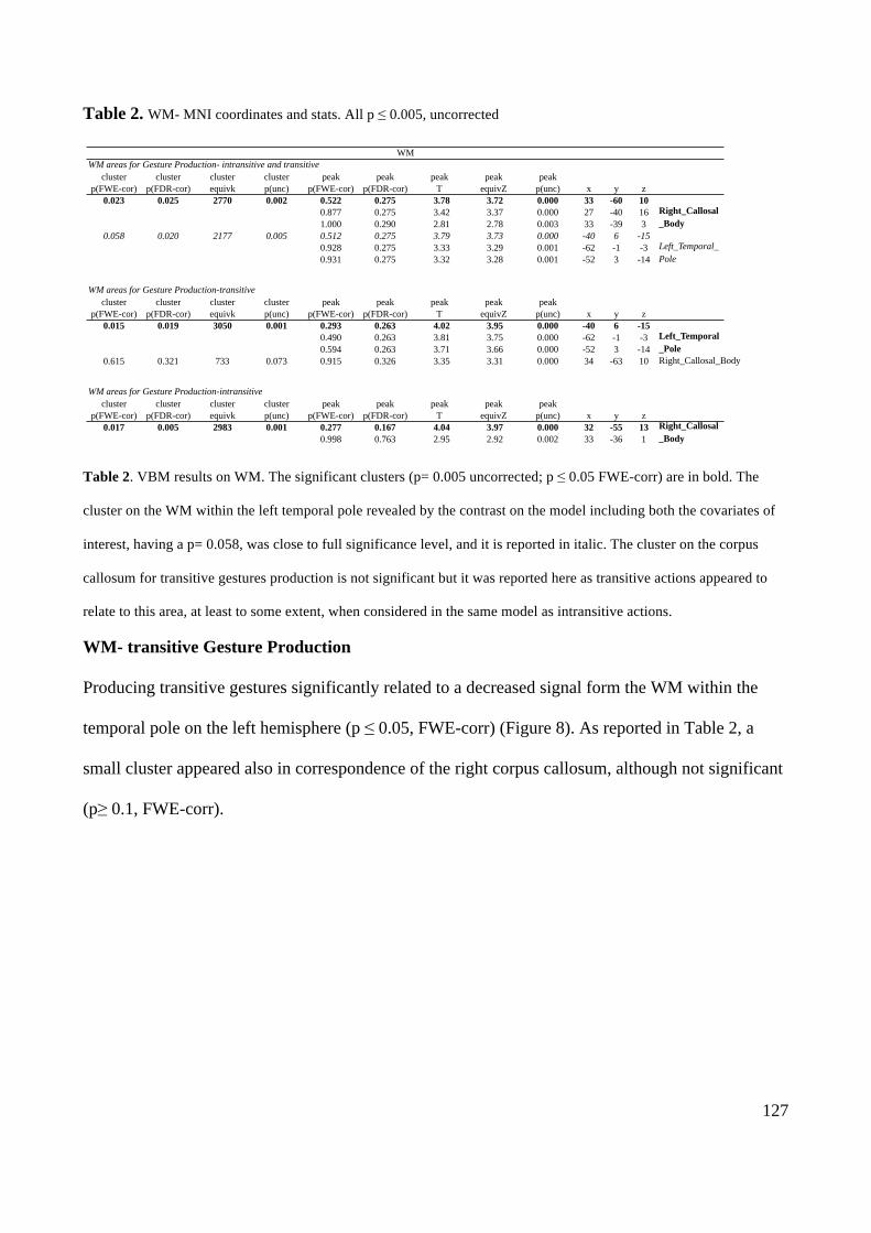
127
Table 2. WM- MNI coordinates and stats. All p ≤ 0.005, uncorrected
Table 2. VBM results on WM. The significant clusters (p= 0.005 uncorrected; p ≤ 0.05 FWE-corr) are in bold. The
cluster on the WM within the left temporal pole revealed by the contrast on the model including both the covariates of
interest, having a p= 0.058, was close to full significance level, and it is reported in italic. The cluster on the corpus
callosum for transitive gestures production is not significant but it was reported here as transitive actions appeared to
relate to this area, at least to some extent, when considered in the same model as intransitive actions.
WM- transitive Gesture Production
Producing transitive gestures significantly related to a decreased signal form the WM within the
temporal pole on the left hemisphere (p ≤ 0.05, FWE-corr) (Figure 8). As reported in Table 2, a
small cluster appeared also in correspondence of the right corpus callosum, although not significant
(p≥ 0.1, FWE-corr).
WM_p=0.005
Page 1
cluster cluster cluster cluster peak peak peak peak peakp(FWE-cor) p(FDR-cor) equivk p(unc) p(FWE-cor) p(FDR-cor) T equivZ p(unc) x y z
0.023 0.025 2770 0.002 0.522 0.275 3.78 3.72 0.000 33 -60 100.877 0.275 3.42 3.37 0.000 27 -40 161.000 0.290 2.81 2.78 0.003 33 -39 3
0.058 0.020 2177 0.005 0.512 0.275 3.79 3.73 0.000 -40 6 -150.928 0.275 3.33 3.29 0.001 -62 -1 -30.931 0.275 3.32 3.28 0.001 -52 3 -14
cluster cluster cluster cluster peak peak peak peak peakp(FWE-cor) p(FDR-cor) equivk p(unc) p(FWE-cor) p(FDR-cor) T equivZ p(unc) x y z
0.015 0.019 3050 0.001 0.293 0.263 4.02 3.95 0.000 -40 6 -150.490 0.263 3.81 3.75 0.000 -62 -1 -30.594 0.263 3.71 3.66 0.000 -52 3 -14
0.615 0.321 733 0.073 0.915 0.326 3.35 3.31 0.000 34 -63 10
cluster cluster cluster cluster peak peak peak peak peakp(FWE-cor) p(FDR-cor) equivk p(unc) p(FWE-cor) p(FDR-cor) T equivZ p(unc) x y z
0.017 0.005 2983 0.001 0.277 0.167 4.04 3.97 0.000 32 -55 130.998 0.763 2.95 2.92 0.002 33 -36 1
Right_Callosal_Body
WM areas for Gesture Production-intransitive
Right_Callosal_Body
WMWM areas for Gesture Production- intransitive and transitive
WM areas for Gesture Production-transitive
Right_Callosal_Body
Left_Temporal_Pole
Left_Temporal_Pole
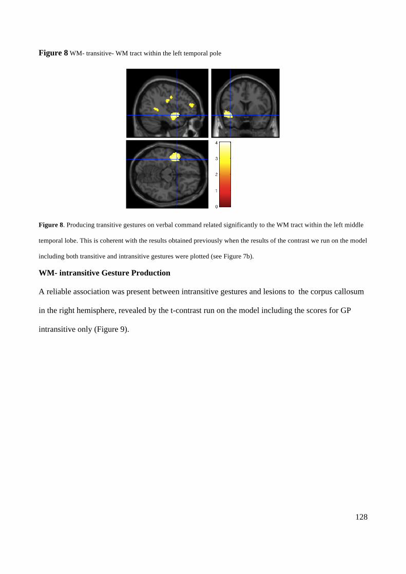
128
Figure 8 WM- transitive- WM tract within the left temporal pole
Figure 8. Producing transitive gestures on verbal command related significantly to the WM tract within the left middle
temporal lobe. This is coherent with the results obtained previously when the results of the contrast we run on the model
including both transitive and intransitive gestures were plotted (see Figure 7b).
WM- intransitive Gesture Production
A reliable association was present between intransitive gestures and lesions to the corpus callosum
in the right hemisphere, revealed by the t-contrast run on the model including the scores for GP
intransitive only (Figure 9).
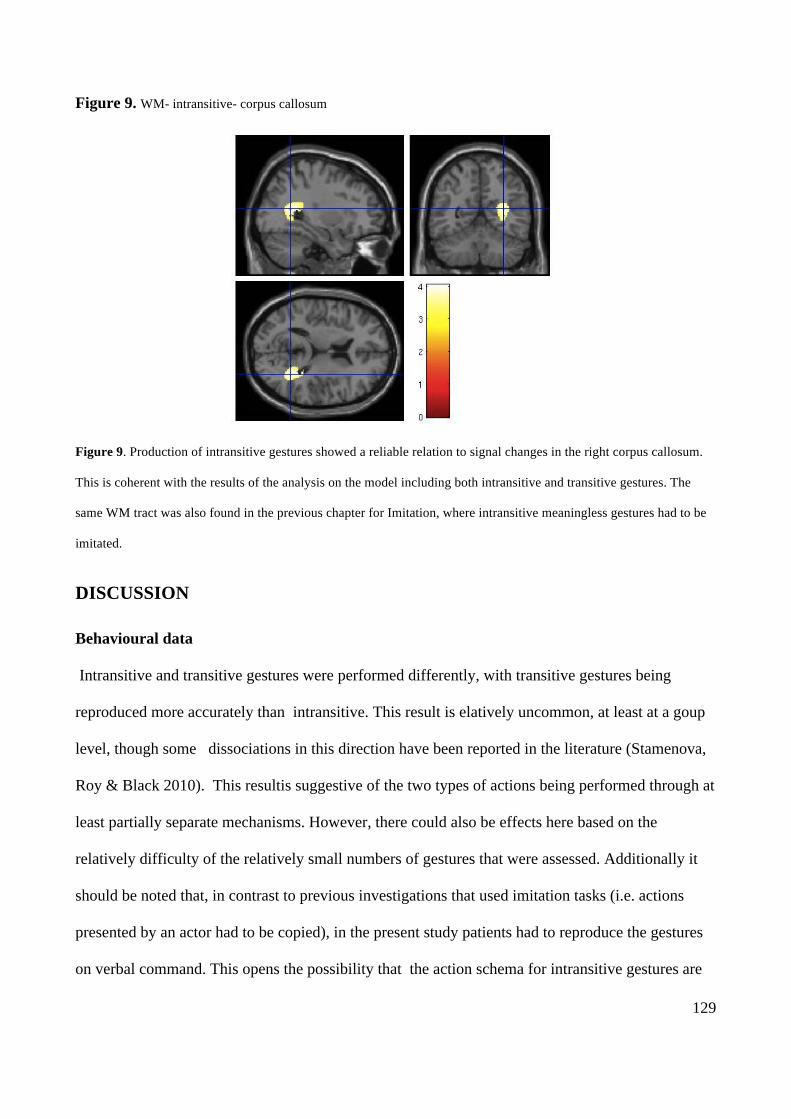
129
Figure 9. WM- intransitive- corpus callosum
Figure 9. Production of intransitive gestures showed a reliable relation to signal changes in the right corpus callosum.
This is coherent with the results of the analysis on the model including both intransitive and transitive gestures. The
same WM tract was also found in the previous chapter for Imitation, where intransitive meaningless gestures had to be
imitated.
DISCUSSION
Behavioural data
Intransitive and transitive gestures were performed differently, with transitive gestures being
reproduced more accurately than intransitive. This result is elatively uncommon, at least at a goup
level, though some dissociations in this direction have been reported in the literature (Stamenova,
Roy & Black 2010). This resultis suggestive of the two types of actions being performed through at
least partially separate mechanisms. However, there could also be effects here based on the
relatively difficulty of the relatively small numbers of gestures that were assessed. Additionally it
should be noted that, in contrast to previous investigations that used imitation tasks (i.e. actions
presented by an actor had to be copied), in the present study patients had to reproduce the gestures
on verbal command. This opens the possibility that the action schema for intransitive gestures are

130
not generally more robust than those for transitive gestures but that the representations of the two
types of gestures are more easy to be accessed by different input systems, i.e. visual or verbal
respectively.
VBM
The contrasts examined areas associated with greater deficits on transitive than intransitive actions,
and vice versa. The analyses revealed that contrasting lesion patterns were associated with the
different problems, along with some overlapping brain regions. Most notably, there was evidence
for subcortical regions involving the left inferior insula being associated with deficits in producing
transitive actions to a name. In contrast, deficits in producing intransitive actions were linked to
frontal and parietal lesions, although this result was not strong. Differences emerged also in terms
of WM damage, with transitive gestures being related to WM lesions within the left temporal
cortex, while the corpus callosum on the right hemisphere appeared to have a role in processing
intransitive gestures. Transitive gestures also appeared to have at least some correlation with the
signal intensity on the right corpus callosum.
Beside those differences, overlapping areas seemed to relate to both types of gesture in the left
supramarginal gyrus (most strongly related to intransitive gestures).
The evidence for distinct neural sites associated with impairments in transitive and intransitive
actions is interesting and supports the case for these two classes of action being supported by
different functional and neural processes.
In terms of GM damage, the results indicated that transitive actions were selectively affected by
lesions to sub-cortical structures (basal ganglia and thalamus). This result is compatible with
findings on Parkinson’s patients who have been found to have poorer performance than controls
when reproducing MF pantomimes but not intransitive gestures (Leiguarda et al. 1997, 2001). The
role of the basal ganglia in action execution will be discussed further in the next chapter. Beside
subcortical areas, the cluster found to relate significantly to transitive gesture production also
131
extended to the left middle temporal cortex, an area that was reliably associated also to the BcoS
Actions Recognition task analysed in the previous chapter (see chapter 5). This same area has also
been found to be active in a PET study when participants observed meaningful but not meaningless
actions (Decety et al. 1997), and it has been reported among the lesions of patients with a deficit in
action production (Price et al. 2010). Moreover, two of the patients reported by Price et al. (2010)
were impaired in gesturing object use, on presentation of the pictures of the target objects, but still
retained the ability to recognize the objects (Price et al. 2010). As already suggested in the previous
chapter, those data indicate a role of the left middle temporal cortex in retrieving information about
actions that are stored in long-term memory. Also, as here, the same area did not appear for the
intransitive gestures. This suggests that the left middle temporal cortex may be more crucial for
actions involving tools.
For the production of intransitive gestures the results in the GM analysis indicated a role of the
inferior frontal cortex and the supramarginal gyrus in the left hemisphere. These results were not
strong, but they do replicate the findings in the previous chapter for the BCoS Imitation task, where
intransitive meaningless actions visually presented by the examiner had to be imitated. Also, in the
present chapter, the cluster centred on the left supramarginal gyrus appeared to relate, at least to
some extent, to both transitive and intransitive gestures, even though the relation was stronger for
the intransitive gestures. The cluster on the frontal area instead was not reported for transitive
actions. However it should be recognized that neither the frontal nor the parietal areas related to
Action Recognition in Chapter 5.
The involvement of the left supramarginal gyrus for transitive, intransitive meaningful and
intransitive meaningless gesture production and imitation supports the role of this area in gesture
execution, regardless the meaning or the modality of presentation of the action (i.e. demonstration
by an actor or verbal command), and these data fit with reports that associate lesions in this region
with ideomotor apraxia (e.g. Heliman et al- 1982). The left inferior frontal regions, that were shared
132
by the intransitive meaningful actions used here and the intransitive meaningless actions used in
chapter 5, may have a role in coding the motor properties of intransitive gestures only. For instance
intransitive gestures may be linked to the need to compute fine-grained spatio-temporal action
codes to generate intransitive actions (Buxbaum et al. 2007), regardless the meaning and the way
they are presented. The schema for transitive gestures instead may be triggered automatically by
the information about the object itself, even in pantomimes where an actual object is not present
(e.g. to put the hand in the grasping posture may automatically trigger the whole action sequence for
the target object). However, as stated above, the results obtained in the present study for intransitive
gestures on GM were not robust enough to draw strong conclusions, and future studies, using for
instance a longer list of both MF and ML stimuli having different levels of difficulty, are needed to
clarify this issue.
Results were obtained also when the analyses were run on models based on WM damage. To date
just the main WM tracts have been mapped, and a only few studies have tried to explore WM
involvement in gesture execution, and cognitive functions in general. However a few points about
the present data can be discussed. For instance, transitive gestures appear to relate significantly to
WM tracts within the left temporal pole, while the intransitive related significantly to the signal
from the corpus callosum. The cluster in the corpus callosum was also related to transitive gestures,
though the relation was lower than it was for intransitive gestures. Moreover the signal in the corpus
callosum correlated with the scores at the BCoS Imitation task in the previous chapter (chapter 5).
More studies, for instance using MRI scans on GM together with Diffusion Tensor Imaging (DTI)
sequences, should be run to look at the areas that are connected by this portion of the corpus
callosum and the possible role of this circuit in action execution.
In contrast with a previous fMRI study on healthy participants (e.g. Kroliczak & Fray, 2009) the
current data point in the direction of differential processes in the production of intransitive and
intransitive gestures. This remains to be clarified, nonetheless it should be said that in prior fMRI
133
studies only relatively limited distal movements of the fingers have been used (due to the
constraints on movement in the scanner). It may be possible these restrictions mean that the stimuli
were not clearly tapping more “typical” transitive and intransitive actions (e.g. hammering
(transitive), waving hello (intransitive). In contrast, the present study used naturalistic actions and
tested for the necessary role of the brain regions affected in the patients.
Finally, as in the previous chapter, the data were analysed using a statistical threshold with a p≤
0.005, instead of p≤ 0.001. This was done in order to reduce the risk of losing information from the
data. The reasons guiding this choice have been elaborated in chapter 5.
In chapter 5 we suggested a follow up on the same patients in order to understand how the pattern
of association between behaviour and brain lesions evolved for the effect of time and rehabilitation
(see discussion in chapter 5). The follow up should consider the gesture production task as a whole,
as well as the split into transitive and intransitive gesture production sub-tasks.
134
7. The Role of the Basal Ganglia in Action Imitation:
Neuropsychological Evidence from Parkinson’s disease Patients
ABSTRACT
Though previous studies have suggested that the basal ganglia are necessarily involved in gesture
imitation, their precise role is unclear. Leiguarda et al. (1997, 2001) report that Parkinson’s Disease
patients (PD) have poor execution of observed meaningful (MF) transitive (tool-related) actions,
despite normal performance with intransitive (non tool-related) MF and meaningless (ML) gestures.
In contrast, Tessari et al. (2007) described two right –brain damaged patients with ideomotor
apraxia, whose lesions involved the basal ganglia, who were more impaired in imitating ML as
compared to meaningful MF transitive pantomimes. This inconsistency was assessed here by testing
a group of PD patients with MF transitive and intransitive pantomimes and matched ML
movements. The data indicate a double dissociation, with PD reporting higher scores when
imitating MF transitive actions than ML matched gestures, while performing better with the ML
than the MF intransitive actions. Performance on ML intransitive gestures also related to the
performance of the patients on the Corsi block test of visuo-spatial memory. The results are
discussed in terms of the factors that load on visual memory for gesture reproduction, as well as the
possible role of the basal ganglia in communicative gestures (for MF intransitive actions).
INTRODUCTION
Previous studies have suggested that the basal ganglia are necessarily involved when participants
perform gestures on verbal command or on imitation (e.g. Pramstaller et al. 1996; Roy et al. 2000;
Leiguarda et al.; 1997, Leiguarda et al. 2001; Tessari, Canessa, Ukmar, Rumiati, 2007). For
instance, Tessari et al. (2007) described two right –brain damaged patients whose lesions involved
the basal ganglia. These individuals were more impaired in imitating meaningless (ML) as
135
compared to meaningful (MF) transitive (or tool-related) pantomimes and whose lesions overlapped
on basal ganglia. In contrast to this, though, Leiguarda et al. (1997, 2001) report that Parkinson’s
disease (PD) patients were impaired on the execution of transitive MF pantomimes both on
imitation and on verbal command, while performance with both symbolic (MF) and non-symbolic
(ML) intransitive gestures were normal. Unlike Tessari et al. (2007), performance on ML transitive
gestures was not tested.
Rumiati et al. (2002) proposed a ‘dual route’ model of action imitation in which learned actions can
be imitated via either of two ‘routes’ – a lexical route based on the recognition of a learned action
and the retrieval of a learned action programme, and a non-lexical route based on direct mapping
between perceived motor actions and output motor commands, that does not depend on having
learned representations for the actions (see Figure 1 in the general introduction). According to
Tessari et al. (2007), the basal ganglia modulate the direct (non-lexical) route to action. In contrast
they suggest that the lexical route operates through posterior parietal and pre-frontal brain regions,
particularly in the left hemisphere (see Rothi et al. 1991, 1997; Peigneux et al. 2001, for a similar
account). These arguments are supported by data from functional brain imaging (Peigneux et al.
2004; Kareken et al. 1998) and from lesion overlap analyses of patients with problems in imitating
meaningful or meaning less gestures (Tessari et al. 2007). On the other hand, the data from
Leiguarda et al. (1997, 2001) suggest that the basal ganglia are necessary for the imitation of
transitive actions, while intransitive actions may operate through cortical regions – with this holding
even for meaningless actions based on intransitive gestures.
To attempt to reconcile these apparently conflicting results, we examined a group of PD patients on
tasks requiring the imitation of both MF and ML transitive and intransitive gestures. Is the
involvement of the basal ganglia most apparent in the performance of ML relative to MF gestures,
or are they most required to carry out transitive MF gestures? The performance of these different
gestures was tested here using sets of matched stimuli, and the relations between imitation scores,
136
object and action recognition, neuropsychological assessment and neurological exam were
considered.
METHODS
In this chapter we tested a sample of PD patients in imitation of MF and ML transitive and
intransitive gestures. The PD patients’ scores were also related to an index of their peripheral
neurological symptoms and to the neuropsychological evaluation.
The PD patients and controls sample and the tasks are described in the method chapter (see pgs. 21-
38).
The tests employed in the neuropsychological evaluation are described in Appendix 3.
RESULTS
Figure 1 illustrates the proportion accuracy scores for imitating transitive and intransitive,
meaningful and meaningless gestures, for the PD patients and the controls. For the gesture and
object recognition tasks the PD patients scored at ceiling. This indicates that any problems in
imitating MF gestures were not due to poor perceptual processing or access to stored visual-
perceptual knowledge. Since separate control participants were used for the transitive and
intransitive gestures the data were analysed first for transitive and intransitive gestures separately,
comparing the PD patients and each set of controls; subsequently the data were compared between
transitive and intransitive gestures for the PD patients and the controls separately.
Imitating transitive (object-related) gestures
Accuracy scores15
15 The Cohen’s k agreement coefficient was calculated on the scores provided by the two independent raters. The
coefficient was computed for MF and ML actions taken separately, and for the total action scores. The analysis was
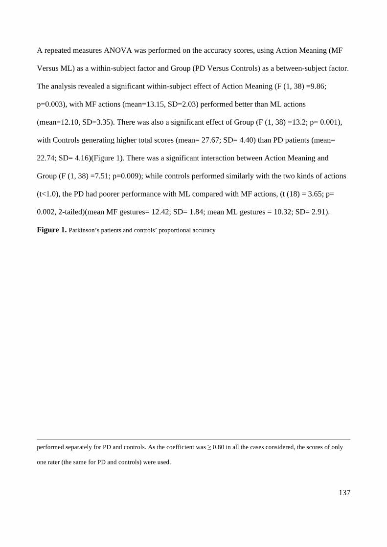
137
A repeated measures ANOVA was performed on the accuracy scores, using Action Meaning (MF
Versus ML) as a within-subject factor and Group (PD Versus Controls) as a between-subject factor.
The analysis revealed a significant within-subject effect of Action Meaning (F (1, 38) =9.86;
p=0.003), with MF actions (mean=13.15, SD=2.03) performed better than ML actions
(mean=12.10, SD=3.35). There was also a significant effect of Group (F (1, 38) =13.2; p= 0.001),
with Controls generating higher total scores (mean= 27.67; SD= 4.40) than PD patients (mean=
22.74; SD= 4.16)(Figure 1). There was a significant interaction between Action Meaning and
Group (F (1, 38) =7.51; p=0.009); while controls performed similarly with the two kinds of actions
(t<1.0), the PD had poorer performance with ML compared with MF actions, (t (18) = 3.65; p=
0.002, 2-tailed)(mean MF gestures= 12.42; SD= 1.84; mean ML gestures = 10.32; SD= 2.91).
Figure 1. Parkinson’s patients and controls’ proportional accuracy
performed separately for PD and controls. As the coefficient was ≥ 0.80 in all the cases considered, the scores of only
one rater (the same for PD and controls) were used.
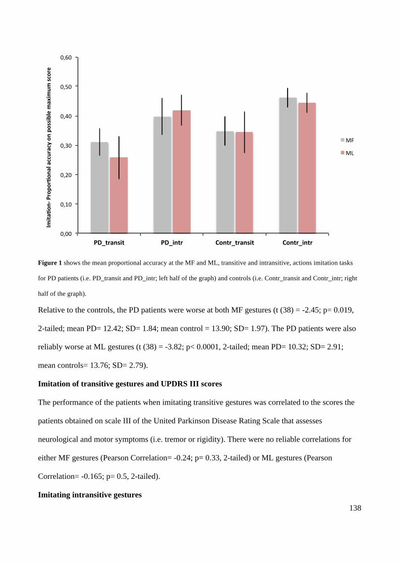
138
Figure 1 shows the mean proportional accuracy at the MF and ML, transitive and intransitive, actions imitation tasks
for PD patients (i.e. PD_transit and PD_intr; left half of the graph) and controls (i.e. Contr_transit and Contr_intr; right
half of the graph).
Relative to the controls, the PD patients were worse at both MF gestures (t (38) = -2.45; p= 0.019,
2-tailed; mean PD= 12.42; SD= 1.84; mean control = 13.90; SD= 1.97). The PD patients were also
reliably worse at ML gestures (t (38) = -3.82; p< 0.0001, 2-tailed; mean PD= 10.32; SD= 2.91;
mean controls= 13.76; SD= 2.79).
Imitation of transitive gestures and UPDRS III scores
The performance of the patients when imitating transitive gestures was correlated to the scores the
patients obtained on scale III of the United Parkinson Disease Rating Scale that assesses
neurological and motor symptoms (i.e. tremor or rigidity). There were no reliable correlations for
either MF gestures (Pearson Correlation= -0.24; p= 0.33, 2-tailed) or ML gestures (Pearson
Correlation= -0.165; p= 0.5, 2-tailed).
Imitating intransitive gestures
0,00#
0,10#
0,20#
0,30#
0,40#
0,50#
0,60#
PD_transit* PD_intr* Contr_transit* Contr_intr*
Imita
/on0*Propo
r/on
al*accuracy*on
*possible*maxim
um*sc
ore*
MF#
ML#
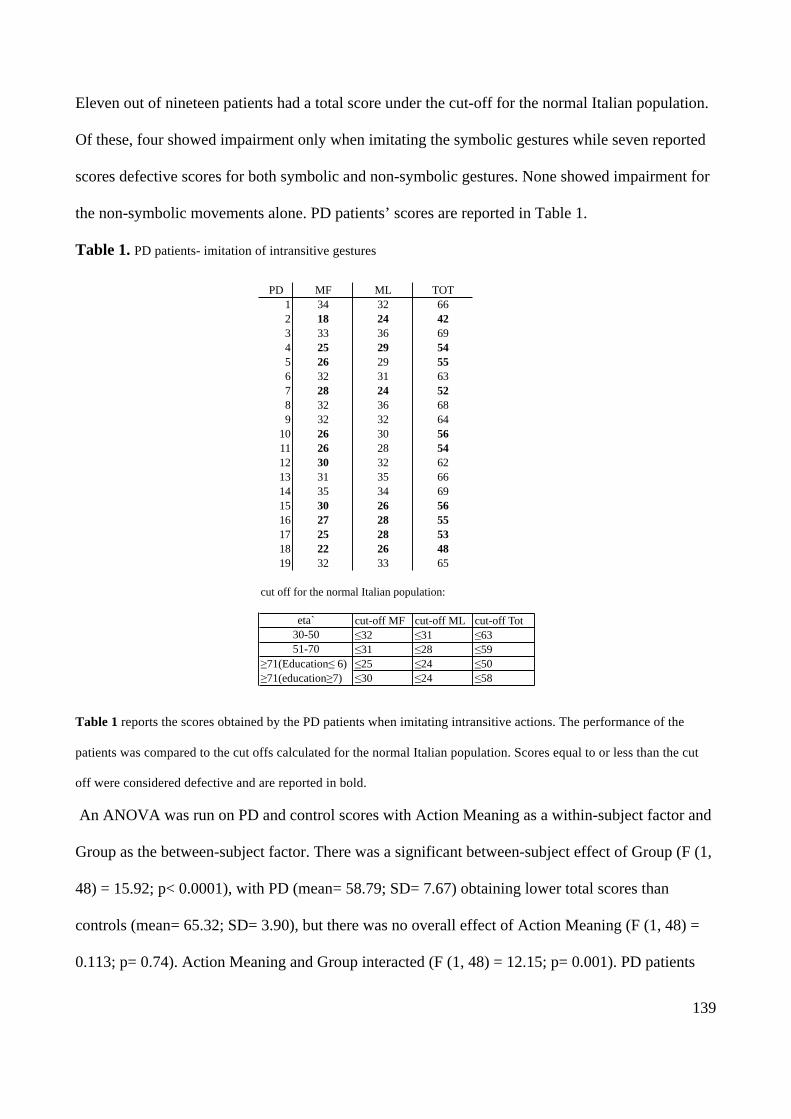
139
Eleven out of nineteen patients had a total score under the cut-off for the normal Italian population.
Of these, four showed impairment only when imitating the symbolic gestures while seven reported
scores defective scores for both symbolic and non-symbolic gestures. None showed impairment for
the non-symbolic movements alone. PD patients’ scores are reported in Table 1.
Table 1. PD patients- imitation of intransitive gestures
Table 1 reports the scores obtained by the PD patients when imitating intransitive actions. The performance of the
patients was compared to the cut offs calculated for the normal Italian population. Scores equal to or less than the cut
off were considered defective and are reported in bold.
An ANOVA was run on PD and control scores with Action Meaning as a within-subject factor and
Group as the between-subject factor. There was a significant between-subject effect of Group (F (1,
48) = 15.92; p< 0.0001), with PD (mean= 58.79; SD= 7.67) obtaining lower total scores than
controls (mean= 65.32; SD= 3.90), but there was no overall effect of Action Meaning (F (1, 48) =
0.113; p= 0.74). Action Meaning and Group interacted (F (1, 48) = 12.15; p= 0.001). PD patients
PD MF ML TOT1 34 32 662 18 24 423 33 36 694 25 29 545 26 29 556 32 31 637 28 24 528 32 36 689 32 32 64
10 26 30 5611 26 28 5412 30 32 6213 31 35 6614 35 34 6915 30 26 5616 27 28 5517 25 28 5318 22 26 4819 32 33 65
cut off for the normal Italian population:
cut-off MF cut-off ML cut-off Tot≤32 ≤31 ≤63≤31 ≤28 ≤59≤25 ≤24 ≤50≤30 ≤24 ≤58
≥71(Education≤ 6)≥71(education≥7)
eta`30-5051-70

140
generated better scores when imitating ML (mean= 30.16; SD= 3.75) than MF gestures (mean=
26.63; SD= 3.75)(t (18) = -2.33; p= 0.032, 2-tailed). In contrast, controls performed better with MF
(mean= 33.29; SD= 2.36) than ML gestures (mean= 32.03; SD= 2.37) (t (30) = 2.62; p= 0.014, 2-
tailed).
Further comparisons between the PD and controls when PD and Controls confirmed that the control
advantage was reliable for MF actions alone (t (corr. df = 24.4) = 4.25; p< 0.0001, 2-tailed), while
there was a borderline effect for ML scores (t (corr. df = 26.97; p= 0.061, 2-tailed).
Intransitive actions and UPDRS III scores
There was no significant correlation between the part 3 of the United Parkinson’s Disease Rating
Scale (UPDRS III) and the total imitation score for intransitive gestures (Pearson Correlation= -
0.36; p= 0.14, 2-tailed); nor were there effects for MF or ML considered alone (MF: Pearson
Correlation= -0.38; p= 0.11; ML: Pearson Correlation= -0.29; p= 0.23, both 2-tailed).
Comparison between transitive and intransitive actions
As the scores for the transitive and intransitive action tasks were based on different scales, a
proportional score was calculated based on accuracy relative to the total maximum score for
transitive and intransitive gestures, summed over the MF and ML stimuli (maximum scores= 40 and
72 respectively), MF and ML The patients’ results, after being transformed as described above,
were subjected to 2 X 2 Analysis of Variance (ANOVA), with Type of Action (Transitive vs
Intransitive) and Action Meaning (MF versus ML) as within-subjects factors. The analysis revealed
both significant main effects of the Type of Action (F (1, 18) =119.05; p < 0.0001)(transitive mean=
0.57; SD= 0.1; intransitive mean= 0.82; SD= 0.11), and of Action Meaning (F (1, 18) = 4.54; p=
0.047) (MF mean= 0.35; SD= 0.07; ML mean= 0.34; SD= 0.1) actions. Also the interaction
between Type of Action and Action Meaning was significant (F (1, 18) = 16.28; p= 0.001). For
transitive gestures there was more accurate performance with MF than ML actions (t(18)= 3.78),
141
while for intransitive actions there were more accurate gestures to ML than MF stimuli (t(18)= -
2.14) (both p<0.05).
The data for the controls were first transformed as for the patients and then entered into a 2-factor
ANOVA with Type of Action (Transitive versus Intransitive), Action Meaning (MF versus ML) as
between-subjects factors. The analysis revealed a significant between-subject effect of Type of
Action (F (1, 50) = 81.91; p< 0.0001) and a trend for an effect of Action Meaning (F (1, 50) = 3.37;
p= 0.072). The interaction between the two factors was not significant (F (1, 50) = 1.45; p= 0.23).
Intransitive actions (mean= 0.91; SD= 0.05) were imitated more accurately than transitive action
(mean= 0.69; SD= 0.11). MF actions tended to be reproduced more accurately than ML actions
(mean MF= 0.42; SD= 0.07; mean ML= 0.40; SD= 0.07) actions.
Gesture imitation and neuropsychological assessment
The relations between imitation performance and the general neuropsychological screening tests
were also considered. Relative to the norms established for the standard neuropsychological tests,
the PD patients did not show an impairment when evaluated for general intelligence, language
functions, visual perceptive functions, short-term memory for verbal material and locations in
space, and attention (see Table 1, also Appendix 1). However 2 PD patients showed a borderline
score at the Corsi block task, and 3 showed borderline performance at the Verbal Fluency test
(FAS), so correlations between those tests and the imitation tasks were conducted. For the
correlation only the patients having available scores in the Corsi block tast and FAS were
considered.
Corsi block task, FAS and gesture imitation
A significant correlation emerged between total scores on imitating transitive actions and the Corsi
test (Pearson Correlation (n=13) = 0.63; p= 0.02, 2-tailed). This result was confined to ML actions
(Pearson Correlation (n=13) = 0.49; p< 0.001), and the correlation was not reliable for MF
142
transitive actions (Pearson Correlation (n=13) = 0.38; p= 0.21). The correlations between transitive
actions and verbal fluency were not reliable (p>0.05).
For intransitive actions (MF and ML), none of the correlations were reliable (all p>0.05).
DISCUSSION
The present results were collected from a group of PD patients who showed good recognition of
gestures and objects implied in transitive actions, but who were nevertheless impaired at action
imitation. Our results match previous findings, whilst also painting a more complex picture of
imitation deficits in PD. Similarly to Tessari et al. (2007) we found that the PD patients were
impaired at imitating ML relative to MF transitive gestures. Tessari et al. used this result to argue
for PD patients having an impaired direct, non-lexical route in imitation, inferring from this that the
direct, non-lexical route is mediated by the basal ganglia. In contrast, Leiguarda et al. (1992, 2001)
reported that PD patients were impaired at imitating transitive relative to intransitive gestures.
Consistent with this, our PD patients were poor at transitive gestures and obtained higher accuracy
scores on intransitive gestures. Our data go beyond the previous results though by assessing both
the type of action (transitive or intransitive) and the familiarity of the gesture (MF vs. ML).
Through this we were able to demonstrate a form of double dissociation in which ML gestures were
impaired for transitive actions whilst MF intransitive gestures were disrupted more than ML
gestures. This pattern of results goes against a simple account of PD patients as either having an
impaired direct, non-lexical route to imitation, or against them having a deficit specific to transitive
actions. We consider both points.
While the results for transitive gestures concur with the argument that PD patients have a deficit in
a direct, non-lexical route to imitation (MF>ML), the results for intransitive actions do not (since
ML>MF). We also found that the ability to imitate ML transitive actions correlated with
performance on the Corsi blocks task – a test of visuo-spatial memory. This last finding suggests
143
that transitive ML stimuli placed a particular load on visuo-spatial memory, and that the PD may
have had difficulty reproducing these actions for this reason. The lack of correlation for ML
intransitive actions may have occurred because these actions placed a lesser load on Visuo-spatial
STM. A previous study found some PD patient to be impaired in an n-back task tapping visuo-
spatial STM (Costa et al. 2010;, as well as in a delayed-response test in a spatial condition as
compared with a non-spatial (object) condition (Postle et al. 1997). This also meshes with controls
also finding these actions the most difficult to re-produce (see Figure 1). It is possible that demands
on visuo-spatial memory contributed to previously reported deficits in PD patients imitating ML
actions (Tessari et al. 2007).
In contrast to their performance with transitive actions, the patients and controls generally
performed better with intransitive actions. This could be due to these actions being less complex
than transitive actions, despite our attempts to match the action types. Alongside this, the PD
patients were relatively impaired, compared with the controls, at re-producing MF intransitive
actions. In absolute terms, this effect was small and so we need to be cautious in any interpretations.
However, it is suggestive that the basal ganglia may be particularly involved either in
communicative gestures or in making gestures in relation to the body as opposed to another object
(as required for transitive actions). Consistent with this, a previous study reported that PD patients
were impaired in their pragmatic communication abilities and this deficit correlated with measures
of frontal lobe functions (Mc Namara et al. 2002).
In sum, the current data go against an account of initiation deficits in PD purely in terms of a loss of
a direct, non-lexical route to action. The results suggest instead that factors such as load on visuo-
spatial memory and requirements either to make communicative or body-related gestures may be
particularly important in eliciting problems in imitation.
144
8. GENERAL DISCUSSION
The goal of the present work was to try to assess the functional and neural mechanisms underlying
action production and recognition, and in doing this, to reconcile some inconsistencies in the
literature on praxis. One specific aim was to assess the relations between the processes involved in
the reproduction of different types of gesture (transitive and intransitive, MF or ML), and a second
was to evaluate the relations between gesture recognition, action production and imitation.
The first empirical chapter (Chapter 3) presented a neuropsychological investigation of a group of
patients having unilateral lesions either in the left (Left Brain Damaged, LBD) or the right (Right
Brain Damaged, RBD) hemisphere. Previous similar investigations have typically involved only a
small range of tasks (e.g. comparing imitation of MF pantomimes with imitation of ML intransitive
actions, or the recognition MF gestures and imitation of only MF gestures) and often assessed only
small groups of patients. In contrast to this I sampled a large group of patients, and give them a
battery of tasks including MF and ML transitive and MF and ML intransitive actions, as well as two
tasks where they had to demonstrate knowledge of the MF actions they previously imitated.
Imitation and recognition tasks were also administered to a group of healthy participants and
compared to the LBD and RBD patients. In this study all patients and controls were right handed.
The results showed that the patients generally had poorer action production than the controls,
regardless of the side of the lesion, while on average the patients did not differ significantly from
controls when they had to recognize MF actions. On imitation, LBD and RBD differed in their total
scores with transitive gestures, although this difference was driven by the performance with the
transitive ML only, as the two groups did not differ when they had to reproduce transitive MF
pantomimes. Also there were no reliable differences between the two groups for the intransitive
gestures. At single case level more LBD patients fell below the norms for control participants, but
in almost all cases there were instances where at least one RBD patient performed the same way.
145
Significant differences between MF and ML transitive gestures emerged within both the patient and
control groups, while intransitive actions did not show a significant difference between MF and ML
gestures (neither for the patients nor the controls). In addition, intransitive gestures were always
easier to perform than the transitive gestures, regardless whether the actions were MF or ML, and
there was also better recognition of intransitive gestures for both patients and controls.
Although there were dissociations between the imitation tasks, in the patient group the performance
of MF and ML transitive actions correlated with each other and the MF transitive also correlated
with MF and ML intransitive actions. MF and ML intransitive actions also correlated within each
other. Within controls MF and ML transitive actions correlated with each other as were MF and ML
intransitive actions, but performance on transitive and intransitive actions did not correlate (note
though that this result can rise because the intransitive actions were found particularly easy to
perform by controls). Also correlations emerged between the imitation and recognition tasks.
Finally the generation of MF transitive and MF intransitive actions correlated with Picture Naming
from the BCoS, a task that uses mixtures of animate and inanimate objects. Also the accuracy of
MF transitive gestures correlated with scores on the BCoS Action Recognition test, suggesting that
the retrieval of action meaning is crucial for imitating those actions. In contrast ML intransitive
gesture performance correlated with the BCoS Figure Copy task, indicating the importance of
spatial coding when ML actions have to be reproduced.
The second study (Chapter 4) employed scores on the same MF and ML transitive and intransitive
imitation tasks as used in Chapter 3 as predictors of changes in voxel intensity in MRI T1 images of
segmented grey matter (GM) in a group of brain damaged patients with acquired brain lesions. The
data were then analyzed using voxel-based morphometry. For all kinds of gestures apart from the
MF intransitive actions, reliable correlations were found between impairments in gesturing and
neural changes in both the left and the right hemispheres. MF and ML transitive and ML
intransitive gestures related to the signal in the left superior parietal cortex involving the precuneus,
146
and, for the ML transitive actions, the cluster also extended into the left occipital cortex. Moreover,
MF and ML transitive, as well as ML intransitive actions, related to clusters in the right frontal
cortex and SMA (though for the ML transitive actions this cluster did not reach full significance
after correction). In chapter 4 results were also obtained in the WM, but only for the ML transitive
gestures, with the contrast revealing a large significant cluster in the corticospinal tract running
within the left hemisphere. MF intransitive gestures failed to give any significant result and this
may reflect a lack of variation for these stimuli, due to generally good performance across the
patients.
The brain areas involved in the production of MF gestures on verbal command, imitation of ML
actions and action recognition were investigated in the third empirical chapter (Chapter 5). A VBM
approach was again used on a large sample of acute stroke patients, but in this case combining the
data for learned transitive and intransitive actions. The analysis used CT scans as images and the
scores at Gesture Production, Imitation and Action Recognition from the BCoS as predictors of
changes in GM signal intensity. In this study reliable results were obtained only for Action
Recognition, which was associated with the signal from the left anterior cingulate cortex and the left
middle temporal lobe. The results obtained for Gesture Production and Imitation did not survive to
FEW/FDR corrections, suggesting that other factors (e.g. general impairment) may determine
performances. Nonetheless it can be noticed that Gesture Production related to a cluster that also
involved the left middle temporal cortex - so partially overlapping the results obtained for Action
Recognition. Moreover Gesture Production and Imitation both indicated effects related to the
superior parietal cortex in the left hemisphere. However these results must be treated cautiously.
The study presented in chapter 5 also highlighted significant results in WM. Both Action
Recognition and Gesture Production significantly related to WM change within the left superior
temporal pole. Gesture Production also significantly correlated with the signal from the posterior
147
part of the corpus callosum, and a cluster in the same WM tract was also revealed for Imitation
(although it did not reach the full significance level after correction).
The fourth empirical chapter (Chapter 6) focused on the Gesture Production task from the BCoS
that was split into two subtests: i) production of MF transitive pantomimes, and ii) production of
intransitive MF symbolic gestures. The same patient sample was used as in the previous chapter.
The aim was to look at different neural correlates of production on verbal command of transitive
and intransitive gestures. A preliminary analysis compared the patients’ accuracy in producing the
two types of gestures, highlighting an advantage for transitive with respect to the intransitive
actions. This is strange if compared with the results in chapter 3, where intransitive gestures were
produced more accurately than the transitive. However the tasks in chapter 3 and 6 differed in that
in chapter 3 patients imitated visually presented gestures, while in chapter 6 they had to execute the
gestures on verbal command. At a neural level, only the transitive gestures gave results surviving
cluster correction. This analysis revealed a cluster centred on the insula in the left hemisphere. Also
production of intransitive gestures revealed two clusters in the left supramarginal gyrus and left
frontal cortex. Though these last results they did not survive cluster correction, they did overlap the
results found for Imitation (chapter 5) (where intransitive ML gestures had to be produced in
imitation). The left supramarginal gyrus was also associated to intransitive ML action imitation in
chapter 4. Note however that all the results for intransitive Gesture Production, BCoS Imitation
(chapter 5) and intransitive ML actions imitation in chapter 4 were not significant after correction
and they must be taken cautiously.
In the final empirical study (Chapter 7) a group of patients with idiopathic Parkinson’s Disease
(PD) with minimal cognitive decline, was tested. The imitation of MF and ML transitive and
intransitive gestures was probed. Generally the PD patients showed lower performance than the
controls but also demonstrated different patterns of performance with transitive and intransitive
actions. There was worse performance with the ML transitive actions as compared to the MF
148
transitive actions, and worse performance with the MF intransitive than the ML intransitive actions.
The performance of these patients did not relate to their peripheral neurological symptoms but may
(to some degree) reflect the differential overall difficulty of the different tasks.
Implications for models and the neuroanatomy of action recognition and production
Generally the results obtained in the five studies indicate that tasks involving praxis functions are
highly correlated and may differentiate mainly because of the difficulty of the task. These data do
not fit terribly well with the existence of separate processes for action production to name, imitation
or command. At group level, in Chapter 3, the results mainly suggested that the different tasks
varied in difficulty, even though some patients behaving opposite way to the rest of the group were
reported (i.e. one right-brain damaged patient who imitated better transitive than intransitive
actions; one patient left-brain damaged patients being at control level for imitation of transitive MF
gestures but who was impaired in recognizing them; one left-brain damaged patient who was
impaired in recognition but not on imitation of MF intransitive gestures; one left-brain damaged
patient having normal scores when imitating ML intransitive actions but being below the cut off for
MF intransitive gestures) suggesting the involvement of at least some separate process. However,
these behaviours were reported only on few patients and they cannot be considered as final proof in
favour of the existence of a dual process. At group level, the comparison between transitive and
intransitive gestures in chapter 6 revealed an advantage for transitive actions, with this result going
in the opposite direction to the data in chapter 3 (and from the majority of the other studies) where
an advantage for intransitive gestures has been noted. However, in chapter 6 the gestures had to be
produced on verbal command while in chapter 3 they had to be imitated. It is possible that
differences in performance may depend by on how the stimuli are presented. It is possible that
actions schemas for intransitive gestures are more easily activated from a visual input while the
transitive are more easily activated via auditory input. Nonetheless this also suggests that different
types of information may be crucial for the execution of transitive or intransitive gestures, with one
149
type of information more automatically linked to the visual system (intransitive) while the other
type (for transitive) is linked to the auditory system. This remains speculative however, especially
given that transitive gestures reflect tool use, and there have been shown to be strong visually-
driven responses to tools in various imaging studies (REFS).
Also, the analyses in chapter 4 (VBM on MRI scans and imitation scores of chronic patients)
revealed common areas, as well as some differences in brain regions, involved in the processing of
different action types. The right frontal cortex and SMA were associated with MF and ML transitive
as well as ML intransitive gestures. Also, the cluster for MF transitive gestures mainly involved
those frontal areas in the right hemisphere, only slightly extending to the left posterior cingulate
cortex. This result is not standard in relation to the literature, and I have proposed that it was mainly
driven by a few patients having a right frontal lesion who failed in imitation because of an
attentional deficit or an impairment in controlling their motor behaviour, when imitating both MF or
ML, transitive or intransitive actions. ML transitive and ML intransitive gestures were also related
to a cluster in the parietal cortex having its peak in the precuneus. The precuneus was previously
reported to sustain the ability to shift attention between different locations in space and spatially
guided behaviours (see Cavanna et al. 2006 for a review). The intervention of visuo-spatial
processes in ML intransitive gestures is compatible with the correlation found at behavioural level
in chapter 3 between imitation of those gestures and the BCoS Figure Copy. Though a correlation
with Figure Copy was not found for ML transitive gestures, ML transitive gestures shared a cluster
with ML intransitive gesture performance in this brain area putatively involved in visuo-spatial
processing. Moreover, BCoS Figure Copy taps the ability to put elements in their correct position in
order to give to the figure its proper configuration. This ability may be important in intransitive
gestures, as they mainly require moving the limb in a required direction in order to reach the target
configuration. In contrast, transitive gestures mainly require repeated movements with the limb

150
never stopping in a precise configuration). So, it is possible that both transitive and intransitive ML
actions share those processes needed to correctly understand the direction of action.
In Chapter 5 (VBM on CT scans and BCoS scores of acute patients) Action Recognition was the
only task giving strong results – with an association to the left anterior cingulate cortex (ACC) and
the middle temporal gyrus. The ACC is an area that has interconnections with the hippocampus and
the structures belonging to the limbic system and has been reported to have a role in cognitive
functions as well as in emotion and motivation (see Bush et al. 2000 for a review). We proposed
two possible explanations of our result for BCoS Action Recognition: i) the lesion in the ACC
contributed to a deficit in recognizing actions because of its relation to the hippocampus where
information about known actions may be stored; ii) the ACC has a role in identifying the motivation
or the intention of the actions and this may be crucial for correctly recognize them. Gesture
Production (on verbal command) and Imitation did not give strong results; nonetheless they may
suggest something about the processes through which actions are executed. Indeed a cluster for
Gesture Production extended into the left middle temporal cortex, which was also found for Action
Recognition, but not for Imitation. As Action Recognition and Gesture Production both used MF
actions as stimuli it may be possible that the left middle temporal cortex had a role in retrieving
long-term memories for gestures. The cluster found for Gesture Production had its peak on the left
supramarginal gyrus and this was also found for Imitation but not for Action Recognition. Also,
when Gesture Production was split in its transitive and intransitive components (chapter 6), it
appeared that the intransitive but not the transitive gestures related to the left supramarginal gyrus.
Summarizing, Imitation of ML intransitive actions and production of MF intransitive gestures on
verbal command related to clusters on the left supramarginal gyrus on sub-acute patients (chapters 5
and 6), and the same cluster was also found for chronic patients imitating ML intransitive in chapter
4. Additionally impairments for the BCoS Imitation of ML intransitive gestures task, and the
intransitive MF Gestures Production task, showed a cluster on the frontal cortex (chapter 5 and 6).
151
In all three studies these clusters did not survive to corrections, nonetheless they suggest that frontal
and parietal areas may have a role in the execution of intransitive gestures, regardless of their
meaning and the modality of stimulus presentation. However this conclusion must be made very
cautiously, and further investigation is needed, for instance testing patients in imitation and
production on verbal command using the same set of MF and ML intransitive actions. Also it would
be useful to carry out those studies on the same patients in both acute and chronic phases to assess
the stability of the results..
Stronger findings were obtained in chapter 6 (VBM with CT scans and BCoS Gesture Production,
transitive and intransitive, scores of acute patients) for the production of MF transitive gestures,
which were related to a cluster centered on the insula in the left hemisphere. This result matched
findings in chapter 7 where Parkinson’s patients were tested on the imitation of MF and ML,
transitive and intransitive gestures. The study on Parkinson’s patients gave the results that best
supported the dual route model for action imitation, with patients showing opposite patterns of
performance according to the type of gestures that had to be executed. PD patients generally had
lower gesture scores than controls, but while they did worse with the ML transitive than the MF
transitive pantomimes, the pattern was the opposite when intransitive actions were considered
(ML>MF). This is difficult to explain in terms of overall task difficulty but suggests the possibility
of a further subdivision of action imitation according to other characteristics of the actions.
However even if several pieces of data in the present work are consistent with a distinction between
MF and ML actions it is still unclear if a distinction exists also between actions involving or not a
tool (transitive vs. intransitive actions). Some results pointed in that direction. To assess the
dissociation between transitive and intransitive actions the stimuli tended to differ in difficulty – a
problem that pervades the literature and not just the current data. In particular, intransitive actions
tended to be easier than transitive, for controls as well as patients, and this may make it difficult to
demonstrate a double dissociation (as opposed to the one-way dissociation in favour of intransitive
152
actions. It may be easier to generate double dissociations between patients (as groups and not only
at single subject level) if transitive and intransitive gestures could be more balanced in terms of
difficulty. The current analysis was limited in this respect since it used a standard
neuropsychological screening test for at least some of the analyses.
Some methodological issues
VBM was used in the investigations of the neural correlates of action production, an approach that
uses the general linear model to statistically assess the relations between brain tissue integrity and
behavioral performance. Here, the MF and ML imitation scores were used as predictors of change
in the signal intensity coming from each voxel across the whole brain in a group of consecutively
sampled patients, not pre-selected on the basis of having apraxia. In addition the analysis used
continuous scores on tests of gesture imitation in order to characterize imitation abilities. This
approach also allows controls over covariates of no interests such as age, gender, neglect, which
may otherwise contaminate the results. VBM also has the advantage to allow offline testing (at least
compared with on-line measurement techniques, such as fMRI). This can facilitate investigations
about praxis when the tasks require to execute actual movements as, differently form the most part
of the others neuroimaging approaches, where movements create artifacts.
However VBM can fail to give results if the data are not normally distributed, a quite common
characteristic of data with patients. There can also be problems in segmenting and normalizing
patients with large lesions or where there has been substantial enlargement of the ventricles. In
general, though, these problems proved relatively slight. The scans were also independently verified
by eye, to ensure that segmentation and normalization was accurate.
One of the interesting aspects of the VBM analyses here is that CT as well as MRI was used. CT
may have the disadvantage of having more noise in the signal and being lower resolution although it
also has some benefits (e.g., less distortion). Despite the possible noise, the current study indicates
that CT analyses can be useful, at least when large numbers of patients are involved. Also, the aim
153
of the chapter where CT scans were used was to test a sample of sub-acute patients who had jus
survived from a stroke, also in order to explore the neural correlates of a new neuropsychological
screen that is mainly aimed at obtaining quick, but efficient, evaluations of patients when they are
admitted following a stroke. Only CT scans were available for those patients, taken as routine when
patients were first seen in the hospital. Since the scans were taken from a very large sample of
patients, it is likely to be quite representative of the stroke population.
Other implications
Understanding how movement production works may be critical for creating tests able to assess
movement deficits that are due to a cognitive (as opposed to motor) impairment and that can go
undiagnosed (because the test is too easy or because it does not tap the component that is impaired.
For instance if the double dissociation between MF and ML gestures and the existence of a double
route mechanism for imitation is true, and if the direct route allows the subject to reproduce both
MF and ML gestures, then a problem to the semantic route could remain undiagnosed as the patient
is still able to imitate the MF actions through the direct route. Nevertheless the patient can suffer
from subtle manifestations of the deficit in everyday life when actions need to be produced from
long-term memory. The BCoS test is interesting in this respect because it attempts to tap different
parts of the action system, using ML actions for imitation and MF actions for production from
name, as well as action recognition. In extensions of the current work it would be interesting to
assess how well the analyses of ideomotor apraxia in patients predict their function in everyday life,
when objects are handled rather than pantomimed in action. Very often patients perform better
when using real objects than when pantomiming (Chainay & Humphreys, 2002), but nevertheless
subtle problems may emerge particularly when the tasks become more taxing (e.g., with multi-
steps). This should be analysed.
In addition, it would be interesting to evaluate whether there is differential success in rehabilitating
the different symptoms in patients – can training on one route transfer to performance using the
154
other route? If this is the case, then an argument can be presented that the routes are not modular but
more distributed, so that retraining one component can generalise to other forms of action. This
suggests that fine-grained analyses of action rehabilitation may be a useful way to further probe the
nature of the action processing system.
Finally, the data suggest that gesture execution is a quite complex function where different core
processes may intervene. Also, it is possible that gestures themselves can be divided into
components. In the same way spoken language is composed of single letters that combine together
according to language rules to give shape to morphemes, lexemes, syllables, and words etc.,
movements may be divided into minimal motor units that become complete gestures having
different meaning according to the rules through which they are associated together.
155
9. SUMMARY
The present work investigated production and recognition of meaningful and meaningless transitive
and intransitive gestures.
The first experimental chapter reported a neuropsychological investigation on patients with
unilateral lesions on the right or on the left side of the brain revealing a praxic deficit for patients
after both left and right hemisphere lesions. The second study used voxel-based morphometry to
look at the neural correlates of actions imitation. This study showed common areas in the right
frontal cortex associated with meaningful transitive, meaningless transitive and meaningless
intransitive gestures. Moreover, the left precuneus related to meaningless transitive and meaningless
intransitive gestures. Imitation of meaningless intransitive gestures also showed a cluster in the left
supramarginal gyrus. Although not significant after correction this result was replicated in the third
and fourth experimental studies (voxel based morphometry on CT scans of sub-acute patients) for
imitation of meaningless intransitive actions, along with imitation and gesture production for
meaningful intransitive actions – two tasks taken from the Birmingham Cognitive Screen. This
suggests that this area may have a role for intransitive gestures, regardless of their meaning. None
of those results on the supramarginal gyrus survived the corrections and must be treated very
cautiously.
The third and fourth experiments also gave significant results for Action Recognition and transitive
Gesture Production, again taking data from the Birmingham Cognitive Screen. Action Recognition
related to clusters centered on the Anterior Cingulate cortex and the middle temporal cortex, with
this last cluster also present (although not significant) for Gesture Production (performances for
transitive and intransitive gestures summed up together). Transitive Gesture Production was
associated to a significant cluster centered on the insula. This result also fits with one of the finding
in the last experimental study that was carried out on a sample of Parkinson’s patients that proved to
156
be impaired in imitation of transitive gestures. Beside this, Parkinson’s patients also had
significantly different performance when imitating different types of actions. Overall, the results
point to a complex network of structures than support action, depending on factors such as the
meaningfulness of the stimuli, whether actions are communicative or tool-based (for intransitive
and transitive actions), whether they are imitative, made to command to simply recognized.
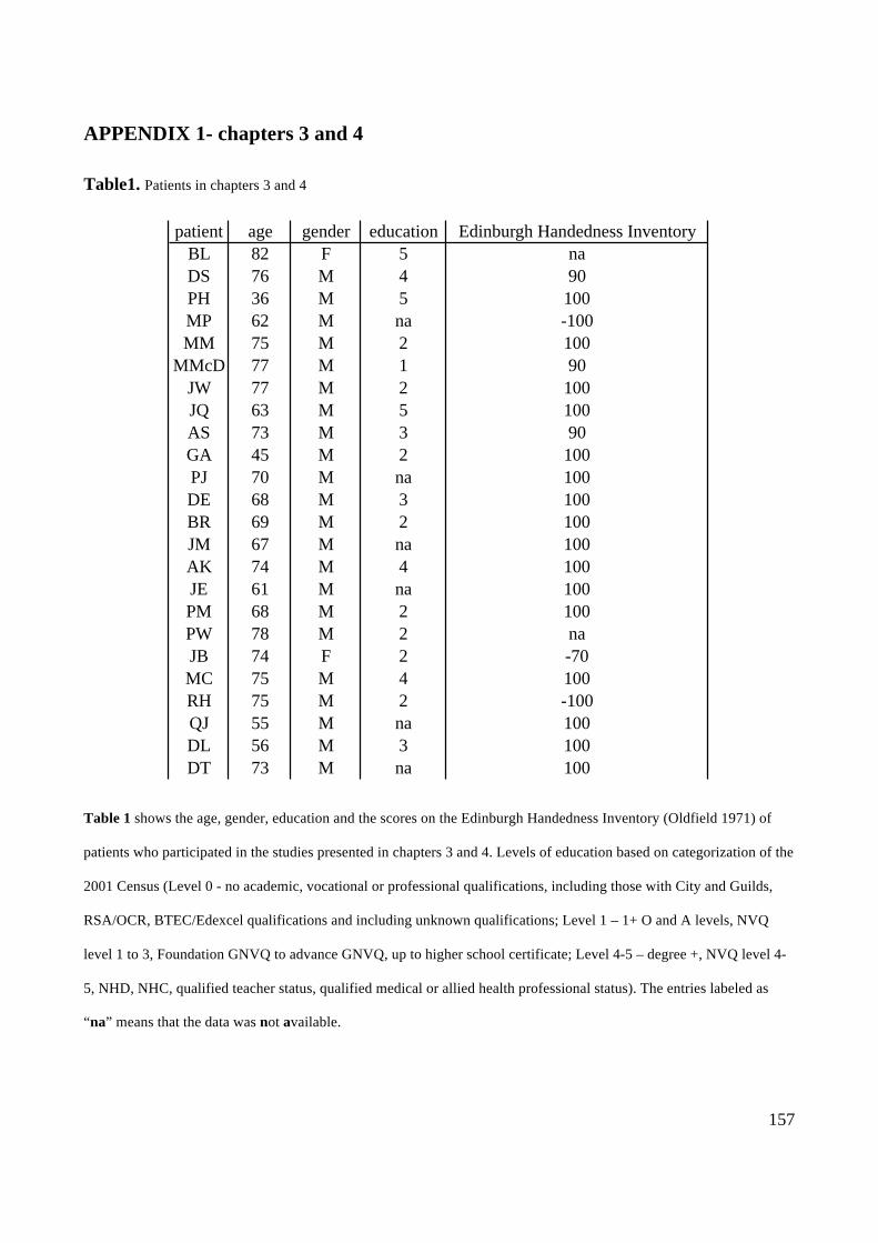
157
APPENDIX 1- chapters 3 and 4
Table1. Patients in chapters 3 and 4
Table 1 shows the age, gender, education and the scores on the Edinburgh Handedness Inventory (Oldfield 1971) of
patients who participated in the studies presented in chapters 3 and 4. Levels of education based on categorization of the
2001 Census (Level 0 - no academic, vocational or professional qualifications, including those with City and Guilds,
RSA/OCR, BTEC/Edexcel qualifications and including unknown qualifications; Level 1 – 1+ O and A levels, NVQ
level 1 to 3, Foundation GNVQ to advance GNVQ, up to higher school certificate; Level 4-5 – degree +, NVQ level 4-
5, NHD, NHC, qualified teacher status, qualified medical or allied health professional status). The entries labeled as
“na” means that the data was not available.
patient age gender education Edinburgh Handedness InventoryBL 82 F 5 naDS 76 M 4 90PH 36 M 5 100MP 62 M na -100MM 75 M 2 100
MMcD 77 M 1 90JW 77 M 2 100JQ 63 M 5 100AS 73 M 3 90GA 45 M 2 100PJ 70 M na 100DE 68 M 3 100BR 69 M 2 100JM 67 M na 100AK 74 M 4 100JE 61 M na 100PM 68 M 2 100PW 78 M 2 naJB 74 F 2 -70MC 75 M 4 100RH 75 M 2 -100QJ 55 M na 100DL 56 M 3 100DT 73 M na 100

158
Birmingham Cognitive Screen- Tasks administered to patients in chapter 3
Normative sample
The Birmingham Cognitive Screen (BCoS) was administered to adult healthy participants that had
been selected according to age, gender and education level, in proportions that should be
representative of the English adults population according to the 2001 Census (Office for National
Statistic website accessed on 18th July 2008). Age and education level were divided in ranges as
follows: i) age: 50-64, 65-74, 75 or above; ii) education: Level 0 - no academic, vocational or
professional qualifications, including those with City and Guilds, RSA/OCR, BTEC/Edexcel
qualifications and including unknown qualifications; Level 1 – 1+ O and A levels, NVQ level 1 to
3, Foundation GNVQ to advance GNVQ, up to higher school certificate; Level 4-5 – degree +,
NVQ level 4-5, NHD, NHC, qualified teacher status, qualified medical or allied health professional
status. The education levels were based on the categorization of the 2001 Census.
The cut off were obtained from 100 participants divided according to the three age range: 50-64
(N= 34); 65-74 (N= 33); 75 or above (N= 33). The cut offs for the tasks considered in the present
study are reported in Figure 1.
Figure 1. Cut offs at the BCoS tasks administered to patients in chapter 3
Figure 1. Performance is considered as defective in falling below the scores reported for each age range.
159
BCoS- Patients testing
The following subtests from the Birmingham Cognitive Screen (BCoS) (Humphreys, Bickerton,
Samson and Riddock 2012) were administered to the patients who took part in the present study:
Gesture Production, Imitation and Action Recognition are described in the methods chapter (chapter
2).
Long-term memory16- Personal Information: This subtest is composed by eight questions
concerning age, job, home address etc.. Each question is presented verbally, while the patient can
answer in a verbal or written form, in case of unreliable verbal production. For written answers the
patient can use his/her favourite hand.
Each correct answer is scored 1, incorrect answers of answers delayed more than 15 seconds are
scored 0, for a maximum score = 8.
Long-term memory-Time and Space: it assesses the patient’s time and space awareness. It is
composed by 6 questions, 3 concerning the actual moment and place (i.e. where are you right now;
in which city; what time of the day is it now); the other 3 refer to a more general temporal
dimension (i.e. what month is it; what day of the week is it; what year is it). The patient should
answer verbally, but in case of error or non-response multiple-choice questions are presented, with
the right response presented among two distracters
Each correct answer is scored 1, while incorrect of delayed (<15secs) answers are scored 0, for a
maximum score = 0.
Picture Naming: The task includes fourteen pictures of objects belonging to different categories
(i.e. seven inanimate objects, seven living things (3 fruits; 2vegetables; 2animals)). The items were
16 Both Personal Orientation and Time and Space consist of open verbal questions (e.g., where are you now? What
month is it?). For the orientation in time and space, in addition, there is a multiple choice (4 choice responses) to be
presented in the case of a non-response by the patient, an error in the initial question or in cases where aphasia prevents
answers to the open questions.
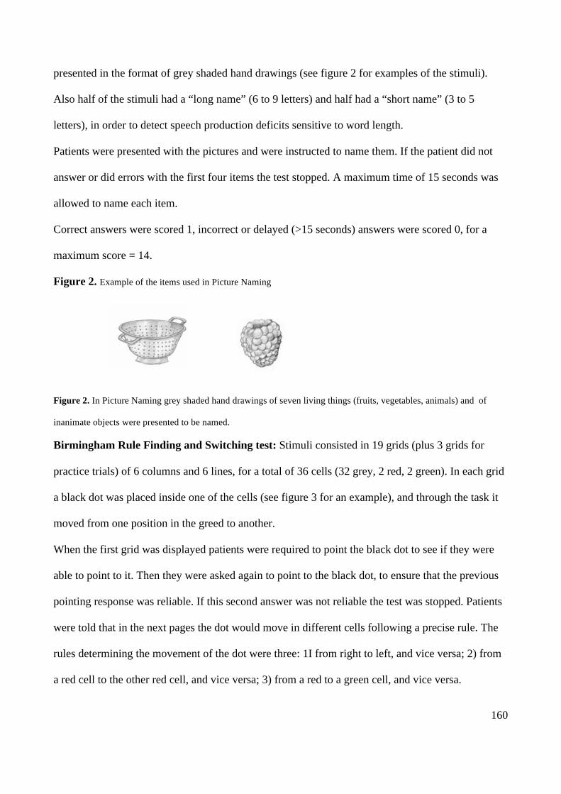
160
presented in the format of grey shaded hand drawings (see figure 2 for examples of the stimuli).
Also half of the stimuli had a “long name” (6 to 9 letters) and half had a “short name” (3 to 5
letters), in order to detect speech production deficits sensitive to word length.
Patients were presented with the pictures and were instructed to name them. If the patient did not
answer or did errors with the first four items the test stopped. A maximum time of 15 seconds was
allowed to name each item.
Correct answers were scored 1, incorrect or delayed (>15 seconds) answers were scored 0, for a
maximum score = 14.
Figure 2. Example of the items used in Picture Naming
Figure 2. In Picture Naming grey shaded hand drawings of seven living things (fruits, vegetables, animals) and of
inanimate objects were presented to be named.
Birmingham Rule Finding and Switching test: Stimuli consisted in 19 grids (plus 3 grids for
practice trials) of 6 columns and 6 lines, for a total of 36 cells (32 grey, 2 red, 2 green). In each grid
a black dot was placed inside one of the cells (see figure 3 for an example), and through the task it
moved from one position in the greed to another.
When the first grid was displayed patients were required to point the black dot to see if they were
able to point to it. Then they were asked again to point to the black dot, to ensure that the previous
pointing response was reliable. If this second answer was not reliable the test was stopped. Patients
were told that in the next pages the dot would move in different cells following a precise rule. The
rules determining the movement of the dot were three: 1I from right to left, and vice versa; 2) from
a red cell to the other red cell, and vice versa; 3) from a red to a green cell, and vice versa.

161
The rule by which the dot moved sometimes changed, but patients were not warned about that.
Patients were asked to predict the position of the dot in each consequent grid. To do this they had to
find the rule determining the position of the dot in the next item, as well as to detect if the rule had
changed.
Each grid was presented one at a time. The preceding grid was always left visible in order to reduce
memory problems.
The task started with a practice trial: a first grid was followed by a second where the dot had
changed its position. The patient was asked to indicate where the dot would have moved in the
subsequent grid. Then the third grid was showed, for feedback. If necessary the patient response
was corrected by the experimenter. The actual task followed.
Each correct answer was scored 1, incorrect or delayed (> 15 seconds) answers were scored 0, for a
maximum score = 18 (i.e. Accuracy). Each correct rule detection was scored 1, for a maximum
score = 3 (i.e. Rule Detection).
Figure 3. Example of the items used in Birmingham Rule Finding and Switching test
Figure 3. Patients had to predict the position of the dot on subsequent grids on the basis of the previous moves of the
dot. The rule by which the dot moved sometimes could change.
Multi-Step Object Use: This task required the patients to perform a sequence of actions on objects
in order to carry out the task. Two target objects and three distracters were displayed: matches
(objects placed nearest to patient), batteries, glue stick, screwdriver, torch (object placed furthest
from patient). The task was to light the torch.
A B C D E F
2
3
4
5
6
1
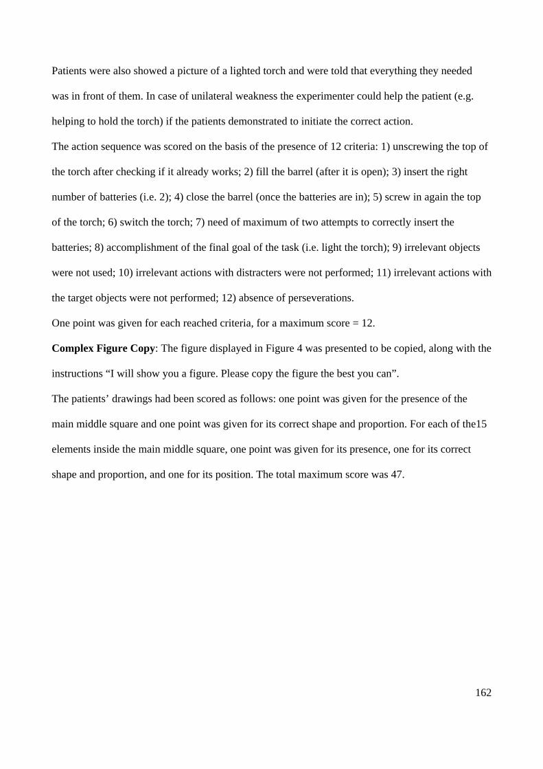
162
Patients were also showed a picture of a lighted torch and were told that everything they needed
was in front of them. In case of unilateral weakness the experimenter could help the patient (e.g.
helping to hold the torch) if the patients demonstrated to initiate the correct action.
The action sequence was scored on the basis of the presence of 12 criteria: 1) unscrewing the top of
the torch after checking if it already works; 2) fill the barrel (after it is open); 3) insert the right
number of batteries (i.e. 2); 4) close the barrel (once the batteries are in); 5) screw in again the top
of the torch; 6) switch the torch; 7) need of maximum of two attempts to correctly insert the
batteries; 8) accomplishment of the final goal of the task (i.e. light the torch); 9) irrelevant objects
were not used; 10) irrelevant actions with distracters were not performed; 11) irrelevant actions with
the target objects were not performed; 12) absence of perseverations.
One point was given for each reached criteria, for a maximum score = 12.
Complex Figure Copy: The figure displayed in Figure 4 was presented to be copied, along with the
instructions “I will show you a figure. Please copy the figure the best you can”.
The patients’ drawings had been scored as follows: one point was given for the presence of the
main middle square and one point was given for its correct shape and proportion. For each of the15
elements inside the main middle square, one point was given for its presence, one for its correct
shape and proportion, and one for its position. The total maximum score was 47.
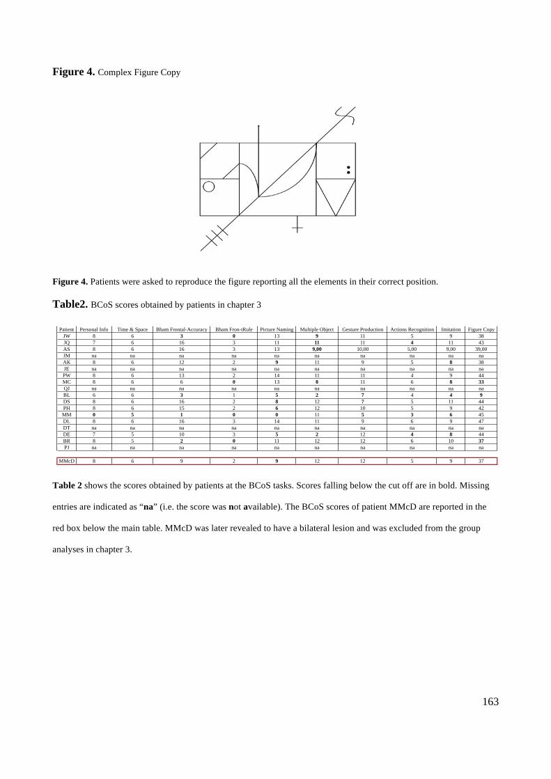
163
Figure 4. Complex Figure Copy
Figure 4. Patients were asked to reproduce the figure reporting all the elements in their correct position.
Table2. BCoS scores obtained by patients in chapter 3
Table 2 shows the scores obtained by patients at the BCoS tasks. Scores falling below the cut off are in bold. Missing
entries are indicated as “na” (i.e. the score was not available). The BCoS scores of patient MMcD are reported in the
red box below the main table. MMcD was later revealed to have a bilateral lesion and was excluded from the group
analyses in chapter 3.
Patient Personal Info Time & Space Bham Frontal-Accuracy Bham Fron-tRule Picture Naming Multiple Object Gesture Production Actions Recognition Imitation Figure CopyJW 8 6 3 0 13 9 11 5 9 38JQ 7 6 16 3 11 11 11 4 11 43AS 8 6 16 3 13 9,00 10,00 5,00 9,00 39,00JM na na na na na na na na na naAK 8 6 12 2 9 11 9 5 8 38JE na na na na na na na na na naPW 8 6 13 2 14 11 11 4 9 44MC 8 6 6 0 13 8 11 6 8 33QJ na na na na na na na na na naBL 6 6 3 1 5 2 7 4 4 9DS 8 6 16 2 8 12 7 5 11 44PH 8 6 15 2 6 12 10 5 9 42MM 0 5 1 0 0 11 5 3 6 45DL 8 6 16 3 14 11 9 6 9 47DT na na na na na na na na na naDE 7 5 10 3 5 2 12 4 8 44BR 8 5 2 0 11 12 12 6 10 37PJ na na na na na na na na na na
MMcD 8 6 9 2 9 12 12 5 9 37

164
APPENDIX 2- chapters 5 and 6
Table 1. Patients in chapter 5 and 6
Table1 summarize gender (male female), hand used for imitation and educations level of the 233 acute patients whose
data were used in chapter 5 and 6. Notice that all the patients in this sample were right-handed. Education levels were
based on the categorization of the 2001 Census. The information on the education of 7 patients was not available (i.e.
“na”).
Male Female Right left level1 level2 level3 level4 level5 na
N 110 123 191 42 6 169 30 7 14 7
N Tot= 233
GENDER HAND USED EDUCATION
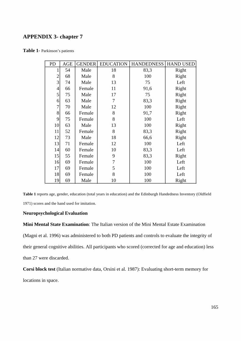
165
APPENDIX 3- chapter 7
Table 1- Parkinson’s patients
Table 1 reports age, gender, education (total years in education) and the Edinburgh Handedness Inventory (Oldfield
1971) scores and the hand used for imitation.
Neuropsychological Evaluation
Mini Mental State Examination: The Italian version of the Mini Mental Estate Examination
(Magni et al. 1996) was administered to both PD patients and controls to evaluate the integrity of
their general cognitive abilities. All participants who scored (corrected for age and education) less
than 27 were discarded.
Corsi block test (Italian normative data, Orsini et al. 1987): Evaluating short-term memory for
locations in space.
PD AGE GENDER EDUCATION HANDEDNESS HAND USED1 54 Male 18 83,3 Right2 68 Male 8 100 Right3 74 Male 13 75 Left4 66 Female 11 91,6 Right5 75 Male 17 75 Right6 63 Male 7 83,3 Right7 70 Male 12 100 Right8 66 Female 8 91,7 Right9 75 Female 8 100 Left
10 63 Male 13 100 Right11 52 Female 8 83,3 Right12 73 Male 18 66,6 Right13 71 Female 12 100 Left14 60 Female 10 83,3 Left15 55 Female 9 83,3 Right16 69 Female 7 100 Left17 69 Female 5 100 Left18 69 Female 8 100 Left19 69 Male 10 100 Right
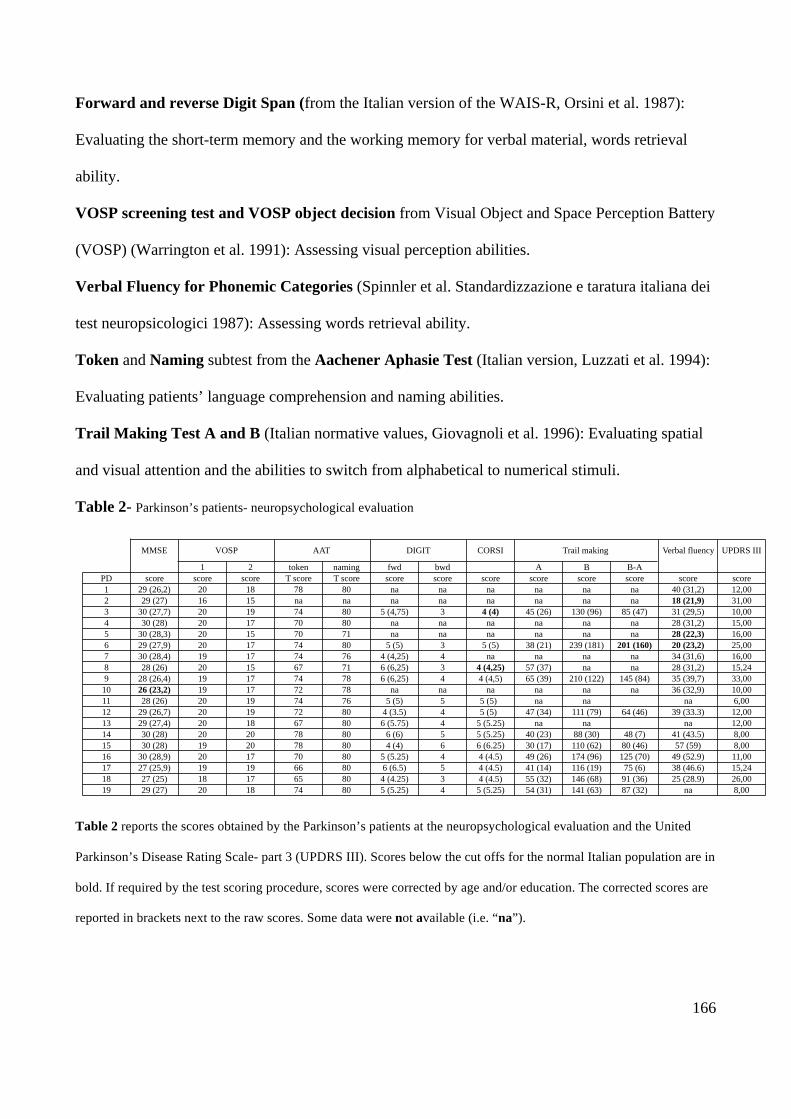
166
Forward and reverse Digit Span (from the Italian version of the WAIS-R, Orsini et al. 1987):
Evaluating the short-term memory and the working memory for verbal material, words retrieval
ability.
VOSP screening test and VOSP object decision from Visual Object and Space Perception Battery
(VOSP) (Warrington et al. 1991): Assessing visual perception abilities.
Verbal Fluency for Phonemic Categories (Spinnler et al. Standardizzazione e taratura italiana dei
test neuropsicologici 1987): Assessing words retrieval ability.
Token and Naming subtest from the Aachener Aphasie Test (Italian version, Luzzati et al. 1994):
Evaluating patients’ language comprehension and naming abilities.
Trail Making Test A and B (Italian normative values, Giovagnoli et al. 1996): Evaluating spatial
and visual attention and the abilities to switch from alphabetical to numerical stimuli.
Table 2- Parkinson’s patients- neuropsychological evaluation
Table 2 reports the scores obtained by the Parkinson’s patients at the neuropsychological evaluation and the United
Parkinson’s Disease Rating Scale- part 3 (UPDRS III). Scores below the cut offs for the normal Italian population are in
bold. If required by the test scoring procedure, scores were corrected by age and/or education. The corrected scores are
reported in brackets next to the raw scores. Some data were not available (i.e. “na”).
1 2 token naming fwd bwd A B B-APD score score score T score T score score score score score score score score score1 29 (26,2) 20 18 78 80 na na na na na na 40 (31,2) 12,002 29 (27) 16 15 na na na na na na na na 18 (21,9) 31,003 30 (27,7) 20 19 74 80 5 (4,75) 3 4 (4) 45 (26) 130 (96) 85 (47) 31 (29,5) 10,004 30 (28) 20 17 70 80 na na na na na na 28 (31,2) 15,005 30 (28,3) 20 15 70 71 na na na na na na 28 (22,3) 16,006 29 (27,9) 20 17 74 80 5 (5) 3 5 (5) 38 (21) 239 (181) 201 (160) 20 (23,2) 25,007 30 (28,4) 19 17 74 76 4 (4,25) 4 na na na na 34 (31,6) 16,008 28 (26) 20 15 67 71 6 (6,25) 3 4 (4,25) 57 (37) na na 28 (31,2) 15,249 28 (26,4) 19 17 74 78 6 (6,25) 4 4 (4,5) 65 (39) 210 (122) 145 (84) 35 (39,7) 33,00
10 26 (23,2) 19 17 72 78 na na na na na na 36 (32,9) 10,0011 28 (26) 20 19 74 76 5 (5) 5 5 (5) na na na 6,0012 29 (26,7) 20 19 72 80 4 (3.5) 4 5 (5) 47 (34) 111 (79) 64 (46) 39 (33.3) 12,0013 29 (27,4) 20 18 67 80 6 (5.75) 4 5 (5.25) na na na 12,0014 30 (28) 20 20 78 80 6 (6) 5 5 (5.25) 40 (23) 88 (30) 48 (7) 41 (43.5) 8,0015 30 (28) 19 20 78 80 4 (4) 6 6 (6.25) 30 (17) 110 (62) 80 (46) 57 (59) 8,0016 30 (28,9) 20 17 70 80 5 (5.25) 4 4 (4.5) 49 (26) 174 (96) 125 (70) 49 (52.9) 11,0017 27 (25,9) 19 19 66 80 6 (6.5) 5 4 (4.5) 41 (14) 116 (19) 75 (6) 38 (46.6) 15,2418 27 (25) 18 17 65 80 4 (4.25) 3 4 (4.5) 55 (32) 146 (68) 91 (36) 25 (28.9) 26,0019 29 (27) 20 18 74 80 5 (5.25) 4 5 (5.25) 54 (31) 141 (63) 87 (32) na 8,00
MMSE VOSP AAT DIGIT CORSI Trail making Verbal fluency UPDRS III
167
10. References
Altschuler EL, Vankov A, Hubbard EM, Roberts E, Ramachandran VS, Pineda JA (1997). See,
Person Do: Human Cortical Electrophysiological Correlates of Monkey See Monkey Do Cells,
Poster Session Presented at the 27th Annual Meeting of the Society for Neuroscience, New Orleans,
LA.
Altschuler EL, Vankov A, Hubbard EM, Roberts E, Ramachandran VS, Pineda JA (200). Mu wave
blocking by observer of movement and its possible use as a tool to study theory of other minds,
Poster session presented at the 30th annual meeting of the society for neuroscience, New Orleans,
LA.
Ashburner J, Friston KJ (2003). Spatial normalization using basis functions, in: Frackowiak RSJ,
Friston KJ, Frith CD, Dolan RJ, Price CJ, Ashburner J, Penny WD, Zeki S (Eds.), Human Brain
Function, Academic Press, San Diego.
Ashburner J, Friston KJ. 2005. Unified segmentation. Neuroimage 26: 839–851.
Ashburner, J, Friston, KJ (2000). Voxel-based morphometry-The methods. Neuroimage 11: 805–
821.
Bard KA., & Russell CL (1999). Evolutionary foundations of imitation: social cognitive and
developmental aspects of imitative processes in non-human primates. In J. Nadel & G. Butterworth
(Eds.), Imitation in infancy (pp. 89–123). Cambridge: Cambridge University Press.
Bartolo A., Cubelli, R, Della Sala S, Drei S, Marchetti C. (2001). Double dissociation between
meaningful and meaningless gesture reproduction in apraxia. Cortex 37, 696–699.
Bell BD (1994). Pantomime recognition impairment in aphasia: an analysis of error types. Brain
Language; 47: 269-78.
Bush G, Luu P, and Posner MI (2000). Cognitive and emotional influences in anterior cingulate
cortex. TICS 4: 215–222.
168
Buxbaum L. J., Kyle, K., Grossman, M., Branch Coslett, B. Left Inferior Parietal Representations
for Skilled Hand-Object Interactions: Evidence from Stroke and Corticobasal Degeneration. Cortex
(2007) 43 (3): 411-423.
Buxbaum LJ, Kalénine S (2010) Action knowledge, visuomotor activation, and embodiment in the
two action systems. Ann N Y Acad Sci 1191:201–218.
Buxbaum, L. J. (2001). Ideomotor apraxia: A call to action. Neurocase, 445–458.
Buxbaum, L.J., Kyle, K.M., & Menon, R. On beyond mirror neurons: internal representations
subserving imitation and recognition of skilled object-related actions in humans. Cognitive Brain
Research 25, 226-239 (2005).
Canavan, A.G.M., Passingham, R.E., Marsden, C.D., Quinn, Wyke, M., and Polkey, C.E. (1989).
The performance on learning tasks of patients in the early stages of Parkinson’s disease.
Neuropsychologia 27, 141–156.
Carmo JC, Rumiati RI (2009) Imitation of transitive and intransitive actions in healthy individuals.
Brain Cogn. 69: 460–464.
Catani, M., Jones, D., Ffichte, D., 2005. Perysilvian language networks of the human (completare)
Cavanna AE, Trimble MR. The precuneus: a review of its functional anatomy and behavioural
correlates. Brain 2009; 129, 564–583.
Chainay H. ; Humphreys G.W. Neuropsychological evidence for a convergent route model for
action. Cognitive Neuropsychology 2002; 19 (1) 67 – 93.
Chechlacz M, Rotshtein P, Bickerton WL, Hansen PC, Deb S, Humphreys GW (2010). Separating
neural correlates of allocentric and egocentric neglect: Distinct cortical sites and common white
matter disconnections', Cognitive Neuropsychology, 27: 3, 277 — 303
Chong, T. T., Cunnington, R., Williams, M. A., Kanwisher, N., & Mattingley, J. B. (2008). fMRI
adaptation reveals mirror neurons in human inferior parietal cortex. Current Biology,18, 1576–
1580.
169
Cochin S, Barthelemy C, Lejeune B, Roux S, Martineau J. 1998. Perception of motion and qEEG
activity in human adults. Electroen- cephalogr. Clin. Neurophysiol. 107:287–95
Cochin S, Barthelemy C, Roux S, Martineau J. 1999. Observation and execution of move- ment:
similarities demonstrated by quanti- fied electroencephalograpy. Eur. J. Neurosci. 11:1839–42
Cohen-Seat G, Gastaut H, Faure J, Heuyer G.1954. Etudes exp´erimentales de l’activité nerveuse
pendant la projection cinématographique. Rev. Int. Filmologie 5:7–64
Costa A, Peppe A, Dell’Agnello G, Carlesimo GA, Murri L, Bonuccelli U, Caltagirone C (2003)
Dopaminergic modulation of visual-spatial working memory in Parkinson’s disease. Dement
Geriatr Cogn Disord 15:55–66.
Crawford, J.R., & Garthwaite, P.H. Investigation of the single case in neuropsychology: confidence
limits on the abnormality of test scores and test score differences. Neuropsychologia 40 (2002)
1196–1208
Crawford, J.R., & Garthwaite, P.H. Testing for suspected impairments and dissociations in single-
case studies in neuropsychology: Evaluation of alternatives using Monte Carlo simulations and
revised tests for dissociations", Neuropsychology 19, 318-331 (2005).
Cubelli R, Bartolo A, Nichelli P, Della Sala S. List effect in apraxia assessment. Neuroscience Lett
2006; 407: 118-20.
Cubelli, R., C. Marchetti, G. Boscolo & S. Della Sala. 2000. Cognition in action: testing a model of
limb apraxia. Brain Cogn. 44: 144–165.
De Renzi, E., F. Cavalleri & S. Facchini. 1996. Imitation and utilization behaviour. J. Neurol.
Neurosurg. Psychiatry 61: 396–400.
Decety, J., and Grèzes, J. Neural mechanisms subserving the perception of human actions. Trends
in Cognitive Sciences 3, 172-178 (1999).

170
Decety,J., Grèzes, J., Costes, N., Perani, D, Jeannerod, M., Procyk, E., Grassi, F., and Fazio F.
Brain activity during observation of actionsInfluence of action content and subject’s strategy. Brain
(1997), 120, 1763–1777
Dehaene, S. et al. (1994). Localization of a neural system for error detection and compensation.
Psychol. Sci. 5, 303–305
Della Sala S., Faglioni P., Motto C., and Spinnler H. Hemisphere asymmetry for imitation of hand
and finger movements, Goldenberg's hypothesis reworked (2006). Neuropsychologia, 44 (8), 1496-
1500.
Dinstein, I., Hasson, U., Rubin, N., & Heeger, D. J. (2007). Brain areas selective for both observed
and executed movements. Journal of Neurophysiology, 98, 1415–1427.
Duvernoy, H. M. The human brain: surface, blood supply and three-dimensional sectional anatomy.
Wien: Springer-Verlag; 1991.
Fadiga, L., L. Fogassi, G. Pavesi & G. Rizzolatti. 1995. Motor facilitation during action
observation: a magnetic stimulation study. J. Neurophysiol. 73: 2608–2611.
Ferrari, P. F., Visalberghi, E., Fogassi, L., Ruggiero, A., & Suomi, S. J. (2006). Neonatal imitation
in rhesus macaques. PLoS Biology, 4, 1501–1508.
Friston, K. J., Ashburner, J., Kiebel, S. J., Nichols, T. E., & Penny, W. (2007). Statistical parametric
mapping: The analysis of functional brain images (1st ed.). Amsterdam, The Netherlands:
Elsevier/Academic Press.
Gallese V, Fadiga L, Fogassi L, Rizzolatti G. Action recognition in the premotor cortex. Brain
1996; 119: 593–609.
Gastaut HJ, Bert J. 1954. EEG changes during cinematographic presentation. Elec- troencephalogr.
Clin. Neurophysiol. 6:433– 44
Gazzola V, van der Worp H, Mulder T, Wicker B, Rizzolatti G, Keysers C. (2007). Aplasics born
without hands mirror the goal of hand actions with their feet. Curr Biol. 17:1235--1240.
171
Gazzola, V., Aziz-Zadeh, L., and Keysers, C. (2006). Empathy and the somatotopic auditory mirror
system in human. Curr. Biol. 16, 1824–1829.
Geschwind, N. (1976). The apraxias: Neural mechanisms of disorders of learned movement.
American Scientist, 63, 188-195.
Gillebert C.R.; Humphreys G.W. 2010. The decomposition of visual binding over time:
Neuropsychological evidence from illusory conjunctions after posterior parietal damage. Visual
Cognition Volume 18, Issue 7.
Goldenbeg, G., Hermsdorfer, J., Glindermann, R., 2007. Pantomime of tool use depends on
integrity of left inferior frontal cortex. Cereb Cortex. 17:2769-2776.
Goldenberg G, Strauss S (2002) Hemisphere asymmetries for imitation of novel gestures.
Neurology 59:893– 897.
Goldenberg G. Apraxia and the parietal lobes. Neuropsychologia, 47: 1449e1459, 2009.
Goldenberg G. Supplementary motor area structure and function: review and hypotheses. Behav
Brain Sci 1985; 8: 567-615.
Goldenberg G., Karnath H. The Neural Basis of Imitation is Body Part Specific. The Journal of
Neuroscience, June 7, 2006, 26(23):6282-6287.
Goldenberg, G. (2008). Apraxia. In G. Goldenberg & B. Miller (Eds.), Handbook of clinical
neurology, 3rd Series, Vol. 88: Neuropsychology and behavioral neurology (pp. 323–338).
Edinburgh: Elsevier.
Goldenberg, G. & S. Hagmann. 1997. The meaning of meaningless gestures: a study of visuo-
imitative apraxia. Neuropsychologia 35: 333–341.
Grèzes J, Costes N, Decety J (1998): Top down effect of the strategy to imitate on the brain areas
engaged in perception of biological motion: a PET investigation. Cogn Neuropsychol 15:553–582.
Haaland K.Y., Falherty D. The Different Types of Limb Apraxia Errors Made by Patients by
patients with left vs. right hemisphere damage (1984). Brain and Cognition, 3, 4, 370-384.
172
Haaland K.Y., Harrington D.L and Knight R.T.. Neural representations of skilled movement. Brain
(2000) 123 (11): 2306-2313
Hanna-Pladdy, B., Daniels, S. K., Fieselman, M. A.,Thompson, K., Vasterling, J. J., Heilman, K.
M, Foundas, A. L. 2001 Praxis lateralization: errors in rightand left hemisphere stroke. Cortex 37,
219–230.
Hari R, Forss N, Avikainen S, Kirveskari S, Salenius S, Rizzolatti G. 1998. Activation of human
primary motor cortex during action observation: a neuromagnetic study. Proc. Natl. Acad. Sci. USA
95:15061–65
Hari R, Salmelin R (1997) Human cortical oscillations: a neuromagnetic view through the skull.
Trends Neurosci 20:44–49.
Heilman KM, Rothi LJ, Valenstein E (1982) Two forms of ideomotor apraxia. Neurology 32:342–
346
Hickok, G. Eight problems for the mirror neuron theory of action understanding in monkeys and
humans. J. Cogn. Neurosci. 21, 1229–1243 (2009).
Howard, R.J. et al. (1996) A direct demonstration of functional specialization within motion-related
visual and auditory cortex of the human brain Curr. Biol. 6, 1015–1019.
Humphreys, Bickerton, Samson and Riddock. Birmingham Cognitive Screen (BCoS). 2012
Iacoboni M, Woods RP, Brass M, Bekkering H, Mazziotta JC, Rizzolatti G. 1999. Cortical
mechanisms of human imitation. Science 286:2526–28
Iacoboni, M. et al. Grasping the intentions of others with one’s own mirror neuron system. PLoS
Biol. 3, e79 (2005).
Kareken DA, Unverzagt F, Caldemeyer K, Farlow MR, Hutchins GD (1998) Functional Brain
Imaging in Apraxia. Archives of Neurology 55: 107-113.
Koski L, Iacoboni M, Dubeau MC, Woods RP, Mazziotta JC. 2003. Modulation of cortical activity
during different imitative behaviors. J. Neurophysiol. 89:460–71
173
Koski L, Wohlschlager A, Bekkering H, Woods RP, Dubeau MC. 2002. Modulation of mo- tor and
premotor activity during imitation of target-directed actions. Cereb. Cortex 12: 847–55
Króliczak, G. & S.H. Frey. 2009. A common network in the left cerebral hemisphere represents
planning of tool use pantomimes and familiar intransitive gestures at the hand-independent level.
Cereb. Cortex 19: 2396–2410.
Lange K W, Robbins TW, Marsden CD, James M, Owen AM, Paul GM (1992) L-Dopa withdrawal
in Parkinson’s disease selectively impairs cognitive performance in tests sensitive to frontal lobe
dysfunction. Psychopharmacology 107: 394-404.
Leiguarda R (2001) Limb apraxia: cortical or subcortical. Neuroimage 14: 137-41.
Leiguarda R, Pramstaller P, Marello M, Starkstein S, Lees AJ, Marsden CD (1997) Apraxia in
Parkinson’s disease, progressive supranuclear palsy, multiple system atrophy and neuroleptic-
induced parkinsonism. Brain 120: 75-90.
Lewis JW, Brefczynski JA, Phinney RE, Janik JJ, DeYoe EA. 2005. Distinct cortical pathways for
processing tool versus animal sounds. J. Neurosci. 25:5148–58
Liepmann, H. (1900). Das Krankheitshildder der Apraxie (Motorischen/Asymbolie). Monatschrift
für Psychiatrie und Neurologie, 8, 15–44, 102–132.
Liepmann, H. 1905. Die linke Hemisphere und das Handeln. Munch. Med. Wochenschr. 49: 2322–
2326, 2375–2378.
Luzzati C, Willmes K, De Bleser R, Bianchi A, Chiesa G, De Tanti A, Gonnella ML, Lorenzi L,
Bozzoli C (1994) Nuovi dati normative per la versione italiana dell’ Aachener Aphasie Test (AAT).
Arch. Psicol. Neurol. Psichiatria 55: 1086- 1131.
Mahon B. z., Caramazza A. A critical look at the embodied cognition hypothesis and a new
proposal for grounding conceptual content. Journal of Physiology 102 (2008) 59–70.
Mahon, B.Z. & A. Caramazza. 2005. The orchestration of the sensory-motor systems: clues from
neuropsychology. Cogn. Neuropsychol. 22: 480–494.
174
McNamara P, Durso R (2003) Pragmatic communication skills in patients with Parkinson’ s
disease. Brain Lang 84: 414–423.
Mehler, M.F. 1987. Visuo-imitative apraxia. Neurology 37: 129.
Meltzoff, A. N. and Moore, M. K. (1977). Imitation of facial and manual gestures by human
neonates. Science, 198, 75±78.
Meltzoff, A. N. and Moore, M. K. (1983). Newborn infants imitate adult facial gestures. Child
Development, 54, 702±809
Meltzoff, A. N. and Moore, M. K. (1989). Imitation in newborn infants: exploring the range of
gestures imitated and the underlying mechanisms. Developmental Psychology, 25, 954±962
Merians AS, Clark M, Poizner H, Macauley B, Gonzalez Rothi LJ, Heilman KL. Visual-imitative
dissociation apraxia. Neuropsychologia 1997; 35: 1485–90.
Mozaz, M., Rothi, L., Anderson, J., Crucian, G., Heilman, K., 2002. Postural knowledge of
transitive pantomimes and intransitive gestures. J. Int. Neuropsychol. Soc. 8, 958– 962.
Myowa-Yamakoshi, M., Yamaguchi, M.K., Tomonaga, M., Tanaka, M., & Matsuzawa, T. (2005).
Development of face recognition in infant chimpanzees (Pan troglodytes). Cognitive Development,
20, 49–63.
Myowa, M. (1996). Imitation of facial gestures by an infant chimpanzee. Primates, 37, 207–213.
Negri, G.A.L. et al . 2007. What is the role of motor simulation in action and object recognition?
Evidence from apraxia. Cogn. Neuropsych. 24: 795–816.
Oldfield R.C. The assessment and analysis of handedness: The Edinburgh inventory.
Neuropsychologia. Volume 9, Issue 1, March 1971, 97-113
Owen AM, Iddon JL, Hodges JR, Summers BA, and Robbins TW (1997) Spatial and non-spatial
working memory at different stages of Parkinson’s disease. Neuropsychologia 35 (4): 519-532.
175
Pardo, J.V., Pardo, P.J., Janer, K.W., Raichle, M.E., 1990. The anterior cingulate cortex mediates
processing selection in the Stroop attentional conflict paradigm. Proc. Natl. Acad. Sci. U. S. A. 87,
256–259.
Pazzaglia, M. et al. 2008. Neural underpinnings of gesture discrimination in patients with limb
apraxia. J. Neurosci. 28: 3030–3041.
Pazzaglia, M., Smania, N., Corato, E., & Aglioti, S. M. (2008). Neural underpinnings of gesture
discrimination in patients with limb apraxia. Journal of Neuroscience, 28, 3030–3041.
Peigneux P, Salmon E, Garraux, G, Laureys S, Willems S, Dujardin K, Degueldre C, Lemaire C,
Luxen A, Moonen G, Franck G, Destee A, Van der Linden M (2001) Neural and cognitive bases of
upper limb apraxia in corticobasal degeneration. Neurology 57:1259–1268.
Peigneux P, Van der Linden M, Garraux G, Laureys S, Delgueldre C, Aerts J, Del Fiore G, Moonen
G, Luxen A, Salmon E (2004) Imaging a cognitive model of apraxia: the neural substrate of action-
specific cognitive processes. Hum. Brain Mapp. 21: 119–142.
Peigneux, P., Van der Linden, M., Andres-Benito, P., Sadzot, B., Franck, G., & Salmon, E.
(2000). Exploration neuropsychologique et par imagerie fonctionelle cérébrale d’une apraxie visuo-
imitative. Revue Neurologique, 156, 459–472.
Pramstaller PP, and Marsden CD (1996) The basal ganglia and apraxia [review]. Brain 119: 319–
340.
Price C.J., Crinion J.T., Leff A.P., Richardson F.M., Schofield T., Prejawa S., Ramsden S.,
Gazarian K., Lawrence M., Ambridge L., Andric M., Small S.L., Seghier M.L. Lesion sites that
predict the ability to gesture how an object is used. Arch. Ital. Biol., 2010. 148: 243-258
Rapcsak S.Z., Ochipa C, Beeson PM, Rubens A.B (1993). Praxis and the right hemisphere. Brain
Cogn; 23, 181-201.
Rizzolati, G. & Sinigaglia, C. The functional role of the parieto-frontal mirror circuit:
interpretations and misinterpretations. Nature Rev. Neurosci. 11, 264–274 (2010).
176
Rizzolatti, G & Craighero L. The mirror-neuron system. Annu. Rev. Neurosci. 27, 169–192 (2004).
Rizzolatti, G. et al . 1996. Localization of grasp representations in humans by PET: 1. Observation
versus excecution. Exp. Brain Res. 111: 246–252.
Rizzolatti, G., L. Fogassi & V. Gallese. 2001. Neurophysiological mechanisms underlying the
understanding and imitation of action. Nat. Rev. Neurosci. 2: 661–670.
Rorden C, Karnath HO. Using human brain lesions to infer function: a relic from a past era in the
fMRI age? Nat Rev Neurosci 2004; 5: 813–9.
Rorden, C. MRIcro. http://www.sph.sc.edu/comd/rorden/mricro.html, 2005.
Rothi LJG, Ochipa C, Heilman KM (1991) A cognitive neuropsychological model of limb praxis.
Cogn Neuropsychol 8: 443-58.
Rothi, L. J. G., Mack, L. & Heilman, K. M. (1986) Pantomime agnosia. Journal of Neurology,
Neurosurgery, and Psychiatry 49:451–54.
Rothi, LJG, Heliman, KM (1997) Apraxia. The Neuropsychology of Action. Psychology Press, East
Sussex, UK.
Roy AE (2000) Apraxia in diseases of the basal ganglia. Movement Disord 15 (4): 598-600.
Rumiati RI, Tessari A (2002) Imitation of novel and well-known actions: the role of short-term
memory. Exp Brain Res 142: 425-33.
Rumiati RI, Weiss PH, Tessari A, Assmuss A, Zilles K, Herzog H (2005). Common and differential
neural mechanisms supporting imitation of meaningful and meaningless actions. Journal of
Cognitive Neuroscience ; 17: 1420-31.
Rumiati, R. I., Papeo, L. and Corradi-Dell’Acqua, C. (2010), Higher-level motor processes. Annals
of the New York Academy of Sciences, 1191: 219–241.
Rumiati, R.I., J.C. Carmo & C. Corradi-Dell’Acqua. (2009). Neuropsychological perspectives on
the mechanisms of imitation. Philos. Trans. R. Soc. Lond., B Biol. Sci. B 364: 2337–2347.
177
Salenius S., Portin K., Kajola M., Salmelin, R., and Hari R. Cortical control of human motoneuron
firing during isometric contraction. J. Neurophysiol. 77: 3401–3405, 1997.
Salmelin R., Hari R., Spatiotemporal characteristics of sensorimotor neuromagnetic rhythms related
to thumb movement, Neuroscience 60 (1994) 537– 550.
Seghier, M.L., Ramlackhansingh, A., Crinion, J., Leff, A.P., Price, C.J., 2008. Lesion identification
using unified segmentation-normalisation models and fuzzy clus- tering. NeuroImage 41, 1253–
1266.
Spinler H, Tognoni G (1987) Standardizzazione e taratura italiana di test neuropsicologici. Ital J
Neurol Sci 6 (8): 12-120.
Stamenova V, Roy E.A., Black S.E. (2009). A Model-Based Approach to Understanding Apraxia in
Corticobasal Syndrome. Neuropsychology Review 19,1 , 47-63.
Stamenova V, Roy E.A., Black S.E. (2010). Associations and dissociations of transitive and
intransitive gestures in left and right hemisphere stroke patients. Brain and Cognition, 72 (3), 483-
490.
Stamenova V, Roya EA, Blacka SE (2011). Limb apraxia in corticobasal syndrome. Cortex, 47 (4),
460–472.
Tessari A, Bosanac D, Rumiati RI (2006) Effect of learning on imitation of new actions:
implications for a memory model. Exp Brain Res 173 (3): 507-13.
Tessari A, Canessa N, Ukmar M, Rumiati RI (2007) Neuropsychological evidence for a strategic
control of multiple routes in imitation. Brain 130: 1111-26.
Tessari A, Lunardelli A, Zadini A, Rumiati RI (submitted) Prova standardizzata per la diagnosi del
disturbo aprassico ideomotorio selettivo per tipo di gesto e tipo di effettore. Ricerche di Psicologia.
Tessari A, Rumiati RI (2004) The strategic control of multiple routes in imitation of actions. J Exp
Psychol Hum Percept Perform 30: 1107-16.
178
Tubridy S, Davachi L (2011). Medial Temporal Lobe Contributions to Episodic Sequence
Encoding. Cerebral Cortex, 21, 272-280.
Weiss PH, Fink GR (2010). Structural and functional neuroima- ging of the pathophysiology of
apraxia. Nervenarzt, 81, 1444-1449.
Worsley KJ, Friston KJ (1995). Analysis of fMRI time-series revised again. Neuroimage, 2, 173-
181.


![The Practice of Neuropsychological Assessment - … · rated into the neuropsychological test canon ... Poppelreuter, 1990 [1917]; W.R. Russell ... 1 THE PRACTICE OF NEUROPSYCHOLOGICAL](https://static.fdocuments.us/doc/165x107/5b9c7f2609d3f272468cc5a2/the-practice-of-neuropsychological-assessment-rated-into-the-neuropsychological.jpg)


