ACSM's Handbook for the Team .. Physician · avulsion fracture and on manual muscle testing...
21
, ( ACSM's Handbook for the Team . Physician Edited by W. Ben Kibler, MD Medical Director Lexington Clinic Sports Medicine Center Lexington, Kentucky Williams & F A WAVERLY BALTIMORE • PHI!ADELPHIA • LONDON • PARIS •. BANGKOK BUENOS AIRES • HONG KONG • MUNICH• SYDNEY• TOKYO • WROCL\W 1996
-
Upload
nguyendang -
Category
Documents
-
view
219 -
download
0
Transcript of ACSM's Handbook for the Team .. Physician · avulsion fracture and on manual muscle testing...

,
(
ACSM's Handbook
for the Team .. Physician
Edited by W. Ben Kibler, MD Medical Director
Lexington Clinic Sports Medicine Center
Lexington, Kentucky
Williams & Wilkins~. F A WAVERLY COMPANY~
BALTIMORE • PHI!ADELPHIA • LONDON • PARIS •. BANGKOK BUENOS AIRES • HONG KONG • MUNICH• SYDNEY• TOKYO • WROCL\W
1996

Editor: Donna Balado Managing Editor: Victoria Vaughn Production Coordinator: Alethea H. Elkins Book Project Editor: Arlene Sheir-Allen Designer: Arlene Putterman Typesetter: Maryland Composition Printer: Courier Binder: Courier
Copyright © 1996 Williams & Wilkins
351 West Camden Street Baltimore, Maryland 21201-2436 USA
Rose Tree Corporate Center 1400 North Providence Road Building 11, Suite 5025 Media, Pennsylvania 19063-2043 USA
ISBN
I I 9 780683 000283
All rights reserved. This book is protected by copyright. No part of this book may be reproduced in any form or by any means, including photocopying or utilized by any information storage and retrieval system without written permission from the copyright owner.
Accurate indications, adverse reactions, and dosage schedules for drugs are provided in this book, but it is possible that they may change. The reader is urged to review the package information data of the manufacturers of the medications mentioned.
Printed in the United States of America
Library of Congress Cataloging in Publication Data
ACSM's handbook for the team physician I [edited by] W. Ben Kibler. p. cm.
Includes bibliographical references and index. ISBN 0-683-00028-4 1. Sports medicine-Handbooks, manuals, etc. I. Kibler, W. Ben.
II. American College of Sports Medicine. [DNLM: 1. Sports Medicine-handbooks. QT 29 A 187 1996]
RC1211 .A27 1966 617.1 '027-dc20 DNLM/DLC for Library of Congress 95-25993
CIP
The Publishers have made every effort to trace the copyright holders for borrowed material. If they have inadvertently overlooked any, they will be pleased to make the necessary arrangements at the first opportunity.
96 97 98 99 2 3 4 5 6 7 8 9 10
Reprints of chapters may be purchased from Williams & Wilkins in quantities of 100 or more. Call the Special Sales Department, (800) 358-3583.
There are many books on sr the clinical practitioner in tho tion why another book is pre to serve a slightly different that is truly a handbook, a \· be readily available and ca1 the practitioner on the field American College of Sports provide a resource book for teams as a part of the tot
designed to be the first rel athletic-related problems as in the office. It is the purpos· in this format and being ffi l
fill a gap between the large . goals of this book are to pn problems that occur in tea1
that it is easily accessed in '
up-to-date information fror who are actually engaged i College of Sports Medicin successor and enlargement and as a complementary ' This continues the Ameril

23 Lower Leg Injuries
in Athletes Mary Lloyd Ireland
Calf injuries are "uncommonly good-" They occur often but are
rarely serious. Early diagnosis is key. History and physical examina
tion with particular attention to anatomy and injury patterns enables
the practitioner to diagnose early and institute appropriate treatment.
Classification of leg pain can be by location: anterior, anterolateral, and posterior (Table 23-1). The diagnosis can also be classified
by the anatomic part involved: muscle, muscle tendon junction, fascia, bone, periosteum, nerve, artery, and vein.
ALIGNMENT
Lower extremity alignment, flexibility, and muscular development
and strength all play key roles in development of injury. Although
the alignment of the individual cannot truly be changed, treatment is addressed toward improving flexibility, improving strength, and
balancing the musculature in the anterior and posterior compartments of the calf, assessment of the foot and orthotics to improve
contact forces or shock absorption. This can be accomplished with a clinical assessment of viewing the lower extremity. One can im
prove the surface area of contact in a cavus foot with an orthotic
and the shock absorption by increasing the medial arch with a soft
333

334 ACSM's Handbook for the Team Physician
Table 23-.1. Differential Diagnosis Location
Diagnosis
Medial Medial tibial stress syndrome
(MTSS) (Shin splints)
Tendinitis/Dysfunction Posterior tibial
Flexor hallucis longus Achilles
Fracture Tibia stress
Acute Medial malleolus
Tarsal Tunnel Syndrome
Fascial Defect
Posterior Strain
Gastrocnemius Medial Lateral
Rupture Gastrocnemius
Medial or lateral Proximal head
Achilles tendon
Plantaris
Compartment syndrome Location
Superficial Deep posterior
Timing Acute vs. chronic
exertional (CECS)
Workup
PE: Diffuse pain in medial tibia
Pain posterior ankle on resistance
Great toe dorsiflexion Pain on plantar flexion
PE: Localized pain Plain radiographs
Cone and Marked Possible bone scan
PE: Pain and swelling
PE: Palpable defect usually musculotendinous junction
Thomas test, palpable defect; history pop
History pop PE: Posterior knee pain
Compartment Pressure Measurement
(continued on next pag&,
Lower Leg Injuries in Athletes
Table 23-1. (continued)
Diagnosis
Fracture Acute OS trigonum Posterior tibia
Stress Posterior medial tibia
Vascular Vein
Deep vein thrombosis Superficial
Phlebitis
Artery Popliteal artery
Entrapment Arterial insufficiency
Fascial Defect
Anterolateral Ankle sprain
High
Strain/ tendinitis Anterior tibialis
Peroneal tendons
Fracture Fibula acute whip kick
Stress
Workup
Radiographs Plain Marked cone views
Bone scan
Venogram Plethysmograph
Doppler studies Arteriogram History claudication
PE: Pain dorsiflexion Eversion of ankle lnterosseous pain
335
Pain compression tibia fibula
PE: Pain on dorsiflexion foot . Localized swelling
PE: Pain on eversion
PE: Localized tenderness Plain radiographs
PE: Localized tenderness Cone, AP radiographs Marked
(cont!fJUed on next page')
i • I I l
1 l I'
i-·
336 ACSM's Handbook for tile Team Physician
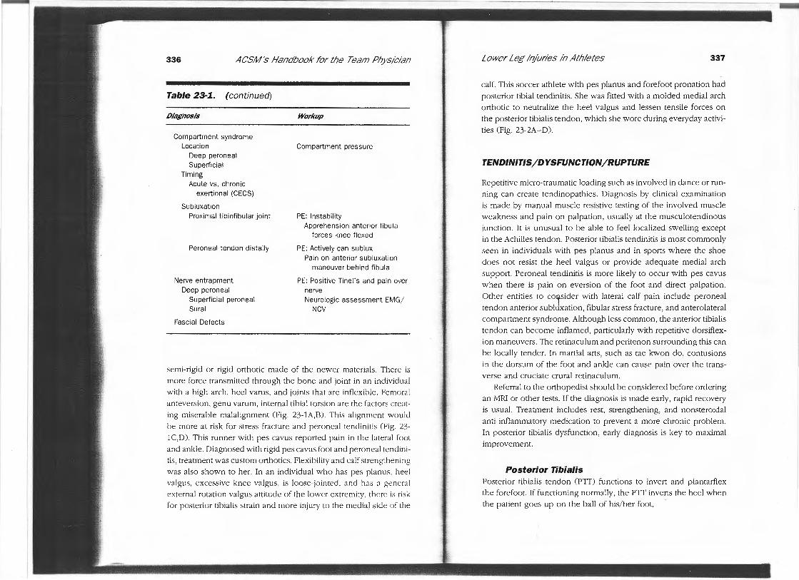
Table 23-1. (continued)
Diagnosis
Compartment syndrome Location
Deep peroneal Superficial
Timing Acute vs. chronic
exertional (CECS)
Subluxation Proximal tibiofibular joint
Peroneal tendon distally
Nerve entrapment Deep peroneal
Superficial peroneal Sura I
Fascia! Defects
Workup
Compartment pressure
PE: Instability Apprehension anterior fibula
forces knee flexed
PE: Actively can sublux Pain on anterior subluxation
maneuver behind fibula
PE: Positive Tinel's and pain over nerve Neurologic assessment EMG/
NCV
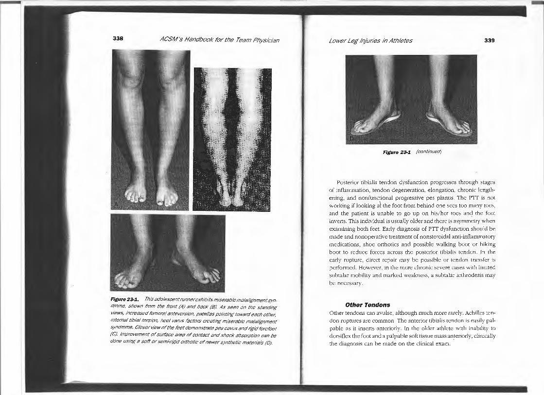
semi-rigid or rigid orthotic made of the newer materials. There is more force transmitted through the bone and joint in an individual with a high arch, heel varus, and joints that are inflexible. Femoral anteversion , genu varum, internal tibial torsion are the factors creating miserable malalignment (Fig. 23-lA,B). This alignment wou ld be more at risk for stress fracture and peroneal tendinitis (Fig. 23-1 C,D). This runner with pes cavus reported pain in the late ral foot and ankle. Diagnosed with rigid pes cavus foot and peroneal tendinitis, treatment was custom orthotics. Flexibility and calf strengthening was also shown to her. In an individual who has pes planus , heel va lgus, excessive knee valgus, is loose- jo inted , and has a general external rotation valgus attitude of the lower extremity, there is risk for poste rior tibialis strain and more injury to the medial side of the
Lower Leg Injuries in At/J/etes 337
calf. This soccer athlete with pes planus and forefoot pronation had posterior tibial tendinitis. She was fitted with a molded medial arch orthotic to neutralize the heel valgus and lessen tensile forces on the posterior tibialis tendon, which she wore during everyday activities (Fig. 23-2A-D).
TENDINITIS/DYSFUNCTION/RUPTURE
Repetitive micro-traumatic loading such as involved in dance or running can create tendinopathies. Diagnosis by clinical examination is made by manual muscle resistive testing of the involved muscle weakness and pain on palpation, usually at the musculotendinous junction. It is unusual to be able to feel localized swelling except in the Achilles tendon. Posterior tibialis tendinitis is most commonly seen in individuals with pes planus and in sports where the shoe does not resist the heel valgus or provide adequate medial arch support. Peroneal teodinitis is more likely to occur with pes cavus when there is pain on eversion of the foot and direct palpation. Other entities to co7sider with lateral calf pain include peroneal tendon anterior subluxation, fibular stress fracture, and anterolateral compartment syndrome. Although less common, the anterior tibialis tendon can become inflamed, particularly with repetitive dorsiflexion maneuvers. The retinaculum and peritenon surrounding this can be locally tender. In martial arts, such as tae kwon do, contusions in the dorsum of the foot and ankle can cause pain over the transverse and cruciate crural retinaculum.
Referral to the orthopedist should be considered before ordering an MRI or other tests . If the diagnosis is made early, rapid recovery is usual. Treatment includes rest , strengthening, and nonsteroidal anti-inflammatory medication to prevent a more chronic problem. In posterior tibialis dysfunction , early diagnosis is key to maximal improvement.
Posterior Tibialis Posterior tibialis tendon (PTT) functions to invert and plantarflex the forefoot. If functioning normally, the PTT inverts the heel when the patient goes up on the ball of his/her foot .
338 ACSM's Handbook for tile Team Physician
Figure 23-1. This adolescent runner exhibits miserable ma/alignment syndrome, shown from the front (A) and back (BJ. As seen on the standing views, increased femoral ante version, patellas pointing toward each other,
internal tibial torsion, heel varus factors creating miserable mala!tgnment syndrome. Closer view of the feet demonstrate pes cavus and rigid forefoot (C). Improvement of surface area of contact and shock absorption can be
done using a soft or semkigtd orthotic of newer synthetic materials (D).
••
Lower Leg Injuries in Athletes 339
Figure 23-1 (continued)
Posterior tibialis tendon dysfunction progresses through stages of inflammation, tendon degeneration, elongation, chronic lengthening, and nonfunctional progressive pes planus. The PTT is not working if looking al the foot from behind one sees too many toes, and the patient is unable to go up on his/her toes and the foot inverts. This individual is usually older and there is asymmet1y when examining both feet. Early diagnosis of PTT dysfunction should be made and nonoperative treatment of nonsteroidal anti-inflammatory medications, shoe orthotics and possible walking boot or hiking boot to reduce forces across the posterior tibialis tendon. In the early rupture, direct repair may be possible or tendon transfer is performed. However, in the more chronic severe cases with limited subtalar mobility and marked weakness, a subtalar arthrodesis may be necessary.
Other Tendons Other tendons can avulse, although much more rarely. Achilles tendon ruptures are common. The anterior tibialis tendon is easily palpable as it inse1ts anteriorly. In the older athlete with inability to dorsiflex the foot and a palpable soft tissue mass anteriorly, clinically the diagnosis can be made on the clinical exam.

340 ACSM's Ha17dbook ror t/Je Team P/Jysician
Figure 23-2. T/7/s male soccer athlete was having pain in tile medial as
pect or Ills root and diagnosed as having posterior tibia/is tendinitis. Tile roreroot pronation (AJ and l!ee/ valgus seen !Tom behind (BJ showing tile
lack or medial arcl! and first toe visible or tile root, wl!icl! ls at risk due to
being more l7ex1b!e and going into excessive valgvs. A semk1j[;d ortl!otic
to decrease l!ee/ valgus and by less posterior tibial!'s tensile rorces l!e/ped return tl71's athlete to ru// soccer activities as seen !Tom beside (CJ and !Tom bel7ind (0)
Lower Leg !rz/uries /17 Athletes 341
Figure 23-2 (continued')
Peroneal longus tendon rupture, although rare, can occur with ankle sprain. The level is at the distal fibula. Partial tears can exist and are treated with immobilization. MRI scan can be helpful in this diagnosis, which may be confusing. Direct repair of the tendon is best in acute rupture.
Peroneus brevis can detach from the tendon insertion at the base of the 5th metatarsal. This can be clinically apparent by a small avulsion fracture and on manual muscle testing inability to evert the foot. '

342 ACSM's Handbook for the Team Physician
STRAINS
The most common calf strain involves the medial head of the gastrocnemius in an activity involving plantar flexion and has been coined "tennis leg." A sudden pop is felt in the mid-calf. Controversy exists whether plantaris rupture actually occurs. A pop fe lt in the posterior calf when doing an explosive push-off maneuver usually involves the gastrocnemius. Lateral gastrocnemius can also be involved. If the pain is deeper, soleus strain also is considered. Other entities in the differential diagnosis include acute posterior compartment syndrome and deep vein thrombosis if there is acute onset of calf
pain.
MEDIAL TIBIAL STRESS SYNDROME
This syndrome presents with diffuse medial tibial pain of differing degrees of severity. Other previous names for this are shin splints, tibial periostitis, or medial tibial syndrome. Unlike a tibial stress fracture , the pain is diffuse over the medial aspect of the tibia on direct palpation, usually the middle third. Plain radiographs may show thickening of the medial tibial cortex. Technetium bone scan will show a diffuse, less intense activity than the more intense focal uptake seen in a tibial stress fracture. This entity is secondary to repetitive loading on pronated extremity resulting in changes in attachments and inflammation of the soleus and posterior tibial is tendon and tibial periostitis. Medial tibial stress syndrome is more common in the foot with pronation, heel in valgus, and differing degrees of rigidity. The more typical foot attitude is pes planus as in Figure 23-2. Treatment is rest, strengthening of the musculature and assurance of proper medial plantar and heel support, and nonsteroidal
anti-inflammatory medications.
FRACTURES
Stress fractures result from fatigue or insufficiency. Fatigue fractures are caused by repetitive muscular stresses and torque on normal
Lower Leg //'!furies in Athletes 343
bone. The synchrony of load transmitted through bone and dynamic muscular ability to dissipate forces are disturbed. Controversy exists
if the muscle fatigues first , then bone load increases or if the muscle tension and attaching forces cause the bone to fail.
Insufficiency fractures occur when the bone does not have normal e lastic resistance or mineral. This can be due to many factors, including previous immobilization, age, gender, menstrual history,
medical condition , certain medications, level of fitness, anatomic alignment, biomechanical factors, and prev ious surgeries on the
lower extremities. Wolff's law describes the bone adapting to the stresses placed on it. If the elastic res istance is abnormal , weakening ma y occur rather than strengthening. If loading forces exceed the bone integrity, a stress fracture resu lts.
The frequ ency of stress fractures in runners was reported by McBryde's series as tibia 34%, fibula 24%, metatarsals 18%, femur
14%, pelvis 6%, and other bones 4%. Fibula stress fractures are being seen at greater rates in female athletes in sports that involve landing and cutting-gymnastics and basketball. If a stress fracture is diagnosed, workup should include a detailed history of distance and
intensity of traini~g, nutritional assessment with completion of daily intakes, observation of eating by a medical staff, and menstrual history . Physical examination is also important to assess lower extremity alignment, leg length discrepancy, foot mobility, pes cavus, pes
planus, and wear on the sole of the shoe. In a repetitive sport such as running, the longer extremity is more
likely to be injured and at the calf will tend to have increased valgus
and pronation movements. The longer leg has an increased risk of injury. Runners who are running on the side of the road will have
more problems with their downside leg.
Running on a hard surface such as concrete, or alignment
problems such as tibia vara or pes cavus, increase the forces that must be absorbed by the bone. When a runner is on a banked
track or running in the road that has the ability for drainage of water, a leg length discrepancy and abnormality of forces exist. The lower or then essentially longer leg must externally rotate ,
creating increased posteromedial knee joint forces and abnormal loading of bone.

344 ACSM's Ha!7dbook !or t/Je Team P/Jysic/a17
Stress fractures are more common in female long distance runners who have irregular menstrual periods and eating disorders or poor nutrition. Barrow and Saha reviewed female long distance runners
and found significant increase in stress fractures in runners with very ll' irregular menstrual periods. Association of menstrual irregularities and eating behavior disorder was also found to be very high.
Tibia
The most common location of tibial stress fracture is the mid-distal third junction. A test for tibial stress fracture involves 3-point pressure and distraction at fracture site (Fig. 23-3). This premenarchal runner began running cross country immediately with 20 miles per week. After 2 weeks, she developed localized pain to palpation directly over the medial tibia. Marked radiographs of the left tibia/ fibula show localized periosteal reaction consistent with a healing stress fracture 4 weeks after the initial symptoms (Fig. 23-4A).
Bone scan was not performed because the patient clinically had a small localized area of pain. The plain radiographs showed callus and medial tibial cortical thickening. She was treated with rest, swimming exercises, and returned to nmning activities at 4
months. She was then postmenarchal and nutrition assessment had been done with improvement of her dietary habits. One year after the tibial stress fracture, she was playing soccer and running
cross country. She was seen for bilateral calf pain, and radiographs showed the fracture healed (arrow) and no new stress fracture (marker) (Fig. 23-4B).
ANTERIOR CORTEX
The fracture of the anterior tibial cortex or the dreaded black line occurs in jumping athletes who have an anterior tibial bow and repetitive flexor musculature activities involved in the sport which
place tensile forces along the anterior tibial cortex. This basketball athlete had localized pain and firmness in the midanterior tibial cortex with pain localized over area on palpation. He had 6 weeks of pain and had started intensive plyometrics. The figures show the
Lower Leg lq/uries /17 Athletes 345
PAIN
Figure 23-3. Easy clinJ a1 test to diagnose tibial stress fracture is shown diagrammatically using 3 point pressure techniques and distraction (Cour
tesy of Peter .Joki, MD., Chief, Section of Sports Medicine, Yale Sports Medicine Center, Department of Orthopaedics and Rehabilitation).
dreaded black line in a very thick cortex and alignment, which showed an anterior tibial bow (Fig. 23-5). Treatment for this fracture was rest, water conditioning, and return to activities at 6 weeks when the fracture clinically was no longer tender. This fracture can be very bothersome with delayed nonunions and may require surgery.
MEO/AL MALLEOLUS STRESS FRACTURE
This fracture is seen in jumping sports. This basketball athlete sustained a second medial malleolus fracture after the initial one was healed with casting. The acute reinjury films show radiolucency of the medial malleolus just at the corner of the tibial plafond (Fig. 23-6A). Aggressive treatment with internal fixation without exposing
i. ll
ij1' I· 1·, ~~ I
i: :· ii ; 1
I 't !
I I I' :I
I
346 ACSM's Ha17dbook ror t/Je Team P/Jysic/a17
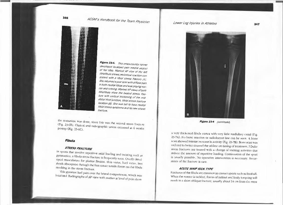
Figure 23-4. This cross-count;y runner
developed localized pain medial aspect
of her tibia. Marked AP view of the left
t1biollbula shows periosteal reaction con
sistent with a tibial stress fracture (A)
She returned a year later w1tl7 diffuse pain
in both medial t1bias and was playing soc
cer and running. Marked AP views of both
t1biollbula snow the healed stress frac
ture wit/7 cortical thickening of the mid
distal third/unction, tibial stress fracture
location (BJ. She was felt to /Jave medial
tibial stress syndrome and no new stress fracture.
th e nonunion was done, since this was the second stress fra cture (Fig. 23-68). Clinical and radiographic union occurred at 6 weeks postop (Fig. 23-6C).
Fibula
STRESS FRACTURE
In sports that involve repetitive axial loading and twisting such as gymnastics, a fibula stress fracture is frequently seen. Overly devel
oped musculature for plantar flexion, tibia varus, heel varus, less shock absorption through the foot create tensile forces on th e fibula resulting in the stress fracture.
This gymnast had pain over the lateral compartment, which was loca li zed. Radiographs of AP view with marker at level of pain show
Lower Leg 117/uries /17 At/J/etes 347
Figure 23-4 (continued)
a very thickened fibula cortex with very little medullary canal (Fig. 23-7 A). No bone reaction or radiolucent line can be seen. A bone scan showed intense increase in activity (Fig. 23-78). Bone scan was o rdered to better counsel the athlete on timing of treatment. Fibular stress fra ctures are treated with a change of training activities that red uce the amount of repetitive loading. Continuation of the sport is usually possible. No operative intervention is necessary. Recurrence of the fracture is rare.
ACUTE WHIP KICK TYPE
Fractures of the fibula are common in contact sports such as football. When the runner is tackled, forces of helmet and body torquing will result in a short ohlique fracture , usually about 14 cm from the most

348 ACSM's Handbook !"or the Team Physician
Figure 23-5. Cone lateral view of the tibia shows the dreaded black line or anterior tibial cortex stress fracture. This is typical/y seen in athletes with
anterior tibial bow and can_ be difficult to heal and can require intermedullaty rodding for late or nonunion.
proximal aspect of the fibul a. This football nmning back was tackled
and sustained a direct blow with shoulder pads in the lateral leg. Fracture orientation and location is classic (Fig. 23-SA). The fracture
hea ls quickly as shown in AP view at 4 weeks post injury (Fig. 23-SB). Return to sport is based on pain and can be as early as a couple of weeks.
!,.
Lower Leg Injuries in Athletes
Figure 23-6. Medial
malleolus stress fracture
is seen with lucency going
obliquely in the medial
malleolus at the tibial plafond level. There is no sig
nificant periosteal reac
tion due to the cancellous
bone and intra-articular nature of this fracture (A).
Rxation was done as this was the second medial
malleolar stress fracture
with two cancellous screws placed without the fracture being taken down
(BJ. Healing was complete
at 6 weeks postop as shown(CJ.
349

-350
ACSM's Handbook !or tl7e Team P17ysician
Figure 23-6 (continued)
Pathologic Fracture
Particularly in adolescents, careful scrutiny of the radiographs should be done to rule out any other reasons for fracture such as an underlying tumor. This soccer athlete sustained a pathologic fracture
through an underlying nonossifying fibroma of the distal tibia . The fracture occurred when his foot was planted as he was going for the ball in a noncontact mechanism. He had no previous knowledge of the cyst or pain in the tibia. The long oblique comminuted fra cture
is shown (Fig. 23-9A). He was treated in a long leg cast (Fig. 23-98). After 8 weeks of cast immobilization, the fracture had completely healed. Follow-up radiographs at 6 months postop show the fracture completely healed, and the cyst was smaller on AP (Fig. 23-9C) and lateral (Fig. 23-90) views. The fracture itself and bone response reduce the size of the cyst. Careful attention should be
Lower Leg /f?/Uries in Athletes
Figure 23-7. Marked AP
view of the tibloflbula show the
cortical thickening diffusely of
the flbula without much medul
la!)/ canal (A). Stress fracture
was clinically diagnosed by the localized pain and the nature
of her sport of repetitive axial
loading In gymnastics. A bone scan (8) was performed to doc
ument the diagnosis since there was no periosteal reaction on plain radiograph.
351
352 ACSM's Haadbook ror t/Je Team P/Jysiciaa
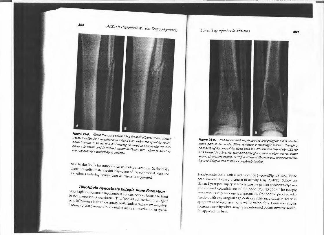
Figure 23-8. Rbula fracture occurred in a l'ootbal/ athlete, short, oblique (ypical location !'or a w/7/pkick-(ype il?IUIY 14 cm below tile t1;o or tile fibula.
Acute fracture is shown in A and Ilea/Ing occurred at !'our weeks (B). This fracture is stable and is treated symptomatica/(y, wtt/7 return to sport as soon as running coml'ortab/y is possible.
paid to the fibula for tumors such as Ewing's sarcoma. fn skele tally immature individuals, carefu l inspection of the epiphyseal plate and sometimes ordering comparison AP views is suggested.
Tibiofibula Synostosis Ectopic Bone Formation With high interosseous ligamentous spra ins ectopic bone can form in the interosseous membrane. This football athlete had prolonged pain following a high ankle sprain . Initial radiographs were negative. Radiographs at 3 months following his injury showed a fibular synos-
~ rr7SaW¥7t~~~~~~-;::~~~::::c
Lower Leg /!?Juries /17 At/Jletes 353
Figure 23-9. Tilts soccer at/7/ete planted l71s root going !'or a ball and felt
acute pain in l71s ankle. Films reviewed a pathologic fracture tllroug/7 a
nonossi(ying l'ibroma or tile distal tibia (A), AP wew and lateral view (B). He
was treated in a long leg cast and lleal1i7g occurred at eigllt weeks. Views
shown six months postop, AP (C), and lateral (DJ show cyst to be consolidating and lill1i7g 1i7 and fracture completely healed.
tosis/ ecropic bone with a radiolucency (arrow)(Fig. 23-lOA). Bone scan showed intense increase in activity (Fig. 23-lOB). Follow-up film at 1 year post injury at which time the patient was nonsymptomatic showed consolidation of the bone (Fig. 23-IOC). The ectopic bone will usually become asymptomatic. One should proceed with caution with any surgical exploration as this may cause increase in symptoms and recurrent bone will develop if the bone scan shows increased activity when surgery is performed. A conservative watchfu l approach is best.

354 ACSM's Handbook !'or tl7e Team P17ysician
Figure 23-9. (continued)
PROXIMAL TIBIOFIBULAR INJURIES
Proximal tibiofibular joint sprains, subluxations, and dislocations, although unusual, occur. Injuries involving this jo int are no t we JJ appreciated or reported. The mechanism is foot in plantar fl exion
and inversion, knee fl exed and leg adducted. Initial management if strained involves anterior pad on the fibular head to decrease
chances of anterior subJuxation. Symptoms can recur with recurrent anterolateral dislocations. Operative intervention to stabilize this obliquely oriented joint may be unsuccessful. This football athlete anteriorly subluxed the proximal tib-fib joint clinically. Radiographs show (Fig. 23-11) an oblique joint with no fracture or subluxa tion. Variations of the slope of the lateral tibial metaphysis and proximal
Lower Leg Injuries in At/7/etes
Figure 23-10. Football athlete with prolonged pain in the interos
seous membrane. Football athlete had prolonged pain following high
ankle sprain. His initial radiographs were negative. At 3 months, radiographs showed a tib-1ofibular synostosis as seen on cone AP v;ew (A). Bone scan showed intense increase in activity in this area indicative of continued bone formation in the interosseous membrane (B). Follow up at 1 year following his inju;y shows consol1dat1on of the ectopic bone (C) and the athlete is asymptomatic. Care should be taken to not aggressively treat these lesions su;gically while bone formation is present or there will be reformation of bone and continued pain.
)
B
355
P.

356 ACSM's Handbook !'or t/Je Team Physician
Figure 23-10. (continued')
aspect of the tibia are common. The more horizontal tibia and less elongated fibular head are inherently more stable. This cheerleader was noted to have asymptomatic instability during pre-season physicals (Fig. 23-12). This joint is best examined with the knee fl exed and compared with the opposite side.
FASCIAL DEFECTS
With increased pressure in a compartment the muscle may bulge through the fascia. This is a cosmetic problem, not functional. No surgical repair is indicated. If symptomatic, treatment is a compressive calf sleeve and ice.
Lower Leg lq/uries In Athletes 357
Figure 23-11. Proximal t1biofibular joint injuries occur more often than thought. This football athlete anteriorly sub/uxed his proximal ftbula anter1:
orly. Radiographs of an oblique view show no fracture and the joint was re
reduced. Variations in contour of the lateral tibial plateau and proximal ftbula influence the joint stability.
358 ACSM's Ha!7dbook !'or tl7e Team P17ysician
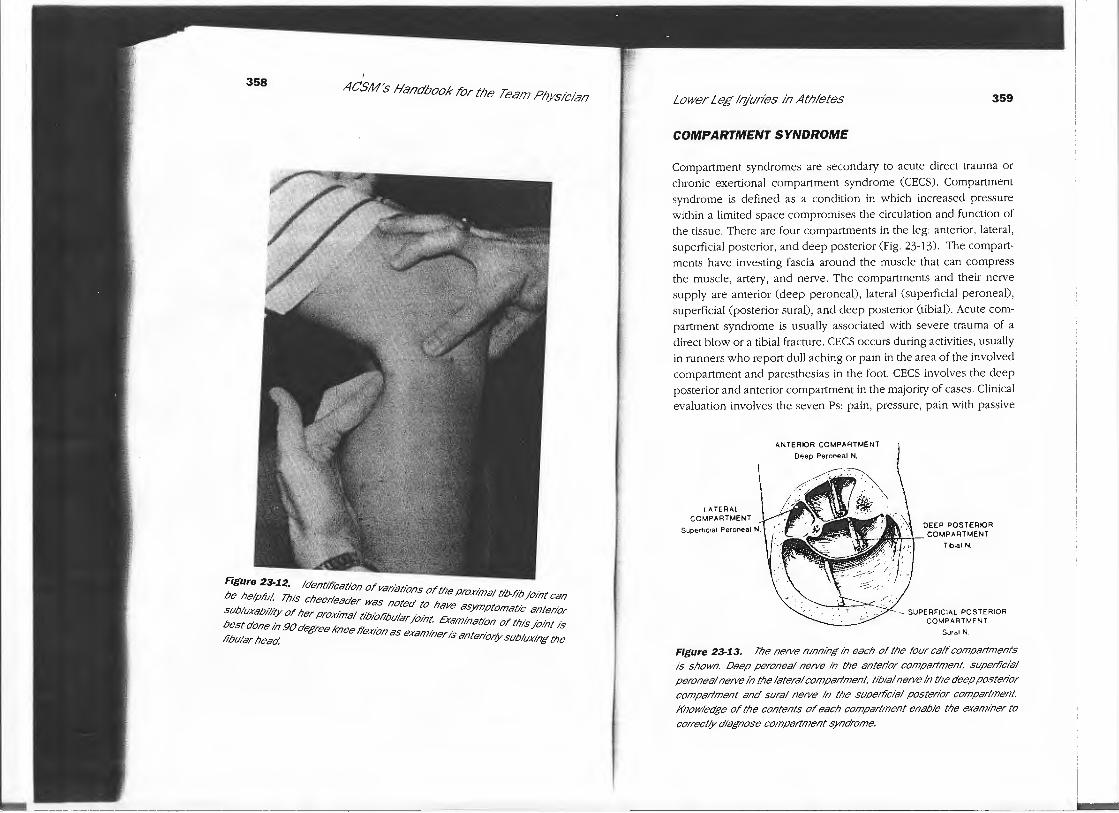
Figure 23-12. Identification or variations or the prox1inal t1b-fib /Dint can
be he/pru1. This cheerleader was noted to have asymptomatl"c antenor subluxab1/i(Y or her prox1inal t1b101'ibular /Dint. Examination or this jo1i?t is
best done 1i7 90 degree knee ti'ex;On as examiner is anter1or(Y sublux!i?g the fibular head.
Lower Leg Injuries in Athletes 359
COMPARTMENT SYNDROME
Compartment syndromes are secondary to acute direct trauma or chronic exertional compartment syndrome (CECS). Compartment syndrome is defined as a condition in which increased pressure within a limited space compromises the circulation and function of the tissue. There are four compartments in the leg: anterior, lateral, superficial posterior, and deep posterior (Fig. 23-13). The compartments have investing fascia around the muscle that can compress the muscle, artery, and nerve. The compartments and their nerve supply are anterior (deep peroneal), lateral (superficial peroneal), superficial (posterior sural), and deep posterior (tibial). Acute compartment syndrome is usually associated with severe trauma of a direct blow or a tibial fracture. CECS occurs during activities, usually in runners who report dull aching or pain in the area of the involved compartment and paresthesias in the foot. CECS involves the deep posterior and anterior compartment in the majority of cases. Clinical evaluation involves the seven Ps: pain, pressure, pain with passive
LATERAL COMPARTMENT
Super11c1al Peroneal N.
ANTERIOR COMPARTMENT
Deep Peroneal N.
DEEP POSTERIOR
1%, lif,f/!, . \ COMPARTMENT Tibial N.
- SUPERFICIAL POSTERIOR COMPARTMENT
Sura! N
Figure 23-13. The nerve running in each of the rour calr compartments
is shown. Deep peroneal nerve in the anterior compartment, superficial
peroneal nerve in the lateral compartment, tibial nerve ii? the deep posterior compartment and sural nerve in the superficial posterior compartment. Knowledge or the contents or each compartment enable the examiner to
correct(Y diagnose compartment syndrome.
---~~L

360
Table 23-2.
Rel{/ on
Anterior
Lateral
Superficial posterior
Deep posterior
ACSM's Handbook for the Team P17ysician
Anatomy Calf Compartments
Muscle
Tibialis anterior Extensor digitorum
longus, extensor hallucis longus
Peroneus tertius
Peroneus longus Peroneus brevis
Gastrocnemius-Soleus
Plantaris
Tibialis posterior, flexor hallucis longus
Flexor digitorum
l\ ngus
Artery
Anterior tibial
Peroneal
Tibial
Ne1J1e
Deep peroneal
Deep peroneal
Superficial peroneal
Sura I
Tibial
stretch, paresis or paralysis, paresthesia, pulselessness, and power.
The diagnosis is made by measurement of the intracompartmental pressures. The contents of the specific compartments are summarized in Table 23-2. Measurements of intracompartmental pressures before and after exercise are performed. Post exercise compartment pressures above 30 mm Hg is believed to be significant. If accepta ble to the patient, treatment for CECS is modification of activities, stopping before symptoms develop. However, the management of a documented compartment syndrome is surgical with release of that specific compartment. The superficial and deep posterior compartments are released through a single incision off of the medial tibia, and the anterior and lateral compartments are released using an incision just anterolaterally. This athlete developed a bilateral foot drop and lateral calf pain a certain distance into her run. The difference in her resting and post exercise pressures was 40 mm Hg.
She underwent anterior and ante rolateral fasciotomies . Operative exposure of the anterolatera l calf shows fascia! investment (Fig. 23-
Lower Leg Injuries in Athletes 361
14A), scissors performing release (Fig. 23-14B), and healthy muscle
under no further pressure (Fig. 23-14C).
Nerve Entrapment The three most common nerves to be entrapped by fascia are the deep peroneal nerve, superficial peroneal nerve, and sural nerve
(Fig. 23-15). The common peroneal nerve can be entrapped by peroneus longus origin. This has been reported in runners in activities that involve plantar fl exion inversion force at the ankle, causing the nerve to be under pressure due to the repetitive muscular contracture and sharp fibrous edge of the peroneus longus. The common peroneal nerve can also have pressure applied from osteochondroma or bony lesion of the proximal fibula , proximal tib-fib dislocation, knee injuries, and localized lateral menisca l cyst. Deta iled physical exam is necessary.
The deep peroneal nerve is most commonly entrapped as it trav
els between the extensor digitorum longus and extensor hallucis
longus, approximately 5 cm above the ankle joint just below the extensor retinaculum (Fig. 23-15). This entrapment is described as
ante rior tarsal tunnel syndrome. Symptoms include positive Tinel 's
sign and pain in the first web space. Initial treatment is suggestions on changing of shoes, making sure there is no direct pressure on
the anterior part of the ankle or repetitive stresses involving plantar fl exion inversion .
Superficial peroneal nerve is most commonly entrapped as it exits
from the fascia 10 cm proximal to the lateral malleolus. Styf described
a test fo r superficial peroneal nerve entrapment as positive Tinel's sign at level of exit from the deep fascia, pain on passive ankle plantar fl exion inversion , and tenderness on palpation with resistive
ankle dorsiflexion and eversion.
The sural nerve emerges between the two heads of the gastrocnemius in the mid-calf of the leg. Entrapment may occur at any level of the latera l calf commonly as the branches exit a couple of centimeters above the ankle. Suspicion of entrapment is made based on positive Tinel 's sign directly over the su ral nerve course (Fig. 23-15).

362 ACSM's Handbook /"or tile Team P/Jysician :~'
~.
Figure 23-J.4. (continued)
Lower Leg Injuries in At!Jletes 363
Figure 23-1.4. (Continued). This athlete developed bi lateral footdrop when she would run a set distance. Her compartment pressures shou ldn't be a difference of 40 mg. mercury before and after exercise. Release of the anterior and anterolateral compartments was done through an 8 cm incision in the mid-third lateral to the anterior aspect of the tibia. Exposure before fascia l release is shown (A), release using Metzenbaum scissors of the anterior compartment (8), and post release showing the normal appear
ance of the muscle following fascial release (C).
Other conditions associated of sural nerve entrapment are recurrent ankle sprains, ganglion of the peroneal sheath or ankle, and Achilles' tendinitis. Nerve entrapments are unusual but should be considered in the differential diagnosis, particularly of localized pain in the anterolateral aspect of the leg.
Consideration of lumbar spine involvement with radiculopathy shou ld always be done and is more common than these localized nerve entrapment syndromes. Systemic conditions of diabetes , alcohol , and double crush syndrome should also be considered.
Tarsal Tunnel Syndrome High tarsal tunnel syndrome refers to pressure on the posterior tibialis nerve as it emerges at the musculotendinous junction of the calf. Electromyelograms and nerve conduction velocities should be
364
Common~ Peroneal N.
Sura/ N.
Intermediate Dorsal Cutaneous N.
ACSM'S Haodbook ror tile Team Pllysiciao
Deep Peroneal N.
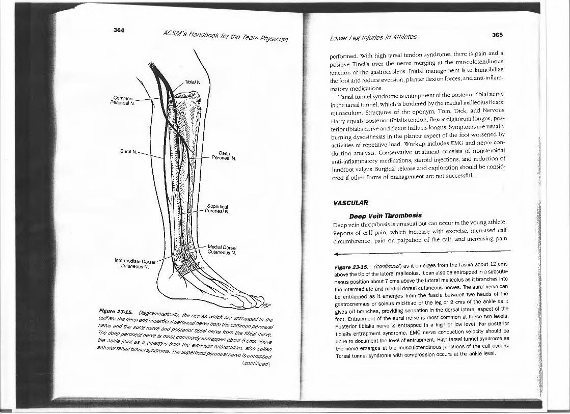
Figure 23·15. Diagrammatically, tl7e ne1Ves w/7/c/7 are entrapped in tl7e
calr are tl7e deep and supertlcial peroneal ne1Ve from tl7e common peroneal
ne1Ve and tl7e sural ne1Ve and posterior tibial ne1Ve from tl7e tibial ne1Ve.
Tl7e deep peroneal ne1Ve is most common(y entrapped about 5 ems above
tl7e ankle /Dint as ti emetges from tl7e extensor retinaculum, also called an tenor tarsal tunnel syndrome. Tl7e supemcial peroneal ne1Ve is entrapped
(continued)
Lower Leff /ojuries io Athletes 365
performed. With high tarsal tendon syndrome, there is pain and a positive Tinel's over the nerve merging at the musculotendinous junction of the gastrocsoleus. Initial management is to immobilize the foot and reduce eversion , plantar flexion forces, and anti-inflam-
matory medications. Tarsal tunnel syndrome is entrapment of the posterior tibial nerve
in the tarsal tunnel, which is bordered by the medial malleolus flexor retinaculum. Structures of the eponym, Tom, Dick, and Nervous Harry equals posterior tibialis tendon, flexor digitorum longus, posterior tibialis nerve and flexor hallucis longus. Symptoms are usually burning dysesthesias in the plantar aspect of the foot worsened by activities of repetitive load. Workup includes EMG and nerve conduction analysis. Conservative treatment consists of nonsteroidal anti-inflammatory medications, steroid injections, and reduction of hindfoot valgus. Surgical release and exploration should be consid
ered if other forms of management are not successful.
VASCULAR
Deep Vein Thrombosis Deep vein thrombosis is unusual but can occur in the young athlete . Reports of calf pain, which increase with exercise, increased calf circumference, pain on palpation of the calf, and increasing pain
Figure 23-15. (continued) as it emerges from the fascia about 12 ems above the tip of the lateral malleolus. It can also be entrapped in a subcutaneous position about 7 ems above the lateral malleolus as it branches into
the intermediate and medial dorsal cutaneous nerves. The sural nerve can be entrapped as it emerges from the fascia between two heads of the gastrocnemius or soleus mid-third of the leg or 2 ems of the ankle as it gives off branches, providing sensation in the dorsal lateral aspect of the foot. Entrapment of the sural nerve is most common at these two levels . Posterior tibialis nerve is entrapped in a high or low level. For posterior tibialis entrapment syndrome, EMG nerve conduction velocity should be done to document the level of entrapment. High tarsal tunnel syndrome as the nerve emerges at the musculotendinous junctions of the calf occurs.
Tarsal tunnel syndrome with compression occurs at the ankle level.
I 1 !
·I
:j I
Ii tl •1 u ~ ·I I ,,
366 ACSM's Handbook for t/Je Team Physician
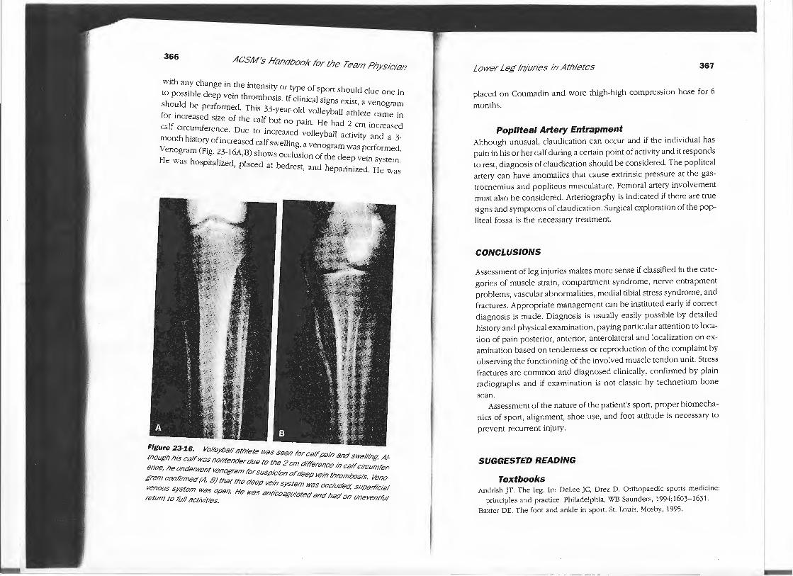
with any change in the intensity or type of sport should clue one in to possible deep vein thrombosis. If clinical signs exist, a venogram should be performed. This 33-year-old volleyball athlete came in for increased size of the calf but no pain. He had 2 cm increased ca lf circumference. Due to increased volleyball activity and a 3-month history of increased calf swelling, a venogram was performed. Venogram (Fig. 23-16A,B) shows occlusion of the deep vein system. He was hospitalized, placed at bedrest, and heparinized . He was
Figure 23-1.6. Volleyball athlete was seen for calf pain and swelling. Al
tl7ougl717is calf was nontender due to tile 2 cm difference in calf circumfer
ence, Ile underwent venogram for suspicion of deep vein thrombosis. Veno
gram cont?rmed (A, BJ that tile deep vein system was occluded, supelfi'cial
venous system was open. He was ant/coagulated and had an uneventl'u/ return to l'u!/ activities.
:.:
Lower Leg Injuries in Athletes 367
placed on Coumadin and wore thigh-high compression hose for 6
months.
Popliteal Artery Entrapment Although unusual, claudication can occur and if the individual has pain in his or her calf during a certain point of activity and it responds to rest, diagnosis of claudication should be considered. The popliteal artery can have anomalies that cause extrinsic pressure at the gastrocnemius and popliteus musculature. Femoral artery involvement must also be considered. Arteriography is indicated if there are true signs and symptoms of claudication. Surgical exploration of the pop
liteal fossa is the necessary treatment.
CONCLUSIONS
Assessment of leg injuries makes more sense if classified in the categories of muscle strain, compartment syndrome, nerve entrapment problems, vascular abnormalities, medial tibial stress syndrome, and fractures. Appropriate management can be instituted early if correct diagnosis is made. Diagnosis is usually easily possible by detailed histo1y and physical examination, paying particular attention to location of pain posterior, anterior, anterolateral and localization on examination based on tenderness or reproduction of the complaint by observing the functioning of the involved muscle tendon unit. Stress fractures are common and diagnosed clinically, confirmed by plain radiographs and if examination is not class ic by technetium bone
scan. Assessment of the nature of the patient's sport, proper biomecha
nics of sport, alignment, shoe use, and foot attitude is necessary to
prevent recurrent injury.
SUGGESTED READING
Textbooks Andrish JT. The leg. In: Delee JC, Drez D. Orthopaedic sports medicine:
principles and practice. Philadelphia, WB Saunders, 1994;1603-1631-Baxter DE. The foot and ankle in sport. St. Louis, Mosby, 1995.

368 ACSM's Handbook f'or t/Je Team P/Jysician
Mubarak S, Hargens A. Compartment syndromes and Volkmann's contractures. Philadelphia, WB Saunders, 1981.
Fu FH, Stone DA. Sports injuries: Mechanism, prevention,
and treatment, 2nd ed. Baltimore, Williams & Wilkins, 1994.
Nicholas ]A. The lower extremity and spine in sports medicine, Vol I. St. Louis, CV Mosby Co., 1986.
Epidemiology
Mechelen WV. Running injuries: A review of the epidemiological literature. Sports Med 1992;14:320-335.
Stress Fractures
McBryde AM. Stress fractures in runners. In: D'Ambrosia R, Drez D, eds.
Prevention and treatment of running injuries. Thorofare, New j ersey: Slack, 1982: 21-42.
Matheson GO, Clement DB, McKenzie DC, et al. Stress fractures in athletes: A study of 320 easel Sports Med 1987; 15: 46-58.
Barrow GW, Saha S. Menstrual irregularity and stress fractures in collegiate female distance runl)ers. Am] Sports Med 1988;16:209-216.
Orava S, Hulkko A. Stress fractures in athlete. Int] Sports Med 1987;8: 221-226.
Rettig A, Shelbourne D, Mccarroll], et al. The natural history and treatment
of delayed union stress fractures of the anterior cortex of the tibia. Am .J Sports Med 1988;16:250-255.
Hershman EB, Mailly T. Stress fractures. Clin Sports Med 1990;9:183-214.
Sterling JC, Edelstein DW, Calvo RD, et al. Stress fractures in the athle1e: Diagnosis and management. Sports Med 1992;14:336- 346.
Nerve Entrapment
Leach R, Purnell M, Saito A. Pe roneal nerve entrapment in runners. Am .I Sports Med 17(2):287-291.
Styf). Entrapment of the superficial peroneal nerve . .I Bone Joint Surg (Br) 1989;71: 131-135.
;n if
Lower Leg Injuries in Athletes 369
Proximal Tibiofibular Injuries Resnick D, Newell], Guerra ], et al. Proximal tibiofibular joint. Anatomic
pathologic-radiographic correlation. Am] Roentgenol 1978; 131 : 133-138.
Compartment Syndrome Hutchinson MR, Ireland ML. Common compartment syndromes in athletes:
Treatment and rehabilitation. Spo1ts Med 1994;17:200-208. Matsen F, Winquist R, Krugmire R. Diagnosis and management of compart
mental syndromes.] Bone Joint Surg 1980;62A:286- 291.
Mubarak S, Hargens A. Acute compartment syndromes. Surg Clin North Am
1983;63: 539-565. Mubarak S. A practical approach to compartment syndromes- Part II. Inst
Course Leet 1983;32:92-102. Eisele SA, Sammarco GJ. Chronic exertional compartment syndrome. Instr
Course Leet, Vol. 42, American Academy of Orthopaedic Surgeons, 1993.
Styf]. Diagnosis of exercise induced pain in the anterior aspect of the lower
leg. Am] Sports Med 1988;16:165-169.