Acquired Valvular Heart Disease -...
42
Copyright ©2005 Lippincott Williams & Wilkins Woods, Susan L., Froelicher, Erika Sivarajan, Motzer, Sandra Adams (Underhill), Bridges, Elizabeth J. Cardiac Nursing, 5th Edition 33 Acquired Valvular Heart Disease Denise Ledoux DATABASE FOR NURSING MANAGEMENT Definition, Classification, and Epidemiology Valvular heart disease continues to be a common source of cardiac dysfunction and mortality. Competent cardiac valves maintain a unidirectional flow of blood through the heart as well as to the pulmonary and systemic circulations. Diseased cardiac valves that restrict the forward flow of blood because they are unable to open fully are referred to as stenotic . Stenotic valves elevate afterload and cause hypertrophy of the atria or ventricles pumping against the increased pressure. Cardiac valves that close incompetently and permit the backward flow of blood are referred to as regurgitant, incompetent , or insufficient . Regurgitant valves cause an elevated volume load and dilation of the cardiac chambers receiving the blood reflux. Valvular dysfunction may be primarily stenotic or regurgitant, or it is a “mixed” lesion, a valve that neither opens nor closes adequately. Valvular heart disease is usually described by the duration of the dysfunction (acute vs. chronic), the valves involved, and the nature of the valvular dysfunction (stenosis, insufficiency, or a combination of stenosis and insufficiency). The degree of cardiac dysfunction is defined by the New York Heart Association's (NYHA) Functional and Therapeutic Classification. Acquired valvular heart disease most commonly affects, and is most symptomatic with, the aortic and mitral valves. This chapter focuses on the mitral and aortic valves, with a brief discussion of tricuspid valve disease. Because the cause of pulmonic disease is primarily congenital, it is not presented (see Chapter 35). Causes of Acquired Valvular Heart Disease Rheumatic Heart Disease Rheumatic fever is the most commonly acquired cause of valvular heart disease in childhood (Khan, 1996). Tissues involved in rheumatic fever include the lining and valves of the heart, skin, and connective tissue (Fig. 33-1). Rheumatic fever results as a complication of group A streptococcal upper respiratory tract infections, occurring in approximately 3% of those with streptococcal pharyngitis 2 to 3 weeks after acute rheumatic fever. Rheumatic fever is an acute systemic, inflammatory disease that occurs as a response to streptococcal infections (Bhola & Gill, 2001). Group A streptococcal throat infection is responsible for initial and recurrent attacks of rheumatic fever. Lymphatic Página 1 de 42 Ovid: Cardiac Nursing 06/04/05 http://gateway.ut.ovid.com/gw2/ovidweb.cgi
Transcript of Acquired Valvular Heart Disease -...

Copyright ©2005 Lippincott Wil l iams & Wilkins Woods, Susan L., Froelicher, Erika Sivarajan, Motzer, Sandra Adams (Underhil l), Bridges, Elizabeth J. Cardiac Nursing, 5th Edit ion
33 Acquired Valvular Heart Disease
Denise Ledoux
DATABASE FOR NURSING MANAGEMENT
Definition, Classification, and Epidemiology Valvular heart disease continues to be a common source of cardiac dysfunction and mortality. Competent cardiac valves maintain a unidirectional f low of blood through the heart as well as to the pulmonary and systemic circulations. Diseased cardiac valves that restrict the forward f low of blood because they are unable to open ful ly are referred to as stenotic . Stenotic valves elevate afterload and cause hypertrophy of the atria or ventricles pumping against the increased pressure. Cardiac valves that close incompetently and permit the backward f low of blood are referred to as regurgitant, incompetent, or insuff icient . Regurgitant valves cause an elevated volume load and dilation of the cardiac chambers receiving the blood reflux. Valvular dysfunction may be primari ly stenotic or regurgitant, or i t is a “mixed” lesion, a valve that neither opens nor closes adequately. Valvular heart disease is usually described by the duration of the dysfunction (acute vs. chronic), the valves involved, and the nature of the valvular dysfunction (stenosis, insuff iciency, or a combination of stenosis and insufficiency). The degree of cardiac dysfunction is defined by the New York Heart Association's (NYHA) Functional and Therapeutic Classif ication. Acquired valvular heart disease most commonly affects, and is most symptomatic with, the aortic and mitral valves. This chapter focuses on the mitral and aortic valves, with a brief discussion of tr icuspid valve disease. Because the cause of pulmonic disease is primari ly congenital, i t is not presented (see Chapter 35).
Causes of Acquired Valvular Heart Disease
Rheumatic Heart Disease Rheumatic fever is the most commonly acquired cause of valvular heart disease in childhood (Khan, 1996). Tissues involved in rheumatic fever include the l ining and valves of the heart, skin, and connective t issue (Fig. 33-1). Rheumatic fever results as a complication of group A streptococcal upper respiratory tract infections, occurring in approximately 3% of those with streptococcal pharyngit is 2 to 3 weeks after acute rheumatic fever. Rheumatic fever is an acute systemic, inflammatory disease that occurs as a response to streptococcal infections (Bhola & Gil l, 2001). Group A streptococcal throat infection is responsible for init ial and recurrent attacks of rheumatic fever. Lymphatic
Página 1 de 42Ovid: Cardiac Nursing
06/04/05http://gateway.ut.ovid.com/gw2/ovidweb.cgi
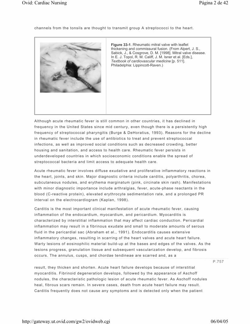
channels from the tonsils are thought to transmit group A streptococci to the heart.
Although acute rheumatic fever is sti l l common in other countries, i t has declined in frequency in the United States since mid century, even though there is a persistently high frequency of streptococcal pharyngit is (Burge & DeHoratius, 1993). Reasons for the decline in rheumatic fever include the use of antibiotics to treat and prevent streptococcal infections, as well as improved social condit ions such as decreased crowding, better housing and sanitation, and access to health care. Rheumatic fever persists in underdeveloped countries in which socioeconomic condit ions enable the spread of streptococcal bacteria and l imit access to adequate health care.
Acute rheumatic fever involves diffuse exudative and proliferative inflammatory reactions in the heart, joints, and skin. Major diagnostic criter ia include cardit is, polyarthrit is, chorea, subcutaneous nodules, and erythema marginatum (pink, circinate skin rash). Manifestations with minor diagnostic importance include arthralgias, fever, acute-phase reactants in the blood (C-reactive protein), elevated erythrocyte sedimentation rate, and a prolonged PR interval on the electrocardiogram (Kaplan, 1998).
Cardit is is the most important cl inical manifestation of acute rheumatic fever, causing inflammation of the endocardium, myocardium, and pericardium. Myocardit is is characterized by interstit ial inflammation that may affect cardiac conduction. Pericardial inflammation may result in a f ibrinous exudate and small to moderate amounts of serous f luid in the pericardial sac (Abraham et al., 1991). Endocardit is causes extensive inflammatory changes, result ing in scarring of the heart valves and acute heart fai lure. Warty lesions of eosinophil ic material build-up at the bases and edges of the valves. As the lesions progress, granulation t issue and subsequent vascularization develop, and fibrosis occurs. The annulus, cusps, and chordae tendineae are scarred and, as a result, they thicken and shorten. Acute heart fai lure develops because of interstit ial myocardit is. Fibrinoid degeneration develops, fol lowed by the appearance of Aschoff nodules, the characteristic pathologic lesion of acute rheumatic fever. As Aschoff nodules heal, f ibrous scars remain. In severe cases, death from acute heart fai lure may result. Cardit is frequently does not cause any symptoms and is detected only when the patient
Figure 33-1. Rheumatic mitral valve with leaflet thickening and commissural fusion. (From Alpert, J. S., Sabick, J., & Cosgrove, D. M. [1998]. Mitral valve disease. In E. J. Topol, R. M. Califf, J. M. Isner et al. [Eds.], Textbook of cardiovascular medicine [p. 511]. Philadelphia: Lippincott-Raven.)
P.757
Página 2 de 42Ovid: Cardiac Nursing
06/04/05http://gateway.ut.ovid.com/gw2/ovidweb.cgi

seeks help because of arthrit is or chorea.
Auscultatory signs of aortic and mitral insuff iciency are frequently apparent. In more than 90% of patients with cardit is, the mitral valve is affected. When the mitral valve is affected, there may be a high-pitched, blowing, pansystolic murmur. A Carey-Coombs murmur, a low-pitched, mid-diastolic murmur of short duration, may be noted at the apex. The Carey-Coombs murmur may be attr ibuted to swell ing and stif fening of mitral valve leaflets, increased flow across the valve, and alteration in left ventricular compliance. The regurgitation of the aortic valve results in diastolic murmurs, whereas involvement of the tr icuspid valve is rarely appreciated during the acute phase (Abraham et al., 1991).
Rheumatic fever can be prevented by aggressive treatment of the init ial episode of streptococcal pharyngit is: penici l l in G, 500 mg as the f irst dose and then 250 mg four t imes daily for a duration of 10 days. If a patient is penici l l in al lergic, then clarithromycin 500 mg twice daily for 7 to 14 days or cl indamycin 150 mg every 8 hours can be substituted (Khan, 1996).
Infective Endocarditis Infective endocardit is is an endovascular infection that supports continuous bacteremia from the source of the infection, usually a vegetation on a heart valve (Towns & Reller, 2003). Although incidence of infective endocardit is is low, between 1.5 and 6 cases per 100 cases per year, morbidity and mortality are high (Sexton & Spelman, 2003). Rheumatic heart disease, as well as other cardiac lesions such as calcif ic aortic stenosis, hypertrophic cardiomyopathy, and congenital heart disease, and the presence of prosthetic heart valves predispose to endocardit is. Intravenous drug abusers are at r isk for infective endocardit is caused by recurrent bacteremias related to injection from contaminated needles and localized infections at injection sites. Patients with long-term intravenous l ines or dialysis catheters are also at increased risk. Acute endocardit is can also occur in normal heart valves from infection somewhere else in the body. In patients with community-acquired, native valve endocardit is, Staphylococcus aureus is the predominant cause of acute disease (Karchmer, 1998). Pathogens that are most commonly responsible for subacute endocardit is include streptococci, enterococci, coagulase-negative staphylococci, and the HACEK group of organisms (Hemophilus species, Actinobacil lus actinomycetemcomitans, Cardiobacterium hominis, Eikenella species, and Kingella kingae). The S. aureus infecting intravenous drug users is frequently methici ll in-resistant (Karchmer, 1998). Clinical presentations of endocardit is range from fever and malaise to symptoms related to systemic emboli (Table 33-1).
Table 33-1 CLINICAL MANIFESTATIONS OF INFECTIVE ENDOCARDITIS
Symptoms Physical Examination Findings
Página 3 de 42Ovid: Cardiac Nursing
06/04/05http://gateway.ut.ovid.com/gw2/ovidweb.cgi

The pathologic process of endocardit is requires that several condit ions exist to permit infection to grow in the heart and to promote an environment that supports growth on the endocardial surface. For endocardit is to occur, there must be: (1) endocardial surface injury; (2) thrombus formation at the site of injury; (3) bacteria in the circulation; and (4) bacterial adherence to the injured endocardial surface (Chan et al., 1993). The complications of infective endocardit is include congestive heart fai lure, paravalvular abscess formation, and embolic events to the brain or other organs, sepsis, pericardit is, renal fai lure, and metastatic abscesses (Sexton & Spelman, 2003). The reduction in mortality for infective endocardit is over the past 30 years from 25% to 30% down to 10% to 20% may be largely related to aggressive surgical intervention in cases complicated by congestive heart fai lure, invasive abscesses, and prosthetic valve infections (Olaison & Pettersson, 2003).
Blood cultures are an essential diagnostic tool in infective endocardit is. Three separate sets of blood cultures drawn from different venipuncture sites, obtained over 24 hours, usually identify the organism. Patients with infective endocardit is that remain culture-negative may have fastidious organisms or may have received intravenous antibiotics before blood samples were drawn. In acute endocardit is, antibiotic therapy
Fever Fever
Chills and sweats Changing or new heart murmur
Malaise Evidence of systemic emboli
Weigth loss Splenomegaly
Anorexia Stroke symptoms Myalgias
Janeway lesions (small hemorrhages on palms or soles of feet)
Arthralgias Confusion Splinter hemorrhages (hemorrhagic streaks at fingernail trips)
Congestive heart failure Osler' nodules (small, tender nodules on finger or toe pads)
P.758
Página 4 de 42Ovid: Cardiac Nursing
06/04/05http://gateway.ut.ovid.com/gw2/ovidweb.cgi

should be started after blood cultures have been obtained using strict aseptic technique and optimal skin prep (Towns & Reller, 2003). The cl inical approach in acute endocardit is includes appropriate antibiotics and monitoring for complications (Display 33-1). The usual course is 6 ful l weeks of intravenous antibiotics. Patients who do not respond well to standard antibiotic therapy may be referred for surgical valve replacement (Display 33-2).
DISPLAY 33-1 CLINICAL APPROACH TO ENDOCARDITIS Establish diagnosis
Blood cultures
Physical examination f indings
Echocardiography
Establish source that seeded endocardit is
Start appropriate antibiotics based on blood cultures
Monitor telemetry for conduction defects
Treat valvular regurgitation with afterload reduction agents
Repeat blood cultures 3 days after antibiotics started to ensure response
Insert long-term intravenous access for antibiotics
Monitor drug levels when appropriate
Monitor for systemic emboli
Echocardiography is frequently used to verify the presence of vegetations on the valves (Fig. 33-2). Transthoracic echocardiography (TTE) is less sensit ive than transesophageal (TEE) echocardiography in identifying vegetations (45% to 75% vs. 90% to 94%) (Karchmer, 1998). Transesophageal echocardiography is also useful to identify paravalvular leaks and annular abscesses seen in prosthetic valve endocardit is. Although TEE is more sensit ive, some clinicians recommend that TTE be obtained first and to perform TEE only if the TTE images are inadequate or suspicion of infective endocardit is remains high and the init ial TTE was negative (Sachdev et al., 2003).
Figure 33-2. Two-dimensional echocardiogram view of vegetation on tricuspid valve in 27-year-old woman with endocarditis (arrow).
Página 5 de 42Ovid: Cardiac Nursing
06/04/05http://gateway.ut.ovid.com/gw2/ovidweb.cgi

DISPLAY 33-2 INDICATIONS FOR CARDIAC SURGERY IN INFECTIVE ENDOCARDITIS Heart fai lure with hemodynamic instabil i ty
Persistent bacteremia and fever despite optimal antibiotic therapy
Paravalvular abscess or f istula
Recurrence of endocardit is after ful l course of antibiotics
Systemic emboli
Heart fai lure due to prosthetic valvular dysfunction
Valve dehiscence (in prosthetic valvular endocardit is)
New conduction system defects
Fungal endocardit is
Prevention of endocardit is in high-risk populations, such as those with rheumatic heart disease or structural valve disease, is essential. Patients at r isk (Display 33-3) who are undergoing procedures that may cause a transient bacteremia, such as dental or genitourinary procedures, should be treated prophylactically using recommended guidelines (for complete guidelines, refer to Prevention of Bacterial Endocardit is: Recommendations by the American Heart Association) (Dajani et al., 1997). The treatment for infective endocardit is is prolonged high-dose antibiotic therapy and valve replacement for those who have evidence of severe valve dysfunction (Chan, 1993).
Miscellaneous Causes of Valvular Disease In addit ion to rheumatic fever and endocardit is, there are other causes of acquired valvular heart disease. Degenerative changes of the t issue, such as myxomatous degeneration, calcif ication, and those associated with Marfan syndrome, can cause valvular dysfunction. Trauma or infection may affect the supportive or subvalvular apparatus. Dilation of the ventricles caused by chronically elevated preloading may dilate an atrioventricular valve opening to the point that the leaflets no longer approximate and the valve becomes incompetent. Coronary heart disease (CHD) and myocardial infarction (MI) can affect the papil lary muscles of the right and left ventricles, causing either dysfunction caused by ischemia or frank f lail of atrioventricular valve leaflets caused by papil lary muscle rupture. Systemic diseases such as lupus erythematosus and scleroderma may also cause valvular dysfunction.
DISPLAY 33-3 RISK FACTORS FOR INFECTIVE ENDOCARDITIS Recent dental procedure or periodontal disease
P.759
Página 6 de 42Ovid: Cardiac Nursing
06/04/05http://gateway.ut.ovid.com/gw2/ovidweb.cgi

History of congentital heart disease
History of valvular heart disease
Long-term in-dwell ing intravenous l ine
Genitourinary infections or instrumentation
Prosthetic valve (mechanical or biologic)
History of intravenous drug abuse
Hemodialysis
Diagnostic Testing for Valvular Heart Disease The diagnosis of valvular heart disease is based on patient history, physical assessment, and diagnostic testing. Some tests, such as the ECG and the chest radiograph, may be relatively insensit ive in diagnosing valvular heart disease, even though they are part of standard screening tests in patients with heart dysfunction. Diagnostic tests that are more specif ic and quantitative for valvular dysfunction include echocardiography mode, two-dimensional, Doppler ultrasonography, or transesophageal), r ight and left heart catheterization, nuclear imaging, and exercise testing (Buccino et al., 1993). Diagnostic f indings for specif ic valvular lesions are noted in the sections discussing each abnormality.
Mitral Stenosis
Cause The predominant cause of mitral stenosis is rheumatic fever. The mitral valve is the valve most often damaged by rheumatic cardit is (Dalen & Fenster, 2000). Rheumatic fever causes thickening and decreased mobil ity of the mitral valve leaflets associated with fusion of the commissures. Uncommon, nonrheumatic causes of mitral stenosis include malignant carcinoid syndrome, severe mitral annular or leaflet calcif ication, congenital absence of one of the papil lary muscles result ing in a parachute deformity of the mitral valve, neoplasm, endocardial vegetations, and degenerative calcif ication of an implanted tissue prosthetic heart valve (Fann et al., 1997).
Pathology The rheumatic process causes the mitral valve to become fibrinous, result ing in leaflet thickening, commissural or chordal fusion, and calcif ication. As a result, the mitral valve apparatus becomes funnel-shaped with a narrowed orif ice. Fusion of the mitral valve commissures results in narrowing of the principal orif ice, whereas interchordal fusion obliterates the secondary orif ices.
Pathophysiology The normal mitral valve area is 4 to 6 cm2. Once the cross-sectional area of the mitral valve is reduced to 2 cm2 or less, a pressure gradient between the left atrium and left ventricle occurs. The reduced orif ice impedes left atrial emptying. Increased left atrial
Página 7 de 42Ovid: Cardiac Nursing
06/04/05http://gateway.ut.ovid.com/gw2/ovidweb.cgi

pressure and dilation occurs along with left atrial hypertrophy in an attempt to maintain normal diastolic f low into the left ventricle. Increased left atrial pressure is transmitted to the pulmonary circuit, result ing in pulmonary hypertension and pulmonary congestion. As the left atrium distends and pressure rises, atrial conduction f ibers are stretched, stimulating onset of atrial f ibri l lation (Chan et al., 1993). Patients have left-sided congestive heart fai lure (CHF) without left ventricular dysfunction. Mitral stenosis has a sparing effect on the left ventricle. Symptoms of mitral stenosis are usually related to obstruction of the mitral valve rather than ventricular dysfunction. As pulmonary pressure increases, r ight-sided heart fai lure may occur.
Clinical Manifestations Women have mitral stenosis four-t imes more frequently than men do (Braunwald, 1998). Women who had previously been asymptomatic with mitral stenosis may become symptomatic and even experience severe hemodynamic decompensation during pregnancy (Teerl ink & Foster, 1998). Most patients remain asymptomatic for several years and may not have symptoms unti l the fourth or f i f th decades of l i fe (Carabello, 1998).
Mild dyspnea on exertion occurs as the most common symptom of mild mitral stenosis (valve area of 1.6 to 2.0 cm2). As mitral stenosis becomes more severe (valve area of 1 to 1.5 cm2), dyspnea, fatigue, paroxysmal nocturnal dyspnea, and atrial f ibri l lation may occur. When mitral stenosis becomes severe (valve area of 1 cm2 or less), symptoms include fatigue and dyspnea with mild exertion or rest. Patients often have a cough or hoarseness and may have hemoptysis (Khan, 1996). With advanced mitral stenosis, pulmonary hypertension and symptoms of r ight-sided heart fai lure occur (i.e., edema, hepatomegaly, ascites, elevated jugular venous pressure). Increased left atrial pressure, atrial f ibri l lation, and stagnation of left atrial blood flow can result in formation of mural thrombi, with resultant embolic events, including cerebral vascular accidents.
Physical Assessment In severe mitral stenosis, on auscultation, there are four typical f indings, including: (1) an accentuated S1; (2) an opening diastolic snap; (3) a mid-diastolic rumble noted best at the
apex (in sinus rhythm, followed by presystolic accentuation); and (4) an increased pulmonic S2 intensity associated with pulmonary hypertension (Table 33-2). It usually takes 2 or
more years after the rheumatic episode for development of the typical murmur of mitral stenosis (Khan, 1996).
Página 8 de 42Ovid: Cardiac Nursing
06/04/05http://gateway.ut.ovid.com/gw2/ovidweb.cgi

Patients with mitral stenosis may exhibit malar blush (pink discoloration of the cheeks). Patients with severe mitral stenosis may have weak pulses secondary to reduced cardiac output. The apical pulse is tapping in quality and is nondisplaced. A lower left parasternal l i f t or heave caused by right ventricular hypertrophy may be present. Cardiac rhythm is often irregular, indicating atrial f ibri l lation.
Diagnostic Tests Echocardiography is used in the evaluation of mitral stenosis to: (1) quantify the valve area and gradient; (2) quantify the degree of mitral insuff iciency; (3) define the degree of left atrial enlargement; (4) assess mitral annular calcif icat ion; (5) assess pulmonary artery pressures and degree of pulmonary hypertension; and (6) evaluate right- and left-sided ventricular function. Transesophageal echocardiography provides better detail of the mitral valve and provides better visualization of atrial thrombus (Brady, 2003).
Cardiac catheterization is used less in diagnosis of mitral stenosis as echocardiography techniques improve. Cardiac catheterization does allow for accurate assessment of valve area and can also identify associated mitral regurgitation. For patients with known or suspected CHD, coronary angiography can delineate coronary anatomy. Right heart catheterization can evaluate right heart and pulmonary artery pressures.
Electrocardiography is nonspecif ic and does not indicate the severity of mitral stenosis. If the patient remains in sinus rhythm and left atrial enlargement has occurred, characteristic P mitrale (broad, bif id P waves in leads II and V1) may be identif ied. Right axis deviation
and right ventricular hypertrophy may be noted in severe mitral stenosis. Atrial f ibri l lation is common in patients with long-standing mitral stenosis and is usually coarse in appearance.
Chest radiography correlates with the degree of mitral stenosis. As mitral stenosis becomes more severe, the chest radiograph demonstrates straightening of the left heart border caused by left atrial enlargement, elevation of the left mainstem bronchus caused by distention of the left atrium, and distribution of blood flow from the lower to upper lobes. Although heart size remains normal, central pulmonary arteries become prominent. Kerley B l ines and interstit ial edema are often present.
Medical Management Medical therapy for mitral stenosis is aimed at preventing the complications of systemic embolization and bacterial endocardit is, as well as atrial f ibri l lation if i t occurs (Dalen & Fenster, 2000). Patients who have asymptomatic mitral stenosis require only antibiotic prophylaxis. Patients with mild pulmonary congestion can be managed with diuretics alone. Beta-blockers can be used to reduce heart rate and improve diastolic f i l l ing t ime. When patients have atrial f ibri l lation, digoxin, beta-blockers, or calcium channel blockers can be
P.760
Página 9 de 42Ovid: Cardiac Nursing
06/04/05http://gateway.ut.ovid.com/gw2/ovidweb.cgi

used for ventricular response rate control. Patients with atrial f ibri l lation require anticoagulation to prevent thrombus formation in the atrium. Once the patient has symptoms of NYHA functional class III or IV despite adequate medical management, mechanical correction of mitral stenosis by balloon valvuloplasty or surgery should be performed.
Interventional and Surgical Management
Percutaneous Mitral Catheter Balloon Valvuloplasty Percutaneous mitral catheter balloon valvuloplasty is an alternative, less invasive procedure than surgical treatment for mitral stenosis. Balloon valvuloplasty is performed in the cardiac catheterization laboratory by a cardiologist experienced with invasive techniques. A small balloon valvuloplasty catheter is introduced percutaneously at the femoral vein and passed into the right atrium. The catheter is then directed transseptally and posit ioned across the mitral valve.
Inflation of either one large balloon (23 to 25 mm) or two smaller balloons (12 to 18 mm) stretches the valve leaflets (Fig. 33-3). Separation of the commissures and fracture of nodule calcium are the apparent mechanisms that improve valve movement and function (Braunwald, 1992). The best results from this technique to date have been in patients with rheumatic mitral stenosis with commissural fusion. An echocardiographic scoring system rates leaflet thickening, leaflet mobil i ty, calcif ication, and subvalvular deformity, with a maximum score of 4 in each division. Patients with a total echo score of ≤8 respond most favorably (Palacios et al., 2002). Balloon valvuloplasty has been associated with complications including systemic embolization (1% to 3%), severe mitral regurgitation (3% to 5%), and death (0% to 1%) (Mayes et al., 1999). An atrial septal defect may also occur in as many as 10% of patients undergoing balloon valvuloplasty as a result of the transseptal approach, but this closes or decreases in most patients (Mazur et al., 1999). Results have been promising, with the average gradient reduction being approximately 18 to 6 mm Hg and, on the average, an increase in calculated valve area of 50% to 100%. Mitral balloon valvuloplasty is reserved for patients who continue to be symptomatic despite adequate medical therapy.
P.761
Figure 33-3. Mitral valvuloplasty: Inoue's technique. (A) Inflation of distal portion of balloon, which is then pulled back and anchored at the mitral valve. (B) Inflation of proximal and middle portions of balloon. At full inflation, the narrowed “waist” of the balloon has disappeared. (From Vahanian, A. S. [1998]. Valvuloplasty. In E. J. Topol, R. M. Califf, J. M. Isner et al. [Eds.], Textbook of cardiovascular medicine [p. 2157]. Philadelphia: Lippincott-Raven.)
Página 10 de 42Ovid: Cardiac Nursing
06/04/05http://gateway.ut.ovid.com/gw2/ovidweb.cgi

Balloon valvuloplasty has become increasingly useful in women who experienced hemodynamic decompensation during pregnancy (Teerl ink & Foster, 1998).
Surgical Treatment Surgical replacement of the mitral valve is required when there is severe mitral regurgitation coexisting with mitral stenosis or i f the mitral stenosis is not amenable to percutaneous balloon valvuloplasty. Although some valves with mitral stenosis may be repaired by open commissurotomy and reconstruction, heavily calcif ied rheumatic mitral valves often are beyond the point of repair. The usual prosthetic valve of choice in mitral stenosis is a mechanical prosthesis, because patients already require l ife-long anticoagulation because of atrial f ibri l lat ion. For young women who wish to become pregnant, a bioprosthesis may be recommended.
Tricuspid Valve Disease Tricuspid regurgitation is primarily “functional” rather than structural and occurs secondary to di lation of the right ventricle and the annulus of the tr icuspid valve. Functional tr icuspid regurgitation frequently accompanies mitral stenosis and pulmonary hypertension because of the increased pressure and volume load on the right ventricle. Symptoms include signs of r ight-sided heart fai lure, large V waves in their r ight atrial or central venous pressure trace, and pulsati le neck veins. Other causes of tr icuspid regurgitation include trauma, infective endocardit is, r ight atrial tumor, and tr icuspid valve prolapse. The murmur of tr icuspid regurgitation is a holosystolic murmur heard along the left sternal border and may extend over the precordium, sounding l ike the murmurs of mitral regurgitation and ventricular septal defect (Table 33-3). Patients with mild tr icuspid regurgitation normally do not require treatment. Medical treatment is aimed at reducing pulmonary artery pressures and right heart afterload. If tr icuspid regurgitation is severe and symptomatic, surgical intervention with annuloplasty repair may be performed. Tricuspid valve replacement is performed only if repair is not feasible or fails (Otto, 1999).
P.762
Página 11 de 42Ovid: Cardiac Nursing
06/04/05http://gateway.ut.ovid.com/gw2/ovidweb.cgi
Acquired tr icuspid stenosis is uncommon, recognized in approximately 5% of patients with rheumatic heart disease, and usually does not occur without involvement of the mitral valve (Ewy, 2000). Tricuspid stenosis has a pathologic process similar to that of mitral stenosis. The murmur of tr icuspid stenosis is comparable to the murmur of mitral stenosis, including an opening snap fol lowed by a diastolic rumble. The murmur of tr icuspid stenosis is a diastolic decrescendo murmur along the left sternal border (see Table 33-2). Common symptoms of tr icuspid stenosis include fatigue, minimal orthopnea, paroxysmal nocturnal dyspnea, hepatomegaly, and anasarca. Although there is l imited experience with balloon valvuloplasty, when tricuspid stenosis is severe, surgical repair with direct commissurotomy and annuloplasty is usually performed at the same time as mitral valve intervention (Otto, 1999).
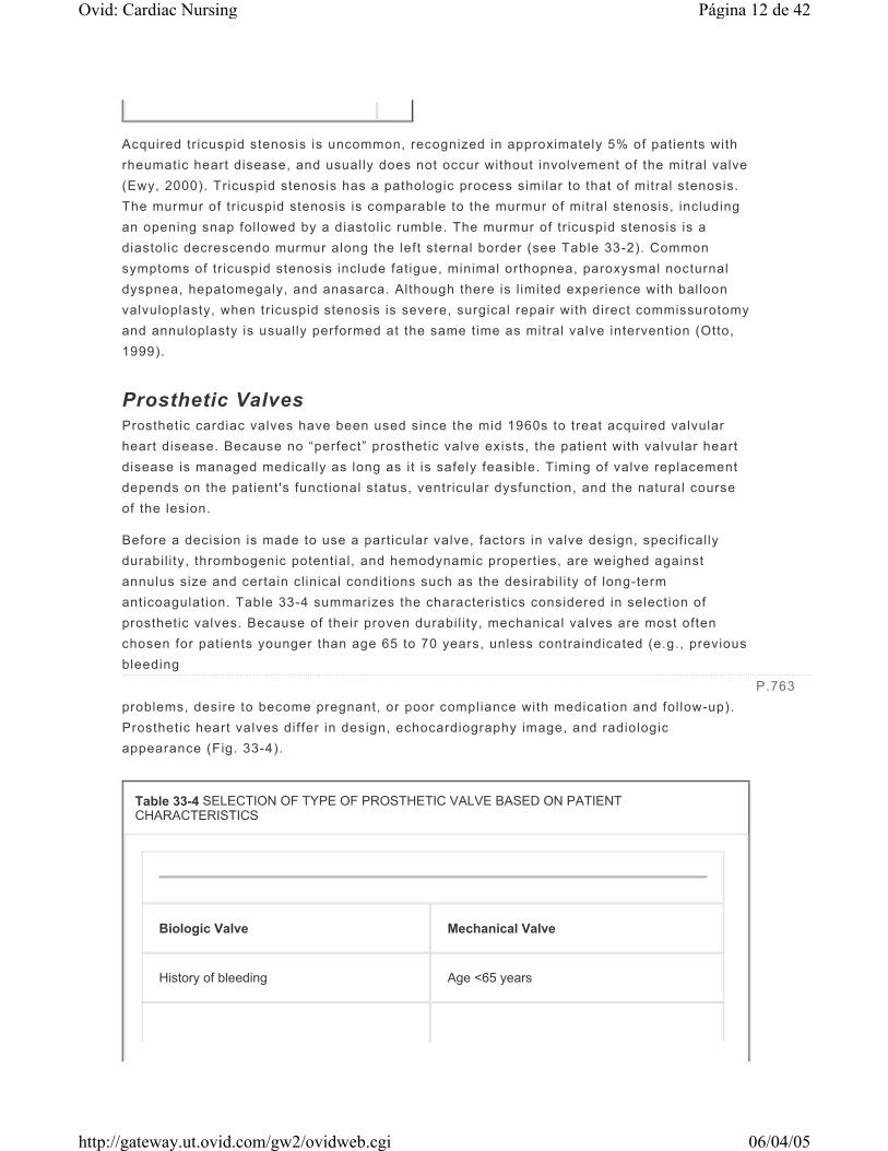
Prosthetic Valves Prosthetic cardiac valves have been used since the mid 1960s to treat acquired valvular heart disease. Because no “perfect” prosthetic valve exists, the patient with valvular heart disease is managed medically as long as it is safely feasible. Timing of valve replacement depends on the patient's functional status, ventricular dysfunction, and the natural course of the lesion.
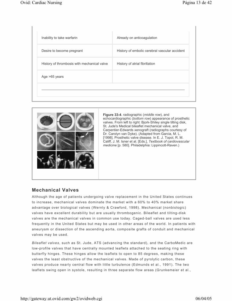
Before a decision is made to use a particular valve, factors in valve design, specif ically durabili ty, thrombogenic potential, and hemodynamic properties, are weighed against annulus size and certain clinical condit ions such as the desirabil i ty of long-term anticoagulation. Table 33-4 summarizes the characteristics considered in selection of prosthetic valves. Because of their proven durabil ity, mechanical valves are most often chosen for patients younger than age 65 to 70 years, unless contraindicated (e.g., previous bleeding problems, desire to become pregnant, or poor compliance with medication and fol low-up). Prosthetic heart valves differ in design, echocardiography image, and radiologic appearance (Fig. 33-4).
P.763
Table 33-4 SELECTION OF TYPE OF PROSTHETIC VALVE BASED ON PATIENT CHARACTERISTICS
Biologic Valve Mechanical Valve
History of bleeding Age <65 years
Página 12 de 42Ovid: Cardiac Nursing
06/04/05http://gateway.ut.ovid.com/gw2/ovidweb.cgi
Mechanical Valves Although the age of patients undergoing valve replacement in the United States continues to increase, mechanical valves dominate the market with a 60% to 40% market share advantage over biological valves (Wernly & Crawford, 1998). Mechanical (nonbiologic) valves have excellent durabili ty but are usually thrombogenic. Bileaflet and ti l t ing-disk valves are the mechanical valves in common use today. Caged-ball valves are used less frequently in the United States but may be used in other areas of the world. In patients with aneurysm or dissection of the ascending aorta, composite grafts of conduit and mechanical valves may be used.
Bileaflet valves , such as St. Jude, ATS (advancing the standard), and the CarboMedic are low-profi le valves that have centrally mounted leaflets attached to the seating ring with butterf ly hinges. These hinges allow the leaflets to open to 85 degrees, making these valves the least obstructive of the mechanical valves. Made of pyrolytic carbon, these valves produce nearly central f low with l i t t le turbulence (Edmunds et al., 1991). The two leaflets swing open in systole, result ing in three separate f low areas (Grunkemeier et al.,
Inability to take warfarin Already on anticoagulation
Desire to become pregnant History of embolic cerebral vascular accident
History of thrombosis with mechanical valve History of atrial fibrillation
Age >65 years
Figure 33-4. radiographic (middle row), and echocardiographic (bottom row) appearance of prosthetic valves. From left to right: Bjork-Shiley single tilting disk, St. Jude's Medical bileaflet mechanical valve, and Carpentier-Edwards xenograft (radiographs courtesy of Dr. Carolyn van Dyke). (Adapted from Garcia, M. L. [1998]. Prosthetic valve disease. In E. J. Topol, R. M. Califf, J. M. Isner et al. [Eds.], Textbook of cardiovascular medicine [p. 580]. Philadelphia: Lippincott-Raven.)
Página 13 de 42Ovid: Cardiac Nursing
06/04/05http://gateway.ut.ovid.com/gw2/ovidweb.cgi

1994). With adequate anticoagulation, thromboembolic r isk is low with bi leaflet valves.
The t i l t ing-disk valve is a low-profi le valve consisting of a disk that sits in a seating ring; the f lat or convexoconcave disk t i l ts in response to pressure changes. The Medtronic Hall valve is a t i l t ing-disk valve commonly used today. Tilt ing-disk valves open to an angle of 60 to 75 degrees in relation to the seating ring. When open, t i l t ing-disk valves produce a minor and major orif ice for blood to pass through. Tilt ing disks have more central f low, but usually more turbulence, than caged-ball valves.
Tilt ing disks close with an audible cl ick. The technology for production of t i l t ing-disk valves has evolved so that a single piece of metal is used to avoid welded struts. In the past, welded struts fractured and caused fatal results, as did the older Bjork-Shiley convexoconcave valve, which is no longer, manufactured or implanted (Edmunds et al., 1991).
Caged-ball valves have been used since the 1960s and have an excellent durabil i ty record. Changes in pressure cause the ball to move forward and back within its caged structure. Flow is directed laterally through the valve rather than centrally. Because of i ts high profi le, the caged-ball valve prosthesis can become obstructive, especially when used in patients with small aortic roots or small left ventricles. The Starr-Edwards and the Sutter (formerly SmeloffCutter) are two of the most common caged-ball valves used. Caged-ball prostheses have been largely abandoned in favor of lower-profi le bi leaflet valves.
Tissue Valves Tissue (biologic) valves are characterized by having low rates of thrombotic episodes associated with their use. Porcine or bovine tissue is strengthened and made nonviable by treatment with glutaraldehyde. Homografts are t issue valves from cadavers. They are preserved cryogenically, but are diff icult to procure, and their longevity has not been well proven. The main advantages of t issue valves are the associated low rates of thromboembolism and the subsequent decrease in patient morbidity when anticoagulant therapy is not required. Nonthrombogenicity is particularly important for those patients in whom long-term anticoagulation should be avoided, such as children, young adult women, patients older than age 70 years, or people with a history of bleeding.
The Hancock porcine valve, the Medtronic Mosaic porcine bioprosthesis (treated with alpha oleic acid to retard calcif ication), and the Carpentier-Edwards porcine valve are xenografts using porcine aortic valves preserved with glutaraldehyde under pressure, mounted on a stent (Jamieson, 2003). The Carpentier-Edwards pericardial bioprosthesis is made of leaflets fashioned from bovine pericardium fixed without pressure in glutaraldehyde.
Stentless bioprosthetic porcine xenograft valves such as the St. Jude Medical-Toronto, the Medtronic Freestyle Stentless, and the Edwards Prima Plus porcine bioprosthesis have been developed to improve the durabil i ty and enhance the hemodynamic performance of porcine aortic valves. Stentless aortic biological valves were developed secondary to the recognit ion that conventional bioprosthesis have l imitations of long-term durabil i ty and residual obstruction that may impede left ventricular mass regression (Goldman & Mall idi,
P.764
Página 14 de 42Ovid: Cardiac Nursing
06/04/05http://gateway.ut.ovid.com/gw2/ovidweb.cgi

2003). Because of the structural similarity to aortic al lografts, stentless bioprostheses adapt to the aortic root and reproduce the anatomy of the native aortic valve (Luciani et al., 1998). Use of the stentless aortic bioprosthesis has resulted in enhanced survival and hemodynamic superiority (David, 1998). It is expected that reducing mechanical stress on valve leaflets, and the associated degeneration of the bioprosthesis, may be slowed. Thus, stentless xenografts may prove more durable than commonly used stented valves (Luciani et al., 1998).
Homografts or allografts from human cadavers are virtually free of any associated thrombosis. They are especially useful in patients with small aortic roots or in patients with active endocardit is. Earl ier homografts were preserved with glutaraldehyde and demonstrated early fai lure. Homografts are now stored “fresh” after harvesting in an antibiotic solution and are then cryopreserved, increasing their longevity to at least 10 years. Valve failure is uncommon and usually the result of progressive valve incompetence (Doty et al., 1998). Even though the homografts are human tissue, there does not appear to be any problem with antigenicity (Whitt lesay & Geha, 1991). Aortic al lografts have demonstrated excellent freedom from thromboembolism, endocardit is, and progressive valve incompetence (Doty et al., 1998). Because of lack of availabil i ty, use of homografts has been l imited.
In the Ross procedure (also known as pulmonary autograft), the aortic valve is replaced with a pulmonary autograft, and the native pulmonary valve is replaced with a pulmonic allograft. Although this procedure introduced by Donald Ross in 1967 (Elkins, 2003) was originally developed for pediatric application, i t has been expanded to adult surgery as well. In patients undergoing the Ross procedure, the native pulmonary valve is excised and then implanted in the aortic posit ion (autograft); a pulmonary homograft (al lograft) is implanted into the pulmonic posit ion (Fig. 33-5). The pulmonary autograft has been shown to be resistant to degeneration and calcif ication (Ross, 1987). Potential clinical and hemodynamic advantages of the pulmonary autograft over the aortic homograft include potential for growth when used in the pediatric population, increased cellular viabil i ty, enhanced durabil i ty, and possibly internal innervation of the cusps (Santini, 1997). The 30-day mortality for the Ross procedure as reported by the International Ross Registry is 3.3% (140 of 4,197 patients) (International Ross Registry, 2003). The actuarial freedom from pulmonary autograft valve replacement is 90% ± 3% at 13 years (Elkins, 2003). Although the Ross procedure is gaining acceptance, especially in young adults who wish to avoid anticoagulation, there is concern that i t offers no better result than the aortic homograft, which is a simpler procedure, with less morbidity. Pulmonary autografts require signif icantly longer operating t ime but do not seem to affect early and midterm outcomes compared with aortic homografts (Santini, 1997).
Figure 33-5. Illustration of Ross procedure. Suture line of pulmonary homograft is shown. (From Elkins, R. C. [1998]. Valve repair and valve replacement in children, including the Ross procedure. In L. R. Kaiser, I. L. Kron, & T. L. Spray [Eds.], Mastery of cardiothoracic surgery [p. 947]. Philadelphia: Lippincott-Raven.)
Página 15 de 42Ovid: Cardiac Nursing
06/04/05http://gateway.ut.ovid.com/gw2/ovidweb.cgi

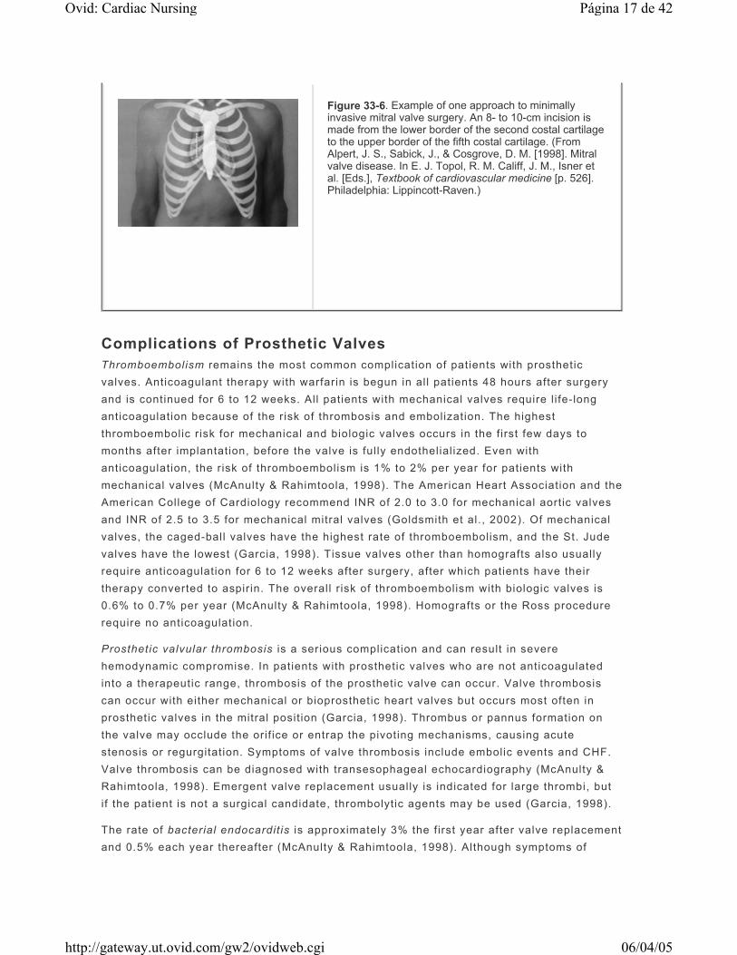
Minimally Invasive Valve Surgery Minimally invasive valve surgery is now used for both aortic valve replacement and mitral valve repair and replacement. Minimally invasive surgical approaches are possible because a wide assortment of technological advances, such as endoscopic and surgical equipment, have been developed. Although patients undergoing minimally invasive valve surgery sti l l require cardiopulmonary bypass, classic median sternotomy may be avoided, thus reducing pain, improving cosmetic results, and expedit ing recovery. These patients have a lower requirement for erythrocytes, express greater satisfaction, and have lower hospital charges (approximately 20% less than in patients with standard mitral valve and aortic valve approaches) (Cohn et al., 1997). As minimally invasive valve surgery continues to evolve, it wil l l ikely become a mainstay in the treatment of valvular heart surgery.
Aortic valve replacement can be performed through an upper “T” mini-sternotomy without intraoperative diff icult ies. Postoperative pain is reduced and recovery is expedited, with patients discharged to home as early as postoperative day 3 (Izzat et al., 1998). Two minimally invasive techniques for mitral valve surgery have been used: a right parasternal approach (Fig. 33-6) developed at the Cleveland Clinic and a mini-thoracotomy (Chitwood et al., 1997). Compared with patients with median sternotomy, patients undergoing mitral valve replacement through the right parasternal approach had a shortened length of stay and reduced direct hospital costs (Cosgrove et al., 1998). The mini-thoracotomy approach with video assistance can be used safely in patients undergoing mitral valve repair or replacement; compared with standard techniques, the mini-thoracotomy resulted in less morbidity, earl ier discharge, and lower cost (Chitwood et al., 1997). The majority of cl inical series demonstrates that minimally invasive port-access approach to mitral valve surgery has low morbidity and mortality, with echocardiographic outcomes equivalent to conventional mitral valve surgery (Sharony et al., 2003). More recently, minimally invasive mitral valve surgery has evolved to include computed-assisted robotic techniques in current cl inical tr ials. The daVinci Surgical System allows the surgeon to operate from a console through an end-affector using micro wrist instruments, which are mounted on robotic arms, inserted through the chest wall (Chitwood, 2003).
P.765
Página 16 de 42Ovid: Cardiac Nursing
06/04/05http://gateway.ut.ovid.com/gw2/ovidweb.cgi
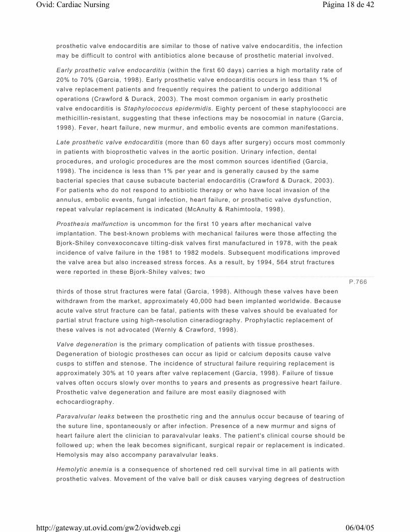
Complications of Prosthetic Valves Thromboembolism remains the most common complication of patients with prosthetic valves. Anticoagulant therapy with warfarin is begun in all patients 48 hours after surgery and is continued for 6 to 12 weeks. All patients with mechanical valves require l i fe-long anticoagulation because of the risk of thrombosis and embolization. The highest thromboembolic risk for mechanical and biologic valves occurs in the f irst few days to months after implantation, before the valve is ful ly endothelial ized. Even with anticoagulation, the risk of thromboembolism is 1% to 2% per year for patients with mechanical valves (McAnulty & Rahimtoola, 1998). The American Heart Association and the American College of Cardiology recommend INR of 2.0 to 3.0 for mechanical aortic valves and INR of 2.5 to 3.5 for mechanical mitral valves (Goldsmith et al., 2002). Of mechanical valves, the caged-ball valves have the highest rate of thromboembolism, and the St. Jude valves have the lowest (Garcia, 1998). Tissue valves other than homografts also usually require anticoagulation for 6 to 12 weeks after surgery, after which patients have their therapy converted to aspirin. The overall r isk of thromboembolism with biologic valves is 0.6% to 0.7% per year (McAnulty & Rahimtoola, 1998). Homografts or the Ross procedure require no anticoagulation.
Prosthetic valvular thrombosis is a serious complication and can result in severe hemodynamic compromise. In patients with prosthetic valves who are not anticoagulated into a therapeutic range, thrombosis of the prosthetic valve can occur. Valve thrombosis can occur with either mechanical or bioprosthetic heart valves but occurs most often in prosthetic valves in the mitral posit ion (Garcia, 1998). Thrombus or pannus formation on the valve may occlude the orif ice or entrap the pivoting mechanisms, causing acute stenosis or regurgitation. Symptoms of valve thrombosis include embolic events and CHF. Valve thrombosis can be diagnosed with transesophageal echocardiography (McAnulty & Rahimtoola, 1998). Emergent valve replacement usually is indicated for large thrombi, but if the patient is not a surgical candidate, thrombolytic agents may be used (Garcia, 1998).
The rate of bacterial endocardit is is approximately 3% the f irst year after valve replacement and 0.5% each year thereafter (McAnulty & Rahimtoola, 1998). Although symptoms of
Figure 33-6. Example of one approach to minimally invasive mitral valve surgery. An 8- to 10-cm incision is made from the lower border of the second costal cartilage to the upper border of the fifth costal cartilage. (From Alpert, J. S., Sabick, J., & Cosgrove, D. M. [1998]. Mitral valve disease. In E. J. Topol, R. M. Califf, J. M., Isner et al. [Eds.], Textbook of cardiovascular medicine [p. 526]. Philadelphia: Lippincott-Raven.)
Página 17 de 42Ovid: Cardiac Nursing
06/04/05http://gateway.ut.ovid.com/gw2/ovidweb.cgi
prosthetic valve endocardit is are similar to those of native valve endocardit is, the infection may be diff icult to control with antibiotics alone because of prosthetic material involved.
Early prosthetic valve endocardit is (within the f irst 60 days) carries a high mortality rate of 20% to 70% (Garcia, 1998). Early prosthetic valve endocardit is occurs in less than 1% of valve replacement patients and frequently requires the patient to undergo addit ional operations (Crawford & Durack, 2003). The most common organism in early prosthetic valve endocardit is is Staphylococcus epidermidis . Eighty percent of these staphylococci are methici l l in-resistant, suggesting that these infections may be nosocomial in nature (Garcia, 1998). Fever, heart fai lure, new murmur, and embolic events are common manifestations.
Late prosthetic valve endocardit is (more than 60 days after surgery) occurs most commonly in patients with bioprosthetic valves in the aortic posit ion. Urinary infection, dental procedures, and urologic procedures are the most common sources identif ied (Garcia, 1998). The incidence is less than 1% per year and is generally caused by the same bacterial species that cause subacute bacterial endocardit is (Crawford & Durack, 2003). For patients who do not respond to antibiotic therapy or who have local invasion of the annulus, embolic events, fungal infection, heart fai lure, or prosthetic valve dysfunction, repeat valvular replacement is indicated (McAnulty & Rahimtoola, 1998).
Prosthesis malfunction is uncommon for the f irst 10 years after mechanical valve implantation. The best-known problems with mechanical failures were those affecting the Bjork-Shiley convexoconcave ti l t ing-disk valves f irst manufactured in 1978, with the peak incidence of valve fai lure in the 1981 to 1982 models. Subsequent modif ications improved the valve area but also increased stress forces. As a result, by 1994, 564 strut fractures were reported in these Bjork-Shiley valves; two thirds of those strut fractures were fatal (Garcia, 1998). Although these valves have been withdrawn from the market, approximately 40,000 had been implanted worldwide. Because acute valve strut fracture can be fatal, patients with these valves should be evaluated for partial strut fracture using high-resolution cineradiography. Prophylactic replacement of these valves is not advocated (Wernly & Crawford, 1998).
Valve degeneration is the primary complication of patients with t issue prostheses. Degeneration of biologic prostheses can occur as l ipid or calcium deposits cause valve cusps to stiffen and stenose. The incidence of structural fai lure requiring replacement is approximately 30% at 10 years after valve replacement (Garcia, 1998). Failure of t issue valves often occurs slowly over months to years and presents as progressive heart fai lure. Prosthetic valve degeneration and failure are most easily diagnosed with echocardiography.
Paravalvular leaks between the prosthetic ring and the annulus occur because of tearing of the suture l ine, spontaneously or after infection. Presence of a new murmur and signs of heart fai lure alert the cl inician to paravalvular leaks. The patient's cl inical course should be followed up; when the leak becomes signif icant, surgical repair or replacement is indicated. Hemolysis may also accompany paravalvular leaks.
Hemolytic anemia is a consequence of shortened red cell survival t ime in all patients with prosthetic valves. Movement of the valve ball or disk causes varying degrees of destruction
P.766
Página 18 de 42Ovid: Cardiac Nursing
06/04/05http://gateway.ut.ovid.com/gw2/ovidweb.cgi

of the red blood cells. Hemolysis may also occur with paravalvular leak. Commonly, hemolysis is mild and the patient can compensate by increasing red blood cell production. Rarely, hemolytic anemia occurs. Chronic intravascular hemolysis results in loss of iron in the urine; iron deficiency anemia may result after several years.
Mitral Insufficiency
Cause Mitral insuff iciency (also termed regurgitation) may be either chronic or acute (Table 33-5). Acute mitral regurgitation is caused by chordal rupture, MI, trauma, myxomatous valvular degeneration, mitral valve prolapse, or endocardit is (Carabello, 2000). Chronic mitral regurgitation may be the result of a number of abnormalit ies including, but not l imited to, rheumatic heart disease, injury after radiation, cardiomyopathies, infi l trative disease, ischemic damage to the subvalvular apparatus, infective endocardit is, myxomatous degeneration, hypertrophic cardiomyopathy, diet-drug–induced lesions, or marked left ventricular di lation (Enriquez-Sarano et al, 2000).
Table 33-5 ETIOLOGIES OF ACQUIRED MITRAL REGURGIATION
Chronic Mitral Reguritation Acute Mitral Reguritation
Rheumatic heart disease Myocardial infarction causing: Papillary muscle rupture or dysfunction
Ischemia to subvalvular apparatus
Infective endocarditis Rupture of chordae
Myxomatous degeneration Infective endocarditis
Hypertrophic cardiomyopathy Trauma
Myxomatous degeneration with chordal rupture
Left ventricular dilation
Página 19 de 42Ovid: Cardiac Nursing
06/04/05http://gateway.ut.ovid.com/gw2/ovidweb.cgi
Pathology Primary mitral regurgitation occurs when the mitral valve annulus, leaflets, chordae, or papil lary muscles are affected by ischemia, collagen disease, infection, calcif ication, trauma, or degenerative changes, causing incompetent coaptation of the mitral leaflets. Secondary mitral regurgitation occurs with ventricular di lation when ventricular geometry is changed, causing malalignment of the papil lary muscles. Although it is sometimes diff icult to distinguish between primary and secondary regurgitation, primary regurgitation is often more severe than insuff iciency secondary to annular di lation (Braunwald, 1992).
Pathophysiology Mitral regurgitation occurs as the result of inadequate closure of the mitral valve, al lowing regurgitant f low back into the left atrium during each left ventricular systole. Its severity depends on the volume of regurgitant f low. Regurgitant f low into the left atrium reduces forward f low, stroke volume, and cardiac output (Khan, 1996). Regurgitant f low also increases left atrial pressure, causing left atrial di lation and pulmonary congestion. During diastole, the regurgitant volume returns to the left ventricle and increases its volume load.
In chronic mitral regurgitation, persistent volume overload results in progressive ventricular di lation and mild hypertrophy. Although ventricular di lation and hypertrophy are init ial ly
Systemic lupus erythematosus
Marfan's syndrome
Calcification of annulus
Ankylosing spondylitis
Scleroderma
Ehlers-Danlos syndrome
Prosthetic paravalvular leak
Deterioration of prosthetic mittal valve
Página 20 de 42Ovid: Cardiac Nursing
06/04/05http://gateway.ut.ovid.com/gw2/ovidweb.cgi
compensatory, over t ime, chronic volume overload may result in decreased systolic function of the left ventricle and lead to heart fai lure (Khan, 1996). In acute mitral regurgitation, neither the left atrium nor the ventricle has had sufficient t ime to adjust to the increased volume load. Left atrial pressure rises quickly, result ing in pulmonary congestion and edema.
Clinical Manifestations Patients with acute versus chronic mitral regurgitation vary in cl inical presentation and physical examination f indings. In acute mitral regurgitation, symptoms progress rapidly. Symptoms are typically those of left ventricular fai lure. The patient is usually tachycardic to compensate for the reduced forward stroke volume. Patients are dyspneic secondary to pulmonary congestion and edema; they are often orthopneic and have paroxysmal nocturnal dyspnea and poor exercise tolerance. Patients may also have signs of biventricular fai lure because r ight-sided fai lure may occur secondary to pulmonary hypertension. Patients in acute mitral regurgitation often present to the emergency room with reports of sudden inabil ity to breathe. New-onset atrial f ibri l lation can occur. Patients with ischemic mitral insuff iciency or papil lary muscle rupture may also report chest pain.
During the compensatory phase of chronic mitral regurgitation, patients may be relatively asymptomatic for years. Init ial signs of mitral regurgitation include exertional dyspnea, orthopnea, paroxysmal nocturnal dyspnea, cough, palpitations, new atrial f ibri l lat ion, and lower extremity edema. Symptoms may occur so gradually that patients may present subacutely to the cl inic with symptoms as vague as fatigue and inabil i ty to sleep.
Physical Assessment On examination, the most easily noted characteristic of either chronic or acute mitral regurgitation is the holosystolic murmur, which is heard best at the apex and radiates to the axil la (see Table 33-3). The murmur of mitral regurgitation may vary somewhat depending on the underlying cause. Patients may have an S3 gallop in moderate to severe regurgitation caused by high diastolic f low into the ventricle. An S4 gallop is uncommon in chronic mitral regurgitation. However, in acute mitral regurgitation, an S4 gallop is common
because the left atrium and ventricle are noncompliant. The patient with rheumatic heart disease may also have a diastolic murmur related to coexisting mitral stenosis.
Because of left ventricular di lation, patients with chronic mitral regurgitation have an easily palpated, left laterally displaced point of maximal impulse. Patients with a markedly enlarged left atrium may have a left parasternal l i f t because of anterior displacement of the apex. Patients with acute or decompensated chronic mitral regurgitation may be anxious and diaphoretic because of left ventricular fai lure. Blood pressure may be normal to low and pulse pressure may be narrowed secondary to decreased stroke volume. Jugular venous pressure can be normal or elevated in the patient with right-sided heart fai lure. Breath sounds can range from basilar crackles to dullness secondary to pleural effusion. In addit ion, hepatosplenomegaly, hepatojugular reflux, peripheral edema, and ascites may be
P.767
Página 21 de 42Ovid: Cardiac Nursing
06/04/05http://gateway.ut.ovid.com/gw2/ovidweb.cgi

present in the patient with right-sided heart fai lure.
Diagnostic Tests Transthoracic echocardiography can identify the structural cause of the mitral regurgitation as well as gauge left atrial size, left ventricular dimensions and performance, pulmonary artery pressures, and right heart function. Color f low Doppler al lows for assessment of severity of regurgitation. Transesophageal echocardiography is better than transthoracic echocardiography for defining mitral valve anatomy and discriminating prosthetic valves and paravalvular leaks.
Cardiac catheterization is used to identify coexisting coronary artery disease and to grade the severity of mitral regurgitation. Left ventriculography can assess left ventricular function and distinguish any wall motion abnormalit ies. Right heart catheterization quantif ies pulmonary artery pressures and allows for evaluation of the large V waves in the pulmonary artery wedge tracing.
Electrocardiography in chronic mitral regurgitation may demonstrate left ventricular hypertrophy and left atrial enlargement or P mitrale (characterized by M-shaped P waves). Atrial f ibri l lation may occur with acute and chronic mitral regurgitation. Patients with ischemic papil lary muscle dysfunction may demonstrate ischemic changes, and patients with papil lary muscle rupture can show acute inferior, posterior, or anterior MI.
Chest radiography in chronic mitral regurgitation shows left ventricular hypertrophy and left atrial enlargement. Calcif ication of the mitral valve annulus and apparatus may also be seen. In acute or decompensated chronic mitral regurgitation, pulmonary vascular redistribution and pulmonary edema can be observed. If the heart is of normal size, the degree of mitral regurgitation is so mild or so acute that eccentric left ventricular hypertrophy has not had time to develop.
Medical Management Medical therapy for mitral regurgitation is geared toward afterload reduction to promote forward f low and minimize regurgitation back into the left atrium and pulmonary vasculature. In patients with acute or decompensated chronic mitral regurgitation, intravenous vasodilators such as nitroprusside can reduce fi l l ing pressures and ventricular cavity size and promote forward f low with afterload reduction. Intravenous diuretics are used to reduce volume overload. In acutely i l l patients refractory to medications, intra-aortic balloon counterpulsation can be used further to reduce afterload while maintaining coronary perfusion with diastolic augmentation.
In patients with chronic mitral regurgitation or those in acute heart fai lure who are being weaned from intravenous inotropes and vasodilators, other afterload-reducing agents, such as angiotensin-converting enzyme (ACE) inhibitors, nitrates, or hydralazine, may be used. Diuretics can treat chronic and acute volume overload. Some practit ioners continue to advocate the use of digoxin, especially for patients in atrial f ibri l lat ion. In the patient with chronic but compensated mitral valve regurgitation, mitral surgery can be safely deferred or avoided. The patient should be carefully monitored, however, and referred for mitral valve repair or replacement before signif icant left ventricular dysfunction or pulmonary
Página 22 de 42Ovid: Cardiac Nursing
06/04/05http://gateway.ut.ovid.com/gw2/ovidweb.cgi

hypertension occurs.
Surgical Management
Surgical Intervention Two surgical approaches are used to treat mitral regurgitation. Mitral valve repair uses reconstructive techniques as well as a rigid prosthetic r ing to repair the mitral valve apparatus, thus sparing the valve and avoiding the consequences of valve replacement (Fig. 33-7). Mitral valve replacement involves implantation of a prosthetic valve with attempted preservation of at least part of the mitral valve apparatus (Reardon & David, 1998), which contributes to left ventricular function (Fig. 33-8).
Figure 33-7. (Top) Regurgitant mitral valve (note large primary orifice). (Bottom) Completed mitral valve annuloplasty with ring sutured in place.
Página 23 de 42Ovid: Cardiac Nursing
06/04/05http://gateway.ut.ovid.com/gw2/ovidweb.cgi

In patients with chronic mitral regurgitation, mitral replacement should occur before the patient has had irreversible left ventricular dysfunction. Mitral valve replacement or repair can preserve left ventricular function and ejection fraction. Patients with NYHA class II symptoms should be considered for surgery. Factors contributing to increased operative risk include reduced left ventricular ejection fraction, increased left ventricular end-systolic volume, older age, concomitant coronary artery disease, previous cardiac surgery, and pulmonary hypertension (Fann et al., 1997).
Mitral Valve Repair In selected patients, mitral valve repair may be undertaken for patients with mitral insuff iciency as an alternative to replacement. Surgical techniques involve reconstructing the leaflets and annulus in such a way as to narrow the orif ice. These procedures consist of direct suture of the valve cusps, repair of the elongated or ruptured chordae tendineae (chordoplasty), or repair of the valve annulus (annuloplasty). With an annuloplasty, the incompetent valve is remodeled using a ring prosthesis that is attached to the leaflets and the annulus. Mitral valve repair has demonstrated excellent short-term and long-term results with low perioperative mortality rate (not >2% in most reported series). Carpentier and associates report 94% and 92% freedom from re-operations at 10 and 20 years, respectively (Hampton & Verrier, 2003).
Mitral Valve Replacement In patients with acute mitral regurgitation secondary to MI, coronary angiography should be performed to define coronary anatomy for concomitant coronary bypass surgery at the t ime of mitral valve repair or replacement. In patients with acute mitral regurgitation secondary to MI, the mortali ty rate can be as high as 50% secondary to acute left ventricular fai lure (Fann et al., 1997).
Mitral Valve Prolapse
Figure 33-8. Valve replacement with chordal preservation. (From Chitwood, W. R. [1998]. Mitral valve repair: Ischemic. In L. R. Kaiser, I. L. Kron, & T. L. Spray [Eds.], Mastery of cardiothoracic surgery [p. 321]. Philadelphia: Lippincott-Raven.)
P.768
Página 24 de 42Ovid: Cardiac Nursing
06/04/05http://gateway.ut.ovid.com/gw2/ovidweb.cgi

Cause Mitral valve prolapse refers to a number of condit ions in which one or both of the mitral valve leaflets becomes superior to the plane of the annulus during systole (Braunwald, 1998). The posterior leaflet is most often affected. Mitral valve prolapse (MVP) is also known as Barlow syndrome or click-murmur syndrome. It is the most common cause of signif icant isolated mitral regurgitation (Karon, 1997) and has been reported to be one of the most common heart disorders, with an overall prevalence of 2.4% (Playford & Weyman, 2001). Although MVP occurs most commonly in women, with a peak incidence in the fourth decade of l i fe, severe mitral regurgitation associated with mitral valve prolapse is more common in men (Fann et al., 1997). The most common cause of MVP is myxomatous degeneration. Marfan syndrome, Ehlers-Danlos syndrome, rheumatic heart disease, and ischemic papil lary muscle dysfunction also cause mitral valve prolapse. In addit ion, MVP has a hereditary component transmitted as an autosomal dominant trait (Braunwald, 1992).
Pathology Patients with MVP have redundant myxomatous t issue with excess deposits of proteoglycans in the middle or spongiosa layer of the valve.
Histologically, collagen fragmentation and disorganization as well as elastic f iber are present. Acid mucopolysaccharide material accumulates in the valve leaflets. The mitral valve leaflets, annulus, and chordae tendineae may also demonstrate disrupted collagen structure and extensive myxomatous change. Myxomatous changes may also occur in the tr icuspid, aortic, and pulmonic valves (Alpert et al., 1998).
Pathophysiology Enlargement of the valve leaflets related to myxomatous degeneration causes systolic prolapse of one or both leaflets into the left atrium. Patients with MVP may have mitral regurgitation ranging in severity from none to severe. Persistent bil lowing of the valve causes stress to the underlying chordae and papil lary muscles. Progressive mitral valvular degeneration can result in increasingly severe mitral regurgitation. If chordal rupture occurs, severe mitral regurgitation develops.
Supraventricular tachycardias (i.e., premature atrial contractions and paroxysmal supraventricular tachycardias) and ventricular arrhythmias may occur in patients with MVP. Although some patients with MVP have had sudden cardiac death, it is unclear what role MVP has in the cause. Some investigators believe that patients with MVP, history of syncope, complex ventricular arrhythmias, signif icant mitral regurgitation, and prolonged QT interval are at increased risk for sudden death (Kligfield & Devereux, 1995). Patients with MVP may also have autonomic nervous system dysfunction; specif ically, mid-brain control of adrenergic and vagal responses may be abnormal. Heightened sympathetic nervous system tone may lead to a decease in left ventricular preload, result ing in MVP (Alpert et al., 1998).
P.769
Página 25 de 42Ovid: Cardiac Nursing
06/04/05http://gateway.ut.ovid.com/gw2/ovidweb.cgi

Clinical Manifestations Most patients with MVP are asymptomatic. Patients may have sharp, localized chest pain that is usually brief in duration. Although the cause of this chest pain is unclear if the patient does not have CHD, some authorit ies have suggested that the chest pain is cardiac in origin and is related to abnormal traction and tension on the papil lary muscles (O'Rourke, 1998). Patients may have equivocal symptoms of anxiety, fatigue, palpitations, and orthostatic hypotension. As mitral regurgitation progresses, patients may note increasing dyspnea, fatigue, decreased exercise tolerance, orthopnea, and paroxysmal nocturnal dyspnea. Ruptured chordae with leaflet f lai l and acute mitral regurgitation result in symptoms of severe left ventricular fai lure.
Physical Assessment The classic auscultatory f inding of mitral valve prolapse is mid-systolic cl ick with mid to late systolic murmur (see Table 33-3). The click of MVP occurs when the elongated mitral valve apparatus reaches the end of i ts tether in mid systole (Carabello, 1998). The murmur occurs secondary to regurgitant f low when the mitral valve leaflets fai l to approximate. Patients with mitral valve prolapse may have the murmur or cl ick, or both. Findings may also vary over t ime. When the degree of mitral regurgitation is mild to moderate or less, heart rate and blood pressure may be normal. Addit ional physical f indings may include thin body habitus, pectus excavatum, straight-back syndrome, and scoliosis.
Diagnostic Tests Echocardiography plays a key role in the diagnosis of MVP. Abnormal systolic motion of one or both of the mitral valve leaflets superior to the annular plane can be seen (Fig. 33-9). Doppler echocardiography gives addit ional evidence of valve regurgitation. Transesophageal echocardiography provides a more detailed look at the mitral valve and chordal structures (Brady, 2003).
Figure 33-9. (A) Long-axis echocardiographic view of mitral valve with bileaflet prolapse above the annular plane into the left atrium. (B) Illustration corresponding to echocardiogram. RV, right ventricle; LV, left ventricle; AP, annular plane; PL, prolapsing leaflet.
Página 26 de 42Ovid: Cardiac Nursing
06/04/05http://gateway.ut.ovid.com/gw2/ovidweb.cgi

Cardiac catheterization can be used to rule out CHD as the origin of chest pain. Left ventriculography can demonstrate abnormal motion of the mitral valve and help determine the degree of regurgitation.
Electrocardiography is nondiagnostic. The ECG may be normal or have nonspecif ic ST-T-wave changes in the inferior leads (II, III, and aVF) and occasionally in the anterolateral leads (V4 through V6). The ST-T-wave changes may become more notable with exercise.
Premature atrial and ventricular complexes may also be identif ied. Exercise testing may be used to help rule out the cause of the chest pain.
Chest radiography is often normal and is usually nondiagnostic for MVP. Patients with acute mitral regurgitation secondary to chordal rupture have pulmonary congestion but not cardiomegaly. Patients with chronic severe mitral regurgitation have an enlarged cardiac si lhouette secondary to left atrial and left ventricular enlargement in addit ion to pulmonary congestion (Fig. 33-10).
P.770
Figure 33-10. Chest radiograph of 51-year-old man with history of mitral valve prolapse and repair (note annuloplasty ring marked with arrow). Patient's valve repair has failed and his mitral regurgitation is now severe. Patient is now in severe heart failure with notable bilateral pleural effusions and cardiomegaly.
Página 27 de 42Ovid: Cardiac Nursing
06/04/05http://gateway.ut.ovid.com/gw2/ovidweb.cgi

Medical and Surgical Management
Medical Treatment Asymptomatic patients with MVP require no therapy other than antibiotic prophylaxis. Opinions remain divided on whether patients with isolated cl ick without murmur require antibiotic prophylaxis. Patients with the murmur of mitral regurgitation or echocardiographic evidence of mitral regurgitation are recommended to have antibiotic prophylaxis. Beta-blockers or calcium channel blockers may be used to help alleviate palpitations or chest pain syndrome.
Surgical Treatment Patients with MVP and severe mitral regurgitation or f lai l leaflets should be evaluated for surgery. They can often undergo repair rather than replacement. For discussion of surgical options, refer to the section on surgical intervention for mitral regurgitation.
Prognosis MVP is usually a benign condit ion. Most patients remain asymptomatic for their entire l ives. However, in a small subset of patients, sudden cardiac death may occur secondary to arrhythmias. Patients with palpitations, syncope, or dizziness should be further evaluated and considered for treatment of arrhythmias (Shah, 1991).
Aortic Stenosis
Cause Aortic stenosis is characterized by obstruction of the left ventricular outf low tract. Most commonly, left ventricular outf low obstruction is valvular, but i t may be either supravalvular or subvalvular. The age at which aortic stenosis becomes symptomatic is determined by the underlying cause. Aortic stenosis occurring from ages 1 to 30 years usually represents
Página 28 de 42Ovid: Cardiac Nursing
06/04/05http://gateway.ut.ovid.com/gw2/ovidweb.cgi

congenital aortic stenosis. Aortic stenosis presenting at the ages of 40 to 60 years is primari ly rheumatic in origin or secondary to calcif ic aortic stenosis in a congenital ly bicusp aortic valve. Past the age of 60 to 70 years, calcif ic degenerative stenosis is the most prevalent cause. Of the causes of aortic stenosis, senile/degenerative calcif ic aortic stenosis is most common.
Pathology In senile/degenerative calcif ic aortic stenosis, cumulative wear and tear leads to calcif ication on an otherwise normal aortic valve. Calcif ic deposits prevent the cusps from opening normally in systole, result ing in stenosis. Risk factors for development of calcif ic aortic stenosis include male gender, elevated l ipoprotein(a), height, hypertension, smoking, elevated low-density l ipoprotein cholesterol, raised serum calcium, raised serum creatinine, and diabetes (Rajamannan et al., 2003). In patients with congenital ly bicuspid aortic valves, abnormal f low through the valve leads to calcium deposit ion and restrict ion of cusp opening. In rheumatic aortic stenosis, inflammation and fibrosis of the valve result in fusion of the commissures as well as calcif ied masses in the aortic cusp (Chan, 1993).
Pathophysiology Aortic stenosis typically progresses over a period of years. As the valve cusps become less mobile, the valve orif ice decreases in size, result ing in an increasingly higher left ventricular systolic pressure necessary to eject blood across the stenosed valve. This increased left ventricular afterload results f irst in compensatory concentric left ventricular hypertrophy. Although init ial ly adaptive in aortic stenosis, left ventricular hypertrophy leads to decreased ventricular compliance and diastolic dysfunction. As aortic stenosis becomes severe, left ventricular systolic function may also decline, result ing in CHF. Late in the course, any coexisting mitral regurgitation increases because of an increased pressure gradient, which drives blood from the left ventricle into the left atrium (Braunwald, 1992).
Angina may result even in the absence of CHD because of an imbalance in myocardial oxygen supply and demand. Myocardial oxygen demand is increased secondary to increased left ventricular wall stress and muscle mass. Myocardial oxygen delivery is reduced as a result of decreased coronary perfusion pressure.
Syncope or near syncope can result secondary to reduced cerebral perfusion pressure, inappropriate left ventricular baroreceptor response, or arrhythmia. Orthostatic blood pressure changes may occur during exertion when arterial pressure drops because of systemic vasodilation in the sett ing of a f ixed cardiac output. Increased left ventricular pressure may result in inappropriate baroreceptor response. Rapid atrial arrhythmias or ventricular arrhythmias may also cause “graying out” spells or frank syncopal episodes (Braunwald, 1992).
Clinical Manifestations Patients with mild to moderate aortic stenosis are usually asymptomatic. As severe aortic
P.771
Página 29 de 42Ovid: Cardiac Nursing
06/04/05http://gateway.ut.ovid.com/gw2/ovidweb.cgi
stenosis develops, the most common init ial symptom is dyspnea on exertion, fol lowed by angina and near syncope or syncope. CHF also may occur as a result of ventricular dysfunction or increasing mitral regurgitation. Less commonly, sudden death, probably caused by ventricular f ibril lation, may be the presenting cl inical feature.
Physical Assessment Aortic stenosis is most readily detected by auscultation of i ts classic mid-systolic (systolic ejection) murmur (see Table 33-3). As aortic stenosis progresses, the murmur peaks progressively later in systole and decreases in intensity as cardiac output fal ls. The murmur may decrease or disappear over the sternum and reappear at the apex, causing the incorrect impression of mitral regurgitation (Gallavardin phenomenon) (Carabello, 1998). An S4 gallop is usually present. The point of maximal intensity is sustained but may not be
displaced. Blood pressure is normal to hypertensive unti l late in the disease progress. Jugular venous pressure is normal in most patients except those with severe aortic stenosis associated with heart failure. Reduction in stroke volume and cardiac output may cause diminished carotid upstrokes and late systolic peak (tardus) in severe or crit ical aortic stenosis.
Diagnostic Tests Echocardiography is the principal modality used to diagnose and quantify aortic stenosis. Two-dimensional echocardiography defines valve leaflet thickening and cusp movement restriction as well as gauging left ventricular hypertrophy and evaluating ventricular function (Carabello, 1998). Aortic valve pressure gradient can be measured, aortic valve area calculated, and pulmonary artery pressures estimated. Echocardiography is the most important diagnostic imaging technique used to diagnose and follow aortic stenosis (Brady, 2003).
Cardiac catheterization is performed in patients with aortic stenosis primarily to rule out concomitant CHD. Left ventriculography can quanti fy the left ventricular ejection fraction. The transvalvular gradient can be established by direct pressure measurement. Right heart catheterization can better quantify pulmonary artery pressures and cardiac output.
Electrocardiography often shows a pattern of left ventricular hypertrophy, although its absence does not exclude the presence of crit ical aortic stenosis. In addit ion to QRS amplitude changes typically associated with left ventricular hypertrophy, the patient with aortic stenosis may demonstrate ST-T-wave changes typical of left ventricular strain (Braunwald, 1992).
Exercise testing in patients with mild to moderate aortic stenosis with equivocal symptoms may be cautiously accomplished in the hands of a cardiologist and can provide relevant information regarding exercise tolerance. In patients with known severe aortic stenosis and classic symptoms such as syncope, dyspnea, and chest pain, exercise testing carries increased risk of ventricular tachyarrhythmias and ventricular f ibri l lation and should not be preformed.
Gated blood pool radionuclide scans provide information regarding ventricular function similar to echocardiography and left ventriculography. Gated pool scans may be useful in
Página 30 de 42Ovid: Cardiac Nursing
06/04/05http://gateway.ut.ovid.com/gw2/ovidweb.cgi

patients in whom left ventriculography cannot be performed (i.e., patients with elevated creatinine), or in those in whom the left ventricle cannot be clearly imaged with echocardiography.
Chest radiography may be negative even in advanced disease. Heart size may be normal or only minimally enlarged. The left ventricular border and apex may be rounded, demonstrating a boot-shaped silhouette. Identif iable calcif ication of the aortic valve and aorta may be present. As disease progresses, left atrial enlargement, pulmonary hypertension, and CHF may become evident. Poststenotic dilation of the proximal ascending aorta may be noted along the right heart border in the posteroanterior chest radiograph (Fig. 33-11).
Medical Management Aside from antibiotic prophylaxis to prevent endocardit is, there is no effective medical management of aortic stenosis (Carabello, 1998). Because the course of aortic stenosis varies in its progression, patients should be carefully fol lowed-up by their health care providers with serial physical examinations and periodic echocardiography. Patients with mild aortic stenosis undergo echocardiography every 2 years. Patients with asymptomatic severe aortic stenosis are followed-up with serial echocardiograms every 6 to 12 months. Patients are instructed regarding symptoms of aortic stenosis, including dyspnea, decreased exercise tolerance, shortness of breath, chest pain, near syncope, and syncope (Braunwald, 1992). Beta-blockers, diuretics, nitrates, and ACE inhibitors must be used with caution, because they may precipitate syncope or even cardiovascular collapse in the patient with severe aortic stenosis (Carabello, 1998).
Interventional and Surgical Management
Percutaneous Aortic Catheter Balloon Valvuloplasty Percutaneous aortic catheter balloon valvuloplasty is accomplished by passing a guide wire across the stenotic aortic valve into
Figure 33-11. (A) Posteroanterior chest radiograph showing rounded border of left ventricle (arrows). (B) Lateral chest radiograph showing calcified aortic valve (arrowheads) and filling of the retrosternal airspace with dilated ascending aorta (arrows). (From Boxt, L. M. [1998]. Plain film examination of the chest. In E. J. Topol, R. M. Califf, J. M. Isner et al. [Eds.], Textbook of cardiovascular medicine [p. 511]. Philadelphia: Lippincott-Raven.)
P.772
Página 31 de 42Ovid: Cardiac Nursing
06/04/05http://gateway.ut.ovid.com/gw2/ovidweb.cgi

the apex of the left ventricle. A balloon-tipped catheter is advanced retrograde across the stenotic valve. The balloon is inflated, fracturing calcif ied nodules and separating the fused commissures. The aortic valve ring is also stretched to increase the size of the aortic valve orif ice. Although results vary, aortic balloon valvuloplasty has been shown, on average, to increase aortic valve area from 0.5 to 0.8 cm2 and to decrease the gradient from 60 to 30 mm Hg. In addit ion, left ventricular ejection fraction tends to rise in those patients with depressed left ventricular function (Braunwald, 1992).
Restenosis is a major problem in balloon aortic valvuloplasty in adults, occurring in approximately half of the patients within 6 months. Approximately one third of patients have recurrence of symptoms within 6 months. Because of the high restenosis rate, aortic balloon valvuloplasty in adults is reserved for those candidates unsuitable for surgery (i.e., the elderly with heart fai lure or pregnant women). Aortic balloon valvuloplasty may be used as a bridge or pall iative procedure in these populations, and surgical replacement may be considered for a later t ime (Braunwald, 1992).
Aortic Valve Replacement Aortic valve replacement is the only effective treatment for advanced aortic stenosis (Chan, 1993). The natural history of aortic stenosis is used as a guide to determine the t iming of aortic valve replacement surgery. Patients with asymptomatic aortic stenosis have nearly the same survival rate as the age-matched general population. Once the patient experiences symptoms of angina, syncope, or heart fai lure, there is an abrupt decline in survival rate. In patients presenting with CHF, only 50% survive 2 years. For patients who present with syncope, the 3-year survival rate is only 50% without aortic valve replacement. The average l ife expectancy of patients with apnea is only 5 years without aortic valve replacement (Ross, 1968). Aortic valve replacement is recommended in all patients with severe, symptomatic aortic stenosis. Although the average perioperative mortali ty rate in most centers ranges from 2% to 8% (Braunwald, 1992), perioperative mortality rates range from less than 5% in young, healthy patients to as high as 30% in the frail elderly (Nishimura, 1997). Factors that increase the mortali ty r isk at the t ime of aortic valve replacement include class III or IV heart fai lure, emergency surgery, aortic insuff iciency, and cardiomegaly (Shine & Howland-Gradman, 1996). Even in patients with severe left ventricular dysfunction, improvement of symptoms and left ventricular ejection fraction occurs in most (Connolly et al, 1997). Selection of aortic valve prostheses was discussed earl ier in this chapter.
Aortic Insufficiency
Cause Aortic regurgitation may be caused by either intrinsic abnormalit ies of the aortic valve leaflets or disease of the aortic root. In rheumatic fever and endocardit is, the aortic leaflets are directly affected. In congenital ly bicuspid valves, the larger cusp may become redundant, result ing in diastolic prolapse and progressive aortic regurgitation. The aortic valve may
P.773
Página 32 de 42Ovid: Cardiac Nursing
06/04/05http://gateway.ut.ovid.com/gw2/ovidweb.cgi

also become incompetent because of aortic root di lation. As the aortic root di lates, the aortic annulus becomes so large that the valve cusps no longer approximate, result ing in regurgitation.
Aortic root di lation is seen in patients with Marfan syndrome, rheumatic arthrit is, ankylosing spondylit is, annuloaortic ectasia (associated with hypertension and aging), aortic dissection, syphil i t ic aortit is, and collagen vascular disease.
Pathology Rheumatic fever leads to f ibrinous infi l trates on the valve cusps, causing them to contract and become malaligned and incompetent. Patients with rheumatic disease may have a “mixed lesion” that includes both aortic regurgitation and aortic stenosis. In acute or subacute infective endocardit is of the aortic valve, t issue destruction of the leaflets causes cusp perforation or prolapse. Vegetations adherent to the aortic valve may also interfere with valve closure, causing incompetence. In patients with aortic root di lation or ascending aortic dissection, the aortic annulus becomes greatly enlarged, the aortic leaflets separate, and aortic incompetence fol lows.
Pathophysiology Volume overload occurs secondary to regurgitant volume reentering the left ventricle from the aorta through the incompetent aortic valve. Retrograde flow occurs during diastole when left ventricular pressure is low and aortic pressure is high. The left ventricle is forced to pump the normal volume received from the left atrium as well as the regurgitant volume from the aorta.
Similar to mitral regurgitation, the hemodynamic presentation and the heart 's abil ity to compensate differ depending on whether the aortic insuff iciency is acute or chronic. In chronic aortic regurgitation, the left ventricle is subjected to pressure and volume overload. As a result, the left ventricle develops mild concentric hypertrophy to accommodate the pressure load and eccentric hypertrophy to compensate for the increased volume load. Patients with chronic aortic regurgitation may remain asymptomatic for years, unti l progressive left ventricular di lation and dysfunction result in CHF. In patients with acute aortic regurgitation, the left ventricle has not had time to compensate with either concentric or eccentric hypertrophy and cannot accommodate the large volume caused by acute aortic regurgitation. As a result, left ventricular and left atrial pressures rise sharply, causing acute CHF and pulmonary edema. Patients with acute aortic regurgitation usually require surgical intervention.
Clinical Manifestations Patients with chronic aortic regurgitation are often asymptomatic for many years. Common symptoms of aortic regurgitation include fatigue and exertional dyspnea. Patients may report palpitations, dizziness, and the sensation of a forceful heartbeat, especially when lying on their left side. Angina may also be noted, but i t occurs less frequently in aortic regurgitation than in aortic stenosis. As heart fai lure ensures, patients experience orthopnea, paroxysmal nocturnal dyspnea, and cough related to left-sided heart fai lure.
Página 33 de 42Ovid: Cardiac Nursing
06/04/05http://gateway.ut.ovid.com/gw2/ovidweb.cgi

With acute aortic regurgitation, symptoms of left-sided heart fai lure develop rapidly.
Physical Assessment The typical murmur of aortic regurgitation is a high-pitched, early diastolic decrescendo murmur with a blowing quality (see Table 33-2). Patients may also have a physiologic murmur of mitral stenosis caused by the regurgitant aortic jet, which partial ly prevents mitral valve closure (Austin Flint murmur). As the severity of aortic regurgitation increases, the murmur becomes louder and longer. In chronic aortic regurgitation, the point of maximal impulse is displaced laterally. Systolic hypertension and decreased diastolic pressure create a widened pulse pressure. Patients with chronic aortic regurgitation may have a host of other physical f indings that may not be present in acute aortic regurgitation (Table 33-6).
Table 33-6 SPECIFIC PHYSICAL EXAMINATION FINDINGS IN AORTIC REGURGITATION
Sign Phsical Description
Quincke's sign Pulsatile flushing/blanching of nail bed with application of gentle pressure
de Musset's sign Bobbing of head with each pulse
Corrigan's pulse (waterhammer)
Sharp systolic upstroke and diastolic collapse of pulse
Müller's sign Bobbing of uvula with each pulse
Traube's sign “Pistol shot” sound auscultated over the femoral arteries
Duroziez's sign Biphasic femoral bruit auscultated with mild pressure
Hill's sign Blood pressure higher in arms than legs
Página 34 de 42Ovid: Cardiac Nursing
06/04/05http://gateway.ut.ovid.com/gw2/ovidweb.cgi

Diagnostic Tests Echocardiography is helpful in identifying the cause of aortic regurgitation. Echocardiography can indicate left ventricular volume overload by the increased internal diameter of the ventricular chamber during systole and diastole. Doppler echocardiography is the best noninvasive means to detect aortic regurgitation. Transesophageal echocardiography is especially useful in imaging the ascending and descending aorta in patients with suspected aortic dissection.
Cardiac catheterization should be performed to visualize and quantify the extent of regurgitation before surgery. However, physical f indings and noninvasive tests are suff icient to establish the diagnosis of aortic insuff iciency. In patients with known or suspected CHD, coronary angiography should be performed. In patients with aortic root di lation, aortic root angiography may be performed concurrently with coronary angiography.
Radionuclide imaging can be used to estimate ejection fraction and determine myocardial perfusion defects in patients with concomitant CHD.
Exercise testing may be used to establish exercise tolerance and to evaluate asymptomatic patients.
Electrocardiography may be normal in patients with acute aortic regurgitation or in patients with mild to moderate chronic regurgitation. Patients with moderate to severe chronic regurgitation may have left-axis deviation and a pattern of left ventricular strain (Q waves in I, aVL, and V3 to V6, with small R wave in V1). Intraventricular conduction defects may occur with left ventricular
dysfunction or annular abscess.
Chest radiography may show only CHF in the patient with acute aortic regurgitation because compensatory left ventricular di lation has not yet occurred. In chronic aortic regurgitation, the chest radiograph demonstrates marked cardiomegaly with inferior and leftward displacement of the apex (Braunwald, 1992). Dilation of the ascending aorta and a widened mediastinum may be noted in patients with aortic dissection. In patients with a dilated aortic root or dissection, computed tomography or magnetic resonance imaging may be necessary to better-delineate the ascending aorta, transverse arch, and proximal descending aorta.
Medical Management Patients who have asymptomatic aortic regurgitation should receive appropriate antibiotic prophylaxis as well as afterload reduction with vasodilators. In patients with asymptomatic, chronic, or severe aortic regurgitation and normal left ventricular function, nifedipine reduced left ventricular size and mass and further improved left ventricular function (Scognamiglio et al., 1994), thus reducing and delaying the need for aortic valve replacement. Nifedipine is superior to hydralazine, which does not decrease left ventricular mass or reduce left ventricular dimension (Khan, 1996). Dilt iazem and verapamil are contraindicated in aortic regurgitation because they have a more potent negative inotropic effect and may produce bradycardia, which may worsen heart fai lure. It is probably
P.774
Página 35 de 42Ovid: Cardiac Nursing
06/04/05http://gateway.ut.ovid.com/gw2/ovidweb.cgi
reasonable to substitute other dihydropyridine calcium channel blockers if nifedipine is poorly tolerated (Carbello, 1998). ACE inhibitors may be used to reduce afterload, although they are not as well studied in this population of patients.
Patients with moderate to severe aortic regurgitation should not participate in vigorous exercise or competit ive sports. Patients with chronic severe aortic regurgitation should be followed-up with physical examination and echocardiography every 6 to 12 months (Rahimtoola, 1998). Sodium nitroprusside reduces preload and afterload and can be used to stabil ize patients with acute aortic regurgitation before surgery. Intra-aortic balloon counterpulsation cannot be used because inflation of the balloon during diastole would increase the regurgitant volume into the left ventricle, which acutely worsens left ventricular di lation and heart fai lure.
Surgical Management Acute aortic regurgitation requires urgent aortic valve replacement. Without adequate t ime for compensatory mechanisms to develop, aortic regurgitation tr iggers rapid onset of CHF, tachycardia, and diminished cardiac output. It is desirable to treat patients with acute aortic regurgitation secondary to infective endocardit is with a minimum of 48 hours of appropriate intravenous antibiotics before implanting a prosthetic valve. In patients with active endocardit is who are hemodynamically unstable, use of cadaveric human aortic homografts may minimize the risk of prosthetic valve endocardit is. Patients who have aortic regurgitation caused by ascending aortic dissection or di lation require replacement of the ascending aorta as well.
In chronic aortic regurgitation, the aortic valve must be replaced before irreversible left ventricular dysfunction. In asymptomatic patients, i t is usually recommended that the aortic valve be replaced when left ventricular function begins to deteriorate. Surgery is recommended when the echocardiographic left ventricular ejection fraction is.55 or less, the end-diastolic diameter approaches 75 mm, or the end-systolic diameter reaches 50 mm. When symptoms of heart fai lure develop, aortic valve surgery should be performed regardless of echocardiography findings, because new-onset heart fai lure indicates that the heart has met the l imits of compensation (Carbello, 1998).
REFERENCES
Alpert, J. S., Sabick, J., & Cosgrove, D. M. (1998). Mitral valve disease. In E. J. Topol, R. M. Califf , J. M. Isner et al. (Eds.), Textbook of cardiovascular medicine (pp. 503–532). Philadelphia: Lippincott-Raven.
Abraham, M. T., & Cherin, G. (1991). Rheumatic fever. In K. Chatterjee, M. D. Cheit l in, J. Karl iner et al. (Eds.), Cardiology: An i l lustrated text/reference (pp. 10.98–10.106). Philadelphia: JB Lippincott.
Bhola, R., & Gil l , E. (2001). Rheumatic heart disease and mitral stenosis. In O. V.
Página 36 de 42Ovid: Cardiac Nursing
06/04/05http://gateway.ut.ovid.com/gw2/ovidweb.cgi
Adair, Cardiology secrets (2nd ed., pp. 226–235).
Brady, T. J., Grist, T. M., Westra, S. J., et al. (2003). Valvular. In T. J. Brady, T. M. Grist, S. J. Westra, et al, Pocket radiologist: Cardiac top 100 diagnoses (pp. 53–75). Salt Lake, UT: Amirsys.
Braunwald, E. (1992). Valvular heart disease. In E. Braunwald (Ed.), Heart disease: A textbook of cardiovascular medicine (4th ed., pp. 1007–1077). Philadelphia: WB Saunders.
Braunwald, E. (1998). Valvular heart disease. In A. S. Fauci, E. Braunwald, & K. J. Isselbacher (Eds.), Harrison's principles of internal medicine (14th ed., pp. 1311–1324). New York: McGraw-Hil l .
Buccino, D., Tu, A. S., & Come, P. C. (1993). Diagnostic imaging and catheterization techniques. In L. S. Lil ly (Ed.), Pathophysiology of heart disease (pp. 130–146). Philadelphia: Lea & Febiger.
Burge, D. J., & DeHoratius, R. J. (1993). Acute rheumatic fever. In A. N. Brest (Ed.), Valvular heart disease: Comprehensive evaluation and treatment (2nd ed., pp. 3–7). Philadelphia: F. A. Davis.
Carabello, B. A. (1998). Recognit ion and management of patients with valvular heart disease. In L. Goldman, & E. Braunwald (Eds.), Primary cardiology (pp. 370–389). Philadelphia: WB Saunders.
Carabello, B. A. (2000). Acute mitral regurgitation. In J. S. Alpert, J. E. Dalen, & S. H. Rahimtoola (Eds.), Valvular heart disease (3rd ed., pp. 143–155). Philadelphia: Lippincott Wil l iams & Wilkins.
Chan. E., Duh, E., Stidham, B. et al. (1993). Valvular heart disease. In L. S. Li l ly (Ed.), Pathophysiology of heart disease (pp. 130–146). Philadelphia: Lea & Febiger.
Chitwood, W.R., Wixon, C. L., & Elbeery, J. R. (1997). Video-assisted minimally invasive mitral valve surgery. Journal of Thoracic & Cardiovascular Surgery , 114, 773–781.
Chitwood, W. R. (2003). Robot-assisted mitral valve surgery. In K. L. Franco, & E. D. Verrier (Eds.), Advanced therapy in cardiac surgery (pp. 220–229). Hamilton, Ontario:
Página 37 de 42Ovid: Cardiac Nursing
06/04/05http://gateway.ut.ovid.com/gw2/ovidweb.cgi

BC Decker.
Cohn, L. H., Adams, D. H., Couper, G. S., et al. (1997). Minimally invasive cardiac valve surgery improves patient satisfaction while reducing costs of cardiac valve replacement and repair. Annals of Surgery , 226, 421–426.
Connolly, H. M., Oh, J. K., Orszulak, T. A., et al. (1997). Aortic valve replacement for aortic stenosis with severe left ventricular dysfunction: Prognostic Indicators. Circulation , 95, 2395–2399.
Cosgrove, D. M., Sabik, J. F., & Navia, J. L. (1998). Minimally invasive valve operations. Annals of Thoracic Surgery , 65, 1538–1539.
Crawford, M. H. & Durack, D. T. (2003). Clinical presentation of infective endocardit is. Cardiology Clinics , 21, 159–166.
Dajani, A. S., Taubert, K. A., Wilson, W., et al (1997). Prevention of bacterial endocardit is: Recommendations by the American Heart Association. Circulation , 96, 358–366.
Dalen, J. E., & Fenster, P. E. (2000). Mitral stenosis. In J. S. Alpert, J. E. Dalen, & S. H. Rahimtoola (Eds.), Valvular heart disease (3rd ed., pp. 75–112). Philadelphia: Lippincott Wil l iams & Wilkins.
David, T. E., Puschmann, R., Ivanov, J., et al. (1998). Aortic valve replacement with the stentless and stented porcine valves: A case-match study. Journal of Thoracic & Cardiovascular Surgery , 116, 236–240.
Doty, J. R., Salazar, J. D., Liddicoat, J. R., et al. (1998). Aortic valve replacement with cryopreserved aortic al lograft: Ten year experience. Journal of Thoracic Cardiovascular Surgery , 115, 371–379.
Edmunds, L. H., Addonizio, V. P., & Tepe, N. A. (1991). Valvular heart disease: Prosthetic valve replacement. In K. Chatterjee, M. D. Cheitl in, & J. Karl iner et al. (Eds.), Cardiology: An i l lustrated text/rReference (pp. 940–941). Philadelphia: JB Lippincott.
Elkins, R. C. (2003). Pulmonary autograft. In K. L. Franco & E. D. Verrier (Eds.),
P.775
Página 38 de 42Ovid: Cardiac Nursing
06/04/05http://gateway.ut.ovid.com/gw2/ovidweb.cgi

Advanced therapy in cardiac surgery (pp. 156–167). Hamilton, Ontario: BC Decker.
Enriquez-Sarano, M., Schaff, H. V., Tajik, A. J. & Frye, R. L. (2000). Chronic mitral regurgitation. In J. S. Alpert, J. E. Dalen, & S. H. Rahimtoola (Eds.), Valvular heart disease (3rd ed., pp. 113–141). Philadelphia: Lippincott Wil l iams & Wilkins.
Ewy, G. A. (2000). Tricuspid valve disease. In J. S. Alpert, J. E. Dalen, & S. H. Rahimtoola (Eds.), Valvular heart disease (3rd ed., pp. 377–392). Philadelphia: Lippincott Wil l iams & Wilkins.
Fann, J. I., Ingels, N. B., & Mil ler, D. C. (1997). Pathophysiology of mitral valve disease and operative indications. In L. H. Edmunds (Ed.), Cardiac surgery in the adult (pp. 959–990). New York: McGraw-Hil l .
Garcia, M. J. (1998). Prosthetic valve disease. In E. J. Topol, R. M. Califf, J. M. Isner et al. (Eds.), Textbook of cardiovascular medicine (pp. 579–605). Philadelphia: Lippincott-Raven.
Goldman, B. S., & Mall idi, H. (2003). Update on Stentless valves. In K. L. Franco & E. D. Verrier (Eds.), Advanced therapy in cardiac surgery (pp. 196–206). Hamilton, Ontario: BC Decker.
Goldsmith, I., Turpie, A. C. G., & Lip, G. Y. (2002). ABC of Antithrombotic therapy: valvular heart disease and prosthetic heart valves. BMJ, 325, 1228–1231.
Grunkemeier, G. L., Starr, A., & Rahimtoola, S. H. (1994). Replacement heart valves. In R. A. O'Rourke (Ed.), Hurst's the heart: Update I (pp. 98–123). New York: McGraw-Hil l .
Hamptom, C. R., & Verrier, E. D. (2003). Mitral valve repair. In K. L. Franco & E. D. Verrier (Eds.), Advanced therapy in cardiac surgery (pp. 207–219). Hamilton, Ontario: BC Decker.
International Ross Registry. (2003). Follow-up data. [Online]. Available: http://www.rossregistry.com (Accessed 8/28/03).
Izzat, M. B., Yim, A. P., El-Zufari, et al. (1998). Upper T mini-sternotomy for aortic valve operations. Chest, 114, 291–294.
Página 39 de 42Ovid: Cardiac Nursing
06/04/05http://gateway.ut.ovid.com/gw2/ovidweb.cgi

Jamieson, W. R. E. (2003). Update on new tissue valves. In K. L. Franco & E. D. Verrier (Eds.), Advanced therapy in cardiac surgery (pp. 177–195). Hamilton, Ontario: BC Decker.
Kaplan, E. L. (1998). Acute rheumatic fever. In R. W. Alexander, R. C. Sclant, & V. Fuster (Eds.), Hurst's the heart (9th ed., pp. 1759–1787). New York: McGraw-Hil l .
Karchmer, A. W. (1998). Approach to the patient with infective endocardit is. In L. Goldman & E. Braunwald (Eds), Primary cardiology (pp. 201–218). Philadelphia: WB Saunders.
Karon, B. L. (1997). Valvular regurgitation. In J. G. Murphy (Ed.), Mayo Clinic cardiology review (pp. 533–554). Armonk, NY: Futura.
Khan, M. G. (1996). Valvular heart disease and rheumatic fever. In M. G. Khan (Ed), Heart Disease Diagnosis and Therapy: A Practical Approach (pp. 415–460). Balt imore: Wil l iams & Wilkins.
Kligfield, P., & Devereux, R. B. (1995). Arrhythmia in mitral valve prolapse. In P. R. Podrid & P. R. Kowey (Eds.), Cardiac arrhythmia: Mechanisms, diagnosis, and management (pp. 1253–1260). Balt imore: Wil l iams & Wilkins.
Luciani, G. B., Bertolini, P., Vecchi, B. et al. (1998). Midterm results after aortic valve replacement with freehand stentless xenografts: A comparison of three prostheses. Journal of Thoracic and Cardiovascular Surgery, 115, 1287–1296.
Mayes, C. E., Cigarroa, J. E., Lange, R. A., et al. (1999). Percutaneous mitral balloon valvuloplasty. Clinics in Cardiology, 22, 501–503.
Mazur, W., Pari lak, L. D., Kaluza, G., et al. (1999). Balloon valvuloplasty for mitral stenosis. Current Opinion in Cardiology , 14, 95–103.
McAnulty, J. H., & Rahimtoola, S. H. (1998). Antithrombotic therapy and valvular heart disease. In R. W. Alexander, R. C. Sclant, & V. Fuster (Eds.), Hurst's the heart (9th ed., pp. 1759–1787). New York: McGraw-Hil l .
Nishimura, R. A. (1997). Valvular stenosis. In J. G. Murphy (Ed.), Mayo Clinic cardiology review (pp. 521–532). Armonk, NY: Futura.
Página 40 de 42Ovid: Cardiac Nursing
06/04/05http://gateway.ut.ovid.com/gw2/ovidweb.cgi

Olaison, L., & Pettersson, G. (2003) Current best practices and guidelines: Indications for surgical intervention in infective endocardit is. Cardiology Clinics , 21, 235–251.
O'Rourke, R. A. (1998). Mitral valve prolapse syndrome. In R. W. Alexander, R. C. Sclant, & V. Fuster (Eds.), Hurst's the heart (9th ed., pp. 1821–1831). New York: McGraw-Hil l .
Otto, C. M. (1998). Aortic stenosis: Clinical evaluation and optimal t iming of surgery. Cardiology Clinics , 16(3), 353–373.
Otto, C. M. (1999). Right-sided valve disease. In C. M. Otto, Valvular heart disease (pp. 362–379). Philadelphia: W.B. Saunders.
Palacios, I. F., Sanchez, P. L. Harrell, L. C., et al. (2002). Which patients benefit from percutaneous mitral balloon valvuloplasty? Circulation , 105, 1465–1471.
Playford, D., & Weyman, A. E. (2001). Mitral valve prolapse: t ime for a fresh look. Reviews in Cardiovascular Medicine , 2(2), 73–81.
Rahimtoola, S. H. (1998). Aortic valve disease. In R. W. Alexander, R. C. Sclant, & V. Fuster (Eds.), Hurst's the heart (9th ed., pp. 1759–1787). New York: McGraw-Hil l .
Rajamannan, N. M., Gersh, B., & Bonow, R. O. (2003). Calcif ic aortic stenosis: from bench to bedside- emerging cl inical and cellular concepts. Heart, 89, 801–805.
Reardon, M. J., & David, T. E. (1998). Mitral valve replacement with preservation of the subvalvular apparatus. Current Opinion in Cardiology , 14, 104–110.
Ross, J. Jr., & Braunwald, E. (1968). Aortic stenosis. Circulation (Suppl. V), 38, 61–67.
Ross, D. N., Jackson, M., & Davies, J. (1987). Pulmonary autograft aortic valve replacement. Circulation , 75, 895–901.
Sachdev, M., Peterson, G. E., & Joll is, J. G. (2003). Imaging techniques for diagnosis of infective endocardit is. Cardiology Clinics , 21, 185–195.
Santini, F., Dyke, C., Edwards, S., et al. (1997). Pulmonary autograft versus homograft
Página 41 de 42Ovid: Cardiac Nursing
06/04/05http://gateway.ut.ovid.com/gw2/ovidweb.cgi

replacement of the aortic valve: A prospective randomized trial. Journal of Thoracic and Cardiovascular Surgery, 113, 894–899.
Scognamiglio, R., Rahimtoola, S. H., Fasoli, G., et al. (1994). Nifedipine in asymptomatic patients with severe aortic regurgitation and normal left ventricular function. New England Journal of Medicine , 331, 689–694.
Sexton, D. J., & Spelman, D. (2003). Current best practices and guidelines: Assessment and management of complicat ions in infective endocardit is. Cardiology Clinics , 21, 273–282.
Shah, P. M. (1991). Mitral valve prolapse. In K. Chatterjee, M. D. Cheit l in, J. Karl iner et al. (Eds.), Cardiology: An i l lustrated text/reference (pp. 9.30–9.39). Philadelphia: JB Lippincott.
Sharony, R., Grossi, E. A. Ribakove, G. H., et al. (2003). Minimally invasive cardiac valve surgery. In K. L. Franco & E. D. Verrier (Eds.), Advanced therapy in cardiac surgery (pp.147–155). Hamilton, Ontario: BC Decker.
Shine, L., & Howland-Gradman, J. (1996). Aortic stenosis in the elderly: Valvuloplasty vs surgery. American Journal of Nursing (Suppl.), 7–11.
Teerl ink, J. R., & Foster, E. (1998). Valvular heart disease in pregnancy. Cardiology Clinics , 16 (3), 573–594.
Towns, M. L., & Reller, L. B. (2003). Diagnostic methods: current best practices and guidelines for isolation of bacteria and fungi in infective endocardit is. Cardiology Clinics , 21, 197–205.
Wernly, J. A., & Crawford, M. H. (1998). Choosing a prosthetic heart valve. Cardiology Clinics , 16 (3), 491–504.
Whitt lesay, D., & Geha, A. S. (1991). Selection and complications of cardiac valvular prosthesis. In A. E. Baue, A. S. Geha, G. L. Hammond et al. (Eds.), Glenn's thoracic and cardiovascular surgery (5th ed., pp. 1719–1728). Norwalk, CT: Appleton & Lange.
Página 42 de 42Ovid: Cardiac Nursing
06/04/05http://gateway.ut.ovid.com/gw2/ovidweb.cgi