ACL Injuries: Do We Know the Mechanisms?
33
Cleveland State University Cleveland State University EngagedScholarship@CSU EngagedScholarship@CSU Mechanical Engineering Faculty Publications Mechanical Engineering Department 1-2007 ACL Injuries: Do We Know the Mechanisms? ACL Injuries: Do We Know the Mechanisms? Antonie J. van den Bogert Cleveland State University, [email protected] Scott G. McLean Cleveland Clinic Follow this and additional works at: https://engagedscholarship.csuohio.edu/enme_facpub Part of the Biomechanical Engineering Commons How does access to this work benefit you? Let us know! How does access to this work benefit you? Let us know! Original Citation Original Citation van den Bogert, A. J., McLean, S. G., 2007, "ACL Injuries: Do we Know the Mechanisms?" The Journal of Orthopaedic and Sports Physical Therapy, 37(2), pp. A8-9. This Article is brought to you for free and open access by the Mechanical Engineering Department at EngagedScholarship@CSU. It has been accepted for inclusion in Mechanical Engineering Faculty Publications by an authorized administrator of EngagedScholarship@CSU. For more information, please contact [email protected].
Transcript of ACL Injuries: Do We Know the Mechanisms?
Cleveland State University Cleveland State University
EngagedScholarship@CSU EngagedScholarship@CSU
Mechanical Engineering Faculty Publications Mechanical Engineering Department
1-2007
ACL Injuries: Do We Know the Mechanisms? ACL Injuries: Do We Know the Mechanisms?
Antonie J. van den Bogert Cleveland State University, [email protected]
Scott G. McLean Cleveland Clinic
Follow this and additional works at: https://engagedscholarship.csuohio.edu/enme_facpub
Part of the Biomechanical Engineering Commons
How does access to this work benefit you? Let us know! How does access to this work benefit you? Let us know!
Original Citation Original Citation van den Bogert, A. J., McLean, S. G., 2007, "ACL Injuries: Do we Know the Mechanisms?" The Journal of Orthopaedic and Sports Physical Therapy, 37(2), pp. A8-9.
This Article is brought to you for free and open access by the Mechanical Engineering Department at EngagedScholarship@CSU. It has been accepted for inclusion in Mechanical Engineering Faculty Publications by an authorized administrator of EngagedScholarship@CSU. For more information, please contact [email protected].

ACL Supplement: Introduction
This was the third research retreat focused on gender bias in anterior cruciate ligament (ACL) injuries. The first 2 took place in Lexington, KY in April of 2001 and 2003. The purpose of this third retreat was to continue to examine the factors thought to be associated with gender bias in ACL injuries. In addition, we sought to revisit and update the consensus statement
from 2003.1 A call for abstracts for the retreat was announced in the summer of 2005. All received abstracts were then peer-reviewed for scientific merit and relevance to the retreat topic. There was a 50% increase in the number of abstract submissions this year. In the end, 33 abstracts were accepted.
These were grouped into sessions addressing structural, neuromuscular, biomechanical, and hormonal factors that may influence the gender bias in ACL injury in-cidence. It was interesting to note that the majority of abstracts submitted were in the area of neuromuscular and biomechanical factors, with only a few in the ar-eas of structural and hormonal factors. This suggests a trend in the research focus towards the more modifiable factors. The retreat was cohosted by Kentucky Sports Medicine and Drayer Physical Therapy Institute and sponsored by DonJoy, Aircast, Bluegrass Bracing, and Smith and Nephew. To accommodate the increase in number of high-quality abstracts, the meeting was ex-tended to 2 full days. The retreat was attended by both clinicians and scientists with a common interest in the ACL injury gender bias. The 60-plus participants in-cluded registrants from across the United States as well as Canada and Australia. As with the previous retreats, the group consisted of physicians, physical therapists, athletic trainers, and scientists in the areas of biome-chanics, motor control, and neuromuscular function.
1 University of Delaware, Newark, DE.
2 Drayer Physical Therapy Institute, Hummelstown, PA.
3 Kentucky Sports Medicine Center, Lexington, KY.
4 University of Kentucky, Lexington, KY.
GuesT ediTors: irene davis, PT, PhD1,2 • Mary LLoyd ireLand, MD3 • saori Hanaki, MS, ATC4
Thirty percent of the participants in the 2006 retreat were participants in a previous retreat as well. The for-mat of the meeting included 1 keynote presentation per day, along with 15-minute podium presentations made by some of the participants. The keynote presenters were chosen for their scientific contribution to the un-derstanding of factors associated with the gender bias seen in the incidence of ACL injuries. Ton van den Bo-gert, PhD, from the Department of Biomedical Engi-neering at the Cleveland Clinic gave the first keynote titled “ACL Injuries: Do We Know the Mechanisms?” The second keynote presenter was William Garrett, MD, PhD, from the Department of Orthopedics at Duke University, whose talk was titled “Anterior Cruciate Lig-ament Injury Mechanisms and Risk Factors.” Following all of the presentations, a consensus development ses-sion was held. In the following pages, you will find the consensus statement and an abstract on each of the 33 presentations made at the conference, organized by the topics listed above. t
ACL Injuries—The Gender BiasResearch Retreat III
ACL Supplement: Consensus Statement
The consensus statement was developed with input from all of the participants. Once all papers were presented, the participants formed into groups, based upon each of the factors discussed during the meeting. As with previous retreats, the consensus was formed through discussion of what we know, which was grounded in the recent literature and information
presented at the current retreat. The group then identified what is still unknown (what we don’t know) about each factor’s contribution to the gender bias. This led to the final part of the consensus, in which suggestions for future research directions were made. Each group was charged
with providing a draft consensus statement that was then presented to the group for further discussion. Participants agreed that the consensus should be viewed as the present state of thought about anterior cruciate ligament (ACL) in-juries, based upon current knowledge. As with previous re-treats, it was acknowledged that what we know will likely evolve with time.
Participants continue to agree that, while there is a gender bias in ACL injuries, some of the identified factors (struc-tural, neuromuscular, and biomechanical) may not be purely gender specific. Some males may also exhibit these factors and be at greater risk for ACL injury. The question of whether females should move like men or whether there is a different optimal pattern for them was also again discussed, but not resolved. However, it is apparent that the current movement patterns of females place them at increased risk for injury. Finally, there was a group consensus that more ecological studies are needed to better replicate the environments in which the injuries typically occur.
New to this year’s retreat were presentations on the use of 2-dimensional techniques to assess movement patterns. These approaches are important as they can be easily imple-mented in the clinic. They also provide a means to conduct large-scale screening studies to predict injury risk and as-sess the effect of intervention programs. Another new topic discussed this year was gender differences in variability of movement. This is an emerging topic in the biomechanics community, with ongoing discussions of the importance of variability in reducing injury risk. However, excessive vari-
1 University of Delaware, Newark, DE.
2 Drayer Physical Therapy Institute, Hummelstown, PA.
3 Kentucky Sports Medicine Center, Lexington, KY.
4 University of Kentucky, Lexington, KY.
GuesT ediTors: irene davis, PT, PhD1,2 • Mary LLoyd ireLand, MD3 • saori Hanaki, MS, ATC4
ability of movement may be detrimental. Therefore, the op-timal level of variability requires further defining.
Following are the sections of the consensus statement for each of the factors thought to be associated with the ACL injury gender bias, as well as for the section on interven-tion programs. We realize that these lists are not all inclu-sive; however, they do represent the collective opinions of the participants in this retreat. Many of the questions from the previous retreat remain unanswered. It is our hope that this consensus statement will promote research studies in the suggested areas so that some of these gaps in the literature might be filled by the next ACL research retreat.
BioMeCHaniCaL FaCTors
What We know1. Females (adults and children) have higher knee valgus
angles and moments during a variety of cutting, landing, and squatting tasks.8,9,10,19,23,24
2. These increased valgus angles and moments have been shown to be associated with ACL injury risk both experi-mentally5 and in a computational model.13
3. Females demonstrate decreased hip flexion angles9,12 and knee flexion stiffness15 during cutting tasks.
4. Hip adduction angles during a variety of activities are greater in females3,4,16,23 and are positively related to knee valgus angles.14
5. Hip transverse10,20 and frontal plane angles20 are related to knee valgus moments during cutting tasks.
ACL Injuries—The Gender Bias
6. Knee frontal and sagittal plane moments at both slow and fast speeds are influenced by anticipation.1,6 Thus, tasks that incorporate more ecological conditions may result in differences in knee biomechanical variables.11,18,21
7. Frontal plane trunk adaptations are associated with al-terations in frontal plane knee moments.7
8. 2-D methods under specific conditions may be used to predict static and dynamic knee valgus.2,22
What We don’t know1. How do multiplanar mechanics of the lower extremity
combine during movement to induce ACL injury?2. How do posture and balance of the trunk influence lower
extremity mechanics?3. What are the most useful measures of variability (ie, joint
angles, continuous relative phase angles, joint kinetics)? Is there an optimal window of variability in terms of in-jury risk and can variability be altered with training?
4. How do maturation levels influence mechanics during dynamic activities?
5. To what extent do mechanics observed in the lab relate to and reflect those observed in the real world (ie, game situations)?
6. Is the 2-D knee valgus angle predictive of those at risk for ACL injury?
Where do We Go From Here?1. Further explore the influence of the trunk on lower ex-
tremity mechanics.2. Determine when maturation of biomechanical patterns
occurs.3. Explore biomechanical definitions of variability as a risk
factor for ACL injury.4. Further explore the association of biomechanical vari-
ables to predict injury.5. Develop biomechanical models to explore the role of me-
chanics in ACL injury.6. Identify specific methods that have ecological validity for
evaluating injury risk. In addition, develop methods to assess dynamics in real life (ie, game) situations.
7. Examine whether simple 2-D measures are predictive of injury risk.
ACL Supplement: Consensus Statement
reFerenCes
1. Besier TF, Lloyd DG, Ackland TR, Cochrane JL. Anticipatory effects on knee joint loading during running and cutting maneuvers. Med Sci Sports Exerc. 2001;33:1176-1181.
2. Earl JE, Monteiro SK, Geiser CF. Reliability and validity of a 2-D approach to assessing frontal plane dynamic alignment of the lower extremity [ab-stract]. J Orthop Sports Phys Ther. 2006;37:xxx-xxx.
3. Ferber R, Davis IM, Williams DS, 3rd. Gender differences in lower extremity
mechanics during running. Clin Biomech (Bristol, Avon). 2003;18:350-357.4. Ford KR, Myer GD, Toms HE, Hewett TE. Gender differences in the kine-
matics of unanticipated cutting in young athletes. Med Sci Sports Exerc. 2005;37:124-129.
5. Hewett TE, Myer GD, Ford KR, et al. Biomechanical measures of neuromus-cular control and valgus loading of the knee predict anterior cruciate liga-ment injury risk in female athletes: a prospective study. Am J Sports Med. 2005;33:492-501.
6. Houck J, De Haven K, Maloney M. Associations between planning time and kinetic variables during an unexpected cut tasks [abstract]. J Orthop Sports Phys Ther. 2007;37:A14-A15.
7. Houck HR, Duncan A, De Havenc KE. Comparison of frontal plane trunk kinematics and hip and knee moments during anticipated and unanticipated walking and side step cutting tasks. Gait Posture. 2006;24:314-322.
8. Malinzak RA, Colby SM, Kirkendall DT, Yu B, Garrett WE. A comparison of knee joint motion patterns between men and women in selected athletic tasks. Clin Biomech (Bristol, Avon). 2001;16:438-445.
9. McKean KA, Landry SC, Hubley-Kozey CL, Deluzio KJ, Stanish WD. Kinematic and kinetic differences between male and female soccer players [abstract]. J Orthop Sports Phys Ther. 2007;37:A12.
10. McLean SG, Huang X, van den Bogert AJ. Association between lower extrem-ity posture at contact and peak knee valgus moment during sidestepping: implications for ACL injury. Clin Biomech (Bristol, Avon). 2005;20:863-870.
11. McLean SG, Lipfert SW, Van Den Bogert, AJ. Effect of gender and defensive opponent on the biomechanics of sidestep cutting. Med Sci Sport Exer. 2004; 36(6);1008-1016.
12. McLean SG, Su A, van den Bogert AJ. Development and validation of a 3-D model to predict knee joint loading during dynamic movement. J Biomech Eng. 2003;125:864-874.
13. McLean SG, Walker KB, van den Bogert AJ. Effect of gender on lower extrem-ity kinematics during rapid direction changes: an integrated analysis of three sports movements. J Sci Med Sport. 2005;8:411-422.
14. Padua DA, Marchall SW, Beutler AI, et al. Anterior tibial shear force and knee valgus angle are influenced by lower extremity kinematics, muscle strength, and landing technique [abstract]. J Orthop Sports Phys Ther. 2007;37:A26-A27.
15. Pollard CD, Heiderscheit BC, van Emmerik RE, Hamill J. Gender differences in lower extremity coupling variability during an unanticipated cutting maneu-ver. J Appl Biomech. 2005;21:143-152.
16. Pollard CD, Sigward SM, Ota S, Langford K, Powers CM. The influence of in-season injury prevention training on lower-extremity kinematics during landing in female soccer players. Clin J Sport Med. 2006;16:223-227.
17. Pollard CD, Sigward SM, Powers CM. Gender differences in knee joint torsion-al stiffness during a side-step cutting maneuver [abstract]. J Orthop Sports Phys Ther. 2007;37:A13-A14.
18. Shapiro R, Buhr J, Hanaki S. Effect of condition, executing a cutting maneu-ver or remaining stationary after landing, and gender on landing kinematics [abstract]. J Orthop Sports Phys Ther. 2007;37:A15-A16.
19. Sigward S, Powers CM. The influence of experience on knee mechan-ics during side-step cutting in females. Clin Biomech (Bristol, Avon). 2006;21:740-747.
20. Sigward SM, Powers CM. Loading characteristics of females who exhibit ex-cessive valgus moments during side-step cutting [abstract]. J Orthop Sports Phys Ther. 2007;37:A12-A13.
21. Swartz EE, Metivier SJ, Decoster LC. Gender differences in landing using functional vs. nonfunctional tasks [abstract]. J Orthop Sports Phys Ther. 2007;37A16-A17.
22. Willson JD, Davis I. Utility of the frontal plane projection angle of the knee dur-ing single leg squats [abstract]. J Orthop Sports Phys Ther. 2007;37:A19-A20.
23. Willson JD, Ireland ML, Davis I. Core strength and lower extremity alignment during single leg squats. Med Sci Sports Exerc. 2006;38:945-952.
24. Onate JA, Belez AL, Van Lunen BL, Cortes N. Lower extremity motion pat-terns in youth sports camp participants performing five types of sports-spe-cific stop-jump landing tasks. J Athl Train. 2005;40:S20.

ACL Supplement: Consensus Statement
neuroMusCuLar FaCTors
What We know1. Muscle strength and knee kinematics are weakly to mod-
erately related.4,8,12 This suggests that other neuromuscu-lar factors, such as muscle timing and activation patterns, may also be important.
2 Females have been shown to exhibit different quadriceps, hamstrings, and gastrocnemius muscle activation pat-terns than males.5,7,9,13
3. Females are weaker (normalized to body weight) in hip strength1 as well as quadriceps and hamstring strength6 measures than males.
4. Muscle activation differs during anticipated and unantici-pated tasks.2,5,10
5. Muscle activation patterns can be changed with training.3
What We don’t know1. Whether muscle activation differences between males
and females relate to ACL injury risk.2. Which strength measures best correlate to knee kinemat-
ics and kinetics?3. How activation (ie, amplitude, onset, timing) of specific
muscles at the hip, knee, and ankle influences knee kine-matics and kinetics?
4. How to most effectively alter muscle activation with training?
5. Whether changes in muscle activation induced by train-ing and measured in the lab translate to functional/game-like settings.
Where to Go From Here1. Continue to investigate the influence of strength on knee
mechanics by varying strength testing protocols. These might include eccentric action testing, velocity-specific testing, and angle-specific testing.
2. Determine if there is a critical threshold of strength gain needed to facilitate changes in knee mechanics.
3. Continue to investigate how hip and ankle muscle activa-tion influence knee mechanics.
4. Continue to investigate muscle activation during “sport-specific” tasks associated with ACL injury.
5. Continue to investigate how trunk position and core sta-bility influences knee mechanics.
6. Develop reliable and valid clinical measures of neuromus-cular control.
inTervenTion ProGraMs
What We know1. Various training programs that incorporate elements of
perturbation training, plyometric training, education, strengthening, and feedback have been shown to alter biomechanical and neuromuscular variables thought to contribute to ACL injury.3,5,7,8,9,12,13
2. Various intervention programs have been shown to re-duce the incidence of ACL injuries.4,6,10
3. The protective effects of strength training appear to be transient.1,2,11
What We don’t know1. What are the mechanisms underlying the success of var-
ious injury prevention programs? Specifically, what ele-ments of an injury prevention program (strengthening, plyometrics, etc) produce the desired protective effect?
2. How much training stimulus (ie, duration and timing) is
reFerenCes
1. Anderson AF, Dome DC, Gautam S, Awh MH, Rennirt GW. Correlation of an-thropometric measurements, strength, anterior cruciate ligament size, and intercondylar notch characteristics to sex differences in anterior cruciate ligament tear rates. Am J Sports Med. 2001;29:58-66.
2. Besier TF, Lloyd DG, Cochrane JL, Ackland TR. External loading of the knee joint during running and cutting maneuvers. Med Sci Sports Exerc. 2001;33:1168-1175.
3. Cowan SM, Bennell KL, Crossley KM, Hodges PW, McConnell J. Physical therapy alters recruitment of the vasti in patellofemoral pain syndrome. Med Sci Sports Exerc. 2002;34:1879-1885.
4. Herman DC, Weinhold PS, Guskiewicz KM, Garrett WE, Yu B, Padua DA. The effects of strength training on the neuromuscular characteristics of a stop-jump task in female recreational athletes [abstract]. J Orthop Sports Phys Ther. 2007;37:A24.
5. Landry SC, McKean KA, Hubley-Kozey CL, Deluzio KJ, Stanish WD. Differ-ences exist in neuromuscular patterns during unanticipated running and cutting maneuvers between elite male and female soccer players [ab-stract]. J Orthop Sports Phys Ther. 2007;37:A23.
6. Lephart SM, Ferris CM, Riemann BL, Myers JB, Fu FH. Gender differences in strength and lower extremity kinematics during landing. Clin Orthop Relat Res. 2002;162-169.
7. Malinzak RA, Colby SM, Kirkendall DT, Yu B, Garrett WE. A comparison of knee joint motion patterns between men and women in selected athletic tasks. Clin Biomech (Bristol, Avon). 2001;16:438-445.
8. McGinn PA, Mattacola CG, Malone TR, Johnson DL, Shapiro R. Strength training for 6-weeks does not significantly alter landing mechanics of female collegiate basketball athletes [abstract]. J Orthop Sports Phys Ther. 2007;37:A24.
9. Myer GD, Ford KR, Hewett TE. The effects of gender on quadriceps muscle activation strategies during a maneuver that mimics a high ACL injury risk position. J Electromyogr Kinesiol. 2005;15:181-189.
10. Saunders N, Otago L, McLean SG. Differences in lower-limb neuromuscular control between sports movements executed in laboratory and game set-tings. J Orthop Sports Phys Ther. 2007;37:A22.
11. Sell TC, Ferris CM, Abt JP, et al. The effect of direction and reaction on the neuromuscular and biomechanical characteristics of the knee during tasks that simulate the noncontact anterior cruciate ligament injury mechanism. Am J Sports Med. 2006;34:43-54.
12. Shimokochi Y, Lee SY, Schmitz RJ, Shultz SJ. Eccentric thigh strength and ankle eversion motion predict tibial internal rotation at the knee [abstract]. J Orthop Sports Phys Ther. 2007;37:A20-A21.
13. Sigward S, Powers CM. The influence of experience on knee mechan-ics during side-step cutting in females. Clin Biomech (Bristol, Avon). 2006;21:740-747.
ACL Supplement: Consensus Statement
required to produce the desired protective effect and how long does the effect last?
3. At what age should an injury prevention program be implemented to produce maximum effects?
4. Do intervention programs need to be tailored to specific sports, specific ages, or an individual athlete’s needs?
5. Do intervention programs influence athletic performance?
Where do We Go From Here?1. Evaluate various intervention modalities (individually
or in combination) to determine the optimal approach to alter biomechanical and neuromuscular risk factors thought to contribute to ACL injury.
2. Develop screening tools to identify at-risk individuals who would most benefit from an intervention program.
3. Evaluate how athletes of different stages of maturation respond to injury prevention programs.
4. Determine the optimal timing of an intervention with respect to the competitive season.
5. Evaluate whether injury prevention programs affect ath-letic performance.
6. Continue conducting prospective, randomized, double-blind studies to evaluate the ability of prevention strate-gies to prevent ACL injuries.
sTruCTuraL FaCTors
What We know1. Female ACL is smaller in length, cross-sectional area, and
volume when compared to male.4 The difference persists even after adjusting for body anthropometry.
2. Notch height is smaller and notch angle larger in the male population when compared to the female population (these differences may influence the notch impingement theory).4
3. Notch width is a good predictor of ACL size (area and volume) in men but not in women. Notch angle is a good predictor of ACL size in females but not in males.4
4. The female ACL is less stiff (lower modulus of elasticity) and fails at a lower load level (lower failure strength).3 These differences persist even after adjusting for age, body anthropometrics, and ACL size. The lower stiffness of the female ACL generally supports the higher observed laxity of the female knee.
5. The ultrastructural analysis of the ACL shows that the percent of the area occupied by collagen fiber (area of col-lagen fibers/total area of the micrograph) is lower in fe-male when adjusted for age and body anthropometrics.5
What We don’t know1. The impact of activity level on the strength and properties
of the ACL.2. The impact of exercise and weight training on the laxity of
the knee. Results of current studies1,6,8,9 are inconclusive.3. The impact of meniscus geometry on ACL strain during
activity.4. Which activities (cutting, jumping, stop-jumping, etc)
cause the highest load and strain on the ACL. The few studies that have been conducted are inconclusive.2,7,11
Where do We Go From Here1. Determine whether we can simulate ACL failure in the
laboratory environment under physiological loading.2. Demonstrate how the ACL and the overall knee joint
behave under different dynamic conditions.3. Investigate the relationship between muscle-strength-
ening programs and ACL response to such programs through knee laxity measurements.
reFerenCes
1. Graves JE, Pollock ML, Leggett SH, Braith RW, Carpenter DM, Bishop LE. Ef-fect of reduced training frequency on muscular strength. Int J Sports Med. 1988;9:316-319.
2. Hakkinen K, Alen M, Kallinen M, Newton RU, Kraemer WJ. Neuromus-cular adaptation during prolonged strength training, detraining and re-strength-training in middle-aged and elderly people. Eur J Appl Physiol. 2000;83:51-62.
3. Herman DC, Weinhold PS, Guskiewicz KM, Garrett WE, Yu B, Padua DA. The effect of strength training on the neuromuscular characteristics of a stop-jump task in female recreational athletes [abstract]. J Orthop Sports Phys Ther. 2007;37:A24.
4. Hewett TE, Lindenfeld TN, Riccobene JV, Noyes FR. The effect of neuromus-cular training on the incidence of knee injury in female athletes. A prospec-tive study. Am J Sports Med. 1999;27:699-706.
5. Hurd WJ, Chmielewski TL, Snyder-Mackler L. Perturbation-enhanced neu-romuscular training alters muscle activity in female athletes. Knee Surg Sports Traumatol Arthrosc. 2006;14:60-69.
6. Mandelbaum BR, Silvers HJ, Watanabe DS, et al. Effectiveness of a neuro-muscular and proprioceptive training program in preventing anterior cruci-ate ligament injuries in female athletes: 2-year follow-up. Am J Sports Med. 2005;33:1003-1010.
7. Myer GD, Ford KR, McLean SG, Hewett TE. The effects of plyometric versus dynamic stabilization and balance training on lower extremity biomechan-ics. Am J Sports Med. 2006;34:445-455.
8. Myer GD, Ford KR, Palumbo JP, Hewett TE. Neuromuscular training im-proves performance and lower-extremity biomechanics in female athletes. J Strength Cond Res. 2005;19:51-60.
9. Onate JA, Guskiewicz KM, Marshall SW, Giuliani C, Yu B, Garrett WE. Instruction of jump-landing technique using videotape feedback: altering lower extremity motion patterns. Am J Sports Med. 2005;33:831-842.
10. Petersen W, Braun C, Bock W, et al. A controlled prospective case control study of a prevention training program in female team hand-ball players: the German experience. Arch Orthop Trauma Surg.
2005;125:614-621.11. Petibois C, Deleris G. Effects of short- and long-term detraining on
the metabolic response to endurance exercise. Int J Sports Med. 2003;24:320-325.
12. Pollard CD, Sigward SM, Ota S, Langford K, Powers CM. The influence of in-season injury prevention training on lower-extremity kinematics during landing in female soccer players. Clin J Sport Med. 2006;16:223-227.
13. Trowbridge CA, Ricard MD, Knight KL, Hopkins JT, Draper DO, Hilton SC. The effects of strength and plyometric training on joint position and joint moments of the female knee [abstract]. J Orthop Sports Phys Ther. 2007;37:A25.
ACL Supplement: Consensus Statement
HorMonaL FaCTors
What We know1. The mechanical and molecular properties of the ACL are
likely influenced by, not only estrogen, but by the inter-action of several sex hormones, secondary messengers, remodeling proteins, and stresses.7,10,11,13,16,19,20
2. There is a time dependency effect for sex hormones and other remodeling agents to influence a change in ACL tissue characteristics.7,11
3. There are receptors for estrogen and testosterone in the anterior cruciate ligament.6,7
What We don’t know1. How do ACL injury rates vary in women using oral
contraceptives?2. Do sex hormones have effects on different types of tissues
(eg, muscle, tendons etc.) that can also increase injury risk?
3. What are the interactions between mechanical stress, hormones, and altered ACL structure?
4. What are the ACL injury risks in women with abnormal menstrual cycles?
5. Are there genetic differences in the structural and mo-lecular properties of the ACL that predispose women or certain women to ACL injury?
6. The time course of collagen remodeling from expres-sion of mRNA and protein to measurable changes in the strength of the ligament.
7. How does the interaction of hormonal fluctuations during the menstrual cycle affect the neuromuscular control?
Where do We Go From Here?1. Further study characteristics of the ACL and ACL injury
risk in oral contraceptive users. The type of contraceptive should be documented, and both the endogenous and ex-ogenous levels of sex hormones examined.
2. In future analyses, there is a need to focus more on indi-vidual results, rather than mean values as there is much variability in individual menstrual cycle characteristics.
3. Develop improved methods of measuring individual hor-mone profiles, need to test more subjects, need to verify phases of the cycle with some levels of hormones, need to consider all relevant hormones to include estrogen, pro-gesterone, and possibly others.
4. Define the mechanisms by which sex hormones mediate gender-specific differences in collagen remodeling and ACL strength.
5. Develop precise methods of measuring hormone profiles across all phases of the menstrual cycle in conjunction with objective indices of neuromuscular control. t
reFerenCes
1. Belanger MJ, Moore DC, Crisco JJ, 3rd, Fadale PD, Hulstyn MJ, Ehrlich MG. Knee laxity does not vary with the menstrual cycle, before or after exercise. Am J Sports Med. 2004;32:1150-1157.
2. Cerulli G, Benoit DL, Lamontagne M, Caraffa A, Liti A. In vivo anterior cruci-ate ligament strain behaviour during a rapid deceleration movement: case report. Knee Surg Sports Traumatol Arthrosc. 2003;11:307-311.
3. Chandrashekar N, Mansouri H, Slauterbeck J, Hashemi J. Sex-based differ-ences in the tensile properties of the human anterior cruciate ligament. J Biomech. 2006;39:2943-2950.
4. Chandrashekar N, Slauterbeck J, Hashemi J. Sex-based differences in the anthropometric characteristics of the anterior cruciate ligament and its relation to intercondylar notch geometry: a cadaveric study. Am J Sports Med. 2005;33:1492-1498.
5. Hashemi J, Chandrashekar N, Mansouri H, Hardy DM, Slauterbeck JR. Ul-trastructure, Size, and Material Properties of the Human Anterior Cruciate Ligament: A Sex-Based Assessment [abstract]. J Orthop Sports Phys Ther. 2007;37:A31.
6. Nawata K, Teshima R, Morio Y, Hagino H, Enokida M, Yamamoto K. An-terior-posterior knee laxity increased by exercise. Quantitative evalua-tion of physiologic changes. Acta Orthop Scand. 1999;70:261-264.
7. Shin CS, Chaudhari AM, Andriacchi TP. The influence of deceleration forces on ACL strain during single-leg landing: A simulation study. J Biomech. 2006;
8. Skinner HB, Wyatt MP, Stone ML, Hodgdon JA, Barrack RL. Exercise-related knee joint laxity. Am J Sports Med. 1986;14:30-34.
9. Steele JR, Milburn PD, Roger GJ. Warm-up effect on active and pas-sive arthrometric assessment of knee laxity. Arch Phys Med Rehabil. 1999;80:829-836.
10. Steiner ME, Grana WA, Chillag K, Schelberg-Karnes E. The effect of exercise on anterior-posterior knee laxity. Am J Sports Med. 1986;14:24-29.
11. Withrow TJ, Huston LJ, Wojtys EM, Ashton-Miller JA. The effect of an impulsive knee valgus moment on in vitro relative ACL strain during a simulated jump landing. Clin Biomech (Bristol, Avon). 2006;21:977-983.
reFerenCes
1. Arendt EA, Bershadsky B, Agel J. Periodicity of noncontact anterior cruci-ate ligament injuries during the menstrual cycle. J Gend Specif Med. 2002;5:19-26.
2. Deie M, Sakamaki Y, Sumen Y, Urabe Y, Ikuta Y. Anterior knee lax-ity in young women varies with their menstrual cycle. Int Orthop. 2002;26:154-156.
3. Fleming BC, Beynnon BD, Nichols CE, Johnson RJ, Pope MH. An in vivo comparison of anterior tibial translation and strain in the anteromedial band of the anterior cruciate ligament. J Biomech. 1993;26:51-58.
4. Heitz NA, Eisenman PA, Beck CL, Walker JA. Hormonal Changes Through-out the Menstrual Cycle and Increased Anterior Cruciate Ligament Laxity in Females. J Athl Train. 1999;34:144-149.
5. Liu SH, al-Shaikh R, Panossian V, et al. Primary immunolocalization of estrogen and progesterone target cells in the human anterior cruciate ligament. J Orthop Res. 1996;14:526-533.
6. Liu SH, Al-Shaikh RA, Panossian V, Finerman GA, Lane JM. Estrogen affects the cellular metabolism of the anterior cruciate ligament. A potential explanation for female athletic injury. Am J Sports Med. 1997;25:704-709.
7. Lovering RM, Romani WA. Effect of testosterone on the female anterior cruciate ligament. Am J Physiol Regul Integr Comp Physiol. 2005;289:R15-22.
8. Martineau PA, Al-Jassir F, Lenczner E, Burman ML. Effect of the oral contraceptive pill on ligamentous laxity. Clin J Sport Med. 2004;14:281-286.
9. Rau MD, Renouf D, Benfield D, et al. Examination of the failure proper-
ACL Supplement: Consensus Statement
ties of the anterior cruciate ligament during the estrous cycle. Knee. 2005;12:37-40.
10. Romani W, Patrie J, Curl LA, Flaws JA. The correlations between estradiol, estrone, estriol, progesterone, and sex hormone-binding globulin and anterior cruciate ligament stiffness in healthy, active females. J Womens Health (Larchmt). 2003;12:287-298.
11. Shultz SJ, Kirk SE, Johnson ML, Sander TC, Perrin DH. Relationship be-tween sex hormones and anterior knee laxity across the menstrual cycle. Med Sci Sports Exerc. 2004;36:1165-1174.
12. Slauterbeck J, Clevenger C, Lundberg W, Burchfield DM. Estrogen level alters the failure load of the rabbit anterior cruciate ligament. J Orthop Res. 1999;17:405-408.
13. Slauterbeck JR, Hickox JR, Beynnon B, Hardy DM. Anterior cruciate liga-ment biology and its relationship to injury forces. Orthop Clin North Am. 2006;37:585-591.
14. Slauterbeck JR, Pankratz K, Xu KT, Bozeman SC, Hardy DM. Canine ovar-iohysterectomy and orchiectomy increases the prevalence of ACL injury. Clin Orthop Relat Res. 2004;301-305.
15. Strickland SM, Belknap TW, Turner SA, Wright TM, Hannafin JA. Lack of
hormonal influences on mechanical properties of sheep knee ligaments. Am J Sports Med. 2003;31:210-215.
16. Tipton CM, Tcheng TK, Mergner W. Ligamentous strength measurements from hypophysectomized rats. Am J Physiol. 1971;221:1144-1150.
17. Wreje U, Brynhildsen J, Aberg H, Bystrom B, Hammar M, von Schoultz B. Collagen metabolism markers as a reflection of bone and soft tissue turnover during the menstrual cycle and oral contraceptive use. Contra-ception. 2000;61:265-270.
18. Yu WD, Liu SH, Hatch JD, Panossian V, Finerman GA. Effect of estrogen on cellular metabolism of the human anterior cruciate ligament. Clin Orthop Relat Res. 1999;229-238.
19. Yu WD, Panossian V, Hatch JD, Liu SH, Finerman GA. Combined effects of estrogen and progesterone on the anterior cruciate ligament. Clin Orthop Relat Res. 2001;268-281.
20. Zazulak BT, Paterno M, Myer GD, Romani WA, Hewett TE. The effects of the menstrual cycle on anterior knee laxity: a systematic review. Sports Med. 2006;36:847-862.

ACL Supplement: Keynotes
Injuries of the anterior cruciate ligament (ACL) remain a frequent occurrence in many sports activities. Recent research has identified risk factors, and prevention strategies are being developed based on these findings. There is, however, still a limited understanding of the actual injury mechanisms. In this presentation we will review the current knowledge
on ACL injury mechanisms, and identify needs and opportunities for further research.
Risk Factors and PreventionInspired by the well-known gender difference in ACL in-
jury risk, numerous studies were designed to test for gender differences in anatomical and neuromuscular variables that have been proposed as risk factors. Some of the key anatomi-cal findings were that females have a smaller intercondylar notch, smaller ACL, lower muscle strength.1 In movement analysis studies, it was found that females perform sports movements with less knee flexion and more knee valgus.13 As there are many gender differences in anatomy and neuro-muscular control that are not related to ACL injury, conclu-sions from these studies often rely less on the data than on a good prior understanding of ACL injury mechanisms.
Stronger study designs are obtained when proposed risk factors are correlated to actual injury risk within athletes of the same gender. Key risk factors identified by these stud-ies were: joint laxity, intercondylar notch width, body mass index,12 maximal knee flexion during landing, and initial and maximal knee valgus.5 Before incorporating these findings into prevention programs, some caution is advised. Statisti-cal analysis will detect association, not necessarily causation. Unless we understand the mechanism of injury well, we do not know whether an abnormal movement pattern, found to be associated with ACL injury risk, might in fact be a desir-
1 Department of Biomedical Engineering, Orthopaedic Research Center, Cleveland Clinic Foundation, OH
AntonIe J. (ton) vAn den BogeRt1 • Scott g. McLeAn1
able neuromuscular adaptation of the athlete to compensate for an intrinsic risk, such as abnormal joint laxity.
Injury MechanismsA proper understanding of injury mechanisms requires 2
components: (1) failure load for the tissue or structure, and (2) the load placed upon the tissue or structure during the movements of interest.
The sagittal plane injury mechanism is well understood. About 2000 N of force is required to tear the ACL. In the sag-ittal plane, ACL force is nearly equal to the anterior drawer force acting on the tibia, and hence, we typically use 2000 N of anterior load as an injury criterion for the sagittal plane mechanism. The main contributor to this anterior force is the patellar tendon, especially near full extension. This quadriceps-induced anterior drawer force is counteracted by the hamstrings, and by the ground reaction force (except in skiing). This injury mechanism was theoretically known but only recently demonstrated in a cadaver model.2 In vivo experiments are not possible, but forward dynamic com-puter simulation is an increasingly viable alternative. Such simulations have demonstrated that the sagittal plane injury mechanism can occur in skiing4 but not in cutting, landing, and pivoting movements.6 These findings can be explained
keynote address i
ACL Injuries: Do We Know the Mechanisms?

by the dynamic relationships between musculoskeletal forces and movements during sports activities.
Observations of ACL injury events often suggest that there is substantial nonsagittal loading, such as valgus and rota-tional moments.8 There is limited knowledge of their impact on ACL injury. Early cadaver experiments showed that injury can occur at 125 to 210 Nm of valgus torque or 35 to 80 Nm of internal rotation torque,11 but there is no comparable data on more complex loading conditions. During isolated ACL injuries, external rotation and valgus are often seen in combi-nation, and this is counterintuitive to the understanding that the ACL is only loaded during internal rotation. This particu-lar injury mechanism may be related to impingement of the ACL on the lateral wall of the intercondylar notch, as recently shown by Fung et al.3 A narrow notch and lax knee would directly contribute to this mechanism. A complicating factor in cadaveric models is that knee joint loading is described by 6 independent variables and a full understanding requires systematic exploration of a large number of load combina-tions. Such cadaveric studies have been done, to some extent, in subfailure conditions, but extrapolation to much higher injurious loads may not be feasible. Direct observation of ACL failure can only be done once in each specimen, and this has rarely been done, with some exceptions.1,11 There is limited knowledge of valgus loading during sports activities. In laboratory studies, valgus moments typically remain well below injury levels.7 In forward dynamic computer simula-tions, random variations in neuromuscular control can cause valgus moments to rise above injurious levels.6 These simula-tions also demonstrated that females have a higher probabil-ity of excessive valgus loading than males.
computational ModelingForward dynamics models have provided a basic under-
standing of injury mechanisms but are still limited in their ability to fully represent the effects of musculoskeletal dy-namics on ACL injuries. Because of limitations in computer speed, only 1 limb was modeled, the knee was represented by a hinge, the foot is rigid.6 A next generation of movement simulations should overcome these limitations.
The limitations of cadaveric injury models can be over-come by further development of computational joint models. Generic models already exist and have been used to simulate ACL reconstruction techniques9 and ACL loading during noninjurious movements,10 but not to simulate ACL injury events. Once validated against cadaveric experiments, these
computational models can be injured again and again until the mechanisms are understood. Computational joint mod-els can eventually incorporate subject-specific joint geometry and tissue properties, and can become a tool for evaluation of the intrinsic risk factors. The long-term goal can be that a personalized strategy is developed in which neuromuscular factors and protective equipment are designed for an indi-vidual athlete to eliminate injury risk with consideration of their intrinsic risk factors and with minimal impact on sports performance. t
ACL Supplement: Keynotes
ReFeRenceS
1. Anderson AF, Dome DC, Gautam S, Awh MH, Rennirt GW. Correlation of an-thropometric measurements, strength, anterior cruciate ligament size, and intercondylar notch characteristics to sex differences in anterior cruciate liga-ment tear rates. Am J Sports Med. 2001;29:58-66.
2. DeMorat G, Weinhold P, Blackburn T, Chudik S, Garrett W. Aggressive quadri-ceps loading can induce noncontact anterior cruciate ligament injury. Am J Sports Med. 2004;32:477-483.
3. Fung DT, Zhang LQ. Modeling of ACL impingement against the intercondylar notch. Clin Biomech (Bristol, Avon). 2003;18:933-941.
4. Gerritsen KG, Nachbauer W, van den Bogert AJ. Computer simulation of landing movement in downhill skiing: anterior cruciate ligament injuries. J Biomech. 1996;29:845-854.
5. Hewett TE, Myer GD, Ford KR, et al. Biomechanical measures of neuromus-cular control and valgus loading of the knee predict anterior cruciate liga-ment injury risk in female athletes: a prospective study. Am J Sports Med. 2005;33:492-501.
6. McLean SG, Huang X, Su A, Van Den Bogert AJ. Sagittal plane biomechanics cannot injure the ACL during sidestep cutting. Clin Biomech (Bristol, Avon). 2004;19:828-838.
7. McLean SG, Huang X, van den Bogert AJ. Association between lower extrem-ity posture at contact and peak knee valgus moment during sidestepping: implications for ACL injury. Clin Biomech (Bristol, Avon). 2005;20:863-870.
8. Olsen OE, Myklebust G, Engebretsen L, Bahr R. Injury mechanisms for anterior cruciate ligament injuries in team handball: a systematic video analysis. Am J Sports Med. 2004;32:1002-1012.
9. Pena E, Calvo B, Martinez MA, Palanca D, Doblare M. Influence of the tunnel angle in ACL reconstructions on the biomechanics of the knee joint. Clin Biomech (Bristol, Avon). 2006;21:508-516.
10. Pollard CD, Davis IM, Hamill J. Influence of gender on hip and knee mechan-ics during a randomly cued cutting maneuver. Clin Biomech (Bristol, Avon). 2004;19:1022-1031.
11. Seering WP, Piziali RL, Nagel DA, Schurman DJ. The function of the pri-mary ligaments of the knee in varus-valgus and axial rotation. J Biomech. 1980;13:785-794.
12. Uhorchak JM, Scoville CR, Williams GN, Arciero RA, St Pierre P, Taylor DC. Risk factors associated with noncontact injury of the anterior cruciate ligament: a prospective four-year evaluation of 859 West Point cadets. Am J Sports Med. 2003;31:831-842.
13. Yu B, McClure SB, Onate JA, Guskiewicz KM, Kirkendall DT, Garrett WE. Age and gender effects on lower extremity kinematics of youth soccer players in a stop-jump task. Am J Sports Med. 2005;33:1356-1364.

ACL Supplement: Keynotes
The research effort to determine risk factors of sustaining noncontact ACL injuries is increasing as the concerns of increased incidents and cost for treatment, and serious consequences of non-contact ACL injuries are growing. Prospective cohort studies are commonly used epidemiological research designs for determining risk factors of injuries and diseases, and are being used in
determining risk factors of sustaining noncontact ACL injuries. The results of epidemiological studies with cohort designs, however, are descriptive in nature and lack cause-and-effect relationship between identified risk factors and the injury. Without a good understanding of the injury mechanisms, the
risk factors of sustaining noncontact ACL injuries identified from epidemiological studies could be misinterpreted and lead to the selection of nonoptimal injury prevention programs.
Current literature shows that proximal tibia anterior shear force, valgus/varus moment, and internal/external rotation moments are 3 possible ACL loading mechanisms. Several in vitro studies demonstrate that proximal tibia anterior shear force significantly affect ACL loading, while knee valgus, var-us, and internal rotation moments have significant effects on ACL loading only when proximal tibia shear force is applied. In vitro studies also demonstrate that ACL loading decreases as knee flexion angle increases. In vivo studies demonstrate that ACL strain significantly increases as proximal tibia an-terior shear force increases under a weight bearing condition but has essentially no responses to knee valgus, varus, and external rotation moment loading. These studies suggest that proximal tibia anterior shear force is the major ACL loading mechanisms. This indicates that sagittal plane biomechanical factors such as excessive quadriceps muscle force, small knee flexion angle, and large posterior ground reaction force may be major risk factors of sustaining noncontact ACL injuries.
Excessive quadriceps muscle force can result in great pa-
1 Sports Medicine Center, Duke University Medical Center, Durham, NC. 2Center for Human Movement Science, University of North Carolina at Chapel Hill, Chapel Hill, NC
William E. GarrETT, MD, PhD1 • BinG Yu, PhD2
tella tendon force and proximal tibial anterior shear force to load the ACL. A recent cadaver study shows that a 4500-N quadriceps muscle force at 20° knee flexion angle can result in complete or partial ACL tears, and in significant increase in knee anterior laxity. Small knee flexion angle can increase ACL loading by increasing patella tendon-tibia shaft angle and ACL elevation angle. Recent studies demonstrate that patella tendon-tibia shaft angle and ACL elevation angle in-crease as knee flexion angle decrease. Increased patella ten-don-tibia shaft angle would increase proximal tibia anterior shears, thus increase ACL loading. Increased ACL elevation angle increases ACL axial loading with a constant proximal tibia anterior shear force. Increased peak posterior ground reaction force increases knee extension moment demand and quadriceps muscle force. Increased quadriceps muscle force would increase ACL loading. A recent study demonstrates that peak ACL loading occurs at peak impact ground reac-tion forces.
Literature also shows that individuals at high risk of sus-taining noncontact ACL injuries have smaller knee flexion angle during athletic tasks than individuals at low risk do. Epidemiological studies show that female athletes are at
keynote address ii
Anterior Cruciate Ligament Injury Mechanisms and Risk Factors

higher risk of sustaining noncontact ACL injuries than their male counterpart do. Recent biomechanical studies dem-onstrated that female recreational athletes exhibited small knee flexion angles in running, jumping, and cutting tasks. Studies also demonstrate that female adolescent athletes had a sharply increased ACL injury rate after 13 years of age. A recent biomechanical study showed that female adolescent soccer players started decreasing their knee flexion angle during a stop-jump task after 13 years of age. These results combined together suggest that small knee flexion angle dur-ing landing tasks may be a risk factor of sustaining noncon-tact ACL injuries.
Literature shows that individuals at a high risk of sus-taining noncontact ACL injuries have greater peak poste-rior ground reaction forces in athletic tasks. Recent studies showed that female recreational athletes had greater peak impact posterior ground reaction force, peak resultant proxi-mal tibia anterior shear force, and peak knee joint resultant extension moment during landings of stop jump tasks than did male recreational athletes. The resultant peak proximal tibia anterior shear force was positively correlated to the peak posterior ground reaction force, and ACL loading.
A recent epidemiological study revealed an association of knee valgus moment loading with ACL injuries. The interpre-tation of this association, however, needs to be cautious. The literature demonstrates that knee valgus moment loading does not significantly affect ACL loading unless a significant proximal tibia anterior shear force is applied. The literature also demonstrates that the cruciate ligaments are not major knee valgus-varus moment bearing structure when the collat-eral ligaments are intact. The literature further demonstrates that it would be unlikely to have a complete ACL rupture due to knee valgus moment loading alone without a com-plete MCL rupture. Future studies are needed to understand the roles of nonsagittal plane biomechanics in ACL injury mechanisms and risk factors. t
ACL Supplement: Keynotes
rEFErEnCES
1. Bendjaballah MZ, Shirazi-Adl A, Zukor DJ. Finite element analysis of human knee joint in varus-valgus. Clin Biomech (Bristol, Avon). 1997;12:139-148.
2. Berns GS, Hull ML, Patterson HA. Strain in the anteromedial bundle of the anterior cruciate ligament under combination loading. J Orthop Res. 1992;10:167-176.
3. Cerulli G, Benoit DL, Lamontagne M, Caraffa A, Liti A. In vivo anterior cruci-ate ligament strain behaviour during a rapid deceleration movement: case report. Knee Surg Sports Traumatol Arthrosc. 2003;11:307-311.
4. Chappell JD, Yu B, Kirkendall DT, Garrett WE. A comparison of knee kinetics between male and female recreational athletes in stop-jump tasks. Am J Sports Med. 2002;30:261-267.
5. DeMorat G, Weinhold P, Blackburn T, Chudik S, Garrett W. Aggressive quad-riceps loading can induce noncontact anterior cruciate ligament injury. Am J Sports Med. 2004;32:477-483.
6. Fleming BC, Renstrom PA, Beynnon BD, et al. The effect of weightbear-ing and external loading on anterior cruciate ligament strain. J Biomech. 2001;34:163-170.
7. Hewett TE, Myer GD, Ford KR, et al. Biomechanical measures of neuromus-cular control and valgus loading of the knee predict anterior cruciate liga-ment injury risk in female athletes: a prospective study. Am J Sports Med. 2005;33:492-501.
8. Li G, Defrate LE, Rubash HE, Gill TJ. In vivo kinematics of the ACL during weight-bearing knee flexion. J Orthop Res. 2005;23:340-344.
9. Malinzak RA, Colby SM, Kirkendall DT, Yu B, Garrett WE. A comparison of knee joint motion patterns between men and women in selected athletic tasks. Clin Biomech (Bristol, Avon). 2001;16:438-445.
10. Markolf KL, Burchfield DM, Shapiro MM, Shepard MF, Finerman GA, Slauter-beck JL. Combined knee loading states that generate high anterior cruciate ligament forces. J Orthop Res. 1995;13:930-935.
11. Matsumoto H, Suda Y, Otani T, Niki Y, Seedhom BB, Fujikawa K. Roles of the anterior cruciate ligament and the medial collateral ligament in preventing valgus instability. J Orthop Sci. 2001;6:28-32.
12. Mazzocca AD, Nissen CW, Geary M, Adams DJ. Valgus medial collateral ligament rupture causes concomitant loading and damage of the anterior cruciate ligament. J Knee Surg. 2003;16:148-151.
13. Nunley RM, Wright DW, Renner JB, Yu B, Garrett WE. Gender comparison of patella-tendon tibial shaft angle with weight bearing. Res Sports Med. 2003;11:173-185.
14. van den Bogert AJ, McLean SG, Yu B, Chappell JJ, Garrett WE. Authors’ response. Am J Sports Med. 2006;37:312-315.
15. Yu B, Lin CF, Garrett WE. Lower extremity biomechanics during the landing of a stop-jump task. Clin Biomech (Bristol, Avon). 2006;21:297-305.

KINEMATIC AND KINETIC DIFFERENCES BETWEEN MALE AND FEMALE SOCCER PLAYERSMcKean K, Landry S, Hubley-Kozey C, Deluzio K, Stanish W School of Biomedical Engineering, School of Physiotherapy, Dalhousie University, Halifax, Canada; Department of Surgery, Division of Ortho-paedics, Dalhousie University, Halifax, CanadaINTRODuCTION: Anterior cruciate ligament injuries occur 2 to 8 times more often in females than males.1,2 Over 70% of these injuries occur in a noncontact situation including cutting, pivoting, and landing from a jump.3 The purpose of this study was to detect differences in kinetics and kinematics during cutting maneuvers that may contribute to this gender predisposition.METhODS: Twenty elite male and 20 elite female soccer players between the ages of 14 to 18 years underwent a complete 3-D kinematic, kinetic, and electomyographic (EMG) analysis of the lower limb during unantic-ipated running and cutting maneuvers. Hip, knee, and ankle angles and moments were collected during the stance phase of each maneuver. Sub-jects were instructed to run down the walkway of the lab at 3.5 0.2 m/s. Just prior to their right foot landing on the force plate, a light system randomly directed the individuals to either (1) cut to the left (side-cut), (2) continue running straight, or (3) cut to the right (cross-cut) until 5 successful trials were obtained for each direction. All cutting maneuvers were made at a 45° to 60° angle. The kinematic and kinetic waveforms for the entire stance phase of each task were analyzed using principal component analysis.4
RESuLTS: There was no significant difference between males and fe-males in age, body mass index (BMI), years of soccer experience, or speed of the cutting maneuvers. However, the males were significant-ly taller and heavier than the females (P,.01). For the cross-cut and side-cut conditions females exhibited less hip flexion than males (P = .01). In the cross-cut maneuver females exhibited a larger knee adduc-tion moment (P = .03) than males. When the moments were normal-ized to both body mass and height the females still exhibited a larg-er knee adduction moment than males. However, using an ANCOVA model with height and weight as covariates height was significantly related to the adduction moment (P = .02) and weight was borderline significant (P = .06). Athletes that were taller and heavier had a small-er knee adduction moment.DISCuSSION: Anthropometric differences between males and females help explain some of the biomechanical differences between the 2 groups during cutting maneuvers. It has been hypothesized by oth-ers that young athletes may exhibit altered biomechanics during mat-uration due to increased limb growth without adequate neuromus-cular development.5 While this is a hypothesis at this point we have found biomechanical differences that are affected by anthropomet-ric differences between male and female soccer players at a high risk age.
ACL Supplement: Abstracts
CONCLuSIONS: Kinematic and kinetic differences exist between males and females during cutting maneuvers. Females cut with less hip flexion. Fe-males also exhibit a larger knee adduction moment both when normal-ized to body mass and body mass times height. This difference was not due to gender alone, but was influenced by differences in height and weight between the groups.
LOADING ChARACTERISTICS OF FEMALES WhO EXhIBIT EXCESSIVE VALGuS MO-MENTS DuRING SIDE-STEP CuTTINGSigward SM, Powers CM Musculoskeletal Biomechanics Research Laboratory, Department of Biokinesiology and Physical Therapy, University of Southern California, Los Angles, CAINTRODuCTION: Although excessive knee valgus moments are considered a risk factor for non contact anterior cruciate ligament (ACL) injuries in female athletes, little is known about the biomechanical factors that contribute to this loading pattern. The purpose of this study was to com-pare lower extremity kinematics, foot position, and ground reaction forc-es (GRF) between female soccer players who demonstrate normal frontal plane moments with those who demonstrate excessive frontal plane mo-ments at the knee during a cutting task.METhODS: Sixty-one female soccer players between the ages of 14 and 18, with no history of ACL injury, participated. Each performed a side-step cutting maneuver at a speed of 5.5 to 7.0 m/s by planting their right foot and changing direction to the left at a 45° angle. Anthropometric data, 3-D knee kinematics (6 camera, VICON Motion System, 120 Hz), and ground reaction forces (AMTI force plate, 2400 Hz) were used to cal-culate frontal plane moments at the knee (inverse dynamics equations). Peak knee frontal plane moments during the early deceleration phase of the cut cycle (0%-20%) were used to place subjects into normal and ex-cessive valgus moment groups. The definition of excessive valgus mo-ments was based on previous work in our laboratory evaluating male col-legiate soccer players performing the same side-step cutting task. Females
ACL Injuries—The Gender Bias Abstracts
REFERENCES:1. ArendtE,DickR.Kneeinjurypatternsamongmenandwomenincollegiate
basketballandsoccer.NCAAdataandreviewofliterature.Am J Sports Med.1995;23:694-701.
2. EliasSR.10-yeartrendinUSACupsoccerinjuries:1988-1997.Med Sci Sports Ex-erc.2001;33:359-367.
3. NylandJA,ShapiroR,StineRL,HornTS,IrelandML.Relationshipoffatiguedrunandrapidstoptogroundreactionforces,lowerextremitykinematics,andmuscleactivation.J Orthop Sports Phys Ther.1994;20:132-137.
4. DeluzioKJ,WyssUP,CostiganPA,SorbieC,ZeeB.Principalcomponentmodelsofkneekinematicsandkinetics-normalvspathologicalgaitpatterns.Hum Move-ment Sci.1999;16:201-217.
5. HewettTE,MyerGD,FordKR.Decreaseinneuromuscularcontrolaboutthekneewithmaturationinfemaleathletes.J Bone Joint Surg Am.2004;86-A:1601-1608.
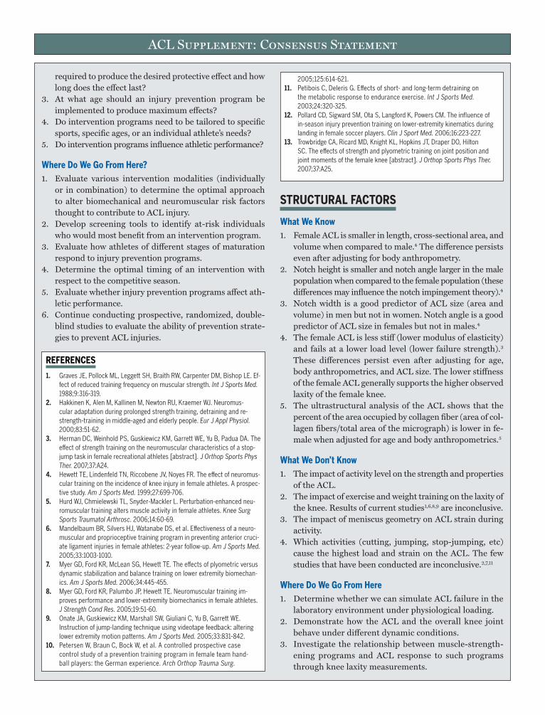
ing strategy than those who exhibit normal knee frontal plane moments. While it is not known why a particular strategy would be chosen, it is ap-parent that these patterns differ in their effect on the knee joint. For ex-ample, after accounting for the forces and moments acting at the foot segment, a laterally directed GRF would impose a laterally directed in-tersegmental force at the distal tibia (FIGuRE 1). In addition, the combi-nation of greater degrees of hip abduction and internal rotation would position the center of pressure further from the center of mass of the tib-ia thereby creating a larger moment arm for the vertical intersegmen-tal force at the distal tibia (FIGuRE 2). Together, a large laterally directed ground reaction force and increased hip abduction and internal rota-tion would create a greater valgus moment about the center of mass of the tibia.CONCLuSIONS: These results provide insight into potentially injurious loading strategies and support the premise that interventions designed to encourage loading of the lower extremity in a more neutral alignment may work to decrease frontal plane loading at the knee.
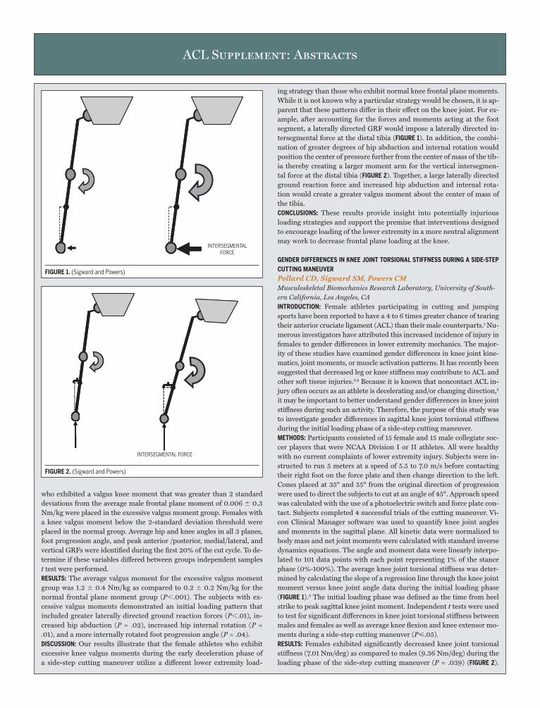
GENDER DIFFERENCES IN KNEE JOINT TORSIONAL STIFFNESS DuRING A SIDE-STEP CuTTING MANEuVERPollard CD, Sigward SM, Powers CM Musculoskeletal Biomechanics Research Laboratory, University of South-ern California, Los Angeles, CAINTRODuCTION: Female athletes participating in cutting and jumping sports have been reported to have a 4 to 6 times greater chance of tearing their anterior cruciate ligament (ACL) than their male counterparts.1 Nu-merous investigators have attributed this increased incidence of injury in females to gender differences in lower extremity mechanics. The major-ity of these studies have examined gender differences in knee joint kine-matics, joint moments, or muscle activation patterns. It has recently been suggested that decreased leg or knee stiffness may contribute to ACL and other soft tissue injuries.3,4 Because it is known that noncontact ACL in-jury often occurs as an athlete is decelerating and/or changing direction,2 it may be important to better understand gender differences in knee joint stiffness during such an activity. Therefore, the purpose of this study was to investigate gender differences in sagittal knee joint torsional stiffness during the initial loading phase of a side-step cutting maneuver.METhODS: Participants consisted of 15 female and 15 male collegiate soc-cer players that were NCAA Division I or II athletes. All were healthy with no current complaints of lower extremity injury. Subjects were in-structed to run 5 meters at a speed of 5.5 to 7.0 m/s before contacting their right foot on the force plate and then change direction to the left. Cones placed at 35° and 55° from the original direction of progression were used to direct the subjects to cut at an angle of 45°. Approach speed was calculated with the use of a photoelectric switch and force plate con-tact. Subjects completed 4 successful trials of the cutting maneuver. Vi-con Clinical Manager software was used to quantify knee joint angles and moments in the sagittal plane. All kinetic data were normalized to body mass and net joint moments were calculated with standard inverse dynamics equations. The angle and moment data were linearly interpo-lated to 101 data points with each point representing 1% of the stance phase (0%-100%). The average knee joint torsional stiffness was deter-mined by calculating the slope of a regression line through the knee joint moment versus knee joint angle data during the initial loading phase (FIGuRE 1).3 The initial loading phase was defined as the time from heel strike to peak sagittal knee joint moment. Independent t tests were used to test for significant differences in knee joint torsional stiffness between males and females as well as average knee flexion and knee extensor mo-ments during a side-step cutting maneuver (P<.05).RESuLTS: Females exhibited significantly decreased knee joint torsional stiffness (7.01 Nm/deg) as compared to males (9.36 Nm/deg) during the loading phase of the side-step cutting maneuver (P = .039) (FIGuRE 2).
who exhibited a valgus knee moment that was greater than 2 standard deviations from the average male frontal plane moment of 0.006 0.3 Nm/kg were placed in the excessive valgus moment group. Females with a knee valgus moment below the 2-standard deviation threshold were placed in the normal group. Average hip and knee angles in all 3 planes, foot progression angle, and peak anterior /posterior, medial/lateral, and vertical GRFs were identified during the first 20% of the cut cycle. To de-termine if these variables differed between groups independent samples t test were performed.RESuLTS: The average valgus moment for the excessive valgus moment group was 1.2 0.4 Nm/kg as compared to 0.2 0.2 Nm/kg for the normal frontal plane moment group (P,.001). The subjects with ex-cessive valgus moments demonstrated an initial loading pattern that included greater laterally directed ground reaction forces (P,.01), in-creased hip abduction (P = .02), increased hip internal rotation (P = .01), and a more internally rotated foot progression angle (P = .04).DISCuSSION: Our results illustrate that the female athletes who exhibit excessive knee valgus moments during the early deceleration phase of a side-step cutting maneuver utilize a different lower extremity load-
INTERSEGMENTAL FORCE
INTERSEGMENTAL FORCE
ACL Supplement: Abstracts
FIGuRE 1.(SigwardandPowers)
FIGuRE 2.(SigwardandPowers)

ACL Supplement: Abstracts
There were no significant differences in average knee flexion angles or knee extensor moments during the loading phase of cutting.DISCuSSION: Our results support the premise that gender differences in sag-ittal knee joint torsional stiffness exist during the performance of a side-step cutting maneuver. These findings are similar to those of Granata et al (2002) who reported that females exhibited significantly lower leg stiff-ness during a hopping task when compared to males. Interestingly, the focus of ACL injury prevention programs contradicts this concept in that females are taught “soft” landing techniques (ie, decreasing lower extrem-ity stiffness). At this time it remains unclear how decreased stiffness could predispose females to soft tissue injury; particularly in the current study where there were no group differences in average knee flexion angles or ex-tensor moments. However, because of the gender disparity in ACL injury, the results of this investigation combined with previous work4 suggest that sagittal knee joint torsional stiffness may be an underlying factor related to the increased incidence of ACL injury in female athletes.CONCLuSIONS: In summary, the concept that increased knee joint stiffness exhibited by males during the side-step cutting maneuver serves to pro-tect the soft tissues of the knee joint, including the ACL, needs to be fur-ther explored. In addition, future investigations are needed to examine the influence of successful intervention programs on knee joint stiffness.
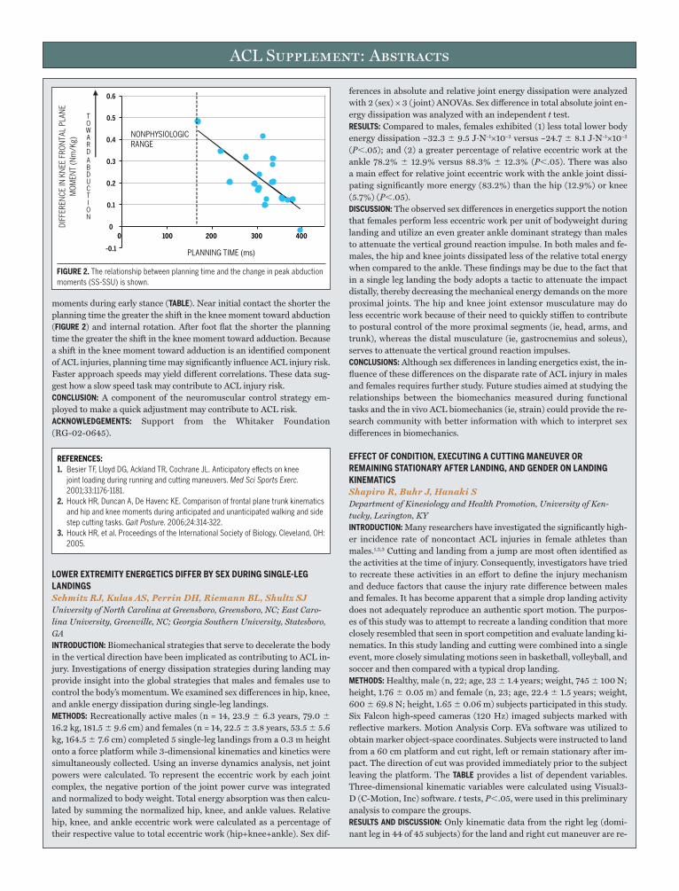
ASSOCIATIONS BETWEEN PLANNING TIME AND KINETIC VARIABLES DuRING AN uNEXPECTED CuT TASKSHouck J, De Haven K, Maloney M Ithaca College, Rochester Campus, Rochester, NY; University of Rochester Medical Center, Department of Orthopedics Rochester, NYINTRODuCTION: During sports play quick movements such as cut tasks are believed to place athletes at risk. Motor control responses to cued ad-justments in direction influence neuromuscular control. Specific neuro-muscular control strategies associated with cued adjustments may lead to greater risk of anterior cruciate injury.1,2 In previous analyses, online adjustments during an unexpected walking cut task led to a shift in the knee moments toward adduction2 and greater plantar flexors moments.3 This analysis examines the relationship between changes in knee kinetic variables (TABLE) due to anticipation and planning time during an unex-pected walking cut task.
METhODS: Twenty healthy subjects (22.6 5.5 years old, 172.8 9.0 cm, and 71.9 14.3 kg) participated in this study. Data were collected us-ing an Optotrak Motion Analysis System (Northern Digital, Inc) and force plate (Kistler) integrated with Motion Monitor Software (Innsport Training, Inc) to generate kinetic variables. Position data were sampled at 100 Hz and force and analogue data at 1000 Hz. Each testing ses-sion included expected tasks, straight walking (ST), and 45° side-step cut (SS), followed by a set of unexpected straight walking (STU) and un-expected side-step cut (SSU) tasks in a random order. For all tasks speed was maintained at 2 m/s. To assess the change due to anticipation, the difference between peak variables of the SS and SSU task (SS-SSU) were calculated. Planning time is defined in FIGuRE 1. Relationships between the change in kinetic variables (TABLE) and planning time were examined using SPSS 10.0.RESuLTS: See TABLE.DISCuSSION: The findings of this analysis suggest that planning time is mod-erately correlated with frontal and transverse plane but not sagittal plane
VARIABLE/% STANCE INTERVAL r VALuE P VALuE
ChangeinMoments(SS-SSU)
Adduction(Nm/kg)/(0%–10%) 0.72 ,.01
Abduction(Nm/Kg)/(10%–30%) 0.50 .03
ExternalRotation(Nm/Kg)/(5%–30%) 0.52 ,.01
Correlations of Selected Variables
-1
0
1
2
3
4
STIFFNESS =� MOMENT
� ANGLE
KNEE FLEXION ANGLE (DEGREES)
KNEE
JO
INT
MO
MEN
T (N
m/k
g)FL
EXO
R (-
) EXT
ENSO
R (+
)
0 10 20 30 40 50 60
FIGuRE 1. Exampleofkneetorsionalstiffnesscalculation(exemplardatatakenfrom1femalesubject).
KNEE
JO
INT
STIF
FNES
S(N
m/d
eg)
2
0
4
6
8
10
12
14
MALES FEMALES
*
FIGuRE 2.Malesexhibitedsignificantlygreaterkneejointstiffnessthanfemales.
0.5-0.5 1.5 2.5
PLANNING TIME
INITIAL CONTACT
(GRF)
VISUAL CUE
1600
1200
800
400
0
-400
TIME (SECONDS)
VOLT
S (m
V)FO
RCE
(N)
FIGuRE 1. Subjectswentstraightorcutwhengivenavisualcuetoturn.Planningtimewastheintervalbetweenananaloguesignalsynchronizedwiththelightcueandinitialcontactdeterminedfromthegroundreactionforcedata.
REFERENCES:1. ArendtE,DickR.Kneeinjurypatternsamongmenandwomenincollegiate
basketballandsoccer.NCAAdataandreviewofliterature.Am J Sports Med.1995;23:694-701.
2. BodenBP,DeanGS,FeaginJA,Jr.,GarrettWE,Jr.Mechanismsofanteriorcruciateligamentinjury.Orthopedics.2000;23:573-578.
3. ButlerRJ,CrowellHP,3rd,DavisIM.Lowerextremitystiffness:implicationsforper-formanceandinjury.Clin Biomech (Bristol, Avon).2003;18:511-517.
4. GranataKP,WilsonSE,PaduaDA.Genderdifferencesinactivemusculoskeletalstiffness.PartI.Quantificationincontrolledmeasurementsofkneejointdynamics.J Electromyogr Kinesiol.2002;12:119-126.
ACL Supplement: Abstracts
moments during early stance (TABLE). Near initial contact the shorter the planning time the greater the shift in the knee moment toward abduction (FIGuRE 2) and internal rotation. After foot flat the shorter the planning time the greater the shift in the knee moment toward adduction. Because a shift in the knee moment toward adduction is an identified component of ACL injuries, planning time may significantly influence ACL injury risk. Faster approach speeds may yield different correlations. These data sug-gest how a slow speed task may contribute to ACL injury risk.CONCLuSION: A component of the neuromuscular control strategy em-ployed to make a quick adjustment may contribute to ACL risk.ACKNOWLEDGEMENTS: Support from the Whitaker Foundation (RG-02-0645).
LOWER EXTREMITY ENERGETICS DIFFER BY SEX DuRING SINGLE-LEG LANDINGSSchmitz RJ, Kulas AS, Perrin DH, Riemann BL, Shultz SJ University of North Carolina at Greensboro, Greensboro, NC; East Caro-lina University, Greenville, NC; Georgia Southern University, Statesboro, GAINTRODuCTION: Biomechanical strategies that serve to decelerate the body in the vertical direction have been implicated as contributing to ACL in-jury. Investigations of energy dissipation strategies during landing may provide insight into the global strategies that males and females use to control the body’s momentum. We examined sex differences in hip, knee, and ankle energy dissipation during single-leg landings.METhODS: Recreationally active males (n = 14, 23.9 6.3 years, 79.0 16.2 kg, 181.5 9.6 cm) and females (n = 14, 22.5 3.8 years, 53.5 5.6 kg, 164.5 7.6 cm) completed 5 single-leg landings from a 0.3 m height onto a force platform while 3-dimensional kinematics and kinetics were simultaneously collected. Using an inverse dynamics analysis, net joint powers were calculated. To represent the eccentric work by each joint complex, the negative portion of the joint power curve was integrated and normalized to body weight. Total energy absorption was then calcu-lated by summing the normalized hip, knee, and ankle values. Relative hip, knee, and ankle eccentric work were calculated as a percentage of their respective value to total eccentric work (hip+knee+ankle). Sex dif-
ferences in absolute and relative joint energy dissipation were analyzed with 2 (sex) × 3 ( joint) ANOVAs. Sex difference in total absolute joint en-ergy dissipation was analyzed with an independent t test.RESuLTS: Compared to males, females exhibited (1) less total lower body energy dissipation –32.3 9.5 J·N–1×10⁻² versus –24.7 8.1 J·N–1×10⁻² (P,.05); and (2) a greater percentage of relative eccentric work at the ankle 78.2% 12.9% versus 88.3% 12.3% (P,.05). There was also a main effect for relative joint eccentric work with the ankle joint dissi-pating significantly more energy (83.2%) than the hip (12.9%) or knee (5.7%) (P,.05).DISCuSSION: The observed sex differences in energetics support the notion that females perform less eccentric work per unit of bodyweight during landing and utilize an even greater ankle dominant strategy than males to attenuate the vertical ground reaction impulse. In both males and fe-males, the hip and knee joints dissipated less of the relative total energy when compared to the ankle. These findings may be due to the fact that in a single leg landing the body adopts a tactic to attenuate the impact distally, thereby decreasing the mechanical energy demands on the more proximal joints. The hip and knee joint extensor musculature may do less eccentric work because of their need to quickly stiffen to contribute to postural control of the more proximal segments (ie, head, arms, and trunk), whereas the distal musculature (ie, gastrocnemius and soleus), serves to attenuate the vertical ground reaction impulses.CONCLuSIONS: Although sex differences in landing energetics exist, the in-fluence of these differences on the disparate rate of ACL injury in males and females requires further study. Future studies aimed at studying the relationships between the biomechanics measured during functional tasks and the in vivo ACL biomechanics (ie, strain) could provide the re-search community with better information with which to interpret sex differences in biomechanics.
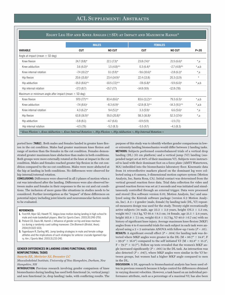
EFFECT OF CONDITION, EXECuTING A CuTTING MANEuVER OR REMAINING STATIONARY AFTER LANDING, AND GENDER ON LANDING KINEMATICSShapiro R, Buhr J, Hanaki S Department of Kinesiology and Health Promotion, University of Ken-tucky, Lexington, KYINTRODuCTION: Many researchers have investigated the significantly high-er incidence rate of noncontact ACL injuries in female athletes than males.1,2,3 Cutting and landing from a jump are most often identified as the activities at the time of injury. Consequently, investigators have tried to recreate these activities in an effort to define the injury mechanism and deduce factors that cause the injury rate difference between males and females. It has become apparent that a simple drop landing activity does not adequately reproduce an authentic sport motion. The purpos-es of this study was to attempt to recreate a landing condition that more closely resembled that seen in sport competition and evaluate landing ki-nematics. In this study landing and cutting were combined into a single event, more closely simulating motions seen in basketball, volleyball, and soccer and then compared with a typical drop landing.METhODS: Healthy, male (n, 22; age, 23 1.4 years; weight, 745 100 N; height, 1.76 0.05 m) and female (n, 23; age, 22.4 1.5 years; weight, 600 69.8 N; height, 1.65 0.06 m) subjects participated in this study. Six Falcon high-speed cameras (120 Hz) imaged subjects marked with reflective markers. Motion Analysis Corp. EVa software was utilized to obtain marker object-space coordinates. Subjects were instructed to land from a 60 cm platform and cut right, left or remain stationary after im-pact. The direction of cut was provided immediately prior to the subject leaving the platform. The TABLE provides a list of dependent variables. Three-dimensional kinematic variables were calculated using Visual3-D (C-Motion, Inc) software. t tests, P,.05, were used in this preliminary analysis to compare the groups.RESuLTS AND DISCuSSION: Only kinematic data from the right leg (domi-nant leg in 44 of 45 subjects) for the land and right cut maneuver are re-
-0.1
0
0.1
0.2
0.3
0.4
0.5
0.6
DIFF
EREN
CE IN
KN
EE F
RON
TAL
PLAN
EM
OM
ENT
(Nm
/Kg)
TOWARD
ABDUCTION
0 100 200 300 400
PLANNING TIME (ms)
NONPHYSIOLOGIC RANGE
FIGuRE 2. Therelationshipbetweenplanningtimeandthechangeinpeakabductionmoments(SS-SSU)isshown.
REFERENCES:1. BesierTF,LloydDG,AcklandTR,CochraneJL.Anticipatoryeffectsonknee
jointloadingduringrunningandcuttingmaneuvers.Med Sci Sports Exerc.2001;33:1176-1181.
2. HouckHR,DuncanA,DeHavencKE.Comparisonoffrontalplanetrunkkinematicsandhipandkneemomentsduringanticipatedandunanticipatedwalkingandsidestepcuttingtasks.Gait Posture.2006;24:314-322.
3. HouckHR,etal.ProceedingsoftheInternationalSocietyofBiology.Cleveland,OH:2005.
ACL Supplement: Abstracts
ported here (TABLE). Both males and females landed in greater knee flex-ion in the cut condition. Males had greater maximum knee flexion and range of motion than the females in the cut condition. Females demon-strated greater maximum knee abduction than males in both conditions. Both groups were more externally rotated at the knee at impact in the cut condition. Males and females reached greater hip flexion in the cut con-dition compared to the no-cut conditions. Males were more abducted at the hip at landing in both conditions. No differences were observed for hip internal/external rotation.CONCLuSIONS: Differences were observed in all 3 planes of motion when a cut was introduced after the landing. Differences were also observed be-tween males and females in their responses to the no cut and cut condi-tions. The inclusion of more game-like situations in studies needs to be considered. Further investigation as the “impact” of these differences on potential injury including joint kinetic and neuromuscular factors needs to be evaluated.
GENDER DIFFERENCES IN LANDING uSING FuNCTIONAL VERSuS NONFuNCTIONAL TASKSSwartz EE, Metivier SJ, Decoster LC Musculoskeletal Institute, University of New Hampshire, Durham, New Hampshire, NHINTRODuCTION: Previous research involving gender comparison of knee biomechanics during landing has used both functional (ie, vertical jump) and non functional (ie, drop landing) tasks, with conflicting results. The
purpose of this study was to identify whether gender comparisons in low-er extremity landing biomechanics would differ between 2 landing tasks.METhODS: Subjects performed counterbalanced trials of a vertical drop landing (DL) (61 cm platform) and a vertical jump (VJ) landing (sus-pended target set at 80% of their maximum VJ). Subjects were instruct-ed to land with their dominant foot on a force plate (AMTI Watertown, MA) imbedded into the biomechanics laboratory floor. Kinematic data from 14 retroreflective markers placed on the dominant leg were col-lected using a 6 camera, 3-dimensional motion capture system (Motion Analysis, Inc, Santa Rosa, CA). Initial contact was determined from the vertical ground reaction force data. Trial data collection for video and ground reaction forces was set at 3 seconds and was initiated and simul-taneously controlled through an external trigger. Data were processed and stored (Eva software version 6.01; Motion Analysis, Inc) and ana-lyzed using the Kintrak software package (version 6.2 Motion Analy-sis, Inc). A 2 × 2 gender (male, female) by landing task (DL, VJ) repeat-ed-measures design was used for the study. Twenty-eight recreationally active subjects (14 male, age 23.2 2.9 years, height 176.3 5.3 cm, weight 80.7 9.5 kg, VJ 60.4 8.5 cm; 14 female, age 21.5 2.4 years, height 165.8 7.1 cm, weight 65.8 11.7 kg, VJ 40.0 6.7 cm) with no history of significant knee injury. Average maximum knee flexion (MKF) angles from 4 to 6 successful trials for each subject in each task were an-alyzed using a 2 × 2 univariate ANOVA with follow-up t tests (P = .05).RESuLTS: A significant overall effect (P = .004) for landing task was de-tected where MKF angles were greater in the DL (M = 86.7° 14.8°, F = 99.9° 16.8°) compared to the self initiated VJ (M = 82.8° 15.2°, F = 79.7° 12.7°). Follow up tests revealed that the women’s MKF an-gle increased significantly (P = .001) in the DL task. An interaction was also detected (P = .047), where MKF angles were similar in the VJ be-tween groups, but women had a higher MKF angle compared to men in the DL.DISCuSSION: A DL approach to biomechanical analysis has been used of-ten in previous research because it helps control for differences obtained in varying descent velocities. However, a task based on an individual per-formance attribute, such as a percentage of a maximal VJ, has also been
Right Leg Hip and Knee Angles (SD) at Impact and Maximum Range*
REFERENCES:1. FordKR,MyerGD,HewettTE.Valguskneemotionduringlandinginhighschoolfe-
maleandmalebasketballplayers.Med Sci Sports Exerc.2003;35:1745-1750.2. PollardCD,DavisIM,HamillJ.Influenceofgenderonhipandkneemechan-
icsduringarandomlycuedcuttingmaneuver.Clin Biomech (Bristol, Avon).2004;19:1022-1031.
3. FagenbaumR,DarlingWG.Jumplandingstrategiesinmaleandfemalecollegeathletesandtheimplicationsofsuchstrategiesforanteriorcruciateligamentinju-ry. Am J Sports Med.2003;31:233-240.
MALES FEMALES
VARIABLE CuT NO CuT CuT NO CuT P<.05
Angleatimpact(meanSDdeg)
Kneeflexion 24.7(9.8)* 22.1(7.3)* 23.8(7.4)ª 21.5(6.6)ª *,a
Kneeadduction 3.6(6.0)* 1.5(4.8)*,b 0.3(6.4)ª –2.7(4.8)ª,b *,a,b
Kneeinternalrotation –7.4(10.2)* 0.1(5.9)* –9.6(10.6)ª –2.8(6.1)ª *,a
Hipflexion 20.6(15.8)* 23.4(14.9)* 22.4(13.8) 20.3(11.9) *
Hipadduction –15.0(8.6)*,a –10.5(7.2)*,b –7.8(6.8)ª –5.9(6.0)b *,a,b
Hipinternalrotation –17.3(8.7) –15.7(7.7) –14.8(9.9) –12.8(7.8)
Maximumorminimumangleafterimpact(meanSDdeg)
Kneeflexion 97.0(7.7)*,b 83.4(8.6)* 83.6(11.2)a,b 79.3(6.5)a *,a,b
Kneeadduction –7.4(8.6)a –8.3(6.9)b –12.8(8.3)*,a –14.3(9.1)*,b *,a,b
Kneeinternalrotation 4.3(6.2)* 9.4(5.1)* 3.3(5.9)a 6.6(5.6)a *,a
Hipflexion 63.8(16.9)* 55.0(20.8)* 58.3(16.8)a 52.3(17.4)a *,a
Hipadduction –3.8(8.1) –4.7(6.6) –0.9(5.9) –1.5(7.1)
Hipinternalrotation –4.5(7.9) –5.2(8.3) –5.5(9.7) –4.1(8.3)
* Knee Flexion +, Knee Adduction +, Knee Internal Rotation +, Hip Flexion +, Hip Adduction +, Hip Internal Rotation +
ACL Supplement: Abstracts
used due to its obvious functional similarities. In this study, the same analysis variable (MKF) revealed conflicting results in the same 2 groups of subjects that performed a landing task under 2 different conditions. This highlights a concern regarding the tasks subjects perform during biomechanical analyses and may also help explain why results from re-search utilizing various tasks yield conflicting results.CONCLuSIONS: When landing from a VJ, subjects MKF angles were simi-lar. However, women responded differently to the DL compared to men. Investigators should consider the appropriateness of the landing task when deciding on research methodology.
ThE INFLuENCE OF A FOOT ORThOTIC ON LOWER EXTREMITY TRANSVERSE PLANE KINEMATICS IN COLLEGIATE FEMALE AThLETES WITh LARGE NAVICuLAR DROP SCORES DuRING LANDINGCarcia CR, Drouin JM, Houglum PA Duquesne University; Slippery Rock University, Slippery Rock, PAINTRODuCTION: Large navicular drop (LND) scores have been associat-ed with both increased foot pronation and medial tibial rotation. As increased medial tibial rotation heightens anterior cruciate ligament (ACL) strain, investigators have suggested female athletes with a LND may be at greater risk for noncontact ACL injury. While a rear-foot me-dially posted foot orthotic has been shown to decrease medial tibial ro-tation during walking, its effect during landing in female athletes with LND scores is unknown. Therefore, the purpose of our study was to com-pare absolute tibial and relative tibiofemoral rotations in female ath-letes with LND scores with and without a foot orthotic during 2 land-ing tasks.METhODS: Using a repeated measures (RM) counterbalanced design, 20 Division I and II female athletes (age, 20.1 1.0 years; height, 169.3 9.9 cm; mass, 69.7 9.7 kg) with LND scores (mean, 9.6 2.5 mm) completed 3 trials of a hopping and landing task with and without a prefabricated foot orthotic. The prefabricated foot orthotic (Interpod, St Kilda, Australia) was rigid and included a 6° extrinsic rear-foot var-us post. Participants performed a single-leg hop at a distance equal to 45% of their height onto the center of a force plate. They also performed a single-leg landing from a box (height, 25 cm) onto the center of the force plate. An electromagnetic tracking system measured 3-dimensional lower extremity kinematics while the force plate identified ground con-tact. Absolute tibial and relative tibiofemoral rotations were calculated for each trial. A mean of the 3 trials for both activities and conditions was used for data analysis. Hop and landing data were analyzed with separate RM ANOVA with 1 within factor at 2 levels (foot orthotic; no foot orthot-ic). P values were set a priori at ,.05.RESuLTS: There were no differences in absolute tibial (P = .549) or rela-tive tibiofemoral rotations (P = .893) with or without the foot orthotic during the landing task. Similarly, no differences in absolute tibial (P = .161) or relative tibiofemoral rotations (P = .935) were identified during the hopping task.DISCuSSION: Results suggest prefabricated orthotics with a 6° rear-foot varus post do not alter absolute or relative tibiofemoral rotation in fe-male athletes with large navicular drop scores during select single-leg landing tasks. Several explanations likely account for these findings. It is possible, the 6° post was simply too small to alter transverse plane tibi-al kinematics. Additionally, midfoot or forefoot landings would have in-fluenced the potential effects of this type of orthotic. Furthermore, na-vicular drop is a static measure of foot pronation and may not relate to active pronation during dynamic activity.CONCLuSION: The present study does not support the hypothesis that foot orthoses alter transverse plane kinematics during landing in female ath-letes with LND scores. However, additional study investigating other types of foot orthoses (ie, inverted, forefoot post) on lower extremity ki-nematics as well as kinetics (ie, tibiofemoral moments) is warranted be-fore firm conclusions can be drawn regarding the influence of this type of intervention in this population.
EFFECTS OF INCREASED BODY MASS INDEX ON LOWER EXTREMITY MOTION PATTERNS IN A STOP-JuMP TASKBrown CN, Yu B, Kirkendall DT, Garrett WE Center for Human Movement Science, The University of North Carolina, Chapel Hill, NCINTRODuCTION: Anterior cruciate ligament (ACL) injuries are common in sports. Altered neuromuscular control has been identified as a possible risk factor for ACL injury and may be a consequence of increased body mass index (BMI) and decreased relative strength. The purpose of this study was to test the effects of increased BMI on lower extremity kine-matics and kinetics of male and female recreational athletes performing a stop-jump task.METhODS: A cross-sectional design was used to test the effects of BMI on landing patterns. All data were collected in a biomechanics labora-tory. A weight vest with 10% of the subject’s body weight evenly distrib-uted around the chest was used to increase BMI and decrease relative strength. Subjects performed 3 practice and 7 test trials of the stop-jump task for each of the weighted and nonweighted conditions. The testing order of conditions was randomized. Twenty college aged recreational athletes were recruited (10 females, 10 males). Average height, mass, and age was 1.68 0.06 m, 63.2 7.5 kg, and 22.6 3.8 years for females, and 1.78 0.06 m, 77.5 9.7 kg, and 22.8 2.4 years for males. A real time 3-dimensional (3-D) videographic data acquisition system was used to collect 3-D coordinate data at a sampling rate of 120 frames/second. Two force plates were used to collect ground reaction force and moment data at 1200 samples/second. Selected lower extremity motion pattern measures were reduced from the 3-D videographic and force plate data including joint angles, moments, and angular velocities for knee and hip flexion-extension, valgus-varus (abduction-adduction), and internal-ex-ternal rotation. These variables were identified at initial ground contact and at their maximum point during the stance phase. Data from the first 3 analyzable trials of each subject were used. Mixed factorial ANOVAs were performed to compare selected knee and hip kinematics and kinet-ics between weighted and nonweighted conditions with consideration of possible gender effects (a = .05).RESuLTS: With increased BMI, subjects exhibited significantly decreased hip flexion angle (P = .01) and knee flexion angular velocity (P = .01) at initial ground contact, and increased peak knee extension moment dur-ing landing (P = .02). The effects of BMI on other knee and hip motion pattern measures were not consistent across subjects.DISCuSSION: Decreasing relative strength by increasing BMI resulted in a more extended lower extremity position with decreased knee flexion ve-locity at landing for college aged recreational athletes. Previous studies have linked extended lower extremity position at landing with ACL inju-ry, therefore increased BMI and decreased relative strength may increase the risk of ACL injuries.CONCLuSIONS: Decreased relative strength resulted in deleterious chang-es in landing patterns. Strength training may need to be included in fu-ture ACL injury prevention programs. This abstract was presented at the National Athletic Trainers’ Association Annual Meeting as a Free Com-munication in Indianapolis, IN, June 2005. Reproduced with permission from the Journal of Athletic Training. 40(2 Suppl):S32, 2005. ©2005 National Athletic Trainers’ Association, Inc.
LOWER EXTREMITY MOTION PATTERNS IN YOuTh SPORTS CAMP PARTICIPANTS PERFORMING 5 TYPES OF SPORT-SPECIFIC STOP-JuMP LANDING TASKSOñate JA, Belez AL, Van Lunen BL, Cortes N Old Dominion University, Norfolk, VAINTRODuCTION: Youth female athletes have a greater risk for incurring an-terior cruciate ligament (ACL) injuries as compared to their male coun-terparts. The purpose of this study was to analyze the differences in knee kinematics between gender and movement task during the stop-jump phase of 5 types of jump-landing tasks.

ACL Supplement: Abstracts
METhODS: Experimental design study conducted during a youth sports camp in a university biomechanics research lab. Participants performed 5 standardized stop-jump landing task approaches: (1) 3-step, (2) 5-m run, (3) volleyball block, (4) 33-cm box-drop, and (5) 5-m deceleration soccer pass and retreat, in a counterbalanced order. Eighteen healthy males (n = 10, height = 149.0 9.6 cm, weight = 47.6 12.5 kg) and fe-males (n = 8, height = 152.2 9.3 cm, weight = 52.4 15.6 kg) between the ages of 10 to 12 years old participated in this study. All subjects were participants in a youth sports program and played a minimum of 1 sea-son of organized basketball, volleyball, or soccer. Two force-plates and a 3-D electromagnetic tracking system collected kinematics and kinet-ics data during the tasks. The stop-jump phase, defined as initial ground contact to maximum knee flexion, was analyzed for each task. The de-pendent variables measured were knee flexion angle at initial contact and maximum range of motion, knee valgus (+)/varus (–) angle at ini-tial contact and at maximum knee flexion angle of the preferred stance leg during the stop-jump phase of each task. Four separate 2 (gender) × 5 (task) repeated-measures analysis of variance (P<.05) were conducted, with Tukey post hoc testing used for comparisons.RESuLTS: Significant differences were found between tasks for knee flex-ion angle at initial contact and knee flexion range of motion (P = .001, respectively), with no gender differences noted. Individuals performing the stop-jump phase of a 5-m deceleration soccer pass and retreat landed with the knee more extended and had less knee flexion range of motion as compared to their knee position in the other landing tasks. Females performed each task in a greater knee valgus position at initial ground contact (P = .038) as compared to their male counterparts who landed in a varus position. Males landed in a greater knee varus position at maxi-mum knee flexion during the deceleration soccer pass (P = .002) as com-pared to knee position in the other tasks.DISCuSSION: The most important finding of this study was that female youth sports camp participants tended to land in a greater valgus posi-tion at initial ground contact as compare to their male counterparts. Ad-ditionally, this trend occurred across 5 different types of jump-landing tasks resulting in consistency of landing techniques regardless of the type of jump-landing task performed.CONCLuSIONS: This information may have implications in the design of ex-perimental testing strategies to evaluate lower extremity motion patterns and instructional strategies aimed at reducing ACL injuries in youth ath-letes. Presented recently at the National Athletic Trainers’ Association Annual Meeting and Symposia, 2005. Reproduced with permission from the Journal of Athletic Training. 40(2 Suppl):S20, 2005. ©2005 Nation-al Athletic Trainers’ Association, Inc.
DETERMINING ThE ACCuRACY AND RELIABILITY OF A DIGITAL PhOTOGRAPhIC-GONIOMETRIC METhOD FOR LOWER EXTREMITY MEASuRESMoncrieff MJ, Jenkins JL, Livingston LA Dalhousie University; University of North Carolina-Greensboro, Greens-boro, NCINTRODuCTION: Alignment of the lower limb has been studied extensively as a potential risk factor for ACL injury.1,3,4 Comparison between inves-tigations is difficult, however, as there is little to no agreement in the lit-erature about the characterization of abnormal alignment or the meth-ods of measuring it.2 Therefore, the purpose of this study was to estimate the accuracy and reliability characteristics of a 2-dimensional (2-D) dig-ital photographic-goniometric measurement instrument, in conjunction with standardized measurement protocols, for deriving static noninva-sive measures of lower limb alignment and segment length.METhODS: Instrumentation accuracy and reliability characteristics were estimated for goniometric measurements (n = 35) extracted from digital photographs of markers positioned on a rigid, life-sized planar measure-ment jig to anatomically simulate the quadriceps (Q) angle, tibiofemoral (TF) angle, and the distal and proximal ends of the femur. Measurements
were taken from identical sets of photographs, by 2 testers, during 2 in-dependent measurement sessions. Intratester and intertester reliability characteristics were then established for the same measurements taken from digital photographs of anatomical markers positioned, using stan-dardized protocols, on human participants (n = 20) by the same 2 tes-ters. Photographs were captured of each individual while adopting 2 dif-ferent stance positions (ie, self selected, Romberg) during 2 independent testing sessions with each tester. The testers then derived measurements from each other’s photographs to distinguish between the error generat-ed by anatomical marker placement versus tester error associated with manipulation of the goniometer on the photograph.RESuLTS: The digital photographic-goniometric instrumentation method yielded measurements with extremely high degrees of instrumentation accuracy (ICC2,1 = 0.97-0.99), intertester reliability (ICC2,1 = 0.98-0.99), and intratester reliability (ICC2,1 = 0.98-0.99) characteristics from the 2-D measurement jig. For measurements derived from photographs of markers placed on human participants, the reliability characteristics (ie, intratester ICC2,1 = 0.458-0.958; intertester ICC2,1 = 0.257-0.944) ranged from fair to almost perfect, with values for measures of femur length con-sistently stronger than that of the TF and Q angle, respectively. In gener-al, the reliability of measures derived in the Romberg stance was higher than those taken in a self-selected stance. Between-tester measurement differences appeared to be due to differences in anatomical marker place-ment rather than goniometer manipulation techniques.DISCuSSION: The digital photographic-goniometric method of measure-ment yielded almost perfect accuracy and reliability characteristics for measures derived from the planar measurement jig, and generated fair to almost perfect reliability characteristics for measures derived from hu-man participants. Higher reliability coefficients were generally associat-ed with measurements derived while participants adopted a standard-ized stance position, and for those measures requiring the use of fewer anatomical markers.CONCLuSIONS: The digital photographic-goniometric method of measure-ment, used in conjunction with standardized measurement protocols, may yield accurate and reliable 2-D measures of the Q angle, TF angle, and femur length. The key is to reduce between tester differences in ana-tomical marker application.
RELIABILITY AND VALIDITY OF A 2-D APPROACh TO ASSESSING FRONTAL PLANE DYNAMIC ALIGNMENT OF ThE LOWER EXTREMITYEarl JE, Monteiro SK, Geiser CF, Alexander PT University of Wisconsin-Milwaukee, Milwaukee, WIINTRODuCTION: The Sport Motion Analysis Review Tool (SMART) is a fast, inexpensive, and not technically demanding software package that is used for basic movement analysis. This could be used in a large pro-spective study screening the frontal plane lower extremity dynamic align-ment of athletes to examine whether or not poor dynamic alignment is a risk factor for ACL injury. The purpose of this study was to determine the reliability of the SMART program in assessing frontal plane kinemat-ics, and to determine its validity by comparing it to the gold standard 3-D motion analysis system.
REFERENCES:1. McLeanSG,WalkerK,FordKR,MyerGD,HewettTE,vandenBogertAJ.Evaluation
ofatwodimensionalanalysismethodasascreeningandevaluationtoolforante-riorcruciateligamentinjury.Br J Sports Med.2005;39:355-362.
2. MurphyDF,ConnollyDA,BeynnonBD.Riskfactorsforlowerextremityinjury:are-viewoftheliterature. Br J Sports Med.2003;37:13-29.
3. NylandJ,SmithS,BeickmanK,ArmseyT,CabornDN.Frontalplanekneeangleaf-fectsdynamicposturalcontrolstrategyduringunilateralstance.Med Sci Sports Exerc. 2002;34:1150-1157.
4. SodermanK,AlfredsonH,PietilaT,WernerS.Riskfactorsforleginjuriesinfemalesoccerplayers:aprospectiveinvestigationduringoneout-doorseason.Knee Surg Sports Traumatol Arthrosc.2001;9:313-321.
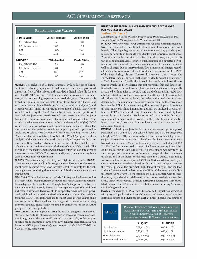
ACL Supplement: Abstracts
METhODS: The right leg of 10 female subjects, with no history of signifi-cant lower extremity injury was tested. A video camera was positioned directly in front of the subject and recorded a digital video file for use with the SMART program. 3-D kinematic data were collected concur-rently via a 7-camera high-speed motion analysis system. Data were col-lected during a jump-landing task (drop off the front of a block, land with both feet, and immediately perform a maximal vertical jump), and a stepdown task (stand on your right leg on top of a block, slowly lower your left foot to tap the floor). Each subject performed 3 repetitions of each task. Subjects were tested a second time 1 week later. For the jump landing, the variables were knee valgus angle, and valgus distance (lin-ear distance between the markers on the lateral femoral condyles). ROM values were determined from foot contact to maximum knee valgus. For the step-down the variables were knee valgus angle, and hip adduction angle. ROM values were determined from quiet standing to toe touch. These variables were obtained from both the SMART program, and via analysis of the 3-D data. The trials were analyzed separately by 3 re-searchers. Between day (intratester), and between tester reliability were calculated using the intraclass correlation coefficient (ICC) statistic. The precision of the measurements was analyzed using the standard error of the measurement (SEM). Concurrent validity was calculated using Pear-son’s product moment correlation.RESuLTS: The between day reliability was high for all variables (TABLE). The SEM values are small, indicating an acceptable amount of measure-ment error. Pearson’s correlation revealed excellent validity for the val-gus angle measure during the step-down and for the valgus distance dur-ing the jump.DISCuSSION: This technique using the SMART program has been found to be reliable in assessing frontal plane lower extremity alignment both be-tween days and between testers. Though this 2-D approach is attractive for use in a multisite study because it is inexpensive, portable, and does not require advanced technical skills to operate, it had not been previ-ously validated to the gold standard 3-D motion analysis. The variables from the SMART program that are the most valid are the valgus angle excursion during the step-down, and valgus distance excursion during the vertical jump. These variables should be considered for use in future prospective screening studies.CONCLuSION: This 2-D approach using the SMART program is an accept-able alternative to 3-D kinematic analysis in assessing frontal plane dy-namic alignment. This tool could be used in a large scale, multisite, pro-spective study examining lower extremity dynamic alignment as a risk factor for ACL injury. This study was presented at the 2005 GLATA An-nual Meeting, Toledo, OH.
uTILITY OF ThE FRONTAL PLANE PROJECTION ANGLE OF ThE KNEE DuRING SINGLE LEG SQuATSWillson JD, Davis I Department of Physical Therapy, University of Delaware, Newark, DE; Drayer Physical Therapy Institute, Hummelstown, PAINTRODuCTION: Abnormal lower extremity mechanics during athletic ac-tivities are believed to contribute to the etiology of numerous knee joint injuries. The single leg squat test is commonly used by practicing cli-nicians to identify individuals who display such abnormal mechanics. Presently, due to the restraints of typical clinical settings, analysis of this test is done qualitatively. However, quantification of a patient’s perfor-mance on this test would facilitate documentation of these mechanics as well as changes due to interventions. Two-dimensional images record-ed by a digital camera reveal the frontal plane projection angle (FPPA) of the knee during this test. However, it is unclear to what extent the FPPA determined using such methods is related to actual 3-dimension-al (3-D) kinematics. Specifically, it would be beneficial to know the ex-tent to which the FPPA during this test represents hip and knee rota-tion in the transverse and frontal planes as such rotations are frequently associated with injuries to the ACL and patellofemoral joint. Addition-ally, the extent to which performance on the SL squat test is associated with these rotations during faster, more demanding tasks has not been determined. The purpose of this study was to examine the correlation between the FPPA of the knee during SL squats and hip and knee fron-tal and transverse plane kinematics. Second, we analyzed to what ex-tent the FPPA of the knee during SL squats reflects knee and hip kine-matics during a SL landing. We hypothesized that the FPPA during SL squats would be significantly correlated with greater hip adduction, hip internal rotation, knee abduction, and knee external rotation during SL squats and landings.METhODS: 20 healthy subjects (11 female, 9 male; mean age, 26.4 years) performed 5 SL squats to a self-selected depth and 5 SL landings from a height of 23 cm. All trials were collected for the dominant leg of each subject. Retroreflective markers placed on the lower extremity were tracked by a 6 camera Vicon motion analysis system collecting at 120 Hz. V3-D software was used to determine lower extremity kinematics. Additionally, during each squat trial, a digital image was recorded by a camera placed 2 m anterior to the subject, perpendicular to the fron-tal plane, and at the height of the knee joint in SL stance. Each image was recorded as the subject passed 45° knee flexion as determined by an electrogoniometer. Markers placed on the leg of each subject bisecting the frontal plane of the proximal thigh, femoral condyles, and malleoli at the ankle were used to determine the FPPA of the knee in each digi-tal image (CorelDraw). To synchronize the digital camera with the mo-tion analysis, a signal was delivered to the motion analysis workstation as the image was recorded. Pearson correlation coefficients were calcu-lated between the FPPA and selected 3-D kinematics during SL stance and landing conditions.RESuLTS: The change in FPPA from SL stance to SL squat was associated with greater hip adduction, knee abduction, and knee external rotation during SL squats and SL landings (TABLE 1). Three-dimensional rotations
Reliability and Validity
JuMP LANDING VALGuS DISTANCE VALGuS ANGLE
ICC1,kbetweendays .74 .88
ICC2,kbetweentesters .80 .90
SEM .02m 3.6°
Pearsonr –.88 .53
STEPDOWN VALGuS ANGLE PELVIS ANGLE
ICC1,kbetweendays .95 .85
ICC2,kbetweentesters .84 .91
SEM 1.6° 2.1°
Pearsonr .87 –.01
Pearson Correlation Coefficients for the Association Between the FPPA Excursion
During SL Squats and 3-D Rotation Excursions During SL Squats and Landings
TABLE 1
SL SQuAT SL LANDING
Hipadduction 0.58,P=.008 0.67,P=.001
Hipinternalrotation 0.35,P=.16 0.16,P=.51
Kneeabduction 0.71,P=.001 0.58,P=.008
Kneeexternalrotation 0.77,P<.001 0.54,P=.015
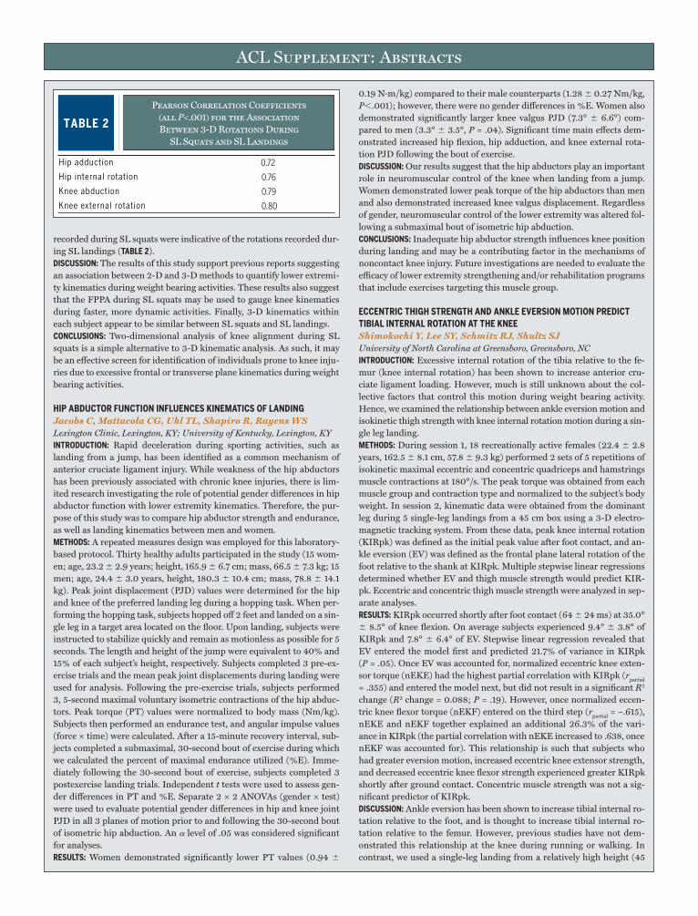
ACL Supplement: Abstracts
recorded during SL squats were indicative of the rotations recorded dur-ing SL landings (TABLE 2).DISCuSSION: The results of this study support previous reports suggesting an association between 2-D and 3-D methods to quantify lower extremi-ty kinematics during weight bearing activities. These results also suggest that the FPPA during SL squats may be used to gauge knee kinematics during faster, more dynamic activities. Finally, 3-D kinematics within each subject appear to be similar between SL squats and SL landings.CONCLuSIONS: Two-dimensional analysis of knee alignment during SL squats is a simple alternative to 3-D kinematic analysis. As such, it may be an effective screen for identification of individuals prone to knee inju-ries due to excessive frontal or transverse plane kinematics during weight bearing activities.
hIP ABDuCTOR FuNCTION INFLuENCES KINEMATICS OF LANDINGJacobs C, Mattacola CG, Uhl TL, Shapiro R, Rayens WS Lexington Clinic, Lexington, KY; University of Kentucky, Lexington, KYINTRODuCTION: Rapid deceleration during sporting activities, such as landing from a jump, has been identified as a common mechanism of anterior cruciate ligament injury. While weakness of the hip abductors has been previously associated with chronic knee injuries, there is lim-ited research investigating the role of potential gender differences in hip abductor function with lower extremity kinematics. Therefore, the pur-pose of this study was to compare hip abductor strength and endurance, as well as landing kinematics between men and women.METhODS: A repeated measures design was employed for this laboratory-based protocol. Thirty healthy adults participated in the study (15 wom-en; age, 23.2 2.9 years; height, 165.9 6.7 cm; mass, 66.5 7.3 kg; 15 men; age, 24.4 3.0 years, height, 180.3 10.4 cm; mass, 78.8 14.1 kg). Peak joint displacement (PJD) values were determined for the hip and knee of the preferred landing leg during a hopping task. When per-forming the hopping task, subjects hopped off 2 feet and landed on a sin-gle leg in a target area located on the floor. Upon landing, subjects were instructed to stabilize quickly and remain as motionless as possible for 5 seconds. The length and height of the jump were equivalent to 40% and 15% of each subject’s height, respectively. Subjects completed 3 pre-ex-ercise trials and the mean peak joint displacements during landing were used for analysis. Following the pre-exercise trials, subjects performed 3, 5-second maximal voluntary isometric contractions of the hip abduc-tors. Peak torque (PT) values were normalized to body mass (Nm/kg). Subjects then performed an endurance test, and angular impulse values (force × time) were calculated. After a 15-minute recovery interval, sub-jects completed a submaximal, 30-second bout of exercise during which we calculated the percent of maximal endurance utilized (%E). Imme-diately following the 30-second bout of exercise, subjects completed 3 postexercise landing trials. Independent t tests were used to assess gen-der differences in PT and %E. Separate 2 × 2 ANOVAs (gender × test) were used to evaluate potential gender differences in hip and knee joint PJD in all 3 planes of motion prior to and following the 30-second bout of isometric hip abduction. An a level of .05 was considered significant for analyses.RESuLTS: Women demonstrated significantly lower PT values (0.94
0.19 N·m/kg) compared to their male counterparts (1.28 0.27 Nm/kg, P,.001); however, there were no gender differences in %E. Women also demonstrated significantly larger knee valgus PJD (7.3° 6.6°) com-pared to men (3.3° 3.5°, P = .04). Significant time main effects dem-onstrated increased hip flexion, hip adduction, and knee external rota-tion PJD following the bout of exercise.DISCuSSION: Our results suggest that the hip abductors play an important role in neuromuscular control of the knee when landing from a jump. Women demonstrated lower peak torque of the hip abductors than men and also demonstrated increased knee valgus displacement. Regardless of gender, neuromuscular control of the lower extremity was altered fol-lowing a submaximal bout of isometric hip abduction.CONCLuSIONS: Inadequate hip abductor strength influences knee position during landing and may be a contributing factor in the mechanisms of noncontact knee injury. Future investigations are needed to evaluate the efficacy of lower extremity strengthening and/or rehabilitation programs that include exercises targeting this muscle group.
ECCENTRIC ThIGh STRENGTh AND ANKLE EVERSION MOTION PREDICT TIBIAL INTERNAL ROTATION AT ThE KNEEShimokochi Y, Lee SY, Schmitz RJ, Shultz SJ University of North Carolina at Greensboro, Greensboro, NCINTRODuCTION: Excessive internal rotation of the tibia relative to the fe-mur (knee internal rotation) has been shown to increase anterior cru-ciate ligament loading. However, much is still unknown about the col-lective factors that control this motion during weight bearing activity. Hence, we examined the relationship between ankle eversion motion and isokinetic thigh strength with knee internal rotation motion during a sin-gle leg landing.METhODS: During session 1, 18 recreationally active females (22.4 2.8 years, 162.5 8.1 cm, 57.8 9.3 kg) performed 2 sets of 5 repetitions of isokinetic maximal eccentric and concentric quadriceps and hamstrings muscle contractions at 180°/s. The peak torque was obtained from each muscle group and contraction type and normalized to the subject’s body weight. In session 2, kinematic data were obtained from the dominant leg during 5 single-leg landings from a 45 cm box using a 3-D electro-magnetic tracking system. From these data, peak knee internal rotation (KIRpk) was defined as the initial peak value after foot contact, and an-kle eversion (EV) was defined as the frontal plane lateral rotation of the foot relative to the shank at KIRpk. Multiple stepwise linear regressions determined whether EV and thigh muscle strength would predict KIR-pk. Eccentric and concentric thigh muscle strength were analyzed in sep-arate analyses.RESuLTS: KIRpk occurred shortly after foot contact (64 24 ms) at 35.0° 8.5° of knee flexion. On average subjects experienced 9.4° 3.8° of KIRpk and 7.8° 6.4° of EV. Stepwise linear regression revealed that EV entered the model first and predicted 21.7% of variance in KIRpk (P = .05). Once EV was accounted for, normalized eccentric knee exten-sor torque (nEKE) had the highest partial correlation with KIRpk (rpartial
= .355) and entered the model next, but did not result in a significant R2 change (R2 change = 0.088; P = .19). However, once normalized eccen-tric knee flexor torque (nEKF) entered on the third step (rpartial = –.615), nEKE and nEKF together explained an additional 26.3% of the vari-ance in KIRpk (the partial correlation with nEKE increased to .638, once nEKF was accounted for). This relationship is such that subjects who had greater eversion motion, increased eccentric knee extensor strength, and decreased eccentric knee flexor strength experienced greater KIRpk shortly after ground contact. Concentric muscle strength was not a sig-nificant predictor of KIRpk.DISCuSSION: Ankle eversion has been shown to increase tibial internal ro-tation relative to the foot, and is thought to increase tibial internal ro-tation relative to the femur. However, previous studies have not dem-onstrated this relationship at the knee during running or walking. In contrast, we used a single-leg landing from a relatively high height (45
Pearson Correlation Coefficients (all P<.001) for the Association Between 3-D Rotations During
SL Squats and SL Landings
TABLE 2
Hipadduction 0.72
Hipinternalrotation 0.76
Kneeabduction 0.79
Kneeexternalrotation 0.80

ACL Supplement: Abstracts
cm). Given the substantially higher peak ground reaction force and force attenuation demands associated with this task, it is likely that greater levels of EV, thus greater KIRpk were realized. A secondary finding is that the relationship between thigh strength and KIRpk was stronger with maximal eccentric versus concentric contractions. This may in part be due to the fact that maximal eccentric isokinetic muscle strength has been shown to be less affected by angular velocity than maximal con-centric isokinetic muscle strength. Although adding only nEKE did not alter the r2 significantly, an additional 26.3% of the variance in KIRpk was explained once both nEKE and nEKF were accounted for. Hence, the absolute strength balance between nEKE and nEKF may be a criti-cal factor in controlling KIRpk.CONCLuSIONS: The ACL has been shown to be loaded with the applica-tion of internal rotation torque to the knee joint, especially during shal-low knee flexion angles (ie, below ~45°) and a forceful quadriceps muscle contraction. Our findings suggest that preventing excessive ankle ever-sion may reduce the amount of knee internal rotation that occurs with force attenuation upon landing, and lend further support to the impor-tance of balanced strength of quadriceps and hamstring muscles in knee stability.
BIOMEChANICS OF ANTICIPATED AND uNANTICIPATED SIDE-STEP CuTTING IN MALE AND FEMALE BASKETBALL PLAYERS DuRING GAME-LIKE SCENARIOS AND LOWER EXTREMITY STRENGThCarlstedt KE, Brandner RD, Kernozek TW Rehabilitation Institute of Chicago, Northshore Outpatient Clinic, Chicago, IL; University of Wisconsin-La Crosse, Department of Health Professions, La Crosse, WI; Luther Hospital, Eau Claire, WIINTRODuCTION: The anterior cruciate ligament (ACL) is one of the most commonly injured ligaments in the knee. The majority of these injuries occur due to noncontact mechanisms such as, sudden acceleration/decel-eration, change of direction, or landing from a jump. Most of the current studies examining sidestep cutting has been performed on male athletes with none evaluating the athletes’ strength concurrently. The aim of this study was to determine the effects of unanticipated game like scenarios on the lower extremity joint kinematics and kinetics during the stance phase of side-step cutting maneuvers as well as to determine any gender differences in strength.METhODS: Nineteen females and 19 males NCAA division III basketball players were tested. All athletes had no prior history of serious knee in-jury. Subjects were analyzed using a 3 dimensional motion analysis sys-tem and 2 force platforms during 4 different game like cutting condi-tions: anticipated cut, unanticipated pass to left side, unanticipated fake to left side, and unanticipated straight ahead pass. An infrared timing light system was used to determine approach speed and to cue the pass-er to throw a ball to another player. The subjects were instructed to fol-low the movement of the ball and to intercept the pass as they would in a game situation. Each subject performed 5 trials of each scenario list-ed previously in a randomized order for both dominant and nondom-inant legs. All ground reaction force data, moment data, and strength data were normalized (percent body weight and Nm/kg of body weight). A series of repeated measures analysis of variance were used on each of the kinematic and kinetic dependent variables (a = .05). Bonferroni post hoc tests were used if tests were significant. t tests were used to compare strength values between genders (a = .05).RESuLTS: Overall, cutting conditions resulted in greater maximum knee valgus angles for females compared to males during stance. In gener-al, females exhibited significantly more knee valgus throughout stance, significantly less hip flexion at initial contact, and significantly less hip extension during the stance phase of all cutting conditions. Maximum internal knee extensor moment was significantly greater in males than females. Maximum internal hip abductor moment was significantly greater in females, while maximum internal hip adduction moment was significantly greater in males. Stance time was not different across con-
ditions. The knee extensor and knee flexor isokinetic strength measure-ments were significantly less in females. In addition, weight normalized hip abductor strength values were significantly lower in females.DISCuSSION: There were no significant differences found between an-ticipated and unanticipated cutting scenarios. There were several gen-der differences observed between male and female athletes. Overall, female players demonstrated greater knee valgus angles across condi-tions as well as greater maximum internal hip abduction moments than males. This increased internal hip abductor torque must be generated by females to avoid movement into a position of increased hip adduc-tion and further knee valgus. This has been indicated as a high-risk po-sition for ACL injuries. Greater internal maximum hip adduction mo-ments in male players and larger maximum hip abduction moments in females during cutting maneuvers may correspond to underlying ana-tomical gender differences in hip structure or differences in lumbopelvic strength and stability. Further research is needed to examine the poten-tial factors that place female athletes at higher risk for ACL injury dur-ing cutting maneuvers in sport.CONCLuSIONS: Unanticipated game-like cutting conditions produced few changes in knee and hip kinematics and kinetics as compared to antici-pated cutting maneuvers. Female basketball players demonstrated sig-nificantly larger knee valgus angles and greater internal hip abductor moments than males throughout the stance phase across all game-like scenarios. Females with weak hip abductor strength may not be able to generate a large enough internal hip abductor moment to avoid high-risk knee positions for ACL injury during cutting maneuvers. Further research is needed to examine the relationship of lumbopelvic strength to cutting mechanics in male and female athletes during game-like scenarios.
GENDER AND AGE COMPARISONS IN NEuROMuSCuLAR AND BIOMEChANICAL ChARACTERISTICS OF ThE KNEE IN YOuNG AThLETESSell TC, Abt JP, Nagai T, Myers JB, Fu FH, Lephart SM Neuromuscular Research Laboratory, University of Pittsburgh, Pittsburgh, PAINTRODuCTION: The majority of research examining gender differenc-es related to noncontact ACL injuries has focused on high school and collegiate athletes due to the disparity in injury rates between genders. Yet, current evidence suggests that injury rate differences also occur in younger, school-aged athletes. In addition, the potential contributors to these injury rate differences in school-aged athletes are largely unknown. The purpose of this study was to examine and compare the gender and age group strength, balance, jump height, and landing kinematics char-acteristics in high school and school-aged athletes.METhODS: Forty healthy high school (16.8 0.8 years) and 40 school-aged (10.8 0.8 years), athletes with an equal distribution of males and females within each group participated in the study. Each subject under-went an isokinetic strength test (knee flexion and extension at 60°/s), single-leg balance assessment (eyes open and eyes closed), vertical jump height test, and a kinematic analysis (knee flexion and valgus angle at initial contact) during a single-leg vertical stop jump task. A 2-way ANO-VA (age × gender) was utilized to examine age group (within gender) and gender (within age group) differences.RESuLTS: Significant (P,.05) gender differences were revealed in strength, balance, and jump height performance in the high school group. Specifi-cally, males had greater strength (flexion and extension) and higher jump height whereas females had better balance. No differences were observed in the school-aged group in any of the variables analyzed. Significant age group differences were observed within both genders for strength (flex-ion and extension) and jump height such that high school athletes had greater strength and jump height performance. School-aged males had better balance than high school males. No significant differences were observed in landing kinematics for any of the comparisons made.DISCuSSION: Previous studies have demonstrated significant kinematic differences during sports tasks in high school and college-aged athletes.
ACL Supplement: Abstracts
The current study did not reveal kinematic differences but did reveal strength differences which may partially explain the differences in inju-ry rates in these athletes.CONCLuSIONS: The lack of gender differences within school-aged athletes does not help to explain the gender differences in knee injuries described in the literature. Research should continue to examine the neuromuscu-lar and biomechanical characteristics of young athletes.
DIFFERENCES IN LOWER-LIMB NEuROMuSCuLAR CONTROL BETWEEN SPORTS MOVEMENTS EXECuTED IN LABORATORY AND GAME SETTINGSSaunders N, Otago L, McLean S School of Human Movement and Sport Sciences, University of Ballarat, Ballarat, Australia; The Cleveland Clinic, Cleveland, OHINTRODuCTION: Anterior cruciate ligament (ACL) injury is a common and traumatic sports injury. While the underlying mechanisms remain un-clear, neuromuscular control elicited during high-risk sports movements has become increasingly viewed as a primary risk factor.1 To date, neuro-muscular predictors of ACL injury have typically arisen from lab-based assessments of these movements, as a means to counter the inherently random and unpredictable nature of the true game setting. It is possi-ble however, that this approach excludes important components of actu-al game-play that contribute directly to the chosen movement response and resultant injury risk.3 A game-based assessment of high-risk sports movements may thus afford more reliable neuromuscular injury predic-tors, and hence, more effective injury screening and prevention strat-egies. With this in mind, the purpose of the current investigation was to compare lab and game-based measures of lower limb neuromuscular control during high-risk sports movements.METhODS: Ten female subjects (age 24.3 9.5 years) had lower limb EMG data recorded continuously during a fixtured netball game. At a subsequent session, occurring in the lab, EMG data were also recorded for 3 chosen conditions (as below). The movement chosen for investiga-tion was a “leap” land, which is commonly employed in netball and in-volves taking off on a single leg and landing on the opposite leg. For each subject, bilateral EMG (Mespec 4000, MegaWin) data, sampled at 1000 Hz, were recorded telemetrically for rectus femoris (RF), biceps femoris (BF), medial hamstring (MH), and gluteus medius (GM) muscles. Dur-ing lab testing, subjects performed 5 leap land trials for each leg, for 3 specific movement conditions of increasing complexity, namely: (1) run and leap land (Land); (2) run, leap land whilst catching a ball, pivot and pass to a teammate in the same movement (No Def); (3) break from a defender, run, leap land while catching a ball, pivot and pass to a team-mate in the same movement (Def). The point of initial contact (IC) of the land leg, for both the game and lab trials was first determined via video camera recordings. EMG data were then analyzed for each trial to deter-mine muscle onset relative to IC (onset to IC), and the resultant muscle burst duration that occurred concurrent with IC. Specifically, the onset level was defined as the point where muscle activation exceeded baseline levels by at least 1 SD for a minimum of 10 milliseconds.2 All dependent measures were subsequently submitted to a 2-way ANOVA to determine for the main effects of test condition and leg.RESuLTS: Onset to IC for the RF was observed to be significantly differ-ent between the game and lab conditions (Game versus Land P = .050, Game versus No Def P = .016, Game versus Def P = .027). Onset of ac-tivation occurred much closer to IC during the game (48 35 millisec-onds) compared to the lab (Land: 87 22 milliseconds; No Def: 84 44 milliseconds; Def: 80 31 milliseconds) conditions. Comparisons of muscle burst duration data failed to yield significant results for all sta-tistical comparisons. No difference was found between approach speed for each condition.DISCuSSION: The following implications of a delay in RF onset during a game are proposed. The delay in RF onset during a game may impact knee stability at landing and therefore the potential to counteract po-tentially hazardous joint load states. Conversely, a delayed onset of RF
may demonstrate an adaptive strategy for knee stability that is necessary for the inherently random nature of a netball game. The delay in RF on-set may be unrelated to injury risk and indicative of RF behavior to op-timize performance in a game setting. Finally, the delay in RF onset may be a reflection of the demand for hip and upper body control necessary during game play.CONCLuSIONS: When examining high risk sporting maneuvers, there is some evidence that what we test in the lab may reflect the game. How-ever more research is required to ensure that any subtle differences be-tween the game and laboratory setting are not key components to under-standing injury mechanisms. This abstract was presented as a poster at ISB 05 in Cleveland.
CASE REPORT: CAN NEuROMuSCuLAR MEASuRES PREDICT ACL INJuRY?Otago L, Saunders N, McLean S School of Human Movement and Sport Sciences, University of Ballarat, Australia; The Cleveland Clinic, Cleveland, OHINTRODuCTION: Neuromuscular control elicited during high-risk sports movements has become increasingly viewed as a primary risk factor for noncontact ACL injuries.1 Understanding neuromuscular characteristics prior to an ACL injury could be invaluable in elucidating injury mecha-nisms and contributing to the development of accurate screening meth-ods. However, prospective neuromuscular-based studies in an attempt to identify predictors of ACL injury would be very difficult because any time lapse between testing and injury may deem results questionable and in-valid, as neuromuscular measures can be influenced, for example, with training. This case report discusses the findings from a subject who sus-tained an ACL injury 6 days post testing (from the study described be-low), which indicates possible neuromuscular dysfunction that may have contributed to the ACL injury.METhODS: Ten female subjects (age, 24.3 9.5 years) had lower limb EMG data recorded for 3 chosen conditions (as below). The movement chosen for investigation was a “leap” land, which is commonly employed in netball and involves taking off on a single leg and landing on the op-posite leg. For each subject, bilateral EMG (Mespec 4000, MegaWin) data, sampled at 1000 Hz, were recorded telemetrically for rectus femo-ris (RF), biceps femoris (BF), medial hamstring (MH), and gluteus me-dius (GM) muscles. During lab testing, subjects performed 5 leap land trials for each leg, for 3 specific movement conditions of increasing com-plexity, namely: (1) Run and leap land (Land); (2) Run, leap land while catching a ball, pivot and pass to a teammate in the same movement (No Def); (3) Break from a defender, run, leap land while catching a ball, pivot and pass to a teammate in the same movement (Def). The point of initial contact (IC) of the land leg, was first determined via video cam-era recordings. EMG data were then analysed for each trial to determine muscle onset relative to IC, and the resultant muscle burst duration that occurred concurrent with IC. Specifically, the onset level was defined as the point where muscle activation exceeded baseline levels by at least 1 SD for a minimum of 10 ms.3
INJuRY INCIDENT: The player ran for a ball that was going out of court. As she reached the court boundary she performed an abrupt 1-legged stop. As her foot was planted, she twisted/attempted to pivot whilst bringing the ball back into play when she heard a snap. She attempted to continue pivoting, however her leg gave way and she fell to the floor.
REFERENCES:1. GriffinLY,AgelJ,AlbohmMJ,ArendtEA.Noncontactanteriorcruciateligamentin-juries:riskfactorsandprevention.J Am Acad Orthop Surg.2000;8:141-150.2. HodgesPW,BuiBH.Acomparisonofcomputer-basedmethodsforthedetermina-tionofonsetofmusclecontractionusingelectromyography.Electroencephalogr Clin Neurophysiol. 1996;101:511-519.3. McLeanSG,LipfertSW,vandenBogertAJ.Effectofgenderanddefen-siveopponentonthebiomechanicsofsidestepcutting.Med Sci Sports Exerc.2004;36:1008-1016.

ACL Supplement: Abstracts
RESuLTS: The ACL injured subject was the only player to record an on-set post IC within her trials. These results were only found in the injured limb compared with the noninjured limb. A total of 6 onset post IC trials were recorded within the Def condition across all 4 muscles out of a pos-sible 15 lands. Five onset post IC trials were recorded within the No Def condition across RF and BF muscles only. No onset post IC trial was re-corded for the least complex, Land condition.DISCuSSION: A player will often sustain an ACL injury while performing a common, albeit high-risk task. Results presented for this subject high-light the potential for this to be explained. It has been found that muscle activation patterns are pre-planned and activate prior to loading to coun-ter potentially injurious moments.4 Therefore an onset post IC potential-ly renders the knee joint extremely vulnerable to injury, relying on pre-dominantly passive restraints to counter loading during high-risk tasks. The ACL injured subject in this study performed the high-risk, landing task the majority of times using an activation strategy prior to IC. During the simple and less ‘game like’ landings, this activation strategy prior to IC was consistent. However with increasing and more ‘game-like’ com-plexity, the injured subject’s results suggest that she was unable to recruit a functional level of activation prior to IC for up to 40% of her landings, potentially increasing her chance of ligament injury.CONCLuSION: While this is a single subject case report, it does provide valuable insight into potential neuromuscular dysfunction prior to ACL injury.
DIFFERENCES EXIST IN NEuROMuSCuLAR PATTERNS DuRING uNANTICIPATED RuNNING AND CuTTING MANEuVERS BETWEEN ELITE MALE AND FEMALE SOCCER PLAYERSLandry SC, McKean KA, Hubley-Kozey CL, Deluzio KJ, Stanish WD School of Biomedical Engineering, Dalhousie University, Halifax, Canada; School of Physiotherapy, Dalhousie University, Halifax, Canada; Department of Surgery (Orthopaedics), Dalhousie University, Halifax, CanadaINTRODuCTION: The anterior cruciate ligament (ACL) is important for knee joint stability and proper function, particularly during sporting activi-ties that involve running, stopping, jumping, and cutting. Approximate-ly 70% to 80% of these injuries are noncontact in nature and females are 2 to 8 times more likely of sustaining an ACL injury than males. The noncontact mechanism of this injury most often involves landing from a jump or cutting to change directions during activities such as basketball or soccer. The purpose of this study was to identify gender related neuro-muscular differences of the lower limb in unanticipated running and cut-ting maneuvers within an elite adolescent soccer population.METhODS: Twenty-one male and 21 female elite adolescent soccer play-ers underwent a complete 3-D kinematic, kinetic, and electromyograph-ic (EMG) analysis of the lower limb, with only the EMG findings being discussed in this abstract. Subjects were required to run at 3.5 0.2 m/s down the laboratory runway and just prior to the right foot landing on the force plate, a light system randomly cued the individuals to either (1) cut to the left (side-cut), (2) continue running straight, or (3) cut to the right (cross-cut). For the stance portion of the maneuver, muscle activa-
tion patterns of the rectus femoris, vastus medialis and lateralis, lateral and medial hamstring, and medial and lateral gastrocnemius were col-lected and then normalized to maximum voluntary isometric contrac-tions (MVIC) against a Cybex dynamometer. Both cuts were made at 35° to 60° from the direction of travel and muscle activation waveforms were analyzed for gender and medial-lateral site differences using principal component analysis (PCA).RESuLTS: Both male (17.0 0.6 years) and female (16.6 1.1 years) players had several years of soccer experience (M = 10.71.7 years, F = 9.82.1) and had played or trained with the Nova Scotia provincial and/or Canada Games soccer program. All players were injury free at the time of testing, however, many reported previous minor lower limb in-juries in their soccer careers (M = 62%, F = 86%). The males were sig-nificantly taller (M = 1.77 0.04 m, F = 1.65 0.1 m) and heavier (M = 69.6 6.6 kg, F = 60.8 5.5 kg) than their female counterparts. Gas-trocnemius activity during the 3 tasks was significantly greater in the female group. Females demonstrated higher lateral gastrocnemius ac-tivity compared to their medial gastrocnemius (P,.02) and also com-pared to the lateral gastrocnemius of males during the first 60% to 70% of stance (P,.002). There were no statistical differences between lateral and medial gastrocnemius activity in the male group. The differences in the hamstring muscles were more variable between the individual tasks. The side-cut produced a statistically significant medial-lateral hamstring imbalance in the males only, with males having a higher overall lateral hamstring activation level compared to the medial hamstring (P = .02). For the straight run, the only hamstring difference was that males had higher lateral hamstring activity compared to females (P = .01) and for the cross-cut, males demonstrated both higher lateral and medial ham-string activity compared to the females (P,.05). With respect to gender quadriceps differences, females had higher rectus femoris activity for all 3 tasks (P,.05) and higher vastus medialis and lateralis activity for the straight run only (P,.05).DISCuSSION: The most notable gender difference for all 3 unanticipated tasks was that females had higher lateral gastrocnemius activity than males and females also demonstrated a medial-lateral gastrocnemius muscle imbalance during early to midstance that was not present in males. Studies using a computer based model2 and a differential vari-able transducer implanted on the ACL1 have shown that contraction of the gastrocnemii alone or in combination with the quadriceps is able to increase the load in the ACL. If the assumption that higher muscle acti-vations result in larger muscle forces is followed, then the higher female gastrocnemius muscle force in this study may be putting the ACL under greater load compared to males, thereby increasing the likelihood of in-jury to the ACL. The medial-lateral gastrocnemius imbalance present in only females may also be contributing to less internal/external rotary sta-bility, a motion that the ACL has an important role in resisting. In addi-tion to the gastrocnemius gender differences between athletic tasks, the females also tended to have higher activated quadriceps and/or lower activated hamstrings. These findings may further increase the ACL load and place the ligament at a higher risk of being injured in females.CONCLuSION: Unanticipated athletic tasks have identified gastrocnemius, quadriceps, and hamstrings muscle activation differences that are po-tential contributors to the gender bias in ACL injury rates. The gas-trocnemius imbalance and higher activity identified in this study for females has not been previously reported in the literature. Similar Ab-stract Presented At: ISB 2005 (Cleveland, Ohio) and CASM 2005 (Otta-wa, Ontario).
REFERENCES:1. GriffinLY,AgelJ,AlbohmMJ,ArendtEA.Noncontactanteriorcruciateligamentin-
juries:riskfactorsandprevention.J Am Acad Orthop Surg.2000;8:141-150.2. McLeanSG,WalkerK,FordKR,MyerGD,HewettTE,vandenBogertAJ.Evaluation
ofatwodimensionalanalysismethodasascreeningandevaluationtoolforante-riorcruciateligamentinjury.Br J Sports Med.2005;39:355-362.
3. HodgesPW,BuiBH.Acomparisonofcomputer-basedmethodsforthedetermina-tionofonsetofmusclecontractionusingelectromyography.Electroencephalogr Clin Neurophysiol.1996;101:511-519.
4. BesierTF,LloydDG,AcklandTR.Muscleactivationstrategiesatthekneeduringrunningandcuttingmaneuvers.Med Sci Sports Exerc.2003;35:119-127.
REFERENCES:1. FlemingBC,RenstromPA,OhlenG,etal.Thegastrocnemiusmuscleisanantago-
nistoftheanteriorcruciateligament.J Orthop Res.2001;19:1178-1184.2. O’ConnorJJ.Canmuscleco-contractionprotectkneeligamentsafterinjuryorre-
pair?J Bone Joint Surg Br.1993;75:41-48.

ACL Supplement: Abstracts
STRENGTh TRAINING FOR 6-WEEKS DOES NOT SIGNIFICANTLY ALTER LANDING MEChANICS OF FEMALE COLLEGIATE BASKETBALL AThLETESMcGinn PA, Mattacola CG, Malone TR, Johnson DL, Shapiro R Nova Southeastern University, Ft Lauderdale, FL; University of Kentucky, Lexington, KYINTRODuCTION: Landing mechanics employed by female athletes have been considered a potential risk factor for lower extremity injuries. This project compared the landing mechanics of 2 groups of collegiate female basketball athletes (trained, untrained) to determine if changes in lower extremity strength would alter landing mechanics.METhODS: Data were collected in the University Biodynamics Labora-tory and analyzed using a repeated measures design. Fifteen collegiate (NCAA Division III and NAIA) female, right leg dominant, basketball athletes volunteered for participation (age, 18.8 0.1 years; height, 175.1 6.1 cm; mass, 67.0 5.6 kg), and reported no history of low-er extremity surgery or occurrence of a lower extremity injury within 3 months prior to testing. Baseline 3-dimensional hip and knee joint ki-nematic data (120 FPS), and bilateral quadriceps (concentric, eccen-tric) and hamstring (concentric) isokinetic strength data (180°/s) were collected for each participant (pre-test, post test). Landing mechan-ics were evaluated during 2 single-limb landing tasks (hop, step). The trained group (n = 7) participated in supervised biweekly weight train-ing sessions (squats, lunges, leg press, step-ups, hamstring curls, dead-lifts) for 6-weeks, the untrained group (n = 8) did not. Kinematic data used in reduction began 50 milliseconds prior to impact and ended 350 milliseconds after impact, to include the landing phase. KinTrack software was used for post processing of kinematic data. All kinemat-ic and strength data were statistically analyzed using SPSS, alpha lev-el P,.05 (a priori).RESuLTS: Data were evaluated using 2 × 2 repeated-measures ANO-VA. Significant increases in strength occurred for the trained group. Concentric hamstring and concentric quadriceps peak torque values improved 14% (P = .012) and 8% (P = .004) from pretest, respective-ly. Landing tasks yielded significant limb differences at the knee and hip joint. The dominant limb landed with less hip flexion (4.9° 4.9°, 10.1° 3.6°) (P,.001), less knee flexion (12.2° 3.5°, 15.2° 4.0°) (P = .007), more knee valgus (9.1° 4.2°, 6.2° 3.3°) (P = .013), and with a larger medial/lateral ground reaction force when compared to the nondominant limb (P = .011). Kinematic hip and knee joint data were not significantly different after 6 weeks for either group.DISCuSSION: The evidence presented from this project, supports increas-es in concentric hamstring and quadriceps PT can occur after 6-weeks of isotonic strength training; however these increases were not suffi-cient to cause a change in landing mechanics during the posttest. Inter-estingly, regardless of task, the nondominant limb landed in a position, which placed less stress on the knee joint, compared to the dominant limb. This leads the authors to question whether we need to re-eval-uate the definition of “dominance.” Although evidence suggests that comprehensive ACL intervention programs decrease ACL injuries be-cause of training, it is hypothesized that the strength-training com-ponent of these programs may not be the most influential factor in their success.CONCLuSIONS: Our findings suggest that strength training alone does not alter landing mechanics. However, based on previous literature our findings imply that neuromuscular training, specifically focused on motor control patterns, may be necessary for improvements in land-ing kinematics. Presented at the National Athletic Trainers’ Association Meeting, June 2006 and as a poster presentation at the Southeastern Athletic Trainers’ Association Meeting, March 2005. Reproduced with permission from the Journal of Athletic Training. 42(2), 2006. ©2006 National Athletic Trainers’ Association, Inc.
ThE EFFECTS OF STRENGTh TRAINING ON ThE NEuROMuSCuLAR ChARACTERISTICS OF A STOP-JuMP TASK IN FEMALE RECREATIONAL AThLETESHerman DC, Weinhold PS, Guskiewicz KM, Garrett WE, Yu B, Padua DA University of North Carolina, Chapel Hill, NC; Duke University, Durham, NCINTRODuCTION: Previous research has shown that females exhibit altered neuromuscular characteristics at the knee and adjacent joints com-pared to males during athletic tasks; furthermore, these differences have been shown to increase the load and deformation of the anterior cruciate ligament (ACL). ACL injury prevention protocols employing various combinations of proprioception, balance, plyometric, strength, and technique training have previously shown positive ACL injury pre-vention effects; however, little is known about the relative contribu-tions of these various training components to ACL injury prevention. Furthermore, little is known about how these protocols alter the spe-cific movement patterns and loading properties associated with ACL injury. Due to the incidence, cost, and long-term disability of ACL in-jury, it is important to understand the relative contribution and im-portance of each component in injury prevention protocols to develop maximal efficiency and effectiveness. This research focuses on the role of strength training, which is a common modality in intervention pro-tocols, in altering neuromuscular characteristics. We hypothesize that a strength training protocol focusing on the knee and hip musculature will alter the baseline neuromuscular characteristics in female subjects during the landing phase of an athletic task (the stop-jump) that is as-sociated with ACL injury.METhODS: Three-dimensional videography, force plate, and maximum voluntary isometric contraction (MVIC) dynamometry data were col-lected for 11 healthy female recreational athletes (age, 24.8 2.7 years; height, 1.65 .07 m; weight, 63.8 11.4 kg) before and after complet-ing a 6-week strength training protocol. The strength training protocol lasted 6 weeks with 3 training sessions per week lasting approximate-ly 40 minutes each. The protocol employed 4 exercises targeting the quadriceps, hamstrings, gluteus medius, and gluteus maximus. All ses-sions were monitored to ensure proper technique, progression of resis-tance, and compliance. Kinetic and kinematic parameters at the knee and hip were estimated using an inverse dynamic procedure instru-mented in a MS3-D computer program package. Dependent sample t tests (a = .05) were employed to determine statistical significance.RESuLTS: The quadriceps, hamstrings, gluteus medius, and gluteus max-imus muscles increased in MVIC strength subsequent to the strength training protocol (P,.001 for all muscles). Knee anterior shear force (P = .026) decreased and hip abduction angle (P = .005), hip abduc-tion moment (.034), and hip flexion moment (P = .009) increased sub-sequent to the strength training protocol.DISCuSSION: The increased muscular strength about the knee and hip we observed may act to help stabilize the knee joint during landing. De-creased anterior knee shear force and increased hip flexion moment may allow subjects to adopt a more controlled landing strategy rather than using a “ligament dominant” landing strategy. Furthermore, in-creased strength may allow for an “amplification effect” of other ACL injury prevention modalities by providing the physical capacity for al-tered motion patterns.CONCLuSIONS: The results indicate that a 6-week single-modality inter-vention using strength training can positively alter critical neuromus-cular characteristics in female recreational athletes. Ultimately, this re-search will help to provide clinicians with a foundation on which to design effective ACL injury prevention protocols.

ACL Supplement: Abstracts
ThE EFFECTS OF STRENGTh AND PLYOMETRIC TRAINING ON JOINT POSITION AND JOINT MOMENTS OF ThE FEMALE KNEETrowbridge CA, Ricard MD, Knight KL, Hopkins JT, Draper DO, Hilton SC University of Texas at Arlington, Arlington, TX; Brigham Young University, Provo, UTINTRODuCTION: Female athletes experience a 2 to 6 times greater chance of noncontact anterior cruciate ligament (ACL) injury. Previous research has indicated that specific prevention training programs can reduce the number of female knee injuries; however, we do not know how different training programs affect the specific mechanics of different sport activi-ties. The purpose of this study was to determine the effects of plyomet-ric and strength training programs on the kinematics and kinetics of the knee joint during various performance conditions.METhODS: A 2 × 3 × 3 × 6 factorial design was used. The factors were time (pretest and posttest), training program (control, plyometric training, and traditional strength training), trial replication (3 trials), and perfor-mance condition (run, deceleration run, cut 45°, deceleration cut 45°, drop landing, and single-leg hop). Time, replication, and performance condition were repeated measures. Thirty-six healthy female recreation-al athletes (age, 21.4 0.1 years; height, 165.1 2.6 cm; mass, 63.3 2.9 kg) with the inability to maintain knee joint stabilization when per-forming functional tasks were the subjects. Separate 6-week plyometric and strength training programs were used. Pretesting and posttesting oc-curred within a week of the assigned training program. The order of per-formance conditions was counterbalanced using a 6 × 6 balanced-Latin square. Running and cutting speed (4.0 to 5.0 m/s) were monitored by timing lights. Drop landing was from 46 cm and single-leg hop was per-formed at a distance of 45% of subject’s height. The right foot was the plant foot for all conditions. All data were collected in the Human Per-formance Research Center Laboratory. Kinetic data were collected using an imbedded force plate. Kinematic data were collected using a 5-cam-era phase locked motion analysis system integrated with the force plate. Dependent variables included joint angles and internal joint moments in 3 planes during the weight acceptance phase. Weight acceptance was the average of all the points between the initial contact and maximum knee flexion. Custom software used the Euler angle convention to calculate knee joint angles and used inverse dynamics to calculate internal joint moments. Change scores (posttest-pretest) were analyzed with 3 × 6 re-peated measure factorial ANOVAs. Mixed models accounted for the vari-ance between subjects. Alpha level was set a priori to .05. If group-by-condition differences were detected among the training programs, then post hoc testing was used.RESuLTS: The plyometric group had significant increases in their knee flexion angles for the deceleration cut (P,.003), the drop landing (P,.0001), and the single-leg hop (P,.0007) when compared to the con-trol group. Significant results were obtained when the plyometric group was compared to the strength group for the drop landing (P,.0006) and the single-leg hop (P,.0006). The plyometric group increased their av-erage flexion angle up to 8° for both the drop landing and single-leg hop and up to 4° for the deceleration cut. The plyometric group also demon-strated a greater extensor torque at the posttest for the 45° cut than the control (P,.03) and strength (P,.0003) groups. The control and plyo-metric groups exhibited lesser valgus moments during posttesting, but only the plyometric groups’ decreased valgus moment for the 45° cut cor-responded with an angular change.DISCuSSION: It appears that plyometrics can modify technique and mus-cle force during cutting and landing conditions. The clinical application of these findings is relevant to the prevention of the “position of no re-turn,” because an increase in flexion angle at foot planting or landing, an increase in extensor moment, and a reduction in the valgus moment and the external rotary moment might prevent the knee from collapsing into the position of injury. Therefore, plyometric training may be used to treat ligament dominance, quadriceps dominance, and leg dominance, which
are often seen in female athletes.CONCLuSIONS: These data support the role of plyometric training in im-proving deceleration and landing. Our findings indicate that the plyo-metric group might have improved functional joint stability more than the strength group. Further research is encouraged to investigate the role of different prevention programs in the establishment of functional joint stability. This abstract was presented at National Athletic Trainers’ Asso-ciation Annual Meeting and Symposium, June 2005, Indianapolis, IN. Reproduced with permission from the Journal of Athletic Training. 40(2 Suppl):S90, 2005. ©2005 National Athletic Trainers’ Association, Inc.
SEX DIFFERENCES AND TRIAL-TO-TRIAL VARIABILITY IN LANDING PARAMETERS IN BASKETBALL REBOuNDING WITh ChILDRENWinter TJ, Orlik JR, Livingston LA Dalhousie University, Halifax, CanadaINTRODuCTION: Given the additional complications of orthopaedic treat-ment for ACL rupture in skeletally immature patients,1 injury preven-tion is imperative.2 Considerable effort has been devoted to examining the sex difference in ACL injury in teenagers and adults, yet few have addressed the same issue in children.3 The purpose of this study was to investigate sex differences and variability in landing impact forces and CoP characteristics during the completion of a simulated and con-trolled, yet ecologically valid, basketball rebounding task with 8- to 10-year-old children.METhODS: Children aged 8 to 10 years (10 boys, 10 girls) performed re-peated trials of a simulated rebounding task. Traditional measures of impact forces (ie, normalized peak vertical ground reaction force (NPvGRF), loading rate [NvGRFLR]) and centre of pressure (CoP) pa-rameters (ie, total CoP excursion path length (DCoP), elapsed time for CoP excursion (CoPt), and velocity of CoP excursion [CoPvel]) were the landing parameters of interest, while video cameras were used to cap-ture images of the participants’ jumping techniques. Between-group (male, female) and within-participant (trial) variability was quantified to investigate variability patterns for each dependent variable. Descrip-tive statistics and 1-way ANOVAs were used to evaluate between-sex differences and individual extreme-case comparisons for all variables. Qualitative methods were used to interpret CoP trajectory patterns and base-of-support (BoS) characteristics.RESuLTS: No significant between-group differences were detected for any of the landing impact or CoP variables. Significant differences were noted, however, between individuals demonstrating high within-par-ticipant variability when contrasted to participants demonstrating low within-participant variability. Significant differences in extreme cases of within-participant variability were noted for PvGRF (F1,6 = 102.882, P,.001), DCoP (F1,10 = 20.462, P,.001), CoPt (F1,10 = 26.411, P,.001), and CoPvel (F1,8 = 30.369, P,.001). Differences in extreme cases of within-participant variability of NvGRFLR (F1,8 = 5.808, P,.053) ap-proached significance. These individual differences were supported by qualitative observations of variability in patterns of impact measures and CoP parameters. Some individuals demonstrating more variabili-ty in the dependent measures also seemed to demonstrate less mature jumping form than individuals who demonstrated less variablity (ie, consistency) in performance across trials. No consistent sex differences were noted for CoP trajectory or BoS characteristics although individu-al differences in variability across trials were evident.DISCuSSION: A trend of higher PNvGRF and NvGRFLR magnitude was observed for females, although males tended to demonstrate larger vari-ability measures for the same variables. Females also tended to demon-strate larger CoP displacement, which may indicate lower balance con-trol than male participants. Observed impact forces were proportionally higher than in previous drop-jump studies, indicating that drop jumping may be an overly conservative representation of a high-risk movement. Extreme-case comparisons highlighted differences in experimental vari-ables and landing technique. Participants demonstrating low within-par-

ACL Supplement: Abstracts
ticipant variability tended toward a more mature, controlled jumping technique. Limitations in experimental task control, temporal normal-ization of CoP variables, and small sample size may have contributed to a lack of significant findings.CONCLuSIONS: No significant sex differences were observed, however the data provide an important baseline against which the results of future studies with children may be compared. In addition, positive participant reports and qualitative observations suggest that the experimental task provided a valid simulation of a true basketball situation.
VARIABILITY IN COORDINATION OF LEG KINEMATICS IN MALES AND FEMALES DuRING TREADMILL RuNNINGMullineaux DR, Dierks TA, Davis IS Lexington KY, Indiana University, Bloomington, IN; Drayer Physical Therapy Institute, Milford, PA; University of Delaware, Newark, DEINTRODuCTION: Sex differences in movement patterns have been associ-ated with increased risk of anterior cruciate ligament (ACL) injuries in females. The variability in movement is becoming of increasing interest to biomechanists. In the past, greater variability has been viewed as in-dicative of poor technique (Davids et al, 1997) or associated with a more adaptable and healthy state (Hamill et al, 1999). Decreased variability may result in a less flexible system and greater consistency in loading that may contribute to an increased risk for injury. Therefore, the aim of this study was to compare variability in coordination between males and females during treadmill running. It was hypothesized that females would exhibit less variability in coordination compared to their male counterparts.METhODS: Five female (age, 23.6 5.5 years; mass, 60.7 7.9 kg) and 5 male (age, 24.4 9.4 years; mass, 71.9 1.8 kg) healthy recreational runners participated in this study. Kinematic data for 3 consecutive tri-als running at 2.68 0.36 m.s-1 on a treadmill were recorded at 120 Hz using a 6-camera 3-D VICON motion analysis system (Oxford Metrics Ltd, UK). Data were low-pass filtered at 8 Hz using a fourth order zero
lag Butterworth filter. Data from 5 frames before and after stance were normalized to 101 data points. Knee internal rotation (KIR), hip internal rotation (HIR), and rearfoot eversion (REV) were calculated. Root mean square difference (RMSD ratio of 2 variables), vector coding (VC cooeffi-cient, Tepavac and Field-Fote 2001) and continuous relative phase stan-dard deviation (CRPsd phase angle, Hamill et al, 1999) were used to an-alyze variability in the coordination of leg kinematics. The variability in the coordination between KIR with HIR and KIR with REV were calcu-lated for each of the 101 data points and averaged. Sex differences were presented as percentage differences (% diff) and effect sizes.RESuLTS: See TABLE.DISCuSSION: Variability in coordination between various knee kinemat-ics with either ankle or hip joint kinematics was consistently lower in fe-male runners. The variability was at least 15% less in females when cal-culated using RMSD or CRPsd, although this difference was negligible when calculated using VC. Generally greater effect sizes of at least mod-erate size existed for the REV-KIR coupling suggesting that the variabil-ity difference between sexes was greater for the rearfoot-knee coupling as opposed to the hip-knee coupling.CONCLuSION: The data support that variability in leg kinematics is less for females than males during treadmill running. Further work, including using larger sample sizes, is required to verify the findings and to assess the importance of coordination variability on ACL injuries.
ANTERIOR TIBIAL ShEAR FORCE AND KNEE VALGuS ANGLE ARE INFLuENCED BY LOWER EXTREMITY KINEMATICS, MuSCLE STRENGTh, AND LANDING TEChNIQuEPadua DA, Marshall SW, Beutler AI, DeMaio M, Yu B, Thigpen CA, Boling MC, Garrett WE University of North Carolina at Chapel Hill, Chapel Hill, NC; United States Naval Academy, Annapolis, MD; Duke University, Durham, NCINTRODuCTION: Anterior tibial shear force (ATSF) and knee valgus angle (KVA) are described to increase ACL loading, thus may be important factors that influence ACL injury risk. Previous research demonstrates gender differences in peak ATSF and KVA during cutting and jumping tasks. Understanding how factors other than gender influence ATSF and KVA may lead to the development of injury prevention interventions to reduce the risk of ACL injury. Therefore, the purpose of this study was to identify the influence of lower extremity kinematics, muscle strength, and landing technique on ATSF and KVA.METhODS: A total 117 participants took part in this study (males, 63; fe-males, 54). An electromagnetic tracking system and nonconductive force plate were used to measure joint kinematics and ATSF (normalized to body weight) during a jump-landing task. A hand-held dynamometer was used to assess the force produced (normalized to body weight) dur-ing isometric contraction for the following muscles: gluteus maximus, gluteus medius, hip external rotators, hip internal rotators, quadriceps, and hamstrings. The Landing Error Scoring System (LESS), which is a novel clinical movement assessment tool, was used to quantify an indi-vidual’s jump-landing technique based on review of frontal and sagittal plane video imagery. Separate forward stepwise multiple regression anal-yses were performed to determine the relationship for ATSF and KVA with knee and hip kinematics as well as muscle strength. To investigate the influence of landing technique on ATSF and KVA the sample was di-
REFERENCES:1. DavidsK,etal.In:LidorR,Bar-EliM,eds. Innovations in Sport Psychology: Linking
Theory and Practice.Netanya,Israel:InternationalSocietyofSportsPsychology;1997.
2. HamillJ,vanEmmerikRE,HeiderscheitBC,LiL.Adynamicalsystemsapproachtolowerextremityrunninginjuries.Clin Biomech (Bristol, Avon).1999;14:297-308.
3. Field-FoteEC,TepavacD.Improvedintralimbcoordinationinpeoplewithincom-pletespinalcordinjuryfollowingtrainingwithbodyweightsupportandelectricalstimulation.Phys Ther.2002;82:707-715.
Results for the 3 Methods of Coordination Variability
KIR-hIR
RMSD VC CRPsd
Females 45 0.730.07 136
Males 57 0.730.08 1910
%diff –20 0 –32
Effectsize 0.17 0.00 0.75
KIR-REV
RMSD VC CRPsd
Females 34 0.690.11 166
Males 76 0.710.03 1914
%diff –57 –3 –16
Effectsize 0.80 0.29 0.30
REFERENCES:1. KocherMS,SaxonHS,HovisWD,HawkinsRJ.Managementandcomplicationsof
anteriorcruciateligamentinjuriesinskeletallyimmaturepatients:surveyoftheHerodicusSocietyandTheACLStudyGroup.J Pediatr Orthop.2002;22:452-457.
2. HewettTE,MyerGD,FordKR.Preventionofanteriorcruciateligamentinjuries. Curr Womens Health Rep.2001;1:218-224.
3. LoudKJ,MicheliLJ.Commonathleticinjuriesinadolescentgirls.Curr Opin Pedi-atr.2001;13:317-322.

ACL Supplement: Abstracts
chotomized into groups: high error and low error. Subjects whose LESS scores were in the lower 25th percentile were assigned to the low error group (n = 28). Subjects whose LESS scores were in the upper 25th per-centile were assigned to high error group (n = 30). Separate 1-way ANO-VAs were used to compare ATSF and KVA between the high and low er-ror groups (a,.05).RESuLTS: Regression analyses for peak ATSF revealed that both knee and hip flexion at initial contact were significant predictors of peak ATSF (R2 = 0.461, P = .001). However, muscle strength was not shown to be pre-dictive of peak ATSF (P..05). Regression analyses for KVA involving hip and knee kinematics revealed that both hip adduction and hip ro-tation were significant predictors of knee valgus at initial contact (R2 = .67, P,.05) and peak knee valgus (R2 = .48, P,.05). In addition, gluteus medius strength was the only significant predictor of KVA at initial con-tact (R2 = .06, P,.05) and peak knee valgus (R2 = .07, P,.05), although this may be considered low predictive power. Landing technique also in-fluenced peak ATSF as the high error group demonstrated significantly greater peak ATSF (P,.05) compared to the low error group, however there was no difference in KVA (P..05) between groups.DISCuSSION: Nearly half of the variance in peak proximal ATSF is ex-plained by knee and hip flexion angle at initial contact during a jump-landing task. Individuals displaying less knee and hip flexion experienced greater peak ATSF. Also, individuals who displayed poor landing tech-nique experienced greater peak ATSF than those with good landing tech-nique. Thus, knee and hip flexion and landing technique appear to be important factors in controlling peak proximal ATSF. Individuals with greater hip adduction and rotation and reduced gluteus medius strength demonstrated greater KVA. These findings demonstrate the importance of hip motion and strength for controlling KVA.CONCLuSION: Several factors other than gender influence peak ATSF and KVA during a jump-landing task. Specifically, lower extremity kinemat-ics, muscle strength, and landing technique were all shown to influence peak ATSF and KVA. These findings may have implications in under-standing ACL injury risk factors and in the design of ACL injury preven-tion programs.(Funded by the National Institutes of Arthritis and Mus-culoskeletal and Skin Disorders Division of NIH (RO1-AR050461001) and the American Orthopaedic Society for Sports Medicine).
NON-WEIGhT BEARING ANTERIOR KNEE LAXITY IS RELATED TO ANTERIOR TIBIAL TRANSLATION DuRING TRANSITION FROM NONWEIGhT BEARING TO WEIGhT BEARINGShultz SJ, Shimokochi Y, Nguyen AH, Ambegaonkar JP, Schmitz RJ, Beynnon BD, Perrin DH University of North Carolina at Greensboro, Greensboro, NCINTRODuCTION: Females have greater anterior knee laxity (AKL) compared to males, yet the effect of this increased laxity on knee joint mechanics during weight bearing has not been examined. We examine the relation-ship between nonweight bearing AKL and the anterior translation of the tibia relative to the femur (ATT) that is produced when a subject transi-tions from nonweight bearing to weight bearing postures.METhODS: The right knee of 21 subjects (11 M, 10 F; 25.3 4.0 years, 170.4 11.5 cm, 72.4 16.9 kg) was measured for AKL and ATT on 2 occasions approximately 24-48 hours apart. AKL was measured at 133 N with the KT-2000 (MEDmetric Corp; San Diego, CA). ATT was measured with the subject supine in the Vermont Knee Laxity Device (VKLD; University of Vermont, Burlington, VT). The VKLD allows con-trolled loading of the tibiofemoral joint by first creating a zero shear load condition across the knee while it is unweighted to establish an initial neutral position of the tibia relative to the femur, then applying a com-pressive load of 40% body weight through the ankle and hip axes to sim-ulate weight-bearing. Electromagnetic position sensors (Ascension Tech-nologies, Colchester, VT) attached to the patella and the anteromedial aspect of the tibia measured anterior-posterior displacement of the tibia relative to the femur. The average of 3 trials for each measure was ana-
lyzed. Intraclass Correlation Coefficients (ICC2,k) and Standard Error of Measurements (SEM) confirmed day to day measurement consistency, and linear regression examined the relationship between AKL and ATT.RESuLTS: Means SD for AKL and ATT respectively were 7.4 2.6 mm and 7.2 2.3 mm for day 1, and 7.5 2.3 mm and 6.5 2.5 mm for day 2. Measurement consistency was high for AKL (ICC = .97; SEM = 0.4 mm) and ATT (ICC = .88, SEM = 0.8 mm). Linear regression indicated that AKL predicted 35.3% of the variance in ATT (P = .005), with a pre-diction equation of YATT = 3.22 + .531 (XAKL).DISCuSSION: While knee joint laxity has been identified as a predictor of ACL injury risk in females, the mechanism for this relationship has re-ceived little attention. Our findings indicate that this mechanism may in part be a biomechanical phenomenon, with greater anterior knee laxity potentially leading to greater anterior displacement of the tibia relative to the femur upon initial ground contact. Whether this modest increase in joint displacement in an otherwise healthy knee is sufficient to disrupt normal joint biomechanics and increase risk of ligament injury requires further study. It is also recognized that other factors can influence ante-rior-posterior displacement behavior of the knee during weight bearing, (thus the relationship between AKL and ATT), namely muscle activity and joint geometry (eg, knee flexion angle and the anterior-to-posterior slope of the tibia). Future work is planned to clarify the relationship be-tween AKL and ATT while accounting for these variables, and compar-ing these relationships in males and females.CONCLuSIONS: These data suggest that increased AKL is associated with increased anterior displacement of the tibia relative to the femur at ini-tial ground contact. The potential for disruption of normal knee biome-chanics during activities such as landing from a jump, or the foot strike phase of gait deserves further study. Previously presented at the First World Congress of Sport Injury Prevention, Oslo, Norway, July, 2005. Reproduced with permission from the British Journal of Sports Medicine. 39(6):401, 2005. ©2005 BMJ Publishing Group.
GENDER DIMORPhISM IN KNEE JOINT MEChANICS AFFECTS ACL LOADINGMizuno K, Andrish JT, van den Bogert AJ, McLean SG Department of Biomedical Engineering and Orthopaedic Research Center, Cleveland Clinic, OHINTRODuCTION: The gender disparity in ACL injuries is commonly attrib-uted to a series of gender-based neuromuscular variations.1,2 Current in-jury prevention programs thus predominantly teach females to adopt what are considered safer “male-like” movement strategies. The poten-tial for gender differences in ACL loading, and hence injury risk, stem-ming from gender dimorphic knee mechanics however, has not been ad-dressed. If this is indeed the case, then a global prevention strategy may ultimately contribute to, rather than reduce injury risk in many females. The current study aimed to determine whether the ACL in the female knee experiences larger strain than its male counterpart, under applica-tion of the same 3-dimensional (3-D) knee joint loading state.METhODS: Continuously varying joint loads were applied to 5 male (57.5 13.4 years) and 5 female (58.3 11.5 years) cadaveric specimens via a custom-designed manual-loading device and were simultaneously re-corded with a 6 DOF load cell fixed to the tibia (FIGuRE 1). Synchronous knee flexion data were recorded via an electro-goniometer secured across the joint. ACL strain was also recorded via a microminiature DVRT (Mi-crostrain, Burlington, VT) attached to the anterior-medial bundle of the ligament (AMB). In each specimen, data were collected at 10 Hz while flexion angle and loads were varied continuously for approximately 15 minutes. Regression analyses were applied to data outputs to determine the relationship between ACL strain and 7 independent factors: ante-rior-posterior, medial-lateral and compression force, flexion-extension, varus-valgus and internal-external rotation torques, and knee flexion an-gle. Each series of specimen data were fit to the following model:
7 7
01 1 1
i
i i ij i ji i j
a b x c x xε= = =
= + +∑ ∑∑

ACL Supplement: Abstracts
(1) where, corresponds to AMB strain and xi is the ith independent load-ing variable. After least-squares fitting to obtain specimen-specific regres-sion coefficients, model efficacies were evaluated via cross validation. A clinically relevant 3-D knee joint loading state, comprising combined val-gus (10 Nm) and internal rotation torque (10 Nm) was then input into each validated model, at flexion angles of 0°, 15°, 30°, 60°, and 90°.3 Re-sulting ACL strains were computed using equation (1) and submitted to a 2-way ANOVA to test for main effects of gender and flexion angle.RESuLTS: Male and female regression models predicted ACL strain with-in 0.51% 0.10% and 0.52% 0.07% of the measured data respective-ly, and in each case explained more than 75% of the variance. The strain response in each model was consistent with previous research, with fe-males eliciting significantly larger (P,.05) strains than males (FIGuRE 2)DISCuSSION: Current results show that the female ACL undergoes greater loading than the male ACL for the same external knee load state. Train-ing females to adopt male neuromuscular patterns may be thus unable to eliminate the gender disparity in injury rates. Rather, movement strate-gies accommodating for gender, if not individual differences in underlying joint mechanics, may be necessary. It is not known which joint mechani-cal factors precipitate larger ACL strains in women. It is possible that the female ACL experiences the same force as the male, but is more compli-ant and undergoes larger strains. Another possibility is that female knee geometry potentiates larger ACL forces when subjected to a combined
valgus and internal rotation load compared to males. Further work is re-quired to elucidate how these combined factors manifest within the non-contact ACL injury mechanism in both men and women.CONCLuSION: Gender dimorphic knee joint mechanics are evident under application of combined valgus and internal rotation knee loading. This outcome may contribute directly to the increased risk of noncontact ACL injuries in women and should be considered within current prevention paradigms.ACKNOWLEDGEMENT: Funded by CCF RPC (RPC 07206).
GENDER DIFFERENCE IN PASSIVE KNEE JOINT PROPERTIES uNDER TIBIAL ROTATIONPark HS, Ahn C, Zhang L-Q Rehabilitation Institute of Chicago, Northwestern University, Chicago, ILINTRODuCTION: The anterior cruciate ligament (ACL) is an important structure for controlling knee joint movement and maintaining stabili-ty. The incidence rate of ACL injury is 2 to 8 times higher in women than in men.1 ACL impingement against intercondylar notch during excessive tibial external rotation and abduction has been considered as 1 of the mechanisms of ACL injury, especially in women.2 On one hand, the in-tercondylar notch geometry (“narrower,” loosely speaking) makes it more likely for female knees to have ACL impingement than male knees.2 On
CLAMP
LED MARKERS
EXTERNAL
INTERNALANKLE CAST
6-AXIS FORCE SENSOR
SERVO MOTOR
FIGuRE 1. (Parketal.)
00
1
2
3
4
5
6
7
15 30 60 90
KNEE FLEXION (deg)
PRED
ICTE
D AC
L ST
RAIN
(%) *
**
*
FIGuRE 2. Gendercomparisonsofpredictedstrainsunderapplicationofacombined10-Nmvalgusand10-Nminternaltibialtorques.
MALE FEMALE
REFERENCES:1. HewettTE,MyerGD,FordKR,etal.Biomechanicalmeasuresofneuromuscular
controlandvalgusloadingofthekneepredictanteriorcruciateligamentinjuryriskinfemaleathletes:aprospectivestudy.Am J Sports Med.2005;33:492-501.
2. McLeanSG,HuangX,vandenBogertAJ.Associationbetweenlowerextremitypostureatcontactandpeakkneevalgusmomentduringsidestepping:implica-tionsforACLinjury.Clin Biomech (Bristol, Avon).2005;20:863-870.
3. KanamoriA,WooSL,MaCB,etal.Theforcesintheanteriorcruciateligamentandkneekinematicsduringasimulatedpivotshifttest:Ahumancadavericstudyus-ingrobotictechnology. Arthroscopy. 2000;16:633-639.
HANDLE
COMPRESSION FORCE
LOAD CELL
FIGuRE 1.Illustrationofmanualloadingdevice.

ACL Supplement: Abstracts
the other hand, women may have larger laxity in tibial rotation and ab-duction than men, which also makes it more likely for women to have ACL impingement than men under the same knee loading. The purpose of this study was to evaluate passive knee joint properties in tibial rota-tion in male and female subjects.METhODS: Twenty subjects (10 men and 10 women) were seated in a cus-tom designed joint driving device (FIGuRE 1). The knee and hip flexion angles were 60° and 80°, respectively. The ankle was cast in the neutral position and coupled to 1 end of an L-shaped aluminum angle located distal and posterior to the foot-ankle cast. The L-shaped attachment was mounted onto the motor shaft through a 6-axis force sensor. To measure tibial rotation angles, LED markers were attached on the bony and flat anteromedial surface of the tibia, and the 3-D movements of the mark-ers were measured by an Optotrak system at 100 Hz. The lateral move-ment of femur was constrained by clamping the lateral and medial femo-ral condyles to the seat (FIGuRE 1). The zero tibial rotation was taken with the second toe pointing forward. With the position and torque limits set at the internal and external directions, the motor rotated the tibia at the constant speed of 1.5°/s within the limits. Joint laxity in tibial rotation is measured in 2 ways: (1) terminal rotation measured at 7 Nm torque, and (2) intermediate rotation measured at the slope of 2 deg/Nm.3 Joint stiffness and energy loss in tibial rotation were measured at 15° rotation (shaded area in FIGuRE 2). FIGuRE 1: Experimental setup for knee axial ro-tation. The servo motor rotates the knee about the tibial long axis with the 6-axis forces/moments measured by the JR3 force sensor. The ankle
was cast for tight coupling. Optotrak markers on the flat tibia surface measure the angle of tibial rotation.RESuLTS AND DISCuSSION: Under the controlled load, women showed sig-nificantly higher laxity in tibial external rotation with larger terminal ro-tation (26.7° 7.1° versus 19.2° 3.8°, P = .005) and larger intermediate rotation (23.3° 7.6° in women versus 14.8° 4.5° in men, P = .0017). Women also showed lower joint stiffness in external rotation (35.0 11.9 Nm/rad versus 44.5 6.6 Nm/rad in men, P = .028). In contrast, there was no significant difference in the passive knee properties in tib-ial internal rotation between the men and women. Intermediate rota-tion at 2 deg/Nm, terminal rotation at 7 Nm, stiffness and energy loss at 7 Nm are shown.CONCLuSIONS: With the narrower notch and larger laxity in tibial exter-nal rotation, female knees are more prone to having ACL impingement and thus ACL injury. This study presents an in vivo and accurate charac-terization of knee biomechanical properties in tibial rotation in men and women, which may help us understand the mechanisms underlying the several fold higher ACL injury rate in women than in men. Clinically, re-habilitation protocols may be developed accordingly to strengthen mus-cles crossing the knee and modify joint properties in tibial external rota-tion, especially in women, to reduce ACL injuries.ACKNOWLEDGEMENT: This work was supported in part by NIH and NIDRR.
3-D ANALYSIS OF ACL IMPINGEMENT BASED ON INDIVIDuAL PATIENTS’ MRI DATAFung DT, Hendrix RW, Koh JL, Zhang L-Q Sensory Motor Performance Program, Rehabilitation of Chicago; Depart-ments of Biomedical Engineering, Radiology, Orthopaedic Surgery, and Physical Medicine & Rehabilitation, Northwestern University, Chicago, ILINTRODuCTION: While many noncontact ACL injuries may have resulted from direct over-stretching of the ligament, the ACL may also be injured due to its impingement against the intercondylar notch. Previous stud-ies presented mixed views on whether the shape of the notch as seen on planar radiographic and CT images predisposes the injury,1 with gender-specific implications. Considering the complex shape of the notch, the oblique alignment of the ACL, and anatomical variations among individ-uals, there is a need to evaluate ACL impingement in 3-D space accurate-ly in individual knees. The purpose of this study was to develop a model-ing method to evaluate/predict ACL impingement in individual subjects using 3-D MRI data of their injured/uninjured knees with corroboration of laser-scanned and MRI data from cadaver specimens.METhODS: MR images of the knee were acquired from 1 female cadaver-ic specimen (F1) 6 human subjects (4 female: F2-F5; 2 males: M1, M2) in the axial plane at full knee extension with 120 slices spaced at 1 mm and a FOV of 160 × 160 mm in images of 512 × 512 elements. The con-tour curves that outline the planar shape of the femur and the tibia in-cluding articular cartilage, and the ACL, partitioned into its AM, I, and PL bands,2 were defined using the spline function in the scanned axi-al images, and the sagittal and coronal images derived from the volu-metric MRI data. Interpolated images were generated on oblique planes along the ACL’s orientation, allowing clear visualization of its broad at-tachment sites and multiband morphology (FIGuRE 1). Additional con-tours describing the ACL were defined in these obliquely oriented im-ages. Displaying the spatial data from all orthogonal and oblique planes
Energy Loss0Nm~7Nm
Intermediate Rotationat 2 deg/Nm
Stiffnessat 7Nm
Terminal Rotationat 7Nm
Male Female
-30 -20 -10 0 10 20 30
-8
-6
-4
-2
0
2
4
6
8
AXIA
L TO
RQU
E (N
m)
JOINT ANGLE (deg)
INTERMEDIATE ROTATION AT 2 deg/Nm
STIFFNESS AT 7 Nm
TERMINAL ROTATION AT 7 Nm
ENERGY LOSS0 Nm�7 Nm
FIGuRE 2. Typicaltibialrotationtorqueanglerelationshipinamanandawoman.
MALE FEMALE
0
10
20
30
40
50
60
70
INTERNAL ROTATIONEXTERNAL ROTATION
T.R. (deg)
INT. R. (deg)
STIFFNESS (Nm/rad)
E.LOSS (10-2 J)
T.R. (deg)
INT. R. (deg)
STIFFNESS (Nm/rad)
E.LOSS (10-2 J)
* *
*
FIGuRE 3. Passivekneejointpropertiesintibialrotationinmenandwomen.
MALE FEMALE
REFERENCES:1. GwinnDE,WilckensJH,McDevittER,RossG,KaoTC.Therelativeincidenceofan-
teriorcruciateligamentinjuryinmenandwomenattheUnitedStatesNavalAcad-emy.Am J Sports Med.2000;28:98-102.
2. FungDT,ZhangLQ.ModelingofACLimpingementagainsttheintercondylarnotch.Clin Biomech (Bristol, Avon).2003;18:933-941.
3. MillsOS,HullML.Rotationalflexibilityofthehumankneeduetovarus/valgusandaxialmomentsinvivo.J Biomech.1991;24:673-690
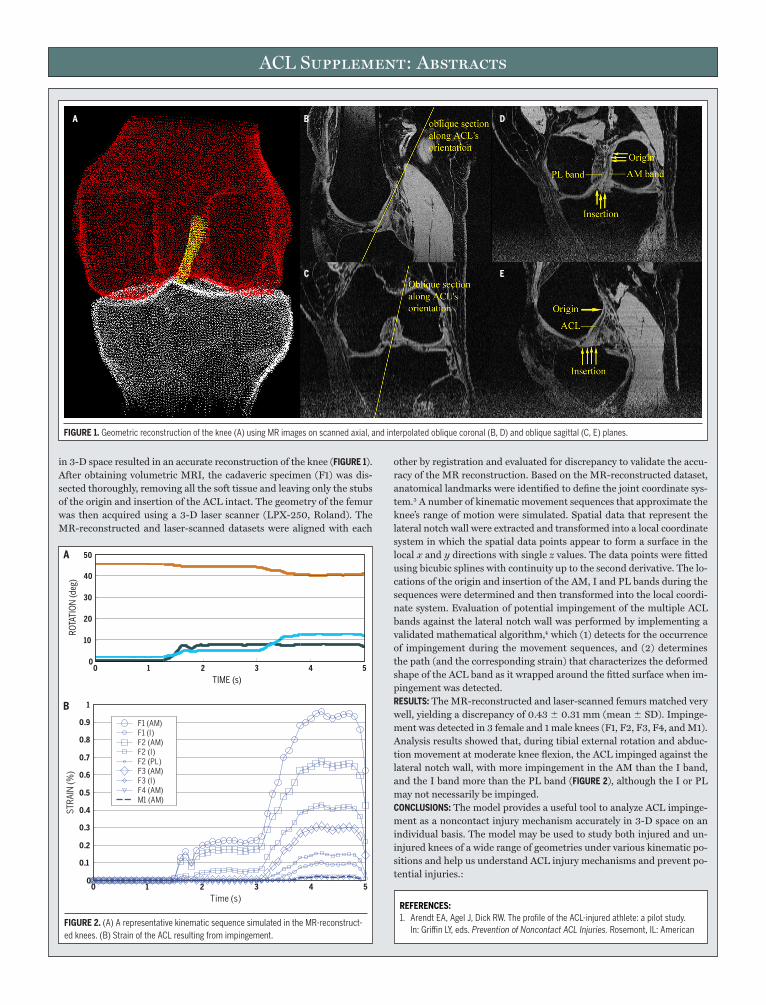
ACL Supplement: Abstracts
in 3-D space resulted in an accurate reconstruction of the knee (FIGuRE 1). After obtaining volumetric MRI, the cadaveric specimen (F1) was dis-sected thoroughly, removing all the soft tissue and leaving only the stubs of the origin and insertion of the ACL intact. The geometry of the femur was then acquired using a 3-D laser scanner (LPX-250, Roland). The MR-reconstructed and laser-scanned datasets were aligned with each
other by registration and evaluated for discrepancy to validate the accu-racy of the MR reconstruction. Based on the MR-reconstructed dataset, anatomical landmarks were identified to define the joint coordinate sys-tem.3 A number of kinematic movement sequences that approximate the knee’s range of motion were simulated. Spatial data that represent the lateral notch wall were extracted and transformed into a local coordinate system in which the spatial data points appear to form a surface in the local x and y directions with single z values. The data points were fitted using bicubic splines with continuity up to the second derivative. The lo-cations of the origin and insertion of the AM, I and PL bands during the sequences were determined and then transformed into the local coordi-nate system. Evaluation of potential impingement of the multiple ACL bands against the lateral notch wall was performed by implementing a validated mathematical algorithm,4 which (1) detects for the occurrence of impingement during the movement sequences, and (2) determines the path (and the corresponding strain) that characterizes the deformed shape of the ACL band as it wrapped around the fitted surface when im-pingement was detected.RESuLTS: The MR-reconstructed and laser-scanned femurs matched very well, yielding a discrepancy of 0.43 0.31 mm (mean SD). Impinge-ment was detected in 3 female and 1 male knees (F1, F2, F3, F4, and M1). Analysis results showed that, during tibial external rotation and abduc-tion movement at moderate knee flexion, the ACL impinged against the lateral notch wall, with more impingement in the AM than the I band, and the I band more than the PL band (FIGuRE 2), although the I or PL may not necessarily be impinged.CONCLuSIONS: The model provides a useful tool to analyze ACL impinge-ment as a noncontact injury mechanism accurately in 3-D space on an individual basis. The model may be used to study both injured and un-injured knees of a wide range of geometries under various kinematic po-sitions and help us understand ACL injury mechanisms and prevent po-tential injuries.:
FLEX ABD. ER
FIGuRE 1.Geometricreconstructionoftheknee(A)usingMRimagesonscannedaxial,andinterpolatedobliquecoronal(B,D)andobliquesagittal(C,E)planes.
A B D
C E
TIME (s)
ROTA
TIO
N (d
eg)
0 1 2 3 4 50
10
20
30
40
50
FIGuRE 2.(A)ArepresentativekinematicsequencesimulatedintheMR-reconstruct-edknees.(B)StrainoftheACLresultingfromimpingement.
0 1 2 3 4 50
0.1
0.2
0.3
0.4
0.5
0.6
0.7
0.8
0.9
1
Time (s)
STRA
IN (%
)
F1 (AM)F1 (I)F2 (AM)F2 (I)F2 (PL)F3 (AM)F3 (I)F4 (AM)M1 (AM)
A
B
REFERENCES:1. ArendtEA,AgelJ,DickRW.TheprofileoftheACL-injuredathlete:apilotstudy.
In:GriffinLY,eds.Prevention of Noncontact ACL Injuries.Rosemont,IL:American

ACL Supplement: Abstracts
uLTRASTRuCTuRE, SIZE, AND MATERIAL PROPERTIES OF ThE huMAN ANTERIOR CRuCIATE LIGAMENT: A SEX-BASED ASSESSMENTHashemi J, Chandrashekar N, Mansouri H, Hardy D, Slauter-beck JR Texas Tech University and Health Sciences Center, Lubbock, TX; University of Vermont, Burlington, VTINTRODuCTION: Despite intense focus and numerous research initiatives over the past decade, the sex-based disparity in the rate of anterior cru-ciate ligament (ACL) injuries has not diminished, as reported by Agel et al (Agel et al, 2005). This recent report also indicates that, none of the existing theories completely explains the disparity nor offers a so-lution. Clearly, before a solution may be offered, the cause or causes for the disparity should be identified; the authors propose that a root cause (central factor) in conjunction with neuromuscular and hormonal the-ories, feeds the existing sex-based disparity. This root cause is centered around the differences in the geometry and properties of the ACL based on sex. Several factors regarding sex differences in the performance of the ACL have been recently reported: (i) female ACL is smaller than the male ACL (Chandrashekar et al, 2005) even after adjustments made due to body anthropometry; (ii) smaller ACLs are in fact weaker than larg-er ACLs, when body anthropometric measurements are considered in the analysis; (iii) if one considers male and female ACLs of essentially the same size, in our recent study (Chandrashekar et al, in press), it was shown that the female ACL has a lower mechanical quality (a measure of mechanical properties) when compared to male ACL. What could be the cause for this difference in properties? There is no evidence in the liter-ature of a sex-based difference in the ultrastructural parameters of the ACL. Any such difference could point to differences in the remodeling re-sponse of the ACL. In this study, we hypothesize that female ACL has a lower collagen fibril density, lower percentage of area occupied by fibrils and lower mean diameter than male ACL after adjustments have been made to age and donor anthropometry.METhODS: Twelve ACL (6 male and 6 female) which were a subset of those ACLs used for the study of mechanical properties, were randomly select-ed to study the sex based differences in their ultrastructure. The ACLs were dissected from fresh-frozen knees. The mean ages of male and fe-male donors were 34.5 and 36.5 years, respectively. The anteromedial and posterolateral bundles of the ACL were separated. Small pieces (3 mm in length and 1 mm in width and thickness) of the ACL from the proximal, distal, and middle portions were removed from each bundle and resulted in a minimum of 18 micrographs for each ACL. These mi-crographs were then scanned using a negative scanner. The images were analyzed using ImageJ software provided by NIH. The mean fibril di-ameter, number of fibrils/square micron area and the percent cross-sec-tional area occupied by fibrils were then calculated using all 18 micro-graphs but omitting those regions of unusual absence of fibrils. These ultrastructural parameters were compared between sexes using multi-variate regression analysis considering the donor age and donor anthro-pometry as covariates. The level of statistical significance (alpha) was set to 0.05 a priori. RESuLTS: There was no evidence of any sex based differences in the aver-age fibril diameter and fibrils/square microns between sexes. We found that male compare to female ACL has a higher percentage of collagen
fibrils occupying a defined cross-sectional area (male 45.2%, female 40.6%, P = .049). DISCuSSION: Since collagen fibrils are primary load bearing members of the ACL, higher collagen content can manifest itself in a tissue of high-er mechanical quality. This is consistent with our earlier result showing lower mechanical quality of the female ACL. The total number of fibrils is generally associated with the stiffness of the ligaments. Accordingly, higher number of collagen fibrils per square area relates with the mod-ulus of elasticity of the ligament. Higher modulus of elasticity in male ACLs (Chandrashekar et al, 2006) might not be due to higher fibril den-sity in males. CONCLuSION: Our findings regarding sex differences in size, properties, and ultrastructure, collectively, offer a root cause in part describing the existing disparity in ACL failure. This could explain how some of the dif-ferences in neuromuscular response between athletes could have an im-pact on the ACL failure rate. Female ACL has lower percentage collagen content. This might be a reason for higher incidence of ACL injuries in females. Such ultrastructural differences might show possible remodel-ing differences between the sexes. A study with higher sample size can provide more accurate results.
ACuTE EFFECTS OF STEROID SEX hORMONES ON TYPE 1 COLLAGEN MRNA EXPRESSIONRomani WA University of Maryland School of Medicine, Dept. of Physical Therapy and Rehabilitation ScienceINTRODuCTION: Type 1 alpha-1 (T1C) and Type 3 collagen are crucial struc-tural components within the extracellular matrix of the ACL and pro-vide the tensile and elastic strength of the ligament. Estradiol (E2) has been shown to reduce in vitro collagen proliferation and in-vivo load to failure in the rabbit ACL, but studies on other animal models and on the differences in ACL injury rate throughout a normal menstrual cycle have been inconclusive. We have identified relationships between knee stiff-ness and E2 concentrations near ovulation but no significant changes be-tween menstrual cycle stages. To date, most of the attention on the role of sex hormones and ACL injuries has focused on the female sex hormones, E2 and progesterone. Since the male sex hormone, testosterone (T) plays a key role in the aromatization of estrogen and collagen proliferation in other connective tissues it is possible that T may also influence collagen remodeling and the strength of the ACL. The purpose of this study was to determine the acute influence of male and female steroid sex hormones on the expression of a key component of ligament strength, type 1 colla-gen. We hypothesized that T1C mRNA expression would be lower in ani-mals injected with E2 and higher in animals injected with T as compared to gonadectomized controls.METhODS: Two groups of male (n = 7, 7) and 2 groups of female (n = 6, 7) 12-week-old Sprague-Dawley rats were gonadectomized. After a 15 day hormone clearance period the female rats were subcutaneously inject-ed on 2 consecutive days at 8 am with 2.5 ug of E2 dissolved in 1 ml of sesame oil or with sesame oil alone. Male rats were injected on the same schedule with the sesame oil vehicle or 2 mg of dissolved Testosterone Propionate. Rats were euthanized 6 hours after the second injection and
AcademyofOrthopaedicSurgeons;2001.2. AmisAA,DawkinsGP.Functionalanatomyoftheanteriorcruciateligament.Fibre
bundleactionsrelatedtoligamentreplacementsandinjuries.J Bone Joint Surg Br.1991;73:260-267.
3. GroodES,SuntayWJ.Ajointcoordinatesystemfortheclinicaldescrip-tionofthree-dimensionalmotions:applicationtotheknee.J Biomech Eng.1983;105:136-144.
4. FungDT,ZhangLQ.ModelingofACLimpingementagainsttheintercondylarnotch.Clin Biomech (Bristol, Avon).2003;18:933-941.
REFERENCES:1. AgelJ,ArendtEA,BershadskyB.Anteriorcruciateligamentinjuryinnationalcol-
legiateathleticassociationbasketballandsoccer:a13-yearreview.Am J Sports Med.2005;33:524-530.
2. ChandrashekarN,SlauterbeckJ,HashemiJ.Sex-baseddifferencesinthean-thropometriccharacteristicsoftheanteriorcruciateligamentanditsrela-tiontointercondylarnotchgeometry:acadavericstudy.Am J Sports Med.2005;33:1492-1498.
3. ChandrashekarN,MansouriH,SlauterbeckJ,HashemiJ.Sex-baseddifferenc-esinthetensilepropertiesofthehumananteriorcruciateligament.J Biomech.2006;39:2943-2950.
ACL Supplement: Abstracts
a32
tissue was stored in RNAlater at –20°C until homogenized and isolat-ed. Reverse transcriptase generation of cDNA was performed and qPCR protocol to amplify T1C as per our lab’s protocol. Quantitative analysis was carried out with an MJ SYBR green kit and an MJ Chromo 4 Real Time Detector. Beta-Actin was used as an internal control. Differences in mRNA expression were determined based on the relative difference (or fold change) in cycle number (DCt) necessary to achieve the same level of fluorescence as the reference control. Two separate paired t tests were used to determine differences in mRNA expression between hor-mone treated and control groups. Significance was set at P,.05RESuLTS: As expected male rats treated with TP had higher mRNA ex-pression than male controls (P = .025). Similarly, female rats treated with TP had higher trends for mRNA expression than female controls (P = .088).DISCuSSION AND CONCLuSIONS: These results indicate that steroid sex hor-mones acutely influenced the expression of T1C mRNA in gonadecto-mized male and female rats. Although T1C likely plays a role in the ten-sile strength of the ACL, changes in the expression of mRNA do not necessarily indicate a change in protein content or strength of the ACL. In fact, studies in our and other laboratories have indicated that chang-es in sex hormone concentrations do not acutely influence the strength of the ACL. However, cyclic changes in hormone concentrations may ini-tiate a process of collagen production or turnover that over a longer pe-riod of time, may influence the strength of the ACL. Further studies are needed to determine the long term influence of steroid sex hormones on other markers for ligament strength and metabolism such as type 3 col-lagen and matrix metalloproteinases.
ThE EFFECT OF SEX hORMONES ON ANTERIOR TIBIAL DISPLACEMENT AND ANTERIOR TIBIAL ShEAR IN NORMAL OVuLATING WOMEN AND ORAL CONTRACEPTIVE uSERSVerscheure S, Osternig L, Drayer B, Fuller L University of Oregon, Eugene, OR; Loyola Marymount, Los Angeles, CA; National College of Naturopathic Medicine, Portland, ORINTRODuCTION: The purpose of this study was to determine the effect of fluc-tuating hormone levels on anterior tibial displacement (ATD) and anterior tibial shear (ATS) in both ovulating women and oral contraceptive users.METhODS: Eighteen uninjured, ovulating women (NORM) and 20 oral
contraceptive (OC) users participated. The NORM subjects were test-ed for ATD, ATS, estrogen, and progesterone at 3 time periods during 1 complete menstrual cycle including: (1) the immediate onset of menses (M1), (2) the estimated occurrence of estrogen peak (E), and (3) the im-mediate onset of the next menses (M2). OC subjects were tested after in-gesting estrogen supplements for 7 days (E), and when no supplement was taken (M1, M2). ATD was produced by instrumented forces (KT-2000) and ATS was recorded on a closed-chain dynamometer. Hormone levels were assessed for each test time in the NORM group. ANOVAs were used to identify significant differences in ATD and ATS (P,.05), if any, between test periods, groups, and conditions. Post hoc analysis in-cluded the identification of the predominant pattern of change of ATD and ATS across test time for each group.RESuLTS: No significant differences in ATD or ATS were found, howev-er, the effect of test time on normalized ATD approached significance (P,.08). The predominant pattern of ATD and ATS change across time period in the NORM group (7 of 18 subjects) was an increase from M1 to E and a decrease from E to M2. The predominant pattern of ATD (6 of 20 subjects) and ATS (9 of 20 subjects) change across test time observed for the OC group was a decrease from M2 to E and an increase from E to M2, the opposite of the NORM group.DISCuSSION: Peak blood estrogen levels occurring during ovulation may be linked to an increase in ATD and ATS in some ovulating women, and in-gested synthetic hormones may be linked to a decrease in ATD and ATS in some OC users. Changes in ATD may be due to a temporary effect of estrogen or synthetic progestin on ACL tissue in some women, and changes in ATS may be due to a transient effect of natural or synthetic hormones on quadriceps activation in some women.CONCLuSIONS: These data may indicate an effect of fluctuating hormone levels on anterior tibial displacement (ATD) and anterior tibial shear (ATS) in some ovulating women and oral contraceptive users, but these results were only noted when the patterns of change in the dependent variables (across time) for individual subjects were considered. Note: Portions of these data have been presented at the following conferenc-es: International Society of Biomechanics, Dunedin, New Zealand, July 2003; Northwest American College of Sports Medicine - Missoula, Mon-tana, April 2003; Canadian Athletic Therapists’ Association, Victoria, British Columbia, May 2003 and 2005.
libuser
Typewritten Text
Post-print standardized by MSL Academic Endeavors, the imprint of the Michael Schwartz Library at Cleveland State University, 2014