Achalasia - IAGHiagh.org/Portals/44fa7561-56f7-47e4-a228-477ca071e439/Monthly... · Achalasia The...
39
Achalasia Prof. Javad Mikaeli
Transcript of Achalasia - IAGHiagh.org/Portals/44fa7561-56f7-47e4-a228-477ca071e439/Monthly... · Achalasia The...

Achalasia
Prof. Javad Mikaeli

Achalasia
The most recognized motor disorder .
The cause of achalasia remains unknown .
The most important pathophysiologic defect:Reduction of NANC inhibitory ganglionic cells.

Achalasia
l An annual incidence is approximately 1.6 case per 100,000.
l Men and women are affected with equal frequency.
l Achalasia is typically diagnosed in patients who are between the ages of 25 and 60 years.

Demographic Features in 700 Achalasia Patients in Iran
l 380 patients (54.3%) were male.l 320 patients (45.7%) were female.
l The mean age of our patients was 38 years.l Approximately 60% of patients (420) were
between the ages of 21-50 years.

l The observations that achalasia is associated with HLA-DQw1 and that affected patients often have circulating antibodies to myenteric neurons suggest that achalasia may be an autoimmune disorder.
Achalasia (etiology)

Achalasia- Etiology
l Initial insult may be a viral infection or other environmental factor, results in myenteric plexus inflammation.
l The inflammation then leads to an autoimmune response in a genetically susceptible population.
l Subsequently, chronic inflammation leads to destruction of the inhibitory myenteric ganglion cells.

Achalasia- Etiology
l Cytotoxic T cells, isolated from the LES of achalasia patients, respond to human herpes virus-1 with γ –IFN and IL-2 production and clonal proliferation.
l In addition, HSV-1 DNA was demonstrated in the vast majority of patients, but also in controls.
Achalasia- Etiology
l These data suggest that a latent infection with HSV-1 leads to persistent immune activation and destruction of esophageal neurons.
l Achalasia may results from chronic infections with herpes zoster or measlesviruses but data have been inconclusive.

Achalasia- Etiology
l This inflammatory degeneration involves the nitric oxide-producing, inhibitory neurons that affect the relaxation of the LES.
l The cholinergic neurons that cause smooth muscle contraction are relatively spared.
l In the esophageal body, the loss of inhibitory neurons results in aperistalsis.
Achalasia
l An association with adrenal insufficiency and alacrima (triple A syndrome or Allgrove syndrome) has been reported in children with achalasia.
l In our study among 25 children with achalasia (4.5 months-14 years) there were 5 cases (20%) of triple A syndrome and double A syndrome (Achalasia and Alacrimia) (3 cases with Double A and 2 cases with Triple A). They were sisters and brothers from two families.

Achalasia
l It is possible that a variant of double A syndrome is present in adults since in one report, 4 of 20 (20 percent) of adult patients with achalasia were found to have deficient tear production.
Achalasia(Diagnosis)
1- Clinical history2- B. swallow (timed esophagogram)3- Endoscopy4- Manometry

Achalasia- Manometry
l Although clinical and radiographic findings may strongly suggest the diagnosis of achalasia, a manometric examination is suggested for confirmation in virtually all cases.

ACHALASIA
l If the clinical and radiographic features are
typical, the manometry can be deferred
and endoscopy can be performed for diagnostic
and therapeutic purposes.

l The symptoms of achalasia often are insidious in onset and gradual in progression.
l In one series of 87 consecutive patients with newly diagnosed achalasia, the mean duration of symptoms was 4.7 years.
l The delay in diagnosis was due to misinterpretation of symptoms by physicians and sometimes atypical clinical manifestations.
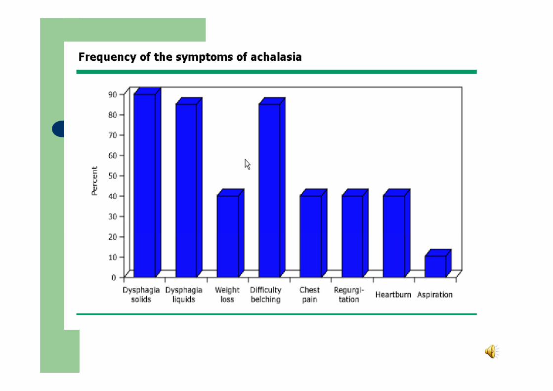
Achalasia (Symptoms)

Achalasia (Symptoms)
l Dysphagia (solids) 99.4%l Dysphagia (liquids) 93.1%l Active regurgitation 83.9%l Passive regurgitation 67.7%l Weight loss 61.4%l Chest pain 58.6%l Respiratory symptoms 45.8%

l Although dysphagia for liquids can occur in patients with other esophageal motility disorders (eg, progressive systemic sclerosis), this symptom is most characteristic of achalasia and strongly suggests the diagnosis.
Achalasia (Symptoms)

Aggravating and Alleviating factors of dysphagia
Water drinking 59%
Back straightening 48%
Standing &walking 24%
Valsalva manuever 7%
Massage on sternum 7%
Deep breathing 6%
Alleviating factors
Stress during eating 73%
Hurried eating 63%
Aggravaiting factors

l Weight loss, regurgitation, chest pain, and heartburn occur in approximately 40 to 60 percent of patients.
l The degree of weight loss is usually in the range of 5 to 10 kg. Profound weight loss can also be seen.
l Chest pain is more common in younger patients, and tends to diminish over several years.
Achalasia (Symptoms)

Achalasia (Symptoms)
l Chest pain is more common in younger patients, does not completely improve after pneumatic dilatation and tends to diminish over several years.
l In our study Chest pain was the only symptom which was significantly more complained by women than men (70.9% vs. 54.5% P value= 0.03).

Erroneous Diagnosis of Gastroesophageal Reflux Disease in
Achalasia
Department of Gastroenterology and Hepatology, Academic Medical Center Amsterdam, Amsterdam, The NetherlandsCLINICAL GASTROENTEROLOGY AND HEPATOLOGY 2011;9:1020–1024
l Heartburn occurs frequently in achalasia (38%–75%) and patients with heartburn have lower LES pressures than those without it.
l Heartburn may also result from other causes like direct irritation of the esophageal lining by food,pills, or lactate production by bacterial fermentation of retained carbohydrate.
.
Erroneous Diagnosis of …

l Furthermore, heartburn may disappear with the onset of dysphagia, suggesting that achalasia developed in patients with underlying GERD.
l Heartburn and regurgitation are typical symptoms of GERD, and dysphagia is not only encountered in achalasia but is also frequently reported by GERD patients.
Erroneous Diagnosis of …

l Therefore, achalasia can present with symptoms that potentially suggest GERD.
l Nearly all of achalasia patients are receiving PPIs for the initial diagnosis of GERD by the time they are referred to tertiary centers.
Erroneous Diagnosis of …

l Pathologic acid exposure times during pH measurement have been described in 20% of achalasia patients.
l The AGA states that manometry should be used to evaluate patients with suspected GERD who have not responded to an empiric trial of twice-daily PPI therapy and have normal findings on endoscopy.
Erroneous Diagnosis of …

UES dysfunction in achalasia
l In normal subjects, esophageal air injection causes UES relaxation that is accompanied by an audible belch.
l In achalasia, however, air injection frequently causes a paradoxical increase in UES pressure without a belch.

l This abnormal reflex presumably results from the loss of inhibitory neurons.
l The inability to burp may contribute to the esophageal distention and chest pain.
UES dysfunction

l The patient was given 50 mg prednisolone once daily; the symptoms improved dramatically, and HRIM showed complete recovery of manometric abnormalities.
l It is therefore important to control the inflammatory process in patients with idiopathic achalasia, which is likely to result from an autoimmune reaction.
l CLINICAL GASTROENTEROLOGY AND HEPATOLOGY 2011;9:1104–1106
Achalasia With Dense Eosinophilic Infiltrate Responds to Steroid Therapy (case report)

Pseudoachalasia (due to malignancy)
l Duration of symptoms less than six months l Presentation after age 60l Excessive weight loss in relation to the duration of
symptomsl Difficult passage of the endoscope through the
gastroesophageal junctionl These are generally much less common than
idiopathic achalasia, except for Chagas' disease in endemic areas in Central and South America.
Pseudoachalasia (due to malignancy)
l Malignancy — Malignancy is the most common cause of pseudoachalasia.
l Malignancy can cause pseudoachalasia either by invading the esophageal neural plexuses directly, or as a part of paraneoplastic syndrome.
l In addition to gastric carcinoma, other tumors which can produce the syndrome include cancer of the esophagus, carcinoma of the lung, lymphoma and pancreatic carcinoma.
Discrimination of achalasia from pseudoachalasia.
l Esophageal manometry does not help. l CT scan may suggest malignancy if there is marked
(greater than 10 mm) and/or asymmetric esophageal wall thickening.
l Endoscopy with biopsy is not always diagnostic because the tumors are often infiltrative and do not extend through the mucosa to the lumen.
l Repeated evaluations and biopsies is recommended

l EUS may show thickening of the longitudinal and circular smooth muscle layers of the LES, but this finding is not sufficiently specific.
l EUS is useful for characterizing tumors of the distal esophagus and cardia, and is recommended when pseudoachalasia is suspected.
l The accuracy of EUS for distinguishing achalasia from pseudoachalasia is not yet established.
Endoscopic ultrasonography (EUS) in achalasia

l Patients with achalasia are generally thought to be at increased risk for developing squamous cell carcinoma, although studies have also shown an increased risk of esophageal adenocarcinoma.
l Over the long-term, the risk was increased 16-fold compared with population controls.
Achalasia and Esophageal Cancer

Achalasia and Esophageal Cancer
l Cancer was diagnosed an average of 14 years after the diagnosis of achalasia.
l Some series note no increase in risk, particularly with early treatment of achalasia.
l We do not feel that the risk is high enough to warrant regular endoscopic surveillance.

l A study from the Netherlands included 448 patients with achalasia with a median follow-up of 9.6 years.
l Esophageal cancer developed in 15 patients (3.3 percent) after a mean symptom duration of 13 years (hazard ratio 28 compared with an age- and sex-matched cohort).
Achalasia and Esophageal Cancer

l A guideline issued by the ASGE suggests that there are insufficient data to support routine endoscopic surveillance for patients with achalasia.
l If surveillance were to be considered it would be reasonable to initiate it 15 years after onset of symptoms, but the subsequent surveillance interval is not defined.
Achalasia and Esophageal Cancer