ACG Clinical Guideline: Epidemiology, Risk Factors ... · 18 nature publishing group The American...
27
nature publishing group 18 The American Journal of GASTROENTEROLOGY VOLUME 110 | JANUARY 2015 www.amjgastro.com PRACTICE GUIDELINES INTRODUCTION is clinical guideline was designed to address colon ischemia (CI) including its definition, epidemiology, risk factors, presenta- tions, methods of diagnosis, and therapeutic interventions. Each section of the document will present key recommendations or summary statements followed by a comprehensive summary of supporting evidence. An overall summary of all recommenda- tions is listed in Table 1. A search of MEDLINE (1946 to present) and EMBASE (1980 to present) with language restriction to English was conducted using the search terms ischemic colitis, ischaemic colitis, colon ischemia, colonic ischemia, colon ischaemia, colonic ischaemia, colon gang- rene, colonic gangrene, colon infarction, colonic infarction, rectal ischemia, rectal ischaemia, ischemic proctitis, ischaemic proctitis, cecal ischemia, cecal ischaemia, ischemic colon stricture, ischae- mic colon stricture, ischemic colonic stricture, ischaemic colonic stricture, ischemic megacolon, ischaemic megacolon, colon cast, and colonic cast. e references obtained were reviewed and the best studies were included as evidence for guideline statements or in the absence of quality evidence, expert opinion was offered. e GRADE system (Grading of Recommendations Assessment, Development, and Evaluation) was used to evaluate the quality of evidence and strength of recommendations (1,2). e level of evidence ranged from “high” (implying that further research was unlikely to change the authors’ confidence in the estimate of the effect) to “moderate” (further research would be likely to have an impact on the authors’ confidence in the estimate of effect) to “low” (further research would be expected to have an important impact on the authors’ confidence in the estimate of the effect and would be likely to change the estimate) to “very low” (any estimate of effect is very uncertain). e strength of a recommendation was graded as “strong” when the desirable effects of an intervention clearly outweighed the undesirable effects and as “conditional” when there was uncertainty about the tradeoffs between the desir- able and undesirable effects of an intervention. Of note, in this clinical guideline there are several sections focusing on factors associated with prognosis in CI. Because the GRADE system cur- rently is not designed to rate the quality of the literature for these topics, we have preceded each of these sections with “summary statements” that detail the most important concepts regarding each area, but without a GRADE rating. DEFINITION CI is the condition that results when blood flow to the colon is reduced to a level insufficient to maintain cellular metabolic func- tion. e end result of this process is that colonocytes become acidotic, dysfunctional, lose their integrity and, ultimately, die. Although the etymologic root of the word ischemia is from the Greek iskhaimos, meaning a “stopping of the blood,” we now know that blood flow need not stop but only diminish significantly to cause ischemic damage. Moreover, ischemia may be followed by reperfusion injury and, for relatively brief periods of ischemia, this combined injury may produce more damage than just reduc- tion of blood flow without reperfusion. e degree to which colonic blood flow must diminish before ischemia results varies with the acuteness of the event, the degree of preexisting vascular collateralization, and the length of time the low flow state persists. CI may manifest with reversible or irreversible damage. Revers- ible damage includes colopathy, i.e., subepithelial hemorrhage or edema, and colitis; colitis reflects an evolutionary stage in which the overlying mucosa ulcerates as the subepithelial edema and blood are resorbed. In reversible disease, such resorption occurs rather promptly, usually within 3 days. Ulcerations may persist for several months before resolving, although during this time, the patient usually is asymptomatic. Irreversible manifestations of ACG Clinical Guideline: Epidemiology, Risk Factors, Patterns of Presentation, Diagnosis, and Management of Colon Ischemia (CI) Lawrence J. Brandt, MD, MACG, AGAF, FASGE 1 , Paul Feuerstadt, MD, FACG 2 , George F. Longstreth, MD, FACG, AGAF 3 and Scott J. Boley, MD, FACS 4 Am J Gastroenterol 2015; 110:18–44; doi:10.1038/ajg.2014.395; published online 23 December 2014 1 Division of Gastroenterology, Montefiore Medical Center, Albert Einstein College of Medicine, Bronx, New York, USA; 2 Gastroenterology Center of Connecticut, Yale University School of Medicine, Hamden, Connecticut, USA; 3 Department of Gastroenterology, Kaiser Permanent Medical Care Program, San Diego, California, USA; 4 Division of Pediatric Surgery, Montefiore Medical Center, Albert Einstein College of Medicine, Bronx, New York, USA. Correspondence: Lawrence J. Brandt, MD, MACG, AGAF, FASGE, Division of Gastroenterology, Montefiore Medical Center, Albert Einstein College of Medicine, Bronx, New York 10467, USA. E-mail: lbrandt@montefiore.org Received 24 February 2014; accepted 7 November 2014 CME CME
Transcript of ACG Clinical Guideline: Epidemiology, Risk Factors ... · 18 nature publishing group The American...

nature publishing group18
The American Journal of GASTROENTEROLOGY VOLUME 110 | JANUARY 2015 www.amjgastro.com
PRACTICE GUIDELINES
INTRODUCTION
Th is clinical guideline was designed to address colon ischemia
(CI) including its defi nition, epidemiology, risk factors, presenta-
tions, methods of diagnosis, and therapeutic interventions. Each
section of the document will present key recommendations or
summary statements followed by a comprehensive summary of
supporting evidence. An overall summary of all recommenda-
tions is listed in Table 1 .
A search of MEDLINE (1946 to present) and EMBASE (1980 to
present) with language restriction to English was conducted using
the search terms ischemic colitis, ischaemic colitis, colon ischemia,
colonic ischemia, colon ischaemia, colonic ischaemia, colon gang-
rene, colonic gangrene, colon infarction, colonic infarction, rectal
ischemia, rectal ischaemia, ischemic proctitis, ischaemic proctitis,
cecal ischemia, cecal ischaemia, ischemic colon stricture, ischae-
mic colon stricture, ischemic colonic stricture, ischaemic colonic
stricture, ischemic megacolon, ischaemic megacolon, colon cast,
and colonic cast. Th e references obtained were reviewed and the
best studies were included as evidence for guideline statements or
in the absence of quality evidence, expert opinion was off ered.
Th e GRADE system (Grading of Recommendations Assessment,
Development, and Evaluation) was used to evaluate the quality
of evidence and strength of recommendations ( 1,2 ). Th e level of
evidence ranged from “high” (implying that further research was
unlikely to change the authors’ confi dence in the estimate of the
eff ect) to “moderate” (further research would be likely to have
an impact on the authors’ confi dence in the estimate of eff ect) to
“low” (further research would be expected to have an important
impact on the authors’ confi dence in the estimate of the eff ect and
would be likely to change the estimate) to “very low” (any estimate
of eff ect is very uncertain). Th e strength of a recommendation was
graded as “strong” when the desirable eff ects of an intervention
clearly outweighed the undesirable eff ects and as “conditional”
when there was uncertainty about the tradeoff s between the desir-
able and undesirable eff ects of an intervention. Of note, in this
clinical guideline there are several sections focusing on factors
associated with prognosis in CI. Because the GRADE system cur-
rently is not designed to rate the quality of the literature for these
topics, we have preceded each of these sections with “summary
statements” that detail the most important concepts regarding
each area, but without a GRADE rating.
DEFINITION
CI is the condition that results when blood fl ow to the colon is
reduced to a level insuffi cient to maintain cellular metabolic func-
tion. Th e end result of this process is that colonocytes become
acidotic, dysfunctional, lose their integrity and, ultimately, die.
Although the etymologic root of the word ischemia is from the
Greek iskhaimos , meaning a “stopping of the blood,” we now know
that blood fl ow need not stop but only diminish signifi cantly to
cause ischemic damage. Moreover, ischemia may be followed by
reperfusion injury and, for relatively brief periods of ischemia,
this combined injury may produce more damage than just reduc-
tion of blood fl ow without reperfusion. Th e degree to which
colonic blood fl ow must diminish before ischemia results varies
with the acuteness of the event, the degree of preexisting vascular
collateralization, and the length of time the low fl ow state persists.
CI may manifest with reversible or irreversible damage. Revers-
ible damage includes colopathy, i.e., subepithelial hemorrhage or
edema, and colitis; colitis refl ects an evolutionary stage in which
the overlying mucosa ulcerates as the subepithelial edema and
blood are resorbed. In reversible disease, such resorption occurs
rather promptly, usually within 3 days. Ulcerations may persist
for several months before resolving, although during this time,
the patient usually is asymptomatic. Irreversible manifestations of
ACG Clinical Guideline: Epidemiology, Risk Factors,
Patterns of Presentation, Diagnosis, and Management
of Colon Ischemia (CI)
Lawrence J. Brandt , MD, MACG, AGAF, FASGE 1 , Paul Feuerstadt , MD, FACG 2 , George F. Longstreth , MD, FACG, AGAF 3 and
Scott J. Boley , MD, FACS 4
Am J Gastroenterol 2015; 110:18–44; doi: 10.1038/ajg.2014.395 ; published online 23 December 2014
1 Division of Gastroenterology, Montefi ore Medical Center, Albert Einstein College of Medicine , Bronx , New York , USA ; 2 Gastroenterology Center of Connecticut,
Yale University School of Medicine , Hamden , Connecticut , USA ; 3 Department of Gastroenterology, Kaiser Permanent Medical Care Program , San Diego ,
California , USA ; 4 Division of Pediatric Surgery, Montefi ore Medical Center, Albert Einstein College of Medicine , Bronx , New York , USA . Correspondence:
Lawrence J. Brandt, MD, MACG, AGAF, FASGE, Division of Gastroenterology, Montefi ore Medical Center, Albert Einstein College of Medicine , Bronx , New York
10467 , USA . E-mail: lbrandt@montefi ore.org Received 24 February 2014 ; accepted 7 November 2014
CMECME

ACG Clinical Guideline
© 2015 by the American College of Gastroenterology The American Journal of GASTROENTEROLOGY
19
Table 1 . Recommendations and summary statements
Colon Ischemia Recommendations and Best Practice Summary Statements
Recommendation and Best Practice Statements
Clinical Presentation
1. The diagnosis of CI is usually established in the presence of symptoms including sudden cramping, mild, abdominal pain; an urgent desire to defecate;
and passage within 24 h of bright red or maroon blood or bloody diarrhea. (Strong recommendation, very low level of evidence) ( 7,9,17 )
2. A diagnosis of non-isolated right colon ischemia (non-IRCI) should be considered when patients present with hematochezia. (Strong recommendation,
very low level of evidence) ( 7,9,17 )
Imaging of CI
1. CT with intravenous and oral contrast should be the fi rst imaging modality of choice for patients with suspected CI to assess the distribution and phase
of colitis. (Strong recommendation, moderate level of evidence) ( 111–113 )
2. The diagnosis of CI can be suggested based on CT fi ndings (e.g., bowel wall thickening, edema, thumbprinting). (Strong recommendation, moderate
evidence) ( 111–113 )
3. Multiphasic CTA should be performed on any patient with suspected IRCI or in any patient in whom the possibility of AMI cannot be excluded. (Strong
recommendation, moderate level of evidence) ( 113,114 )
4. CT or MRI fi ndings of colonic pneumatosis and porto-mesenteric venous gas can be used to predict the presence of transmural colonic infarction.
(Strong recommendation, moderate level of evidence) ( 115 )
5. In a patient in whom the presentation of CI may be a heralding sign of AMI (e.g., IRCI, severe pain without bleeding, atrial fi brillation), and the
multiphasic CT is negative for vascular occlusive disease, traditional splanchnic angiography should be considered for further assessment. (Conditional
recommendation, low level of evidence) ( 114 )
Colonoscopy in the Diagnosis of CI
1. Early colonoscopy (within 48 h of presentation) should be performed in suspected CI to confi rm the diagnosis. (Strong recommendation, low level of
evidence) ( 17 )
2. When performing colonoscopy on a patient with suspected CI, the colon should be insuffl ated minimally. (Conditional recommendation, very low level of
evidence) ( 69,135 )
3. In patients with severe CI, CT should be used to evaluate the distribution of disease. Limited colonoscopy is appropriate to confi rm the nature of the CT
abnormality. Colonoscopy should be halted at the distalmost extent of the disease. (Strong recommendation, low level of evidence)
4. Biopsies of the colonic mucosa should be obtained except in cases of gangrene. (Strong recommendation, very low level of evidence)
5. Colonoscopy should not be performed in patients who have signs of acute peritonitis or evidence of irreversible ischemic damage (i.e., gangrene and
pneumatosis). (Strong recommendation, very low level of evidence)
Severity and Treatment of CI
1. Most cases of CI resolve spontaneously and do not require specifi c therapy. (Strong recommendation, low quality of evidence) ( 107,108,139 )
2. Surgical intervention should be considered in the presence of CI accompanied by hypotension, tachycardia, and abdominal pain without rectal
bleeding; for IRCI and pan-colonic CI; and in the presence of gangrene. (Strong recommendation, moderate level of evidence) ( 17,107,108 )
3. Antimicrobial therapy should be considered for patients with moderate or severe disease. (Strong recommendation, very low level of evidence)
( 107,108,140 )
Summary Statements (GRADE System not applicable)
Risk Factors
1. Comorbid cardiovascular disease and diabetes mellitus should increase consideration of CI in patients with typical clinical features ( 14,15,20 )
2. A history of IBS and constipation should be sought in patients suspected to have CI ( 8,13,15 )
3. Selective cardiology consultation is justifi ed in patients with CI, particularly if a cardiac source of embolism is suspected ( 134 )
4. Chronic kidney disease is associated with increased mortality from CI ( 7,24,25 )
5. Evaluation for thrombophilia should be considered in young patients with CI and all patients with recurrent CI ( 26–28 )
6. Surgical procedures in which the inferior mesenteric artery (IMA) has been sacrifi ced, such as abdominal aortic aneurysm repair and other abdominal
operations, should increase consideration of CI in patients with typical clinical features ( 14,29,30 )
7. In patients suspected of having CI, a history of medication and drug use is important, especially constipation-inducing medications, immunomodulators,
and illicit drugs ( 9,15,31 )
Clinical Presentation
1. IRCI is associated with higher mortality rates compared with other patterns of CI ( 7,17 )
Table 1 continued on followin page

Brandt et al.
The American Journal of GASTROENTEROLOGY VOLUME 110 | JANUARY 2015 www.amjgastro.com
20
especially great aft er age 69 years and that most patients <50 years
old and all patients <40 years old were women ( 9 ). Th ere seems
to be much less female predominance among young Japanese
patients ( 19 ).
Mortality rates in large series range from 4 to 12%, but inclu-
sion criteria, case ascertainment methods, and rates of comor-
bidity and surgery in these studies diff ered ( 7,9,10,17,18,20 ).
Recurrent CI increases over time; for example, estimated cumu-
lative recurrence rates at 1, 2–3, 4, and 5–6 years were 3%, 5%,
6%, and 10%, respectively, in one study ( 9 ) and 3.3% at 2 years
and 7.5% at 5 years in another study ( 10 ). Particular predispos-
ing illnesses have been reported with recurrent disease, such as
hypercoagulable states ( 21 ). Th erefore, in any large survey, recur-
rence will be related to the relative proportions of patients with
spontaneous, idiopathic disease and those with illnesses likely to
foster recurrence.
PATHOPHYSIOLOGY
CI can result from alterations in the systemic circulation or from
anatomic or functional changes in the mesenteric vasculature; the
proximate cause is thought to be local hypoperfusion and reperfu-
sion injury. In most cases, no specifi c cause for ischemia is identi-
fi ed, and such episodes are attributed to localized nonocclusive
ischemia, likely a result of small-vessel disease. Th ese patients are
sometimes classifi ed as having Type I disease. By contrast, in Type
II disease the etiology is identifi ed and most commonly follows
an episode of systemic hypotension, decreased cardiac output,
or aortic surgery ( 22 ). Th is classifi cation schema for CI is infre-
quently used in clinical settings, but in practice, patients with
Type II disease can have therapy targeted toward the underlying
cause, whereas Type I CI is treated in a broader and supportive
manner. An increasing variety of causes of CI is being defi ned (see
“Risk Factors” section).
Abnormalities seen on angiography rarely correlate with clinical
manifestations of CI, and age-related abnormalities in the splanch-
nic vessels are not uncommon, including narrowing of small
vessels, and tortuosity of the long colic arteries; fi bromuscular dys-
plasia of the superior rectal artery has been associated with CI. Th e
colon is particularly susceptible to ischemia, perhaps owing to its
relatively low blood fl ow, its unique decrease in blood fl ow dur-
ing periods of functional activity, and its sensitivity to autonomic
CI include gangrene, fulminant colitis, stricture formation, and,
rarely, chronic ischemic colitis. Recurrent sepsis due to bacterial
translocation is another rare manifestation of irreversibly dam-
aged bowel.
EPIDEMIOLOGY
Th e absence of a unique diagnosis code for acute large bowel
ischemia in the ICD-9-CM (International Classifi cation of Dis-
eases, 9th Revision, Clinical Modifi cation) challenges case fi nding
for research. Th is system, which is commonly used in the United
States, assigns the hospital discharge code 557.0 (acute vascular
insuffi ciency of intestine) and 557.9 (unspecifi ed vascular insuffi -
ciency of intestine) to ischemic colitis as well as many other small
and large bowel entities. Th is limitation persists in the newer ICD-
10-CM classifi cation system. Th erefore, either medical records
must be reviewed carefully or clear stipulations must be applied to
databases to reliably identify patients with CI ( 3 ).
CI, the term we prefer to ischemic colitis because some patients
do not have a documented infl ammatory phase of disease, is the
etiology in 9–24% of all patients hospitalized for acute lower gastro-
intestinal bleeding ( 4–6 ), ranking CI fi rst ( 5 ), second ( 4,7 ), or third
( 6 ) behind colorectal malignancy in large epidemiological surveys.
A national insurance claims-based survey of patients hospitalized
with CI revealed an annual incidence rate of 17.7 cases/100,000
( 8 ). In the population-based, record-review study of patients hos-
pitalized in the Kaiser San Diego Medical Care Program, the esti-
mated annual incidence was 15.6 patients/100,000 (women, 22.6;
men, 8.0) ( 9 ). Because of multiple admissions of some patients, the
hospitalization rate was 16.4/100,000 per year with 6% of episodes
developing aft er hospitalization for surgery or medical treatment
of another disease. A recently published population-based study
yielded an incidence of 16.3 cases/100,000 person-years with a
nearly four fold increase over 34 years ( 10 ).
Children with CI are only rarely reported ( 11,12 ), but CI occurs
in adults of all ages and increases with age, especially aft er age 49
years ( 8,9 ). An insurance claims-based study reported an inci-
dence of only 7.2 cases/100,000 person-years ( 13 ), although few
people of at least 60 years of age were surveyed, possibly explain-
ing this relatively low incidence. CI is more common in women
than in men, and 57–76% of patients in large series have been female
( 8–10,14–18 ). One survey found that female predominance was
Table 1 . Continued
Colon Ischemia Recommendations and Best Practice Summary Statements
Laboratory Tests in CI
1. Laboratory testing should be considered to help predict CI severity ( 17,94,107 )
2. Decreased hemoglobin levels, low serum albumin, and the presence of metabolic acidosis can be used to predict severity of CI ( 141,142 )
Severity and Treatment of CI
1. When considering mortality risk for patients undergoing surgical intervention for acute CI, the Ischemic Colitis Mortality Risk (ICMR) factors should be
utilized ( 141,142 )

ACG Clinical Guideline
© 2015 by the American College of Gastroenterology The American Journal of GASTROENTEROLOGY
21
stimulation. What triggers the episode of CI, however, usually is
not identifi ed.
RISK FACTORS
Summary statements
1 . Comorbid cardiovascular disease and diabetes mellitus
should increase consideration of CI in patients with typical
clinical features ( 10,14,15,20 ).
2 . A history of irritable bowel syndrome (IBS) and constipation
should be sought in patients suspected to have CI ( 8,13,15 ).
3 . Selective cardiology consultation is justifi ed in patients with CI,
particularly if a cardiac source of embolism is suspected ( 23 ).
4 . Chronic kidney disease and chronic obstructive pulmonary
disease are associated with increased mortality from CI
( 7,10,24,25 ).
5 . Evaluation for thrombophilia should be considered in young
patients with CI and in all patients with recurrent CI ( 26–28 ).
6 . Surgical procedures in which the inferior mesenteric artery
(IMA) has been sacrifi ced, such as abdominal aortic aneu-
rysm repair and other abdominal operations, should increase
consideration of CI in patients with typical clinical features
( 14,29,30 ).
7 . In patients suspected of having CI, a history of medication
and drug use should be sought, especially constipation-
inducing medications, immunomodulators, and illicit drugs
( 9,15,31 ).
Summary of evidence
Five large case–control studies of risk factors for CI examined
both associated medical conditions and drug use ( 10,14,15,20,32 ),
and three of them also assessed surgical history ( 10,14,15 ). Th e
multivariate analyses used in these studies considered potential
risk factors together rather than only individually to detect those
that imposed a risk independent of the infl uences of other varia-
bles. Comparisons of these studies thus must be viewed in light of
the variations in factors assessed and other diff erences in research
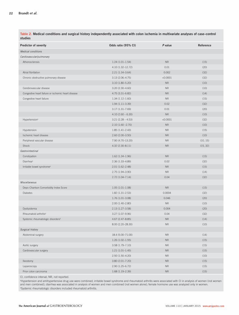
methods. Table 2 displays the entities for which the data seem
most rigorous in view of research methods and consistency of
fi ndings across studies. Study limitations are inclusion of relatively
few elderly patients ( 14 ), reliance on medical claims data without
comprehensive record review ( 14,15 ), reliance on recorded use of
drugs rather than billing or dispensing records ( 10 ), and uncertain
accuracy of diagnoses assessed from ICD-9-CM codes, especially
disorders identifi ed by symptoms only, such as diarrhea ( 32 ),
constipation ( 15 ), and IBS ( 14,15,32 ). Because some studies did
not exclude certain symptoms and entities coded during a short
period before the index date of CI, the authors could have erred
by attributing risk factor status to coded variables that mimicked
or shared acute features of CI, including bloating ( 15 ), dyspepsia
( 15 ), dysentery ( 15 ), rectal bleeding ( 14 ), IBS ( 14,15 ), nonspecifi c
colitis ( 14 ), and use of antidiarrheal drugs ( 14 ) and histamine type
2 receptor antagonists ( 15 ). Th ere are statistical limits on the anal-
ysis of potentially important drugs ( 33 ) that have low frequency of
use (e.g., chemotherapeutic agents or phentermine) or are absent
from controls ( 32 ). Moreover, all studies are limited in analyzing
some over-the-counter drugs, e.g., pseudoephedrine, some laxa-
tives, nonsteroidal anti-infl ammatory drugs, as well as illicit drugs
( 33 ), because pharmacy dispensing data do not comprehensively
capture use and patient history may be inaccurate. Ascertainment
bias also could result in increased detection of CI in patients with
a disorder such as IBS who likely undergo colonoscopy more
oft en than controls ( 34 ). Finally, it is important to understand that
statistical association does not equal causality.
Medical conditions . Cardiovascular and pulmonary risk factors
for CI are refl ected by the comorbidities reported in six series
totaling 1,955 patients ( 7,9,10,17,18,20 ): hypertension (57–72%),
diabetes mellitus (17–28%), coronary artery disease (18–37%),
dyslipidemia (18–33%), chronic obstructive pulmonary disease
(10–18%), congestive heart failure (9–16%), atrial fi brillation
(9–14%), peripheral vascular disease (8–21%), and renal disease
(4–18%). CI also has accompanied type IIIb aortic dissection
( 35 ). Coronary artery disease and atrial fi brillation were approxi-
mately twice as common in patients with isolated right-colon is-
chemia (IRCI) compared with other anatomic patterns of CI that
are generally less severe ( 7 ). Using electrocardiography, Holter
monitoring, and transthoracic echocardiography in patients with
CI, a French group found a “proven” potential cardiac source
of embolism in 35% of patients, primarily those with sustained
or paroxysmal atrial fi brillation ( 36 ). Although they recom-
mended doing all of these tests routinely, many of the patients
had abnormalities that could be detected by physical examina-
tion. Nonetheless, selective diagnostic evaluation and cardiology
consultation seems justifi ed. CI occurred within 3 days of acute
myocardial infarction in 0.13% of patients, and complications
and mortality were higher in patients with both diseases than in
those with either CI or myocardial infarction alone ( 37 ). Among
patients with severe hematochezia, patients with CI more oft en
had moderate or severe lung disease than did patients with other
colonic causes of hemorrhage ( 38 ), and chronic obstructive pul-
monary disease independently predicted mortality in series from
Montefi ore Medical Center in New York and the Mayo Clinic in
Rochester, Minnesota ( 7,10 ).
Hypertension and diabetes mellitus independently predicted CI
among patients with acute lower abdominal pain ( 39 ). Endothelial
dysfunction could contribute to the eff ects of hypertension ( 40 )
and diabetes ( 41 ) and has been off ered to explain the increased
risk of CI associated with rheumatic autoimmune diseases ( 42 ),
including rheumatoid arthritis ( 43 ), although association of hyper-
coagulable states with these diseases is another potential risk factor
( 21,43–45 ).
Interest in IBS as a potential risk factor for CI arose aft er alos-
etron hydrochloride, used to treat women with diarrhea-predom-
inant IBS, was withdrawn from the US market because of reports
of CI among alosetron users during the fi rst few months aft er its
release in 2000. In a medical claims-based study of subjects mainly
<60 years of age, Cole et al. ( 13 ) found the incidence of CI was 3.4
times greater in patients with IBS than those without IBS. In another
study, the relative risk for CI was 3.2 and 2.8 times higher for those
Brandt et al.
The American Journal of GASTROENTEROLOGY VOLUME 110 | JANUARY 2015 www.amjgastro.com
22
Table 2 . Medical conditions and surgical history independently associated with colon ischemia in multivariate analyses of case–control
studies
Predictor of severity Odds ratio (95% CI) P value Reference
Medical conditions
Cardiovascular/pulmonary
Atherosclerosis 1.24 (1.01–1.54) NR ( 15 )
4.10 (1.32–12.72) 0.01 ( 20 )
Atrial fi brillation 2.21 (1.34–3.64) 0.002 ( 32 )
Chronic obstructive pulmonary disease 3.13 (2.06–4.75) <0.0001 ( 32 )
3.10 (1.80–5.20) NR ( 10 )
Cerebrovascular disease 3.20 (2.30–4.60) NR ( 10 )
Congestive heart failure or ischemic heart disease 4.75 (3.31–6.82) NR ( 14 )
Congestive heart failure 1.34 (1.12–1.60) NR ( 15 )
1.94 (1.11–3.39) 0.02 ( 32 )
3.17 (1.31–7.69) 0.01 ( 20 )
4.10 (2.60 – 6.30) NR ( 10 )
Hypertension a 3.21 (2.28 – 4.53) <0.0001 ( 32 )
2.10 (1.60 –2.70) NR ( 10 )
Hypotension 1.85 (1.41–2.43) NR ( 15 )
Ischemic heart disease 2.60 (2.00–3.50) NR ( 10 )
Peripheral vascular disease 7.90 (4.70–13.20) NR ( 10, 15 )
Shock 4.32 (2.30–8.11) NR ( 15, 32 )
Gastrointestinal
Constipation 1.62 (1.34–1.96) NR ( 15 )
Diarrhea a 2.36 (1.33–4.89) 0.02 ( 32 )
Irritable bowel syndrome a 2.01 (1.62–2.48) NR ( 15 )
2.75 (1.94–3.90) NR ( 14 )
2.72 (1.04–7.14) 0.04 ( 32 )
Miscellaneous
Deyo–Charlson Comorbidity Index Score 1.05 (1.01–1.08) NR ( 15 )
Diabetes 1.82 (1.31–2.53) 0.0004 ( 32 )
1.76 (1.01–3.08) 0.046 ( 20 )
2.00 (1.40–2.80) NR ( 10 )
Dyslipidemia 2.13 (1.27–3.58) 0.004 ( 20 )
Rheumatoid arthritis a 3.27 (1.07–9.96) 0.04 ( 32 )
Systemic rheumatologic disorders b 4.67 (2.47–8.85) NR ( 14 )
8.00 (2.20–28.30) NR ( 10 )
Surgical history
Abdominal surgery 18.4 (5.00–71.00) NR ( 14 )
1.26 (1.02–1.55) NR ( 15 )
Aortic surgery 3.58 (1.79–7.10) NR ( 15 )
Cardiovascular surgery 1.21 (1.01–1.45) NR ( 15 )
2.50 (1.50–4.20) NR ( 10 )
Ileostomy 3.80 (2.01–7.20) NR ( 15 )
Laparoscopy 2.90 (1.25–6.72) NR ( 15 )
Prior colon carcinoma 1.68 (1.19–2.39) NR ( 15 )
CI, confi dence interval; NR, not reported.
a Hypertension and antihypertensive drug use were combined; irritable bowel syndrome and rheumatoid arthritis were associated with CI in analysis of women (not women
and men combined); diarrhea was associated in analysis of women and men combined (not women alone); female hormone use was analyzed only in women.
b Systemic rheumatologic disorders included rheumatoid arthritis.
ACG Clinical Guideline
© 2015 by the American College of Gastroenterology The American Journal of GASTROENTEROLOGY
23
with IBS and constipation, respectively, than for those who lacked
these disorders ( 8 ). Subsequently, four case–control studies applied
more extensive multivariate analysis to the assessment of IBS as a
risk factor for CI; three studies confi rmed it did convey increased
risk ( 14,15,32 ), including one study that found an association in
women only ( 32 ), and the fourth one found no statistically sig-
nifi cant risk ( 10 ). Excessive sympathetic activity in IBS ( 46 ) could
impair vasodilation in the mesenteric vessels, the most reactive vas-
cular bed in the body ( 47 ). A retrospective review found that physi-
cians recorded constipation immediately preceding the presenting
features of CI in only 7% of patients ( 9 ), but a prospective study
detected constipation during the 30 days before presentation in 25%
of cases ( 17 ). Constipation could mediate increased risk through
increased intracolonic pressure and reduced blood fl ow ( 48 ) as a
result of fecal impaction, and constipation preceding the symptoms
of CI could be a clinical clue for diff erential diagnosis. Diarrhea
was a risk factor in one study ( 32 ) but, as with constipation ( 15 ), CI
could have been a diagnosis applied to patients with IBS.
Renal disease has also been described as a risk factor for CI from
observational studies, but it has not been identifi ed as an inde-
pendent predictor in case–control studies. In one survey, absence
of dialysis dependency in controls prevented its inclusion in mul-
tivariate analysis ( 32 ). However, severe chronic kidney disease was
present in 11% of cases from Montefi ore Medical Center and was
nearly 3 times as common in patients with IRCI as other cases in
that series ( 7 ), similar to the fi ndings of Taiwanese ( 24 ) and Korean
( 49 ) studies. Th us, 7% of cases of CI reported by Paterno et al. ( 18 )
were dialysis dependent; 33% of the cases described by Flobert
et al. ( 25 ) required hemodialysis, and right-sided disease occurred
in 3 times as many patients on hemodialysis than in other patients.
Th e most obvious causative link, although unproven, in patients
on dialysis in whom CI (predominantly IRCI) develops is fl uctua-
tion in fl uid balance with a decrease in circulating blood volume.
Among patients with severe hematochezia, the mean serum cre-
atinine was twice as high in patients with CI as it was in those with
other colonic causes ( 38 ), and hemodialysis independently pre-
dicted CI in patients with acute lower abdominal pain ( 39 ).
Th rombophilia is another potential risk factor for CI. Th ere are
numerous case reports of various coagulopathies in patients with
CI, including defi ciencies of protein C, protein S, antithrombin III,
and factor V Leiden mutation ( 21,45,50 ). In particular, the “cata-
strophic” variant of the antiphospholipid syndrome causes mul-
tiple vascular occlusions, especially in small vessels, but typically
causes more widespread intestinal ischemia than CI alone ( 44 ).
Th rombophilic abnormalities were described in 72% of frozen
blood samples of ambulatory patients from Crete ( 26 ) and in 28%
of US patients’ blood drawn at least 1 month aft er the diagnosis
of CI ( 27 ). Furthermore, genetic polymorphisms associated with
thrombophilia and vascular hyperactivity were found in more
young patients with CI than in controls ( 28 ). Concerns about
attributing an etiologic role to these fi ndings include uncertainty
regarding whether the clotting abnormalities actually contributed
to CI or were epiphenomena of little signifi cance. For example, it is
diffi cult to reconcile a major etiologic role for coagulopathy in view
of the advanced age of many CI patients and the low recurrence
rate of CI. In one study, 14% of patients with CI had reduced levels
of free protein S ( 26 ) that can occur as a result of its binding to an
acute-phase reactant. 8% of healthy controls had reduced levels of
protein S, and 4% had reduced antithrombin, activated protein C
resistance, or a factor V Leiden mutation, raising concerns about
the assays or study population ( 51 ). At this time, routine testing for
a coagulation disorder in most patients with CI does not appear
justifi ed, although it seems reasonable in young patients with CI
and in patients with recurrent disease ( 21 ).
At least a dozen cases of CI have been reported following long-
distance running in runners who were 26-42 years of age, most of
whom were women. CI involved the right colon in 10 patients and
extended to the left side in 3 of them; in 2 patients CI was limited
to the left colon. Two patients underwent resection of the ischemic
segment, and both survived. Young age, a female predominance,
and lack of mortality contrast with usual series of IRCI ( 52–55 ).
Ischemia is a postulated cause of diarrhea, abdominal pain, hema-
tochezia, and occult bleeding in endurance athletes ( 55–57 ).
Sickle cell crisis with microvascular occlusion can also cause CI.
Of the 7 reported cases, patients were 5–35 years of age and the
distribution varied from sigmoid to pancolonic; 3 patients under-
went colon resection and 2 died ( 58–61 ). Two reported cases of
CI occurred in patients with sickle cell trait: a 44-year-old woman
with diastolic dysfunction survived two episodes of CI of undocu-
mented distribution ( 62 ), and a 52-year-old woman who also had
Sjogren’s disease survived sigmoid CI ( 63 ). Sickle cell disease is
only rarely mentioned as an etiology for CI, but the rate of this
disease causing CI could be higher in populations with a greater
proportion of patients who are of African ancestry ( 9 ).
CI has also rarely followed colonoscopy ( 64–68 ); such reports
include one patient who had other predisposing features ( 68 ).
Postcolonoscopy CI could result from reduced colonic blood fl ow
as a consequence of luminal distention and increased intraluminal
pressure, both of which are more pronounced with insuffl ation by
air than by carbon dioxide ( 69 ). Th ere have been various other
associations with CI including carbon monoxide poisoning ( 70 ),
pheochromocytoma ( 71 ), air embolism aft er deep-sea diving ( 72 ),
colon carcinoma ( 73 ), and, aft er airplane fl ights ( 74 ), a pit viper
bite ( 75 ), blunt trauma to the abdomen ( 76 ), and repetitive jack-
hammer trauma ( 77 ).
Surgical history . A survey of 89,967 admissions for abdominal
aortic aneurysm (AAA) repair revealed an overall rate of postop-
erative CI of 2.2%: with repair of ruptured AAA, 8.9%; with open
elective AAA repair, 1.9%; and with endovascular AAA repair,
0.5% ( 29 ). Routine postoperative sigmoidoscopy aft er AAA repair
reveals that mild CI occurs much more oft en than does sympto-
matic CI ( 30 ). Repair of AAA had preceded CI in only <1–2% of
all cases of CI in reported series ( 7,9,20 ). A major mechanism for
CI aft er these vascular procedures is sacrifi ce of the IMA. One
study identifi ed prior laparoscopy as a risk (with a wide confi -
dence interval) but did not specify the surgery performed (odds
ratio 17.67, 95% confi dence interval 1.91–163.57) ( 15 ). Ligation
of the IMA could also explain CI in patients who have undergone
surgery for colon carcinoma ( 78,79 ).

Brandt et al.
The American Journal of GASTROENTEROLOGY VOLUME 110 | JANUARY 2015 www.amjgastro.com
24
Table 3 . Drugs proposed to predispose to CI, estimate of evidence level, and postulated pathogenesis
Drug Evidence Postulated pathogenesis
Moderate evidence
Constipation-inducing
drugs
Predicted CI in patients with abdominal pain, 2.8 (1.1–7.1); ( 39 )
All drugs: C-CS; increased risk 0.68 (0.62–1.27); ( 14 )
Opioids: C-CS; increased risk 1.96 (1.43–2.67); ( 32 )
Nonopioids: C-CS; increased risk 1.75 (1.25–2.44); ( 32 )
Reduced blood fl ow, increased intraluminal
pressure ( 48 )
Immunomodulator drugs Antitumor necrosis factor-α inhibitors for rheumatoid arthritis from US FDA
AERS: 17 probable, 18 possible cases; median age, 62 years a ( 157 )
Type 1 interferon-α for hepatitis C: 13 probable, 4 possible cases; median age,
51 years a
11 other reported cases ( 158 )
Type 1 interferon-β for multiple sclerosis: 19 probable, 20 possible cases;
median age, 56 years
10 other reported cases of interferon for hepatitis C, 8 age <55 years ( 159 )
Cytokines affecting thrombogenesis ( 33 )
Illicit drugs Amphetamines: 5 reported cases; age 42–50 years ( 33,160 )
Cocaine: used by 19 of 97 (20%) CI patients at 2 inner-city hospitals; age 44–56
years; 37% right-sided and 16% small bowel disease; 26% mortality ( 31 )
Many other reported cases of multiple ischemic organs ( 33 )
Vasoconstriction, hypercoagulation, direct
endothelial injury ( 33 )
Low evidence
Antibiotics Antibiotic-associated colitis resembles CI, usually right-sided ( 33 )
C-CS: increased risk CI, 3.3 (2.19–4.96); ( 32 )
Altered gut microbiome, e.g.,
Klebsiella oxytoca ( 161 )
Appetite suppressants Bitter orange (resembles ephedra): 1 reported case ( 162 )
Hydroxycut : 1 probable case a ( 163 )
Ma huang (mainly ephedrine): 1 reported case ( 164 )
Phentermine : 2 reported cases (1 with fenfl uramine) ( 33,165 )
Xenadrine (bitter orange, ma huang, caffeine, salicin): 1 reported case ( 166 )
3 of 5 cases age <50 years
Vasoconstriction ( 33 )
Chemotherapeutic drugs R-CHOP : 1 reported case ( 167 )
Taxanes : 10 reported cases ( 33,168–170 )
Vinorelbine/cisplatin : 1 reported case ( 33 )
C-CS: increased use of taxanes or vinca alkaloids on univariate analysis ( 32 )
Direct epithelial toxicity, inhibited repair of
vascular injury ( 33 )
Decongestants Pseudoephedrine: 9 reported cases, 6 age <50 years ( 33,171 )
C-CS: risk unaffected 1.1 (0.3–3.9); ( 10 )
Phenylephrine: 1 reported case ( 172 )
Vasoconstriction ( 33 )
Diuretics C-CS: increased risk 1.6 (1.2–2.1); ( 10 ) Extracellular volume defi cit, lower peripheral
vascular resistance, vasoconstriction ( 33 )
Ergot alkaloids (often
combined with caffeine)
20 Reported cases ( 33,173 ) Vasoconstriction ( 33 )
Hormonal therapies Predominance of women among young patients, ( 9,174 ) common use of female
hormones by female patients ( 175 )
Female hormones : C-CS; increased risk 1.88 (1.30–2.73); ( 32 )
Oral contraceptives : C-CS; increased risk 1.05 (1.00–1.10); ( 15 )
risk unaffected 0.59 (0.28–1.33) ( 14 ); 0.7 (0.3–1.5) ( 10 )
Estrogen replacement : C-CS; risk unaffected 0.75 (0.67–1.19) ( 14 )
1.0 (0.7–1.5); ( 10 )
Hypercoagulability, endothelial injury ( 33 )
Laxatives Osmotic agents : 2 reported cases ( 33 )
Bisacodyl : 2 reported cases ( 33 )
Bisacodyl/polyethylene glycol : 1 reported case of 2 episodes ( 176 )
Lubiprostone : 1 reported case ( 177 )
All drugs: C-CS; increased risk 4.73 (3.71–6.02); ( 15 )
Increased motility or rapid intravascular
volume defi cit, reduced perfusion ( 33 )
Psychotropic drugs 6 Reported cases (2 with hypotension) ( 33,178,179 )
10 cases, clinical/pathological data incomplete ( 180 )
C-CS: increased risk 3.7 (1.3–11.0); ( 10 )
Hypotension, constipation
Serotoninergic drugs 5-Hydroxytryptamine 1 receptor agonists: 12 cases from US FDA AERS, 8 age
<50 years ( 33,181–183 )
C-CS: risk unaffected 2.3 (0.8–6.9); ( 10 )
5-hydroxytryptamine 3 receptor antagonist: 1 case of CI/1,000 patient-years of
use ( 35 )
5-hydroxytryptamine 4 partial agonist: 27 reported cases (drug withdrawn) ( 184 )
For 5-hydroxytryptamine 1 receptor agonists
vasoconstriction ( 33 ); for other agents
various factors ( 184 )
Table 3 continued on following page

ACG Clinical Guideline
© 2015 by the American College of Gastroenterology The American Journal of GASTROENTEROLOGY
25
Drug use . When drug-induced CI was reviewed in 2007, there
was evidence to propose various major classes of pharmaco-
logic agents as predisposing to CI ( 33 ). Additional publica-
tions have since provided more evidence for the pathogenic
role of certain drugs, taking into account reported single cases
and other publications, including case–control studies, We be-
lieve that constipation-inducing drugs, immunomodulators,
and illicit drugs are best supported as etiologic agents, but
there is some support for a role of many disparate drug classes
( Table 3 ).
Despite extensive observations on the above clinical factors
associated with CI, there are no specifi c identifi able risk factors for
CI in most patients, and some patients have multiple risk factors.
Th e heterogeneous risk factors for CI support its multifactorial
pathogenesis and indicate the importance of careful assessment of
the medical, surgical, and drug use history in every patient with
CI. Further research into the cause of CI and its risk factors is
needed.
CLINICAL PRESENTATION
Recommendations
1 . Th e diagnosis of CI is usually established because of symp-
toms including sudden cramping, mild, abdominal pain; an
urgent desire to defecate; and passage within 24 h of bright
red or maroon blood per rectum or bloody diarrhea ( 7,9,17 )
(strong recommendation, very low level of evidence).
2 . A diagnosis of non-IRCI should be considered when patients
present with hematochezia (strong recommendation, very
low level of evidence) ( 7,9,17 ).
Summary statement
1 . CI isolated to the right colon (IRCI) is associated with higher
mortality rates compared with other patterns of CI ( 7,17 ).
Summary of evidence
CI generally manifests with sudden cramping, mild, left lower
abdominal pain; an urgent desire to defe cate; and passage within
24 h of bright red or maroon blood per rectum or bloody diar-
rhea ( 7,9,80,81 ). Longstreth and Yao ( 9 ) reported a large ret-
rospective study of 401 patients admitted with CI in which
the most common symptoms of presentation were abdominal
pain (87%), rectal bleeding (84%), diarrhea (56%), and nau-
sea (30%). A separate analysis of 72 Canadian patients with
biopsy-proven CI showed that abdominal pain (77.8%), hema-
tochezia (58.3%), bloody diarrhea (34.7%), and nonbloody
diarrhea (19.4%) were among the most common presenting
features ( 80 ). Abdominal pain, urgent need to defecate, and
bloody diarrhea are the major features, and all three symptoms
occur in this temporal sequence in nearly one-half of cases ( 17 );
vomiting (30%), dizziness (10%), and syncope (6%) occur less
frequently ( 9 ). Pain typically precedes bleeding and is usually
mild to moderate, although robust systematic assessment of its
severity has not been performed to date ( 17 ). Abdominal ten-
derness is usually present over the involved segment of colon.
Notably, patients with IRCI more commonly have pain than
they do rectal bleeding; only 25–46% of patients with IRCI have
rectal bleeding, and physicians should entertain the diagnosis
of IRCI for patients with acute, severe abdominal pain who
lack hematochezia and/or diarrhea, especially if they have the
clinical scenarios associated with IRCI, such as dialysis, sepsis,
and hypotension or shock ( 9,16,17 ).
Th e duration of symptoms is diffi cult to quantify in patients with
CI given the oft en mild and benign course of disease. One study
from Switzerland looked at the time to presentation for a cohort
of 49 patients with biopsy-proven CI: 57.1% presented within 24 h
of symptom onset and 75.5% within 3 days of symptoms ( 81 ). In
more than half of the cases of CI, the disease is reversible. Symp-
toms of CI generally resolve within 2–3 days and the colon heals
Table 3 . Continued
Drug Evidence Postulated pathogenesis
Very low evidence
Digitalis 1 Reported case (poisoning) ( 33 )
Digoxin : C-CS; increased risk 3.6 (2.1–6.2); ( 10 ); (atrial fi brillation not ana-
lyzed); decreased risk 0.27 (0.083–0.86); ( 20 )
Vasoconstriction
Kayexalate 1 Reported case ( 185 )
44 cases of colon injury with incomplete pathological data, ( 186 )
Direct toxic effect, various nondrug factors
( 185,186 )
NO-Xplode 1 Reported case ( 187 ) Blood shunting to skeletal muscle, hypo-
perfusion, various non-drug factors ( 187 )
NSAIDs Reported cases not clearly distinguishable from NSAID-induced colopathy ( 33 )
C-CS: risk unaffected 0.9 (0.6–1.2); ( 10 ); 0.68 (0.62–1.27); ( 188 )
Inhibition of vasodilating prostaglandins,
vasoconstriction ( 33 )
Statins 2 Reported cases ( 33,189 ) None
Vasopressors 1 Reported case ( 33 ) Vasoconstriction
C-CS, case–control study (followed by odds ratio (95% confi dence interval)); CI, colon ischemia; NSAID, nonsteroidal anti-infl ammatory drug; R-CHOP, rituximab, cyclo-
phosphamide, vincristine, doxorubicine, prednisolone; US FDA AERS, United States Food and Drug Administration Adverse Event Reporting System.
a Classifi ed by the criteria of Naranjo et al. ( 163 ).

Brandt et al.
The American Journal of GASTROENTEROLOGY VOLUME 110 | JANUARY 2015 www.amjgastro.com
26
it was not seen in any patient with severe disease, because these
patients were operated on promptly or had died ( 86 ). Th e quoted
studies lack clear defi nitions of stricture, however, and these stric-
ture rates are likely overestimates. Symptomatic strictures are rare,
but further study is needed to defi ne this frequency compared with
that of silent stricture formation.
Acute severe CI can also mimic infl ammatory bowel disease
(IBD) with chronic ulcerations, crypt abscesses, and pseudopolyps
( 88 ). Th e most severe form of ischemic damage causes transmu-
ral infarction. Gangrenous colitis is characterized by increasing
abdominal tenderness, guarding, rebound tenderness, rising tem-
perature, and paralytic ileus. Abdominal pain is seen in the vast
majority of these patients (86.1%) but rectal bleeding is far less
frequent (30.6%); acute abdominal pain without rectal bleeding
(58.3%) and nonbloody diarrhea (27.8%) are the most common
clinical patterns of presentation ( 17 ).
Sudden onset of a toxic colitis with signs of peritonitis and a rap-
idly progressive course are typical of universal fulminant colitis, a
rare variant of CI. Th e classic sequential triad is seen infrequently
(11.1%) with this CI variant; symptoms of severe abdominal pain
(66.7%) and rectal bleeding (55.6%) with abdominal tenderness on
physical examination (85.9%) are the most characteristic presenta-
tion of universal fulminant colitis ( 17 ).
SEGMENTAL NATURE OF CI
Summary of evidence
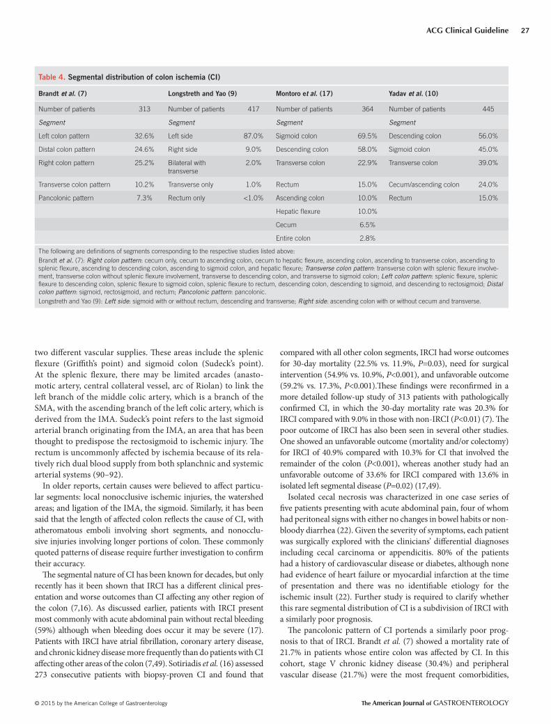
Th e left colon is most commonly aff ected, but no colonic region
is spared from involvement. In a large retrospective study of 313
patients, all of whom had their entire colon evaluated by colo-
noscopy, surgery, or autopsy—alone or in combination—and in
all of whom CI was proven by biopsy, a segmental pattern was
typical: the left colon was aff ected most oft en (32.6%), followed
by the distal colon (24.6%), right colon (25.2%), and entire colon
(7.3%) ( 7 ). In this study, although no specifi c etiology was associ-
ated with any specifi c anatomic distribution, pancolitis and IRCI
were seen frequently in patients with sepsis, and IRCI was asso-
ciated more frequently in patients with coronary artery disease
and chronic kidney disease on hemodialysis ( 7 ). Similar results
were found in large studies in California, Minnesota, and Spain
( Table 4 ) ( 9,10,17 ). Th is pattern of involvement appears to be
universal as a Korean cohort of 59 patients also showed a predom-
inance of left -sided disease (64.1%) over right-sided involvement
(35.9%) ( 49 ), although in this population, there were no diff er-
ences between right- and left -sided CI in clinical characteristic
presentations, cardiovascular risk factors, or the presence of
diabetes mellitus; patients with IRCI, however, were more likely to
have renal failure ( 49 ).
Th e segmental nature of CI can be explained by the vascular
anatomy of the colon and rectum. Colonic blood fl ow is sup-
plied by three vessels: the superior mesenteric artery (SMA),
IMA, and the superior hemorrhoidal artery. Vascular anatomy,
however, is variable and oft en individually unique ( 89 ). Water-
shed areas of the colon are regions that are particularly sus-
ceptible to ischemic insult as a result of their location between
in 1–2 weeks. With severe injury, it may take up to 6 months for
the colon to heal; however, during this time the patient is usually
asymptomatic.
Rectal bleeding is usually mild in CI, but in one retrospective
analysis of 550 patients presenting with severe hematochezia,
11.8% were found to have CI as the cause ( 38 ). Severe bleeding
was seen more frequently in women and in patients with severe
lung disease, elevated creatinine and glucose levels, and those
on anticoagulation. Th e 30-day outcomes for rebleeding, surgi-
cal intervention, and mean number of hospital days were worse
for those with CI compared with other etiologies of lower gas-
trointestinal bleeding ( 38 ). Severe hemorrhage occurs mainly in
patients with gangrenous CI, fulminant pancolitis, and IRCI ( 17 ).
Montoro et al. ( 17 ) reported that 42.6% of their overall popula-
tion of 364 patients with CI had a hemoglobin level of <12 g/dl,
similar to the 36% rate of low hemoglobin in a more recent series
( 10 ). Blood transfusion is required in <5% of patients who present
with CI ( 9,17 ).
Most episodes of CI are benign and self-limited and only a
minority of cases are severe. Th e study of Montoro et al. ( 17 ) found
that gangrenous colitis and universal fulminant colitis are seen in
9.9% and 2.5% of cases, respectively. Symptoms that persist for
more than 2 weeks are also associated with a higher incidence of
acute complications and irreversible disease, such as gangrene and
perforation, segmental ulcerating colitis, or stricture. Symptoms of
patients with severe disease do not necessarily follow the classic
sequence of abdominal pain, urgent desire to defecate, and bloody
diarrhea. Indeed, less than one-third of patients from any of the
groups of Montoro et al. ( 17 ) have classic symptom sequencing.
Only the study of Montoro et al. ( 17 ) has rigorously assessed the
characteristics of presentation of each of these clinical patterns and
that report is used as a guide along with expert opinion for the
clinical pattern presentations discussed below ( 17 ).
Anal passage of an infarcted colonic segment or “colonic cast”
not accompanied by features of peritonitis is a rare complication of
CI that has been described in the literature in 21 patients ( 82–85 ).
Th is complication usually occurs in patients with multiple medi-
cal comorbidities who recently underwent abdominal aortic aneu-
rysm repair or colorectal surgery. All such cases of CI aff ected the
left side of the colon and none have been reported that involve the
right side despite the increasing incidence of right-sided ischemia.
A cast of 25 to 120 cm in length is typically passed 2–4 weeks aft er
the acute ischemic insult ( 82,84,85 ). Casts may consist of mucosa
with or without submucosa or may be full thickness, in which case
a tunnel of infl ammatory tissue is left behind; the latter situation
requires urgent surgical intervention.
Morphologic changes aft er CI vary with the duration and sever-
ity of the injury. Th e mildest injury is mucosal and submucosal
hemorrhage and edema, with or without partial necrosis and
ulceration of the mucosa. Iron-laden macrophages may be found
and with more severe injury, submucosal fi brosis and pseudomem-
branes may develop. In 3.3–9.4% of cases, the muscularis propria
is replaced by fi brous tissue forming a stricture, most of which are
asymptomatic ( 80,86,87 ). Stricture formation is more common in
patients with moderate CI, and was reported in 14.3% of cases;
ACG Clinical Guideline
© 2015 by the American College of Gastroenterology The American Journal of GASTROENTEROLOGY
27
two diff erent vascular supplies. Th ese areas include the splenic
fl exure (Griffi th’s point) and sigmoid colon (Sudeck’s point).
At the splenic fl exure, there may be limited arcades (anasto-
motic artery, central collateral vessel, arc of Riolan) to link the
left branch of the middle colic artery, which is a branch of the
SMA, with the ascending branch of the left colic artery, which is
derived from the IMA. Sudeck’s point refers to the last sigmoid
arterial branch originating from the IMA, an area that has been
thought to predispose the rectosigmoid to ischemic injury. Th e
rectum is uncommonly aff ected by ischemia because of its rela-
tively rich dual blood supply from both splanchnic and systemic
arterial systems ( 90–92 ).
In older reports, certain causes were believed to aff ect particu-
lar segments: local nonocclusive ischemic injuries, the watershed
areas; and ligation of the IMA, the sigmoid. Similarly, it has been
said that the length of aff ected colon refl ects the cause of CI, with
atheromatous emboli involving short segments, and nonocclu-
sive injuries involving longer portions of colon. Th ese commonly
quoted patterns of disease require further investigation to confi rm
their accuracy.
Th e segmental nature of CI has been known for decades, but only
recently has it been shown that IRCI has a diff erent clinical pres-
entation and worse outcomes than CI aff ecting any other region of
the colon ( 7,16 ). As discussed earlier, patients with IRCI present
most commonly with acute abdominal pain without rectal bleeding
(59%) although when bleeding does occur it may be severe ( 17 ).
Patients with IRCI have atrial fi brillation, coronary artery disease,
and chronic kidney disease more frequently than do patients with CI
aff ecting other areas of the colon ( 7,49 ). Sotiriadis et al. ( 16 ) assessed
273 consecutive patients with biopsy-proven CI and found that
compared with all other colon segments, IRCI had worse outcomes
for 30-day mortality (22.5% vs. 11.9%, P =0.03), need for surgical
intervention (54.9% vs. 10.9%, P <0.001), and unfavorable outcome
(59.2% vs. 17.3%, P <0.001).Th ese fi ndings were reconfi rmed in a
more detailed follow-up study of 313 patients with pathologically
confi rmed CI, in which the 30-day mortality rate was 20.3% for
IRCI compared with 9.0% in those with non-IRCI ( P <0.01) ( 7 ). Th e
poor outcome of IRCI has also been seen in several other studies.
One showed an unfavorable outcome (mortality and/or colectomy)
for IRCI of 40.9% compared with 10.3% for CI that involved the
remainder of the colon ( P <0.001), whereas another study had an
unfavorable outcome of 33.6% for IRCI compared with 13.6% in
isolated left segmental disease ( P =0.02) ( 17,49 ).
Isolated cecal necrosis was characterized in one case series of
fi ve patients presenting with acute abdominal pain, four of whom
had peritoneal signs with either no changes in bowel habits or non-
bloody diarrhea ( 22 ). Given the severity of symptoms, each patient
was surgically explored with the clinicians’ diff erential diagnoses
including cecal carcinoma or appendicitis. 80% of the patients
had a history of cardiovascular disease or diabetes, although none
had evidence of heart failure or myocardial infarction at the time
of presentation and there was no identifi able etiology for the
ischemic insult ( 22 ). Further study is required to clarify whether
this rare segmental distribution of CI is a subdivision of IRCI with
a similarly poor prognosis.
Th e pancolonic pattern of CI portends a similarly poor prog-
nosis to that of IRCI. Brandt et al. ( 7 ) showed a mortality rate of
21.7% in patients whose entire colon was aff ected by CI. In this
cohort, stage V chronic kidney disease (30.4%) and peripheral
vascular disease (21.7%) were the most frequent comorbidities,
Table 4 . Segmental distribution of colon ischemia (CI)
Brandt et al . ( 7 ) Longstreth and Yao ( 9 ) Montoro e t al . ( 17 ) Yadav et al. ( 10 )
Number of patients 313 Number of patients 417 Number of patients 364 Number of patients 445
Segment Segment Segment Segment
Left colon pattern 32.6% Left side 87.0% Sigmoid colon 69.5% Descending colon 56.0%
Distal colon pattern 24.6% Right side 9.0% Descending colon 58.0% Sigmoid colon 45.0%
Right colon pattern 25.2% Bilateral with
transverse
2.0% Transverse colon 22.9% Transverse colon 39.0%
Transverse colon pattern 10.2% Transverse only 1.0% Rectum 15.0% Cecum/ascending colon 24.0%
Pancolonic pattern 7.3% Rectum only <1.0% Ascending colon 10.0% Rectum 15.0%
Hepatic fl exure 10.0%
Cecum 6.5%
Entire colon 2.8%
The following are defi nitions of segments corresponding to the respective studies listed above:
Brandt et al. ( 7 ): Right colon pattern : cecum only, cecum to ascending colon, cecum to hepatic fl exure, ascending colon, ascending to transverse colon, ascending to
splenic fl exure, ascending to descending colon, ascending to sigmoid colon, and hepatic fl exure; Transverse colon pattern : transverse colon with splenic fl exure involve-
ment, transverse colon without splenic fl exure involvement, transverse to descending colon, and transverse to sigmoid colon; Left colon pattern : splenic fl exure, splenic
fl exure to descending colon, splenic fl exure to sigmoid colon, splenic fl exure to rectum, descending colon, descending to sigmoid, and descending to rectosigmoid; Distal
colon pattern : sigmoid, rectosigmoid, and rectum; Pancolonic pattern : pancolonic.
Longstreth and Yao ( 9 ): Left side : sigmoid with or without rectum, descending and transverse; Right side : ascending colon with or without cecum and transverse.

Brandt et al.
The American Journal of GASTROENTEROLOGY VOLUME 110 | JANUARY 2015 www.amjgastro.com
28
to be uncommon and the presentation and course seem to be the
same as those of the initial episode; time to recurrence needs to
be assessed further.
Th e question of whether CI can evolve into a chronic colitis
remains controversial because of varying defi nitions of this poten-
tial entity. Chronic segmental colitis should be defi ned clinically by
more than 3 months of typical symptoms and biopsy confi rmation
showing histologic evidence compatible with or characteristic of
CI. Th e cases reported in the literature and presented below do
not employ a uniform defi nition such as the one proposed above
and, therefore, heterogeneity of this defi nition is seen among stud-
ies. Th e classic sequence of abdominal pain and urgent desire to
defecate followed by bloody diarrhea (32.3%) is the most common
presentation, although rectal bleeding without prior abdominal
pains is also seen (30.8%) ( 17 ). Recurrent fever, leukocytosis, and
septicemia suggest presence of an area of segmental colitis that is
continually providing a portal of entry for colonic bacteria.
Estimates of the frequency of chronic ischemic colitis are vari-
able and range from an unquantifi ed “rare” to a controversially
high rate of 25%, and are likely inaccurate (see below) ( 17 ). Mon-
toro et al. ( 17 ) found that 17.9% of their population had chronic
CI, but this study lacked stated criteria for diagnosis, leaving the
frequency of occurrence in question. Other studies detail rates of
up to 20–25%, but these estimates were made in an era predat-
ing colonoscopy, were based on barium enema fi ndings, and likely
overestimated recurrence rates. Pattern diagnosis without histo-
logic confi rmation would not meet the current rigors of modern
diagnostic requirements ( 91 ).
It has been suggested by Wakefi eld and colleagues ( 96–98 ) that
small multifocal gastrointestinal infarction and repetitive throm-
botic mesenteric microvascular occlusion may play an etiologic
role in IBD. A vascular etiology for IBD is supported further by
studies showing that IBD occurs less frequently in patients with
inherited disorders of coagulation (e.g., hemophilia or von Wille-
brand’s disease) and that smoking has a deleterious eff ect on the
progression of Crohn’s disease ( 98–100 ). Almost 50 years ago,
Boley fi rst postulated that one or more bouts of CI might foster
the development of chronic colitis via an autoimmune process.
A study by Aroniadis et al. ( 101 ) of 18 patients with chronic CI
showed that 71% of patients had at least one positive serum marker
from a standard IBD diagnostic Prometheus panel ® . Th is fi nding
in this rare subset of patients supports the concept of an autoim-
mune etiology for chronic CI. In a retrospective study published
in 1981, Brandt et al. ( 102 ) studied 81 patients >50 years old who
had new onset of symptoms of colitis and found that 75% of them
had CI by a set of clinical, radiologic, and pathologic criteria and
that one-half of these patients had been discharged with diagnoses
of ulcerative, Crohn’s disease, or nonspecifi c colitis. A major weak-
ness of this study, however, was its retrospective design and there-
fore the inability to exclude an infectious cause for the segmental
chronic colitis in each case. In the same year, Reeders et al. ( 103 )
also described chronic CI but their study was fl awed by including
patients within 2 weeks of initial symptom onset and failure to pro-
vide information on the timing of development of chronic colitis
compared with the time of initial diagnosis.
and sepsis (70%) was the most common etiology. Longstreth and
Yao ( 9 ) found that 61% of episodes of CI that required surgical
intervention had either IRCI or bilateral (pancolonic) patterns
of ischemia. Th e combination of these two disease distributions
was associated with a hazard ratio of 14.6 ( P <0.001) for CI that
required surgery or led to death ( 9 ). When pancolonic involve-
ment is observed, there probably was hypoperfusion in both the
SMA and IMA circulations and the risk factors associated with
such an episode likely forecast a worse outcome.
RECURRENT AND CHRONIC CI
Summary of evidence
Recurrence of CI is said to occur when a patient has one discrete
episode that resolves and the patient subsequently re-presents
with similar symptoms and has another independent diagnosis of
CI. Defi ning the frequency and timing of recurrences is challeng-
ing, however, given the usually benign self-limited nature of CI
and the fact that many patients with mild disease may not seek
medical attention; there is also a lack of appropriate follow-up
in the current literature. Studies that address long-term follow-
up have signifi cant variability in the time frames assessed, both
among studies and within studies themselves. Some series with
a 5-year follow-up have shown no recurrence ( 86,93 ), whereas
others detail recurrence rates of CI that range from 6.8 to 16.0%
( 9,81,94,95 ). Huguier et al. ( 94 ) looked at a population of 73
patients who were admitted to a surgical service with CI and
found that 6.8% of patients had had a recurrence of their CI with a
mean follow-up of 4.5 years (range: 2–9 years). Of those who had
recurrence, 80% of patients had a benign course and one patient
had a fatal episode. Another study of 49 Swiss patients showed
a recurrence rate of 16% with a median follow-up of 79 months
(range: 6–163 months), but only 4.6% had biopsy-proven colono-
scopic evidence of ischemia. Th is study found that, just as with
the initial presentation, the most common symptoms of recur-
rence are abdominal pain, diarrhea, and hematochezia, although
the frequency of these symptoms was not provided ( 81 ). 8.5% of
118 patients had recurrent disease during a 6-year follow-up in 2
community hospitals in Illinois. When comparing recurrent CI
(70.6% pathologically confi rmed) with nonrecurrent CI (80.6%
histological confi rmation), an abdominal aortic aneurysm (40.0%
vs. 4.7%, P <0.01) and active smoking (50.0% vs. 18.7%, P <0.05)
were more common in the recurrent cohort; no other signifi cant
diff erences in clinical presentation, CT scan fi ndings, comorbidi-
ties, endoscopic features, or use of concomitant medications at
the time of diagnosis were observed ( 95 ). A 16.7% recurrence rate
was seen in a population of 72 Canadian patients with a mean
follow-up of 9.5 months (range: 0–65 months); patients with cor-
onary artery disease and elevated serum creatinine were 3.5- and
1.01-fold more likely to have a recurrence, respectively ( 80 ). Th e
study of Longstreth and Yao of more than 400 patients detailed a
recurrence rate of 10% at the 5–6-year follow-up period, noting
that female gender and left -sided disease were more common in
the recurrent population than in the population with only a single
episode. In sum, although recurrence of CI does occur, it appears
ACG Clinical Guideline
© 2015 by the American College of Gastroenterology The American Journal of GASTROENTEROLOGY
29
Longstreth and Yao ( 9 ) found no evidence of chronic colitis
in their study of 401 patients. Some authors hypothesize that a
chronic colitis might be the intervening process between an initial
diagnosis of CI and the development of a stricture, but the studies
that proposed this failed to characterize the intervening time when
the chronic colitis might be evolving into a stricture. Th ese stud-
ies additionally described the patients as asymptomatic during the
intervening time interval and did not describe treatments of colitis
before stricture development ( 86,104–106 ). At this time, there are
insuffi cient data to support chronic CI as a unique entity and fur-
ther analysis is needed using a more uniform defi nition such as the
one proposed earlier.
LABORATORY TESTING IN CI
Summary statements
1 . Laboratory testing should be considered to help predict CI
severity ( 17,94,107 ).
2 . Decreased hemoglobin levels, low serum albumin, and the
presence of metabolic acidosis can be used to predict severity
of CI ( 17,94,107 ).
Summary of evidence
At this time, there is a shortage of well-controlled trials to assess
the diagnostic and prognostic effi cacy of laboratory tests in CI,
but such testing is a useful tool to determine CI severity. Mosele
et al. ( 107 ) retrospectively assessed laboratory values of 46 geri-
atric patients with biopsy-proven CI and compared the fi ndings
with an age-matched control group. Th ey found that the mean
serum white blood cell count (WBC, P <0.0001), creatinine (Cr,
P =0.003), urea ( P =0.008), and lactate dehydrogenase (LDH,
P <0.0001) were higher in the CI group compared with the controls
( 107 ). Th e authors then compared severe disease (i.e., requiring
surgical intervention or resulting in mortality) with mild disease
(i.e., improving with conservative medical therapy) and found
that only urea (14.5±8.9 vs. 8.2±5.3 mmol/l; P =0.02) and LDH
(459±97 vs. 272±88.7 U/l; P =0.007) were higher in those with
severe disease; there were no statistically signifi cant diff erences
in WBC or Cr ( 107 ). Montoro et al. ( 17 ) prospectively assessed
364 consecutive patients with defi nite or probable CI and found
that WBC >15×10 9 /l, hemoglobin (Hgb) <12 g/dl, and albumin
<2.8 g/l were seen more frequently in patients with severe dis-
ease than in those with mild disease. By retrospectively analyz-
ing 85 consecutive patients with CI, Añón et al. ( 108 ) found that
those with severe disease had higher frequencies of anemia
(Hgb <12 g/dl, 37.5% vs. 10.1%; P =0.012) and hyponatremia
(serum sodium <136 mEql/l, 46.6% vs. 14.9%; P =0.012). One
French study retrospectively evaluated 73 patients admitted to a
surgical service with CI and showed that serum bicarbonate level
<24 mmol was independently associated with severe CI ( P =0.03);
WBC >15,000/mm 3 showed no signifi cant association with disease
severity ( 94 ). By comparing severe disease with mild disease,
these studies sought to identify specifi c blood test alterations that
were associated with poor outcome. Unfortunately, the studies
were limited by size, diff erent patient populations (e.g., geriat-
ric population, surgical admissions), and diff ering study designs,
including variable bloodwork and thresholds for individual tests.
Decreases in Hgb and bicarbonate or increases in WBC or LDH
were most frequently seen in patients with severe CI. More study
is needed to clarify which serologic tests are most strongly asso-
ciated with severe disease, which tests can best prognosticate out-
come, and what threshold values are most sensitive and specifi c.
Th e diff erential diagnosis for patients presenting with abdominal
pain and bloody diarrhea is broad, including Crohn’s disease, ulcera-
tive colitis, infectious colitis, and colonic adenocarcinoma. Accuracy
for the initial diagnosis of CI based upon clinical presentation is
believed to be low. One retrospective study of all patients presenting
to an emergency department in St Louis, Missouri, showed that of
patients who presented with abdominal pain and bloody diarrhea
and who were subsequently diagnosed with CI, only 9% were accu-
rately identifi ed at the time of presentation ( 109 ). Given the broad
diff erential diagnosis and the inaccuracy of diagnosis based upon
clinical presentation, initial evaluation for CI with serology and stool
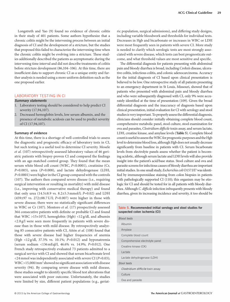
studies is very important. To properly assess the diff erential diagnosis,
clinicians should consider initially obtaining complete blood count,
comprehensive metabolic panel, stool culture, stool examination for
ova and parasites, Clostridium diffi cile toxin assay, and serum lactate,
LDH, creatine kinase, and amylase levels ( Table 5 ). Complete blood
count is useful to assess the WBC for prognostic purposes and the Hgb
level to determine blood loss, although Hgb does not usually decrease
signifi cantly from baseline in patients with CI. Serum bicarbonate
levels from electrolyte panels assess whether the patient is becom-
ing acidotic, although serum lactate and LDH levels will also provide
insight into the patient’s acid/base status. Stool culture and ova and
parasite screens for infectious causes of bloody diarrhea are important
initial studies. In one small study, Escherichia coli O157:H7 was identi-
fi ed by immunoperoxidase staining from colon biopsies in patients
with pathologically supported CI ( 110 ); this organism may be etio-
logic for CI and should be tested for in all patients with bloody diar-
rhea. Although C. diffi cile infection infrequently presents with bloody
diarrhea, given its increasing incidence and severity, it too should be
Table 5 . Recommended initial serology and stool studies for
suspected colon ischemia (CI)
Blood tests
Albumin
Amylase
Complete blood count
Comprehensive electrolyte panel
Creatine kinase (CK)
Lactate
Lactate dehydrogenase (LDH)
Stool tests
Clostridium diffi cile toxin assay
Culture
Ova and parasite

Brandt et al.
The American Journal of GASTROENTEROLOGY VOLUME 110 | JANUARY 2015 www.amjgastro.com
30
part of the initial screening protocol for patients with bloody diarrhea.
Elevations in serum amylase also have been shown to be associated
with acute bowel ischemia ( 50 ). Despite none of these markers having
suffi cient evidence that they can diagnose CI, obtaining them during
the initial workup may provide the clinician deeper insight into the
likelihood and severity of CI.
IMAGING OF CI
Recommendations
1 . CT with intravenous and oral contrast should be ordered as
the imaging modality of choice for patients with suspected
CI, to assess the distribution and phase of colitis (strong
recommendation, moderate level of evidence) ( 111–113 ).
2 . Th e diagnosis of CI can be suggested based on CT fi ndings
(e.g., bowel wall thickening, edema, and thumbprinting)
(strong recommendation, moderate evidence) ( 111–113 ).
3 . Multiphasic CT angiography (CTA) should be performed on
any patient with suspected IRCI or in any patient in whom
the possibility of AMI cannot be excluded (strong recom-
mendation, moderate level of evidence) ( 113,114 ).
4 . CT or magnetic resonance imaging (MRI) fi ndings of colonic
pneumatosis and portomesenteric venous gas can be used to
predict the presence of transmural colonic infarction (strong
recommendation, moderate level of evidence) ( 115 ).
5 . In a patient in whom the presentation of CI may be a heralding
sign of acute mesenteric ischemia (AMI; e.g., IRCI, severe pain
without bleeding, and atrial fi brillation), and the multiphasic CT
is negative for vascular occlusive disease, traditional splanchnic
angiography should be considered for further assessment (con-
ditional recommendation, low level of evidence) ( 114 ).
Summary of evidence
A variety of imaging methods have been used to diagnose CI,
including plain fi lms of the abdomen, barium enema (BE), CT,
ultrasonography (US), and MRI. All can suggest or support the
diagnosis, but none have fi ndings that are specifi c enough to make
a defi nitive diagnosis, except when infarction has occurred.
Plain fi lms of the abdomen that show rounded densities along
the sides of a gas-fi lled distended colon (“thumbprints”) and rigid-
ity with thickening of the colon wall are suggestive of early ischemic
change, whereas intramural gas (pneumatosis linearis), portal
venous gas, and megacolon indicate advanced changes. In one ret-
rospective report, suggestive signs of early disease were present in
21% of 41 patients with CI and no patient had the signs of more
severe disease ( 116 ). When reversible CI was fi rst described in 1963
by Boley et al. ( 117 ), serial BEs together with the patient’s subse-
quent course formed the basis for diagnosis. Th e original radiologic
description of “reversible” CI was of “thumbprints” (pseudotu-
mors) that were caused by subepithelial hemorrhage/edema and
that either resolved in 1–2 weeks or evolved to a segmental ulcera-
tive colitis picture with subsequent normalization over several
months. Today, BE has a limited role in diagnosis and has been
replaced by CT and colonoscopy (see below), largely because of the
latter’s greater accuracy and ability to allow sampling of the mucosa
for pathology. BE is now used primarily to follow the course of
ischemic strictures, although virtual colonography or other imag-
ing tests could be used for this purpose as well. In the usual scenario
wherein the patient becomes asymptomatic aft er the index episode
of CI, there are no data to support a benefi t for repeating colono-
scopy to prove the mucosa has returned to normal.
CT is commonly obtained in the emergency department to assess
abdominal pain. When CT has not been obtained upon admis-
sion, the clinician should consider this modality when patients
are classifi ed as having either moderate or severe CI as defi ned in
Table 6 (“Severity and Treatment of CI” section; see also Figure 1 ).
Th ese patients are at the highest risk for poor outcome and further
characterization of the disease is helpful to determine appropri-
ate management. CT is useful to exclude serious medical condi-
tions other than CI (e.g., diverticulitis), can suggest the diagnosis
of CI, and reveal which areas of the colon are involved. Segmen-
tal wall thickening, thumbprinting, and pericolonic fat stranding
with or without ascites are signs associated with CI, but are not
specifi c enough to make a defi nitive diagnosis. Wolff et al. ( 118 )
assessed patients with abdominal pain (with or without diarrhea
or hematochezia) who had no history of gastrointestinal disease,
who presented to the emergency department or were inpatients,
and who had CT fi ndings consistent with colitis and subsequent
colonoscopy within 30 days of presentation; 36.4% of patients had
ischemia as the etiology and no explanation of colitis was found
in 26.1%. Fat stranding in the presence of bowel wall thickening
was frequently seen in patients with diverticulitis, infectious coli-
tis, and IBD, as well as in CI. Th is study confi rmed that the usual
CT fi ndings of colitis are nonspecifi c and not unique for CI in most
patients with abdominal pain ( 118 ).
Th e appearance of thickened bowel wall in CI usually results
from subepithelial edema and/or hemorrhage and refl ects the
evolving degree of ischemia and the time between the ischemic
insult and performance of CT ( 111,112 ). Balthazar et al. ( 111 )
published a retrospective case series of 54 patients with CI. Th e
study subdivided the CT fi ndings into three morphologically dis-
tinct groups based upon the timing of the examination in relation
to the evolving pathophysiology. A “wet” appearance was seen in
61% of cases and appeared as a loss of colonic haustra with varying
degrees of pericolic streakiness; this correlated to the acute patho-
logic changes aft er reperfusion of ischemic bowel. A “dry” appear-
ance was seen in 33% of cases and manifested as concentric and
symmetric mild mural thickening with homogenous attenuation
of the colon wall and minimal pericolic streakiness; this correlated
to the progression of ischemic damage without reperfusion. Th e
remaining 6% had circumferential intramural air consistent with
pneumatosis coli and presumed colonic necrosis ( 111 ). Of the
overall cohort, 37% had moderate ascites and 24% had a “target
sign” or “double halo” sign showing diff erent attenuations of the
layers of the bowel wall that corresponded to ischemia and reper-
fusion of the involved segment. Th is study attempted to character-
ize the most common fi ndings in segments of colon aff ected by CI,
but it lacked fi rm diagnostic criteria for CI, cohorts were relatively
small, and there was no control group. Similar limitations are seen
in the study of Romano et al. ( 112 ) that looked at patterns of CT
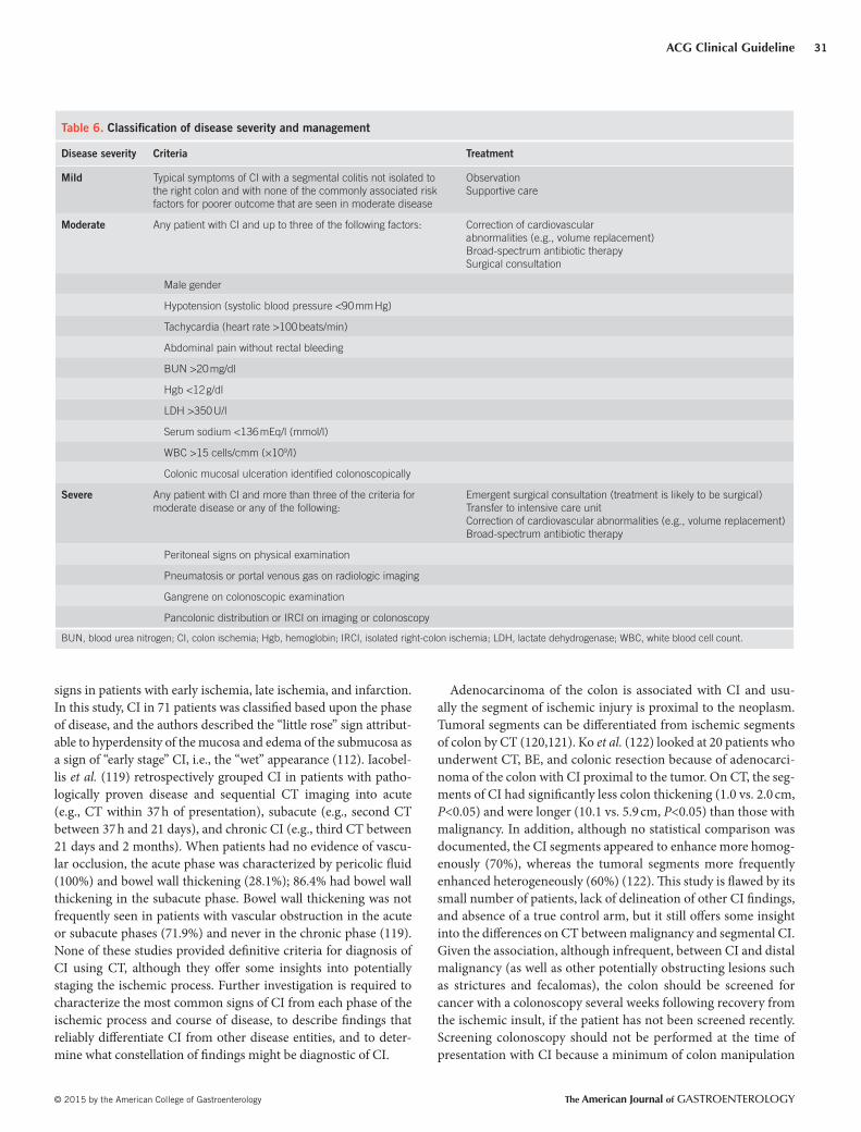
ACG Clinical Guideline
© 2015 by the American College of Gastroenterology The American Journal of GASTROENTEROLOGY
31
signs in patients with early ischemia, late ischemia, and infarction.
In this study, CI in 71 patients was classifi ed based upon the phase
of disease, and the authors described the “little rose” sign attribut-
able to hyperdensity of the mucosa and edema of the submucosa as
a sign of “early stage” CI, i.e., the “wet” appearance ( 112 ). Iacobel-
lis et al. ( 119 ) retrospectively grouped CI in patients with patho-
logically proven disease and sequential CT imaging into acute
(e.g., CT within 37 h of presentation), subacute (e.g., second CT
between 37 h and 21 days), and chronic CI (e.g., third CT between
21 days and 2 months). When patients had no evidence of vascu-
lar occlusion, the acute phase was characterized by pericolic fl uid
(100%) and bowel wall thickening (28.1%); 86.4% had bowel wall
thickening in the subacute phase. Bowel wall thickening was not
frequently seen in patients with vascular obstruction in the acute
or subacute phases (71.9%) and never in the chronic phase ( 119 ).
None of these studies provided defi nitive criteria for diagnosis of
CI using CT, although they off er some insights into potentially
staging the ischemic process. Further investigation is required to
characterize the most common signs of CI from each phase of the
ischemic process and course of disease, to describe fi ndings that
reliably diff erentiate CI from other disease entities, and to deter-
mine what constellation of fi ndings might be diagnostic of CI.
Adenocarcinoma of the colon is associated with CI and usu-
ally the segment of ischemic injury is proximal to the neoplasm.
Tumoral segments can be diff erentiated from ischemic segments
of colon by CT ( 120,121 ). Ko et al. ( 122 ) looked at 20 patients who
underwent CT, BE, and colonic resection because of adenocarci-
noma of the colon with CI proximal to the tumor. On CT, the seg-
ments of CI had signifi cantly less colon thickening (1.0 vs. 2.0 cm,
P <0.05) and were longer (10.1 vs. 5.9 cm, P <0.05) than those with
malignancy. In addition, although no statistical comparison was
documented, the CI segments appeared to enhance more homog-
enously (70%), whereas the tumoral segments more frequently
enhanced heterogeneously (60%) ( 122 ). Th is study is fl awed by its
small number of patients, lack of delineation of other CI fi ndings,
and absence of a true control arm, but it still off ers some insight
into the diff erences on CT between malignancy and segmental CI.
Given the association, although infrequent, between CI and distal
malignancy (as well as other potentially obstructing lesions such
as strictures and fecalomas), the colon should be screened for
cancer with a colonoscopy several weeks following recovery from
the ischemic insult, if the patient has not been screened recently.
Screening colonoscopy should not be performed at the time of
presentation with CI because a minimum of colon manipulation
Table 6 . Classifi cation of disease severity and management
Disease severity Criteria Treatment
Mild Typical symptoms of CI with a segmental colitis not isolated to
the right colon and with none of the commonly associated risk
factors for poorer outcome that are seen in moderate disease
Observation
Supportive care
Moderate Any patient with CI and up to three of the following factors: Correction of cardiovascular
abnormalities (e.g., volume replacement)
Broad-spectrum antibiotic therapy
Surgical consultation
Male gender
Hypotension (systolic blood pressure <90 mm Hg)
Tachycardia (heart rate >100 beats/min)
Abdominal pain without rectal bleeding
BUN >20 mg/dl
Hgb <12 g/dl
LDH >350 U/l
Serum sodium <136 mEq/l (mmol/l)
WBC >15 cells/cmm (×10 9 /l)
Colonic mucosal ulceration identifi ed colonoscopically
Severe Any patient with CI and more than three of the criteria for
moderate disease or any of the following:
Emergent surgical consultation (treatment is likely to be surgical)
Transfer to intensive care unit
Correction of cardiovascular abnormalities (e.g., volume replacement)
Broad-spectrum antibiotic therapy
Peritoneal signs on physical examination
Pneumatosis or portal venous gas on radiologic imaging
Gangrene on colonoscopic examination
Pancolonic distribution or IRCI on imaging or colonoscopy
BUN, blood urea nitrogen; CI, colon ischemia; Hgb, hemoglobin; IRCI, isolated right-colon ischemia; LDH, lactate dehydrogenase; WBC, white blood cell count.
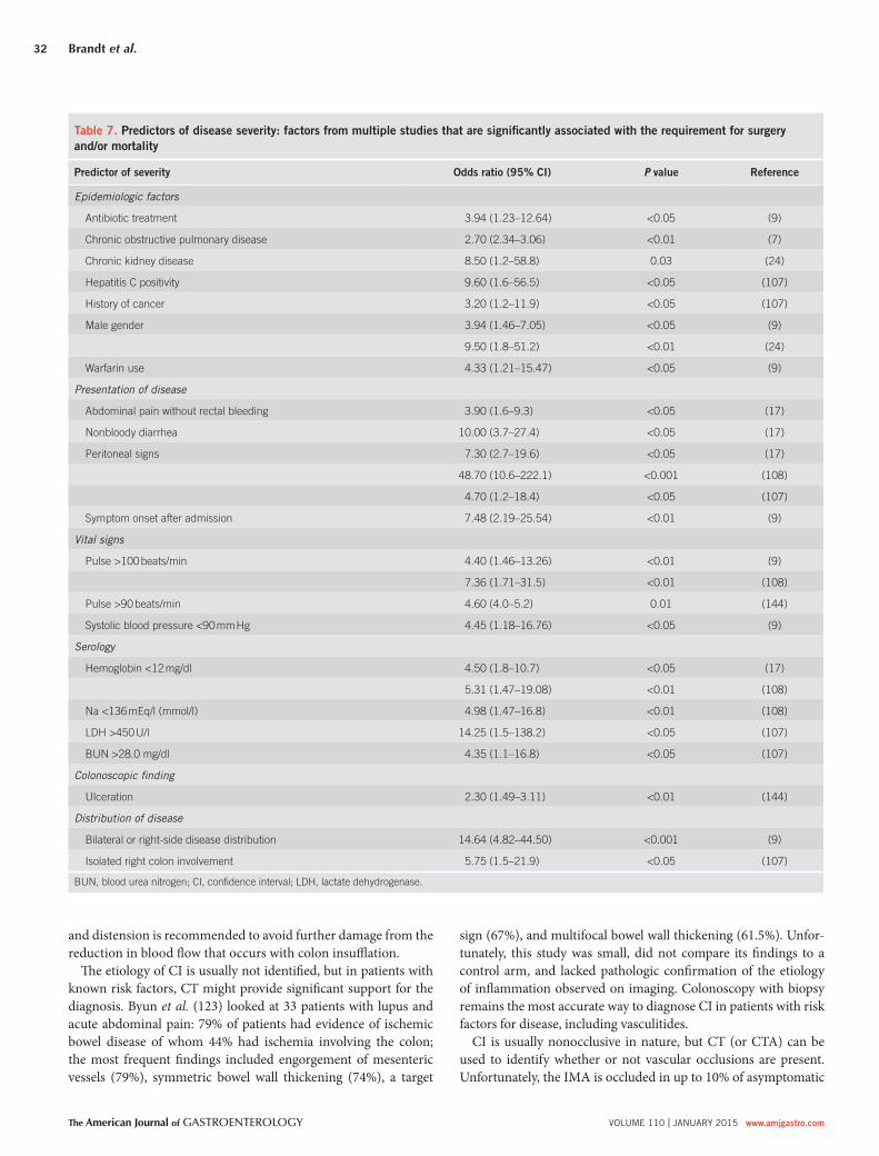
Brandt et al.
The American Journal of GASTROENTEROLOGY VOLUME 110 | JANUARY 2015 www.amjgastro.com
32
sign (67%), and multifocal bowel wall thickening (61.5%). Unfor-
tunately, this study was small, did not compare its fi ndings to a
control arm, and lacked pathologic confi rmation of the etiology
of infl ammation observed on imaging. Colonoscopy with biopsy
remains the most accurate way to diagnose CI in patients with risk
factors for disease, including vasculitides.
CI is usually nonocclusive in nature, but CT (or CTA) can be
used to identify whether or not vascular occlusions are present.
Unfortunately, the IMA is occluded in up to 10% of asymptomatic
and distension is recommended to avoid further damage from the
reduction in blood fl ow that occurs with colon insuffl ation.
Th e etiology of CI is usually not identifi ed, but in patients with
known risk factors, CT might provide signifi cant support for the
diagnosis. Byun et al. ( 123 ) looked at 33 patients with lupus and
acute abdominal pain: 79% of patients had evidence of ischemic
bowel disease of whom 44% had ischemia involving the colon;
the most frequent fi ndings included engorgement of mesenteric
vessels (79%), symmetric bowel wall thickening (74%), a target
Table 7 . Predictors of disease severity: factors from multiple studies that are signifi cantly associated with the requirement for surgery
and/or mortality
Predictor of severity Odds ratio (95% CI) P value Reference
Epidemiologic factors
Antibiotic treatment 3.94 (1.23–12.64) <0.05 ( 9 )
Chronic obstructive pulmonary disease 2.70 (2.34–3.06) <0.01 ( 7 )
Chronic kidney disease 8.50 (1.2–58.8) 0.03 ( 24 )
Hepatitis C positivity 9.60 (1.6–56.5) <0.05 ( 107 )
History of cancer 3.20 (1.2–11.9) <0.05 ( 107 )
Male gender 3.94 (1.46–7.05) <0.05 ( 9 )
9.50 (1.8–51.2) <0.01 ( 24 )
Warfarin use 4.33 (1.21–15.47) <0.05 ( 9 )
Presentation of disease
Abdominal pain without rectal bleeding 3.90 (1.6–9.3) <0.05 ( 17 )
Nonbloody diarrhea 10.00 (3.7–27.4) <0.05 ( 17 )
Peritoneal signs 7.30 (2.7–19.6) <0.05 ( 17 )
48.70 (10.6–222.1) <0.001 ( 108 )
4.70 (1.2–18.4) <0.05 ( 107 )
Symptom onset after admission 7.48 (2.19–25.54) <0.01 ( 9 )
Vital signs
Pulse >100 beats/min 4.40 (1.46–13.26) <0.01 ( 9 )
7.36 (1.71–31.5) <0.01 ( 108 )
Pulse >90 beats/min 4.60 (4.0–5.2) 0.01 ( 144 )
Systolic blood pressure <90 mm Hg 4.45 (1.18–16.76) <0.05 ( 9 )
Serology
Hemoglobin <12 mg/dl 4.50 (1.8–10.7) <0.05 ( 17 )
5.31 (1.47–19.08) <0.01 ( 108 )
Na <136 mEq/l (mmol/l) 4.98 (1.47–16.8) <0.01 ( 108 )
LDH >450 U/l 14.25 (1.5–138.2) <0.05 ( 107 )
BUN >28.0 mg/dl 4.35 (1.1–16.8) <0.05 ( 107 )
Colonoscopic fi nding
Ulceration 2.30 (1.49–3.11) <0.01 ( 144 )
Distribution of disease
Bilateral or right-side disease distribution 14.64 (4.82–44.50) <0.001 ( 9 )
Isolated right colon involvement 5.75 (1.5–21.9) <0.05 ( 107 )
BUN, blood urea nitrogen; CI, confi dence interval; LDH, lactate dehydrogenase.

ACG Clinical Guideline
© 2015 by the American College of Gastroenterology The American Journal of GASTROENTEROLOGY
33
patients over 60 years of age and hence the signifi cance of such
a fi nding is uncertain ( 124 ). With emerging technology, contrast-
enhanced CT can sometimes assess patency of the celiac, SMA,
and IMA without devoted CTA; this usually requires direct com-
munication with the radiology team to review the imaging specifi -
cally with the goal of vascular assessment. Data do not exist on the
sensitivity and specifi city of CT for assessing vascular occlusion
in CI, but a meta-analysis including three prospective and three
retrospective studies that looked at CT in the diagnosis of mes-
enteric arterial embolism, mesenteric arterial thrombosis, and
mesenteric venous thrombosis (i.e., primary vascular AMI) found
the sensitivity and specifi city for diagnosis to be 93.3% and 95.9%,
respectively ( 113 ). Th ese six studies were performed in tertiary
care referral centers in industrialized countries and, therefore,
their high diagnostic accuracy might not refl ect the accuracy of
studies done in small community hospitals or practices; regardless,
CT (or CTA) off ers a high sensitivity and specifi city for vascular
assessment in patients with CI.
In contrast to AMI in which conventional mesenteric angiogra-
phy or CTA plays an essential role, vascular imaging studies are not
indicated in most patients with suspected CI because by the time of
presentation, colon blood fl ow has usually returned to normal and
the observed changes are not from ongoing ischemia but rather
refl ect the ischemic insult with or without reperfusion injury. In
patients with potentially severe, complicated disease, such as those
with IRCI or when the clinician cannot distinguish between AMI
and CI (e.g., in patients with severe abdominal pain and little or no
bleeding), a dedicated multiphasic CT is indicated. In patients with
IRCI, CTA should be performed either initially or before discharge,
as the colonic episode may be the “heralding” indication of SMA
disease and possibly impending AMI. Multiphasic CT scanning,
including a CT without oral contrast but with intravenous con-
trast and attention to the angiographic arterial and portal venous
phases, has become standard for the diagnosis of AMI ( 125–127 ).
One retrospective study that compared triple-phase CT scanning
in 39 patients with pathologically confi rmed AMI and 41 patients
with suspected AMI who ultimately had another diagnosis showed
that the specifi city for detection of SMA thrombus or occlusion
approached 100%, although the sensitivity was poor (26%). Simi-
larly, for celiac and IMA thrombus, the specifi city was estimated
to be 98%, and the sensitivity was 5%. Th is study also showed that
the diagnostic use of oral contrast during CT off ered minimal
additional benefi t to the two vascular phases ( 114 ). Although this
study contained small numbers of patients, it had a control group
and characterized each phase of assessment, thus off ering valuable
data. In any patient in whom the presentation of CI may be a her-
alding sign of AMI (e.g., IRCI), and the multiphasic CT is negative
for vascular occlusive disease, traditional splanchnic angiography
should be considered for further assessment because a negative
triple-phase CT does not absolutely exclude vascular obstruction
as the cause. It is important to note that there are no available pub-
lished data yet analyzing IRCI as a “heralding” indication of SMA
disease. However, our experience has revealed that a select group
of patients with IRCI may present again within weeks, months, or
even a few years aft er the heralding presentation of IRCI with cata-
strophic episodes of AMI, resulting in loss of most of their small
bowel or death. A limited but increasing number of patients with
IRCI have undergone angiography and had occluding thrombi
found in the SMA; stenting and/or thrombectomy resulted in good
outcomes. Conversely, some patients with SMA thrombi did not
undergo angiography or stenting yet had a positive outcome as
well. Although there are no data to yet support our recommenda-
tion, we believe that the benefi t of preventing a potentially cata-
strophic ischemic insult to the small intestine (AMI) far outweighs
the risks of a CTA.
Pneumoperitoneum, portal venous gas, and pneumatosis are
ominous fi ndings in patients suspected of CI. In one Canadian
study of 14 total patients who underwent colectomy with pathol-
ogy showing transmural ischemia, CT showed that 100% had fat
stranding, 71% had pneumatosis, and 36% had portal venous gas
( 128 ). However, the presence of pneumatosis and portal venous gas
does not necessarily indicate nonviable bowel. In one small study
of 23 patients with either small or large bowel ischemia, pneuma-
tosis and portomesenteric venous gas were associated with trans-
mural bowel infarction in 78% and 81% of patients respectively
( 129 ). When patients with an obstructing malignancy presented
with a large bowel obstruction and CT showed cecal pneumato-
sis, surgery revealed transmural necrosis of the cecum in 26% of
patients (6 of 23 total patients) ( 130 ). Milone et al. ( 115 ) assessed
CT fi ndings in 121 patients with transmural colon necrosis and
87 with limited colon necrosis. Th e presence of pneumatosis plus
portomesenteric gas showed good specifi city (83%) for transmural
infarction, but very low sensitivity (17%); the concomitant pres-
ence of the two fi ndings did not achieve statistical signifi cance
in predicting transmural CI ( 115 ). Th ese studies are limited by
their sample size and retrospective nature, but they do show that
patients with CI who also have CT fi ndings of pneumatosis and/
or portomesenteric venous gas are likely to have severe disease,
but not necessarily with full transmural necrosis. Nonetheless, in
the clinical setting of CI, one of these fi ndings is still an indica-
tion for prompt operation. Although CT has become the standard
of care in assessing patients with suspected CI, both US and MRI
also off er some diagnostic utility. In one retrospective case series of
58 patients with CI evaluated by US, the fi ndings were as follows:
symmetric bowel wall thickening (100%), segmental involvement
of the colon (80%), preservation of colon wall stratifi cation (66%),
altered pericolic fat (28%), free fl uid (19%), and pneumatosis
(1.7%). When this cohort was subdivided into transmural necro-
sis and nontransmural necrosis, the only signifi cant diff erence
observed was the presence of abnormal pericolonic fat, which was
seen more frequently in patients with transmural disease (75% vs.
20%, P <0.01) ( 131 ). In another study of 66 patients presenting to
the emergency department with nonspecifi c abdominal pain who
had abdominal US performed as their primary imaging modal-
ity, the group was subdivided based upon diagnosis into infec-
tious colitis, infl ammatory colitis, ischemic colitis, and malignant
tumors. When the US fi ndings were compared, arterial fl ow in the
wall of the involved segments of colon was absent in 43% of those
with CI compared with 12% of those with other diagnoses ( P <0.05)
( 132 ). Th ese US fi ndings do not invalidate the reason why we do

Brandt et al.
The American Journal of GASTROENTEROLOGY VOLUME 110 | JANUARY 2015 www.amjgastro.com
34
not recommend angiography in the evaluation of a fi rst episode of
CI, namely that by the time patients present with CI, colon blood
fl ow has usually returned to normal, because they only measured
signs of arterial fl ow in the bowel wall. Th is study was small and
did not provide any criteria for the diagnosis of CI or information
on the severity of the CI. Moreover, it is not known how many of
the patients had infarcted bowel that, of course, would not have
intramural fl ow. We would be hesitant to challenge this long-held
belief until the results are confi rmed in a robust manner. Experi-
ence with US in the setting of CI is very limited and it is believed
that this technique lacks specifi city for bowel wall thickening and
has a high false negative rate ( 133 ).
MRI has been formally studied in only a small number of
patients with CI; fi ndings are similar to those of CT, but without
the radiation exposure and the need for iodinated contrast agents.
In one prospective study that assessed seven patients with CI, all of
whom had an initial diagnostic CT and a subsequent MRI, simi-
lar segmental involvement and wall thickness were shown on both
modalities ( 134 ). As with CT, such fi ndings usually are not specifi c
enough to make a defi nitive diagnosis. MRI may have a role when
repeat imaging is required or patients have poor renal function.
COLONOSCOPY IN THE DIAGNOSIS OF CI
Recommendations
1 . Early colonoscopy (within 48 h of presentation) should be
performed in suspected CI cases to confi rm the diagnosis
(strong recommendation, low level of evidence) ( 17 ).
2 . When performing colonoscopy on a patient with suspected
CI, the colon should be insuffl ated minimally (conditional
recommendation, very low level of evidence) ( 69,135 ).
3 . In patients with severe CI, CT should be used to evaluate the
distribution of disease. Limited colonoscopy is appropriate
to confi rm the nature of the CT abnormality. Th e endoscopic
procedure should be stopped at the distal-most extent of the
disease (strong recommendation, low level of evidence).
4 . Biopsies of the colonic mucosa should be obtained except in
cases of gangrene (strong recommendation, very low level of
evidence).
5 . Colonoscopy should not be performed in patients who have
signs of acute peritonitis or evidence of irreversible ischemic
damage (i.e., gangrene and pneumatosis) (strong recommen-
dation, very low level of evidence).
Summary of evidence
Colonoscopy has become the primary technique to diagnose CI,
usually aft er CT (see above) has revealed a segment of colon that
is thickened with or without other more specifi c signs of ischem-
ic damage, e.g., pneumatosis linearis and portal venous gas (see
Figure 1 ). BE (see above) had been the main diagnostic mode in
the 1960s but was gradually replaced by colonoscopy as the latter
became increasingly available. Colonoscopy off ers several advan-
tages over radiologic imaging, including the ability to directly ob-
serve the abnormal tissue, to sometimes make a diagnosis just by
appearance, and to obtain tissue samples. Nonspecifi c features of
CI in addition to erythema, edema, and ulceration include aph-
thous ulcers, which may suggest Crohn’s disease, pseudomem-
branes, which are more oft en seen with C. diffi cile infection, and
pseudopolyposis, which may develop with healing. Although the
dusky, cyanotic hue of necrotic mucosa typifi es gangrene and CI
can be assumed in its presence, the validity of this assumption has
never been tested. One retrospective study of 85 consecutive pa-
tients who had a colonoscopy to confi rm diagnosis of CI showed
that the most common fi ndings in transient CI are edematous
and fragile mucosa, segmental erythema, scattered erosions, lon-
gitudinal ulcerations, petechial hemorrhages interspersed with
pale areas, purple hemorrhagic nodules, and a sharply defi ned
segmental involvement ( 136 ). Th is study provides a clear descrip-
tion of the colonoscopic fi ndings seen in patients with mild, tran-
sient CI, but it suff ers from the fl aws of many of the currently
published studies that do not provide frequencies of these fi nd-
ings in the context of disease severity. Montoro et al. ( 17 ) looked
at 297 patients who underwent colonoscopy for all levels of CI
severity: fi ndings were erythema (83.7%), edema (69.9%), fri-
ability (42.6%), superfi cial ulcerations including the single stripe
sign (57.4%), deep ulcerations (21.7%), luminal narrowing and
stenosis (8.4%), intraluminal blood (8.4%), and blue-black nod-
ules with dark-dusky backgrounds suggestive of gangrene (5.5%).
Scattered erosions may also be seen both proximal and distal to
continuous segments of involvement.
Th e colon single-stripe sign (CSSS) is a highly specifi c sign of
CI described by Zuckerman et al. ( 137 ) in 2003. CSSS is defi ned
as a single infl ammatory band of erythema with erosion and/or
ulceration along the longitudinal axis of the colon; all CSSSs in the
study of Zuckerman et al. ( 137 ) were >5 cm in length and 89% of
CSSSs were found in the left colon. Th e 26 patients with CSSS had
a better prognosis than did the 22 patients with circumferential
CI and exhibited a reduced need for surgery (0% vs. 27%) and a
lesser mortality (4% vs. 41%). Although of great interest, the study
of Zuckerman et al. ( 137 ) was retrospective, small, had limited fol-
low-up information, and a cause-and-eff ect relationship between
ischemia and the CSSS was not proven.
Colonoscopy, in addition to detailing specifi c fi ndings, also
enables accurate determination of the anatomic limits of involve-
ment and, therefore, perhaps helps predict prognosis. In 2010,
Brandt et al. ( 7 ) showed CI that was isolated to the right side of
the colon (IRCI) had a worse prognosis than CI involving any
other anatomic segment of colon. In a multicenter Spanish study
led by Montoro et al. ( 17 ) of 364 patients with CI, 345 of whom
had colonoscopy, timing of colonoscopy was found to be impor-
tant for diagnosis: when colonoscopy was performed within
48 h of presentation, typical hemorrhagic nodules were found in
47.1% of cases, but were evident in 33.3% of those in whom the
examination was performed 2–5 days aft er presentation, and in
only 9.7% of patients when colonoscopy was done aft er day 5. In
2011, in a retrospective study of 106 cases of CI divided into two
groups, one with erythema and erosions and another with linear
and circumferential ulcers, Beppu et al. ( 23 ) showed that CI was
more severe with the latter pattern of disease; abdominal pain,
C-reactive protein levels, and length of hospital stay were the only
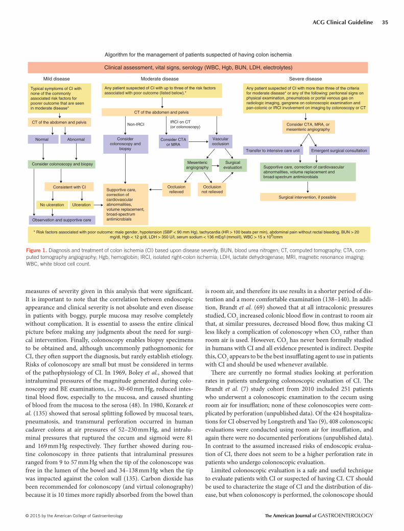
ACG Clinical Guideline
© 2015 by the American College of Gastroenterology The American Journal of GASTROENTEROLOGY
35
measures of severity given in this analysis that were signifi cant.
It is important to note that the correlation between endoscopic
appearance and clinical severity is not absolute and even disease
in patients with boggy, purple mucosa may resolve completely
without complication. It is essential to assess the entire clinical
picture before making any judgments about the need for surgi-
cal intervention. Finally, colonoscopy enables biopsy specimens
to be obtained and, although uncommonly pathognomonic for
CI, they oft en support the diagnosis, but rarely establish etiology.
Risks of colonoscopy are small but must be considered in terms
of the pathophysiology of CI. In 1969, Boley et al. , showed that
intraluminal pressures of the magnitude generated during colo-
noscopy and BE examinations, i.e., 30-60 mm Hg, reduced intes-
tinal blood fl ow, especially to the mucosa, and caused shunting
of blood from the mucosa to the serosa ( 48 ). In 1980, Kozarek et
al. ( 135 ) showed that serosal splitting followed by mucosal tears,
pneumatosis, and transmural perforation occurred in human
cadaver colons at air pressures of 52–230 mm Hg, and intralu-
minal pressures that ruptured the cecum and sigmoid were 81
and 169 mm Hg respectively. Th ey further showed during rou-
tine colonoscopy in three patients that intraluminal pressures
ranged from 9 to 57 mm Hg when the tip of the colonoscope was
free in the lumen of the bowel and 34–138 mm Hg when the tip
was impacted against the colon wall ( 135 ). Carbon dioxide has
been recommended for colonoscopy (and virtual colonography)
because it is 10 times more rapidly absorbed from the bowel than
is room air, and therefore its use results in a shorter period of dis-
tention and a more comfortable examination ( 138–140 ). In addi-
tion, Brandt et al. ( 69 ) showed that at all intracolonic pressures
studied, CO 2 increased colonic blood fl ow in contrast to room air
that, at similar pressures, decreased blood fl ow, thus making CI
less likely a complication of colonoscopy when CO 2 rather than
room air is used. However, CO 2 has never been formally studied
in humans with CI and all evidence presented is indirect. Despite
this, CO 2 appears to be the best insuffl ating agent to use in patients
with CI and should be used whenever available.
Th ere are currently no formal studies looking at perforation
rates in patients undergoing colonoscopic evaluation of CI. Th e
Brandt et al. ( 7 ) study cohort from 2010 included 251 patients
who underwent a colonoscopic examination to the cecum using
room air for insuffl ation; none of these colonoscopies were com-
plicated by perforation (unpublished data). Of the 424 hospitaliza-
tions for CI observed by Longstreth and Yao ( 9 ), 408 colonoscopic
evaluations were conducted using room air for insuffl ation, and
again there were no documented perforations (unpublished data).
In contrast to the assumed increased risks of endoscopic evalua-
tion of CI, there does not seem to be a higher perforation rate in
patients who undergo colonoscopic evaluation.
Limited colonoscopic evaluation is a safe and useful technique
to evaluate patients with CI or suspected of having CI. CT should
be used to characterize the stage of CI and the distribution of dis-
ease, but when colonoscopy is performed, the colonoscope should
Figure 1 . Diagnosis and treatment of colon ischemia (CI) based upon disease severity. BUN, blood urea nitrogen; CT, computed tomography; CTA, com-
puted tomography angiography; Hgb, hemoglobin; IRCI, isolated right-colon ischemia; LDH, lactate dehydrogenase; MRI, magnetic resonance imaging;
WBC, white blood cell count.
Typical symptoms of CI withnone of the commonlyassociated risk factors forpoorer outcome that are seenin moderate disease*
Any patient suspected of CI with up to three of the risk factorsassociated with poor outcome (listed below).*
CT of the abdomen and pelvis
Non-IRCIIRCI on CT(or colonoscopy)
Considercolonoscopy and
biopsy
Consider CTAor MRA
Vascularocclusion
Surgicalevaluation
Mesentericangiography
Occlusionnot relieved
Occlusionrelieved
Transfer to intensive care unit
Supportive care, correction of cardiovascularabnormalities, volume replacement andbroad-spectrum antimicrobials
Surgical intervention, if possible
Emergent surgical consultation
Consider CTA, MRA, ormesenteric angiography
Any patient suspected of CI with more than three of the criteriafor moderate disease* or any of the following: peritoneal signs onphysical examination, pneumatosis or portal venous gas onradiologic imaging, gangrene on colonoscopic examination andpan-colonic or IRCI involvement on imaging by colonoscopy or CT
Severe diseaseModerate diseaseMild disease
Clinical assessment, vital signs, serology (WBC, Hgb, BUN, LDH, electrolytes)
Algorithm for the management of patients suspected of having colon ischemia
CT of the abdomen and pelvis
Normal
Consider colonoscopy and biopsy
Consistent with CI
No ulceration Ulceration
Observation and supportive care
* Risk factors associated with poor outcome: male gender, hypotension (SBP < 90 mm Hg), tachycardia (HR > 100 beats per min), abdominal pain without rectal bleeding, BUN > 20mg/dl, Hgb < 12 g/dl, LDH > 350 U/l, serum sodium < 136 mEq/l (mmol/l), WBC > 15 x 109/cmm
Abnormal
Supportive care,correction ofcardiovascularabnormalities,volume replacement,broad-spectrumantimicrobials

Brandt et al.
The American Journal of GASTROENTEROLOGY VOLUME 110 | JANUARY 2015 www.amjgastro.com
36
be passed only to the distal-most aspect of the aff ected region.
Biopsies should be taken in all cases, except when there is evidence
of gangrene.
Histopathologic specimens obtained colonoscopically are rarely
diagnostic for CI. Pathognomonic features include infarction and
ghost cells, i.e., preserved individual cellular outlines without cell
content, and are infrequently seen ( 138 ). It is more common to see
biopsy specimens showing mucosal and submucosal hemorrhage
and edema and capillary fi brin thrombi with neutrophilic infi ltra-
tion that are consistent with CI which, when taken in the context of
clinical presentation, will support the diagnosis. Most commonly,
pathologic features are nonspecifi c ( 138 ).
The diagnosis of CI, however, cannot be made conclusively
by colonoscopy on just one examination unless mucosal gan-
grene is observed or infarction or ghost cells are seen at histo-
pathology. Montoro et al. ( 17 ) found that of their 297 patients
who underwent colonoscopy with biopsy, ghost cells and
mucosal infarction were each seen in 7.7% of patients; of the
25 patients who had surgical resections, ghost cells and infarc-
tion were seen in 20% and 63%, respectively; thus, these his-
topathologic findings are infrequent and of limited value in
diagnosing CI.
SEVERITY AND TREATMENT OF CI
Recommendations
1 . Most cases of CI resolve spontaneously and do not require
specifi c therapy (strong recommendation, low quality of
evidence) ( 107,108,139 ).
2 . Surgical intervention should be considered in the presence of
CI accompanied by hypotension, tachycardia, and abdominal
pain without rectal bleeding; for IRCI and pancolonic CI;
and in the presence of gangrene (strong recommendation,
moderate level of evidence) ( 17,107,108 ).
3 . Antimicrobial therapy should be considered for patients with
moderate or severe disease (strong recommendation, very
low level of evidence) ( 107,108,140 ).
Summary statement
1 . When considering the mortality risk for patients undergoing
surgical intervention for acute CI, Ischemic Colitis Mortality
Risk (ICMR) factors should be utilized ( 141,142 ).
Summary of evidence
Surgical intervention and mortality are the most commonly used
end points in studies that assess factors associated with poor out-
come in CI and are used to defi ne severe or complicated (poor
outcome) disease in the data discussed below ( Table 7 ). Th e
strength of diagnosis from these studies is based upon the scheme
presented in Table 8 . Longstreth and Yao ( 9 ), Montoro et al. ( 17 ),
and Brandt et al. ( 7 ) assessed large cohorts of patients looking at
predictors of poor outcome, whereas Añón et al. ( 108 ), Huguier
et al. ( 94 ), Lee et al. ( 24 ), O’Neill et al. ( 143 ), and Mosele et al.
( 107 ) each assessed smaller cohorts. Th ese studies all conducted
multivariate analyses and found various signifi cant predictors of
outcome including epidemiologic factors, clinical presentation
of disease, vital signs, serologic values, and disease distribution.
Epidemiologic factors that were associated with poor outcome
included antimicrobial therapy for CI, hepatitis C positivity,
history of cancer, male gender, and warfarin use at the time of
diagnosis ( 9,94,107 ). Th e use of antimicrobial therapy is likely an
indicator of the clinician’s response to disease severity rather than
the antibiotic therapy per se contributing to poor outcome. Pres-
entations of disease, e.g., abdominal pain without rectal bleeding,
nonbloody diarrhea, peritoneal signs on physical examination
and symptom onset aft er admission, are all associated with poor
outcome ( 9,17,94,107,108,143 ). A heart rate of >100 beats per min
and systolic blood pressure of <90 mm Hg at the time of diagno-
sis are associated with the need for surgical intervention and/or
mortality as are Hgb <12 mg/dl, hyponatremia (Na <136 mEq/l
[mmol/l]), LDH >450 U/l, and blood urea nitrogen (>28 mg/dl
[mmol/l]) ( 9,17,107,108 ). As discussed previously, a pancolonic
distribution of disease and IRCI also portend a poor outcome
( 7,9,107,143 ). Th ese studies were heterogeneous in terms of
patient characteristics, physical examination fi ndings, medical
comorbidities, serology, and thresholds of various factors consid-
ered, but several risk factors appeared to be signifi cantly associ-
ated with poor outcome in several of these studies: hypotension,
tachycardia, abdominal pain without rectal bleeding, IRCI, and
pancolonic CI.
Chung et al. ( 144 ) assessed 152 patients with clinically con-
fi rmed CI (74.5% confi rmed pathologically) to analyze signifi -
cant risk factors for poor outcome (e.g., improvement delayed
by >2 weeks, 30-day colectomy, and 30-day mortality) and used
these factors to devise a prognostic scoring system. Th e strongest
Table 8 . Modifi ed Brandt and Boley 190 criteria for strength of diagnosis for colon ischemia (CI) a
Strength of diagnosis Clinical Colonoscopy Pathologic Culture
Defi nite CI Consistent with CI Consistent with CI Consistent with CI Negative
Probable CI Consistent with CI Consistent with CI Not performed or negative
within 72 h
Negative after 12 h of antimi-
crobial therapy
Possible CI Consistent with CI Not performed, negative, or nonspecifi c
changes within 72 h
Not performed or negative
within 72 h
Culture not performed
a Modifi ed from ref. 190.
ACG Clinical Guideline
© 2015 by the American College of Gastroenterology The American Journal of GASTROENTEROLOGY
37
emphasis on the milder cases that are not hospitalized. Th e rec-
ommendations presented in this guideline are mostly based on
small retrospective studies that lack control groups. Most guideline
statements are based upon extrapolation from the available litera-
ture and expert opinion.
One systematic review of the management of CI included 10
retrospective studies comprising 841 patients ( 145 ). Within these
studies there was no standard protocol for management; however,
nonsurgical treatment usually included bowel rest, intravenous
fl uid, electrolyte repletion, and antibiotic usage with occasional
administration of total parenteral nutrition. All articles reviewed
did agree that surgery was indicated for patients with peritonitis
or those who were hemodynamically unstable ( 145 ). Th is system-
atic review had signifi cant limitations including methodologic
anomalies such as excluding some well-structured publications
and also including three studies with patients who had CI as a
complication of vascular procedures despite methodology stating
this as a criterion for exclusion. In addition, the study included
some patients without pathologically confi rmed disease. Th is lack
of uniformity characterizes the CI management literature, but the
abstract’s conclusion reads “Th ere is very little evidence base for
the management of this condition” ( 145 ). We believe this state-
ment is accurate.
O’Neill et al. ( 148 ) conducted a subsequent systematic review
of the management of CI, attempting to improve on the method-
ologies from Diaz-Nieto et al. ( 145 ). Th is review provided a more
rigorous screening evaluation of the studies by including only
biopsy-proven disease in each patient and excluding all studies
with CI resulting from vascular surgery or associated with colonic
adenocarcinoma ( 148 ). Eight retrospective case-series and three
case-control studies comprising 1049 patients were included.
Overall, 80.3% of cases were medically managed without surgical
intervention and had a 6.2% mortality rate; 19.6% were managed
surgically with a mortality rate of 39.3% ( 148 ). Th ese data off er
the best overall insight into medical vs. surgical management of
CI and associated outcomes. Th is analysis is also consistent with
the presumption that those requiring surgery are the sickest and
at highest risk for poor outcome. Medical management most com-
monly included fasting, intravenous fl uids, parenteral nutrition,
antibiotics, and heparin prophylaxis ( 148 ). Requirement for patho-
logic confi rmation of disease is a strength of this study, and is also
a marker of disease because colonoscopy or surgical intervention
isolates those with more advanced disease or when the diagnosis
might be unclear. Th ere was, however, also signifi cant heterogene-
ity among the algorithms used in various studies for medical man-
agement of disease, with some including and others excluding,
antimicrobial therapy, and none detailing the frequency, duration,
or type of antibiotics used.
Indication and timing for antimicrobial use in CI remains
untested. Th ere is no clinical evidence to show benefi cial eff ects
of such therapy and it is unlikely that a randomized controlled
trial will be forthcoming; because the prognosis of CI is excel-
lent in most cases, the number of patients in such a trial needed
to show benefi t (or to disprove a lack of benefi t) would have to
be immense. Previous recommendations for antibiotic usage
predictors in decreasing order of signifi cance were ulceration on
colonoscopy, shock within 24 h of admission, and tachycardia at
the time of diagnosis ( 144 ). If a patient had all three factors, their
risk index for poor outcome was 74.5, whereas if ulceration and
shock were present, but no tachycardia, the risk index decreased
to 39.4; the risk index was 1.00 when none of these factors were
present ( 144 ). Th is simple system is easy to use but requires fur-
ther validation before being broadly applied, given their inclusion
of patients with nonpathologically confi rmed CI and including
a nontraditional end point for severe disease (i.e., improvement
delayed by >2 weeks). Ulceration seen on colonoscopy also has
been associated with poor outcome in a study by Matsumoto
et al. ( 139 ). Th is small study using clinical diagnostic criteria
showed that patients with ulceration on colonoscopy required
longer periods of fasting and had a longer duration of admis-
sion before clinical improvement compared with those without
ulceration. Th is study was very small but is a reinforcement of
the importance of mucosal ulceration as a predictor of outcome
in patients with CI.
In view of the above data and previously described diagnos-
tic options, this guideline proposes redefi ning disease severity to
guide treatment ( Table 6 and Figure 1 ). Th is is currently a non-
validated guide to rating disease severity but incorporates the most
up-to-date knowledge of risk factors for disease severity. Patients
with mild disease are those who have typical symptoms of CI with
a segmental colitis not isolated to the right colon and with none of
the commonly associated risk factors for poorer outcome that are
seen in moderate disease. Moderate disease includes any patient
with up to three of the following factors: male gender, hypotension
(systolic blood pressure <90 mm Hg), tachycardia (heart rate >100
beats/min), abdominal pain without rectal bleeding, blood urea
nitrogen >20 mg/dl, Hgb <12 g/dl, LDH >350 U/l, serum sodium
<136 mEq/l (mmol/l), WBC >15×10 9 /l, or colonoscopically iden-
tifi ed mucosal ulceration. Severe disease is defi ned by more than
three of the previously listed criteria or any of the following: perito-
neal signs on physical examination, pneumatosis on CT, gangrene
on colonoscopy, and a pancolonic distribution or IRCI on CT or
colonoscopy.
Treatment of CI varies with the severity of the disease and its
presentation. In general, many patients have a benign, self-limited
episode of CI that is neither diagnosed nor treated or is managed
in the outpatient setting. Most cases of CI resolve spontaneously
and do not require specifi c therapy; such patients have reversible
ischemic colopathy or transient ischemic colitis ( 91 ). Patients with
more signifi cant symptoms or fi ndings require hospitalization to
enable observation for complications or signs of irreversible dis-
ease. Initial medical management most commonly includes gen-
eral supportive measures, bowel rest, intravenous hydration, and
correction of possible precipitating conditions. Parenteral nutri-
tion may be indicated if a protracted course is anticipated. No
therapeutic modality has ever been tested in a rigorous clinical
trial and the mostly benign nature of CI has led to a shortage of tri-
als assessing various therapies. It is also important to consider that
the currently available literature for the management of CI focuses
on hospitalized patients who have complicated disease with less
Brandt et al.
The American Journal of GASTROENTEROLOGY VOLUME 110 | JANUARY 2015 www.amjgastro.com
38
were based on experimental studies that showed reduction in the
severity and extent of bowel damage when antibiotics were given
before or during an ischemic event, with the majority of these tri-
als being several decades old ( 140,146,147,149,150 ). Yoshiya et al.
( 140 ) pretreated mice with ampicillin, vancomycin, neomycin,
and metronidazole, and subsequently induced intestinal ischemia
with reperfusion by ligating the SMA for 30 min. Th ese mice were
then compared with control mice that also underwent intestinal
ischemia but did not receive antimicrobial therapy. In the mice
pretreated with antibiotic therapy, the expression of Toll-like
receptors 2 and 4 was decreased as was expression of infl amma-
tory markers (e.g., tumor necrosis factor, interleukin-6, and cyclo-
oxygenase-2). As Toll-like receptors normally bind Gram-positive
and Gram-negative bacteria to stimulate infl ammatory responses,
the study supports the theory that antimicrobial therapy decreases
the overall infl ammatory response to ischemic injury. Intestinal
injury resulting from ischemia and reperfusion is also believed to
be stimulated by ischemic cells presenting neoantigens that bind
natural immunoglobulins to activate the complement pathway.
Yoshiya et al. ( 140 ) also showed decreased complement (C3), IgM,
and IgA in ischemic tissue of mice pretreated with antimicrobials
compared with the ischemic tissues of mice not exposed to antimi-
crobial therapy.
Antimicrobial therapy is also believed to prevent poor outcome
through decreased bacterial translocation in the setting of acute
ischemia and reperfusion injury. Th e loss of mucosal integrity as a
result of vigorous infl ammation has been shown in mouse models
to facilitate bacterial translocation and resulting bacteremia ( 151–
153 ). Luo et al. ( 153 ) studied mice that had ischemia induced by
SMA occlusion for 30, 60, or 90 min followed by reperfusion for
30 min. Th ese mice were given varying oral doses of bacteria aft er
the ischemic episode and had blood cultures drawn immediately
and 15 min aft er reperfusion. A control group of mice was fed the
same bacteria but did not have any ischemic episode. Control mice
showed no translocation of bacteria, whereas the mice that were
exposed to even 30 min of ischemia and a low bacterial inoculum
developed bacteremia ( 153 ). Th e studies of Yoshiya et al. ( 140 ) and
Luo et al. ( 153 ) reinforce the theories behind antimicrobial therapy
in the setting of CI in mouse models. Antibiotics are believed to
improve outcome in CI by reducing infl ammatory responses stim-
ulated by the normal fecal microbiome, reducing the antigens that
prompt innate immune response and minimizing bacterial trans-
location through compromised colonic mucosa.
Available human studies of antimicrobial treatment in CI are
few in number, heterogeneous in design, and do not directly
address antimicrobial use for a specifi c therapeutic benefi t. Th ree
studies have presented data regarding broad-spectrum antimicro-
bial therapy of CI and associated outcomes ( 107,108,139 ). Añón
et al. ( 108 ) looked at 85 consecutive patients, of whom 69 had
“mild disease” that was treated with fl uid and electrolyte correction
and “wide-spectrum antibiotics”; all but 1 patient survived (98.6%)
and another 16 patients had severe disease that was initially treated
medically, but subsequently required surgery. Mosele et al. ( 107 )
retrospectively assessed 46 patients who were at least 65 years of
age, of whom 67.4% clinically improved with only medical therapy
consisting of intestinal rest, total parenteral nutrition, and broad-
spectrum antibiotics; 6 of the original cohort received medical
therapy and subsequently died (13.0%), whereas another 9 would
ultimately require surgical intervention (19.6%). Matsumoto et al.
( 139 ) retrospectively assessed 41 patients with CI, with 31.7% of
patients receiving a “combination of antibiotics” along with fasting,
bowel rest, and fl uid replacement; none of these patients required
surgery or died. Th ese studies are similar as they were all small, not
designed to assess the eff ect of antimicrobial therapy on outcome,
mentioned antimicrobial therapy as part of the medical treatment
regimen but mostly did not detail the number of patients within
the “medical therapy” cohort who received antibiotics, and did not
delineate the specifi c antibiotics used or the duration of therapy.
Perhaps most importantly, there were no comparator groups of
patients who did not receive antimicrobials.
By performing multivariate analysis on 401 consecutive
patients, Longstreth and Yao ( 9 ) found that antimicrobial ther-
apy was associated with a 3.94 (confi dence interval: 1.23–164,
P <0.05)-fold increased risk of severe disease as defi ned by need
for surgery or mortality. Th is fi nding speaks to one of the major
weaknesses of all available treatment data, namely that this sta-
tistical association is likely the result of a selection bias by which
patients with the most severe disease are those receiving antimi-
crobial therapy, most of whom are most at risk for poor outcomes
regardless of therapy.
Because there is a shortage of clinical trials of antimicrobial
therapy in CI and there is experimental evidence that these medi-
cations should be benefi cial, this guideline recommends antimicro-
bial therapy for CI patients who have either “moderate” or “severe”
disease as newly classifi ed in a previous section of this guideline
(see also Figure 1 ). Th e criteria for classifi cation are based upon
risk factors for poor outcome in CI that is believed to be mediated
through a vigorous infl ammatory response, bacterial transloca-
tion, and extensive damage to the large bowel. Mouse models have
shown antimicrobial use moderates these processes and we believe
these will improve outcome in the appropriate clinical setting. Th is
recommendation is based upon expert opinion considering the
murine models, retrospective human studies, and personal experi-
ence. As previously discussed, it is highly unlikely that a properly
designed randomized control trial of antibiotics in humans strati-
fi ed for mild, moderate, and severe CI will ever be performed.
Th e question of which antimicrobial agents are most effi cacious
in patients with CI is also unanswered. Plonka et al. ( 154 ) tran-
sected the vascular supply to the colon in mice and then treated
them with saline, gentamicin alone, metronidazole alone, or gen-
tamicin plus metronidazole. Mice receiving metronidazole alone
or gentamicin plus metronidazole showed improved survival. Th is
study supports the role of anaerobic bacteria as a contributing fac-
tor to poor outcome. Because of the risks of bacterial transloca-
tion across compromised colonic mucosa, with infl ammatory and
innate immunologic reactions that are stimulated by the colonic
microbiota, we recommend a broad antimicrobial regimen to
“cover” these organisms. Possible antimicrobial regimens include
an anti-anaerobic agent plus a fl uoroquinolone or an aminoglyco-
side or a third-generation cephalosporin.
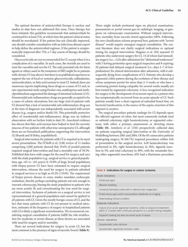
ACG Clinical Guideline
© 2015 by the American College of Gastroenterology The American Journal of GASTROENTEROLOGY
39
Th e optimal duration of antimicrobial therapy is unclear and
studies to date have not addressed this issue. Once therapy has
been initiated, this guideline recommends that antimicrobials be
continued for at least 72 h, at which time the patient’s clinical status
should be reevaluated. If the patient has not clinically improved,
one should consider consultation with an infectious disease expert
to help defi ne the antimicrobial regimen. If the patient is sympto-
matically improved aft er 72 h, a 7-day course of therapy should be
considered.
Glucocorticoids are not recommended for CI, except when it is a
complication of a vasculitis. In such cases, the steroids are used to
treat the vasculitis and not the CI. Th e only potential role for these
agents or others used in the treatment of IBD might be in patients
with chronic CI (see above), but there is no published experience to
support the use of local or systemic glucocorticoids, sulfasalazine,
aminosalicylates, or fatty acid enemas to treat CI. Indeed, there are
numerous reports implicating these drugs as a cause of CI and, in
one experimental study using Fischer rats, azathioprine and meth-
ylprednisolone augmented the damage of intestinal ischemia ( 155 ).
Nonsteroidal anti-infl ammatory drugs are generally accepted to be
a cause of colonic ulcerations, but one large trial of patients with
CI showed that a lack of nonsteroidal anti-infl ammatory drug use
at the time of diagnosis was independently associated with severe
disease (e.g., surgery and/or death) ( 9 ). Th is potentially protective
eff ect of nonsteroidal anti-infl ammatory drugs was an indirect
observation with no further trials to date ( 9 ). Anecdotal evidence
has shown that fatty acid enemas have helped to heal the necrotic
mucosa of two patients with otherwise unresponsive CI, although
there are no formalized publications supporting this intervention
(LJ Brandt and SJ Boley, unpublished).
Surgical intervention for patients with CI is required in its most
severe presentations. Th e O’Neill et al. ( 148 ) review of 11 studies
comprising 1,049 patients showed that 19.6% of pooled patients
required surgical intervention and had a mortality rate of 39.3%.
Published data have wide ranges for the need for surgery and vary
with the study population (e.g., surgical service vs. general popula-
tion; age >65 vs. <65 years); 8–19.8% of large, broad populations
with biopsy-proven CI have been estimated to require surgical
intervention, whereas the need for surgery in patients admitted
to surgical services is as high as 45.2% ( 7,9,94 ). Th e requirement
of biopsy-proven disease in some studies mandates endoscopic
evaluation, thereby perhaps excluding patients not sick enough to
warrant colonoscopy, biasing the study population to patients who
are more acutely ill, and overestimating the true need for surgi-
cal intervention. Inclusion of patients on a surgical service is not
representational of a general population and cannot be applied to
all patients with CI. Given the mostly benign course of CI, and the
fact that many patients with CI do not present to medical atten-
tion, estimates of the frequency of surgery in hospitalized patients
with CI is likely a signifi cant overestimation. We recommend con-
sidering surgical consultation if patients fulfi ll the risk stratifi ca-
tion for moderate or severe disease as these factors are associated
with need for surgery and/or mortality.
Th ere are several indications for surgery in acute CI, but the
most common is the presence of signs of necrotic bowel ( Table 9 ).
Th ese might include peritoneal signs on physical examination,
pneumatosis or portal venous gas on radiologic imaging, or gan-
grene on colonoscopic examination. Without surgical interven-
tion, mortality from necrotic bowel approaches 100%. Following
the new classifi cation scheme proposed here, patients with “severe
disease” would require emergent surgical consultation. Th e cur-
rent literature does not clarify surgical indications or optimal
timing for surgical intervention. Huguier et al. ( 94 ) assessed 33
patients who underwent surgery for CI, 13 of whom had immedi-
ate surgery (i.e., <12 h aft er admission) for “abdominal tenderness,”
with 5 having peritonitis upon surgical inspection and 8 expiring;
20 patients had delayed surgical treatment (i.e., days 2–7 of hos-
pitalization) for clinical deterioration aft er admission, with 6 sub-
sequently dying from complications of CI. Patients who develop a
segmental colitis pattern during the evolution of their disease and
whose symptoms persist for more than 2–3 weeks or who have a
continuing protein-losing colopathy for several weeks usually are
best treated by segmental colectomy. A less-recognized indication
for surgery is the development of recurrent sepsis in a patient who
has symptomatically recovered from an acute episode of CI. Such
patients usually have a short segment of unhealed bowel that, via
bacterial translocation, is the source of the sepsis; resection of this
segment is curative.
Th e surgical procedure patients undergo for CI depends upon
the aff ected segment of colon, but most commonly include total
or subtotal colectomy, right hemicolectomy, or segmental colec-
tomy with either a primary anastomosis or diverting stoma
( Table 10 ). Antolovic et al. ( 156 ) prospectively collected data
on patients requiring surgical intervention at the University of
Heidelberg between 2001 and 2004. Of the 85 consecutive patients
undergoing surgery, 56 (66.7%) required procedures within 24 h
of presentation to the surgical service. Left hemicolectomy was
performed in 8%, right hemicolectomy in 26%, sigmoid resec-
tion in 5%, and total colectomy in 49%, with the remainder hav-
ing other segmental resections; 42% had a Hartmann procedure
Table 9 . Indications for surgery in colonic ischemia
Acute indications
Peritoneal signs
Massive bleeding
Universal fulminant colitis with or without toxic megacolon
Portal venous gas and/or pneumatosis intestinalis on imaging
Deteriorating clinical condition
Subacute indications
Failure of an acute segmental ischemic colitis to respond to treatment
within 2–3 weeks with continued symptoms or a protein-losing colopathy
Apparent healing but with recurrent bouts of sepsis
Chronic indications
Symptomatic colon stricture
Symptomatic segmental ischemic colitis
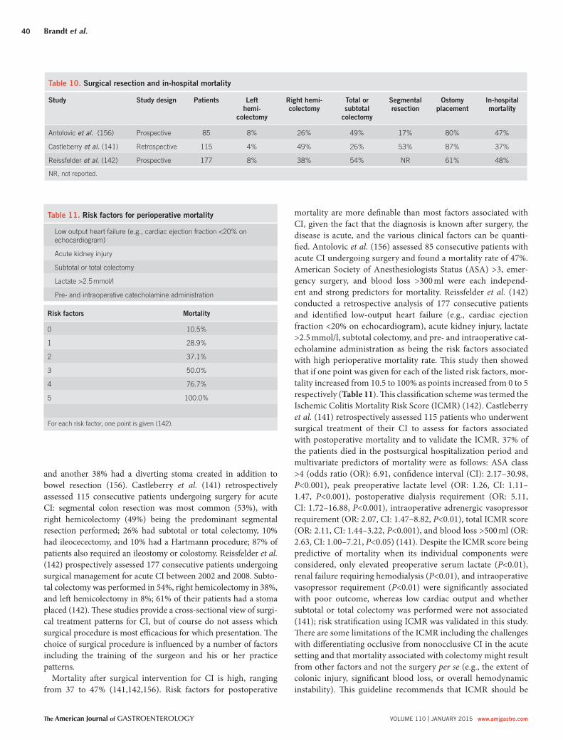
Brandt et al.
The American Journal of GASTROENTEROLOGY VOLUME 110 | JANUARY 2015 www.amjgastro.com
40
and another 38% had a diverting stoma created in addition to
bowel resection ( 156 ). Castleberry et al. ( 141 ) retrospectively
assessed 115 consecutive patients undergoing surgery for acute
CI: segmental colon resection was most common (53%), with
right hemicolectomy (49%) being the predominant segmental
resection performed; 26% had subtotal or total colectomy, 10%
had ileocecectomy, and 10% had a Hartmann procedure; 87% of
patients also required an ileostomy or colostomy. Reissfelder et al.
( 142 ) prospectively assessed 177 consecutive patients undergoing
surgical management for acute CI between 2002 and 2008. Subto-
tal colectomy was performed in 54%, right hemicolectomy in 38%,
and left hemicolectomy in 8%; 61% of their patients had a stoma
placed ( 142 ). Th ese studies provide a cross-sectional view of surgi-
cal treatment patterns for CI, but of course do not assess which
surgical procedure is most effi cacious for which presentation. Th e
choice of surgical procedure is infl uenced by a number of factors
including the training of the surgeon and his or her practice
patterns.
Mortality aft er surgical intervention for CI is high, ranging
from 37 to 47% ( 141,142,156 ). Risk factors for postoperative
mortality are more defi nable than most factors associated with
CI, given the fact that the diagnosis is known aft er surgery, the
disease is acute, and the various clinical factors can be quanti-
fi ed. Antolovic et al. ( 156 ) assessed 85 consecutive patients with
acute CI undergoing surgery and found a mortality rate of 47%.
American Society of Anesthesiologists Status (ASA) >3, emer-
gency surgery, and blood loss >300 ml were each independ-
ent and strong predictors for mortality. Reissfelder et al. ( 142 )
conducted a retrospective analysis of 177 consecutive patients
and identifi ed low-output heart failure (e.g., cardiac ejection
fraction <20% on echocardiogram), acute kidney injury, lactate
>2.5 mmol/l, subtotal colectomy, and pre- and intraoperative cat-
echolamine administration as being the risk factors associated
with high perioperative mortality rate. Th is study then showed
that if one point was given for each of the listed risk factors, mor-
tality increased from 10.5 to 100% as points increased from 0 to 5
respectively ( Table 11 ). Th is classifi cation scheme was termed the
Ischemic Colitis Mortality Risk Score (ICMR) ( 142 ). Castleberry
et al. ( 141 ) retrospectively assessed 115 patients who underwent
surgical treatment of their CI to assess for factors associated
with postoperative mortality and to validate the ICMR. 37% of
the patients died in the postsurgical hospitalization period and
multivariate predictors of mortality were as follows: ASA class
>4 (odds ratio (OR): 6.91, confi dence interval (CI): 2.17–30.98,
P <0.001), peak preoperative lactate level (OR: 1.26, CI: 1.11–
1.47, P <0.001), postoperative dialysis requirement (OR: 5.11,
CI: 1.72–16.88, P <0.001), intraoperative adrenergic vasopressor
requirement (OR: 2.07, CI: 1.47–8.82, P <0.01), total ICMR score
(OR: 2.11, CI: 1.44–3.22, P <0.001), and blood loss >500 ml (OR:
2.63, CI: 1.00–7.21, P <0.05) ( 141 ). Despite the ICMR score being
predictive of mortality when its individual components were
considered, only elevated preoperative serum lactate ( P <0.01),
renal failure requiring hemodialysis ( P <0.01), and intraoperative
vasopressor requirement ( P <0.01) were signifi cantly associated
with poor outcome, whereas low cardiac output and whether
subtotal or total colectomy was performed were not associated
( 141 ); risk stratifi cation using ICMR was validated in this study.
Th ere are some limitations of the ICMR including the challenges
with diff erentiating occlusive from nonocclusive CI in the acute
setting and that mortality associated with colectomy might result
from other factors and not the surgery per se (e.g., the extent of
colonic injury, signifi cant blood loss, or overall hemodynamic
instability). Th is guideline recommends that ICMR should be
Table 10 . Surgical resection and in-hospital mortality
Study Study design Patients Left
hemi-
colectomy
Right hemi-
colectomy
Total or
subtotal
colectomy
Segmental
resection
Ostomy
placement
In-hospital
mortality
Antolovic et al. ( 156 ) Prospective 85 8% 26% 49% 17% 80% 47%
Castleberry et al. ( 141 ) Retrospective 115 4% 49% 26% 53% 87% 37%
Reissfelder et al. ( 142 ) Prospective 177 8% 38% 54% NR 61% 48%
NR, not reported.
Table 11 . Risk factors for perioperative mortality
Low output heart failure (e.g., cardiac ejection fraction <20% on
echocardiogram)
Acute kidney injury
Subtotal or total colectomy
Lactate >2.5 mmol/l
Pre- and intraoperative catecholamine administration
Risk factors Mortality
0 10.5%
1 28.9%
2 37.1%
3 50.0%
4 76.7%
5 100.0%
For each risk factor, one point is given ( 142 ).

ACG Clinical Guideline
© 2015 by the American College of Gastroenterology The American Journal of GASTROENTEROLOGY
41
considered as part of the risk assessment of patients with acute
CI who need surgical intervention.
Colon stricture aft er an episode of CI may be asymptomatic or
even resolve over months to years. Surgery is indicated only when
an ischemic stricture produces symptoms; in such cases, segmental
resection is adequate. Transendoscopic dilation of an ischemic stric-
ture is an alternative to surgery, although an unproven one. Chronic
segmental CI is a more controversial indication for surgery and, as
with other colitides, the decision to abandon medical therapy is a
complex one that must be individualized for each case. Recurrent
CI is uncommon and resection of the involved segment of colon,
while usually curative, does not necessarily protect against recur-
rent CI in other areas of the colon. Th ere are no published data on
the frequency of such recurrence following surgery for recurrence.
Treatment of CI depends upon disease severity at presentation
(see Figure 1 ). Th e overwhelming majority of patients will require
simple conservative measures to manage their disease including
fasting, intravenous fl uids, and correction of underlying condi-
tions. If patients have moderate or severe disease, broad-spectrum
antimicrobial coverage should be instituted along with surgical
evaluation. Surgery should be consulted promptly for patients with
severe disease or colon necrosis.
ACKNOWLEDGMENTS
Th is guideline was produced in collaboration with the Practice
Parameters Committee of the American College of Gastroenterol-
ogy. Th e Committee gives special thanks to Fouad J. Moawad, MD,
FACG, who served as guideline monitor for this document.
CONFLICT OF INTEREST
Guarantor of the article : Lawrence J. Brandt, MD, MACG, AGAF,
FASGE.
Specifi c author contributions: Lawrence J. Brandt, Paul Feuerstadt,
George F. Longstreth, and Scott J. Boley assisted in design, draft ing,
and critical revision of the manuscript for intellectual content.
Financial support: None.
Potential competing interests: None.
REFERENCES 1. Atkins D , Best D , Briss PA et al. Grading quality of evidence and strength
of recommendations . BMJ 2004 ; 328 : 1490 . 2. Guyatt GH , Oxman AD , Vist GE et al. GRADE: an emerging consensus
on rating quality of evidence and strength of recommendations . BMJ 2008 ; 336 : 924 – 6 .
3. Sands BE , Duh MS , Cali C et al. Algorithms to identify colonic ischemia, complications of constipation and irritable bowel syndrome in medical claims data: development and validation . Pharmacoepidemiol Drug Saf 2006 ; 15 : 47 – 56
4. Hreinsson JP , Gumundsson S , Kalaitzakis E et al. Lower gastrointestinal bleeding: incidence, etiology, and outcomes in a population-based setting . Eur J Gastroenterol Hepatol 2013 ; 25 : 37 – 43 .
5. Arroja B , Cremers I , Ramos R et al. Acute lower gastrointestinal bleeding management in Portugal: a multicentric prospective 1-year survey . Eur J Gastroenterol Hepatol 2011 ; 23 : 317 – 22 .
6. Longstreth GF . Epidemiology and outcome of patients hospitalized with acute lower gastrointestinal hemorrhage: a population-based study . Am J Gastroenterol 1997 ; 92 : 419 – 24 .
7. Brandt LJ , Feuerstadt P , Blaszka MC . Anatomic patterns, patient charac-teristics, and clinical outcomes in ischemic colitis: a study of 313 cases supported by histology . Am J Gastroenterol 2010 ; 105 : 2245 – 52 . quiz 2253 .
8. Suh DC , Kahler KH , Choi IS et al. Patients with irritable bowel syndrome or constipation have an increased risk for ischaemic colitis . Aliment Pharmacol Th er 2007 ; 25 : 681 – 92 .
9. Longstreth GF , Yao JF . Epidemiology, clinical features, high-risk factors, and outcome of acute large bowel ischemia . Clin Gastroenterol Hepatol 2009 ; 7 : 1075 – 80 . e1071-1072; quiz 1023 .
10. Yadav S , Dave M , Varayil JE et al. A population-based study of incidence, risk factors, clinical spectrum, and outcomes of ischemic colitis . Clin Gastro-enterol Hepatol 2014 ; e-pub ahead of print 12 August 2014 .
11. Ganguli K , Tanpowpong P , Katz A . Abdominal pain aft er hypovolemic shock in a 5-year old boy . Clin Pediatr 2012 ; 51 : 1202 – 4 .
12. Yanagisawa A , Namai Y , Sekine T et al. Ischemic colitis as a complication in a patient with steroid-dependent nephrotic syndrome . Pediatr Nephrol 2008 ; 23 : 655 – 7 .
13. Cole J , Cook S , Sands B et al. Occurrence of colon ischemia in relation to irritable bowel syndrome . Am J Gastroenterol 2004 ; 99 : 486 – 91 .
14. Walker A , Bohn R , Cali C et al. Risk factors for colon ischemia . Am J Gastroenterol 2004 ; 99 : 1333 – 7 .
15. Chang L , Kahler KH , Sarawate C et al. Assessment of potential risk factors associated with ischaemic colitis . Neurogastroenterol Motil 2008 ; 20 : 36 – 42
16. Sotiriadis J , Brandt L , Behin D et al. Ischemic colitis has a worse prog-nosis when isolated to the right side of the colon . Am J Gastroenterol 2007 ; 102 : 2247 – 52 .
17. Montoro MA , Brandt LJ , Santolaria S et al. Clinical patterns and outcomes of ischaemic colitis: results of the Working Group for the Study of Ischae-mic Colitis in Spain (CIE study) . Scand J Gastroenterol 2011 ; 46 : 236 – 46 .
18. Paterno F , McGillicuddy EA , Schuster KM et al. Ischemic colitis: risk fac-tors for eventual surgery . Am J Surg 2010 ; 200 : 646 – 50 .
19. Kimura T , Shinji A , Horiuchi A et al. Clinical characteristics of young-onset ischemic colitis . Dig Dis Sci 2012 ; 57 : 1652 – 9 .
20. Cubiella Fernandez J , Nunez Calvo L , Gonzalez Vazquez E et al. Risk factors associated with the development of ischemic colitis . World J Gas-troenterol 2010 ; 16 : 4564 – 9 .
21. Heyn J , Buhmann S , Ladurner R et al. Recurrent ischemic colitis in a pa-tient with leiden factor V mutation and systemic lupus erythematous with antiphospholipid syndrome . Eur J Med Res. 2008 ; 13 : 182 – 4 .
22. Schuler JG , Hudlin MM . Cecal necrosis: infrequent variant of ischemic colitis. Report of fi ve cases . Dis Colon Rectum 2000 ; 43 : 708 – 12 .
23. Beppu K , Osada T , Nagahara A et al. Relationship between endo-scopic fi ndings and clinical severity in ischemic colitis . Intern Med 2011 ; 50 : 2263 – 7 .
24. Lee TC , Wang HP , Chiu HM et al. Male gender and renal dysfunction are predictors of adverse outcome in nonpostoperative ischemic colitis patients . J Clin Gastroenterol 2010 ; 44 : e96 – 100 .
25. Flobert C , Cellier C , Berger A et al. Right colonic involvement is associat-ed with severe forms of ischemic colitis and occurs frequently in patients with chronic renal failure requiring hemodialysis . Am J Gastroenterol 2000 ; 95 : 195 – 8 .
26. Koutroubakis IE , Sfi ridaki A , Th eodoropoulou A et al. Role of acquired and hereditary thrombotic risk factors in colon ischemia of ambulatory patients . Gastroenterology 2001 ; 121 : 561 – 5 .
27. Midian-Singh R , Polen A , Durishin C et al. Ischemic colitis revisited: a prospective study identifying hypercoagulability as a risk factor . South Med J 2004 ; 97 : 120 – 3 .
28. Th eodoropoulou A , Sfi ridaki A , Oustamanolakis P et al. Genetic risk factors in young patients with ischemic colitis . Clin Gastroenterol Hepatol 2008 ; 6 : 907 – 11 .
29. Perry RJ , Martin MJ , Eckert MJ et al. Colonic ischemia complicating open vs endovascular abdominal aortic aneurysm repair . J Vasc Surg 2008 ; 48 : 272 – 7 .
30. Champagne BJ , Lee EC , Valerian B et al. Incidence of colonic ischemia aft er repair of ruptured abdominal aortic aneurysm with endograft . J Am Coll Surg 2007 ; 204 : 597 – 602
31. Elramah M , Einstein M , Mori N et al. High mortality of cocaine-related ischemic colitis: a hybrid cohort/case-control study . Gastrointest Endosc 2012 ; 75 : 1226 – 32 .
32. Longstreth GF , Yao JF . Diseases and drugs that increase risk of acute large bowel ischemia . Clin Gastroenterol Hepatol 2010 ; 8 : 49 – 54
33. Hass D , Kozuch P , Brandt L . Pharmacologically mediated colon ischemia . Am J Gastroenterol 2007 ; 102 : 1765 – 80 .
34. Chang L , Chey W , Harris L et al. Incidence of ischemic colitis and serious complications of constipation among patients using alosetron: system-atic review of clinical trials and post-marketing surveillance data . Am J Gastroenterol 2006 ; 101 : 1069 – 79 .

Brandt et al.
The American Journal of GASTROENTEROLOGY VOLUME 110 | JANUARY 2015 www.amjgastro.com
42
35. Michael H , Brandt LJ , Hochsztein JG . Ischemic colitis complicating aortic dissection . Gastrointest Endosc 2002 ; 55 : 442 – 4 .
36. Hourmand-Ollivier I , Bouin M , Saloux E et al. Cardiac sources of embo-lism should be routinely screened in ischemic colitis . Am J Gastroenterol 2003 ; 98 : 1573 – 7 .
37. Cappell MS , Mahajan D , Kurupath V . Characterization of ischemic colitis associated with myocardial infarction: an analysis of 23 patients . Am J Med 2006 ; 119 : 527 – e521-529 .
38. Chavalitdhamrong D , Jensen DM , Kovacs TO et al. Ischemic colitis as a cause of severe hematochezia: risk factors and outcomes compared with other colon diagnoses . Gastrointest Endosc 2011 ; 74 : 852 – 7 .
39. Park CJ , Jang MK , Shin WG et al. Can we predict the development of ischemic colitis among patients with lower abdominal pain? Dis Colon Rectum 2007 ; 50 : 232 – 8 .
40. Nadar S , Blann AD , Lip GY . Endothelial dysfunction: methods of assess-ment and application to hypertension . Curr Pharm Des 2004 ; 10 : 3591 – 605 .
41. Su Y , Liu XM , Sun YM et al. Endothelial dysfunction in impaired fasting glycemia, impaired glucose tolerance, and type 2 diabetes mellitus . Am J Cardiol 2008 ; 102 : 497 – 8 .
42. Murdaca G , Colombo BM , Cagnati P et al. Endothelial dysfunction in rheumatic autoimmune diseases . Atherosclerosis 2012 ; 224 : 309 – 17 .
43. Gonzalez-Gay MA , Gonzalez-Juanatey C , Vazquez-Rodriguez TR et al. Endothelial dysfunction, carotid intima-media thickness, and accelerated atherosclerosis in rheumatoid arthritis . Semin Arthritis Rheum 2008 ; 38 : 67 – 70 .
44. Cervera R , Espinosa G , Cordero A et al. Intestinal involvement second-ary to the antiphospholipid syndrome (APS): clinical and immunologic characteristics of 97 patients: comparison of classic and catastrophic APS . Semin Arthritis Rheum 2007 ; 36 : 287 – 96 .
45. Chin BW , Greenberg D , Wilson RB et al. A case of ischemic colitis associ-ated with factor V Leiden mutation: successful treatment with anticoagu-lation . Gastrointest Endosc 2007 ; 66 : 416 – 8 .
46. Manabe N , Tanaka T , Hata J et al. Pathophysiology underlying irritable bowel syndrome--from the viewpoint of dysfunction of autonomic nerv-ous system activity . J Smooth Muscle Res 2009 ; 45 : 15 – 23 .
47. Rosenblum JD , Boyle CM , Schwartz LB . Th e mesenteric circulation. Anatomy and physiology . Surg Clin North Am 1997 ; 77 : 289 – 306 .
48. Boley SJ , Agrawal GP , Warren AR et al. Pathophysiologic eff ects of bowel distention on intestinal blood fl ow . Am J Surg 1969 ; 117 : 228 – 34 .
49. Chang HJ , Chung CW , Ko KH et al. Clinical characteristics of ischemic colitis according to location . J Korean Soc Coloproctol 2011 ; 27 : 282 – 6 .
50. Th eodoropoulou A , Koutroubakis IE . Ischemic colitis: clinical practice in diagnosis and treatment . World J Gastroenterol 2008 ; 14 : 7302 – 8 .
51. Brandt LJ . Th rombophilia and colon ischemia: aura popularis? Gastro-enterology 2001 ; 121 : 724 – 6 .
52. Kyriakos R , Siewert B , Kato E et al. CT fi ndings in runner’s colitis . Abdom Imaging 2006 ; 31 : 54 – 6 .
53. Sanchez LD , Tracy JA , Berkoff D et al. Ischemic colitis in marathon runners: a case-based review . J Emerg Med 2006 ; 30 : 321 – 6 .
54. Cohen DC , Winstanley A , Engledow A et al. Marathon-induced ischemic colitis: why running is not always good for you . Am J Emerg Med 2009 ; 27 : 255 – e255-257 .
55. Lucas W , Schroy PC 3rd . Reversible ischemic colitis in a high endurance athlete . Am J Gastroenterol 1998 ; 93 : 2231 – 4 .
56. Sullivan SN , Wong C . Runners’ diarrhea. Diff erent patterns and associated factors . J Clin Gastroenterol 1992 ; 14 : 101 – 4 .
57. Rudzki SJ , Hazard H , Collinson D . Gastrointestinal blood loss in triath-letes: it’s etiology and relationship to sports anaemia . Aust J Sci Med Sport 1995 ; 27 : 3 – 8 .
58. Green BT , Branch MS . Ischemic colitis in a young adult during sickle cell crisis: case report and review . Gastrointest Endosc 2003 ; 57 : 605 – 7 .
59. Stewart CL , Menard GE . Sickle cell-induced ischemic colitis . J Natl Med Assoc 2009 ; 101 : 726 – 8 .
60. Karim A , Ahmed S , Rossoff LJ et al. Fulminant ischaemic colitis with atypical clinical features complicating sickle cell disease . Postgrad Med J 2002 ; 78 : 370 – 2 .
61. Qureshi A , Lang N , Bevan DH . Sickle cell ‘girdle syndrome’ progress-ing to ischaemic colitis and colonic perforation . Clin Lab Haematol 2006 ; 28 : 60 – 2 .
62. Sada S , Benini L , Pavan C et al. Ischemic colitis sustained by sickle cell trait in young adult patient . Am J Gastroenterol 2005 ; 100 : 2818 – 21 .
63. Manolakis AC , Kapsoritakis AN , Ioannou M et al. Sickle cell trait-related ischemic colitis in a patient with Sjogren’s syndrome . Am J Gastroenterol 2008 ; 103 : 2952 – 4 .
64. Lee SO , Kim SH , Jung SH et al. Colonoscopy-induced ischemic colitis in patients without risk factors . World J Gastroenterol 2014 ; 20 : 3698 – 702 .
65. Yuksel O , Bolat AD , Koklu S et al. Ischemic colitis, an unusual complica-tion of colonoscopy . South Med J 2008 ; 101 : 972 – 3 .
66. Cheng YC , Wu CC , Lee CC et al. Rare complication following screening colonoscopy: ischemic colitis . Dig Endosc 2012 ; 24 : 379 .
67. Arhan M , Onal IK , Odemis B et al. Colonoscopy-induced ischemic colitis in a young patient with no risk factor . Am J Gastroenterol 2009 ; 104 : 250 – 1 .
68. Versaci A , Macri A , Scuderi G et al. Ischemic colitis following colono-scopy in a systemic lupus erythematosus patient: report of a case . Dis Colon Rectum 2005 ; 48 : 866 – 9 .
69. Brandt LJ , Boley SJ , Sammartano R . Carbon dioxide and room air insuf-fl ation of the colon. Eff ects on colonic blood fl ow and intraluminal pres-sure in the dog . Gastrointest Endosc 1986 ; 32 : 324 – 9 .
70. Duenas-Laita A , Mena-Martin FJ , Roquelai-Ruiz P et al. Ischemic colitis associated with acute carbon monoxide poisoning . Clin Toxicol (Phila) 2008 ; 46 : 780 – 1 .
71. Szmulowicz UM , Savoie LM . Ischemic colitis: an uncommon manifesta-tion of pheochromocytoma . Am Surg 2007 ; 73 : 400 – 3 .
72. Payor AD , Tucci V . Acute ischemic colitis secondary to air embolism aft er diving . Int J Crit Illn Inj Sci 2011 ; 1 : 73 – 8 .
73. Low SR , Strugnell N , Nikfarjam M . Ischaemic colitis associated with colonic carcinoma . ANZ J Surg 2008 ; 78 : 319 – 21 .
74. Butcher JH , Davis AJ , Page A et al. Transient ischaemic colitis following an aeroplane fl ight: two case reports and review of the literature . Gut 2002 ; 51 : 746 – 7 .
75. Kim MK , Cho YS , Kim HK et al. Transient ischemic colitis aft er a pit viper bite (Agkistrodon blomhoffi i brevicaudus) . J Clin Gastroenterol 2008 ; 42 : 111 – 2 .
76. Rosenberg H , Beck J . Jujitsu kick to the abdomen: a case of blunt abdomi-nal trauma resulting in hematochezia and transient ischemic colitis . Ann Emerg Med 2011 ; 58 : 189 – 91 .
77. Lepow H , Bernstein LH , Brandt LJ et al. Vascular occlusion and stricture of the sigmoid colon secondary to trauma from a pneumatic hammer . J Trauma 1977 ; 17 : 69 – 73 .
78. Park MG , Hur H , Min BS et al. Colonic ischemia following surgery for sigmoid colon and rectal cancer: a study of 10 cases and a review of the literature . Int J Colorectal Dis 2012 ; 27 : 671 – 5 .
79. Sato H , Koide Y , Shiota M et al. Clinical characteristics of ischemic colitis aft er surgery for colorectal cancer . Surg Today 2014 ; 44 : 1090 – 6 .
80. Mosli M , Parfi tt J , Gregor J . Retrospective analysis of disease associa-tion and outcome in histologically confi rmed ischemic colitis . J Dig Dis 2013 ; 14 : 238 – 43 .
81. Glauser PM , Wermuth P , Cathomas G et al. Ischemic colitis: clinical presentation, localization in relation to risk factors, and long-term results . World J Surg 2011 ; 35 : 2549 – 54 .
82. Fujiogi TKT , Yasuno M . Brown tubular-shaped object rectally expelled in a patient with persistent diarrhea . Gastroenterology 2013 ; 145 : 1205 .
83. Erguney S , Yavuz N , Ersoy YE et al. Passage of “colonic cast” aft er colorec-tal surgery: report of four cases and review of the literature . J Gastrointest Surg. 2007 ; 11 : 1045 – 51 .
84. Longstreth GF , Mottet MD . Passage of a large bowel cast aft er acute large-bowel ischemia . Clin Gastroenterol Hepatol 2009 ; 7 : e59 – e60 .
85. Su TH , Liou JM , Wang HP . Th e passage of a colonic cast . Lancet 2010 ; 375 : 2099 .
86. Medina C , Vilaseca J , Videla S et al. Outcome of patients with ischemic colitis: review of fi ft y-three cases . Dis Colon Rectum 2004 ; 47 : 180 – 4 .
87. Yikilmaz A , Karahan OI , Senol S et al. Value of multislice computed tomography in the diagnosis of acute mesenteric ischemia . Eur J Radiol 2011 ; 80 : 297 – 302 .
88. Brandt L , Boley S , Goldberg L et al. Colitis in the elderly. A reappraisal . Am J Gastroenterol 1981 ; 76 : 239 – 45 .
89. Kornblith PL , Boley SJ , Whitehouse BS . Anatomy of the splanchnic circu-lation . Surg Clin North Am 1992 ; 72 : 1 – 30 .
90. Boley SJ , Brandt LJ , Veith FJ . Ischemic disorders of the intestines . Curr Probl Surg 1978 ; 15 : 1 – 85 .
91. Brandt LJ , Boley SJ . Colonic ischemia . Surg Clin North Am 1992 ; 72 : 203 – 29 .
92. Bharucha AE , Tremaine WJ , Johnson CD et al. Ischemic proctosigmoidi-tis . Am J Gastroenterol 1996 ; 91 : 2305 – 9 .
93. Longo WE , Ballantyne GH , Gusberg RJ . Ischemic colitis: patterns and prognosis . Dis Colon Rectum 1992 ; 35 : 726 – 30 .

ACG Clinical Guideline
© 2015 by the American College of Gastroenterology The American Journal of GASTROENTEROLOGY
43
125. Kirkpatrick I , Kroeker M , Greenberg H . Biphasic CT with mesenteric CT angiography in the evaluation of acute mesenteric ischemia: Initial experience . Radiology 2003 ; 229 : 91 – 8 .
126. Wiesner W , Hauser A , Steinbrich W . Accuracy of multidetector row computed tomography for the diagnosis of acute bowel ischemia in a non-selected study population . Eur Radiol 2004 ; 14 : 2347 – 56 .
127. Aschoff AJ , Stuber G , Becker BW et al. Evaluation of acute mesenteric ischemia: accuracy of biphasic mesenteric multi-detector CT angiography . Abdom Imaging 2009 ; 34 : 345 – 57 .
128. Alturkistany S , Artho G , Maheshwari S et al. Transmural colonic ischemia: clin-ical features and computed tomography fi ndings . Clin Imaging. 2012 ; 36 : 35 – 40
129. Wiesner W , Mortele KJ , Glickman JN et al. Pneumatosis intestinalis and portomesenteric venous gas in intestinal ischemia: correlation of CT fi nd-ings with severity of ischemia and clinical outcome . AJR Am J Roentgenol 2001 ; 177 : 1319 – 23 .
130. Taourel P , Garibaldi F , Arrigoni J et al. Cecal pneumatosis in patients with obstructive colon cancer: correlation of CT fi ndings with bowel viability . AJR Am J Roentgenol 2004 ; 183 : 1667 – 71 .
131. Ripolles T , Simo L , Martinez-Perez MJ et al. Sonographic fi ndings in ischemic colitis in 58 patients . AJR Am J Roentgenol 2005 ; 184 : 777 – 85 .
132. Danse EM , Jamart J , Hoang P et al. Focal bowel wall changes detected with colour Doppler ultrasound: diagnostic value in acute non-diverticu-lar diseases of the colon . Br J Radiol 2004 ; 77 : 917 – 21 .
133. Taourel P , Aufort S , Merigeaud S et al. Imaging of ischemic colitis . Radiol Clin North Am 2008 ; 46 : 909 – 24 . vi .
134. Mazzei MA , Guerrini S , Cioffi Squitieri N et al. Magnetic resonance imaging: is there a role in clinical management for acute ischemic colitis? World J Gastroenterol 2013 ; 19 : 1256 – 63 .
135. Kozarek RA , Earnest DL , Silverstein ME et al. Air-pressure-induced colon injury during diagnostic colonoscopy . Gastroenterology 1980 ; 78 : 7 – 14 .
136. Zou X , Cao J , Yao Y et al. Endoscopic fi ndings and clinicopathologic charac-teristics of ischemic colitis: a report of 85 cases . Dig Dis Sci 2009 ; 54 : 2009 – 15 .
137. Zuckerman G , Prakash C , Merriman R et al. Th e colon single-stripe sign and its relationship to ischemic colitis . Am J Gastroenterol 2003 ; 98 : 2018 – 22 .
138. Mitsudo S , Brandt LJ . Pathology of intestinal ischemia . Surg Clin North America 1992 ; 72 : 43 – 63 .
139. Matsumoto S , Tsuji K , Shirahama S . Clinical investigation of 41 patients with ischemic colitis accompanied by ulcer . World J Gastroenterol 2007 ; 13 : 1236 – 9 .
140. Yoshiya K , Lapchak PH , Th ai TH et al. Depletion of gut commensal bacteria attenuates intestinal ischemia/reperfusion injury . Am J Physiol Gastrointest Liver Physiol 2011 ; 301 : G1020 – G1030 .
141. Castleberry AW , Turley RS , Hanna JM et al. A 10-year longitudinal analy-sis of surgical management for acute ischemic colitis . J Gastrointest Surg 2013 ; 17 : 784 – 92 .
142. Reissfelder C , Sweiti H , Antolovic D et al. Ischemic colitis: who will sur-vive? Surgery 2011 ; 149 : 585 – 92 .
143. O’Neill S , Elder K , Harrison SJ et al. Predictors of severity in ischaemic colitis . Int J Colorectal Dis 2012 ; 27 : 187 – 91 .
144. Chung JW , Cheon JH , Park JJ et al. Development and validation of a novel prognostic scoring model for ischemic colitis . Dis Colon Rectum 2010 ; 53 : 1287 – 94 .
145. Diaz Nieto R , Varcada M , Ogunbiyi OA et al. Systematic review on the treatment of ischaemic colitis . Colorectal Dis 2011 ; 13 : 744 – 7 .
146. Cohn I Jr , Floyd CE , Dresden CF et al. Strangulation obstruction in ger-mfree animals . Ann Surg 1962 ; 156 : 692 – 702 .
147. Path EJ , Mc CJ Jr . Intestinal obstruction; the protective action of sulfasux-idine and sulfathalidine to the ileum following vascular damage . Ann Surg 1950 ; 131 : 159 – 70 . illust .
148. O’Neill S , Yalamarthi S . Systematic review of the management of ischae-mic colitis . Colorectal Dis 2012 ; 14 : e751 – e763 .
149. Sarnoff SJ , Fine J . Th e eff ect of chemotherapy on the ileum subjected to vascular injury . Ann Surg 1945 ; 121 : 74 – 82 .
150. Jamieson W , Pliagus G , Marchuk S et al. Eff ect of antibiotic and fl uid resuscitation upon survival time in experimental intestinal ischemia . Surg Gynecol Obstet 1988 ; 167 : 103 – 8 .
151. Bennion RS , Wilson SE , Williams RA . Early portal anaerobic bacteremia in mesenteric ischemia . Arch Surg 1984 ; 119 : 151 – 5 .
152. Redan JA , Rush BF , McCullough JN et al. Organ distribution of radio-labeled enteric Escherichia coli during and aft er hemorrhagic shock . Ann Surg 1990 ; 211 : 663 – 6 .
153. Luo CC , Shih HH , Chiu CH et al. Translocation of coagulase-negative bacterial staphylococci in rats following intestinal ischemia-reperfusion injury . Biol Neonate 2004 ; 85 : 151 – 4 .
94. Huguier M , Barrier A , Boelle PY et al. Ischemic colitis . Am J Surg 2006 ; 192 : 679 – 84 .
95. Sherid M , Sifuentes H , Samo S et al. Risk factors of recurrent ischemic colitis: a multicenter retrospective study . Korean J Gastroenterol 2014 ; 63 : 283 – 91 .
96. Wakefi eld AJ , Sawyerr AM , Dhillon AP et al. Pathogenesis of Crohn’s disease: multifocal gastrointestinal infarction . Lancet 1989 ; 2 : 1057 – 62 .
97. Pounder RE . Th e pathogenesis of Crohn’s disease . J Gastroenterol 1994 ; 29 : 11 – 15 .
98. Th ompson NP , Wakefi eld AJ , Pounder RE . Inherited disorders of coagula-tion appear to protect against infl ammatory bowel disease . Gastroenterol-ogy 1995 ; 108 : 1011 – 5 .
99. Langman MJ . Can incoagulable blood protect against infl ammatory bowel disease? Gastroenterology 1995 ; 108 : 1305 – 7 .
100. Calkins BM . A meta-analysis of the role of smoking in infl ammatory bowel disease . Dig Dis Sci 1989 ; 34 : 1841 – 54 .
101. Aroniadis O , Feuerstadt P , Brandt LJ . Prevalence and utility of infl amma-tory bowel disease (IBD) markers in colon ischemia . Am J Gastroenterol 2008 ; 103 : S179 .
102. Brandt L , Boley S , Goldberg L et al. Colitis in the elderly. A reappraisal . Am J Gastroenterol 1981 ; 76 : 239 – 45 .
103. Reeders JW , Rosenbusch G , Tytgat GN . Ischaemic colitis associated with carcinoma of the colon . Eur J Radiol 1982 ; 2 : 41 – 7 .
104. Brandt LJ , Boley SJ , Mitsudo S . Clinical characteristics and natural history of colitis in the elderly . Am J Gastroenterol 1982 ; 77 : 382 – 6 .
105. Habu Y , Tahashi Y , Kiyota K et al. Reevaluation of clinical features of ischemic colitis. Analysis of 68 consecutive cases diagnosed by early colonoscopy . Scand J Gastroenterol 1996 ; 31 : 881 – 6 .
106. Longo WE , Ward D , Vernava AM 3rd et al. Outcome of patients with total colonic ischemia . Dis Colon Rectum 1997 ; 40 : 1448 – 54 .
107. Mosele M , Cardin F , Inelmen EM et al. Ischemic colitis in the elderly: pre-dictors of the disease and prognostic factors to negative outcome . Scand J Gastroenterol 2010 ; 45 : 428 – 33 .
108. Añón R , Bosca MM , Sanchiz V et al. Factors predicting poor prognosis in ischemic colitis . World J Gastroenterol 2006 ; 12 : 4875 – 8 .
109. Ullery BS , Boyko AT , Banet GA et al. Colonic ischemia: an under-recog-nized cause of lower gastrointestinal bleeding . J Emerg Med 2004 ; 27 : 1 – 5 .
110. Su C , Brandt LJ , Sigal SH et al. Th e immunohistological diagnosis of E. coli O157:H7 colitis: possible association with colonic ischemia . Am J Gastroenterol 1998 ; 93 : 1055 – 9 .
111. Balthazar E , Yen B , Gordon R . Ischemic colitis: CT evaluation of 54 cases . Radiology 1999 ; 211 : 381 – 8 .
112. Romano S , Romano L , Grassi R . Multidetector row computed tomogra-phy fi ndings from ischemia to infarction of the large bowel . Eur J Radiol 2007 ; 61 : 433 – 41 .
113. Menke J . Diagnostic accuracy of multidetector CT in acute mesenteric ischemia: systematic review and meta-analysis . Radiology 2010 ; 256 : 93 – 101 .
114. Schieda N , Fasih N , Shabana W . Triphasic CT in the diagnosis of acute mesenteric ischaemia . Eur Radiol 2013 ; 23 : 1891 – 900 .
115. Milone M , Di Minno MN , Musella M et al. Computed tomography fi ndings of pneumatosis and portomesenteric venous gas in acute bowel ischemia . World J Gastroenterol 2013 ; 19 : 6579 – 84 .
116. Wittenberg J , Athanasoulis CA , Williams LF Jr et al. Ischemic colitis. Ra-diology and pathophysiology . Am J Roentgenol Radium Th er Nucl Med 1975 ; 123 : 287 – 300 .
117. Boley SJ , Schwartz S , Lash J et al. Reversible vascular occlusion of the colon . Surg Gynecol Obstet 1963 ; 116 : 53 – 60 .
118. Wolff JH , Rubin A , Potter JD et al. Clinical signifi cance of colonoscopic fi ndings associated with colonic thickening on computed tomography: is colonoscopy warranted when thickening is detected? J Clin Gastroenterol 2008 ; 42 : 472 – 5 .
119. Iacobellis F , Berritto D , Fleischmann D et al. CT fi ndings in acute, suba-cute, and chronic ischemic colitis: suggestions for diagnosis . BioMed Res Int 2014 ; 2014 : 895248 .
120. Rutledge RH . Pseudo-ulcerative colitis proximal to obstructing colon carcinoma . Am Surg 1969 ; 35 : 384 – 8 .
121. Toner M , Condell D , O’Briain DS . Obstructive colitis. Ulceroinfl amma-tory lesions occurring proximal to colonic obstruction . Am J Surg Pathol 1990 ; 14 : 719 – 28 .
122. Ko GY , Ha HK , Lee HJ et al. Usefulness of CT in patients with ischemic colitis proximal to colonic cancer . AJR Am J Roentgenol 1997 ; 168 : 951 – 6 .
123. Byun SJ , So BJ . Successful aspiration and thrombolytic therapy for acute superior mesenteric artery occlusion . J Korean Surg Soc 2012 ; 83 : 115 – 8 .
124. Bailey JA , Jacobs DL , Bahadursingh A et al. Endovascular treatment of segmental ischemic colitis . Dig Dis Sci 2005 ; 50 : 774 – 9 .

Brandt et al.
The American Journal of GASTROENTEROLOGY VOLUME 110 | JANUARY 2015 www.amjgastro.com
44
154. Plonka AJ , Schentag JJ , Messinger S et al. Eff ects of enteral and intrave-nous antimicrobial treatment on survival following intestinal ischemia in rats . J Surg Res 1989 ; 46 : 216 – 20 .
155. Gomella LG , Gehrken GA , Hagihara PF et al. Ischemic colitis and immunosuppression. An experimental model . Dis Colon Rectum 1986 ; 29 : 99 – 101 .
156. Antolovic D , Koch M , Hinz U et al. Ischemic colitis: analysis of risk factors for postoperative mortality . Langenbecks Arch Surg. 2008 ; 393 : 507 – 12 .
157. Salk A , Stobaugh DJ , Deepak P et al. Ischaemic colitis in rheumatoid arthritis patients receiving tumour necrosis factor-alpha inhibitors: an analysis of reports to the US FDA Adverse Event Reporting System . Drug Saf 2013 ; 36 : 329 – 34 .
158. Salk A , Stobaugh DJ , Deepak P et al. Ischemic colitis with type I interferons used in the treatment of hepatitis C and multiple sclerosis: an evaluation from the food and drug administration adverse event reporting system and review of the literature . Ann Pharmacother 2013 ; 47 : 537 – 42 .
159. Baik SJ , Kim TH , Yoo K et al. Ischemic colitis during interferon-ribavirin therapy for chronic hepatitis C: a case report . World J Gastroenterol 2012 ; 18 : 4233 – 6 .
160. Holubar SD , Hassinger JP , Dozois EJ et al. Methamphetamine colitis: a rare case of ischemic colitis in a young patient . Arch Surg 2009 ; 144 : 780 – 2 .
161. Hogenauer C , Langner C , Beubler E et al. Klebsiella oxytoca as a causa-tive organism of antibiotic-associated hemorrhagic colitis . N Engl J Med 2006 ; 355 : 2418 – 26 .
162. Sultan S , Spector J , Mitchell RM . Ischemic colitis associated with use of a bitter orange-containing dietary weight-loss supplement . Mayo Clin Proc 2006 ; 81 : 1630 – 1 .
163. Naranjo CA , Busto U , Sellers EM et al. A method for estimating the prob-ability of adverse drug reactions . Clin Pharmacol Th er 1981 ; 30 : 239 – 45 .
164. Song HJ , Shim KN , Ryu KH et al. A case of ischemic colitis associated with the herbal food supplement ma huang . Yonsei Med J 2008 ; 49 : 496 – 9 .
165. Schembre DB , Boynton KK . Appetite-suppressant drugs and primary pulmo-nary hypertension . N Engl J Med 1997 ; 336 : 510 – 1 . author reply 512-513.
166. Ryan CK , Reamy B , Rochester JA . Ischemic colitis associated with herbal product use in a young woman . J Am Board Fam Pract 2002 ; 15 : 309 – 12 .
167. Halm U , Sack S , Zachaus M . Chemotherapy-induced ischemic colitis in a patient with jejunal lymphoma . Case Rep Gastroenterol 2010 ; 4 : 465 – 8 .
168. Sodhi KS , Aiyappan SK , Singh G et al. Colitis and colonic perforation in a patient with breast carcinoma treated with taxane based chemotherapy . Indian J Cancer 2011 ; 48 : 134 – 5 .
169. Hussein MA , Bird BR , O’Sullivan MJ et al. Symptoms in cancer patients and an unusual tumor: Case 2. Docetaxel-related ischemic colitis . J Clin Oncol 2005 ; 23 : 9424 – 5 .
170. Carrion AF , Hosein PJ , Cooper EM et al. Severe colitis associated with docetaxel use: A report of four cases . World J Gastrointest Oncol 2010 ; 2 : 390 – 4 .
171. Sherid M , Samo S , Husein H et al. Pseudoephedrine-induced ischemic colitis: case report and literature review . J Dig Dis 2014 ; 15 : 276 – 80 .
172. Ward PW , Shaneyfelt TM , Roan RM . Acute ischaemic colitis associ-ated with oral phenylephrine decongestant use . BMJ Case Rep 2014 ; doi:10.1136/bcr-2013-202518.
173. Rodman RE , Willson TD , Connolly MM et al. Ischemic colitis secondary to ergotamine use: a case study . Case Rep Gastroenterol 2011 ; 5 : 1 – 4 .
174. Deana D , Dean P . Reversible ischemic colitis in young women. Associa-tion with oral contraceptive use . Am J Surg Pathol 1995 ; 19 : 454 – 62 .
175. Newman JR , Cooper MA . Lower gastrointestinal bleeding and ischemic colitis . Can J Gastroenterol 2002 ; 16 : 597 – 600 .
176. Baudet JS , Castro V , Redondo I . Recurrent ischemic colitis induced by colonoscopy bowel lavage . Am J Gastroenterol 2010 ; 105 : 700 – 1 .
177. Sherid M , Sifuentes H , Samo S et al. Lubiprostone induced ischemic coli-tis . World journal of gastroenterology: WJG 2013 ; 19 : 299 – 303 .
178. Shah V , Anderson J . Clozapine-induced ischaemic colitis . BMJ Case Rep 2013 ; doi:10.1136/bcr-2012-007933.
179. Tsesmeli NE , Savopoulos ChG , Koliouskas DP et al. Colonic toxicity of antidepressants: an unusual case of a 48-year-old patient with transient ischemic colitis . Int J Colorectal Dis 2007 ; 22 : 985 – 6 .
180. Peyriere H , Roux C , Ferard C et al. Antipsychotics-induced ischaemic colitis and gastrointestinal necrosis: a review of the French pharmacovigi-lance database . Pharmacoepidemiol Drug Saf 2009 ; 18 : 948 – 55 .
181. Hodge JA , Hodge KD . Ischemic colitis related to sumatriptan overuse . J Am Board Fam Med 2010 ; 23 : 124 – 7 .
182. Moawad FJ , Goldkind L . An unusual case of colonic ischemia . South Med J 2009 ; 102 : 405 – 7 .
183. Nguyen TQ , Lewis JH . Sumatriptan-associated ischemic colitis: case report and review of the literature and FAERS . Drug Saf 2014 ; 37 : 109 – 21 .
184. Lewis JH . Th e risk of ischaemic colitis in irritable bowel syndrome patients treated with serotonergic therapies . Drug Saf 2011 ; 34 : 545 – 65 .
185. Tapia C , Schneider T , Manz M . From hyperkalemia to ischemic colitis: a resinous way . Clin Gastroenterol Hepatol 2009 ; 7 : e46 – e47 .
186. Harel Z , Harel S , Shah PS et al. Gastrointestinal adverse events with sodium polystyrene sulfonate (Kayexalate) use: a systematic review . Am J Med 2013 ; 126 : 264 – e269-224 .
187. Magee CD , Moawad FJ , Moses F . NO-Xplode: a case of supplement-asso-ciated ischemic colitis . Mil Med 2010 ; 175 : 202 – 5 .
188. Walker AM , Bohn RL , Cali C et al. Risk factors for colon ischemia . Am J Gastroenterol 2004 ; 99 : 1333 – 7 .
189. Tan J , Pretorius CF , Flanagan PV et al. Adverse drug reaction: rosuvasta-tin as a cause for ischaemic colitis in a 64-year-old woman . BMJ Case Rep 2012 ; doi:10.1136/bcr.11.2011.5270.
190. Brandt LJ , Boley SJ . AGA technical review on intestinal ischemia. Ameri-can Gastrointestinal Association . Gastroenterology 2000 ; 118 : 954 – 68 .