ACG Clinical Guideline: Diagnosis and Management of Small ... · 5. Video capsule endoscopy (VCE)...
24
nature publishing group 1265 © 2015 by the American College of Gastroenterology The American Journal of GASTROENTEROLOGY PRACTICE GUIDELINES Bleeding from the small intestine remains a relatively uncom- mon event, accounting for ~5–10% of all patients presenting with gastrointestinal (GI) bleeding (1,2). Known previously as obscure GI hemorrhage (OGIB), we propose in this guideline that the former term referred to as OGIB be reclassified as small bowel bleeding. e reason for this change in terminology is owing to the fact that the cause of bleeding can now be detected in the majority of patients given advances in small bowel imag- ing with video capsule endoscopy (VCE), deep enteroscopy, and radiographic imaging. e term OGIB would then be reserved for patients in whom a source of bleeding cannot be identified anywhere in the GI tract and may represent a source of bleeding outside of the small bowel. e purpose of this guideline will be to review the definition, epidemiology, causes of small bowel bleeding, and therapeu- tic options. e guideline will provide a review of diagnostic modalities for patients with small bowel hemorrhage including VCE, endoscopic evaluation with push and/or deep enteroscopy, and radiographic modalities including cross-sectional imaging (computed tomography (CT) and magnetic resonance (MR)) enterography, angiography, and scintigraphy. Approaches to treat- ment will be reviewed as endoscopic, medical, and surgical options. As part of this guideline preparation, a literature search was conducted using Ovid MEDLINE from 1946 to present, EMBASE 1988 to present, and SCOPUS from 1980 to present using major search terms and subheadings including “obscure” or “occult,” “gastrointestinal hemorrhage,” “iron-deficiency anemia,” “cap- sule endoscopy,” “enteroscopy” “angiography,” “computed tomo- graphic enterography,” “magnetic resonance enterography,” “tagged red blood cell,” “angioectasia,” “Meckel’s diverticulum,” ACG Clinical Guideline: Diagnosis and Management of Small Bowel Bleeding Lauren B. Gerson, MD, MSc, FACG 1 , Jeff L. Fidler, MD 2 , David R. Cave, MD, PhD, FACG 3 and Jonathan A. Leighton, MD, FACG 4 Bleeding from the small intestine remains a relatively uncommon event, accounting for ~5–10% of all patients presenting with gastrointestinal (GI) bleeding. Given advances in small bowel imaging with video capsule endoscopy (VCE), deep enteroscopy, and radiographic imaging, the cause of bleeding in the small bowel can now be identified in most patients. The term small bowel bleeding is therefore proposed as a replacement for the previous classification of obscure GI bleeding (OGIB). We recommend that the term OGIB should be reserved for patients in whom a source of bleeding cannot be identified anywhere in the GI tract. A source of small bowel bleeding should be considered in patients with GI bleeding after performance of a normal upper and lower endoscopic examination. Second-look examinations using upper endoscopy, push enteroscopy, and/or colonoscopy can be performed if indicated before small bowel evaluation. VCE should be considered a first-line procedure for small bowel investigation. Any method of deep enteroscopy can be used when endoscopic evaluation and therapy are required. VCE should be performed before deep enteroscopy if there is no contraindication. Computed tomographic enterography should be performed in patients with suspected obstruction before VCE or after negative VCE examinations. When there is acute overt hemorrhage in the unstable patient, angiography should be performed emergently. In patients with occult hemorrhage or stable patients with active overt bleeding, multiphasic computed tomography should be performed after VCE or CTE to identify the source of bleeding and to guide further management. If a source of bleeding is identified in the small bowel that is associated with significant ongoing anemia and/or active bleeding, the patient should be managed with endoscopic therapy. Conservative management is recommended for patients without a source found after small bowel investigation, whereas repeat diagnostic investigations are recommended for patients with initial negative small bowel evaluations and ongoing overt or occult bleeding. Am J Gastroenterol 2015; 110:1265–1287; doi:10.1038/ajg.2015.246; published online 25 August 2015 1 Division of Gastroenterology, California Pacific Medical Center and Department of Medicine, University of California School of Medicine, San Francisco, California, USA; 2 Division of Radiology, Mayo Clinic School of Medicine, Rochester, Minnesota, USA; 3 Division of Gastroenterology, University of Massachusetts Medical Center, Worcester, Massachusetts, USA; 4 Division of Gastroenterology, Mayo Clinic School of Medicine, Scottsdale, Arizona, USA. Correspondence: Lauren B. Gerson, MD, MSc, Director of Clinical Research, GI Fellowship Program, Division of Gastroenterology, California Pacific Medical Center , 2340 Clay Street, 6th Floor , San Francisco, California 94115, USA. E-mail: [email protected] Received 7 January 2015; accepted 21 June 2015 CME
Transcript of ACG Clinical Guideline: Diagnosis and Management of Small ... · 5. Video capsule endoscopy (VCE)...
nature publishing group 1265
© 2015 by the American College of Gastroenterology The American Journal of GASTROENTEROLOGY
PRACTICE GUIDELINES
Bleeding from the small intestine remains a relatively uncom-
mon event, accounting for ~5–10% of all patients presenting
with gastrointestinal (GI) bleeding ( 1,2 ). Known previously as
obscure GI hemorrhage (OGIB), we propose in this guideline
that the former term referred to as OGIB be reclassifi ed as small
bowel bleeding. Th e reason for this change in terminology is
owing to the fact that the cause of bleeding can now be detected
in the majority of patients given advances in small bowel imag-
ing with video capsule endoscopy (VCE), deep enteroscopy, and
radiographic imaging. Th e term OGIB would then be reserved
for patients in whom a source of bleeding cannot be identifi ed
anywhere in the GI tract and may represent a source of bleeding
outside of the small bowel.
Th e purpose of this guideline will be to review the defi nition,
epidemiology, causes of small bowel bleeding, and therapeu-
tic options. Th e guideline will provide a review of diagnostic
modalities for patients with small bowel hemorrhage including
VCE, endoscopic evaluation with push and/or deep enteroscopy,
and radiographic modalities including cross-sectional imaging
(computed tomography (CT) and magnetic resonance (MR))
enterography, angiography, and scintigraphy. Approaches to treat-
ment will be reviewed as endoscopic, medical, and surgical options.
As part of this guideline preparation, a literature search was
conducted using Ovid MEDLINE from 1946 to present, EMBASE
1988 to present, and SCOPUS from 1980 to present using major
search terms and subheadings including “obscure” or “occult,”
“gastrointestinal hemorrhage,” “iron-defi ciency anemia,” “cap-
sule endoscopy,” “enteroscopy” “angiography,” “computed tomo-
graphic enterography,” “magnetic resonance enterography,”
“tagged red blood cell,” “angioectasia,” “Meckel’s diverticulum,”
ACG Clinical Guideline: Diagnosis and Management of
Small Bowel Bleeding
Lauren B. Gerson , MD, MSc, FACG 1 , Jeff L. Fidler , MD 2 , David R. Cave , MD, PhD, FACG 3 and Jonathan A. Leighton , MD, FACG 4
Bleeding from the small intestine remains a relatively uncommon event, accounting for ~5–10% of all patients
presenting with gastrointestinal (GI) bleeding. Given advances in small bowel imaging with video capsule
endoscopy (VCE), deep enteroscopy, and radiographic imaging, the cause of bleeding in the small bowel can
now be identifi ed in most patients. The term small bowel bleeding is therefore proposed as a replacement for
the previous classifi cation of obscure GI bleeding (OGIB). We recommend that the term OGIB should be reserved
for patients in whom a source of bleeding cannot be identifi ed anywhere in the GI tract. A source of small bowel
bleeding should be considered in patients with GI bleeding after performance of a normal upper and lower
endoscopic examination. Second-look examinations using upper endoscopy, push enteroscopy, and/or colonoscopy
can be performed if indicated before small bowel evaluation. VCE should be considered a fi rst-line procedure
for small bowel investigation. Any method of deep enteroscopy can be used when endoscopic evaluation and
therapy are required. VCE should be performed before deep enteroscopy if there is no contraindication. Computed
tomographic enterography should be performed in patients with suspected obstruction before VCE or after negative
VCE examinations. When there is acute overt hemorrhage in the unstable patient, angiography should be performed
emergently. In patients with occult hemorrhage or stable patients with active overt bleeding, multiphasic computed
tomography should be performed after VCE or CTE to identify the source of bleeding and to guide further management.
If a source of bleeding is identifi ed in the small bowel that is associated with signifi cant ongoing anemia and/or
active bleeding, the patient should be managed with endoscopic therapy. Conservative management is recommended
for patients without a source found after small bowel investigation, whereas repeat diagnostic investigations are
recommended for patients with initial negative small bowel evaluations and ongoing overt or occult bleeding.
Am J Gastroenterol 2015; 110:1265–1287; doi: 10.1038/ajg.2015.246; published online 25 August 2015
1 Division of Gastroenterology, California Pacifi c Medical Center and Department of Medicine, University of California School of Medicine , San Francisco ,
California , USA ; 2 Division of Radiology, Mayo Clinic School of Medicine , Rochester , Minnesota , USA ; 3 Division of Gastroenterology, University of Massachusetts
Medical Center , Worcester , Massachusetts , USA ; 4 Division of Gastroenterology, Mayo Clinic School of Medicine , Scottsdale , Arizona , USA . Correspondence:
Lauren B. Gerson, MD, MSc, Director of Clinical Research, GI Fellowship Program, Division of Gastroenterology, California Pacifi c Medical Center , 2340 Clay
Street, 6th Floor , San Francisco , California 94115 , USA . E-mail: [email protected] Received 7 January 2015 ; accepted 21 June 2015
CME
Gerson et al.
The American Journal of GASTROENTEROLOGY VOLUME 110 | SEPTEMBER 2015 www.amjgastro.com
1266
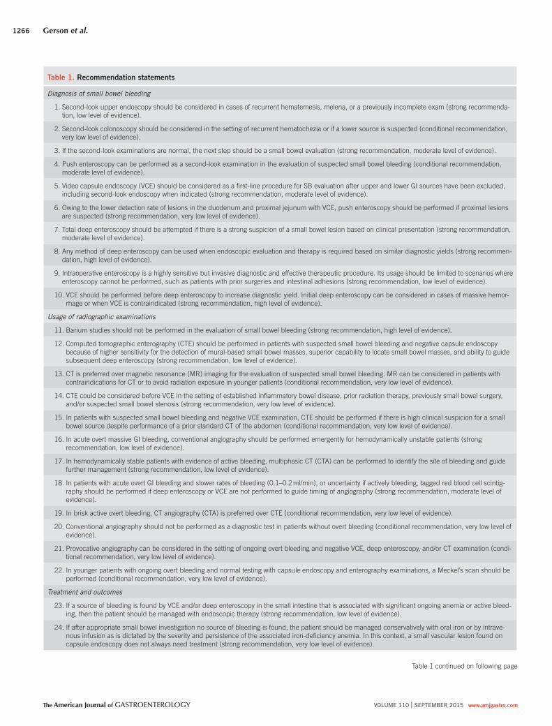
Table 1 . Recommendation statements
Diagnosis of small bowel bleeding
1. Second-look upper endoscopy should be considered in cases of recurrent hematemesis, melena, or a previously incomplete exam (strong recommenda-
tion, low level of evidence).
2. Second-look colonoscopy should be considered in the setting of recurrent hematochezia or if a lower source is suspected (conditional recommendation,
very low level of evidence).
3. If the second-look examinations are normal, the next step should be a small bowel evaluation (strong recommendation, moderate level of evidence).
4. Push enteroscopy can be performed as a second-look examination in the evaluation of suspected small bowel bleeding (conditional recommendation,
moderate level of evidence).
5. Video capsule endoscopy (VCE) should be considered as a fi rst-line procedure for SB evaluation after upper and lower GI sources have been excluded,
including second-look endoscopy when indicated (strong recommendation, moderate level of evidence).
6. Owing to the lower detection rate of lesions in the duodenum and proximal jejunum with VCE, push enteroscopy should be performed if proximal lesions
are suspected (strong recommendation, very low level of evidence).
7. Total deep enteroscopy should be attempted if there is a strong suspicion of a small bowel lesion based on clinical presentation (strong recommendation,
moderate level of evidence).
8. Any method of deep enteroscopy can be used when endoscopic evaluation and therapy is required based on similar diagnostic yields (strong recommen-
dation, high level of evidence).
9. Intraoperative enteroscopy is a highly sensitive but invasive diagnostic and effective therapeutic procedure. Its usage should be limited to scenarios where
enteroscopy cannot be performed, such as patients with prior surgeries and intestinal adhesions (strong recommendation, low level of evidence).
10. VCE should be performed before deep enteroscopy to increase diagnostic yield. Initial deep enteroscopy can be considered in cases of massive hemor-
rhage or when VCE is contraindicated (strong recommendation, high level of evidence).
Usage of radiographic examinations
11. Barium studies should not be performed in the evaluation of small bowel bleeding (strong recommendation, high level of evidence).
12. Computed tomographic enterography (CTE) should be performed in patients with suspected small bowel bleeding and negative capsule endoscopy
because of higher sensitivity for the detection of mural-based small bowel masses, superior capability to locate small bowel masses, and ability to guide
subsequent deep enteroscopy (strong recommendation, low level of evidence).
13. CT is preferred over magnetic resonance (MR) imaging for the evaluation of suspected small bowel bleeding. MR can be considered in patients with
contraindications for CT or to avoid radiation exposure in younger patients (conditional recommendation, very low level of evidence).
14. CTE could be considered before VCE in the setting of established infl ammatory bowel disease, prior radiation therapy, previously small bowel surgery,
and/or suspected small bowel stenosis (strong recommendation, very low level of evidence).
15. In patients with suspected small bowel bleeding and negative VCE examination, CTE should be performed if there is high clinical suspicion for a small
bowel source despite performance of a prior standard CT of the abdomen (conditional recommendation, very low level of evidence).
16. In acute overt massive GI bleeding, conventional angiography should be performed emergently for hemodynamically unstable patients (strong
recommendation, low level of evidence).
17. In hemodynamically stable patients with evidence of active bleeding, multiphasic CT (CTA) can be performed to identify the site of bleeding and guide
further management (strong recommendation, low level of evidence).
18. In patients with acute overt GI bleeding and slower rates of bleeding (0.1–0.2 ml/min), or uncertainty if actively bleeding, tagged red blood cell scintig-
raphy should be performed if deep enteroscopy or VCE are not performed to guide timing of angiography (strong recommendation, moderate level of
evidence).
19. In brisk active overt bleeding, CT angiography (CTA) is preferred over CTE (conditional recommendation, very low level of evidence).
20. Conventional angiography should not be performed as a diagnostic test in patients without overt bleeding (conditional recommendation, very low level of
evidence).
21. Provocative angiography can be considered in the setting of ongoing overt bleeding and negative VCE, deep enteroscopy, and/or CT examination (condi-
tional recommendation, very low level of evidence).
22. In younger patients with ongoing overt bleeding and normal testing with capsule endoscopy and enterography examinations, a Meckel’s scan should be
performed (conditional recommendation, very low level of evidence).
Treatment and outcomes
23. If a source of bleeding is found by VCE and/or deep enteroscopy in the small intestine that is associated with signifi cant ongoing anemia or active bleed-
ing, then the patient should be managed with endoscopic therapy (strong recommendation, low level of evidence).
24. If after appropriate small bowel investigation no source of bleeding is found, the patient should be managed conservatively with oral iron or by intrave-
nous infusion as is dictated by the severity and persistence of the associated iron-defi ciency anemia. In this context, a small vascular lesion found on
capsule endoscopy does not always need treatment (strong recommendation, very low level of evidence).
Table 1 continued on following page
Guidelines for Small Bowel Bleeding
© 2015 by the American College of Gastroenterology The American Journal of GASTROENTEROLOGY
1267
and “telangiectasia.” Th e full literature search strategy is demon-
strated in the Appendix .
To evaluate the level of evidence and strength of recommenda-
tions, we used the Grading of Recommendations Assessment,
Development, and Evaluation (GRADE) system ( 3 ). Th e level of
evidence could range from “high” (implying that further research
was unlikely to change the authors’ confi dence in the estimate of
the eff ect) to “moderate” (further research would be likely to have
an impact on the confi dence in the estimate of eff ect), “low” (fur-
ther research would be expected to have an important impact on
the confi dence in the estimate of the eff ect and would be likely to
change the estimate), or “very low” (any estimate of eff ect is very
uncertain). Th e strength of a recommendation was graded as
“strong” when the desirable eff ects of an intervention clearly out-
weigh the undesirable eff ects and as “conditional” when there is
uncertainty about the trade-off s. We preferentially used meta-anal-
yses or systematic reviews when available, followed by clinical trials
and retrospective cohort studies. To determine the level of evi-
dence, we entered data from the papers of highest evidence into the
GRADE program (accessible at http: // www.gradepro.org ). Th e rec-
ommendation statements from this guideline are shown in Table 1 .
Summary statements, when listed, are designed to be descriptive in
nature without associated evidence-based ratings.
Defi nition of overt or occult small bowel bleeding
Summary statements
1 . A source of small bowel bleeding should be considered in
patients with overt or occult GI hemorrhage aft er perfor-
mance of a normal upper and lower endoscopic examination.
2 . Patients should be classifi ed as having small bowel bleeding
if a source of bleeding is identifi ed distal to the ampulla of
Vater and/or proximal to the ileocecal valve.
3 . Aft er normal upper and lower endoscopic examinations and
before performance of capsule endoscopy, patients should be
classifi ed as having “potential small bowel bleeding.”
4 . “Overt small bowel bleeding” refers to patients presenting
with either melena or hematochezia with a source of
bleeding identifi ed in the small intestine. Th e term “occult
small bowel bleeding” can be reserved for patients presenting
with iron-defi ciency anemia with or without guaiac-positive
stools who are found to have a small bowel source of
bleeding.
5 . Th e term “obscure GI bleeding” should be reserved for
patients not found to have a source of bleeding aft er perfor-
mance of standard upper and lower endoscopic examina-
tions, small bowel evaluation with VCE and/or enteroscopy,
and radiographic testing.
Th e traditional defi nition of “OGIB” before the introduction of
VCE and deep enteroscopy included patients with overt or occult
GI bleeding who underwent normal upper and lower endoscopic
examinations in addition to a small bowel series that did not
reveal a source of bleeding. Patients with overt obscure bleeding
were defi ned as patients presenting with either hematochezia or
melena, whereas patients with occult obscure bleeding were classi-
fi ed based on the presence of a positive fecal occult blood test with
or without iron-defi ciency anemia.
With the introduction of VCE in the United States in 2001 and
deep enteroscopy in 2004, the majority (~75%) of patients previ-
ously classifi ed as having obscure bleeding were found to have
sources of bleeding identifi ed in the small intestine ( 4 ). Th e diag-
nostic yield included any causes of bleeding detected distal to the
ampulla of Vater or proximal to the ileocecal valve by any testing
modality including push enteroscopy, ileoscopy, deep enteroscopy,
VCE, angiography, or an enterography examination. We would
therefore propose that patients with small bowel sources identifi ed
be classifi ed as having small bowel bleeding, reserving the prior
term of OGIB for patients without a source of bleeding identifi ed
aft er comprehensive evaluation of the small bowel as described in
the sections below.
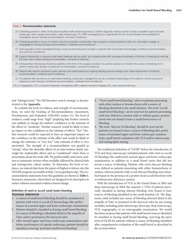
Table 1 . Recommendation statements
25. If bleeding persists in either of the above situations with worsening anemia, a further diagnostic workup should include a repeated upper and lower
endoscopy, video capsule examination, deep enteroscopy, CT or MRI enterography as is appropriate for the clinical situation and availability of
investigative devices (strong recommendation, low level of evidence).
26. If bleeding persists or recurs or a lesion cannot be localized consideration may be given to medical treatment with iron, somostatin analogs, or
antiangiogenic therapy (strong recommendation, moderate level of evidence).
27. Anticoagulation and/or antiplatelet therapy should be discontinued if possible in patients with small bowel hemorrhage (conditional recommendation,
very low level of evidence).
28. Surgical intervention in massive small bowel bleeding may be useful, but is greatly aided with presurgical localization of the site of bleeding by marking
the lesion with a tattoo (strong recommendation, low level of evidence).
29. Intraoperative enteroscopy should be available at the time of the surgical procedure to provide assistance to localize the source of bleeding and to
perform endoscopic therapy (conditional recommendation, low level of evidence).
30. Patients with Heyde’s syndrome (aortic stenosis and angioectasia) and ongoing bleeding should undergo aortic valve replacement (conditional
recommendation, moderate level of evidence).
31. For patients with recurrence of small bowel bleeding, endoscopic management can be considered depending on the patient’s clinical course and
response to prior therapy (conditional recommendation, moderate level of evidence).
CTA, CT angiography; CTE, computed tomographic enterography; MRI, magnetic resonance imaging; VCE, video capsule endoscopy.
Gerson et al.
The American Journal of GASTROENTEROLOGY VOLUME 110 | SEPTEMBER 2015 www.amjgastro.com
1268
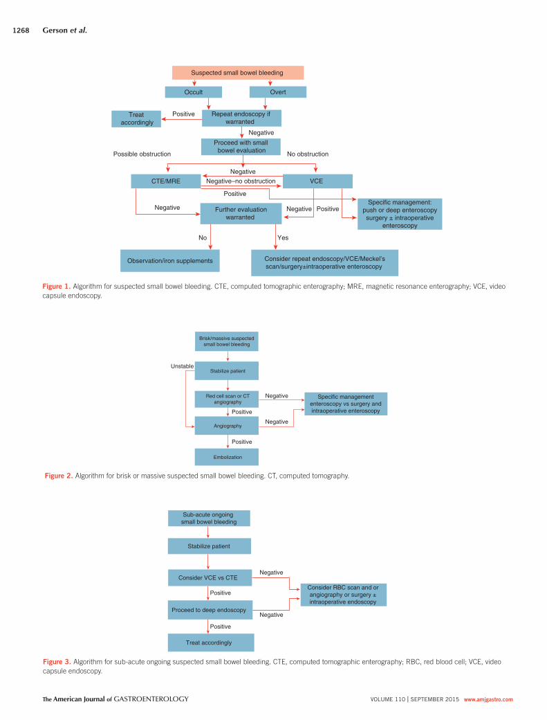
Brisk/massive suspectedsmall bowel bleeding
Stabilize patient
Red cell scan or CTangiography
Angiography
Embolization
Positive
Positive
Specific managemententeroscopy vs surgery andintraoperative enteroscopy
Negative
Negative
Unstable
Figure 2 . Algorithm for brisk or massive suspected small bowel bleeding. CT, computed tomography.
Sub-acute ongoing small bowel bleeding
Stabilize patient
Consider VCE vs CTE
Proceed to deep endoscopy
Treat accordingly
Consider RBC scan and orangiography or surgery ±intraoperative endoscopy
Negative
Negative
Positive
Positive
Figure 3 . Algorithm for sub-acute ongoing suspected small bowel bleeding. CTE, computed tomographic enterography; RBC, red blood cell; VCE, video
capsule endoscopy.
Suspected small bowel bleeding
Occult Overt
Repeat endoscopy ifwarranted
CTE/MRE VCE
Further evaluationwarranted
Observation/iron supplements Consider repeat endoscopy/VCE/Meckel’sscan/surgery±intraoperative enteroscopy
Possible obstruction No obstruction
Specific management:push or deep enteroscopysurgery ± intraoperative
enteroscopy
Negative
PositiveNegative
Positive
YesNo
Negative
Treat accordingly
Positive
Negative
Negative–no obstruction
Proceed with small bowel evaluation
Figure 1 . Algorithm for suspected small bowel bleeding. CTE, computed tomographic enterography; MRE, magnetic resonance enterography; VCE, video
capsule endoscopy.
Guidelines for Small Bowel Bleeding
© 2015 by the American College of Gastroenterology The American Journal of GASTROENTEROLOGY
1269
Epidemiology and natural history of small bowel bleeding
Summary statements
1 . Th e type of lesion responsible for small bowel bleeding is
dependent on patient age but not gender or ethnicity.
2 . Small bowel angioectasia are the most common cause of
small bowel bleeding.
3 . Risk factors for angioectasia include advancing age, presence
of aortic stenosis, chronic renal failure, left ventricular assist
devices, and other hereditary disorders.
4 . Risk factors for recurrent small bowel bleeding from angi-
oectasia include number of lesions, advanced age, presence of
comorbid conditions, and anticoagulant therapy.
Prevalence and etiology of small bowel bleeding . Th e prevalence
of small bowel lesions has been estimated to be ~5–10% in patients
presenting with GI bleeding ( 1,2 ). Details pertaining to the clinical
presentation are critically important in the determination of the
etiology. A history of a bleeding diathesis as with von Willebrand
disease and medication usage including aspirin, nonsteroidal
anti-infl ammatory drugs, anticoagulants, and/or other antiplate-
let agents also can lend clues to the diagnosis. Knowledge of co-
morbidities such as valvular heart disease and prior procedures/
surgeries such as liver biopsy, liver transplantation, abdominal
aortic aneurysm repair, or bowel resection again can be very help-
ful. Common causes of small bowel bleeding are listed in Table
2 and are found in ~75% of patients with suspected small bowel
bleeding ( 5 ). Based on a 2008 meta-analysis combining data from
Western and Asian countries and reporting yields on both VCE
and double-balloon enteroscopy (DBE) ( 4 ), the prevalence of small
bowel vascular lesions based on 10 studies was 24% for both VCE
( N =371) and DBE ( N =364). For infl ammatory fi ndings, the yield
was 18% for VCE ( N =343) and 16% for DBE ( N =336), and the
yield was 11% for mass lesions (VCE, N =343 and DBE, N =336).
An analysis comparing diagnostic yields from Western compared
to Asian countries demonstrated that patients undergoing DBE
in Asian countries were more likely to have neoplastic fi ndings,
whereas angioectasia were more common in Western countries.
Age has been known to be a determinant for the type of small
bowel pathology detected. Patients under the age of 40 years are more
likely to have infl ammatory bowel disease or Meckel’s diverticulum.
Small bowel neoplasms (e.g., GI stromal cell tumor, lymphoma,
carcinoid, adenocarcinoma, or other polypoid lesions) and Dieu-
lafoy’s lesions can occur in both younger and older patient cohorts
( 6–11 ). Angioectasia, other vascular lesions, and ulcers secondary
to anti-infl ammatory agents are more likely in patients over the
age of 40 years. Data regarding ethnicity and small bowel fi ndings
has not been extensively published to date.
Diff erences in fi ndings between patients with overt or occult
small bowel bleeding . Studies using VCE and deep enteroscopy
have demonstrated higher diagnostic yields for patients with
overt bleeding compared with patients with occult bleeding. For
patients with prior overt bleeding, the diagnostic yield was less
than that for current overt bleeders, and decreased substantially
with time. In a 2004 study by Pennazio et al. ( 12 ) of 100 patients
undergoing VCE, the diagnostic yield was 92% for patients with
overt bleeding, 44% for occult bleeders, 67% for patients with pri-
or overt bleeding who were studied within 10–14 days, and 33%
at 3–4 weeks postbleeding episode. In a 2010 study of 200 patients
with bleeding undergoing DBE, the diagnostic yield was 77% for
overt bleeding, 67% for patients with occult hemorrhage, and 59%
for patients with prior overt bleeding ( 13 ).
In addition to higher diagnostic yields for patients with overt
bleeding, recurrence rates may be higher in patients presenting
with overt bleeding. In a multicenter US study assessing long-term
outcomes post-DBE, recurrence of overt bleeding occurred in
34% of patients presenting with overt hemorrhage compared with
13% of patients with occult bleeding at 12 months postprocedure
( P =0.06) ( 14 ). Th ese recurrence rates, however, were not signifi -
cant at 30 months of follow-up (27% vs. 20%, P =NS).
Rare causes and non-small bowel sources of bleeding . Rare
causes of small bowel bleeding are shown in Table 2 . Patients
with disorders associated with portal hypertension and/or with
endoscopic evidence of varices or portal hypertension have also
demonstrated portal hypertensive changes in the small bowel on
VCE or enteroscopy studies ( 15 ). Other rare causes of bleeding
from the small bowel have included Kaposi’s sarcoma associated
with acquired immunodefi ciency syndrome, Plummer–Vinson
syndrome, pseudoxanthoma elasticum, Ehlers–Danlos syndrome,
Henoch–Schoenlein purpura, neurofi bromatosis, malignant
atrophic papulosis, and other inherited polyposis syndromes. A
family history of polyposis syndromes may provide important
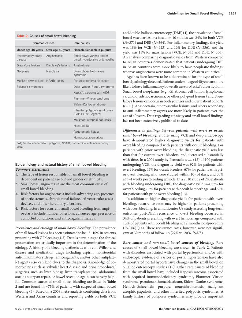
Table 2 . Causes of small bowel bleeding
Common causes Rare causes
Under age 40 years Over age 40 years Henoch–Schoenlein purpura
Infl ammatory bowel
disease
Angioectasia Small bowel varices and/or
portal hypertensive enteropathy
Dieulafoy’s lesions Dieulafoy’s lesions Amyloidosis
Neoplasia Neoplasia Blue rubber bleb nevus
syndrome
Meckel’s diverticulum NSAID ulcers Pseudoxanthoma elasticum
Polyposis syndromes Osler–Weber–Rendu syndrome
Kaposi’s sarcoma with AIDS
Plummer–Vinson syndrome
Ehlers–Danlos syndrome
Inherited polyposis syndromes
(FAP, Peutz–Jeghers)
Malignant atrophic papulosis
Hematobilia
Aorto-enteric fi stula
Hemosuccus entericus
FAP, familial adenomatous polyposis; NSAID, nonsteroidal anti-infl ammatory
drug.
Gerson et al.
The American Journal of GASTROENTEROLOGY VOLUME 110 | SEPTEMBER 2015 www.amjgastro.com
1270
clues to the underlying etiology of small bowel bleeding. Physical
examination, including a detailed dermatological evaluation, may
also be useful in the diagnosis of systemic syndromes, including
hereditary hemorrhagic telangiectasia and blue-rubber bleb ne-
vus syndrome. Uncommon non-small bowel sources of obscure
GI bleeding not shown in the table have included hematobilia,
hemosuccus pancreatitis, and aortoenteric fi stulae.
Prior clinical guidelines have listed celiac disease as a cause of
small bowel bleeding ( 16 ), but there is emerging evidence that
celiac disease leads to iron-defi ciency anemia because of malab-
sorption and not because of the presence of occult GI bleeding
( 17 ). Although complications associated with celiac disease such as
ulcerative jejunitis, lymphoma, and/or adenocarcinoma can cause
bleeding from the small intestine, the entity of celiac disease is no
longer listed as a cause of small bowel bleeding.
Diagnosis of small bowel bleeding ( Figure 1 )
Recommendations
1 . Second-look upper endoscopy should be considered in cases
of recurrent hematemesis, melena, or a previously incomplete
exam (strong recommendation, low level of evidence).
2 . Second-look colonoscopy should be considered in the setting
of recurrent hematochezia or if a lower source is suspected
(conditional recommendation, very low level of evidence).
3 . If the second-look examinations are normal, the next step
should be a small bowel evaluation (strong recommendation,
moderate level of evidence).
4 . Push enteroscopy can be performed as a second-look exami-
nation in the evaluation of suspected small bowel bleeding
(conditional recommendation, moderate level of evidence).
5 . VCE should be considered a fi rst-line procedure for small
bowel (SB) evaluation aft er upper and lower GI sources have
been excluded, including second-look endoscopy when indi-
cated (strong recommendation, moderate level of evidence).
6 . Owing to the lower detection rate of lesions in the duodenum
and proximal jejunum with VCE, push enteroscopy should
be performed if proximal lesions are suspected (strong rec-
ommendation, very low level of evidence).
7 . Total deep enteroscopy should be attempted if there is a
strong suspicion of a small bowel lesion based on clinical
presentation or abnormal VCE study (strong recommenda-
tion, moderate level of evidence).
8 . Any method of deep enteroscopy can be used when endoscopic
evaluation and therapy is required based on similar diagnostic
yields (strong recommendation, high level of evidence).
9 . Intraoperative enteroscopy (IOE) is a highly sensitive but in-
vasive diagnostic and eff ective therapeutic procedure. Its usage
should be limited to scenarios where enteroscopy cannot be
performed, such as patients with prior surgeries and intestinal
adhesions (strong recommendation, low level of evidence).
10 . VCE should be performed before deep enteroscopy to
increase diagnostic yield. Initial deep enteroscopy can be
considered in cases of massive hemorrhage or when VCE
is contraindicated (strong recommendation, high level of
evidence).
Th e main limitations of SB evaluation in the past were related to
its length (>6 m) and limited intubation with conventional endos-
copy; these shortcomings have been largely overcome by recent
advances in endoscopic technology, including VCE, deep enter-
oscopy (including DBE, SB enteroscopy, and spiral enteroscopy),
and radiologic modalities including CT enterography (CTE) and
MR enterography. Th ese new advances, as well as the capacity to
successfully perform endoscopic therapeutic interventions, have
led to signifi cant improvement in the management of patients with
small bowel bleeding, and a decline in invasive surgical procedures
(IOE, laparoscopy, and exploratory laparotomy) ( 18–21 ).
Second-look endoscopy
Most small intestinal bleeding is undramatic in presentation and
either presents as stable overt or occult bleeding. Th e prior litera-
ture demonstrated that a high percentage of patients designated
as having “potential small bowel bleeding” were found to have
missed bleeding sources within reach of conventional upper and
lower endoscopy including diagnostic yields ranging from 2 to
25% in patients undergoing repeat esophagogastroduodenoscopy
and 6 to 23% on repeat colonoscopy ( 22–24 ). More recent stud-
ies using DBE and capsule endoscopy have also confi rmed these
fi ndings ( 25–30 ).
Most overt bleeding can be evaluated fi rst with a second-look
procedure to exclude upper and lower bleeding that can be readily
reached with a standard endoscope. Instead of repeating an upper
endoscopy, a push enteroscopy may be performed to examine the
distal duodenum and proximal jejunum. During the colonoscopy,
every eff ort should be made to intubate the terminal ileum to vis-
ualize the ileal mucosa and to inspect for blood coming from a
more proximal location of the small intestine. For expediency of
work up, it is sometimes appropriate to use VCE as the fi rst-line
test aft er having had a negative upper endoscopy and colonoscopy.
In fact, one study did not show that second-look endoscopy was
cost eff ective ( 31 ). However, the distal duodenum and proximal
jejunum would still need to be examined unless the VCE reveals
the source of the suspected small bowel bleeding.
Push enteroscopy
Push enteroscopy is an extended upper endoscopy performed
with a long endoscope such as a pediatric colonoscope ( 32 ) or
with a commercially available push enteroscope, which is typi-
cally 250 cm in length. Push enteroscopy allows only limited eval-
uation of the proximal SB, ~70 cm distal to the ligament of Treitz.
Push enteroscopy using a colonoscope typically can be passed
45–60 cm beyond the ligament of Treitz ( 33 ). When push enteros-
copy is carried out with the variable stiff ness design, it reaches a
deeper distance of nearly 90 cm ( 34 ). Th e diagnostic yield of push
enteroscopy is reported to range from 3 to 70%, with the major-
ity of SB fi ndings being vascular lesions ( 16,35–38 ). Interestingly,
most of the lesions diagnosed on push enteroscopy have been
found in locations accessible to standard esophagogastroduoden-
oscopy, emphasizing the importance of second-look endoscopy
( 22,39 ). When a dedicated push enteroscope is used, it may be
performed with an overtube designed to reduce looping in the
Guidelines for Small Bowel Bleeding
© 2015 by the American College of Gastroenterology The American Journal of GASTROENTEROLOGY
1271
improved to 60% if a dual camera capsule is used ( 63 ). Nonethe-
less, VCE does miss clinically important duodenal and proximal
jejunal lesions ( 64–67 ), and thus cannot be solely relied upon for
exclusion of bleeding lesions in these areas. However, there are
studies to suggest that repeat VCE may be of benefi t and increase
the diagnostic yield, even when the fi rst study is negative ( 68–70 ).
A prospective study again showed that repeat VCE may be benefi -
cial, particularly when the bleeding changes from occult to overt or
there is a hemoglobin drop ≥4 g/dl ( 71 ).
VCE is very well tolerated by patients ( 72 ). Its main complication
is capsule retention, which may occur in roughly 1.5% of patients
undergoing evaluations for potential small bowel bleeding sources
( 73 ). VCE, however, may be complicated by retention in up to 13%
in Crohn’s disease patients, which limits its use in patients with
suspected obstruction or strictures until patency is documented
( 74,75 ). Screening SB radiographs have not been able to eliminate
this problem, although the patency capsule may be useful ( 76 ).
Th e most serious complication reported with VCE is perforation,
which fortunately has been exceedingly rare ( 77 ).
Deep enteroscopy
Balloon-assisted enteroscopy . Balloon-assisted enteroscopy uses
the principle of push and pull enteroscopy, and includes DBE and
SBE as described further below ( 78 ). As the name suggests, both
of the balloon enteroscopes have an overtube, with balloons at
their distal ends. Th e DBE uses a balloon on the end of the scope
and the overtube. Th e SBE works by using the tip of the scope
as an anchor along with the single balloon. Th e balloons on the
DBE and overtube are composed of latex, whereas the balloon on
the SBE overtube is made of silicone. Th erefore, for patients with
latex allergy, SBE should be performed. Th e enteroscope in both
systems has a working length of 200 cm with an outer diameter of
9.4 mm. Th e overtube is 140 cm in length.
Th e technique of balloon-assisted enteroscopy involves a series
of steps called advancement cycles, described below. Balloon-
assisted enteroscopy can be performed via the oral and rectal
approach. It has been mainly studied in adults between the ages
of 18 and 70 years but appears to be safe in the elderly population
(over 70 years in age), as well as in children ( 79,80 ).
Double-balloon enteroscopy
DBE was fi rst described in 2001 by Yamamoto et al. ( 81 ). Th e
equipment has been available for clinical use in the United States
since 2004. DBE allows deeper intubation of the SB compared
with tradtional endoscopes. It can be advanced a distance of
~240–360 cm distal to the pylorus with the oral approach and
102–140 cm proximal to the ileocecal valve with the rectal
approach. Th is compares to a distance of 90–150 cm with the
push enteroscope and 50–80 cm with ileoscopy ( 51,82 ). It has the
additional advantage over VCE of both diagnostic and therapeu-
tic capabilities, including biopsies, tattoo, hemostasis, polypec-
tomy, dilation, and foreign body removal (including retained
capsules) ( 83–85 ). Th e 2.8 mm accessory channel allows passage
of virtually all standard-caliber, through-the-scope, diagnostic
and therapeutic instruments ( 86 ).
stomach and stiff en the enteroscope for deeper passage ( 40 ).
Although the use of an overtube may allow for deeper SB intuba-
tion up to 150 cm, it does not appear to increase the diagnostic
yield of the test ( 41 ). Th e main disadvantages of this exam include
looping of the enteroscope and patient discomfort. Its role is cur-
rently limited to endoscopic therapeutics in those patients who
have only proximal SB lesions detected on VCE. Although it has
only a limited range, push enteroscopy is an ideal second-look
procedure because of the ability to examine the distal duodenum
and proximal jejunum, a SB segment that is not always well seen
with VCE.
Endoscopic visualization of the small intestine
Video capsule endoscopy . Introduced for clinical use in the United
States in 2001, VCE is now available throughout the world. Th ere
are now four VCE platforms, with three available for clinical use
in the United States. Th e VCE measures 26×11 mm 2 , and has the
capacity to take images at the rate of 2 frames/s, over an 8–12 h
period. Images are transmitted to a recording device, and can be
downloaded and viewed on a computer station with the appro-
priate soft ware. Capsule endoscopy allows noninvasive evaluation
of the entire SB in 79–90% of patients, with a diagnostic yield of
38–83% in patients with suspected small bowel bleeding ( 42 ). Th e
main utility of this test lies in its high positive (94–97%) and nega-
tive predictive value (83–100%) in the evaluation of GI bleeding
( 12,43 ). Findings on VCE leading to endoscopic or surgical inter-
vention or a change in medical management have been reported
in 37–87% of patients ( 12,44 ). In addition, 50–66% of patients
have been reported to remain transfusion free without recurrent
bleed at follow-up, aft er undergoing VCE-directed interventions
( 43,45 ). Th e rebleeding rate ranges from 6 to 27% in patients who
have had a negative capsule study ( 46–48 ).
Th e yield of VCE may be infl uenced by multiple factors, with
a higher likelihood of positive fi ndings in patients with a hemo-
globin <10 g/dl, longer duration of bleeding (>6 months), more
than one episode of bleeding, overt as compared with occult bleed-
ing (60% vs. 46%), and performance of VCE within 2 weeks of the
bleeding episode (91% vs. 34%) ( 49–52 ). Th ere is also evidence
that VCE within 48 to 72 h of overt suspected small bowel bleeding
has the greatest yield for lesion detection ( 53–55 ). A more recent
study confi rmed that overt bleeding was the strongest predictor
of a positive capsule study, but male sex, age >60 years, and in-
patient status were also independent predictors ( 56 ). Other risk
factors for a positive capsule include cardiac and renal comorbidi-
ties. Although usually performed for intermittent overt bleeding,
at least one study suggests that it may be useful in the emergency
situation of severe overt suspected small bowel hemorrhage ( 57 ).
Th e main limitations of VCE include lack of therapeutic capa-
bilities, inability to control its movement through the GI tract, and
the diffi culty in localizing the lesion. Th e other limitations of VCE
include a lack of specifi city with 14% incidental fi ndings in healthy
volunteers ( 58 ) and a 10–36% false-negative rate ( 59,60 ). Finally,
VCE fails to identify the major papilla in a majority of cases ( 61,62 )
and therefore may miss important duodenal lesions because of
rapid transit through the duodenal loop. Th is defi ciency may be
Gerson et al.
The American Journal of GASTROENTEROLOGY VOLUME 110 | SEPTEMBER 2015 www.amjgastro.com
1272
To perform DBE, the enteroscope and overtube are introduced
into the small bowel typically past the ampulla, and the balloon on
the overtube is infl ated. Th e enteroscope is then further advanced
into the small bowel. Th e balloon on the DBE enteroscope is then
infl ated. Th e overtube is subsequently advanced over the entero-
scope. Now both overtube and enteroscope are drawn back (with
both balloons infl ated on DBE), which allows the small bowel to
plicate over the enteroscope. By repeating this series of steps, a
longer distance can be traversed as compared with conventional
endoscopy.
Th e diagnostic yield of DBE ranges from 60 to 80% in patients
with suspected small bowel bleeding and other SB disorders. Suc-
cessful performance of endoscopic therapeutic interventions has
been reported in 40–73% of patients ( 51,87,88 ). A more recent
study confi rms these earlier fi ndings ( 89 ). DBE has generally been
used for small bowel evaluation in the chronic stable or mildly
to moderately active bleeding situation because of its small suc-
tion channel. However, a small recent study actually suggests that
emergency DBE is technically feasible and may facilitate the diag-
nosis and management of patients with massive overt small bowel
hemorrhage ( 90 ). A more recent study also suggests that urgent
DBE is better than non-urgent DBE and is associated with a lower
recurrent bleeding rate ( 91 ). In addition, one study suggests that
repeat DBE from the same direction may also be benefi cial, par-
ticularly if the patient had a prior positive DBE ( 92 ).
Total enteroscopy with DBE is defi ned as complete evaluation
of the small bowel either with a single approach or combined oral
and rectal approach. Th e decision to perform total enteroscopy is
usually dependent on the discretion of the endoscopist, degree of
clinical suspicion for a small bowel lesion, and inability to detect
the lesion using a single approach. Despite the best attempts of the
endoscopist, total enteroscopy may not be feasible in all patients,
with a reported success rate ranging from 16 to 86% ( 81,93 ). A
prospective, randomized study demonstrated that DBE had a sig-
nifi cantly higher total enteroscopy rate than SBE ( 94 ).
Th e main limitations of DBE include its invasive nature, pro-
longed procedure time, and requirement for additional personnel.
Th e reported complication rate for diagnostic procedures is 0.8%,
and up to 4% if therapeutics such as electrocoagulation, polypec-
tomy, or dilation are performed. Th e main complications reported
with this technique are ileus, pancreatitis, and perforation, usu-
ally associated with large polypectomies ( 51,84,95 ). Pancreatitis
is the most common complication of the peroral diagnostic DBE,
occurring in at least 0.3% of patients ( 95 ). Perforation appears
to be more common in patients with intestinal anastomosis and
SB polypectomy ( 96,97 ). Postprocedure bloating and abdominal
pain were once a common occurrence, but they have been rarely
reported by patients as the use of carbon dioxide as the insuffl at-
ing gas because of rapid diff usion of the gas across the intestinal
mucosa ( 98,99 ). A recent large prospective database suggested an
overall complication rate of 1.2% ( 100 ).
Single-balloon enteroscopy
Two years aft er the launch of the commercially available double-
balloon system, SBE was introduced. Th e theory and technique
of SBE are very similar to that of DBE; the key diff erence being
that there is no balloon on the end of the enteroscope with SBE.
During the reduction maneuver with SBE, the overtube balloon is
infl ated and the distal end of the enteroscope hooked over a fold
as the SBE does not have a distal balloon.
Even the dimensions of the enteroscope and the overtubes are
virtually identical to those of DBE. Th e overtube balloon is made
of a silicone material rather than latex. SBEs have a stiff shaft and
the enteroscope can be easily removed and reinserted through the
overtube. Its caliber is similar to that of a standard upper endo-
scope but with more than twice its length (200 cm). Hence, most
endoscopic diagnostic and therapeutic maneuvers are possible to
perform with the SBEs.
A preliminary report of 78 SBE procedures performed in 41
patients, of whom 12 had small bowel bleeding, found that SBE
allowed evaluation of the SB in a safe and eff ective manner, includ-
ing performance of total enteroscopy (25%; 6/24). Th e diagnos-
tic yield in patients with suspected small bowel bleeding sources
was 33% (4/12 patients), and therapeutics such as argon plasma
coagulation could be successfully performed ( 20 ). Another study
evaluated 20 patients with suspected SB disorders, and found a
diagnostic yield of 60% using SBE ( 101 ). More recent studies have
found diagnostic yields between 65 and 74% ( 102–104 ). SBE also
appears to be associated with improved outcomes ( 105 ). A pro-
spective study on 105 patients who underwent at least one oral
SBE procedure found no complications related to the diagnostic
procedures ( 106 ). One perforation occurred aft er stricture dila-
tion. Prospective, sequential amylase testing before and aft er SBE
showed 16% of patients developed elevation of serum amylase but
without any overt clinical evidence of acute pancreatitis. At this
time, it appears that SBE is equivalent to DBE for the evaluation of
small bowel bleeding sources ( 107,108 ).
Spiral enteroscopy
Spiral enteroscopy consists of a unique overtube with an outer
raised spiral ridge at its distal end through which an SBE or a DBE
can be inserted. It is used for enteroscopy via the oral route and
can be used only with enteroscopes <9.4 mm in diameter. Unlike
the balloon enteroscopy techniques, spiral enteroscopy uses the
clockwise motion of the ridged overtube to draw the enteroscope
forward. It is a two-person procedure, with a nurse or physician
rotating the overtube while the endoscopist is keeping the lumen
of the SB in view. Th e duodenojejunal transition poses a technical
challenge because of the sharp angulation that may prevent the
overtube from safely engaging the proximal jejunum for forward
passage. Aside from that, the procedure is rather simple to per-
form and forward progress can complete in about 18 min ( 109 ).
Based on the prior literature, the mean (±s.d.) procedure times for
the anterograde approach have been estimated to be 79±15 min
for DBE (10 studies) ( 51,82–84,87,110–114 ), 65±16 min for SBE
(5 studies) ( 20,106,115–117 ), and 35+6 min for spiral enteroscopy
(4 trials) ( 109,118–120 ). Even though most experts assume that
this technique covers less ground than DBE, there is one case
described in a letter to the editors in which an orally passed spiral
enteroscope reached the cecum in 65 min ( 121 ). Th e diagnostic
Guidelines for Small Bowel Bleeding
© 2015 by the American College of Gastroenterology The American Journal of GASTROENTEROLOGY
1273
recommended as the third test of choice in patients with suspect-
ed small bowel bleeding, who have had a negative esophagogas-
troduodenoscopy and colonoscopy.
DBE compared with push enteroscopy and VCE . A study by May
et al. ( 85 ), which compared DBE to push enteroscopy in 52 pa-
tients with suspected small bowel bleeding, found that DBE not
only allowed a greater depth of intubation (230 vs. 80 cm) but also
had a higher yield for small bowel fi ndings (73% vs. 44%). Fur-
thermore, DBE facilitated detection of additional lesions in the
distal small bowel in patients who had positive fi ndings on push
enteroscopy.
Several studies have compared the yield of VCE with DBE, but
have shown inconsistent results because of their small sample size.
A meta-analysis of 11 studies that compared these modalities in
patients with SB disease (majority with suspected small bowel
bleeding) showed a comparable diagnostic yield (60% vs. 57%;
incremental yield of 3%) for all SB fi ndings. Th e yield with the tests
was also similar for vascular, infl ammatory, and neoplastic lesions
( 4 ). Another meta-analysis of eight studies also found no diff erence
in diagnostic yield between the two tests for the evaluation of SB
disease (odds ratio 1.21, 95% confi dence interval (CI): 0.64–2.29)).
In patients with small bowel bleeding, VCE had a higher yield as
compared with DBE using a single approach (odds ratio 1.61, 95%
CI: 1.07–2.43), but a signifi cantly lower yield as compared with
DBE using a combined antegrade and retrograde approach (odds
ratio 0.12, 95% CI: 0.03–0.52) ( 133 ). Th is fi nding reinforces the
importance of total enteroscopy with DBE in patients with high
clinical suspicion for an SB lesion. Another meta-analysis similarly
showed comparable diagnostic yields, and also suggested that the
diagnostic yield improves if performed in patients with a positive
capsule study ( 134 ). Two more recent meta-analyses again confi rm
the similarity in diagnostic yields between VCE and DBE ( 89,135 ).
VCE has been reported to be useful as a screening tool before
DBE in patients with suspected small bowel bleeding. Th is approach
of a ‘targeted DBE’ has been reported to increase both the diag-
nostic (73–93%) and therapeutic yield (57–73%) of the test ( 136 ).
Furthermore, VCE transit times have been found useful in guiding
the optimal route of DBE. Owing to deeper intubation of the small
bowel and a higher success rate with the oral approach, this is the
preferred route for lesions suspected to lie within the proximal 75%
of the small bowel, whereas the rectal route is used for more distal
lesions. Because of the high negative predictive value of VCE, the
approach of VCE-guided DBE allows avoidance of DBE in patients
with a low pretest probability for SB fi ndings ( 137–139 ).
However, the concept of CE-guided DBE may not be applicable
in all patients. VCE has a false-negative rate of 11% for all SB fi nd-
ings, and more importantly, up to 19% for neoplasms. Additional
fi ndings on repeat VCE have been detected in up to 75% of patients
with suspected small bowel bleeding, thereby leading to a change
in management in 62% ( 69 ). Th ere have also been reports of neo-
plasms missed on VCE and subsequently diagnosed at DBE ( 140 ).
Hence, in patients with a negative VCE, in whom there is a high
clinical suspicion for an SB lesion, DBE should still be pursued,
including consideration for total enteroscopy ( 4 ).
yield of the initial cases of spiral enteroscopy has been reported
to be only 33% ( 122 ). Since that time, a more recent prospective
study suggested that the diagnostic yield in patients with a positive
capsule study was 57% ( 119 ). Furthermore, a prospective cohort
study also found that spiral enteroscopy leads to improved out-
comes in terms of transfusion requirements, iron supplementa-
tion, and additional therapeutic procedures ( 123 ). Th ere is also an
overtube for a rectal approach that can be used for limited ileos-
copy. Questions have been raised about some safety concerns with
regards to bowel trauma and diffi culty in rapid removal during
an emergency. However, there had been no major complication
reported in the early literature ( 120 ). In a series of 75 patients, 12%
of had a sore throat, 27% had superfi cial mucosal trauma, and 7%
had moderate esophageal trauma that did not require any inter-
vention. In a retrospective registry study involving 1750 patients,
the rate of severe complications was reported to be 0.34%, with a
small bowel perforation rate of 0.27% ( 118 ). In the fi rst 850 cases
reported in the literature with spiral enteroscopy, there were no
serious complications ( 124 ).
Intraoperative enteroscopy
IOE involves evaluation of the SB at laparotomy, and may be
performed orally, rectally, or via an enterotomy, wherein the
scope is inserted through a surgical incision in the SB ( 125 ).
Upper endoscopes, colonoscopes, push enteroscopes, and the
newer balloon-assisted scopes have all been used in IOE. Th is
may be the most reliable method to achieve a complete small
bowel evaluation but it is highly invasive. Although the diagnos-
tic yield of IOE has been reported to range from 58 to 88% ( 126 ),
rebleeding may occur in up to 60% of patients ( 127–130 ). Major
complications of IOE include serosal tears, avulsion of mesen-
teric vessels, and prolonged ileus ( 130 ). In addition, the proce-
dure has a high mortality rate of 17%. Owing to these reasons,
IOE should be reserved only for those patients who present with
recurrent bleeds requiring multiple transfusions or hospitaliza-
tions aft er a comprehensive negative evaluation with VCE and
deep enteroscopy or for patients in whom deep enteroscopy
cannot be performed without lysis of adhesions ( 131 ).
Comparison of endoscopic modalities in suspected small
bowel bleeding
Capsule endoscopy compared with push enteroscopy and small
bowel follow-through . Multiple retrospective and prospective
studies have found VCE to be superior to both push enteroscopy
and small bowel series in the evaluation of patients with suspected
small bowel bleeding. A meta-analysis of studies that compared
VCE and push enteroscopy showed that VCE had an incremental
yield of 30% (yield 56% vs. 26%) for clinically signifi cant fi ndings
in patients with small bowel bleeding sources. Similarly, VCE had
an incremental yield of 36% over small bowel series (yield 42%
vs. 6%) ( 132 ). Th e number needed to test with VCE was three,
to establish one additional diagnosis. Based on subanalysis of the
data, VCE had a higher yield for both vascular and infl ammatory
lesions. VCE has hence largely replaced push enteroscopy and
small bowel series in the evaluation of the SB, and is currently

Gerson et al.
The American Journal of GASTROENTEROLOGY VOLUME 110 | SEPTEMBER 2015 www.amjgastro.com
1274
Th e indications for DBE in patients with suspected small
bowel bleeding is broad, and include patients who have a posi-
tive VCE, both for tissue diagnosis and therapeutics; patients in
whom VCE is contraindicated; patients with a negative VCE, but
high clinical suspicion for SB lesion; and in patients with active
bleeding.
Spiral enteroscopy compared with DBE . In a small prospective,
cross-over, single-center trial comparing oral DBE to spiral en-
teroscopy in patients with suspected small bowel vascular mal-
formations, the mean insertion time was signifi cantly quicker for
spiral enteroscopy (43 vs. 65 min; P =0.007). However, more im-
portantly, the depth of insertion was signifi cantly greater for DBE
(310 vs. 250 cm; P =0.004) ( 141 ). A more recent prospective study
found them to be similar in terms of insertion time and distance,
as well as of diagnostic and therapeutic yield ( 142 ).
Cost-eff ectiveness analysis . A cost-eff ectiveness analysis that
compared various diagnostic modalities (push enteroscopy, DBE,
VCE-guided DBE, angiography, and IOE) found that DBE was
not only the most cost-eff ective approach in the evaluation of
overt small bowel bleeding but also had the highest success rate
for bleeding cessation. However, the investigators concluded that
VCE-guided DBE may be associated with better long-term out-
comes as compared with the initial DBE approach, because of
decreased risk for complications and appropriate utilization of
endoscopic resources ( 143 ).
Diagnosis using radiographic techniques
Recommendations
1 . Barium studies should not be performed in the evaluation
of small bowel bleeding (strong recommendation, high level
evidence).
2 . CTE should be performed in patients with suspected small
bowel bleeding and negative capsule endoscopy because of
higher sensitivity for the detection of mural-based small
bowel masses, superior capability to locate small bowel
masses, and ability to guide subsequent deep enteroscopy.
(strong recommendation, low level of evidence).
3 . CT is preferred over MR imaging for the evaluation of
suspected small bowel bleeding. MR can be considered in
patients with contraindications for CT or to avoid radiation
exposure in younger patients (conditional recommendation,
very low level of evidence).
4 . CTE could be considered before VCE in the setting of estab-
lished infl ammatory bowel disease, prior radiation therapy,
previous small bowel surgery, and/or suspected small bowel
stenosis (strong recommendation, very low level of evi-
dence).
5 . In patients with suspected small bowel bleeding and negative
VCE examination, CTE should be performed if there is high
clinical suspicion for a small bowel source despite the perfor-
mance of a prior standard CT of the abdomen (conditional
recommendation, very low level of evidence).
Usage of abdominal imaging . As mentioned previously,
barium examinations of the small bowel have had low yields
(3–17%) for detecting abnormalities in the setting of suspected
small bowel bleeding ( 132,144–146 ), and therefore are not
recommended in the evaluation of patients with suspected small
bowel bleeding.
Cross-sectional imaging techniques optimized for imaging the
small bowel have a larger role in small bowel imaging and have
shown improved performance over routine CT ( 147 ). Advantages
of these techniques include the ability to see all bowel loops without
superimposition and the visualization of extraluminal structures
( 148,149 ). Imaging can be performed using either enterography
technique, which requires ingestion of large volumes of contrast
medium, or enteroclysis with direct administration of enteric fl uid
by a nasoenteric tube. Enteroclysis provides superior small bowel
distension; however, it is not as well tolerated or widely used ( 150 ).
Th e fl uid administered should be a neutral contrast or near water
density to improve detection of hyperenhancing abnormalities
or bleeding. Th ese optimized small bowel techniques can be per-
formed using CT or MR. CT is more widely used in the setting
of GI bleeding because of the superior temporal and spatial reso-
lution compared with MR and is more widely available. Images
obtained during multiple phases of enhancement likely improves
detection and characterization of the site and cause of GI bleed-
ing ( 151–156 ). Overt bleeding can be detected using multiphasic
CT without enterography technique (CT angiography (CTA)).
Patients with overt bleeding may not be able to drink oral contrast
or may be hemodynamically unstable. In addition, the oral con-
trast may dilute the contrast extravasation and make subtle active
bleeding more diffi cult to detect. In stable patients with suspected
small bowel bleeding, enterography with enteric contrast improves
detection of intraluminal masses, which may be the cause of
bleeding.
Multiple studies have demonstrated that the yields for imaging
techniques are higher in the setting of overt bleeding compared
with patients with occult bleeding ( 151,156–159 ).
CT enterography . In a meta-analysis of 18 studies, CTE had a
pooled yield of 40% compared with 53% for VCE ( 160 ). Other
studies have shown similar yields for CTE ( 151,156,158,159 ).
Several studies have shown that VCE has higher yields for
detecting vascular and infl ammatory lesion compared with CTE
( 144,160,161 ). However, some studies have shown that CTE can
detect vascular and infl ammatory abnormalities, which may be
missed on VCE ( 154 ). Th e detection of subtle vascular abnormali-
ties at CTE may be infl uenced by technique and experience.
An advantage of CTE over VCE is the improved detection of
small bowel masses, especially those that are mural-based. In
a study by Huprich et al. ( 154 ), CTE detected 9/9 small bowel
tumors, whereas VCE only detected 3/9 of the lesions.
Th erefore, CTE and VCE are complementary examinations. In
a study of 30 patients with negative CTE, subsequent VCE was
positive in 57% ( 161 ). In another study of 52 patients with non-
diagnostic VCE, subsequent CTE had a 50% positive yield in those
patients with overt small bowel bleeding ( 151 ). Because of the
Guidelines for Small Bowel Bleeding
© 2015 by the American College of Gastroenterology The American Journal of GASTROENTEROLOGY
1275
small number of studies regarding MR enterography ( 150,162 ),
this exam is not routinely recommended in lieu of CTE, but can
be considered in patients aged <40 years because of lower radia-
tion exposure.
Compared with cross-sectional imaging studies, VCE is uni-
formly superior for demonstration of vascular abnormalities
( 144,146,149,150,162,163 ), whereas cross-sectional imaging can
identify masses ( 146,150,163 ) and some infl ammatory changes
( 150 ) missed at VCE.
Another advantage of cross-sectional small bowel
imaging techniques is the ability to screen for contraindica-
tions to capsule endoscopy. In one study, 11% of patients being
evaluated for suspected small bowel bleeding were excluded
from VCE secondary to high-grade strictures identifi ed on MR
enterography ( 150 ).
Overt acute GI bleeding ( Figures 2–3 )
Recommendations
1 . In acute overt massive GI bleeding, conventional angiogra-
phy should be performed emergently for hemodynamically
unstable patients (strong recommendation, low level of
evidence).
2 . In hemodynamically stable patients with evidence of active
bleeding, multiphasic CT (CTA) can be performed to identify
the site of bleeding and guide further management (strong
recommendation, low level of evidence).
3 . In patients with acute overt GI bleeding and slower rates of
bleeding (0.1–0.2 ml/min), or uncertainty if actively bleed-
ing, tagged red blood cell (RBC) scintigraphy should be
performed if deep enteroscopy or VCE are not performed
to guide timing of angiography (strong recommendation,
moderate level of evidence).
4 . In brisk active overt bleeding, CTA is preferred over CTE
(conditional recommendation, very low level of evidence).
5 . Conventional angiography should not be performed as a di-
agnostic test in patients without overt bleeding (conditional
recommendation, very low level of evidence).
6 . Provocative angiography can be considered in the setting of
ongoing overt bleeding and negative VCE, deep enteroscopy,
and/or CT examination (conditional recommendation, very
low level of evidence).
7 . In younger patients with ongoing overt bleeding and normal
testing with VCE and enterography examinations, a Meckel’s
scan should be performed (conditional recommendation,
very low level of evidence).
Radiographic diagnosis for overt GI bleeding
Historically the radiologic diagnosis for acute overt GI bleed-
ing has been performed using Technetium 99m-labeled ( 99m Tc)
RBC scintigraphy and conventional angiography. Promising ini-
tial results have led to increasing utilization of CTA. Given that
the small bowel is the source of GI bleeding only in a minority
of cases, most reported studies on 99m Tc-labeled RBC scintigra-
phy, conventional angiography, and CTA have included upper GI,
small bowel, and colonic data.
CT angiography
Most studies using CT to evaluate GI bleeding are performed
during multiple phases of contrast enhancement with one of
the phases occurring during the arterial phase of enhancement.
When performed with oral contrast, this is referred to as mul-
tiphasic CTE. When no oral contrast is administered, the tech-
nique has been termed multiphasic CT or CTA. Multiphasic CT
or CTA is usually performed to detect the site of active bleeding
in cases of acute overt bleeding, which can occur sporadically or
in the setting of small bowel bleeding. CTA has been shown to be
able to detect bleeding rates as slow as 0.3 ml/min compared with
0.5–1.0 ml/min for conventional angiography and 0.2 ml/min for
99m Tc tagged RBC scintigraphy.
A meta-analysis of 9 studies with 198 patients showed CTA
had a pooled sensitivity of 89% and specifi city of 85% in
diagnosing acute GI bleeding throughout the GI tract ( 164 ).
Several of these studies showed detection by CTA which were
negative by other techniques. CT is widely available and can
be performed rapidly during the time of bleeding, which may
aid in detection compared with other techniques. CT has also
been shown to localize accurately the site of bleeding ( 165 ). Other
studies have shown sensitivities of 79–94% and specifi city of
95–100% for detecting active bleeding throughout the GI tract
( 165–167 ). In a study of 113 consecutive patients with active GI
bleeding, CTA was positive in 80/113 (70.8%), all of which were
confi rmed. Negative studies were seen in 33 patients (29.2%). Out
of 33, 27 of these negative cases did not require further interven-
tion ( 168 ).
In a retrospective analysis of 31 patients with overt suspected
small bowel bleeding, CT had a yield of 45% (86% tumor yield and
33% non-tumor yield) compared with 94% for double-balloon
endoscopy. CT detected 1 of 7 ulcers, 6 of 7 tumors, and both angi-
oectasias seen at DBE. In addition, CT was able to provide correct
guidance for DBE in 100% of cases ( 169 ).
CTA can also be used to help triage patients for further man-
agement. In one study, 64/86 CT angiograms were negative and
92% of these patients required no further intervention. Th ere were
no cases with a negative CTA that had a subsequent positive con-
ventional angiogram within 24 h ( 166 ). Th erefore, some have rec-
ommended watchful waiting in cases with a negative CTA as the
bleeding rate may be low or intermittent and conventional angi-
ography rarely shows an additional site of bleeding. Factors pre-
dictive for a positive conventional angiogram following a positive
CTA include non-diverticular etiologies and lower hemoglobin
levels and should be performed soon aft er the CTA to enhance
detection ( 170 ).
CTA has some limitations however. To detect contrast extravasa-
tion, the patient must be actively bleeding at the time of the scan.
Th e fi ndings of blood within the lumen or sentinel clot may help
to localize the source if the bleeding is subtle or absent. If no active
bleeding or source is identifi ed at the time of the CTA additional
workup may be necessary. In elderly patients with decreased renal
function, the administration of the intravenous contrast for CT
may increase the risk of renal complications if subsequent conven-
tional angiography is required.
Gerson et al.
The American Journal of GASTROENTEROLOGY VOLUME 110 | SEPTEMBER 2015 www.amjgastro.com
1276
Scintigraphy
99m Tc-labeled RBC scintigraphy has been used in the evalua-
tion of overt acute GI bleeding for many years. Advantages of
scintigraphy include the ability to detect lower rates of
bleeding and the ability to perform delayed imaging that can
improve detection of intermittent or delayed bleeding ( 171 ).
Detection of bleeding at angiography may be enhanced by
timing the angiogram to evidence of active bleeding at scintig-
raphy. Th erefore, the examination must be closely monitored so
that the patient can be taken quickly to angiography. Limitations
of scintigraphy include the reported variability in localization
of bleeding, which may be more diffi cult in the foregut and
small bowel ( 172 ), and the inability to characterize the source of
bleeding.
Th ere is a wide range of reported sensitivities (33–93%), speci-
fi city (30–95%), diagnostic yields (26–87%), and localization
accuracy (19–100%) for scintigraphy throughout the GI tract
( 164,171–180 ). Because bleeding is intermittent, scintigraphy may
be helpful in identifying the site of bleeding when other diagnostic
tests have been negative ( 180–182 ).
Negative scintigraphy may also be an indicator of better out-
comes ( 175 ). In some studies, many of the patients with negative
scans may stop bleeding spontaneously and need no further treat-
ment, whereas those with positive scans may need intervention
( 175,176 ).
Because of the large variations in the reported diagnostic yield,
sensitivity, accuracy in localization, and correlation of outcomes
combined with the inability to characterize the source of bleed-
ing, there is considerable controversy on the use of scintigraphy for
acute overt GI bleeding ( 183 ).
In younger patients with ongoing overt bleeding and negative
evaluation with VCE, CTE, or other testing modalities, consid-
eration should be made for testing with a 99m Tc-pertechnetate
scan for detection of Meckel’s diverticulum ( 184 ). Ectopic gastric
mucosa can be seen in 10–60% of Meckel’s diverticulae ( 172,185 ).
Th e results of 99m Tc-labeled pertechnitate scans can be varied and
are dependent on the quantity and functional quality of the het-
erotopic gastric mucosa ( 186 ). Th e diagnostic yields from these
scans appear to be highest when performed in children. Sensi-
tivities have ranged from 50 to 90% with specifi cities from 9 to
95% ( 172,185–187 ). Th ere are several false positives that occur
related to uptake in ulcers, infl ammatory lesion, arteriovenous
malformations, obstruction, intussusceptions, and ectopic gastric
mucosa in other lesions such as duplication cysts ( 172,185 ). False
negatives can occur with anatomic or physiologic cause or other
infl ammation such as ectopic pancreatic mucosa, which can be
present in up to 74% of diverticula ( 186 ).
Angiography
As with scintigraphy, conventional angiography has been used for
many years in patients with active GI bleeding, especially in those
who may be more hemodynamically unstable. An advantage of
angiography is the ability to perform therapeutic intervention
with transarterial embolization at the time of diagnosis and angi-
ography is not hampered by impaired visualization of the source
by intraluminal blood. Limitations of angiography include the
need for higher rates of bleeding (0.5–1.0 ml/min) for detection
and the risk of complications (including renal failure, thrombo-
embolic events, and more commonly infections or bleeding at the
catheter site) that can occur in up to 10% ( 183,188 ). Data from
multiple studies assessing results throughout the GI tract show
yields for angiography in the range of 20–77% with a mean near
50% ( 181,182,189–191 ).
Predictors of positive angiography include hemodynamic insta-
bility, particularly in those who require transfusion of ≥5 U to
achieve hemodynamic stability ( 191 ). A positive yield was shown
to increase to 87% with more massive GI bleeding. Angiographic
yields are highest when the patient is actively bleeding with mini-
mal delay from presentation ( 192 ).
Patients with a negative tagged RBC scan implying a slow bleed-
ing rate or a negative CT angiogram are unlikely to have a positive
conventional angiogram ( 166 ). In patients with a positive CT angi-
ogram, those with non-diverticular etiologies and lower hemo-
globin were more likely to have a subsequent positive conventional
angiogram ( 170 ).
For small vascular abnormalities that require surgical interven-
tion, placement of a catheter in the vessel supplying the vascular
abnormality and dye staining can assist with intraoperative locali-
zation.
Previously, provocative angiography using hemodilution
agents, vasodilators, anticoagulants, and thrombolytics has
been performed in cases of GI bleeding with normal
conventional angiography with good results and low com-
plications rates ( 193 ). However, because of varied results in
clinical practice and newer sensitive techniques, provocative
angiography is rarely used today. Provocative angiography may
be considered when all other diagnostic techniques have been
unrevealing.
Treatment with angiography
Th roughout the years, catheter-based intervention has shown
signifi cant advances with transition from vasopressin infusion to
superselective transarterial embolization, resulting in improved
results and decreased complications. In 15 studies from 1992 to
2006, consisting of 309 patients and using superselective trans-
arterial embolization, there was an 82% success rate, 95% overall
clinical success rate, 76% 30-day success rate, and rebleed rate of
12% ( 194 ). However, the majority of these cases were performed
for bleeding sources outside of the small bowel.
In a recent retrospective study of 70 patients, Hongsakul
et al. ( 195 ) had a 99% technical success rate, 71% primary
clinical success rate, and 79% secondary clinical success rate
aft er repeat embolization. Bowel infarction was seen in 4%,
with the majority of the cases involving bleeding sources out-
side of the small bowel. Predictors of failure to achieve 30-day
hemostasis include hemoglobin <8 g/dl, coagulopathy and
upper GIB, contrast extravasation, and more than one vessel
embolized.
In a retrospective review of 107 angiograms during 83 episodes
of bleeding, angiography eff ectively identifi ed the site of bleeding
Guidelines for Small Bowel Bleeding
© 2015 by the American College of Gastroenterology The American Journal of GASTROENTEROLOGY
1277
in 48% of patients and allowed embolization in 45%. Embolization
achieved clinical success in 76% of patients but repeat embolization
was associated with a high rate of complications. Th e overall mortal-
ity was 7%, with four deaths because of rebleeding and two deaths
because of a medical comorbidity ( 190 ).
Treatment and outcomes
Recommendations
1 . If a source of bleeding is found by VCE and/or deep enteros-
copy in the small intestine that is associated with signifi cant
ongoing anemia or active bleeding, then the patient should
be managed with endoscopic therapy (strong recommenda-
tion, low level of evidence).
2 . If aft er appropriate small bowel investigation no source of
bleeding is found, the patient should be managed conserva-
tively with oral iron or by intravenous infusion as is dictated
by the severity and persistence of the associated iron-
defi ciency anemia. In this context, a small vascular lesion
found on capsule endoscopy does not always need treatment
(strong recommendation, very low level evidence).
3 . If bleeding persists in either of the above situations with
worsening anemia, a further diagnostic workup should
include a repeated upper and lower endoscopy, VCE, deep
enteroscopy, CT, or MRI enterography as is appropriate for
the clinical situation and availability of investigative devices
(strong recommendation, low level evidence).
4 . If bleeding persists or recurs or a lesion cannot be
localized consideration may be given to medical treatment
with iron, somostatin analogs, or antiangiogenic therapy
(strong recommendation, moderate level evidence).
5 . Anticoagulation and/or antiplatelet therapy should be
discontinued if possible in patients with small bowel
hemorrhage (conditional recommendation, very low level
evidence).
6 . Surgical intervention in massive small bowel bleeding may
be useful, but is greatly aided with presurgical localization of
the bleeding site by marking the lesion with a tattoo (strong
recommendation, low level evidence).
7 . IOE should be available at the time of the surgical procedure
to provide assistance to localize the source of bleeding and to
perform endoscopic therapy (conditional recommendation,
low level of evidence).
8 . Patients with Heyde’s syndrome (aortic stenosis and angi-
oectasia) and ongoing bleeding should undergo aortic valve
replacement (conditional recommendation, moderate level of
evidence).
9 . For patients with recurrence of small bowel bleeding,
endoscopic management can be considered depending on
the patient’s clinical course and response to prior therapy
(conditional recommendation, moderate level of evidence).
Th is section will focus primarily on the treatment of vascular
abnormalities in the small intestine. Th e treatment of bleeding
from Crohn’s disease, polyposis syndromes, and small intestinal
neoplasms is beyond the scope of this guideline.
Treatment of small bowel vascular lesions
Evidence from randomized controlled clinical trials as to how best
to treat small bowel bleeding has been very limited. Data from the
precapsule era on angioectasias found in the stomach and colon
demonstrated that non-bleeding lesions were not treated, whereas
those actively bleeding were treated endoscopically ( 196 ). Angi-
oectasias in the stomach and colon may be markers for small
bowel angioectasia. Despite endoscopic therapy, the recurrence
rate aft er treatment of vascular lesions has ranged from 20 to
nearly 50%.
Endoscopic therapy
Data regarding effi cacy of endoscopic therapy for small bowel vas-
cular lesions were limited to studies using push enteroscopy and
surgical intervention before 2001. Despite ongoing usage of push
enteroscopy with heater probe therapy ( 197,198 ) and introduc-
tion of deep enteroscopy aft er 2004, rebleeding rates from vascu-
lar lesions have not declined signifi cantly. In the era before deep
enteroscopy, most angioectasia in the stomach and/or colon were
treated with tools including monopolar and bipolar probes that
provided electrocoagulation, or neodymium yttrium-aluminum-
Garnet laser that provided tissue coagulation. Since 2001, argon
plasma coagulation has been primarily used as the treatment of
choice.
As a general statement, the outcomes associated with treatment
of small bowel sources of bleeding have been disappointing and
there has been a paucity of data regarding outcomes aft er treat-
ment of small bowel angioectasia. To date, there have not been any
published trials comparing endoscopic therapy of angioectasia
compared with sham therapy or trials where only actively bleed-
ing lesions or lesions of a certain size are treated compared with
therapy for all visualized lesions. Given these limitations, recur-
rence of bleeding has been used as a surrogate as to the eff ective-
ness of treatment. Even this strategy is limited because we know
little of whether there are subsets of vascular lesions in the small
intestine that do benefi t from therapy. Two randomized controlled
studies demonstrated lack of benefi t of either intervention, VCE
vs. radiology ( 199 ), or by hormonal therapy ( 200 ) compared with
placebo. Th e placebo arm in both studies demonstrated the natural
history of bleeding from angioectasia. In the radiology study vs.
VCE, the rebleeding rate was 30% in those studied by capsule vs.
24% investigated by radiology, a nonsignifi cant diff erence. Simi-
larly, the rebleeding rate in the study using hormonal therapy vs.
placebo showed a nonsignifi cant 7% diff erence aft er a mean of 412
days of follow-up.
Th ere have been several studies looking at the recurrence of
bleeding aft er endoscopic treatment of vascular lesions in the small
intestine as a measure of its eff ectiveness. Th e most recent was of a
retrospective cohort study carried out at a French tertiary-referral
center between January 2004 and December 2007. Of 261 patients
who presented with suspected small bowel bleeding, 129 of 133
(97%) patients with small bowel vascular lesions were successfully
treated with argon plasma coagulation (using DBE). At 36 months,
rebleeding occurred in 45/98 (46%) patients ( 201 ). A second study
involved 274 patients who had undergone DBE at two diff erent
Gerson et al.
The American Journal of GASTROENTEROLOGY VOLUME 110 | SEPTEMBER 2015 www.amjgastro.com
1278
centers between 2004 and 2006 ( 14 ). At 12 months, 43% of 101
patients reported no further overt bleeding, 23% reported recur-
rent overt bleeding, and 35% reported ongoing iron and/or transfu-
sion requirements. Of the 85 patients who were interviewed at at a
mean of 30 months, 50 (59%) reported no overt bleeding or iron/
transfusion needs, 20 (24%) reported further overt bleeding, and
15 (18%) reported ongoing iron and transfusion requirements. A
recent meta-analysis of 14 studies including 623 subjects with small
bowel angioectasia treated with endoscopic therapy demonstrated
a pooled rebleeding rate of 34% (95% CI: 27–42%) aft er a mean of
22±13 months. Th is rebleeding rate increased to 45% when the 341
patients with small bowel angioectasia were analyzed ( 202 ).
Risk factors for recurrent bleeding from small bowel angioectasia
have included the number of vascular lesions ( 13,201,203 ), age
over 65 years ( 204,205 ), presence of lesions in the jejunum ( 205 ),
presence of cardiac valvular disease ( 65,201 ), chronic renal disease
( 65,204,206 ), usage of anticoagulant medication ( 47 ), and need for
transfusion.
Heyde’s syndrome is a controversial association between
the presence of aortic stenosis and angioectasia, thought to
be secondary to an acquired type 2 von Willebrand defi ciency
( 207,208 ). In support of this relationship is the fact that some
patients with aortic stenosis have demonstrated resolution of GIB
aft er aortic valve replacement ( 202 ). Patients with left ventricular
assist devices have also been demonstrated to be at risk for angi-
oectasia and recurrent bleeding, again secondary to an acquired
von-Willebrand defi ciency syndrome ( 209 ). Pilot studies have
demonstrated that decreased levels of von Willebrand factor are
predictive of recurrent bleeding from small bowel angioectasia in
patients with left ventricular assist devices ( 210 ).
Medical treatment of small bowel bleeding
Supportive care with iron given orally or intravenously is a main-
stay of treatment for mild small intestinal bleeding ( 211 ). Th is not
only helps maintain an adequate level of hemoglobin, but in more
severe cases help reduce the frequency of transfusion. In more
severe bleeding, transfusion of packed RBCs is an essential ele-
ment of treatment, particularly when mechanistic and medical
methods fail.
Although anticoagulation has been associated with an increased
risk of recurrent bleeding ( 47 ), there is no prospective data show-
ing that withdrawal of anticoagulation therapy is benefi cial. In a
2009 assessing 162 patients with small bowel bleeding, risk fac-
tors for recurrent bleeding aft er DBE included the presence of
small bowel vascular disorders and comorbid conditions, but not
the usage of anticoagulants or antiplatelet therapy ( 65 ). Another
follow-up study in 2010 demonstrated that transfusional require-
ments, number, and type of vascular lesions were predictors for
recurrent bleeding, but not anticoagulant usage ( 212 ). Th ere is
no data that cessation of antiplatelet therapy reduces the risk for
recurrent bleeding.
Specifi c medical treatment for small bowel bleeding is poorly
developed. Hormonal therapy has not been shown to be help-
ful. Th alidomide and octreotide have been shown to have some
benefi t.
Hormonal therapy
Th ere have been several trials of hormonal therapy, all in the
pre-capsule era. Th us, the precise nature of what was treated
was largely unknown with respect to the small intestine. Th e
proposed mechanism of action for these agents included short-
ening of the bleeding time contributing to an eff ect on hemo-
stasis ( 213 ). However, other studies suggested that these agents
may instead increase plasma fi brinolysis and lead to recurrent
bleeding ( 214 ). A prospective randomized double-blind placebo-
controlled crossover study performed in Belgium in 1990 cre-
ated enthusiasm for hormonal treatment. Th is was a small study
with 10 patients; it demonstrated a 78% reduction in transfusion
in the patient’s treated with ethinyl estradiol 50 μ g and nore-
thisterone 1 mg daily for 6 months compared with those treated
with placebo. Only one patient on the drug required transfusion
compared with all on the placebo. Th e majority of patients had
chronic renal failure or von Willebrand’s disease ( 215 ), factors
that may not be representative of typical angioectasia patients. A
multicenter double-blind randomized study, in Spain, of the use
of hormonal therapy vs. placebo in patients with GI angioectasia,
showed no benefi t aft er a year of treatment. Th e hormonal therapy
used was ethinyl estradiol 0.01 mg plus norethisterone 2.0 mg or
placebo daily for at least 1 year. Th ere were 35 patients in the pla-
cebo group and 33 in the treatment group. Failure rates for the
treatment and placebo groups were 39% and 46%, respectively,
a nonsignifi cant diff erence ( 200,216 ). Th ere was no diff erence in
the number of bleeding episodes or transfusion requirements over
a mean period of 412±255 days (range 1–3 years). Serious adverse
event occurred in both groups—one pulmonary thromboembolic
event in each group. One patient died of an ischemic stroke in the
placebo group and there was one stroke in the treatment group.
One-third of the women in the treatment group had menorrhagia
in response to the hormonal treatment.
In an earlier study by Lewis et al. ( 217 ), 30 of 64 patients with
small bowel angioectasia received 5–10 mg of norethynodrel either
with mestranol 0.075–0.15 mg 24 patients or in conjunction with
conjugated estrogens 0.625 mg (six patients), whereas the other
half of the cohort did not receive any further treatment. In the
untreated group, 15 of 34 (44%) required no further therapy com-
pared with 15 of 30 (50%) of the treated group of a mean of 16
months ( p =0.8). In summary, hormonal therapy does not appear
to have a role in the treatment of small bowel bleeding.
Somatostatin analogs
Interest in the use of somatostatin analogs for treating angioec-
tasia started in 1999 ( 216 ). Th e proposed mechanism of action
for these agents has included reduction of bleeding by the inhi-
bition of angiogenesis, decrease in splanchnic fl ow, increase in
vascular resistance, and improved platelet aggregation ( 218 ). A
number of case reports were followed by a systematic review
in 2010 ( 219 ) demonstrating a signifi cant reduction in the
need for blood products in 62 patients from three small stud-
ies. Following this systematic review, Bon et al. ( 220 ) reported
response rates for a further 15 patients with angiodyplasias in the
stomach ( n =6), small intestine ( n =9), and colon ( n =3). Th ese
Guidelines for Small Bowel Bleeding
© 2015 by the American College of Gastroenterology The American Journal of GASTROENTEROLOGY
1279
concentration before treatment was 6.5 g/dl and at the end of treat-
ment was 12.1 g/dl. Th ree patients were withdrawn from the study
because of adverse side eff ects.
Radiological treatment
Th is modality is covered in the section on radiological diagnosis.
Surgical treatment
Surgical treatment for small intestinal bleeding is generally
regarded as a last resort or for patients requiring lysis of adhesions
in order to perform successful deep enteroscopy. In the pre-ent-
eroscopic era, a right hemicolectomy was performed as the treat-
ment of choice for recurrent GI bleeding, presumed to originate
from right-sided diverticulosis as the source of bleeding ( 225 ).
Subsequently, surgical treatment of small intestinal bleeding has
been guided by IOE where possible or by a combination of VCE
deep enteroscopy and/or angiographic techniques ( 129,226 ). In a
report by Hartmann et al. ( 226 ), 47 consecutive patients with sus-
pected small bowel bleeding had a negative conventional work-
up followed by VCE studies. Th ese patients then underwent IOE
via an enterotomy; the endoscopist was blind to the results of the
prior VCE study. A bleeding source was identifi ed on IOE in 73%
of all cases. Diagnostic yields were 100% for patients with ongo-
ing overt bleeding, 70% in overt previous bleeding, and 50% in
occult bleeding with an overall mortality rate of 2%. An interest-
ing combined radiological and surgical option has been recently
re-reported involving angiographic localization of small bowel
vascular lesions ( 227 ). Th e angiographic catheter is left in place
and the patient is transferred to the operating room. At laparot-
omy, methylene blue is injected via the angiographic catheter. Th e
dye highlights the vasculature and mesentery related to the intes-
tinal lesion, making it easy for the surgeon to resect the relevant
segment of small intestine. Surgery displays excellent results with
discrete lesions such as tumors or localized arteriovascular mal-
formations. More diff use lesions, such as multiple angioectasias,
are usually treated endoscopically at the time of operation. As the
treatment is the same as that delivered at deep or push enteros-
copy, rebleeding rates can be anticipated to be similar, but there is
no long-term follow-up data.
For patients with Heyde’s syndrome (aortic stenosis and angi-
oectasia), a recent meta-analysis suggested a reduced bleeding
risk aft er aortic valve replacement based on data from two stud-
ies (pooled event rate of 0.16 for rebleeding events (95% CI: 0.05–
0.38).( 202 )
CONCLUSION
Th e occurrence of small bowel bleeding remains a relatively
uncommon event. A signifi cant percentage of patients with
suspected small bowel bleeding will have sources of bleeding
detected upon repeat upper and lower endoscopic examinations.
Th e remainder of the patients will likely demonstrate sources of
bleeding in the small bowel on VCE, deep enteroscopy or CTE
studies. Given the effi cacy of these new imaging modalities, the
prior classifi cation of “obscure GI bleeding” should be reserved
were consecutive patients who had been bleeding for at least 6
months and had endoscopic evaluation with upper endoscopy,
colonoscopy, and VCE, radiological examination with abdominal
CT, and, in some cases, DBE. Most had comorbid diseases listed
by Nardone et al. ( 216 ) as independent co-factors for rebleeding
and some were on anticoagulation. Th ose with refractory bleed-
ing, defi ned as patients requiring >5 U of blood within 3 months
aft er conventional treatment, were given depot octreotide LAR
intramuscularly monthly or Lanreotide 90 mg monthly for a
mean of 12 months (range 6–36 months). Transfusion require-
ments during treatment decreased to 2 (range 0–14) vs. 10 (6–24)
in the period before treatment ( P <0.001). Th e number of patients
experiencing a bleeding episode also decreased to 20% in the
treatment group compared with 73% in the pretreatment phase
( p =0.001).
Most recently, Nardone et al. ( 221 ) performed a retrospective
analysis of the use of octreotide in 98 patients. Th e investigators
demonstrated a reduction of transfusion requirements over a mean
follow-up period of 78 months. Forty percent were categorized as
complete responders, 32% were partial responders, and 26% were
non-responders. Th e protocol used octreotide 100 μ g (three times
a day) subcutaneously for 1 month; at 2 weeks, patients received
an injection of depot preparation of 20 mg monthly for 6 months.
Multivariate analysis showed age over 65 years, male gender, use
of antiplatelet therapy, and the presence of chronic obstructive
pulmonary disease or chronic renal failure were independent
predictors of poor outcome. In summary, a recent meta-analysis
confi rms the value of octreotide and its analogs but provides no
support for hormonal treatment ( 202 ).
Thalidomide
Th alidomide, a drug with a tragic past, has made a resurgence
owing to its properties as an antiangiogenic agent, possibly by its
inhibition by vascular endothelial growth factor ( 222 ). It is also an
antitumor necrosis factor agent and an immune modulator.
Th ere have been several small case studies and one good sized
randomized open-label controlled trial that included a variety of
vascular malformations throughout the GI tract, but predomi-
nantly in the small intestine ( 223 ). Patients enrolled in the rand-
omized open-label controlled trial were required to have at least
six or more bleeding episodes (measured by positive immunoas-
say fecal occult blood test) and received either 25 mg (four times a
day) of thalidomide or 100 mg of iron daily for 4 months, with at
least a 12-month follow-up. Th e primary end point, defi ned as the
proportion of patients showing a reduction of bleeding episodes
by ≥50%, was met in 20/28 (71%) of patients on thalidomide com-
pared with 1/27 (4%) of those on iron supplementation ( p <0.001).
Adverse events including fatigue, constipation, and somnolence
were reported by 73% of the thalidomide group and 34% of the
iron cohort. Levels of vascular endothelial growth factor were con-
sistently and signifi cantly lower in the thalidomide group.
Th e benefi t of thalidomide for patients with small bowel angi-
oectasia failing endoscopic therapy was demonstrated in 9/12
(75%) patients in a study published in 2012 where patients received
daily doses of 200 mg for 4 months ( 224 ). Th e mean hemoglobin
Gerson et al.
The American Journal of GASTROENTEROLOGY VOLUME 110 | SEPTEMBER 2015 www.amjgastro.com
1280
for patients in whom a bleeding source cannot be demonstrated
aft er an extensive evaluation. Small bowel angiodysplastic lesions
remain the most common cause of small bowel bleeding, and
despite endoscopic therapy, demonstrate high recurrence rates.
Medical therapy with somatostatin analogs or antiangiogenic
agents may be an option for refractory patients. Surgical therapy
should be reserved for patients requiring lysis of adhesions for
successful deep enteroscopy, and aortic valve replacement should
be considered for patients with Heyde’s syndrome.
CONFLICT OF INTEREST
Guarantor of the article: Lauren B. Gerson, MD, MSc, FACG.
Specifi c author contributions: All authors were involved in writing
the manuscript and providing critical revision of the manuscript for
important intellectual content.
Financial support: Leighton: Pfi zer; Fidler: Beekley Medical; Cave:
Capsovision and Olympus Tokyo.
Potential competing interests: Gerson has served as a consultant
to Capsovision, Intromedic, Covidien, Given Imaging, and Fujinon.
Leighton has served as a consultant to Olympus, Fujinon, Covidien,
and Given Imaging. Cave has served as a consultant to Olympus
Tokyo and Covidien.
REFERENCES 1. Lau WY , Fan ST , Wong SH et al. Preoperative and intraoperative localisa-
tion of gastrointestinal bleeding of obscure origin . Gut 1987 ; 28 : 869 – 77 . 2. Longstreth GF . Epidemiology and outcome of patients hospitalized with
acute lower gastrointestinal hemorrhage: a population-based study . Am J Gastroenterol 1997 ; 92 : 419 – 24 .
3. Guyatt GH , Oxman AD , Vist GE et al. GRADE: an emerging consensus on rating quality of evidence and strength of recommendations . BMJ 2008 ; 336 : 924 – 6 .
4. Pasha SF , Leighton JA , Das A et al. Double-balloon enteroscopy and cap-sule endoscopy have comparable diagnostic yield in small-bowel disease: a meta-analysis . Clin Gastroenterol Hepatol 2008 ; 6 : 671 – 6 .
5. Mylonaki M , Fritscher-Ravens A , Swain P . Wireless capsule endoscopy: a comparison with push enteroscopy in patients with gastroscopy and colonoscopy negative gastrointestinal bleeding . Gut 2003 ; 52 : 1122 – 6 .
6. Norton ID , Petersen BT , Sorbi D et al. Management and long-term prog-nosis of Dieulafoy lesion . Gastrointest Endosc 1999 ; 50 : 762 – 7 .
7. Sone Y , Kumada T , Toyoda H et al. Endoscopic management and follow up of Dieulafoy lesion in the upper gastrointestinal tract . Endoscopy 2005 ; 37 : 449 – 53 .
8. Romaozinho JM , Pontes JM , Lerias C et al. Dieulafoy's lesion: manage-ment and long-term outcome . Endoscopy 2004 ; 36 : 416 – 20 .
9. Schmulewitz N , Baillie J . Dieulafoy lesions: a review of 6 years of experi-ence at a tertiary referral center . Am J Gastroenterol 2001 ; 96 : 1688 – 94 .
10. Mitsui K , Tanaka S , Yamamoto H et al. Role of double-balloon endoscopy in the diagnosis of small-bowel tumors: the fi rst Japanese multicenter study . Gastrointest Endosc 2009 ; 70 : 498 – 504.
11. Cangemi DJ , Patel MK , Gomez V et al. Small bowel tumors discovered during double-balloon enteroscopy: analysis of a large prospectively col-lected single-center database . J Clin Gastroenterol 2013 ; 47 : 769 – 72 .
12. Pennazio M , Santucci R , Rondonotti E et al. Outcome of patients with obscure gastrointestinal bleeding aft er capsule endoscopy: report of 100 consecutive cases . Gastroenterology 2004 ; 126 : 643 – 53 .
13. Shinozaki S , Yamamoto H , Yano T et al. Long-term outcome of patients with obscure gastrointestinal bleeding investigated by double-balloon endoscopy . Clin Gastroenterol Hepatol 2010 ; 8 : 151 – 8 .
14. Gerson LB , Batenic MA , Newsom SL et al. Long-term outcomes aft er double-balloon enteroscopy for obscure gastrointestinal bleeding . Clin Gastroenterol Hepatol 2009 ; 7 : 664 – 9 .
15. Fix OK , Simon JT , Farraye FA et al. Obscure gastrointestinal hemorrhage from mesenteric varices diagnosed by video capsule endoscopy . Dig Dis Sci 2006 ; 51 : 1169 – 74 .
16. Raju GS , Gerson L , Das A et al. American Gastroenterological Association (AGA) Institute technical review on obscure gastrointestinal bleeding . Gastroenterology 2007 ; 133 : 1697 – 717 .
17. Mant MJ , Bain VG , Maguire CG et al. Prevalence of occult gastrointestinal bleeding in celiac disease . Clin Gastroenterol Hepatol 2006 ; 4 : 451 – 4 .
18. Buchman AL , Wallin A . Videocapsule endoscopy renders obscure gastro-intestinal bleeding no longer obscure . J Clin Gastroenterol 2003 ; 37 : 303 – 6 .
19. Yamamoto H , Yano T , Kita H et al. New system of double-balloon enteros-copy for diagnosis and treatment of small intestinal disorders . Gastroen-terology 2003 ; 125 : 1556 – 57.
20. Tsujikawa T , Saitoh Y , Andoh A et al. Novel single-balloon enteroscopy for diagnosis and treatment of the small intestine: preliminary experiences . Endoscopy 2008 ; 40 : 11 – 15 .
21. Paulsen SR , Huprich JE , Hara AK . CT enterography: noninvasive evalua-tion of Crohn's disease and obscure gastrointestinal bleed . Radiol Clin N Am 2007 ; 45 : 303 – 15 .
22. Zaman A , Katon RM . Push enteroscopy for obscure gastrointestinal bleed-ing yields a high incidence of proximal lesions within reach of a standard endoscope . Gastrointest Endosc 1998 ; 47 : 372 – 6 .
23. Descamps C , Schmit A , Van Gossum A . "Missed" upper gastrointestinal tract lesions may explain "occult" bleeding . Endoscopy 1999 ; 31 : 452 – 5 .
24. Tang SJ , Christodoulou D , Zanati S et al. Wireless capsule endoscopy for obscure gastrointestinal bleeding: a single-centre, one-year experience . Canad J Gastroenterol 2004 ; 18 : 559 – 65 .
25. van Turenhout ST , Jacobs MA , van Weyenberg SJ et al. Diagnostic yield of capsule endoscopy in a tertiary hospital in patients with obscure gastroin-testinal bleeding . Gastrointest Liver Dis 2010 ; 19 : 141 – 5 .
26. Tee HP , Kaff es AJ . Non-small-bowel lesions encountered during double-balloon enteroscopy performed for obscure gastrointestinal bleeding . World J Gastroenterol 2010 ; 16 : 1885 – 9 .
27. Fry LC , Bellutti M , Neumann H et al. Incidence of bleeding lesions within reach of conventional upper and lower endoscopes in patients undergoing double-balloon enteroscopy for obscure gastrointestinal bleeding . Aliment Pharmacol Th er 2009 ; 29 : 342 – 9 .
28. Lorenceau-Savale C , Ben-Soussan E , Ramirez S et al. Outcome of patients with obscure gastrointestinal bleeding aft er negative capsule endoscopy: results of a one-year follow-up study . Gastroenterol Clin Biol 2010 ; 34 : 606 – 11 .
29. Lara LF , Bloomfeld RS , Pineau BC . Th e rate of lesions found within reach of esophagogastroduodenoscopy during push enteroscopy depends on the type of obscure gastrointestinal bleeding . Endoscopy 2005 ; 37 : 745 – 50 .
30. Robinson CA , Jackson C , Condon D et al. Impact of inpatient status and gender on small-bowel capsule endoscopy fi ndings . Gastrointest Endosc 2011 ; 74 : 1061 – 6 .
31. Vlachogiannakos J , Papaxoinis K , Viazis N et al. Bleeding lesions within reach of conventional endoscopy in capsule endoscopy examinations for obscure gastrointestinal bleeding: is repeating endoscopy economically feasible? Dig Dis Sci 2011 ; 56 : 1763 – 8 .
32. Barkin JS , Schonfeld W , Th omsen S et al. Enteroscopy and small bowel biopsy—an improved technique for the diagnosis of small bowel disease . Gastrointest Endosc 1985 ; 31 : 215 – 7 .
33. Foutch PG , Sawyer R , Sanowski RA . Push-enteroscopy for diagnosis of patients with gastrointestinal bleeding of obscure origin . Gastrointest Endosc 1990 ; 36 : 337 – 41 .
34. Harewood GC , Gostout CJ , Farrell MA et al. Prospective controlled assess-ment of variable stiff ness enteroscopy . Gastrointest Endosc 2003 ; 58 : 267 – 71 .
35. Chak A , Koehler MK , Sundaram SN et al. Diagnostic and therapeutic impact of push enteroscopy: analysis of factors associated with positive fi ndings . Gastrointest Endosc 1998 ; 47 : 18 – 22 .
36. Chak A , Cooper GS , Canto MI et al. Enteroscopy for the initial evaluation of iron defi ciency . Gastrointest Endosc 1998 ; 47 : 144 – 8 .
37. Lin S , Branch MS , Shetzline M . Th e importance of indication in the diag-nostic value of push enteroscopy . Endoscopy 2003 ; 35 : 315 – 21 .
38. Hayat M , Axon AT , O'Mahony S . Diagnostic yield and eff ect on clinical outcomes of push enteroscopy in suspected small-bowel bleeding . Endos-copy 2000 ; 32 : 369 – 72 .
39. Linder J , Cheruvattath R , Truss C et al. Diagnostic yield and clinical im-plications of push enteroscopy: results from a nonspecialized center . J Clin Gastroenterol 2002 ; 35 : 383 – 6 .
40. Shimizu S , Tada M , Kawai K . Development of a new insertion technique in push-type enteroscopy . Am J Gastroenterol 1987 ; 82 : 844 – 7 .
41. Taylor AC , Chen RY , Desmond PV . Use of an overtube for enteroscopy—does it increase depth of insertion? A prospective study of enteroscopy with and without an overtube . Endoscopy 2001 ; 33 : 227 – 30 .
Guidelines for Small Bowel Bleeding
© 2015 by the American College of Gastroenterology The American Journal of GASTROENTEROLOGY
1281
66. Bar-Meir S . Video capsule endoscopy or double-balloon enteroscopy: are they equivalent? Gastrointest Endosc 2009 ; 69 : 875 – 6 .
67. Zagorowicz ES , Pietrzak AM , Wronska E et al. Small bowel tumors detected and missed during capsule endoscopy: single center experience . World J Gastroenterol 2013 ; 19 : 9043 – 8 .
68. Svarta S , Segal B , Law J et al. Diagnostic yield of repeat capsule endoscopy and the eff ect on subsequent patient management . Canad J Gastroenterol 2010 ; 24 : 441 – 4 .
69. Jones BH , Fleischer DE , Sharma VK et al. Yield of repeat wireless video capsule endoscopy in patients with obscure gastrointestinal bleeding . Am J Gastroenterol 2005 ; 100 : 1058 – 64 .
70. Min BH , Chang DK , Kim BJ et al. Does back-to-back capsule endoscopy increase the diagnostic yield over a single examination in patients with obscure gastrointestinal bleeding? Gut Liver 2010 ; 4 : 54 – 9 .
71. Viazis N , Papaxoinis K , Vlachogiannakos J et al. Is there a role for second-look capsule endoscopy in patients with obscure GI bleeding aft er a nondiagnostic fi rst test? Gastrointest Endosc 2009 ; 69 : 850 – 6 .
72. Lewis BS , Swain P . Capsule endoscopy in the evaluation of patients with suspected small intestinal bleeding: results of a pilot study . Gastrointest Endosc 2002 ; 56 : 349 – 53 .
73. Pennazio M . Capsule endoscopy: where are we aft er 6 years of clinical use? Dig Liver Dis 2006 ; 38 : 867 – 78 .
74. Fry LC , De Petris G , Swain JM et al. Impaction and fracture of a video capsule in the small bowel requiring laparotomy for removal of the capsule fragments . Endoscopy 2005 ; 37 : 674 – 6 .
75. Cheifetz AS , Kornbluth AA , Legnani P et al. Th e risk of retention of the capsule endoscope in patients with known or suspected Crohn's disease . Am J Gastroenterol 2006 ; 101 : 2218 – 22 .
76. Sachdev MS , Leighton JA , Fleischer DE et al. A prospective study of the utility of abdominal radiographs aft er capsule endoscopy for the diagnosis of capsule retention . Gastrointest Endosc 2007 ; 66 : 894 – 900.
77. Repici A , Barbon V , De Angelis C et al. Acute small-bowel perforation secondary to capsule endoscopy . Gastrointest Endosc 2008 ; 67 : 180 – 3 .
78. Monkemuller K , Fry LC , Bellutti M et al. Balloon-assisted enteroscopy: unifying double-balloon and single-balloon enteroscopy . Endoscopy 2008 ; 40 : 537 – 39.
79. Sidhu R , Sanders DS . Double-balloon enteroscopy in the elderly with obscure gastrointestinal bleeding: safety and feasibility . Eur J Gastroen-terol Hepatol 2013 ; 25 : 1230 – 4 .
80. Urs AN , Martinelli M , Rao P et al. Diagnostic and therapeutic utility of double-balloon enteroscopy in children . J Pediatr Gastroenterol Nutr 2014 ; 58 : 204 – 12 .
81. Yamamoto H , Sekine Y , Sato Y et al. Total enteroscopy with a nonsurgical steerable double-balloon method . Gastrointest Endosc 2001 ; 53 : 216 – 20 .
82. Mehdizadeh S , Ross A , Gerson L et al. What is the learning curve associ-ated with double-balloon enteroscopy? Technical details and early experi-ence in 6 U.S. tertiary care centers . Gastrointest Endosc 2006 ; 64 : 740 – 50 .
83. Nakamura M , Niwa Y , Ohmiya N et al. Preliminary comparison of capsule endoscopy and double-balloon enteroscopy in patients with suspected small-bowel bleeding . Endoscopy 2006 ; 38 : 59 – 66.
84. Heine GD , Hadithi M , Groenen MJ et al. Double-balloon enteroscopy: indications, diagnostic yield, and complications in a series of 275 patients with suspected small-bowel disease . Endoscopy 2006 ; 38 : 42 – 8 .
85. May A , Nachbar L , Schneider M et al. Prospective comparison of push enteroscopy and push-and-pull enteroscopy in patients with suspected small-bowel bleeding . Am J Gastroenterol 2006 ; 101 : 2016 – 24 .
86. Lo SK , Mehdizadeh S . Th erapeutic uses of double-balloon enteroscopy . Gastrointest Endosc Clin N Am 2006 ; 16 : 363 – 76 .
87. Yamamoto H , Kita H , Sunada K et al. Clinical outcomes of double-balloon endoscopy for the diagnosis and treatment of small-intestinal diseases . Clin Gastroenterol Hepatol 2004 ; 2 : 1010 – 6 .
88. Gross SA , Stark ME . Initial experience with double-balloon enteroscopy at a U.S. center . Gastrointest Endosc 2008 ; 67 : 890 – 7 .
89. Zhang Q , He Q , Liu J et al. Combined use of capsule endoscopy and double-balloon enteroscopy in the diagnosis of obscure gastrointestinal bleeding: meta-analysis and pooled analysis . Hepatogastroenterology 2013 ; 60 : 1885 – 91 .
90. Monkemuller K , Neumann H , Meyer F et al. A retrospective analysis of emergency double-balloon enteroscopy for small-bowel bleeding . Endos-copy 2009 ; 41 : 715 – 7 .
91. Aniwan S , Viriyautsahakul V , Rerknimitr R et al. Urgent double balloon endoscopy provides higher yields than non-urgent double balloon endoscopy in overt obscure gastrointestinal bleeding . Endosc Int Open 2014 ; 02 : E90 – 5 .
42. Rondonotti E , Villa F , Mulder CJ et al. Small bowel capsule endos-copy in 2007: indications, risks and limitations . World J Gastroenterol 2007 ; 13 : 6140 – 9 .
43. Delvaux M , Fassler I , Gay G . Clinical usefulness of the endoscopic video capsule as the initial intestinal investigation in patients with obscure digestive bleeding: validation of a diagnostic strategy based on the patient outcome aft er 12 months . Endoscopy 2004 ; 36 : 1067 – 73 .
44. Ben Soussan E , Antonietti M , Herve S et al. Diagnostic yield and thera-peutic implications of capsule endoscopy in obscure gastrointestinal bleeding . Gastroenterol Clin Biol 2004 ; 28 : 1068 – 73 .
45. Estevez E , Gonzalez-Conde B , Vazquez-Iglesias JL et al. Diagnostic yield and clinical outcomes aft er capsule endoscopy in 100 consecutive patients with obscure gastrointestinal bleeding . Eur J Gastroenterol Hepatol 2006 ; 18 : 881 – 8 .
46. Lai LH , Wong GL , Chow DK et al. Long-term follow-up of patients with obscure gastrointestinal bleeding aft er negative capsule endoscopy . Am J Gastroenterol 2006 ; 101 : 1224 – 8 .
47. Koh SJ , Im JP , Kim JW et al. Long-term outcome in patients with obscure gastrointestinal bleeding aft er negative capsule endoscopy . World J Gas-troenterol 2013 ; 19 : 1632 – 8 .
48. Kim JB , Ye BD , Song Y et al. Frequency of rebleeding events in obscure gastrointestinal bleeding with negative capsule endoscopy . J Gastroenterol Hepatol 2013 ; 28 : 834 – 40 .
49. Bresci G , Parisi G , Bertoni M et al. Th e role of video capsule endoscopy for evaluating obscure gastrointestinal bleeding: usefulness of early use . J Gastroenterol 2005 ; 40 : 256 – 9 .
50. Carey EJ , Leighton JA , Heigh RI et al. A single-center experience of 260 consecutive patients undergoing capsule endoscopy for obscure gastroin-testinal bleeding . Am J Gastroenterol 2007 ; 102 : 89 – 95.
51. May A , Nachbar L , Ell C . Double-balloon enteroscopy (push-and-pull enteroscopy) of the small bowel: feasibility and diagnostic and therapeutic yield in patients with suspected small bowel disease . Gastrointest Endosc 2005 ; 62 : 62 – 70.
52. Mishkin DS , Chuttani R , Croffi e J et al. ASGE Technology Status Evaluation Report: wireless capsule endoscopy . Gastrointest Endosc 2006 ; 63 : 539 – 45 .
53. Goenka MK , Majumder S , Kumar S et al. Single center experience of capsule endoscopy in patients with obscure gastrointestinal bleeding . World J Gastroenterol 2011 ; 17 : 774 – 8 .
54. Singh A , Marshall C , Chaudhuri B et al. Timing of video capsule endos-copy relative to overt obscure GI bleeding: implications from a retrospec-tive study . Gastrointest Endosc 2013 ; 77 : 761 – 6 .
55. Yamada A , Watabe H , Kobayashi Y et al. Timing of capsule endoscopy infl uences the diagnosis and outcome in obscure-overt gastrointestinal bleeding . Hepato-gastroenterology 2012 ; 59 : 676 – 9 .
56. Lepileur L , Dray X , Antonietti M et al. Factors associated with diagnosis of obscure gastrointestinal bleeding by video capsule enteroscopy . Clin Gastroenterol Hepatol 2012 ; 10 : 1376 – 80 .
57. Lecleire S , Iwanicki-Caron I , Di-Fiore A et al. Yield and impact of emergency capsule enteroscopy in severe obscure-overt gastrointestinal bleeding . Endoscopy 2012 ; 44 : 337 – 42 .
58. Goldstein JL , Eisen GM , Lewis B et al. Video capsule endoscopy to prospectively assess small bowel injury with celecoxib, naproxen plus omeprazole, and placebo . Clin Gastroenterol Hepatol 2005 ; 3 : 133 – 41 .
59. Lewis BS , Eisen GM , Friedman S . A pooled analysis to evaluate results of capsule endoscopy trials . Endoscopy 2005 ; 37 : 960 – 5 .
60. Appleyard M , Fireman Z , Glukhovsky A et al. A randomized trial compar-ing wireless capsule endoscopy with push enteroscopy for the detection of small-bowel lesions . Gastroenterology 2000 ; 119 : 1431 – 8 .
61. Kong H , Kim YS , Hyun JJ et al. Limited ability of capsule endoscopy to detect normally positioned duodenal papilla . Gastrointest Endosc 2006 ; 64 : 538 – 41 .
62. Clarke JO , Giday SA , Magno P et al. How good is capsule endoscopy for detection of periampullary lesions? Results of a tertiary-referral center . Gastrointest Endosc 2008 ; 68 : 267 – 72 .
63. Karagiannis S , Ducker C , Dautel P et al. Identifi cation of the duodenal papilla by colon capsule endoscope . Z Gastroenterol 2010 ; 48 : 753 – 5 .
64. Postgate A , Despott E , Burling D et al. Signifi cant small-bowel lesions detected by alternative diagnostic modalities aft er negative capsule endos-copy . Gastrointest Endosc 2008 ; 68 : 1209 – 14 .
65. Arakawa D , Ohmiya N , Nakamura M et al. Outcome aft er enteroscopy for patients with obscure GI bleeding: diagnostic comparison between double-balloon endoscopy and videocapsule endoscopy . Gastrointest Endosc 2009 ; 69 : 866 – 74 .
Gerson et al.
The American Journal of GASTROENTEROLOGY VOLUME 110 | SEPTEMBER 2015 www.amjgastro.com
1282
92. Byeon JS , Mann NK , Jamil LH et al. Is a repeat double balloon endoscopy in the same direction useful in patients with recurrent obscure gastroin-testinal bleeding? J Clin Gastroenterol 2013 ; 47 : 496 – 500.
93. Ell C , May A . Mid-gastrointestinal bleeding: capsule endoscopy and push-and-pull enteroscopy give rise to a new medical term . Endoscopy 2006 ; 38 : 73 – 5 .
94. May A , Farber M , Aschmoneit I et al. Prospective multicenter trial com-paring push-and-pull enteroscopy with the single- and double-balloon techniques in patients with small-bowel disorders . Am J Gastroenterol 2010 ; 105 : 575 – 81 .
95. Mensink PB , Haringsma J , Kucharzik T et al. Complications of double bal-loon enteroscopy: a multicenter survey . Endoscopy 2007 ; 39 : 613 – 5 .
96. Gerson LB , Flodin JT , Miyabayashi K . Balloon-assisted enteroscopy: tech-nology and troubleshooting . Gastrointest Endosc 2008 ; 68 : 1158 – 67 .
97. May A , Nachbar L , Pohl J et al. Endoscopic interventions in the small bowel using double balloon enteroscopy: feasibility and limitations . Am J Gastroenterol 2007 ; 102 : 527 – 35 .
98. Domagk D , Bretthauer M , Lenz P et al. Carbon dioxide insuffl ation improves intubation depth in double-balloon enteroscopy: a randomized, controlled, double-blind trial . Endoscopy 2007 ; 39 : 1064 – 7 .
99. Dellon ES , Hawk JS , Grimm IS et al. Th e use of carbon dioxide for insuf-fl ation during GI endoscopy: a systematic review . Gastrointest Endosc 2009 ; 69 : 843 – 9 .
100. Moschler O , May A , Muller MK et al. Complications in and performance of double-balloon enteroscopy (DBE): results from a large prospective DBE database in Germany . Endoscopy 2011 ; 43 : 484 – 9 .
101. Vargo JJ , Upchurch BR , Dumot JA et al. Clinical utility of the Olympus single balloon enteroscope: the initial U.S. experience . Gastrointest endosc 2007 ; 65 : AB90 .
102. Prachayakul V , Deesomsak M , Aswakul P et al. Th e utility of single-balloon enteroscopy for the diagnosis and management of small bowel disorders according to their clinical manifestations: a retrospective review . BMC Gastroenterol 2013 ; 13 : 103 .
103. Zhu M , Zhang J , Tang J et al. Diagnostic value of single balloon endoscopy in obscure gastrointestinal bleeding . World Chin J Digestol 2014 , 1033 – 6 .
104. Manno M , Riccioni ME , Cannizzaro R et al. Diagnostic and therapeutic yield of single balloon enteroscopy in patients with suspected small-bowel disease: results of the Italian multicentre study . Dig Liver Dis 2013 ; 45 : 211 – 5 .
105. Kushnir VM , Tang M , Goodwin J et al. Long-term outcomes aft er single-balloon enteroscopy in patients with obscure gastrointestinal bleeding . Dig Dis Sci 2013 ; 58 : 2572 – 9 .
106. Aktas H , de Ridder L , Haringsma J et al. Complications of single-balloon enteroscopy: a prospective evaluation of 166 procedures . Endoscopy 2010 ; 42 : 365 – 8 .
107. Takano N , Yamada A , Watabe H et al. Single-balloon versus double-bal-loon endoscopy for achieving total enteroscopy: a randomized, controlled trial . Gastrointest Endosc 2011 ; 73 : 734 – 9 .
108. Domagk D , Mensink P , Aktas H et al. Single- vs. double-balloon ent-eroscopy in small-bowel diagnostics: a randomized multicenter trial . Endoscopy 2011 ; 43 : 472 – 6 .
109. Akerman PA , Agrawal D , Cantero D et al. Spiral enteroscopy with the new DSB overtube: a novel technique for deep peroral small-bowel intubation . Endoscopy 2008 ; 40 : 974 – 8 .
110. Ell C , May A , Nachbar L et al. Push-and-pull enteroscopy in the small bowel using the double-balloon technique: results of a prospective Euro-pean multicenter study . Endoscopy 2005 ; 37 : 613 – 6 .
111. Di Caro S , May A , Heine DG et al. Th e European experience with double-balloon enteroscopy: indications, methodology, safety, and clinical impact . Gastrointest Endosc 2005 ; 62 : 545 – 50 .
112. Monkemuller K , Weigt J , Treiber G et al. Diagnostic and therapeutic impact of double-balloon enteroscopy . Endoscopy 2006 ; 38 : 67 – 72.
113. Manabe N , Tanaka S , Fukumoto A et al. Double-balloon enteroscopy in patients with GI bleeding of obscure origin . Gastrointest Endosc 2006 ; 64 : 135 – 40 .
114. Akahoshi K , Kubokawa M , Matsumoto M et al. Double-balloon en-doscopy in the diagnosis and management of GI tract diseases: Meth-odology, indications, safety, and clinical impact . World J Gastroenterol 2006 ; 12 : 7654 – 9 .
115. Kawamura T , Yasuda K , Tanaka K et al. Clinical evaluation of a newly de-veloped single-balloon enteroscope . Gastrointest Endosc 2008 ; 68 : 1112 – 6 .
116. Ramchandani M , Reddy DN , Gupta R et al. Diagnostic yield and thera-peutic impact of single-balloon enteroscopy: series of 106 cases . J Gastro-enterol Hepatol 2009 ; 24 : 1631 – 8 .
117. Upchurch BR , Sanaka MR , Lopez AR et al. Th e clinical utility of single-balloon enteroscopy: a single-center experience of 172 procedures . Gastrointest Endosc 2010 ; 71 : 1218 – 23 .
118. Akerman PA , Cantero D . Severe complications of spiral enteroscopy in the fi rst 1750 patients . Gastrointest endosc 2009 ; 69 : 5 .
119. Buscaglia JM , Richards R , Wilkinson MN et al. Diagnostic yield of spiral enteroscopy when performed for the evaluation of abnormal capsule endoscopy fi ndings . J Clin Gastroenterol 2011 ; 45 : 342 – 6 .
120. Judah JR , Draganov PV , Lam Y et al. Spiral enteroscopy is safe and eff ec-tive for an elderly United States population of patients with numerous comorbidities . Clin Gastroenterol Hepatol 2010 ; 8 : 572 – 6 .
121. Despott EJ , Hughes S , Marden P et al. First cases of spiral enteroscopy in the UK: let's "torque" about it! . Endoscopy 2010 ; 42 : 517 .
122. Schembre DB , Ross AS . Spiral enteroscopy: a new twist on overtube-assisted endoscopy . Gastrointest Endosc 2009 ; 69 : 333 – 6 .
123. Williamson JB , Judah JR , Gaidos JK et al. Prospective evaluation of the long-term outcomes aft er deep small-bowel spiral enteroscopy in patients with obscure GI bleeding . Gastrointest Endosc 2012 ; 76 : 771 – 8 .
124. Akerman PA , Haniff M . Spiral enteroscopy: prime time or for the happy few? Best practice & research . Clin Gastroenterol 2012 ; 26 : 293 – 301.
125. Jakobs R , Hartmann D , Benz C et al. Diagnosis of obscure gastrointestinal bleeding by intra-operative enteroscopy in 81 consecutive patients . World J Gastroenterol 2006 ; 12 : 313 – 6 .
126. Douard R , Wind P , Panis Y et al. Intraoperative enteroscopy for diagnosis and management of unexplained gastrointestinal bleeding . Am J Surg 2000 ; 180 : 181 – 4 .
127. Zaman A , Sheppard B , Katon RM . Total peroral intraoperative enterosco-py for obscure GI bleeding using a dedicated push enteroscope: diagnostic yield and patient outcome . Gastrointest Endosc 1999 ; 50 : 506 – 10 .
128. Lopez MJ , Cooley JS , Petros JG et al. Complete intraoperative small-bowel endoscopy in the evaluation of occult gastrointestinal bleeding using the sonde enteroscope . Arch Surg 1996 ; 131 : 272 – 7 .
129. Cave DR , Cooley JS . Intraoperative enteroscopy. Indications and techniques . Gastrointest Endosc Clin N Am 1996 ; 6 : 793 – 802.
130. Ress AM , Benacci JC , Sarr MG . Effi cacy of intraoperative enteroscopy in diagnosis and prevention of recurrent, occult gastrointestinal bleeding . Am J Surg 1992 ; 163 : 94 – 8 .
131. Leighton JA , Goldstein J , Hirota W et al. Obscure gastrointestinal bleed-ing . Gastrointest Endosc 2003 ; 58 : 650 – 5 .
132. Triester SL , Leighton JA , Leontiadis GI et al. A meta-analysis of the yield of capsule endoscopy compared to other diagnostic modalities in patients with obscure gastrointestinal bleeding . Am J Gastroenterol 2005 ; 100 : 2407 – 18 .
133. Chen X , Ran ZH , Tong JL . A meta-analysis of the yield of capsule endoscopy compared to double-balloon enteroscopy in patients with small bowel diseases . World J Gastroenterol 2007 ; 13 : 4372 – 8 .
134. Teshima CW , Kuipers EJ , van Zanten SV et al. Double balloon enteroscopy and capsule endoscopy for obscure gastrointestinal bleeding: an updated meta-analysis . J Gastroenterol Hepatol 2011 ; 26 : 796 – 801.
135. Wang BQ , Sun GB , Lou WH et al. Double balloon enteroscopy vs capsule endoscopy for obscure gastrointestinal bleeding: a meta-analysis . World Chin J Digestol 2013 ; 21 : 4060 – 5 .
136. Fry LC , Neumann H , Jovanovic I et al. Capsule endoscopy increases the diagnostic yield of double balloon enteroscopy in patients being inves-tigated for obscure gastrointestinal bleeding . Arch Gastroenterohepatol 2012 ; 29 : 9 – 14 .
137. Gay G , Delvaux M , Fassler I . Outcome of capsule endoscopy in deter-mining indication and route for push-and-pull enteroscopy . Endoscopy 2006 ; 38 : 49 – 58.
138. Kaff es AJ , Siah C , Koo JH . Clinical outcomes aft er double-balloon enteroscopy in patients with obscure GI bleeding and a positive capsule endoscopy . Gastrointest Endosc 2007 ; 66 : 304 – 9 .
139. Hendel JW , Vilmann P , Jensen T . Double-balloon endoscopy: Who needs it? Scand J Gastroenterol 2008 ; 43 : 363 – 7 .
140. Ross A , Mehdizadeh S , Tokar J et al. Double balloon enteroscopy detects small bowel mass lesions missed by capsule endoscopy . Dig Dis Sci 2008 ; 53 : 2140 – 3 .
141. May A , Manner H , Aschmoneit I et al. Prospective, cross-over, single-center trial comparing oral double-balloon enteroscopy and oral spiral enteroscopy in patients with suspected small-bowel vascular malforma-tions . Endoscopy 2011 ; 43 : 477 – 83 .
142. Rahmi G , Samaha E , Vahedi K et al. Multicenter comparison of double-balloon enteroscopy and spiral enteroscopy . J Gastroenterol Hepatol 2013 ; 28 : 992 – 8 .
Guidelines for Small Bowel Bleeding
© 2015 by the American College of Gastroenterology The American Journal of GASTROENTEROLOGY
1283
143. Gerson L , Kamal A . Cost-eff ectiveness analysis of management strategies for obscure GI bleeding . Gastrointest Endosc 2008 ; 68 : 920 – 36 .
144. Leighton JA , Triester SL , Sharma VK . Capsule endoscopy: a meta-analysis for use with obscure gastrointestinal bleeding and Crohn's disease . Gastro-intest Endosc Clin N Am 2006 ; 16 : 229 – 50 .
145. Nutter M , Dunston D , Ieyoub J et al. A retrospective analysis compar-ing small bowel follow-through with wireless capsule endoscopy in the evaluation of obscure gastrointestinal bleeding . Gastroenterol Nurs 2010 ; 33 : 298 – 302.
146. Rajesh A , Sandrasegaran K , Jennings SG et al. Comparison of capsule endoscopy with enteroclysis in the investigation of small bowel disease . Abdom Imaging 2009 ; 34 : 459 – 66 .
147. Hara AK , Leighton JA , Sharma VK et al. Small bowel: preliminary comparison of capsule endoscopy with barium study and CT . Radiology 2004 ; 230 : 260 – 5 .
148. Giuff re P , Montalbano C . Comparative evaluation between capsule endoscopy and CT enteroclysis in the study of obscure gastrointestinal bleedings: our experience . Acta Med Mediterr 2013 ; 29 : 467 – 73 .
149. Golder SK , Schreyer AG , Endlicher E et al. Comparison of capsule endos-copy and magnetic resonance (MR) enteroclysis in suspected small bowel disease . Int J Colorectal Dis 2006 ; 21 : 97 – 104 .
150. Wiarda BM , Heine DG , Mensink P et al. Comparison of magnetic resonance enteroclysis and capsule endoscopy with balloon-assisted enteroscopy in patients with obscure gastrointestinal bleeding . Endoscopy 2012 ; 44 : 668 – 73 .
151. Agrawal JR , Travis AC , Mortele KJ et al. Diagnostic yield of dual-phase computed tomography enterography in patients with obscure gastrointes-tinal bleeding and a non-diagnostic capsule endoscopy . J Gastroenterol Hepatol 2012 ; 27 : 751 – 9 .
152. Hara AK , Walker FB , Silva AC et al. Preliminary estimate of triphasic CT enterography performance in hemodynamically stable patients with suspected gastrointestinal bleeding . Am J Roentgenol 2009 ; 193 : 1252 – 60 .
153. Huprich JE , Fletcher JG , Alexander JA et al. Obscure gastrointestinal bleeding: evaluation with 64-section multiphase CT enterography—initial experience . Radiology 2008 ; 246 : 562 – 71 .
154. Huprich JE , Fletcher JG , Fidler JL et al. Prospective blinded comparison of wireless capsule endoscopy and multiphase CT enterography in obscure gastrointestinal bleeding . Radiology 2011 ; 260 : 744 – 51 .
155. Kulkarni C , Moorthy S , Sreekumar K et al. In the workup of patients with obscure gastrointestinal bleed, does 64-slice MDCT have a role? Indian J Radiol Imag 2012 ; 22 : 47 – 53.
156. Lee SS , Oh TS , Kim HJ et al. Obscure gastrointestinal bleeding: diagnostic performance of multidetector CT enterography . Radiology 2011 ; 259 : 739 – 48 .
157. Jain TP , Gulati MS , Makharia GK et al. CT enteroclysis in the diagnosis of obscure gastrointestinal bleeding: initial results . Clin Radiol 2007 ; 62 : 660 – 7 .
158. Eid M , El Sirafy MN , Kassem MI . Role of CT enterography in obscure gastrointestinal bleeding . Egypt J Radiol Nucl Med 2013 ; 44 : 9 – 14 .
159. Sodhi JS , Zargar SA , Rashid W et al. 64-Section multiphase CT enterog-raphy as a diagnostic tool in the evaluation of obscure gastrointestinal bleeding . Indian J Gastroenterol 2012 ; 31 : 61 – 8 .
160. Wang Z , Chen JQ , Liu JL et al. CT enterography in obscure gastrointesti-nal bleeding: a systematic review and meta-analysis . J Med Imag Radiat Oncol 2013 ; 57 : 263 – 73 .
161. Heo HM , Park CH , Lim JS et al. Th e role of capsule endoscopy aft er nega-tive CT enterography in patients with obscure gastrointestinal bleeding . Eur Radiol 2012 ; 22 : 1159 – 66 .
162. Bocker U , Dinter D , Litterer C et al. Comparison of magnetic resonance imaging and video capsule enteroscopy in diagnosing small-bowel pathology: localization-dependent diagnostic yield . Scand J Gastroenterol 2010 ; 45 : 490 – 500.
163. Khalife S , Soyer P , Alatawi A et al. Obscure gastrointestinal bleeding: preliminary comparison of 64-section CT enteroclysis with video capsule endoscopy . Eur Radiol 2011 ; 21 : 79 – 86.
164. Wu LM , Xu JR , Yin Y et al. Usefulness of CT angiography in diagnosing acute gastrointestinal bleeding: a meta-analysis . World J Gastroenterol 2010 ; 16 : 3957 – 63 .
165. Yoon W , Jeong YY , Shin SS et al. Acute massive gastrointestinal bleeding: detection and localization with arterial phase multi-detector row helical CT . Radiology 2006 ; 239 : 160 – 7 .
166. Kennedy DW , Laing CJ , Tseng LH et al. Detection of active gastrointesti-nal hemorrhage with CT angiography: a 4(1/2)-year retrospective review . J Vasc Interv Radiol 2010 ; 21 : 848 – 55 .
167. Palma J , Mihaila M , Pilleul F . Multidetector computed tomography in acute lower gastrointestinal bleeding . Rep Med Imag 2010 ; 3 : 107 – 13 .
168. Sun H , Jin Z , Li X et al. Detection and localization of active gastrointesti-nal bleeding with multidetector row computed tomography angiography: a 5-year prospective study in one medical center . J Clin Gastroenterol 2012 ; 46 : 31 – 41.
169. Yen HH , Chen YY , Yang CW et al. Clinical impact of multidetector computed tomography before double-balloon enteroscopy for obscure gastrointestinal bleeding . World J Gastroenterol 2012 ; 18 : 692 – 7 .
170. Tan KK , Shore T , Strong DH et al. Factors predictive for a positive invasive mesenteric angiogram following a positive CT angiogram in patients with acute lower gastrointestinal haemorrhage . Int J Colorectal Dis 2013 ; 28 : 1715 – 9 .
171. Howarth DM , Tang K , Lees W . Th e clinical utility of nuclear medicine imaging for the detection of occult gastrointestinal haemorrhage . Nucl Med Commun 2002 ; 23 : 591 – 4 .
172. Howarth DM . Th e role of nuclear medicine in the detection of acute gastrointestinal bleeding . Semin Nucl Med 2006 ; 36 : 133 – 46 .
173. Aksoy T . Obscure and occult gastrointestinal bleeding: role of radionu-clide imaging . Abdom Imag 2012 .
174. Asghar AH , Khan A , Khan AA et al. Role of Tc-99m labeled RBC scan in evaluation of gastrointestinal bleed . J Coll Phys Surg Pak 2002 ; 12 : 335 – 7 .
175. Brunnler T , Klebl F , Mundorff S et al. Signifi cance of scintigraphy for the localisation of obscure gastrointestinal bleedings . World J Gastroenterol 2008 ; 14 : 5015 – 9 .
176. Dolezal J , Vizd'a J , Bures J . Detection of acute gastrointestinal bleeding by means of technetium-99m in vivo labelled red blood cells . Nat Med Central Eastern Eur 2002 ; 5 : 151 – 4 .
177. Dolezal J , Vizda J , Kopacova M . Single-photon emission computed tomog-raphy enhanced Tc-99m-pertechnetate disodium-labelled red blood cell scintigraphy in the localization of small intestine bleeding: a single-centre twelve-year study . Digestion 2011 ; 84 : 207 – 11 .
178. Dusold R , Burke K , Carpentier W et al. Th e accuracy of technetium-99m-labeled red cell scintigraphy in localizing gastrointestinal bleeding . Am J Gastroenterol 1994 ; 89 : 345 – 8 .
179. Friebe B , Wieners G . Radiographic techniques for the localization and treatment of gastrointestinal bleeding of obscure origin . Eur J Trauma Emerg Surg 2011 ; 37 : 353 – 63 .
180. Rajnish A , Sudhakar P , Rammurti S et al. Scintigraphic localization of lower gastrointestinal haemmorhage of obscure origin . Asian Ocean J Radiol 2000 ; 5 : 217 – 20 .
181. Rollins ES , Picus D , Hicks ME et al. Angiography is useful in detecting the source of chronic gastrointestinal bleeding of obscure origin . Am J Roentgenol 1991 ; 156 : 385 – 8 .
182. Charbonnet P , Toman J , Buhler L et al. Treatment of gastrointestinal hem-orrhage . Abdom Imag 2005 ; 30 : 719 – 26 .
183. Strate LL , Syngal S . Predictors of utilization of early colonoscopy vs. radiography for severe lower intestinal bleeding . Gastrointest Endosc 2005 ; 61 : 46 – 52.
184. Kong MS , Huang SC , Tzen KY et al. Repeated technetium-99m pertechne-tate scanning for children with obscure gastrointestinal bleeding . J Pediatr Gastroenterol Nutr 1994 ; 18 : 284 – 7 .
185. Spottswood SE , Pfl uger T , Bartold SP et al. SNMMI and EANM practice guideline for meckel diverticulum scintigraphy 2.0 . J Nucl Med Technol 2014 ; 42 : 163 – 9 .
186. Al Janabi M , Samuel M , Kahlenberg A et al. Symptomatic paediatric Meckel’s diverticulum: stratifi ed diagnostic indicators and accuracy of Meckel's scan . Nucl Med Commun 2014 ; 35 : 1162 – 6 .
187. Uppal K , Tubbs RS , Matusz P et al. Meckel's diverticulum: a review . Clin Anat 2011 ; 24 : 416 – 22 .
188. Strate LL , Lower GI . bleeding: epidemiology and diagnosis . Gastroenterol Clin N Am 2005 ; 34 : 643 – 64 .
189. Leung WK , Ho SS , Suen BY et al. Capsule endoscopy or angiography in patients with acute overt obscure gastrointestinal bleeding: a prospective randomized study with long-term follow-up . Am J Gastroenterol 2012 ; 107 : 1370 – 6 .
190. Gillespie CJ , Sutherland AD , Mossop PJ et al. Mesenteric embolization for lower gastrointestinal bleeding . Dis Colon Rectum 2010 ; 53 : 1258 – 64 .
191. Abbas SM , Bissett IP , Holden A et al. Clinical variables associated with positive angiographic localization of lower gastrointestinal bleeding . ANZ J Surg 2005 ; 75 : 953 – 7 .
192. Whitaker SC , Gregson RHS . Th e role of angiography in the investiga-tion of acute or chronic gastrointestinal haemorrhage . Clin Radiol 1993 ; 47 : 382 – 8 .

Gerson et al.
The American Journal of GASTROENTEROLOGY VOLUME 110 | SEPTEMBER 2015 www.amjgastro.com
1284
210. Joyce D , Crow S , Li Z et al. Pilot investigation of a novel testing strategy for bleeding in ventricular assist device recipients . J Heart Lung Transplant 2012 ; 31 : 750 – 6 .
211. Goddard AF , James MW , McIntyre AS et al. Guidelines for the manage-ment of iron defi ciency anaemia . Gut 2011 ; 60 : 1309 – 16 .
212. Shinozaki S , Yamamoto H , Yano T et al. Long-term outcome of patients with obscure gastrointestinal bleeding investigated by double-balloon endoscopy . Clin Gastroenterol Hepatol 2010 ; 8 : 151 – 8 .
213. Liu YK , Kosfeld RE , Marcum SG . Treatment of uraemic bleeding with conjugated oestrogen . Lancet 1984 ; 2 : 887 – 90 .
214. Koh KK , Mincemoyer R , Bui MN et al. Eff ects of hormone-replacement therapy on fi brinolysis in postmenopausal women . N Engl J Med 1997 ; 336 : 683 – 90 .
215. van Cutsem E , Rutgeerts P , Vantrappen G . Treatment of bleeding gastro-intestinal vascular malformations with oestrogen-progesterone . Lancet 1990 ; 335 : 953 – 5 .
216. Nardone G , Rocco A , Balzano T et al. Th e effi cacy of octreotide therapy in chronic bleeding due to vascular abnormalities of the gastrointestinal tract . Aliment Pharmacol Th er 1999 ; 13 : 1429 – 36 .
217. Lewis BS , Salomon P , Rivera-MacMurray S et al. Does hormonal therapy have any benefi t for bleeding angiodysplasia? J Clin Gastroenterol 1992 ; 15 : 99 – 103 .
218. Szilagyi A , Ghali MP . Pharmacological therapy of vascular malformations of the gastrointestinal tract . Can J Gastroenterol 2006 ; 20 : 171 – 8 .
219. Brown C , Subramanian V , Wilcox CM et al. Somatostatin analogues in the treatment of recurrent bleeding from gastrointestinal vascular malforma-tions: an overview and systematic review of prospective observational studies . Dig Dis Sci 2010 ; 55 : 2129 – 34 .
220. Bon C , Aparicio T , Vincent M et al. Long-acting somatostatin analogues decrease blood transfusion requirements in patients with refractory gas-trointestinal bleeding associated with angiodysplasia . Aliment Pharmacol Th er 2012 ; 36 : 587 – 93 .
221. Nardone G , Compare D , Scarpignato C et al. Long acting release-octreo-tide as "rescue" therapy to control angiodysplasia bleeding: a retrospective study of 98 cases . Dig Liver Dis 2014 ; 46 : 688 – 94 .
222. Stephens TD , Bunde CJ , Fillmore BJ . Mechanism of action in thalidomide teratogenesis . Biochem Pharmacol 2000 ; 59 : 1489 – 99 .
223. Ge ZZ , Chen HM , Gao YJ et al. Effi cacy of thalidomide for refractory gastrointestinal bleeding from vascular malformation . Gastroenterology 2011 ; 141 : 1629 – 37 .
224. Garrido A , Sayago M , Lopez J et al. Th alidomide in refractory bleeding due to gastrointestinal angiodysplasias . Rev Esp Enferm Dig 2012 ; 104 : 69 – 71.
225. Milewski PJ , Schofi eld PF . Massive colonic haemorrhage—the case for right hemicolectomy . Ann R Coll Surg Engl 1989 ; 71 : 253 – 9 .
226. Hartmann D , Schmidt H , Bolz G et al. A prospective two-center study comparing wireless capsule endoscopy with intraoperative enteroscopy in patients with obscure GI bleeding . Gastrointest Endosc 2005 ; 61 : 826 – 32 .
227. Frydman J , Bahouth H , Leiderman M et al. Methylene Blue injection via superior mesenteric artery microcatheter for focused enterectomy in the treatment of a bleeding small intestinal arteriovenous malformation . World J Emerg Surg 2014 ; 9 : 17 .
193. Kim CY , Suhocki PV , Miller MJ Jr et al. Provocative mesenteric angiogra-phy for lower gastrointestinal hemorrhage: results from a single-institu-tion study . J Vasc Interv Radiol 2010 ; 21 : 477 – 83 .
194. Weldon DT , Burke SJ , Sun S et al. Interventional management of lower gastrointestinal bleeding . Eur Radiol 2008 ; 18 : 857 – 67 .
195. Hongsakul K , Pakdeejit S , Tanutit P . Outcome and predictive factors of successful transarterial embolization for the treatment of acute gastroin-testinal hemorrhage . Acta Radiol 2014 ; 55 : 186 – 94 .
196. Richter JM , Christensen MR , Colditz GA et al. Angiodysplasia. Natural history and effi cacy of therapeutic interventions . Dig Dis Sci 1989 ; 34 : 1542 – 6 .
197. Landi B , Cellier C , Gaudric M et al. Long-term outcome of patients with gastrointestinal bleeding of obscure origin explored by push enteroscopy . Endoscopy 2002 ; 34 : 355 – 9 .
198. Saurin JC , Delvaux M , Vahedi K et al. Clinical impact of capsule endos-copy compared to push enteroscopy: 1-year follow-up study . Endoscopy 2005 ; 37 : 318 – 23 .
199. Laine L , Sahota A , Shah A . Does capsule endoscopy improve outcomes in obscure gastrointestinal bleeding? Randomized trial versus dedicated small bowel radiography . Gastroenterology 2010 ; 138 : 1673 – 80 .
200. Junquera F , Feu F , Papo M et al. A multicenter, randomized, clinical trial of hormonal therapy in the prevention of rebleeding from gastrointestinal angiodysplasia . Gastroenterology 2001 ; 121 : 1073 – 9 .
201. Samaha E , Rahmi G , Landi B et al. Long-term outcome of patients treated with double balloon enteroscopy for small bowel vascular lesions . Am J Gastroenterol 2012 ; 107 : 240 – 6 .
202. Jackson CS , Gerson LB . Management of gastrointestinal angiodysplastic lesions (GIADs): a systematic review and meta-analysis . Am J Gastroen-terol 2014 ; 109 : 474 – 83 .
203. Rutgeerts P , Van Gompel F , Geboes K et al. Long term results of treatment of vascular malformations of the gastrointestinal tract by neodymium Yag laser photocoagulation . Gut 1985 ; 26 : 586 – 93 .
204. Lepere C , Cuillerier E , Van Gossum A et al. Predictive factors of positive fi ndings in patients explored by push enteroscopy for unexplained GI bleeding . Gastrointest Endosc 2005 ; 61 : 709 – 14 .
205. Fan GW , Chen TH , Lin WP et al. Angiodysplasia and bleeding in the small intestine treated by balloon-assisted enteroscopy . J Dig Dis 2013 ; 14 : 113 – 6 .
206. Karagiannis S , Goulas S , Kosmadakis G et al. Wireless capsule endoscopy in the investigation of patients with chronic renal failure and obscure gastrointestinal bleeding (preliminary data) . World J Gastroenterol 2006 ; 12 : 5182 – 5 .
207. Batur P , Stewart WJ , Isaacson JH . Increased prevalence of aortic stenosis in patients with arteriovenous malformations of the gastrointestinal tract in Heyde syndrome . Arch Intern Med 2003 ; 163 : 1821 – 4 .
208. Warkentin TE , Moore JC , Morgan DG . Aortic stenosis and bleeding gas-trointestinal angiodysplasia: is acquired von Willebrand's disease the link? Lancet 1992 ; 340 : 35 – 7 .
209. Shrode CW , Draper KV , Huang RJ et al. Signifi cantly higher rates of gastrointestinal bleeding and thromboembolic events with left ventricular assist devices . Clin Gastroenterol Hepatol 2014 ; 12 : 1461 – 7 .
APPENDIX
Literature Search
MEDLINE
(1) Hemorrhage, gastrointestinal/ or "gastrointestinal hemorrhage*".mp. or "gastrointestinal haemorrhage*".mp. or melena.mp. or
hematoemisis.mp. or hematochez*.mp. or haematochez*.mp. [mp=title, abstract, original title, name of substance word, subject head-
ing word, keyword heading word, protocol supplementary concept word, rare disease supplementary concept word, unique identifi er]
(2) 1 and (obscure or ogib*).mp. [mp=title, abstract, original title, name of substance word, subject heading word, keyword heading
word, protocol supplementary concept word, rare disease supplementary concept word, unique identifi er]
(3) 1 and overt.mp. and occult.mp. [mp=title, abstract, original title, name of substance word, subject heading word, keyword heading
word, protocol supplementary concept word, rare disease supplementary concept word, unique identifi er]
(4) 2 or 3
(5) exp intestine, small/bs, pa, ra, ri or exp duodenal diseases/pa, co, di, et, ra, ri, eh, ep or exp ileal diseases/pa, co, di, et, ra, ri, eh, ep or
exp jejunal diseases/pa, co, di, et, ra, ri, eh, ep
Guidelines for Small Bowel Bleeding
© 2015 by the American College of Gastroenterology The American Journal of GASTROENTEROLOGY
1285
(6) 1 and 5
(7) 6 and (obscure or occult or overt).mp. [mp=title, abstract, original title, name of substance word, subject heading word, keyword
heading word, protocol supplementary concept word, rare disease supplementary concept word, unique identifi er]
(8) 4 or 7
(9) anemia, iron defi ciency/ or ida.tw. or localiz*.mp. or localis*.mp. or visuali*.mp. or fobt.mp. or occult blood/ or "occult blood".mp.
or missed.mp. or diagnostic errors/ or diagnosis, diff erential/ [mp=title, abstract, original title, name of substance word, subject heading
word, keyword heading word, protocol supplementary concept word, rare disease supplementary concept word, unique identifi er]
(10) 6 and 9
(11) 8 or 10
(12) avms.mp. or meckels diverticulum/ or vascular diseases/ra, di, ri or dieulafoy*.mp. or telangiectasia*.mp. or ectasia*.mp. or heman-
gioma*.mp. or haemangioma*.mp. or angiodysplasi*.mp. [mp=title, abstract, original title, name of substance word, subject heading word,
keyword heading word, protocol supplementary concept word, rare disease supplementary concept word, unique identifi er]
(13) lvad*.mp. or heart assist devices/ or "osler weber".mp. or "blue rubber".mp. or erosion*.mp. or willebrand*.mp. or crohn*.mp.
[mp=title, abstract, original title, name of substance word, subject heading word, keyword heading word, protocol supplementary concept
word, rare disease supplementary concept word, unique identifi er]
(14) 6 and (12 or 13)
(15) exp anticoagulants/ or exp fi brinolytic agents/ or exp platelet aggregation inhibitors/
(16) (exp anticoagulants/ae or exp fi brinolytic agents/ae or exp platelet aggregation inhibitors/ae) and 6
(17) 11 or 14 or 16
(18)...l/ 17 lg=en and hu=y and yr=1980–2014
(19) exp Gastrointestinal Hemorrhage/ci, cl, di, dh, dt, ec, ep, eh, et, mo, pa, pp, pc, ra, ri, rh, su, th, us [Chemically Induced, Classifi -
cation, Diagnosis, Diet Th erapy, Drug Th erapy, Economics, Epidemiology, Ethnology, Etiology, Mortality, Pathology, Physiopathology,
Prevention & Control, Radiography, Radionuclide Imaging, Rehabilitation, Surgery, Th erapy, Ultrasonography]
(20) 18 and 19
(21) 18 and (esophagoduodenoscop*.mp. or endoscopy/ or exp endoscopy, gastrointestinal/ or capsule endoscopy/ or dbe.mp. or "dou-
ble balloon".mp. or enteroscop*.mp. or duodenoscopy/ or esophagoscopy/ or gastroscopy/ or colonoscopy/) [mp=title, abstract, original
title, name of substance word, subject heading word, keyword heading word, protocol supplementary concept word, rare disease supple-
mentary concept word, unique identifi er]
(22). exp angiography/ or exp radiographic image enhancement/ or exp radiographic image interpretation, computer-assisted/ or exp
radiography, abdominal/ or exp radionuclide imaging/ or exp tomography/
(23) diagnostic imaging/ or exp magnetic resonance imaging/
(24) "tagged red blood".mp. or erythrocytes/ri [mp=title, abstract, original title, name of substance word, subject heading word, keyword
heading word, protocol supplementary concept word, rare disease supplementary concept word, unique identifi er]
(25) exp Radionuclide Imaging/
(26) 18 and (22 or 23 or 24 or 25)
(27) exp diagnostic imaging/ae, st, ut, ed
(28) 18 and 27
(29) 18 and (education*.tw. or train*.mp. or simulat*.mp.) [mp=title, abstract, original title, name of substance word, subject heading
word, keyword heading word, protocol supplementary concept word, rare disease supplementary concept word, unique identifi er]
(30) 18 and clinical competence/
(31) 28 or 29 or 30
(32) 20 or 26 or 28 or 31
(33) 18 and manag*.mp. [mp=title, abstract, original title, name of substance word, subject heading word, keyword heading word, pro-
tocol supplementary concept word, rare disease supplementary concept word, unique identifi er]
(34) 18 and (rebleed* or recurr* or yield* or algorithm* or repeat*).mp. [mp=title, abstract, original title, name of substance word,
subject heading word, keyword heading word, protocol supplementary concept word, rare disease supplementary concept word, unique
identifi er]
(35) exp endoscopy/ed, st
(36) 18 and 35
(37) 31 or 36
(38) 32 or 33 or 34 or 37
(39) remove duplicates from 38
(40) 39 and (longitudinal studies/ or follow-up studies/ or cohort*.mp. or series.mp. or prospective*.mp. or retrospective*.mp.) [mp=title,
abstract, original title, name of substance word, subject heading word, keyword heading word, protocol supplementary concept word, rare
disease supplementary concept word, unique identifi er]
Gerson et al.
The American Journal of GASTROENTEROLOGY VOLUME 110 | SEPTEMBER 2015 www.amjgastro.com
1286
(41) limit 39 to (clinical trial, all or clinical trial, phase i or clinical trial, phase ii or clinical trial, phase iii or clinical trial, phase iv or
clinical trial or comparative study or controlled clinical trial or evaluation studies or meta analysis or multicenter study or observational
study or pragmatic clinical trial or practice guideline or randomized controlled trial or "review" or systematic reviews or validation studies)
(42) exp case-control studies/ or exp cohort studies/ or exp cross-sectional studies/ or exp feasibility studies/ or exp intervention studies/
or exp pilot projects/
(43) 39 and 42
(44) 40 or 41 or 43
EMBASE (1) hemorrhage, gastrointestinal/ or "gastrointestinal hemorrhage*".mp. or "gastrointestinal haemorrhage*".mp. or melena.mp.
or hematoemisis.mp. or hematochez*.mp. or haematochez*.mp. [mp=title, abstract, subject headings, heading word, drug trade name,
original title, device manufacturer, drug manufacturer, device trade name, keyword]
(2) 1 and (obscure or ogib*).mp. [mp=title, abstract, subject headings, heading word, drug trade name, original title, device manufac-
turer, drug manufacturer, device trade name, keyword]
(3) 1 and overt.mp. and occult.mp. [mp=title, abstract, subject headings, heading word, drug trade name, original title, device manufac-
turer, drug manufacturer, device trade name, keyword]
(4) 2 or 3
(5) anemia, iron defi ciency/ or ida.tw. or localiz*.mp. or localis*.mp. or visuali*.mp. or fobt.mp. or occult blood/ or "occult blood".mp. or
missed.mp. or diagnostic errors/ or diagnosis, diff erential/ [mp=title, abstract, subject headings, heading word, drug trade name, original
title, device manufacturer, drug manufacturer, device trade name, keyword]
(6) avms.mp. or meckels diverticulum/ or vascular diseases/ra, di, ri or dieulafoy*.mp. or telangiectasia*.mp. or ectasia*.mp. or heman-
gioma*.mp. or haemangioma*.mp. or angiodysplasi*.mp. [mp=title, abstract, subject headings, heading word, drug trade name, original
title, device manufacturer, drug manufacturer, device trade name, keyword]
(7) lvad*.mp. or heart assist devices/ or "osler weber".mp. or "blue rubber".mp. or erosion*.mp. or willebrand*.mp. or crohn*.mp.
[mp=title, abstract, subject headings, heading word, drug trade name, original title, device manufacturer, drug manufacturer, device trade
name, keyword]
(8) exp anticoagulants/ or exp fi brinolytic agents/ or exp platelet aggregation inhibitors/
(9) exp angiography/ or exp radiographic image enhancement/ or exp radiographic image interpretation, computer-assisted/ or exp
radiography, abdominal/ or exp radionuclide imaging/ or exp tomography/
(10) diagnostic imaging/ or exp magnetic resonance imaging/
(11) "tagged red blood".mp. or erythrocytes/ri [mp=title, abstract, subject headings, heading word, drug trade name, original title, device
manufacturer, drug manufacturer, device trade name, keyword]
(12) exp Radionuclide Imaging/
(13) exp case control study/ or exp case study/ or exp clinical trial/ or exp "clinical trial (topic)"/ or exp intervention study/ or exp lon-
gitudinal study/ or exp major clinical study/ or exp prospective study/ or exp retrospective study/
(14) or/5–12
(15) 1 and 14
(16) 15 and (obscure or ogib* or occult or overt).mp. [mp=title, abstract, subject headings, heading word, drug trade name, original title,
device manufacturer, drug manufacturer, device trade name, keyword]
(17) 4 or 16
(18) 13 and 17
(19) exp cohort analysis/ or exp correlational study/ or exp cross-sectional study/ or exp evidence based practice/ or exp practice guide-
line/
(20) 17 and 19
(21) 18 or 20
(22) remove duplicates from 21
(23) exp gastrointestinal hemorrhage/co, di, dm, ep, et, pc, si, su, th [Complication, Diagnosis, Disease Management, Epidemiology,
Etiology, Prevention, Side Eff ect, Surgery, Th erapy]
(24) 22 and 23
(25) exp diagnostic accuracy/ or exp diagnostic error/ or exp diagnostic reasoning/ or exp diagnostic test accuracy study/ or exp diag-
nostic value/ or exp diff erential diagnosis/ or exp endoscopy/
(26) 23 and 25
(27) 17 and 26
(28) (13 or 19) and 27
(29) 24 or 28

Guidelines for Small Bowel Bleeding
© 2015 by the American College of Gastroenterology The American Journal of GASTROENTEROLOGY
1287
SCOPUS
(TITLE-ABS-KEY((obscure OR occult OR overt OR active OR suspect* OR unknown OR acute) AND (gi OR gastrointestinal* OR intes-
tinal) AND (bleed* OR rebleed* OR hemorrhag* OR haemorrhag*) AND (ct OR tomogra* OR enterography OR angiography OR mdct
OR endoscop* OR enteroscop* OR imag*)) ANDPUBYEAR>1979) AND (performance OR useful* OR value OR important OR plan*
OR suggest* OR diagnos* OR accura* OR missed) AND NOT (PMID(1* OR 2* OR3* OR 4* OR 5* OR 6* OR 7* OR 8* OR 9*)) AND
(EXCLUDE(DOCTYPE, "ch") OR EXCLUDE(DOCTYPE, "ip") OR EXCLUDE(DOCTYPE, "sh") OR EXCLUDE(DOCTYPE, "no") OR
EXCLUDE (DOCTYPE, "le") OR EXCLUDE(DOCTYPE, "bk")) AND (LIMIT-TO(LANGUAGE, "English")) 1150 .
AQ2
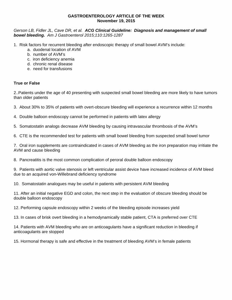
GASTROENTEROLOGY ARTICLE OF THE WEEK November 19, 2015
Gerson LB, Fidler JL, Cave DR, et al. ACG Clinical Guideline: Diagnosis and management of small bowel bleeding. Am J Gastroenterol 2015;110:1265-1287 1. Risk factors for recurrent bleeding after endoscopic therapy of small bowel AVM’s include: a. duodenal location of AVM b. number of AVM’s
c. iron deficiency anemia d. chronic renal disease e. need for transfusions True or False 2..Patients under the age of 40 presenting with suspected small bowel bleeding are more likely to have tumors than older patients 3. About 30% to 35% of patients with overt-obscure bleeding will experience a recurrence within 12 months 4. Double balloon endoscopy cannot be performed in patients with latex allergy 5. Somatostatin analogs decrease AVM bleeding by causing intravascular thrombosis of the AVM’s 6. CTE is the recommended test for patients with small bowel bleeding from suspected small bowel tumor 7. Oral iron supplements are contraindicated in cases of AVM bleeding as the iron preparation may irritiate the AVM and cause bleeding 8. Pancreatitis is the most common complication of peroral double balloon endoscopy 9. Patients with aortic valve stenosis or left ventricular assist device have increased incidence of AVM bleed due to an acquired von-Willebrand deficiency syndrome 10. Somatostatin analogues may be useful in patients with persistent AVM bleeding 11. After an initial negative EGD and colon, the next step in the evaluation of obscure bleeding should be double balloon endoscopy 12. Performing capsule endoscopy within 2 weeks of the bleeding episode increases yield 13. In cases of brisk overt bleeding in a hemodynamically stable patient, CTA is preferred over CTE 14. Patients with AVM bleeding who are on anticoagulants have a significant reduction in bleeding if anticoagulants are stopped 15. Hormonal therapy is safe and effective in the treatment of bleeding AVM’s in female patients