Accute flaccid paralysis
46
ACUTE FLACCID PARALYSIS (AFP) Prepared by;- Dr. SafeerAhmed Jamil RESIDENT FCPS II TRAINEE DEPTT. OF PEDIARICS
-
Upload
safeer-ahmed -
Category
Healthcare
-
view
69 -
download
2
Transcript of Accute flaccid paralysis
ACUTE FLACCID PARALYSIS
(AFP)Prepared by;- Dr. SafeerAhmed Jamil RESIDENT FCPS II TRAINEE DEPTT. OF PEDIARICS

CASE• A 12 yr old male,came to ED with complaints of pain in
the calf of the right leg on Tuesday,followed by weakness of the both lower limbs the next day morning.was bed ridden after the onset of weakness.his bowel and bladder are intact.he is able to feel sensation of clothes over the lower limbs.he felt heaviness of the limbs..taken to local hospital was given treatment..where he noticed weakness in the upper limbs also.
• no symptoms of cranial nerve involvement.• no h/o fever at the onset of weakness.• no h/o recent immunization or no h/o dog bite.• no h/o seizures episodes.• no h/o similar complaints in the past.• no h/o similar complaints in the family.

Clinical examination• Patient was consciuos coherent• no
pallor ,icterus,clubbing,lymphadenopathy,edema.• His BP is 110/70 mm Hg in supine,100/70 mm
Hg on standing.no postural hypotension.• Pulse -82/min regular,all peripheral pulses felt.• Respiratory rate -18 /min.breath holding time –
able to count upto 45 in single breath.
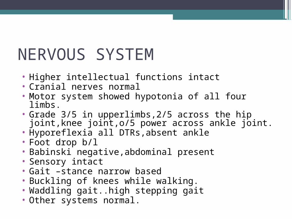
NERVOUS SYSTEM• Higher intellectual functions intact• Cranial nerves normal• Motor system showed hypotonia of all four limbs.• Grade 3/5 in upperlimbs,2/5 across the hip joint,knee
joint,o/5 power across ankle joint.• Hyporeflexia all DTRs,absent ankle• Foot drop b/l• Babinski negative,abdominal present• Sensory intact• Gait –stance narrow based• Buckling of knees while walking.• Waddling gait..high stepping gait• Other systems normal.
Key points
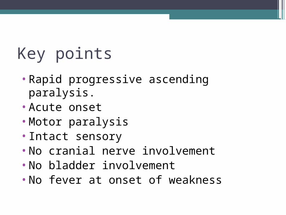
•Rapid progressive ascending paralysis.•Acute onset •Motor paralysis•Intact sensory•No cranial nerve involvement•No bladder involvement•No fever at onset of weakness
Diagnosis………………??????????????????
DEFINITION
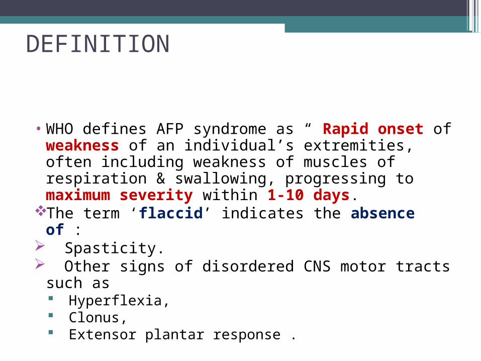
• WHO defines AFP syndrome as “ Rapid onset of weakness of an individual’s extremities, often including weakness of muscles of respiration & swallowing, progressing to maximum severity within 1-10 days.
The term ‘flaccid’ indicates the absence of : Spasticity. Other signs of disordered CNS motor tracts
such as Hyperflexia, Clonus, Extensor plantar response .
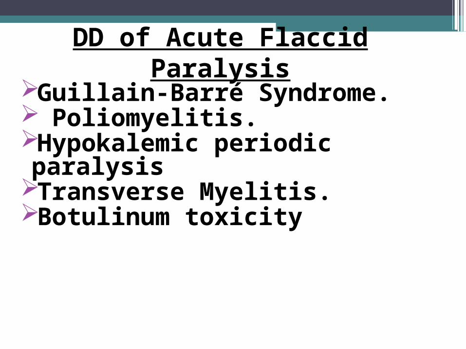
Guillain-Barré Syndrome. Poliomyelitis. Hypokalemic periodic paralysisTransverse Myelitis.Botulinum toxicity
DD of Acute Flaccid Paralysis
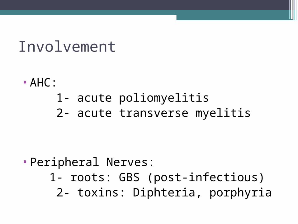
Involvement •AHC: 1- acute poliomyelitis 2- acute transverse myelitis
•Peripheral Nerves: 1- roots: GBS (post-infectious) 2- toxins: Diphteria, porphyria
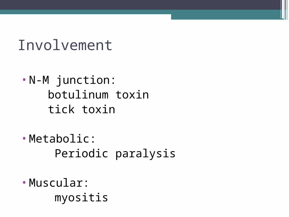
Involvement
•N-M junction: botulinum toxin tick toxin
•Metabolic: Periodic paralysis
•Muscular: myositis
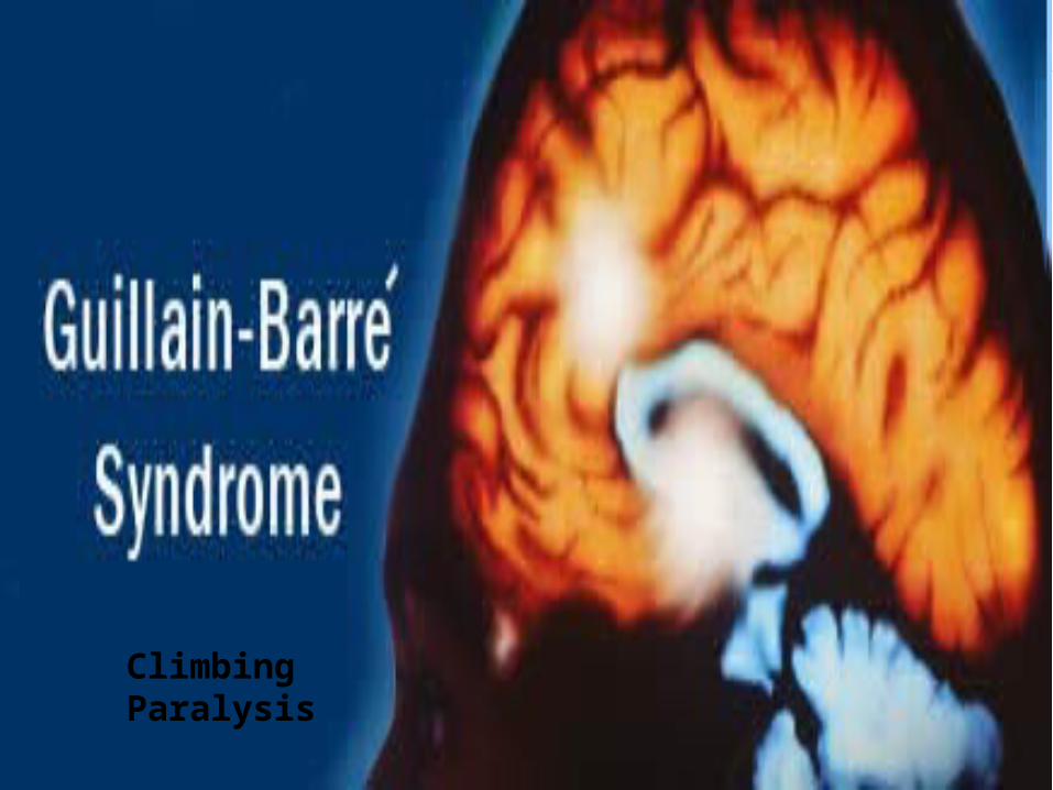
Climbing Paralysis
•It is an acute idiopathic monophasic acquired inflammatory demyelinating polyradiculo-neuropathy.
•Polyradiculopathy refers to damage to multiple nerve roots sufficient to produce neurologic symptoms & signs such as pain, weakness, and sensory affection.
•GBS is the most common cause of acute flaccid paralysis in healthy infants and children.
Guillain-Barré Syndrome :
Guillain Barre Syndrome GBS
•The most common cause of acute flaccid paralysis (AFP) among infants.
•Age : any including newborn •Sex : any ( male > female)•Post-infectious polyneuropathy; ascending
polyneuropathic paralysis
•An acute, rapidly progressing and potentially fatal form of polyneuritis
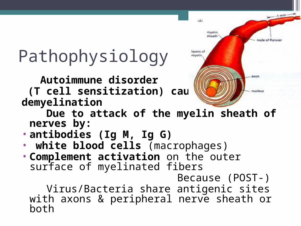
Pathophysiology Autoimmune disorder (T cell sensitization) causes demyelination Due to attack of the myelin sheath of
nerves by:• antibodies (Ig M, Ig G)• white blood cells (macrophages)• Complement activation on the outer
surface of myelinated fibers Because (POST-) Virus/Bacteria share antigenic sites with
axons & peripheral nerve sheath or both

pathophysiology
•inflammation causes leakage of proteins into the CSF causing raised CSF proteins without pleocytosis
•Can involve the peripheral nerves, cranial nerves,dorsal roots, dorsal root ganglia & sympathatic chain

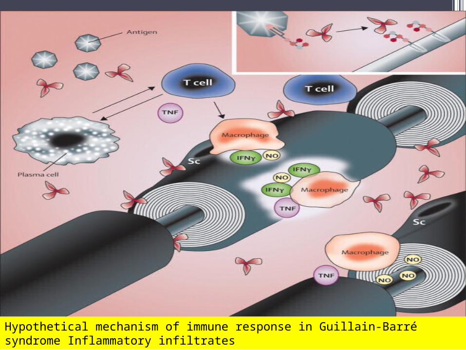
Hypothetical mechanism of immune response in Guillain-Barré syndrome Inflammatory infiltrates

Preceding Events : (1-3 WEEKS)
• Respiratory infections : 1- Viral: CMV, EBV, Varicella virus ,
influenza virus 2- Bacterial: Mycoplasma pneumoniae,
H influenza
• Gastrointestinal infections : Campylobacter-jejuni (Bloody GE)
• Vaccinations• Post surgery

Aetiology ▫Mycoplasma
•Hepatitis B•CMV•EBV•Measles•Mumps•Echovirus•Cocksakie virus•Influenza virus
▫Varicella virus•Compylobacter jejuni

Usually 2 - 4 weeks following respiratory or GI infection.•The classic presentation: * Fine paresthesias in the toes and fingertips. * Lower extremity weakness: symmetric &
ascending(landry ascending paralysis).
* Gait unsteadiness. * Inability to walk. * Respiratory muscles involvement. * Neuropathic pain… low back pain. •Cranial Neuropathy: Facial nerve is most commonly affected, resulting in bilateral facial weakness.
Clinical Features of GBS :
•Symmetric limb weakness. •Absent reflexes. •Vibration and position sensation are
affected in 40% of cases.•Autonomic dysfunction: * Cardiac dysrhythmias. * Orthostatic hypotension, * Hypertension. * Paralytic ileus . * Bladder dysfunction.
Physical Examination :
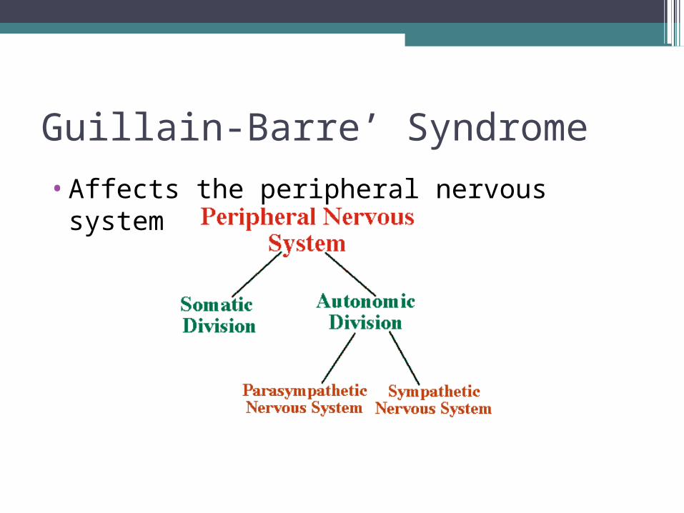
Guillain-Barre’ Syndrome
•Affects the peripheral nervous system

Characteristic “3A”triad:
• ascending weakness : a- bilateral symmetrical weakness b- usually start in LL, then UL c-then, might be affected : i- cranial nerves (Brain
stem) : including glosssopharyngeal and vagus nerves ( difficulty of swallowing even of fluid and water) and III, IV, VI cranial nerves ( eye muscles in Miller Fisher variety), VII Facial nerve ( unilateral or bilateral), and then respiratory muscles
ii- respiratory muscles iii- phrenic nerves
( diaphragm )• areflexia ( Hallmark)• atonia ( hypotonia)
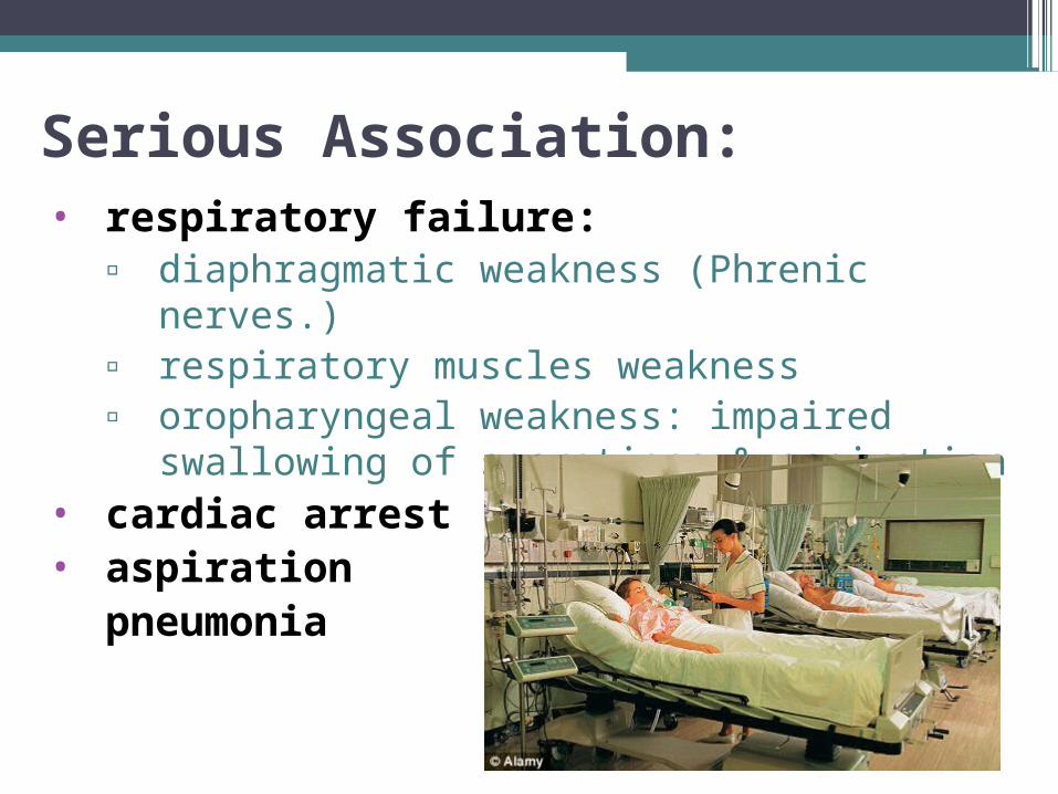
Serious Association:• respiratory failure:
▫ diaphragmatic weakness (Phrenic nerves.)▫ respiratory muscles weakness▫ oropharyngeal weakness: impaired
swallowing of secretions & aspiration• cardiac arrest• aspiration
pneumonia
• Acute inflammatory demyelinating polyneuropathy (AIDP): the most common form in developed countries.
•Acute motor axonal neuropathy: more common in developing countries. More severe with common respiratory involvement. Strong association with campylobacter.
•Acute motor-sensory axonal neuropathy.
•Miller Fisher syndrome: triad of external ophthalmo-plegia, Ataxia, areflexia with muscle weakness.
•Polyneuritis cranialis: associated with CMV infection.
Forms of GBS :
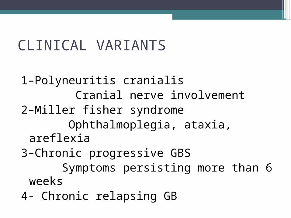
CLINICAL VARIANTS
1–Polyneuritis cranialis Cranial nerve involvement2–Miller fisher syndrome Ophthalmoplegia, ataxia, areflexia3–Chronic progressive GBS Symptoms persisting more than 6
weeks4- Chronic relapsing GB

Differentiation from spinal cord syndrome•Loss of arm reflexes
•Absence of sensory level
•Lack of spinal tenderness
•Normal bowel and bladder function
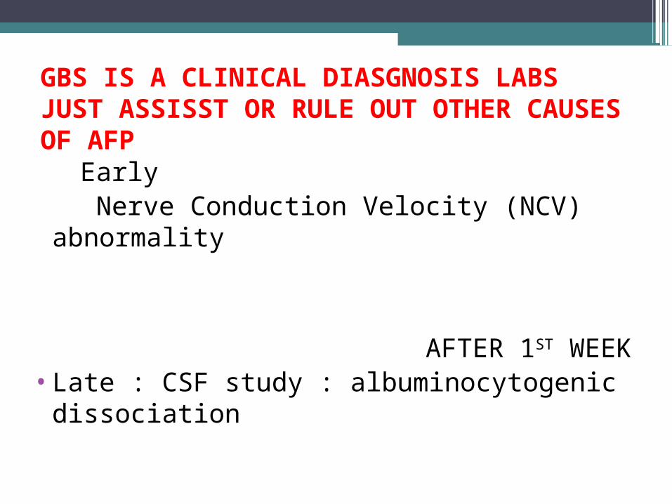
GBS IS A CLINICAL DIASGNOSIS LABS JUST ASSISST OR RULE OUT OTHER CAUSES OF AFP Early Nerve Conduction Velocity (NCV)
abnormality
AFTER 1ST WEEK •Late : CSF study : albuminocytogenic
dissociation

1- NCV/EMG:
i- Early :Delayed or absent F waves or H reflexes ii- slow or block of Nerve conduction velocityiii- normal EMG/ extensive fibrillation showing denervation
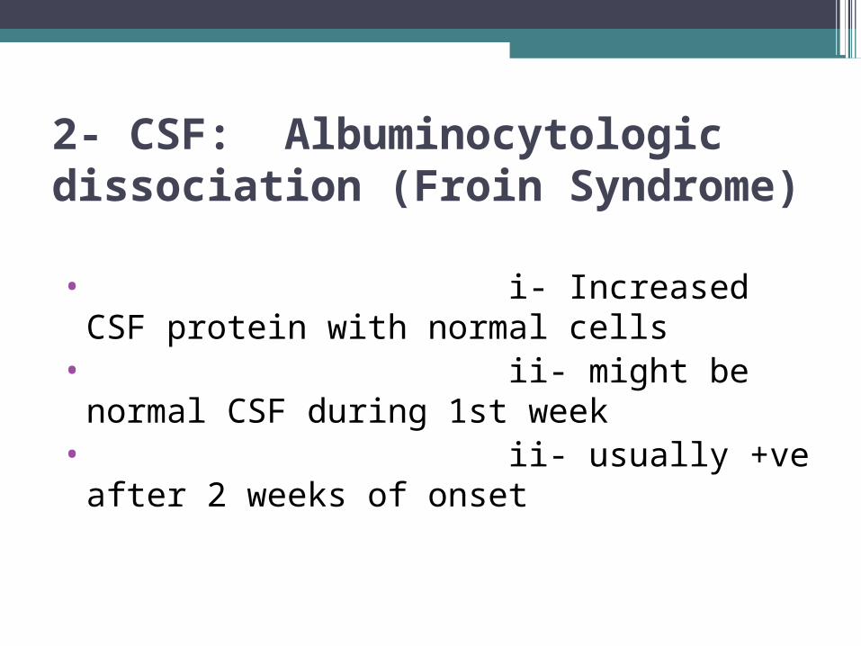
2- CSF: Albuminocytologic dissociation (Froin Syndrome)
• i- Increased CSF protein with normal cells
• ii- might be normal CSF during 1st week
• ii- usually +ve after 2 weeks of onset

Investigations (OTHERS) •Antibody study : Ig M autoantibodies to GM1
and GM2 gangliosides or Spinal MRI or normal CPK
•Anti-GQ1b antibodies are typically found in patients with the Miller Fisher syndrome (Acta Pediatr 2011)
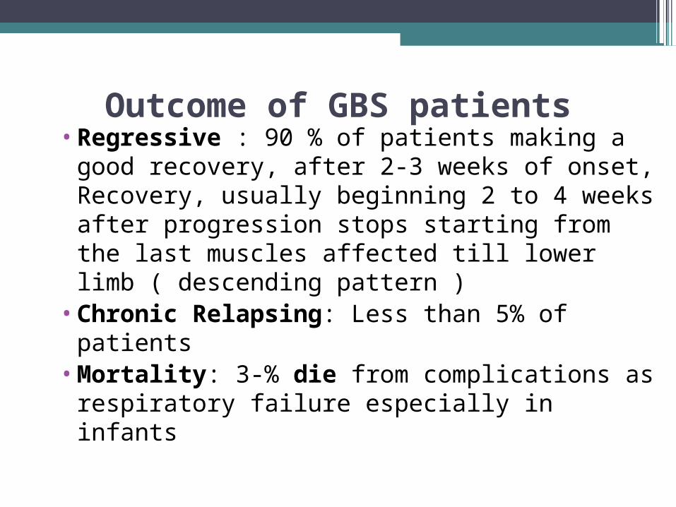
Outcome of GBS patients•Regressive : 90 % of patients making a
good recovery, after 2-3 weeks of onset, Recovery, usually beginning 2 to 4 weeks after progression stops starting from the last muscles affected till lower limb ( descending pattern )
•Chronic Relapsing: Less than 5% of patients
•Mortality: 3-% die from complications as respiratory failure especially in infants

•Marked persistent asymmetry of weakness.•Persistent bladder or bowel dysfunction.
•Bladder or bowel dysfunction at the onset.•Mononuclear leukocytosis in the CSF > 50.
•Sharp sensory level.•Pupillary abnormalities are not seen in GBS.
Doubt the Diagnosis of GBS IF:

Treatment Hospitalization •1- General care •2-- Specific treatment •3- complication treatment
Treatment
•Hospitalization : i- Must be treated in a hospital, never at home
ii- because of a risk of sudden onset of cardiac or respiratory arrest
iii- any hospital ? No, it must be a hospital have a pediatrics ICU
Hospitalization is continued until the child's condition has clearly stabilized.

General care•i- bed sores ii-bowel care iii- nutrition care •monitoring of vital signs
–Nursing care–Repeated spirometries–Bowel and bladder care–Tube feeding–Care for bed sores–Ventilatory support if required

Specific treatment
• i- IV immunoglobin: 2 gm/kg treatment
*at a dose of 0.4 g/kg/day for 5 consecutive days or
* 1gm/kg/day for 2 days• ii- plasmapheresis: 5 exchanges
of 50 ml plasma/ kg on alternate days ( 10 days course).
• iii- both i and ii
complication treatment:
i- artificial respiration for respiratory failure
ii- muscular pain: pain killer as NSAI
iii- chronic relapsing: trial of immunosupprive drugs or corticosteroid

Need for intensive care (PICU)
- Flaccid quadriparesis- Rapidly progressive weakness- Reduced vital capacity (≤20 mL/kg)- Bulbar palsy- Autonomic cardiovascular instability
Need for assisted ventilation — Approximately 20 percent of children with GBS require mechanical ventilation for respiratory failure

Warning signs for RF*- A sustained increase of pCO2 to ≥50 mmHg
(normally 35 to 40 mmHg)- An increasing respiratory rate- Increasing oxygen requirement and increasing
alveolar to arterial oxygen difference (normally 5 to 10 mmHg)
- An increased use of accessory muscles (eg, sternocleidomastoid use, flaring of the ala nasae, intercostal retractions) and decreased or paradoxical diaphragm movements; these reflect restrictive lung-chest wall movement and low lung volumes
- Sweating about the head and neck, wide pulse pressure, and bounding pulses portend CO2 retention.
• Children have less metabolic and muscle reserve than adults. They can deteriorate quite rapidly and become apneic or develop alveolar hypoventilation "right under your nose."
• Sedation and neuromuscular blockade should be avoided in ventilated patients because they obscure the course of the illness.
• Providing scrupulous airway care and chest physiotherapy reduce the risk of pneumonia.
• Tracheostomy may need to be performed if prolonged ventilation is required.
PROGNOSIS
•Mortality 3%•20% of cases need repiratory ve•Recovery - 1 to 6 months, may take 12 months - Delayed recovery may be followed by
permanent neurological sequelPoor prognostic features with sequelae if at
presentation.1. Cranial nerve involvement2. Needs Intubation & vent.3. Maximum disability

Transverse Myelitis:
• ? of immunological disorder• C/P : of AFP ( acute onset of flaccid
hypotonic weakness ) with the following characters: ▫ LL paralysis : paraplegia with areflexia▫ with sensory level of loss of sensation ▫ Later : hyperreflexia

Poliomyelitis :• due to enterovirus affection (polio virus )
( non or inadequate OPV ) • C/P: of AFP with the following characters:
▫ first shock stage ( with generalized hypotonia) then patchy asymmetrical weakness
▫ normal sensation▫ areflexia of affected muscles

Please don’t miss the diagnosis of GBS. By noting: Symptoms begin 2 - 4 weeks following respiratory or GI infection.Diminished or absent reflexes.Symmetric & ascending lower extremity weakness.Sensations intact : Fine paresthesias in the toes and fingertips.No bladder or bowel dysfunction at the onset.