Accurate and Anatomic Midface Filler Injection by Using ... · PDF fileAccurate and Anatomic...
4
Accurate and Anatomic Midface Filler Injection by Using Cheek Implants as an Injection Template JOSEPH NIAMTU, III, DMD Joseph Niamtu, III, DMD, has indicated no significant interest with commercial supporters. C ontemporary cosmetic surgeons appreciate the concept of volume restoration in the rejuvena- tion of the aging face. Injectable fillers and fat have become a mainstay of reproducing youthful facial contours. 1–5 Aging changes in the midface include volume loss from hard and soft tissue changes as well as the de- scent of the SOOF (superficial orbicularis oculi fat) and malar fat complex. 6,7 These changes produce midfacial hollowing and a gaunt appearance. Other exogenous factors such as medication induced lipo- atrophy can mimic the atrophic changes of the aging face. 8,9 Rejuvenation of the midfacial region includes lifting procedures, skin-tightening procedures, im- plant placement, and injectable fillers. Facial filler and or fat injection in the midface has many advantages for the doctor and patient includ- ing ease of placement, avoidance of surgery, adjust- ability, and the ability to individualize and custom contour for each patient. One problem that can exist with midfacial filler augmentation is the decision where to exactly place the filler or how much filler to place. Freehand estimation and injection can pro- duce asymmetry or inconsistent filling and it is ad- vantageous to have a template to better define the areas of desired and intended filling. Midfacial im- plants have undergone many advancements over the past 20 years including the availability of anatom- ically diverse shapes and sizes. These anatomic im- plants were designed to imitate youthful volumetric facial contours. The available selection of implants include those that are intended to augment the submalar area, the malar area, or a combination of both. These implants are anatomic in three-dimen- sional form and are designed to fill atrophic spaces in the midface. The size and shape of the implants can be used for templates on the face to define bound- aries and approximate volume for filler or fat injection. Actual silicone implants or implant sizers (silicone implant analogues intended for try on but not for implantation) can be used to trace the desired area of intended filler injection on the midfacial skin. Implantech (Ventura, CA) manufactures an array of midface implants in multiple shapes and sizes. The submalar implant series is intended to provide augmentation of the atrophic inframalar areas (below the cheek bones and above the lip; Figure 1A). The malar shell implants are intended to provide augmentation more laterally by aug- menting the lateral malar and zygomatic areas (Figure 1B). A combination implant is available that augments both of these areas simultaneously (Figure 1C). The patient is given a mirror and the injector and the patient decides on the desired areas of augmentation. The patient ‘‘tries on’’ the various implant shapes to assist in the treatment area selection. Once decided, the implant is held in place and traced with a surgical skin marker (Figure 1A). & 2007 by the American Society for Dermatologic Surgery, Inc. Published by Blackwell Publishing ISSN: 1076-0512 Dermatol Surg 2008;34:93–96 DOI: 10.1111/j.1524-4725.2007.34018.x 93 Private Practice, Cosmetic Facial surgery, Richmond, Virginia
Transcript of Accurate and Anatomic Midface Filler Injection by Using ... · PDF fileAccurate and Anatomic...
Accurate and Anatomic Midface Filler Injection by UsingCheek Implants as an Injection Template
JOSEPH NIAMTU, III, DMD�
Joseph Niamtu, III, DMD, has indicated no significant interest with commercial supporters.
Contemporary cosmetic surgeons appreciate the
concept of volume restoration in the rejuvena-
tion of the aging face. Injectable fillers and fat have
become a mainstay of reproducing youthful facial
contours.1–5
Aging changes in the midface include volume loss
from hard and soft tissue changes as well as the de-
scent of the SOOF (superficial orbicularis oculi fat)
and malar fat complex.6,7 These changes produce
midfacial hollowing and a gaunt appearance. Other
exogenous factors such as medication induced lipo-
atrophy can mimic the atrophic changes of the aging
face.8,9 Rejuvenation of the midfacial region includes
lifting procedures, skin-tightening procedures, im-
plant placement, and injectable fillers.
Facial filler and or fat injection in the midface has
many advantages for the doctor and patient includ-
ing ease of placement, avoidance of surgery, adjust-
ability, and the ability to individualize and custom
contour for each patient. One problem that can exist
with midfacial filler augmentation is the decision
where to exactly place the filler or how much filler to
place. Freehand estimation and injection can pro-
duce asymmetry or inconsistent filling and it is ad-
vantageous to have a template to better define the
areas of desired and intended filling. Midfacial im-
plants have undergone many advancements over the
past 20 years including the availability of anatom-
ically diverse shapes and sizes. These anatomic im-
plants were designed to imitate youthful volumetric
facial contours. The available selection of implants
include those that are intended to augment the
submalar area, the malar area, or a combination of
both. These implants are anatomic in three-dimen-
sional form and are designed to fill atrophic spaces in
the midface. The size and shape of the implants can
be used for templates on the face to define bound-
aries and approximate volume for filler or fat
injection.
Actual silicone implants or implant sizers (silicone
implant analogues intended for try on but not for
implantation) can be used to trace the desired
area of intended filler injection on the midfacial
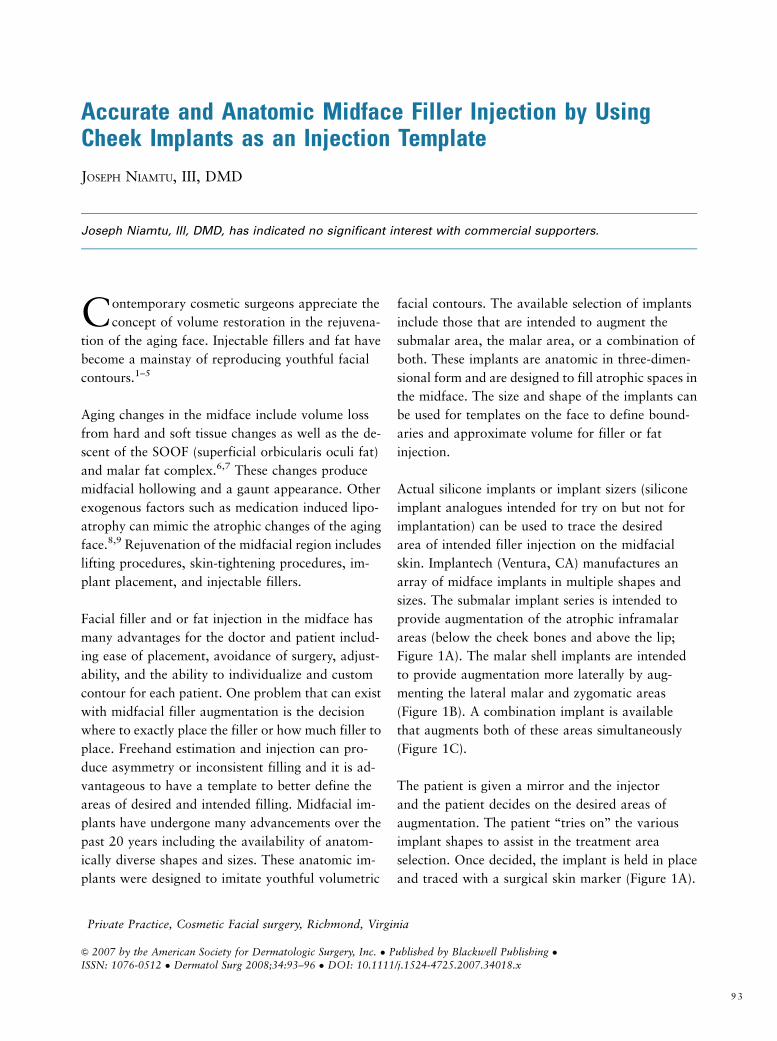
skin. Implantech (Ventura, CA) manufactures an
array of midface implants in multiple shapes and
sizes. The submalar implant series is intended to
provide augmentation of the atrophic inframalar
areas (below the cheek bones and above the lip;
Figure 1A). The malar shell implants are intended
to provide augmentation more laterally by aug-
menting the lateral malar and zygomatic areas
(Figure 1B). A combination implant is available
that augments both of these areas simultaneously
(Figure 1C).
The patient is given a mirror and the injector
and the patient decides on the desired areas of
augmentation. The patient ‘‘tries on’’ the various
implant shapes to assist in the treatment area
selection. Once decided, the implant is held in place
and traced with a surgical skin marker (Figure 1A).
& 2007 by the American Society for Dermatologic Surgery, Inc. � Published by Blackwell Publishing �ISSN: 1076-0512 � Dermatol Surg 2008;34:93–96 � DOI: 10.1111/j.1524-4725.2007.34018.x
9 3
�Private Practice, Cosmetic Facial surgery, Richmond, Virginia
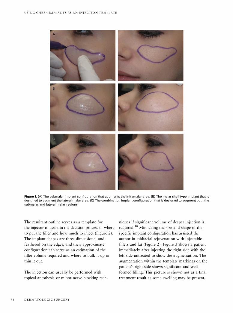
The resultant outline serves as a template for
the injector to assist in the decision process of where
to put the filler and how much to inject (Figure 2).
The implant shapes are three-dimensional and
feathered on the edges, and their approximate
configuration can serve as an estimation of the
filler volume required and where to bulk it up or
thin it out.
The injection can usually be performed with
topical anesthesia or minor nerve-blocking tech-
niques if significant volume of deeper injection is
required.10 Mimicking the size and shape of the
specific implant configuration has assisted the
author in midfacial rejuvenation with injectable
fillers and fat (Figure 2). Figure 3 shows a patient
immediately after injecting the right side with the
left side untreated to show the augmentation. The
augmentation within the template markings on the
patient’s right side shows significant and well-
formed filling. This picture is shown not as a final
treatment result as some swelling may be present,
Figure 1. (A) The submalar implant configuration that augments the inframalar area. (B) The malar shell type implant that isdesigned to augment the lateral malar area. (C) The combination implant configuration that is designed to augment both thesubmalar and lateral malar regions.
D E R M AT O L O G I C S U R G E RY9 4
U S I N G C H E E K I M P L A N T S A S A N I N J E C T I O N T E M P L AT E
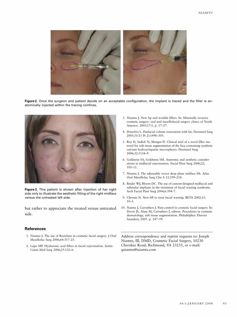
but rather to appreciate the treated versus untreated
side.
References
1. Niamtu J. The use of Restylane in cosmetic facial surgery. J Oral
Maxillofac Surg 2006;64:317–25.
2. Lupo MP. Hyaluronic acid fillers in facial rejuvenation. Semin
Cutan Med Surg 2006;25:122–6.
3. Niamtu J. New lip and wrinkle fillers. In: Minimally invasive
cosmetic surgery: oral and maxillofacial surgery clinics of North
America. 2005;17:1, p. 17–27.
4. Donofrio L. Panfacial volume restoration with fat. Dermatol Surg
2005;31(11 Pt 2):1496–505.
5. Roy D, Sadick N, Mangat D. Clinical trial of a novel filler ma-
terial for soft tissue augmentation of the face containing synthetic
calcium hydroxylapatite microspheres. Dermatol Surg
2006;32:1134–9.
6. Goldstein SA, Goldstein SM. Anatomic and aesthetic consider-
ations in midfacial rejuvenation. Facial Plast Surg 2006;22:
105–11.
7. Niamtu J. The adjustable vector deep plane midface lift. Atlas
Oral Maxillofac Surg Clin A 12:199–214.
8. Binder WJ, Bloom DC. The use of custom-designed midfacial and
submalar implants in the treatment of facial wasting syndrome.
Arch Facial Plast Surg 2004;6:394–7.
9. Cheonis N. New-fill to treat facial wasting. BETA 2002;15:
10–5.
10. Namtu J, Carruthers J. Pain control in cosmetic facial surgery. In:
Dover JS, Alam M, Carruthers J, editors. Procedures in cosmetic
dermatology, soft tissue augmentation. Philadelphia: Elsevier
Saunders; 2005. p. 147–59.
Address correspondence and reprint requests to: JosephNiamtu, III, DMD, Cosmetic Facial Surgery, 10230Cherokee Road, Richmond, VA 23235, or e-mail:[email protected]
Figure 2. Once the surgeon and patient decide on an acceptable configuration, the implant is traced and the filler is an-atomically injected within the tracing confines.
Figure 3. This patient is shown after injection of her rightside only to illustrate the aesthetic filling of the right midfaceversus the untreated left side.
3 4 : 1 : J A N U A RY 2 0 0 8 9 5
N I A M T U
COMMENTARY
The author presents a novel concept to aid injectable midface contouring. Pretreatment freehand marking
of cheek hollows with a surgical marker may give a more tailored approach to a patient’s specific need. In
certain cases, however, using a cheek implant as an injection template may better assist physicians and
patients in visualizing a possible outcome. Hyaluronic acids, calcium hydroxylapetite, poly-L-lactic acid,
and liquid silicone are the most commonly used fillers for cheek contouring and should all be injected into
the immediate subdermal plane or deeper for an optimal cosmetic result in this location. Injecting more
superficially may create dermal contour irregularities.
DEREK JONES, MD
Los Angeles, CA
D E R M AT O L O G I C S U R G E RY9 6
U S I N G C H E E K I M P L A N T S A S A N I N J E C T I O N T E M P L AT E