
Abundance of type I toxin–antitoxin systems in bacteria ...
17
Abundance of type I toxin–antitoxin systems in bacteria: searches for new candidates and discovery of novel families Elizabeth M. Fozo 1 , Kira S. Makarova 2 , Svetlana A. Shabalina 2 , Natalya Yutin 2 , Eugene V. Koonin 2 and Gisela Storz 1, * 1 Eunice Kennedy Shriver National Institute of Child Health and Human Development and 2 National Center for Biotechnology Information, National Institutes of Health, Bethesda, MD 20894, USA Received December 14, 2009; Revised January 18, 2010; Accepted January 19, 2010 ABSTRACT Small, hydrophobic proteins whose synthesis is repressed by small RNAs (sRNAs), denoted type I toxin–antitoxin modules, were first discovered on plasmids where they regulate plasmid stability, but were subsequently found on a few bacterial chromosomes. We used exhaustive PSI-BLAST and TBLASTN searches across 774 bacterial genomes to identify homologs of known type I toxins. These searches substantially expanded the collection of predicted type I toxins, revealed homology of the Ldr and Fst toxins, and suggested that type I toxin–antitoxin loci are not spread by hor- izontal gene transfer. To discover novel type I toxin– antitoxin systems, we developed a set of search parameters based on characteristics of known loci including the presence of tandem repeats and clusters of charged and bulky amino acids at the C-termini of short proteins containing predicted transmembrane regions. We detected sRNAs for three predicted toxins from enterohemorrhagic Escherichia coli and Bacillus subtilis, and showed that two of the respective proteins indeed are toxic when overexpressed. We also demonstrated that the local free-energy minima of RNA folding can be used to detect the positions of the sRNA genes. Our results suggest that type I toxin–anti- toxin modules are much more widely distributed among bacteria than previously appreciated. INTRODUCTION Plasmid maintenance in many bacteria is attributed to the presence of toxin–antitoxin loci on the plasmids. These loci consist of two genes: one encodes a stable toxic protein, and the second an unstable antitoxin. If the plasmid is lost from the cell upon division, the unstable antitoxin is degraded, and the stable toxin is able to kill the cell. This phenomena, referred to as ‘post-segregational killing’ or ‘plasmid addiction’ has been described for plasmids in both Gram negative and Gram positive bacteria. The toxin–antitoxin loci are categorized into two broad classes based on the type of antitoxin: the antitoxin of type I systems is a small RNA (sRNA) which base pairs with the toxin mRNA to prevent protein synthesis, whereas the antitoxin of the type II systems is a protein that binds to and inhibits the toxin protein. Generally, type I toxins are small (under 60 amino acids in length), highly hydrophobic proteins, while type II toxins are slightly larger (100 amino acids) and less hydrophobic. The best-studied type I toxin–antitoxin systems include the hok-sok locus of plasmid R1, and the par locus of plasmid pAD1 of Enterococcus faecalis (1,2). Although the toxin–antitoxin loci were initially described on plasmids, recent studies have shown that many of these gene pairs are also present on bacterial chro- mosomes. The type II toxin–antitoxin systems, in which the antitoxin is a protein, have been documented in diverse bacteria with many genomes carrying dozens of distinct toxin–antitoxin pairs (3). The type II toxins have been shown to degrade RNA or inhibit cellular enzymes such as DNA gyrase (4,5). The physiological role(s) of the type II systems remains a subject of debate; proposed func- tions include stress survival, protection of the bacteria against foreign DNA, and stabilization of chromosomal regions (6,7). Several studies have shown that type I toxin–antitoxin systems, in which the antitoxin is an sRNA, are also present on some bacterial chromosomes. The hok-sok locus from plasmid R1 is encoded in the genomes of several enteric bacteria (8,9). In some strains, the sequences of these loci have degenerated and appear to be non-functional whereas in other cases, the systems are intact. Similarly, the par locus from plasmid pAD1 is *To whom correspondence should be addressed. Tel: + 301 402 0968; Fax: +1 301 402 0078; Email: [email protected]; [email protected] Published online 15 February 2010 Nucleic Acids Research, 2010, Vol. 38, No. 11 3743–3759 doi:10.1093/nar/gkq054 Published by Oxford University Press 2010. This is an Open Access article distributed under the terms of the Creative Commons Attribution Non-Commercial License (http://creativecommons.org/licenses/ by-nc/2.5), which permits unrestricted non-commercial use, distribution, and reproduction in any medium, provided the original work is properly cited.
Transcript of Abundance of type I toxin–antitoxin systems in bacteria ...

Abundance of type I toxin–antitoxin systemsin bacteria: searches for new candidatesand discovery of novel familiesElizabeth M. Fozo1, Kira S. Makarova2, Svetlana A. Shabalina2, Natalya Yutin2,
Eugene V. Koonin2 and Gisela Storz1,*
1Eunice Kennedy Shriver National Institute of Child Health and Human Development and 2National Center forBiotechnology Information, National Institutes of Health, Bethesda, MD 20894, USA
Received December 14, 2009; Revised January 18, 2010; Accepted January 19, 2010
ABSTRACT
Small, hydrophobic proteins whose synthesis isrepressed by small RNAs (sRNAs), denoted type Itoxin–antitoxin modules, were first discovered onplasmids where they regulate plasmid stability,but were subsequently found on a few bacterialchromosomes. We used exhaustive PSI-BLASTand TBLASTN searches across 774 bacterialgenomes to identify homologs of known type Itoxins. These searches substantially expanded thecollection of predicted type I toxins, revealedhomology of the Ldr and Fst toxins, and suggestedthat type I toxin–antitoxin loci are not spread by hor-izontal gene transfer. To discover novel type I toxin–antitoxin systems, we developed a set of searchparameters based on characteristics of known lociincluding the presence of tandem repeats andclusters of charged and bulky amino acids at theC-termini of short proteins containing predictedtransmembrane regions. We detected sRNAs forthree predicted toxins from enterohemorrhagicEscherichia coli and Bacillus subtilis, and showedthat two of the respective proteins indeed aretoxic when overexpressed. We also demonstratedthat the local free-energy minima of RNA foldingcan be used to detect the positions of the sRNAgenes. Our results suggest that type I toxin–anti-toxin modules are much more widely distributedamong bacteria than previously appreciated.
INTRODUCTION
Plasmid maintenance in many bacteria is attributed tothe presence of toxin–antitoxin loci on the plasmids.These loci consist of two genes: one encodes a stabletoxic protein, and the second an unstable antitoxin.
If the plasmid is lost from the cell upon division, theunstable antitoxin is degraded, and the stable toxinis able to kill the cell. This phenomena, referred to as‘post-segregational killing’ or ‘plasmid addiction’ hasbeen described for plasmids in both Gram negative andGram positive bacteria. The toxin–antitoxin loci arecategorized into two broad classes based on the type ofantitoxin: the antitoxin of type I systems is a small RNA(sRNA) which base pairs with the toxin mRNA to preventprotein synthesis, whereas the antitoxin of the type IIsystems is a protein that binds to and inhibits the toxinprotein. Generally, type I toxins are small (under 60 aminoacids in length), highly hydrophobic proteins, while type IItoxins are slightly larger (�100 amino acids) and lesshydrophobic. The best-studied type I toxin–antitoxinsystems include the hok-sok locus of plasmid R1,and the par locus of plasmid pAD1 of Enterococcusfaecalis (1,2).Although the toxin–antitoxin loci were initially
described on plasmids, recent studies have shown thatmany of these gene pairs are also present on bacterial chro-mosomes. The type II toxin–antitoxin systems, in whichthe antitoxin is a protein, have been documented in diversebacteria with many genomes carrying dozens of distincttoxin–antitoxin pairs (3). The type II toxins have beenshown to degrade RNA or inhibit cellular enzymes suchas DNA gyrase (4,5). The physiological role(s) of the typeII systems remains a subject of debate; proposed func-tions include stress survival, protection of the bacteriaagainst foreign DNA, and stabilization of chromosomalregions (6,7).Several studies have shown that type I toxin–antitoxin
systems, in which the antitoxin is an sRNA, are alsopresent on some bacterial chromosomes. The hok-soklocus from plasmid R1 is encoded in the genomes ofseveral enteric bacteria (8,9). In some strains, thesequences of these loci have degenerated and appear tobe non-functional whereas in other cases, the systemsare intact. Similarly, the par locus from plasmid pAD1 is
*To whom correspondence should be addressed. Tel: + 301 402 0968; Fax: +1 301 402 0078; Email: [email protected]; [email protected]
Published online 15 February 2010 Nucleic Acids Research, 2010, Vol. 38, No. 11 3743–3759doi:10.1093/nar/gkq054
Published by Oxford University Press 2010.This is an Open Access article distributed under the terms of the Creative Commons Attribution Non-Commercial License (http://creativecommons.org/licenses/by-nc/2.5), which permits unrestricted non-commercial use, distribution, and reproduction in any medium, provided the original work is properly cited.
present on the chromosomes of E. faecalis, Lactobacilluscasei and a Staphylococcus saprophyticus strain (10).Additional type I toxin–antitoxin loci were foundserendipitously on bacterial chromosomes (1). Theseinclude the ldr-rdl, ibs-sib, tisB-istR-1 and shoB-ohsC lociof Escherichia coli and the txpA-ratA locus of Bacillussubtilis. Interestingly, for these loci, there was noreported homology to known plasmid sequences.However, as for the plasmid-encoded systems, over-production of the corresponding protein leads to celldeath, and this toxicity is repressed by an antisensesRNA regulator. The exact biochemical activities of thesmall, hydrophobic toxin proteins are not known,although similarity to phage holin proteins has beennoted, and overexpression of the proteins is associatedwith membrane depolarization and increased membranepermeability (1). As for the chromosomally-encodedtype II toxin–antitoxin loci, the physiological function(s)of the chromosomally-encoded type I toxin–antitoxinsystems remains unclear.As mentioned above, type II toxin–antitoxin loci
are broadly distributed among diverse bacteria. Wehypothesized that type I systems are also widespread. Totest this, we sought to identify homologs of the knowntype I toxins. Our computational approach identifiedmany more putative toxins than have been previouslyreported. We experimentally validated a homolog of thepar locus encoded in the chromosome of Streptococcuspneumoniae, the first report of a type I toxin–antitoxinsystem in this pathogen.In addition to documenting the distribution of
known type I systems in bacteria, we sought to identifynew type I loci. Given the hydrophobicity and short lengthof type I toxins, and the difficulties in predicting sRNAscomputationally, we developed search parameters basedupon the characteristics of the known type I toxin–anti-toxin systems. For example, given that the ibs-sib andldr-rdl loci of E. coli are duplicated in the same intergenicregion, we hypothesized that a short open reading frame(ORF) encoding a protein with a putative transmembranedomain and repeated in tandem could be a component ofa type I toxin–antitoxin system. We also searched foramino acid sequences containing specific features derivedfrom the analysis of known toxins, such as polarC-terminal residues. Finally, because the known antitoxinsRNAs form complex secondary structures, we developeda computational approach based upon the RNA foldingenergy profile of a putative type I locus to identify thelocation of the antisense sRNAs. Through these multipleapproaches, we identified three new type I toxin–antitoxinloci which were experimentally validated. Our searchesgreatly expand the number of type I toxin–antitoxinsystems known to be encoded in bacterial genomes.
MATERIALS AND METHODS
Computational approaches
Sequence data. All analyzed sequences were from thenon-redundant protein sequence database at the NCBI.For the analysis of completed genomes, the RefSeq v.30
database was used for obtaining genome sequences andannotation (ftp://ftp.ncbi.nih.gov/genomes/Bacteria/).
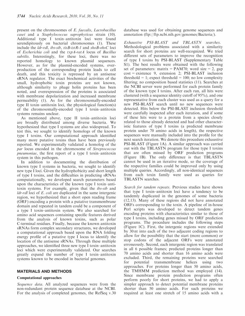
Exhaustive PSI-BLAST and TBLASTN searches.Methodological problems associated with a similaritysearch for short proteins are well-recognized. We trieddifferent sets of parameters to improve the recognitionof type I toxins by PSI-BLAST (Supplementary TableS1). The best results were obtained with the followingset of parameters: matrix=PAM70; word size=2, gapcost=existence 9, extension 2; PSI-BLAST inclusionthreshold=1; expect threshold=100; no low complexityfiltering; no composition based statistics (11). Searches atthe NCBI server were performed for each protein familyof the known type I toxins. After each run, all hits wereclustered (with a sequence identity cutoff of 95%), and onerepresentative from each cluster was used as a query for anew PSI-BLAST search until no new sequences weredetected. Hits below the PSI-BLAST inclusion thresholdwere carefully inspected after each iteration, and if someof these hits were to a protein from a species closelyrelated to those already detected and had other character-istic features of type I toxins (a predicted membraneprotein under 70 amino acids in length), the respectivesequences were manually included into the profile for thenext search iteration. We denote this procedure exhaustivePSI-BLAST (Figure 1A). A similar approach was carriedout with the TBLASTN program for those type I toxinsthat are often missed by ORF prediction programs(Figure 1B). The only difference is that TBLASTNcannot be used in an iterative mode, so the coverage ofthe respective families could be improved only by usingmultiple queries. Accordingly, all non-identical sequencesfrom each toxin family were used as queries forTBLASTN searches.
Search for tandem repeats. Previous studies have shownthat type I toxin–antitoxin loci have a tendency to betandemly duplicated in the genomes of some bacteria(12,13). Many of these regions did not have annotatedORFs corresponding to the toxin. A pipeline of in-housePerl scripts was developed to detect tandem genesencoding proteins with characteristics similar to those oftype I toxins, including genes missed by ORF predictionprograms. The procedure includes the following steps(Figure 1C). First, the intergenic regions were extendedby 30 nt into each of the two adjacent coding regions toallow for the possibility that the start (more common) orstop codons of the adjacent ORFs were annotatederroneously. Second, each intergenic region was translatedin all 6 possible frames; predicted proteins longer than70 amino acids and shorter than 16 amino acids wereexcluded. Third, the remaining proteins were searchedfor potential transmembrane helices using twoapproaches. For proteins longer than 50 amino acids,the TMHMM prediction method was employed (14).Since membrane protein prediction programs oftenperform poorly for short proteins, we had to apply asimpler approach to detect potential membrane proteinsshorter than 50 amino acids. For such proteins werequired at least one stretch of 15 amino acids with a
3744 Nucleic Acids Research, 2010, Vol. 38, No. 11

PSI-BLAST search against NR database
Matrix: PAM70, Word size: 2,
Gap cost: existence 9; extension 2
PSI-BLAST threshold: 1,Expect threshold: 100,
No low complexity filtering
No composition based statistics
Manually add selected sequences for PSI-BLAST
iteration
Remove the 95% identical sequences
All others go to STEP1 as queries until no more new
sequences detected
1
2
3
Exhaustive PSI-BLAST
ExhaustiveTBLASTN
TBLASTN search against nucleotide NR database
Matrix: PAM70, Word size: 2,
Gap cost: existence 9; extension 2
Expect threshold: 100, No low complexity
filteringNo composition based
statistics
Remove the 100% identical sequences
All others go to STEP1 as queries until no more new
sequences detected
Experimentally-characterized type I toxins
1
2
Tandem repeat search
Genome annotation
Filter out ORFs coding for proteins larger than 100 aa and all RNA genes reported
in rnt table
Translate each intergenic spacer in 6 frames;
Filter out predicted ORFs >=70 and <16
Predict membrane proteins; Filter out non-membrane
ORFs
Filter out protein fragments
Find tandemly repeated ORFs
Search for characteristic type I toxin
features
A
B
C
D
Genome annotation
Identify small ORFs (<70 aa)
Predict membrane proteins with one TM region
Identify genes encoded far from neighboring genes
(>400 nt distance upstream and >250 downstream
Identify proteins with composition score (>=300)
Experimentally-characterized type I toxins
Figure 1. Computational approaches used to identify and predict type I toxins. (A) Exhaustive PSI-BLAST to identify homologs of known toxinsnot among previously annotated protein sequences. (B) Exhaustive TBLASTN search to identify homologs of known type I toxins not amongprevious annotated ORFs. (C) Tandem repeat search to identify new type I toxins encoded in the same intergenic region. (D) Search for new type Itoxins based upon characteristics of known toxins.
Nucleic Acids Research, 2010, Vol. 38, No. 11 3745

minimum 10 hydrophobic amino acids (I, V, L, F, C, M,A) as an approximation for a transmembrane region. Allpredicted proteins that did not have a membrane regionpredicted by either approach were discarded. Fourth, toexclude protein fragments or pseudogenes, each remainingpredicted protein was searched against the correspondingproteome using BLASTP. Predicted proteins with a highlysignificant match to previously annotated, longer proteins(e-value <10 and no more than two gaps) were excluded.The remaining proteins were also searched against thegenomic DNA sequence by the TBLASTN program tofind evidence that they might be located in a regionwhich is likely to be non-coding. Those that matched atranslated fragment containing one or more stop codonswere considered as non-coding and discarded. Finally, todetect repeated sequences, the remaining predictedproteins in each intergenic region were grouped usingBLASTCLUST (50% amino acid identity; lengthcoverage 0.7 for at least one ORF). Since the small typeI toxins generally do not have a variable length andinternal gaps within a family, we required that no morethan two internal and no more than one C-terminal gapsoccur in the alignment of the proteins within a cluster.
Identification of characteristic features of type I toxins. Wecombined previously observed features of type I toxinswith the new features identified in this work (for the setof type I toxins identified by PSI-BLAST and TBLASTN)in order to detect putative new toxin loci (Figure 1D,Supplementary Table S2). First, we took into accountthe observation that type I toxins are small (generally �70 amino acids in length) and secondly are membraneproteins. Third, type I toxin genes are separated fromtheir neighboring genes by relatively long intergenicregions. We analyzed the up- and down-stream regionsof the type I toxin genes and calculated the mean valuefor the up- and down-stream distances for all families(Supplementary Table S2). Based on the results of thisanalysis, we set the following thresholds for the distancebetween the putative toxin and its flanking genes: >400 ntbetween the toxin and the gene upstream, and >250 ntbetween the toxin and the gene downstream (Figure1D). Finally, we noticed that many type I proteins haveclusters of charged or bulky amino acids at theirC-terminus. Therefore, in the selected genome set, wecomputed the absolute frequencies (number of occur-rences) of non-hydrophobic amino acids within the10C-terminal amino acids for the combined set of alltype I toxins identified by PSI-BLAST and TBLASTNin those genomes (Supplementary Table S2). We usedthese absolute frequencies to calculate a score for the10C-terminal amino acids for a protein. This was doneby assigning a corresponding value of absolute frequencyfrom the above estimate to each non-hydrophobic aminoacid and calculating the sum of all such values.
Multiple sequence alignment and phylogenetic analysismethods. Multiple alignments of protein sequences wereconstructed using the MUSCLE program (15).Maximum likelihood (ML) phylogenetic trees were con-structed from an alignment by using the MOLPHY
program (16) with the JTT substitution matrix toperform local rearrangement of an original Fitch tree(17). The MOLPHY program was also used to computeRELL bootstrap values. Prediction of transmembranehelices was performed using TMHMM program (14)implemented in the web server (http://www.cbs.dtu.dk/services/TMHMM-2.0/).
Prediction of RNA secondary structure. Sequences of pre-dicted and experimentally detected antisense sRNAs,random sequences from the same genomes and di-shuffledsequences were computationally folded, and the freeenergy of the most stable secondary structure wascalculated using Afold and Mfold, as described previously(18,19). Energy minimization was performed by a dynamicprogramming method that employs nearest neighborparameters to evaluate free energy and finds the secondarystructures with the minimum free energy by summing upthe contributions from stacking, loop length, and otherstructural features, using improved thermodynamicparameters (20–22). The sequence fold variant with thelowest secondary-structure energy was used in ouranalysis. The P-values for randomizations were calculatedusing paired t-tests (18). Results were presented as thefree-energy profiles along the nucleotide sequences ofinterest with window lengths corresponding to thelengths of the antisense sRNAs. Starts and lengths of pre-dicted antisense sRNAs were defined as the local minimaof estimated free-energy profiles in the vicinity of pre-dicted ORFs, taking into account the characteristicfeatures (location and length) of known type I toxinfamilies. The dinucleotide randomization procedurerandomly shuffled all dinucleotides, retaining nucleotidecomposition of native RNAs (18,23).
Molecular approaches
Bacterial strains and plasmids. The strains and plasmidsutilized in this study are listed in Supplementary Table S3,and the sequences of all oligonucleotides are given inSupplementary Table S4.
Growth conditions. E. coli strains were routinely grownin Luria–Burtani (LB) medium (10 g tryptone, 5 g yeastextract and 10 g NaCl per liter) or M9 minimal glucosemedium (1mM MgSO4, 0.1mM CaCl, 1 mg/ml thiamineand 0.2% glucose) at 37�C with shaking. Arabinose wasadded as indicated to a final concentration 0.2%. Bacillussubtilis strains were grown in LB at 37�C with shaking.IPTG was supplemented to a 1mM final concentration asindicated. Enterococcus faecalis OG1RF was grown inBHI medium (Difco) at 37�C. Streptococcus pneumoniaeR6 was grown in BHI at 37�C in an atmosphere contain-ing 5% (vol/vol) CO2/95% air. Antibiotics were added asneeded at the following concentrations: 25 mg/mlchloramphenicol, 100mg/ml spectinomycin, 100mg/mlampicillin.
RNA extraction. For E. coli, total RNA was harvestedfrom cells grown in LB or M9 + 0.2% glucose mediaharvested at OD600 � 0.4 and from overnight cultures(OD600 � 5.0 in LB; OD600 � 2.2 in M9) by the method
3746 Nucleic Acids Research, 2010, Vol. 38, No. 11

of hot acid phenol as previously described (12). For B.subtilis, S. pneumoniae and E. faecalis strains, RNA wasisolated as described (24) with some modifications. Briefly,12-ml aliquots of culture were harvested by centrifuga-tion at 4�C at OD600 � 0.3, 1.0, 1.5 for E. faecalisand S. pneumoniae, and at OD600 � 0.3, 2.0, 3.5 forB. subtilis ssp. subtilis str. 168 and B. subtilis PY79.Pellets were resuspended in 600 ml of Solution GP(50mM Tris–HCl, 10mM EDTA, 1% SDS, 30mMsodium acetate), and transferred to tubes containing 0.5 gsterile glass beads (average diameter �106 mm; Sigma) and650 ml of acid phenol:chloroform. The mixture was beadbeated twice for 45 s at 4�C. The samples were separated bycentrifugation, and the aqueous layer was transferred totubes containing 500 ml of acid phenol: chloroform, andincubated at 65�C for 10min. The supernatant wasextracted two more times with phenol: chloroform, andonce with chloroform. RNAwas then ethanol precipitated,and resuspended in RNase-free water.
Northern analysis. For all antitoxin sRNAs, total RNA(10 mg) was separated on a denaturing 8%polyacrylamide–8M urea gel. For detection of thez3289/z3290 mRNAs, total RNA (10 mg) was separatedon a denaturing 6% polyacrylamide–8M urea gel. RNAwas then transferred to a Zeta-Probe Genomic GTmembrane (Bio-Rad). The membranes were incubatedwith specific oligonucleotide probes labeled with 32P byT4 polynucleotide kinase and washed as previouslydescribed (25).
Primer extension analysis. Total RNA (5 mg) was used forprimer extension analysis as previously described, andcDNA products were separated on a denaturing 8%polyacrylamide–8M urea gel (25). Gene specific primersare found in Supplementary Table S4.
Overproduction of toxic proteins. For the toxicity studiesin E. coli MG1655, potential toxins were cloned behindthe PBAD promoter of the pAZ3 vector (26). As the ends ofthe potential toxin mRNA were unknown, a region con-taining �50 nt upstream of the predicted ribosome bindingsite and 100 nt downstream of the stop codon wasamplified from genomic DNA, digested with EcoRI andHindIII, and cloned into the corresponding sites of pAZ3.For yhzE-2, the amplified fragment and pAZ3 weredigested with EcoRI and XbaI.
To overproduce the toxins in B. subtilis PY79, the sameregions were amplified from genomic DNA, digested withNheI and SphI, and cloned behind the Plac promoter ofpDR111 (27). The resulting plasmids were then used forrecombination into the amyE locus of B. subtilis PY79.Integration was confirmed by PCR and sequencing.
RESULTS
Identification of additional members ofpreviously-characterized type I toxin–antitoxin families
Several studies have used sequence similarity searches,and in particular TBLASTN with default parameters,
to identify chromosomally-encoded type I toxins (9,10).However, given the short lengths of these proteins andthe strict parameters of such similarity searches, we sus-pected that a substantial fraction of homologs mighthave been missed in these studies. Thus, we performed acomprehensive analysis using customized, exhaustivePSI-BLAST and TBLASTN searches for 774 completebacterial genomes (Figure 1A and B, and ‘Materials andMethods’ section). The results, presented in full inSupplementary Tables S5 and S6, and as a condensedlist in Table 1 (for multiple alignments, see SupplementaryFigure S1), substantially expand the number of detectedtype I toxin homologs, especially when compared withresults that would be obtained if default BLAST parame-ters were used (Supplementary Table S1).Some families, such as the Hok (also denoted Gef),
TxpA, Ldr and Fst families, were well represented inprotein databases; the best-annotated group is the Hokfamily in which 72% are correctly named proteins. Bycontrast, others, such as the Ibs, TisB and ShoBfamilies, were often missed by ORF-calling programs.The majority of the putative type I toxins that weidentified with this approach are currently annotated as‘hypothetical proteins’ or are unannotated.To date, type I toxin–antitoxin loci have been experi-
mentally characterized only in a few lineages ofEnteroproteobacteria (Enterobacteria and Vibrionales)and Firmicutes (Bacillus and Enterococcus genus) (1,2).Our searches failed to detect any homologs of theknown type I toxins outside these taxa; however, weidentified previously unnoticed representatives of thesefamilies in many additional lineages of Enteropro-teobacteria and Firmicutes (Supplementary Tables S5and S6). For example, Fst-like sequences were detectedin several Listeriaceae, Staphylococcaceae andClostridiales species, and TxpA-like sequences weredetected in Lactobacillales, Staphylococcaceae andClostridiales species (Supplementary Table S5). Thenumber of type I toxin loci varies greatly between differentspecies and strains. So far the largest number wasidentified in E. coli O157:H7 str. Sakai. This genomecarries 26 toxin–antitoxin loci of six distinct familiesincluding 14 Hok/Gef genes and seven Ibs genes. Takinginto account our previous estimates of the number of typeII toxin–antitoxin loci (3) (given in Supplementary TableS6) on a genome-wide scale we can conclude that type Itoxin–antitoxin system are even more abundant in somegenomes.This approach also allows for the discovery of
non-trivial links between families. Thus, using exhaustivePSI-BLAST, we detected a previously unnoticed connec-tion between the Ldr and Fst families. The multiple aminoacid sequence alignment shows considerable conservationbetween the two families including an apparent super-family signature, a highly conserved tryptophan after apredicted transmembrane helix followed by a cluster ofcharged amino acids (Supplementary Figure S1A). Thisfinding implies that the two families are probably homol-ogous. The Ldr and Fst toxins are widely distributedacross both Firmicutes and Enterobacteria. Given thatrepresentatives of these families are found in potential
Nucleic Acids Research, 2010, Vol. 38, No. 11 3747

Table
1.TypeItoxinsin
selected
completely
sequencedgenomes
Species
Taxonomy
Total
number
of
proteins
Number
of
different
families
Total
count
Ibs
Ldr/Fst
TxpA
Hok
TisB
ShoB
EHEC
YhzE
BacillushaloduransC-125
Bacilli,Bacillales,
Bacillaceae
4066
210
19
BacilluspumilusSAFR-032
Bacilli,Bacillales,
Bacillaceae
3681
27
16
Bacillussubtilisssp.subtilisstr.
168
Bacilli,Bacillales,
Bacillaceae
4105
211
29
Listeriamonocytogenes
str.
4bF2365
Bacilli,Bacillales,
Listeriaceae
2821
11
1Listeriawelshim
eriserovar6b
str.
SLCC5334
Bacilli,Bacillales,
Listeriaceae
2774
12
2
Staphylococcusaureusssp.aureus
NCTC
8325
Bacilli,Bacillales,
Staphylococcaceae
2892
25
32
Staphylococcusepidermidis
RP62A
Bacilli,Bacillales,
Staphylococcaceae
2526
15
5Staphylococcushaem
olyticusJC
SC1435
Bacilli,Bacillales,
Staphylococcaceae
2676
15
5Staphylococcussaprophyticus
ssp.saprophyticusATCC
15305
Bacilli,Bacillales,
Staphylococcaceae
2514
24
31
EnterococcusfaecalisV583
Bacilli,Lactobacillales,
Enterococcaceae
3265
27
16
LactobacilluscaseiATCC
334
Bacilli,Lactobacillales,
Lactobacillaceae
3044
26
24
Leuconostoccitreum
KM20
Bacilli,Lactobacillales,
Leuconostocaceae
1820
12
2Leuconostocmesenteroides
ssp.mesenteroides
ATCC
8293
Bacilli,Lactobacillales,
Leuconostocaceae
2005
12
2
Streptococcuspyogenes
MGAS10270
Bacilli,Lactobacillales,
Streptococcaceae
1986
11
1StreptococcuspneumoniaeCGSP14
Bacilli,Lactobacillales,
Streptococcaceae
2206
13
3Streptococcussuis
05ZYH33
Bacilli,Lactobacillales,
Streptococcaceae
2186
11
1Streptococcusagalactiae2603V/R
Bacilli,Lactobacillales,
Streptococcaceae
2124
11
1StreptococcusthermophilusCNRZ1066
Bacilli,Lactobacillales,
Streptococcaceae
1915
11
1Aeromonassalm
onicida
ssp.salm
onicidaA449
Gammaproteobacteria,Aeromonadales
4437
12
2
Alteromonasmacleodii‘D
eepecotype’
Gammaproteobacteria,Alteromonadales
4072
11
1Escherichia
coliO157:H
7EDL933
Gammaproteobacteria,Enterobacteriales
5411
626
64
12
11
2Salm
onella
enterica
ssp.enterica
serovarParatyphiB
str.
SPB7
Gammaproteobacteria,Enterobacteriales
5592
37
24
1
Shigella
flexneri2astr.
301
Gammaproteobacteria,Enterobacteriales
4440
411
45
11
Klebsiella
pneumoniae342
Gammaproteobacteria,Enterobacteriales
5777
24
31
Shigella
boydiiCDC
3083-94
Gammaproteobacteria,Enterobacteriales
4557
520
74
71
1Citrobacter
koseri
ATCC
BAA-895
Gammaproteobacteria,Enterobacteriales
5008
22
11
Enterobacter
sp.638
Gammaproteobacteria,Enterobacteriales
4240
23
21
Enterobacter
sakazakiiATCC
BAA-894
Gammaproteobacteria,Enterobacteriales
4420
23
21
Escherichia
fergusoniiATCC
35469
Gammaproteobacteria,Enterobacteriales
4269
618
73
41
12
Photorhabdusluminescens
ssp.laumondiiTTO1
Gammaproteobacteria,Enterobacteriales
4683
11
1
Proteusmirabilis
HI4320
Gammaproteobacteria,Enterobacteriales
3662
11
1Salm
onella
enterica
ssp.enterica
serovarTyphistr.
CT18
Gammaproteobacteria,Enterobacteriales
4758
48
22
31
Salm
onella
typhim
urium
LT2
Gammaproteobacteria,Enterobacteriales
4527
35
22
1Serratiaproteamaculans568
Gammaproteobacteria,Enterobacteriales
4942
14
4Shigella
dysenteriaeSd197
Gammaproteobacteria,Enterobacteriales
4503
510
32
31
1Shigella
sonnei
Ss046
Gammaproteobacteria,Enterobacteriales
4471
522
710
31
1Haem
ophilusinfluenzae86-028NP
Gammaproteobacteria,Pasteurellales
1792
11
1Haem
ophilussomnus2336
Gammaproteobacteria,Pasteurellales
1980
17
7VibriovulnificusCMCP6
Gammaproteobacteria,Vibrionales
4472
11
1
3748 Nucleic Acids Research, 2010, Vol. 38, No. 11
vectors for horizontal gene transfer, such as phages andplasmids, we were interested in potential evidence of hor-izontal gene transfer, and reconstructed a phylogenetictree of the Ldr/Fst sequences. Despite limitations in treeconstruction because the sequences are so short, we weresurprised to find that the topology of the tree matched thetaxonomy of the respective bacteria (SupplementaryFigure S2). There was no evidence of recent horizontalgene transfer events between distantly related bacteriafor this family of type I toxins. Instead, we infer thatthere was a duplication of the ancestral toxin–antitoxinlocus in the common ancestor of enterobacteria (LdrD-and LdrB-group) and in at least two distinct clades inStaphylococcaceae. These duplications were followed byother lineage-specific duplication events and a few lossesin some species.
We also constructed a tree for the Ibs family, for whichduplications in several genomes of enterobacteria weredetected as well (Supplementary Figure S3). The analysisof this tree revealed the same trends as those seen inthe Ldr/Fst tree. At least three copies of the ibs genecould have been present in the common ancestor ofEnterobacteriaceae, and two in the Pasteurellaceae andthe Haemophilus clades each. Subsequent duplicationsoccurred independently in Haemophilus somnus andShigella boydii lineages. These observations suggest thatduplications of the type I toxin–antitoxin loci are rela-tively stable in evolution, with the implication that eitherthese loci are prone to duplication and subject to relaxedselection, as in the case of transposons, or that the dupli-cations are functionally important, possibly for stressresistance (28,29), and accordingly are maintained bypurifying selection.
Experimental validation of the predicted Fst homologsin S. pneumoniae
Two Fst homologs (referred to herein as Fst-A and Fst-B),predicted by the exhaustive PSI-BLAST searches, areencoded in tandem in 27 out of the 29S. pneumoniaegenomic sequences deposited in the Microbial genomedatabase at NCBI. These genes were missed by ORF pre-diction programs used for genome annotation in severalstrains including S. pneumoniae R6 (Figure 2A). TheORFs in S. pneumoniae R6 are flanked by fcsR, whichencodes an annotated regulator of the fucose operon,and adcA, an ABC-transporter. We selected one of theseORFs, Fst-B (genomic coordinates 1 965 747–1 965 842),to test whether an antisense sRNA is expressed from thesame locus and whether the product of the ORF is indeedtoxic.
If the S. pneumoniae protein is functionally analogousto Fst, there should be a corresponding antisense sRNAregulator. The Fst protein is the toxin component of thepar locus of the plasmid pAD1. The organization of thepar locus has been well-characterized, and the antisensesRNA regulator (RNA II) overlaps the 30-end of themRNA encoding the toxic protein (10,30,31). TotalRNA was isolated from S. pneumoniae R6 and used fornorthern analysis. A strong signal corresponding to anRNA species of �65–75 nt was detected using an
end-labeled oligonucleotide complementary to the 30 endof fst-B (Figure 2B). This signal is in agreement with thepreviously characterized size and location of the antisensesRNA from pADI, as well as RNA II expressed fromcopies of the par locus encoded in the chromosomes ofother bacteria (10).To test the toxicity of the protein, fst-B from
S. pneumoniae R6 was cloned on a plasmid behind anarabinose-inducible promoter (PBAD) and overproducedin E. coli MG1655. As shown in Figure 2C, induction ofthe protein halted cell growth, and there was a significantdecrease in colony forming units over time, confirmingthat high levels of this protein are indeed toxic.
Finding new type I toxin–antitoxins: tandem repeats
Among the previously identified chromosomally encodedtype I toxin–antitoxin systems, the ibs-sib and ldr-rdl lociare repeated multiple times in the same intergenic region(12,13). Therefore we developed a computational proce-dure to identify tandem repeats encoding potential type Itoxins in intergenic regions of bacterial genomes. We thenexamined a selected set of sequenced genomes in orderto identify new type I toxin families (see Figure 1C, and‘Materials and Methods’ section). The complete searchresults for Proteobacteria and Firmicutes are given inSupplementary Table S7.This approach reproduced some findings obtained
in our exhaustive BLAST search, including theS. pneumoniae Fst toxins described above. The searchalso led to the identification of a duplication of theapparent Ibs homologs in the genome of Helicobacterpylori that were missed by TBLASTN but recently wereidentified experimentally (32). We were particularly inter-ested in further analyzing E. coli and B. subtilis toxin–antitoxin candidates predicted by this approach.
Experimental analysis of new candidate type I toxinsfrom E. coli strain O157:H7
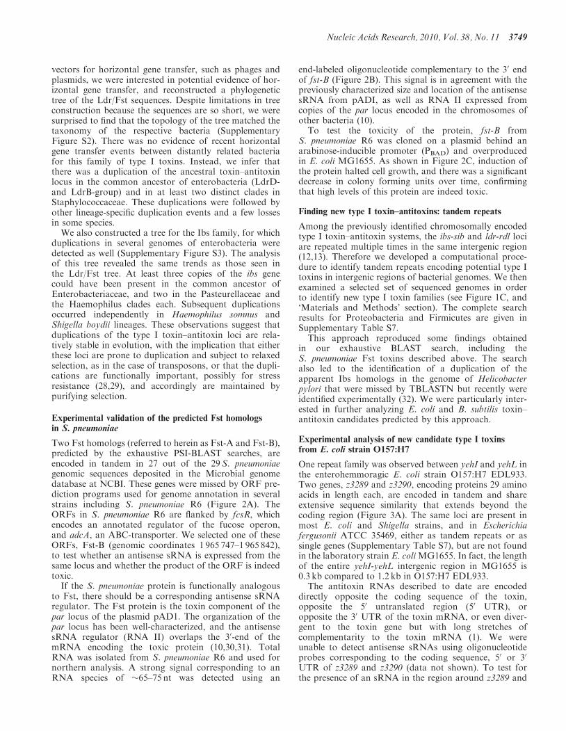
One repeat family was observed between yehI and yehL inthe enterohemmoragic E. coli strain O157:H7 EDL933.Two genes, z3289 and z3290, encoding proteins 29 aminoacids in length each, are encoded in tandem and shareextensive sequence similarity that extends beyond thecoding region (Figure 3A). The same loci are present inmost E. coli and Shigella strains, and in Escherichiafergusonii ATCC 35469, either as tandem repeats or assingle genes (Supplementary Table S7), but are not foundin the laboratory strain E. coliMG1655. In fact, the lengthof the entire yehI-yehL intergenic region in MG1655 is0.3 kb compared to 1.2 kb in O157:H7 EDL933.The antitoxin RNAs described to date are encoded
directly opposite the coding sequence of the toxin,opposite the 50 untranslated region (50 UTR), oropposite the 30 UTR of the toxin mRNA, or even diver-gent to the toxin gene but with long stretches ofcomplementarity to the toxin mRNA (1). We wereunable to detect antisense sRNAs using oligonucleotideprobes corresponding to the coding sequence, 50 or 30
UTR of z3289 and z3290 (data not shown). To test forthe presence of an sRNA in the region around z3289 and
Nucleic Acids Research, 2010, Vol. 38, No. 11 3749

z3290, we carried out northern analysis using threeriboprobes, which together would span a 1 kb segmentencompassing the two small ORFs. We observed astrong band of �80 nt with the probe that spanned theintergenic region between the two genes (data notshown). To further refine the position of the putativesRNA, we calculated the predicted free-energy profile ofthe yehI-yehL intergenic region (see below). This analysisrevealed two local minima of predicted free-energy,corresponding to regions of complex secondary structure,240–300 nt upstream of z3289 and z3290. Upon furtherexamination of these regions, we identified potentialterminators and promoter sequences (Supplementary
Figure 4A). Using oligonucleotides complementary tothese predicted sRNAs, we detected two transcripts of�85 nt in length each. Interestingly, these transcriptswere abundant during the exponential phase in both richand minimal media, but decreased during stationary phase(Figure 3B).
As these sRNA genes were encoded divergent from thetoxin genes, we were interested in whether they had thepotential to base pair with the toxin mRNAs. There isperfect complementarity between the sRNA (sRNA-1)encoded divergent to z3289 and the sequence 72–92 ntupstream of the start codon of the toxin (Figure 3A).Similar complementarity is also observed between z3290
Figure 2. (A) Multiple alignment of selected representatives of the Ldr/Fst family. Proteins studied in this work are denoted by asterisk, experi-mentally characterized type I toxins are shown in bold; the predicted transmembrane region is shaded; conserved small amino acids are colored blue;the conserved tryptophan is colored magenta; charged amino acids (RKDE) are colored red. The consensus was built using CONSENSUS program(http://coot.embl.de/Alignment//consensus.html) for a larger set of Ldr/Fst proteins (see Supplementary Figure S1A). The sequences are denoted byboth the abbreviated species name and the GI number or the coordinates in the corresponding genome in parentheses. Species abbreviations: SP,Streptococcus pneumoniae R6; CB, Clostridium bolteae; EC, Escherichia coli K-12 substr. MG1655; SB, Shigella boydii CDC 3083-94; SE, Salmonellaenterica arizonae z4z23; ECO, Escherichia coli O127:H6 str. E2348/69; SA, Staphylococcus aureus ssp. aureus Mu50; LG, Lactobacillus gasseri MV-22;EF, Enterococcus faecalis plasmid pAD1. (B) Northern blot showing expression of an sRNA antisense to S. pneumoniae fst-B-homolog. Total RNA(10mg) isolated from S. pneumoniae R6 cells grown to OD600 � 0.3 (E), OD600 � 1.0 (L) and OD600 � 1.5 (S) in BHI medium was loaded in each lane.(C) Overproduction of the S. pneumoniae Fst-B homolog in E. coli. MG1655 harboring pAZ3-fst-B was grown in LB medium to OD600 � 0.3. Theculture was split (indicated by the arrow); half was left untreated (blue) while arabinose (0.2% final concentration) was added to the other half (red).Cell dilutions were plated 0 (T0) and 60 (T60)min following arabinose induction.
3750 Nucleic Acids Research, 2010, Vol. 38, No. 11

and the sRNA (sRNA-2) encoded divergent from thisgene (Figure 3A). We carried out primer extensionanalysis to map the transcription start sites of the z3289and z3290 mRNAs and the newly discovered sRNAs(Supplementary Figure S4). The results indicate thatboth toxin mRNAs contain long 50 UTRs (180 nt),similar to what has been reported with other toxins (1).The gene orientations and base pairing potentials are veryreminiscent of the tisB-istR and shoB-ohsC toxin loci. Inthese pairs, the sRNA is encoded divergent, and distantfrom the toxin, but has the potential for extended basepairing with the 50 UTR of the toxin mRNA.
The putative toxin genes were cloned with their nativeribosome binding sites, behind the PBAD promoter on a
multicopy plasmid to measure toxicity. Overproduction ofboth small proteins (Figure 3C and data not shown) in thelaboratory strain E. coli MG1655 led to cell stasis and amild decrease in colony forming units, indicating that theproteins are toxic at high levels.
Experimental analysis of new candidate type Itoxins from B. subtilis
A separate duplication was identified in B. subtilis ssp.subtilis str. 168 genome. The duplicated gene encodes a28 amino acid hydrophobic protein that is conservedacross multiple species of Firmicutes, but it is notsimilar to any of the known type I toxins (Figure 4Aand Supplementary Figure S1C). One of these duplicated
Figure 3. (A) Genomic arrangement of EHEC Z3289 and Z3290. The ORFs are indicated by the black regions of the leftward arrows and theregions of complementarity are indicated by the white boxes. The sequences capable of base pairing are shown below the gene arrangement. (B)Multiple alignment of selected representatives of EHEC family. Most designations are the same as in the Figure 2A. The predicted transmembraneregions is shaded (predicted for E. coli O157:H7 EDL933 proteins and extended for other sequences): small amino acids are colored blue; chargedamino acids (RKDE) are colored red. Species abbreviations (strains are also indicated for E. coli species): EC, E. coli; SB, Shigella boydii CDC3083-94; SF, Shigella flexneri 2a str. 2457T. (C) Expression of the antitoxin RNAs for Z3289 (sRNA-1) and Z3290 (sRNA-2). Total RNA (10 mg)isolated from E. coli O157:H7 EDL933 cells grown to OD600 � 0.4 (E) and OD600 � 5.0 (overnight, S) in LB medium and from cells grown to OD600
� 0.4 (E) and OD600 � 2.2 (overnight, S) in M9 media supplemented with 0.2% glucose was loaded in each lane. (D) Overproduction of Z3290 inMG1655. MG1655 harboring pAZ3-z3290 was grown in LB medium to OD600 � 0.3. The culture was split (indicated by the arrow); half was leftuntreated (blue) while arabinose (0.2% final concentration) was added to the other half (red). Cell dilutions were plated 0 (T0) and 60 (T60)minfollowing arabinose induction.
Nucleic Acids Research, 2010, Vol. 38, No. 11 3751

genes has been annotated as yhzE; herein, we will referto the annotated gene as yhzE-1 and the second copy inthe same intergenic region as yhzE-2. The genes encodingproteins of this family are highly abundant in Firmicutes.For instance, the Bacillus subtilis ssp. subtilis str. 168genome contains eight genes for these proteins(Supplementary Figure S1C and Figure 4A). Analysis ofan alignment of this family reveals a distinct feature: boththe N- and C-termini are highly variable in length but arerich in glycines and aromatic residues (SupplementaryFigure S1C). Genes encoding these proteins are tandemlyduplicated in the genomes of several other bacteria and
are also present in several phages and plasmids.Combined with the sequence features of this proteins,such a distribution makes them possible type I toxincandidates.
To test whether this region has features of a type Itoxin–antitoxin locus, we isolated RNA from B. subtilisPY79 and carried out northern blot analysis. As theB. subtilis ratA antitoxin RNA base pairs at the 30-endof the toxin mRNA, we used an oligonucleotide probethat overlaps the 30-end of the yhzE-2 ORF. A strongsignal, of �110–120 nt in length was detected throughoutgrowth in rich media using this probe (Figure 4B).
Figure 4. (A) Multiple alignment of selected representatives of YhzE family. Most designations are the same as in the Figure 2A. The consensus wasbuilt using CONSENSUS program for a larger set of YhzE family proteins (see Supplementary Figure S1C). The predicted transmembrane regions isshaded (predicted for B. subtilis YhzE protein and extended for other sequences); small amino acids are colored blue; aromatic residues are coloredmagenta. Species abbreviations: Bs, B. subtilis str. 168; Bph, Bacillus phage SPBc2; Gsp, Geobacillus sp. G11MC16; BH, B. halodurans C-125; BA, B.anthracis str. Ames; Psp, Paenibacillus sp. JDR-2; BP, B. pumilus SAFR-032. (B) Expression of an sRNA antisense to yhzE-2. Total RNA (10 mg)isolated from B. subtilis PY79 cells grown to OD600 � 0.3 (E), OD600 � 2.0 (L) and OD600 � 3.5 (S) in LB medium was loaded in each lane. (C)Overproduction of YhzE-2 and TxpA in B. subtilis PY79. YhzE-2 (graph on the left) or TxpA (right) under the control of the Plac promoter wasintegrated into the amyE locus of PY79. The cultures were grown in LB medium to OD600 � 0.3. The cultures were split; (indicated by the arrow);half was left untreated (blue) while IPTG (1mM final concentration) was added to the other half (red).
3752 Nucleic Acids Research, 2010, Vol. 38, No. 11
We initially overexpressed the YhzE-2 protein from aPBAD plasmid in E. coli MG1655 but observed no effectson growth (data not shown). Given that the protein isnative to B. subtilis and not E. coli, we next measured itstoxicity in B. subtilis. The yhzE-2 gene was cloned behindthe Plac promoter of the plasmid pDR111, and the con-struct was integrated into the amyE gene of B. subtilisPY79 (27). As a control, we similarly examined thetoxicity of TxpA, a known type I toxin found in B.subtilis (33). TxpA was highly toxic to B. subtiliswhereas there were no obvious growth defects upon over-production of YhzE-2 (Figure 4C). The lack of YhzE-2mediated toxicity could be due to insufficient levels ofprotein production, possibly because of repression byendogenous antisense sRNAs expressed from themultiple paralogous copies of the locus. Alternatively,the protein may not function as a toxin, even at highlevels.
Finding new type I toxin–antitoxins: characteristicprotein features
In addition to being encoded in tandem repeats, there areother characteristics shared by many type I protein toxins.The described type I toxins are under 70 amino acids inlength, contain a transmembrane region and a smallC-terminal region rich in polar or aromatic residues. Thetoxin–antitoxin loci also are often encoded distant fromtheir flanking genes. We combined these observations intoa set of search parameters (see ‘Materials and Methods’section) taking into account data obtained by the analysisof all known and new toxins described here (Figure 1Dand Supplementary Table S2). Briefly, we identified allproteins under 70 amino acids in length that were pre-dicted to contain at least one transmembrane region. Wethen selected those ORFs that were separated by at least400 nt from the upstream flanking gene and by at least250 nt from the downstream gene. From this set ofproteins, we selected those that contained a C-terminusrich in polar or aromatic residues. Results for theselected genomes are presented in Supplementary TableS8. Using these parameters, we identified, among otherputative novel type I toxins, the 27 amino-acid proteinBH0344 from Bacillus halodurans C-125, which hashomologs in several L. monocytogenes strains and inE. faecalis V583 (protein EF3263), as well as YonTencoded in the B. subtilis ssp. subtilis str. 168 genome. TheEF3263 and YonT proteins were chosen for further analysis.
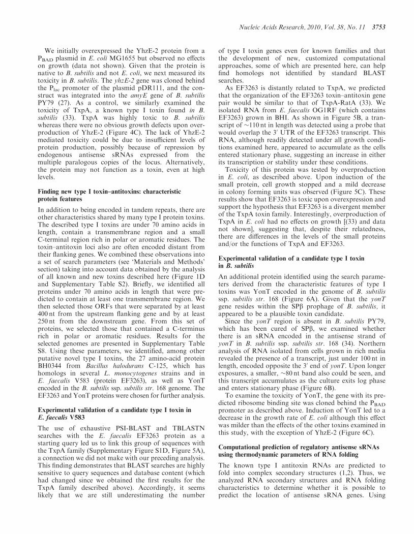
Experimental validation of a candidate type I toxin inE. faecalis V583
The use of exhaustive PSI-BLAST and TBLASTNsearches with the E. faecalis EF3263 protein as astarting query led us to link this group of sequences withthe TxpA family (Supplementary Figure S1D, Figure 5A),a connection we did not make with our preceding analysis.This finding demonstrates that BLAST searches are highlysensitive to query sequences and database content (whichhad changed since we obtained the first results for theTxpA family described above). Accordingly, it seemslikely that we are still underestimating the number
of type I toxin genes even for known families and thatthe development of new, customized computationalapproaches, some of which are presented here, can helpfind homologs not identified by standard BLASTsearches.As EF3263 is distantly related to TxpA, we predicted
that the organization of the EF3263 toxin–antitoxin genepair would be similar to that of TxpA-RatA (33). Weisolated RNA from E. faecalis OG1RF (which containsEF3263) grown in BHI. As shown in Figure 5B, a tran-script of �110 nt in length was detected using a probe thatwould overlap the 30 UTR of the EF3263 transcript. ThisRNA, although readily detected under all growth condi-tions examined here, appeared to accumulate as the cellsentered stationary phase, suggesting an increase in eitherits transcription or stability under these conditions.Toxicity of this protein was tested by overproduction
in E. coli, as described above. Upon induction of thesmall protein, cell growth stopped and a mild decreasein colony forming units was observed (Figure 5C). Theseresults show that EF3263 is toxic upon overexpression andsupport the hypothesis that EF3263 is a divergent memberof the TxpA toxin family. Interestingly, overproduction ofTxpA in E. coli had no effects on growth [(33) and datanot shown], suggesting that, despite their relatedness,there are differences in the levels of the small proteinsand/or the functions of TxpA and EF3263.
Experimental validation of a candidate type I toxinin B. subtilis
An additional protein identified using the search parame-ters derived from the characteristic features of type Itoxins was YonT encoded in the genome of B. subtilisssp. subtilis str. 168 (Figure 6A). Given that the yonTgene resides within the SPb prophage of B. subtilis, itappeared to be a plausible toxin candidate.Since the yonT region is absent in B. subtilis PY79,
which has been cured of SPb, we examined whetherthere is an sRNA encoded in the antisense strand ofyonT in B. subtilis ssp. subtilis str. 168 (34). Northernanalysis of RNA isolated from cells grown in rich mediarevealed the presence of a transcript, just under 100 nt inlength, encoded opposite the 30 end of yonT. Upon longerexposures, a smaller, �80 nt band also could be seen, andthis transcript accumulates as the culture exits log phaseand enters stationary phase (Figure 6B).To examine the toxicity of YonT, the gene with its pre-
dicted ribosome binding site was cloned behind the PBAD
promoter as described above. Induction of YonT led to adecrease in the growth rate of E. coli although this effectwas milder than the effects of the other toxins examined inthis study, with the exception of YhzE-2 (Figure 6C).
Computational prediction of regulatory antisense sRNAsusing thermodynamic parameters of RNA folding
The known type I antitoxin RNAs are predicted tofold into complex secondary structures (1,2). Thus, weanalyzed RNA secondary structures and RNA foldingcharacteristics to determine whether it is possible topredict the location of antisense sRNA genes. Using
Nucleic Acids Research, 2010, Vol. 38, No. 11 3753

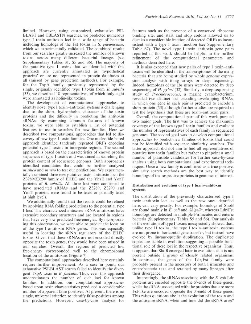
computer algorithms to predict RNA folding and toestimate the free energy for optimal and suboptimal sec-ondary structures (see ‘Materials and Methods’ section),we first created free-energy profiles for the previouslycharacterized antitoxin RNA regions. We found thatthe transcriptional starts for all known antitoxinRNAs (IstR1, Sok, SibA, SibB, RdlD, RatA) arelocated in the local minima of predicted free-energyprofiles (Supplementary Figure S5). Specifically, the dif-ferences in the local minima and the average free-energylevels in the thermodynamic profiles for known antitoxinRNAs compared to those calculated for di-shuffled
sequences and random sequences of comparable lengthsfrom the same genome were statistically significant(P< 0.001).
To validate this approach, we compared the distribu-tion of free-energy values for predicted antitoxin RNAregions for known type I loci identified using BLAST(Supplementary Table S9) with those for randomsequences of comparable lengths taken from elsewhere inthe same genomes, and with randomly shuffled sequenceswith the same dinucleotide content as the RNA antitoxinsequences (‘Materials and Methods’ section). The startsand lengths of the predicted antitoxin RNA were defined
Figure 5. (A) Multiple alignment of selected representatives of the TxpA family. The consensus was built using CONSENSUS program for a largerset of the TxpA family proteins (see Supplementary Figure S1D). Most designations are the same as in the Figure 2A. The predicted transmembraneregions is shaded (predicted for Enterococcus faecalis V583 protein and extended for other sequences). Species abbreviations: Bs, B. subtilis sub.subtilis str. 168; Sph, Staphylococcus phage 42E; EFH, E. faecalis HH22; LC, Lactobacillus casei ATCC 334; LCI, Leuconostoc citreum KM20; LC,Lactobacillus casei ATCC 334; Gs, Geobacillus sp. G11MC16; EFO, E. faecalis OG1RF; BH, B. halodurans C-125; EFV, E. faecalis V583. (B)Northern blot showing expression of an sRNA antisense to EF3263 in E. faecalis OG1RF. Total RNA (10 mg) isolated from E. faecalis OG1RF cellsgrown to OD600 � 0.3 (E), OD600 � 1.0 (L) and OD600 � 1.5 (S) in BHI medium was loaded in each lane. (C) Overproduction of EF3263 in E. coli.MG1655 harboring pAZ3-ef3263 was grown in LB medium to OD600 � 0.3. The culture was split (indicated by the arrow); half was left untreated(blue) while arabinose (0.2% final concentration) was added to the other half (red). Cell dilutions were plated 0 (T0) and 60 (T60)min followingarabinose induction.
3754 Nucleic Acids Research, 2010, Vol. 38, No. 11

based on the characteristic features (location and length)of known type I toxin families. Again the folding freeenergies for the predicted antisense sRNAs were substan-tially lower than those for the di-shuffled sequences(P=9.2E�32; Supplementary Figure S6). Notably,mRNA folding energies for the random sequences weredistributed differently, as compared to those of the anti-toxin RNAs that contain numerous domains capable offolding into highly stable secondary structures. The resultsshow that the predicted antisense sRNA regions generallyhave a propensity to form more stable secondary struc-tures and possess lower free-energy values that therandom genomic sets (P=2.62E�12; SupplementaryFigure S6).
Using this approach, we predicted the locations of thegenes encoding the antitoxin RNAs identified in E. coliO157:H7, B. subtilis and E. faecalis, and experimentallyanalyzed in this study (Figure 7). The predicted energyminima coincided perfectly with the sequences of theoligonucleotides used to detect the sRNAs by northernanalysis. The analysis was particularly helpful for z3289
and z3290 of E. coli O157:H7, where antisense sRNAswere not detectable with oligonucleotide probes corre-sponding to the coding sequence, 50 or 30 UTR of thegenes. For these toxins the locations of putative sRNAgenes were predicted based on the analysis of thefree-energy profile of the yehI-yehL intergenic region.This analysis revealed two local minima of free energy,which corresponded to the regions of complexsecondary-structure upstream of z3289 and z3290(Figure 7B) and were confirmed experimentally toexpress sRNAs.Whenever possible, the lengths of sliding windows were
chosen on the basis of the characteristic location andlength of known type I toxins. For new type I toxinfamilies, we performed a more extensive analysis withsliding windows of varying length. Some of the localfree-energy minima observed outside of predicted ORFscorresponded to annotated transcription terminators orunrelated short ORFs and were excluded from consider-ation. Most of the remaining stable free-energy minima,readily detectable with different window lengths, were
Figure 6. (A) Amino acid sequence of yonT gene product of Bacillus subtilis ssp. subtilis str. 168 (Bs). Charged amino acids (EKR) are colored redand the predicted transmembrane regions is shaded. (B) Expression of an sRNA antisense to yonT. Total RNA (10 mg) isolated from B. subtilis ssp.subtilis str. 168 cells grown to OD600 � 0.3 (E), OD600 � 2.0 (L) and OD600 � 3.5 (S) in LB medium was loaded in each lane. A smaller band of�80 nt can be seen upon overexposure as the cells enter the stationary phase of growth. (C) Overproduction of YonT in E. coli. MG1655 harboringpAZ3-yonT was grown in LB medium to OD600 � 0.3. The culture was split (indicated by the arrow); half was left untreated (blue) while arabinose(0.2% final concentration) was added to the other half (red). Cell dilutions were plated 0 (T0) and 60 (T60) min following arabinose induction.
Nucleic Acids Research, 2010, Vol. 38, No. 11 3755

candidates for experimental evaluation. Our resultssupport the observations that the antitoxins are encodedby highly structured RNAs, which justifies the use of thisparameter to predict new type I loci.
DISCUSSION
Several recent studies have focused on the identificationand characterization of the many type II toxin–antitoxingene pairs in which both the toxin and antitoxin are pro-teins. These loci are broadly distributed across bacteriaand archaea, and the numbers of loci vary extensively
between species. In contrast, little is known about the dis-tribution of type I toxin–antitoxin loci in which the anti-toxin is an antisense sRNA. We thus set out to screen forhomologs of known type I toxin–antitoxin pairs as well asto identify new loci.
Approaches to identify type I toxin–antitoxin systems
Prior to these studies, identification of homologs of knowntype I toxins relied solely upon TBLASTN and PSI-BLAST searches carried out using default parameters.These searches revealed few homologs (9,10), and conse-quently suggested that the distribution of these toxins was
Figure 7. Prediction of antitoxin sRNAs using free-energy profiles for RNA local secondary structures. Free-energy profiles for RNA local second-ary structures along nucleotide sequences in experimentally tested RNA antitoxin systems in S. pneumoniae (A), E. coli (B), B. subtilis (C and E) andE. faecalis (D). The lengths of the sliding window used for free-energy estimations (70 and 100 nt) corresponded to common lengths of the previouslydescribed sRNA antitoxins. Blue arrows show location of predicted ORFs. Red arrows show the positions of the oligonucleotides used to detect theantisense sRNAs. Other local free-energy minima correspond to annotated terminators or unrelated short ORFs. x-axis: nucleotide positions; y-axis:free energy of RNA folding.
3756 Nucleic Acids Research, 2010, Vol. 38, No. 11
limited. However, using customized, exhaustive PSI-BLAST and TBLASTN searches, we predicted numeroustype I toxin–antitoxin loci in a wide range of bacteriaincluding homologs of the Fst toxins in S. pneumoniae,which we experimentally validated. The combined resultsfrom our searches greatly increased the number of knowntoxins across many different bacterial lineages (seeSupplementary Tables S1, S5 and S6). The majority ofthe putative type I toxins that we identified with thisapproach are currently annotated as ‘hypotheticalproteins’ or are not represented in protein databases atall (missed by gene prediction methods). For example,for the TxpA family, previously represented by thesingle, originally identified type I toxin from B. subtilis(33), we describe 118 representatives, of which only eightwere annotated as holin-like toxins.
The development of computational approaches toidentify novel type I toxin–antitoxin systems is challengingdue to the short, hydrophobic character of the toxinproteins and the difficulty in predicting the antitoxinsRNAs. By examining common features of knowntoxins, we were able to establish a list of potentialfeatures to use in searches for new families. Here wedescribed two computational approaches that led to dis-covery of new type I toxin families. One nucleotide-basedapproach identified tandemly repeated ORFs encodingpotential type I toxins in intergenic regions. The secondapproach is based on the characteristics of known proteinsequences of type I toxins and was aimed at searching theprotein content of sequenced genomes. Both approachesidentified candidates that could be further analyzedin silico and in vivo to test our predictions. We experimen-tally examined three new putative toxin–antitoxin loci: theZ3289/Z3290 family of EHEC and the YhzE and YonTproteins of B. subtilis. All three loci were confirmed tohave associated sRNAs and the Z3289, Z3290 andYonT proteins were found to be toxic or partially toxicat high levels.
We additionally found that the results could be refinedby applying RNA folding predictions to the potential typeI loci. The characterized antisense sRNA antitoxins haveextensive secondary structures and are located in regionsthat have very low predicted free-energies. By incorporat-ing this observation, we were able to predict the locationof the type I antitoxin RNA genes. This was especiallyuseful in locating the sRNA regulators of the EHECtoxins. Given that these sRNAs are not encoded directlyopposite the toxin genes, they would have been missed inour searches. Overall, the regions of predicted lowfree-energy corresponded well to the chromosomallocation of the antitoxins (Figure 7).
The computational approaches described here certainlyrequire further improvement. As a case in point, ourexhaustive PSI-BLAST search failed to identify the diver-gent TxpA toxin in E. faecalis. Thus, even this approachunderestimates the number of such loci for knownfamilies. In addition, our computational approachesbased upon toxin characteristics produced a considerablenumber of apparent false positives. At present, there is nosingle, universal criterion to identify false-positives amongthe predictions. However, case-by-case analysis for
features such as the presence of a conserved ribosomebinding site, and start and stop codons allowed us todismiss a considerable fraction of detected ORFs as incon-sistent with a type I toxin function (see SupplementaryTable S7). The novel type I toxin–antitoxin gene pairsreported in this work should be helpful in the furtherrefinement of the computational parameters andmethods described here.It is also expected that new pairs of type I toxin–anti-
toxins will be identified in the transcriptomes of the manybacteria that are being studied by whole genome expres-sion analysis with tiling arrays or deep sequencing.Indeed, homologs of the Ibs genes were detected by deepsequencing of H. pylori (32). Similarly, a deep sequencingstudy of Prochlorococcus, a marine cyanobacterium,revealed two distinct loci encoding overlapping RNAs,in which one gene in each pair is predicted to encode ashort protein (35) although further studies are required totest the hypothesis that these are toxin–antitoxin pairs.Overall, the computational part of this work pursued
two major goals. The first was to achieve the maximumcoverage of the known type I toxin families and evaluatethe number of representatives of each family in sequencedgenomes. The second goal was to develop computationalapproaches to predict new families of toxins that couldnot be identified with sequence similarity searches. Thelatter approach did not aim to find all representatives ofputative new toxin families but rather to pinpoint a smallnumber of plausible candidates for further case-by-caseanalysis using both computational and experimental tech-niques. Once a new family is confirmed as a true positive,similarity search methods are the best way to identifyhomologs of the respective proteins in genomes of interest.
Distribution and evolution of type I toxin–antitoxinsystems
The distribution of the previously characterized type Itoxin–antitoxin loci, as well as the new ones identifiedhere, can vary greatly. For example, homologs of ShoBare found mainly in E. coli and Shigella, whereas Ldr/Fsthomologs are detected in multiple Firmicutes and entericbacteria (Supplementary Tables S5 and S6). Our analysisof the evolution of type I toxins unexpectedly showed that,unlike type II toxins, the type I toxin–antitoxin systemsare not prone to horizontal gene transfer, but instead haveevolved by lineage-specific duplication. The duplicatedcopies are stable in evolution suggesting a possible func-tional role of these loci in the respective organisms. Thus,it appears that ShoB emerged later in evolution as it is notpresent outside a group of closely related organisms.In contrast, the genes of the Ldr/Fst family wereprobably present in the ancestors of both Firmicutes andenterobacteria taxa and retained by many lineages aftertheir divergence.Interestingly, the sRNAs associated with the E. coli Ldr
proteins are encoded opposite the 50-ends of these genes,while the sRNAs associated with the proteins that are moreFst-like are encoded opposite the 30-ends of these genes.This raises questions about the evolution of the toxin andthe antisense sRNA; when and how did the sRNA arise?
Nucleic Acids Research, 2010, Vol. 38, No. 11 3757

Related to this, there could be a difference in the distribu-tion of the ‘traditional’ type I toxin–antitoxin loci wherethe sRNA is encoded opposite the toxin gene versus thefamilies such as ShoB-OhsC, TisB-IstR-1, Z3289-sRNA-1and Z3290-sRNA-2 pairs, where the sRNA is encodeddivergent from the toxin gene, but possesses extensivebase pairing potential. Thus far these loci have only beenfound in E. coli and closely related bacteria. However, pre-dicting new families of this subset of type I toxin–antitoxinmodules is difficult, and consequently it remains unknownwhether other bacteria possess toxin–antitoxin loci withthis divergent gene arrangement.It is likely that still other permutations of type I toxin–
antitoxin loci as well as combinations of type I and IItoxin–antitoxin modules will be found. In E. coli, theSymR antisense sRNA represses the synthesis of thetoxic SymE protein (26). Interestingly, although SymEfunctions like a type II toxin, it actually resembles typeII antitoxins. A number of ‘orphan’ type II toxin geneslacking the adjacent antitoxin gene have been found insearches for type II loci (3); it is quite possible that thesynthesis of these toxins is repressed by antisense sRNAs.In another recent study, an RNA which carries a strikingrepeated sequence and is encoded upstream of the ToxNprotein of Erwinia carotovora was reported to act as anantitoxin by binding to the ToxN protein rather thanblocking synthesis as an antisense sRNA (36).Until now type I toxin–antitoxin gene pairs have only
been experimentally characterized in Firmicutes andg-Proteobacteria, and our searches were based uponwhat is known about these few examples. Thus, theapparent absence of known type I toxins in the genomesof bacteria and archaea other than Firmicutes andg-Proteobacteria may reflect the limits of our methods todetect new families or highly diverged members of knownfamilies. As the methods for computational predictionof type I toxin–antitoxin pairs are refined and moretranscriptome information is obtained and validatedfrom other bacteria, the number of type I toxin–antitoxinfamilies is likely to expand.
Function of type I toxins
Some features of the type I toxin proteins point to paral-lels with phage holins despite the absence of obvioussequence similarity. Both type I toxins and holins are pre-dicted small membrane proteins with charged or aromaticterminal regions. Holin family proteins are extremelydiverse but all appear to retain the same mechanism ofaction, namely, the formation of pores in bacterial mem-branes. It is plausible that type I toxins function throughthe same mechanism as holins (37); killing the cell byforming pores. However, similarities between type Itoxins and other small hydrophobic proteins, such aspeptides that affect ribosome stalling (38), suggest otherpotential modes of action. It is also quite possible thatdifferent families of type I toxin proteins have unique bio-logical activities.The toxic phenotype of many chromosomally encoded
type I toxins has only been reported upon overproductionfrom a multicopy plasmid. There is very little evidence for
toxicity when these proteins are natively expressed fromthe chromosome (1). Thus, as it is unlikely the levels ofthe toxin would reach amounts high enough to causelethality, the main function of these chromosomallyencoded proteins might not be to kill the cell (callinginto question the use of the term ‘toxin’). In addition,although we detected an sRNA encoded antisense toB. subtilis yhzE-2, we were unable to demonstrate thatthe protein was toxic, even in its native species. Thislack of toxicity could be due to insufficient protein pro-duction, or it could suggest that the protein does notfunction to kill B. subtilis.
Support for a biological function other than toxicitycomes from the apparent species specificity in the effectsof type I toxins. For example, TxpA is toxic only uponoverproduction in B. subtilis and is not toxic in E. coli[(33) and data not shown]. This observation could bedue to differences in the amounts of the proteinsoverproduced by different bacteria but might also reflectthe native target/function of TxpA. It has been suggestedthat the function of TxpA is to maintain the skin element,a chromosomal region excised during spore formation,within the B. subtilis genome (33). The E. faecalis TxpAhomolog EF3263 is toxic to E. coli upon overproduction;possibly it has evolved a separate target, that is sharedbetween Enterococcus and E. coli, from the B. subtilisprotein.
We suggest that the distribution and evolutionary con-servation of the type I toxins implies a genuine function inthe bacterial cell. Despite the variation in the proteinsequences across a toxin family, there are distinctsequence signatures that unite the families(Supplementary Figure S1), suggestive of conserved func-tions for the toxins. Such a proposed function does notcontradict the selfish role of plasmid and phage-encodedtype I toxin–antitoxin loci. Although we still lack a clearunderstanding of the role of the type I toxins, our datademonstrates their broad distribution across bacterialspecies. With the discovery of new families, and furtherexperimentation with identified systems, the function ofthese loci will undoubtedly be revealed.
SUPPLEMENTARY DATA
Supplementary Data are available at NAR Online.
ACKNOWLEDGEMENTS
The authors are grateful to D. Friedman, J. Lemos,W. Haas, E. Hobbs, K. Ramamurthy and Y. Wolf forstrains and/or technical advice. Additionally, the authorsthank K. Elkins and S. Gottesman for the use of equip-ment and S. Gottesman for comments on the manuscript.
FUNDING
Intramural Research Programs of the Eunice KennedyShriver National Institute of Child Health and HumanDevelopment (E.M.F. and G.S.) and National Centerfor Biotechnology Information (K.S.M., S.A.S., N.Y.and E.V.K.) and a Research Associateship from the
3758 Nucleic Acids Research, 2010, Vol. 38, No. 11

National Research Council (E.M.F.). Funding for openaccess charge: Intramural program of the EuniceKennedy Shriver National Institute of Child Health andHuman Development.
Conflict of interest statement. None declared.
REFERENCES
1. Fozo,E.M., Hemm,M.R. and Storz,G. (2008) Small toxic proteinsand the antisense RNAs that repress them. Microbiol. Mol. Biol.Rev., 72, 579–589.
2. Gerdes,K. and Wagner,E.G.H. (2007) RNA antitoxins.Curr. Opin. Microbiol., 10, 117–124.
3. Makarova,K.S., Wolf,Y.I. and Koonin,E.V. (2009)Comprehensive comparative-genomic analysis of type 2toxin-antitoxin systems and related mobile stress response systemsin prokaryotes. Biol. Direct, 4, 19.
4. Yamaguchi,Y. and Inouye,M. (2009) mRNA interferases,sequence-specific endoribonucleases from the toxin-antitoxinsystems. Prog. Mol. Biol. Transl. Sci., 85, 467–500.
5. Gerdes,K., Christensen,S.K. and Løbner-Olesen,A. (2005)Prokaryotic toxin-antitoxin stress response loci. Nat. Rev.Microbiol., 3, 371–382.
6. Van Melderen,L. and Saavedra De Bast,M. (2009) Bacterialtoxin-antitoxin systems: more than selfish entities? PLoS Genet.,5, e1000437.
7. Buts,L., Lah,J., Dao-Thi,M.H., Wyns,L. and Loris,R. (2005)Toxin-antitoxin modules as bacterial metabolic stress managers.Trends Biochem. Sci., 30, 672–679.
8. Pedersen,K. and Gerdes,K. (1999) Multiple hok genes on thechromosome of Escherichia coli. Mol. Microbiol., 32, 1090–1102.
9. Faridani,O.R., Nikravesh,A., Pandey,D.P., Gerdes,K. andGood,L. (2006) Competitive inhibition of natural antisenseSok-RNA interactions activates Hok-mediated cell killing inEscherichia coli. Nucleic Acids Res., 34, 5912–5922.
10. Weaver,K.E., Reddy,S.G., Brinkman,C.L., Patel,S., Bayles,K.W.and Endres,J.L. (2009) Identification and characterization of afamily of toxin-antitoxin systems related to the Enterococcusfaecalis plasmid pAD1 par addiction module. Microbiology, 155,2930–2940.
11. Schaffer,A.A., Aravind,L., Madden,T.L., Shavirin,S., Spouge,J.L.,Wolf,Y.I., Koonin,E.V. and Altschul,S.F. (2001) Improving theaccuracy of PSI-BLAST protein database searches withcomposition-based statistics and other refinements. Nucleic AcidsRes., 29, 2994–3005.
12. Kawano,M., Oshima,T., Kasai,H. and Mori,H. (2002) Molecularcharacterization of long direct repeat (LDR) sequences expressinga stable mRNA encoding for a 35-amino-acid cell-killing peptideand a cis-encoded small antisense RNA in Escherichia coli.Mol. Microbiol., 45, 333–349.
13. Fozo,E.M., Kawano,M., Fontaine,F., Kaya,Y., Mendieta,K.S.,Jones,K.L., Ocampo,A., Rudd,K.E. and Storz,G. (2008)Repression of small toxic protein synthesis by the Sib and OhsCsmall RNAs. Mol. Microbiol., 70, 1076–1093.
14. Sonnhammer,E.L., von Heijne,G. and Krogh,A. (1998) A hiddenMarkov model for predicting transmembrane helices in proteinsequences. Proc. Int. Conf. Intell. Syst. Mol. Biol., 6, 175–182.
15. Edgar,R.C. (2004) MUSCLE: a multiple sequence alignmentmethod with reduced time and space complexity.BMC Bioinformatics, 5, 113.
16. Adachi,J. and Hasegawa,M. (1992) Computer Science MonographsNo. 27. Institute of Statistical Mathematics, Tokyo.
17. Felsenstein,J. (1996) Inferring phylogenies from protein sequencesby parsimony, distance, and likelihood methods. MethodsEnzymol., 266, 418–427.
18. Shabalina,S.A., Ogurtsov,A.Y. and Spiridonov,N.A. (2006) Aperiodic pattern of mRNA secondary structure created by thegenetic code. Nucleic Acids Res., 34, 2428–2437.
19. Nackley,A.G., Shabalina,S.A., Tchivileva,I.E., Satterfield,K.,Korchynskyi,O., Makarov,S.S., Maixner,W. and Diatchenko,L.(2006) Human catechol-O-methyltransferase haplotypes modulateprotein expression by altering mRNA secondary structure.Science, 314, 1930–1933.
20. Mathews,D.H., Sabina,J., Zuker,M. and Turner,D.H. (1999)Expanded sequence dependence of thermodynamic parametersimproves prediction of RNA secondary structure. J. Mol. Biol.,288, 911–940.
21. Zuker,M. (2003) Mfold web server for nucleic acid folding andhybridization prediction. Nucleic Acids Res., 31, 3406–3415.
22. Ogurtsov,A.Y., Shabalina,S.A., Kondrashov,A.S. andRoytberg,M.A. (2006) Analysis of internal loops within the RNAsecondary structure in almost quadratic time. Bioinformatics, 22,1317–1324.
23. Kondrashov,A.S. and Shabalina,S.A. (2002) Classification ofcommon conserved sequences in mammalian intergenic regions.Hum. Mol. Genet., 11, 669–674.
24. Abranches,J., Candella,M.M., Wen,Z.T., Baker,H.V. andBurne,R.A. (2006) Different roles of EIIABMan and EIIGlc inregulation of energy metabolism, biofilm development, andcompetence in Streptococcus mutans. J. Bacteriol., 188, 3748–3756.
25. Opdyke,J.A., Kang,J.G. and Storz,G. (2004) GadY, a small-RNAregulator of acid response genes in Escherichia coli. J. Bacteriol.,186, 6698–6705.
26. Kawano,M., Aravind,L. and Storz,G. (2007) An antisense RNAcontrols synthesis of an SOS-induced toxin evolved from anantitoxin. Mol. Microbiol., 64, 738–754.
27. van Ooij,C. and Losick,R. (2003) Subcellular localization of asmall sporulation protein in Bacillus subtilis. J. Bacteriol., 185,1391–1398.
28. Unoson,C. and Wagner,E.G.H. (2008) A small SOS-induced toxinis targeted against the inner membrane in Escherichia coli. Mol.Microbiol., 70, 258–270.
29. Weel-Sneve,R., Bjøras,M. and Kristiansen,K.I. (2008)Overexpression of the LexA-regulated tisAB RNA in E. coliinhibits SOS functions; implications for regulation of the SOSresponse. Nucleic Acids Res., 36, 6249–6259.
30. Greenfield,T.J., Ehli,E., Kirshenmann,T., Franch,T., Gerdes,K.and Weaver,K.E. (2000) The antisense RNA of the par locus ofpAD1 regulates the expression of a 33-amino-acid toxic peptideby an unusual mechanism. Mol. Microbiol., 37, 652–660.
31. Weaver,K.E., Jensen,K.D., Colwell,A. and Sriram,S.I. (1996)Functional analysis of the Enterococcus faecalis plasmid pAD1-encoded stability determinant par. Mol. Microbiol., 20, 53–63.
32. Sharma,C.M., Hoffmann,S., Darfeuille,F., Findeiß,S., Sittka,A.,Reignier,J., Chabas,S., Reiche,K., Hackermuller,J., Stadler,P.F.et al. (2009) The primary transcriptome of the major humanpathogen. Helicobacter pylori, Nature, in press.
33. Silvaggi,J.M., Perkins,J.B. and Losick,R. (2005) Smalluntranslated RNA antitoxin in Bacillus subtilis. J. Bacteriol., 187,6641–6650.
34. Zeigler,D.R., Pragai,Z., Rodriguez,S., Chevreux,B., Muffler,A.,Albert,T., Bai,R., Wyss,M. and Perkins,J.B. (2008) The origins of168, W23, and other Bacillus subtilis legacy strains. J. Bacteriol.,190, 6983–6995.
35. Steglich,C., Futschik,M.E., Lindell,D., Voss,B., Chisholm,S.W.and Hess,W.R. (2008) The challenge of regulation in a minimalphotoautotroph: non-coding RNAs in Prochlorococcus. PLoSGenet., 4, e1000173.
36. Fineran,P.C., Blower,T.R., Foulds,I.J., Humphreys,D.P.,Lilley,K.S. and Salmond,G.P. (2009) The phage abortive infectionsystem, ToxIN, functions as a protein-RNA toxin-antitoxin pair.Proc. Natl Acad. Sci. USA, 106, 894–899.
37. Wang,I.N., Smith,D.L. and Young,R. (2000) Holins: the proteinclocks of bacteriophage infections. Annu. Rev. Microbiol., 54,799–825.
38. Ramu,H., Mankin,A. and Vazquez-Laslop,N. (2009) Programmeddrug-dependent ribosome stalling. Mol. Microbiol., 71, 811–824.
Nucleic Acids Research, 2010, Vol. 38, No. 11 3759


![Type II toxin/antitoxin MqsR/MqsA controls type V toxin ......is ghoT [yjdO, ghost cells (Wang et al., 2012)]. GhoT is a membrane toxin that produces the ghost-cell phenotype (lysed](https://static.fdocuments.us/doc/165x107/5fa80fdce5abf545555543f8/type-ii-toxinantitoxin-mqsrmqsa-controls-type-v-toxin-is-ghot-yjdo-ghost.jpg)