ABSTRACTS of the XXXVI PTCOG MEETING Catania, Italy May 29 ...€¦ · of the XXXVI PTCOG MEETING...
55
ABSTRACTS of the XXXVI PTCOG MEETING Catania, Italy May 29 – 31 2002 P ARTICLE T HERAPY C O- O PERATIVE G ROUP Chair Secretary Gudrun Goitein M. D. Janet Sisterson Ph. D. Paul Scherrer Institute Northeast Proton Therapy Center Division of Radiation Medicine Massachusetts General Hospital Villigen PSI CH-5232 30 Fruit Street Switzerland Boston MA 02114 +41 56 310 35 12 617 724 1942 +41 56 310 35 15 Fax 617 724 9532 Fax [email protected] [email protected]
Transcript of ABSTRACTS of the XXXVI PTCOG MEETING Catania, Italy May 29 ...€¦ · of the XXXVI PTCOG MEETING...

ABSTRACTS
of theXXXVI PTCOG MEETING
Catania, Italy
May 29 – 31 2002
PARTICLETHERAPY
CO-OPERATIVE
GROUP
Chair SecretaryGudrun Goitein M. D. Janet Sisterson Ph. D.Paul Scherrer Institute Northeast Proton Therapy CenterDivision of Radiation Medicine Massachusetts General HospitalVilligen PSI CH-5232 30 Fruit StreetSwitzerland Boston MA 02114+41 56 310 35 12 617 724 1942+41 56 310 35 15 Fax 617 724 9532 [email protected] [email protected]
1
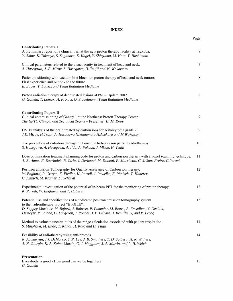
INDEX
Page
Contributing Papers IA preliminary report of a clinical trial at the new proton therapy facility at Tsukuba. 7Y. Akine, K. Tokuuye, S. Sugahara, K. Kagei, Y. Shioyama, M. Hata, T. Hashimoto
Clinical parameters related to the visual acuity in treatment of head and neck. 7A. Hasegawa, J.-E. Mizoe, S. Hasegawa, H. Tsujii and M. Wakaisami
Patient positioning with vacuum bite block for proton therapy of head and neck tumors: 8First experience and outlook to the future.E. Egger, T. Lomax and Team Radiation Medicine
Proton radiation therapy of deep seated lesions at PSI � Update 2002 8G. Goitein, T. Lomax, H. P. Rutz, O. Stadelmann, Team Radiation Medicine
Contributing Papers IIClinical commissioning of Gantry 1 at the Northeast Proton Therapy Center. 9The NPTC Clinical and Technical Teams – Presenter: H. M. Kooy
DVHs analysis of the brain treated by carbon ions for Astrocytoma grade 2. 9J.E. Mizoe, H.Tsujii, A. Hasegawa N.Yamamoto H.Asakura and M.Wakaisami
The prevention of radiation damage on bone due to heavy ion particle radiotherapy. 10S. Hasegawa, A. Hasegawa, A. Iida, A. Fukuda, J. Mizoe, H. Tsujii
Dose optimization treatment planning code for proton and carbon ion therapy with a voxel scanning technique. 11A. Boriano , F. Bourhaleb, R. Cirio, J. Derkaoui, M. Donetti, F. Marchetto, C. J. Sanz Freire, C.Peroni
Positron emission Tomography for Quality Assurance of Carbon ion therapy. 12W. Enghard, P. Crespo, F. Fiedler, K. Parodi, J. Pawelke, F. Pönisch, T. Haberer,C. Kausch, M. Krämer, D. Schardt
Experimental investigation of the potential of in-beam PET for the monitoring of proton therapy. 12K. Parodi, W. Enghardt, and T. Haberer
Potential use and specifications of a dedicated positron emission tomography system 13to the hadrontherapy project �ETOILE�.D. Sappey-Marinier, M. Bajard, J. Balosso, P. Pommier, M. Beuve, A. Emsallem, Y. Declais,Demeyer, P. Jalade, G. Largeron, J. Rochat, J. P. Gérard, J. Remillieux, and P. Lecoq
Method to estimate uncertainties of the range calculation associated with patient respiration. 14S. Minohara, M. Endo, T. Kanai, H. Kato and H. Tsujii
Feasibility of radiotherapy using anti-protons. 14N. Agazaryan, J.J. DeMarco, S. P. Lee, J. B. Smathers, T. D. Solberg, H. R. Withers,A. N. Giorgio, K. A. Kubat-Martin, C. J. Maggiore, J. A. Martin, and L. H. Welch
PresentationEverybody is good - How good can we be together? 15G. Goitein

2
PageFocus Session: DosimetryDosimetric commissioning of the CATANA proton beam for the first patient treatments. 15L. Raffaele, G. A. P. Cirrone, G. Cuttone, P. A. Lo Jacono, S. Lo Nigro, N. Romeo, M. G. Sabini,V. Salamone, L. M. Valastro
New high spatial resolution semiconductor microdosimetry in proton therapy. 16A. B. Rosenfeld, I. M. Cornelius, P. D. Bradley, M. Jackson, and J. Flanz
Investigation about proton beam quality with a mini TEPC. 17V. Cesari, V. Conte, P. Colautti, L. de Nardo, G. Tornielli, N. Iborra, J. Herault, and P. Chauvel
Polystyrene versus water calibration of proton beams. 18M. F. Moyers, A. Vatnitsky and S. M. Vatnitsky
Monte Carlo calculated versus experimental fluence correction factors in plastic phantoms 18for clinical proton beams.H. Palmans, J. E. Symons, E. A. de Kock, D. T. L. Jones, J-M. Denis and S. Vynckier
Analytical linear energy transfer calculations for proton therapy. 19J. J. Wilkens and U. Oelfke
High spatial resolution MOSFET dosimetry: possibility of application in proton therapy. 19A. B. Rosenfeld, G. I. Kaplan, M. L. F. Lerch, T. Kron, M. Jackson, and Y. Takada
An Analytical Model for Monitor Unit Calculations in Spread-Out Bragg Peak Proton Fields. 20H. M Kooy, S. Rosenthal, M. J Schaefer, and T. R Bortfeld
CVD and natural diamond and silicon detectors in proton beam dosimetry. 21G. Cuttone, G. A. P. Cirrone, S. Lo Nigro, L. Raffaele, M. G. Sabini, V. Salamone,M. Bucciolini, M. Bruzzi, S. Mazzocchi, S. Pini, S. Sciortino, S. Onori, C. De Angelis, and M. Pacilio
The Hounsfield look-up table used for treatment planning at gsi: measurement, accuracy and validation. 21E. Rietzel and D. Schardt
The expected radiation failure rate for optical encoders in the mpri treatment rooms. 21V. Anferov and A. N. Schreuder
ReportProgress on DICOM for ion therapy. 22Michael Neumann
Focus Session: Localized prostate tumors: 3D-conformal RT and IMRT vs. proton therapy3D conformal radiotherapy (3D-CRT) and IMRT in organ-confined prostate cancer. 23C. Greco, and R. Orecchia
Conformal proton beam therapy of prostate cancer-report of long-term PSA-based outcomes in over 24twelve hundred patientsC. J. Rossi Jr., J. D. Slater and J. M. Slater
Physical and clinical aspects of prostate IMRT with photons. 24J. Bohsung, D. Böhmer, A. Moys, S. Marnitz, and V. Budach
Real time tumor tracking (TULOC): first clinical measurements and plans for applications in prostate irradiation. 25H. Blattmann, R. K. Muench, P. G. Seiler, A. Sumova, J. Verwey, B. Kaser-Hotz and C. Rohrer Bley

3
Page
Patient positioning for proton therapy of prostate tumors: First experience and outlook to the future. 25E. Egger, A. Lomax and Team Radiation Medicine
Contributing PapersA new superconducting cyclotron to produce 250 MeV light ion beams. 26L. Calabretta, M. Maggiore, and D. Rifuggiato
Status of development of installation for proton conformal therapy mass application. 26V. Balakin
Successful acceleration test of the first module of the proton linac booster �LIBO�. 26P. Berra, E. Rosso, B. Szeless, M. Vretenar, U. Amaldi, K. R. Crandall, M. Mauri, D. Toet, M. Weiss,R. Zennaro, C. Cicardi, C. De Martinis, D. Giove, L. Grilli, D. Davino, M. R. Masullo, V. G. Vaccaro,L.Calabretta and A. Rovelli
Status of TOP Linac Costruction. 27S. Frullani, L. Picardi and C. Ronsivalle
Recent Improvements on the Proteus 230 for a Flawless System Operation. 27G. Gevers
Focus session: Eye melanomaLong term results after proton beam radiotherapy of uveal melanoma. 27E. Egger, G. Goitein, and L. Zografos
Ten years of technical evolutions in the preparation of ocular diseases for protontherapy. 29P. Chauvel, N. Iborra, J. Herault, A.Courdi, C. Mosci, S. Squarcia, and W. Sauerwein
Statistical analysis on melanoma treatment with proton beam in Nice 30C. Mosci, S. Donadio, D. Grosso, B. Mascialino, M. A. Penco, S. Squarcia, P. Chauvel, N. Iborra
Proton beam radiotherapy of iris melanoma: long term follow-up study in UK. 30B. Damato, A Kacperek, M Sheen, and R. D Errington
Results of proton and plaque therapy of choroidal melanoma treated in Vancouver. 31E. W. Blackmore, K. Paton, C. Duzenli, W. Kwa, R. Ma, and T. Pickles
Proton beam therapy for wet macular degeneration: the rationale for adding photodynamic therapy. 31L. T. Yonemoto
Proton therapy of ocular tumors in Berlin. The experience of the first four years. 32N. E. Bechrakis, M. Nausner, H. Fuchs, J. Heese, H. Kluge, W. Hinkelbein, M. H. Foerster
Clinical application of proton beams in the treatment of uveal melanomas: the first therapies 32carried out in Italy (CATANA project)C. Spatola, G. Privitera, G. A. P. Cirrone, G. Cuttone, J. Ott, L. Raffaele, M. L. Rallo, A. Reibaldi,V. Salamone, and M. G. Sabini
The experience of 10 years of eye treatments by protontherapy at CPO: from melanomas to other indications. 33C. Desblancs

4
PageContributing PapersThe Italian experimental radiobiological research related to hadrontherapy. 33P. Scampoli,
Potential reduction of the incidence of radiation-induced second cancers by using proton beams 34in the treatment of pediatric tumors.R. Miralbell, L. Cella, A. Lomax, and U. Schneider
Present status of the proton therapy system in PMRC. 35M. Umezawa and collaboration with PMRC1 and KEK Accelerator Lab.
Proton beam therapy at the Svedberg Laboratory. a status report 2002. 35E. Blomquist, A. Montelius, E. Grusell, U. Isaksson, O. Gudjonsson, G. Nyberg, P. Enblad,E. Ronne-Engström, H. Bolander, B. Glimelius, L. Pellettieri, and H. C:Son Silander
CNA, the Italian national centre for proton and carbon ions and related projects. 36U. Amaldi and S. Rossi
The Italian project for the development of a proton accelerator for oncological therapy (TOP project). 36The TOP Collaboration
The CATANA (Centro di AdroTerapia ed Applicazioni Nucleari Avanzate) 37proton therapy beam delivery system.G. Cuttone, G. A. P. Cirrone, N. Romeo, P. A. Lojacono, S. Lo Nigro, M. G. Sabini, L. M. Valastro,L. Raffaele, and V. Salamone
The control system of the CATANA proton therapy facility. 37A. Rovelli, A. Amato, L. Raffaele, P. Nicotra, E. Pollara, and C. Santoro
Focus session: Active beam scanning systemWhat are the questions we should be asking about scanning? 38J. B. Flanz
Studies of optimization methods for dose delivery with A beam scanning system. 38A. V. Trofimov and T. R. Bortfeld
Beam Scanning Research at LLUMC. 39G. B. Coutrakon, P. Koss, J. Hubbard, A. Ghebremedhin, M. F. Moyers
Optimization in proton scanning beams. 39E. Matsinos, B. Schaffner and W. Kaissl
Status of the development of a pencil beam scanning system at IBA, Belgium. 39C. Brusasco, J. F. de la Hoye, B. Marchand, and D.Prieels
Performances of a pixel ionization chamber to monitor a voxelscan hadron beam. 40A. Boriano, F. Bourhaleb, R. Cirio, M. Donetti, F. Marchetto, and C. Peroni
Treatment Planning for Broad-Beam 3D Irradiation Heavy-Ion Radiotherapy. 40N. Kanematsu, M. Endo, T. Kanai, H. Asakura, Y. Futami, H. Oka, and K. Yusa
Treatment Planning for Carbon ion Therapy. 41O. Jäkel, P. Heeg, C.P. Karger, P. Heeg, D. Schulz-Ertner, J. Debus M. Krämer, and G. Kraft

5
PagePostersAmmonium tartrate and alanine ESR detectorsfor proton beam dosimetry. 42A. Bartolotta, M. C. D’Oca, M. Brai, and A. Kacpereck
Investigation on LET dependence of glow curve characteristics of thermoluminescent materials 42irradiated with proton beams.M. Brai, S.Basile, G.Bruno, A. Bartolotta, and A. Kacpereck
Physical characteristics of lucite compensator used for conjonctival melanoma treatment. 42N. Iborra, J. Herault, P. Chauvel, A. Courdi, C. Mosci, S. Squarcia, and W.Sauerwein
Technical requirements and indications of protontherapy for conjunctival melanomas. 43P. Chauvel, N. Iborra, J. Herault, A. Courdi, C. Mosci, S.Squarcia and W.Sauerwein
Raman spectroscopy of irradiated tissue samples. 43J. de Boer, J. Besserer, S. Froschauer, R. Gerlach, M. Loewe, M. Moosburger, P. Quicken,M. Wuerkner, P. Alexa, A. Synytsya, and K. Volka
Long term results of proton beam radiotherapy for small posterior choroidal melanoma. 44E. Egger, G. Goitein and L. Zografos
Centre for accelerator science imaging and medicine at the Daresbury Laboratory 44in the north west of the United Kingdom.P. Butler, A. Kacperek, J. E. Shaw and R. A. Lewis
Status report of proton treatment facility at NCC (Kashiwa). 45T. Tachikawa, S. Kataoka, I. Kohmura, H. Nonaka, T. Kimura, T. Satoh, T. Nishio, M. Shimbo,T. Ogino, and H. Ikeda
The design and implementation of a heavy ion therapy database. 45H. Koyama-Ito, M. Endo, S. Minohara, J. Mizoe, H. Tsujii, A. Ito and H. Asai
Potential advantage of 99-TC-methoxy-isobutil-isonitrile (MIBI) - spect in association with CT 46and MR imaging in delineation of target volume for high grade gliomas: preliminary results.M. Krengli, G. Loi, G. Sacchetti, E. Inglese, and A. Cotroneo
Development of 3 Tesla Magnet-in-magnet for a compact synchrotron. 46M. Kumada, E. Antokhin, M. Endo, M. Aoki, E. Sugiyama, Y. Iwashita, I. Bolshakoba, and R. Holyaka
Presentation of the IBA Therapy Control Software. 47A. Choppin & D. Leyman
Preliminary test of 123I production with proton induced reaction. 47S. Lo Nigro, G. A. P. Cirrone, G. Cuttone, P. A. Lojacono, I. V. Patti, L. Raffaelle, M. G. Sabini,V. Salamone and L. M. Valastro
Effects of proton beam irradiation on uveal melanoma. 47The experience of the Oncology Eye Center of Genoa, Italy.C. Mosci, M. Nicolò, P. Chauvel, A. Polizzi, S. Squarcia, G. Nicolò, D. Ghiglione, G. Calabria
The physical results for proton treatment at National Cancer Center, Kashiwa. 48T. Nishio, M. Shimbo, T. Ogino, S. Katsuta, S. Kawasaki, and H. Ikeda
The study of a target autoactivation by using a proton beam for therapy - part 2. 49T. Nishio, M. Knazawa, A. Kitagawa, T. Murakami, T. Kanai, T. Tomitani, M. Suda, E. Urakabe,M. Hirai, M. Knazawa, K. Narushima, T. Motobayashi, and H. Mizuno

6
Page
Radiochromic film employed as a dosimeter to characterize a clinical proton beam. 50L. Raffaele, G. A. P. Cirrone, G. Cuttone, P.A. Lojacono, S. Lo Nigro, I. V. Patti, M. G. Sabini,V. Salamone, and L. M. ValastroRelative dosimetry with ionization chambers for the CATANA project. 50N. Romeo, A. Amato, A. Rovelli, L. Raffaele, G. Cuttone, G.A.P. Cirrone, S. Lo Nigro,M.G. Sabini, and V. Salamone
The use of thermoluminescence detectors for proton dose distribution measurements. 51M. G. Sabini, G. A. P. Cirrone, G. Cuttone, P. A. Lojacono, S. Lo Nigro, I. V. Patti, L. Raffaele,V. Salamone, and L. M. Valastro
Respiration synchronized irradiation system and beam efficiency. 51M. Shimbo, T. Nishio, T. Ogino, S. Katsuta, S. Kawasaki and Ikeda
A specialized clinical folder for choroidal melanoma treatment. 52S. Donadio, D. Grosso, B. Mascialino, M. A. Penco, S. Squarcia, C.Mosci and P. Chauvel
Monte Carlo simulations using PTRAN and MCNPX for dose and spectral calculations 52in clinical proton beams.F. Verhaegen, H. Palmans and A. Kacperek
Potential for a Radiotherapeutic Gain from an Anti-Proton Beam. 53S. Lee, W. McBride, T. Solberg, J. DeMarco, N. Agazaryan, J. B. Smathers, H. D. Suit,H. R. Withers, A. J. Giorgio, K. A. Kubat-Martin, C. J. Maggiore, J. A. Martin, and L. H. Welch
7
A preliminary report of a clinical trial at the new proton therapy facility at Tsukuba.Y. Akine, K. Tokuuye, S. Sugahara, K. Kagei, Y. Shioyama, M. Hata, T. Hashimoto, Proton Medical Research Center,
University of Tsukuba, 1-1-1 Tennoudai, Tsukuba, 305-8575, JapanE-mail: [email protected]
We started treating patients with proton therapy at the newly constructed facility in the campus of the University ofTsukuba Hospital in September 2001. Fifty-one patients were entered in the trial as of March 31, 2002. There were 10 menand 32 women. A median age of the patients was 67 years ranging from 11 to 90. Of the patients 17 had hepatocellularcarcinoma, 10 lung cancer, six head & neck (H&N) tumors and remaining patients had miscellaneous tumors. We irradiated59 tumors in the 51 patients. Of the 59 tumors 28 were in the liver, 11 in the lung, 6 in the H&N, remaining 18 in themiscellaneous organs. Of the 59 tumors irradiated 48 were primary tumors and 11 metastatic tumors. Of the 59 tumors 43were irradiated with proton beams alone and remaining 16 with a combined x-rays and proton beams. We irradiated 20hepatic tumors in the 17 patients with proton beams alone. Doses given to the 20 tumors were a median of 64.5 Gy in 15fractions with a range of 50-72 Gy in 10-22 fractions.
We have two treatment rooms with one each rotating gantry. It takes about 15-20 min to irradiate a patient with anaverage 1.5 ports. The treatment time comprised of 2-10 min of irradiation and 8-15 min of preparation. We currentlyirradiate 10-15 patients a day. An estimated maximum number of patients to be irradiated in six hours is about 50 patients.
After we obtain a safety approval by the government for the irradiation apparatus, we will proceed to obtain apermission to charge a patient for proton therapy.
Representative cases treated and clinical protocols at the center will be presented.
*******************************************************
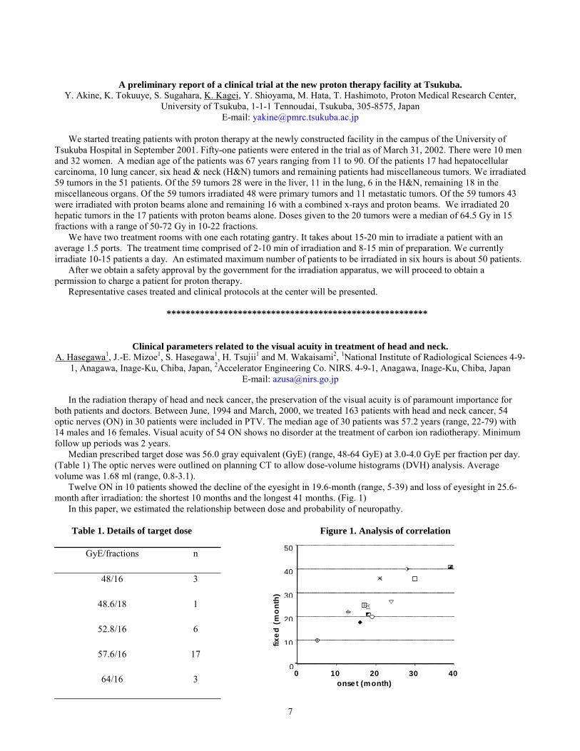
Clinical parameters related to the visual acuity in treatment of head and neck.A. Hasegawa1, J.-E. Mizoe1, S. Hasegawa1, H. Tsujii1 and M. Wakaisami2, 1National Institute of Radiological Sciences 4-9-
1, Anagawa, Inage-Ku, Chiba, Japan, 2Accelerator Engineering Co. NIRS. 4-9-1, Anagawa, Inage-Ku, Chiba, JapanE-mail: [email protected]
In the radiation therapy of head and neck cancer, the preservation of the visual acuity is of paramount importance forboth patients and doctors. Between June, 1994 and March, 2000, we treated 163 patients with head and neck cancer, 54optic nerves (ON) in 30 patients were included in PTV. The median age of 30 patients was 57.2 years (range, 22-79) with14 males and 16 females. Visual acuity of 54 ON shows no disorder at the treatment of carbon ion radiotherapy. Minimumfollow up periods was 2 years.
Median prescribed target dose was 56.0 gray equivalent (GyE) (range, 48-64 GyE) at 3.0-4.0 GyE per fraction per day.(Table 1) The optic nerves were outlined on planning CT to allow dose-volume histograms (DVH) analysis. Averagevolume was 1.68 ml (range, 0.8-3.1).
Twelve ON in 10 patients showed the decline of the eyesight in 19.6-month (range, 5-39) and loss of eyesight in 25.6-month after irradiation: the shortest 10 months and the longest 41 months. (Fig. 1)
In this paper, we estimated the relationship between dose and probability of neuropathy.
Table 1. Details of target dose Figure 1. Analysis of correlation
GyE/fractions n
48/16 3
48.6/18 1
52.8/16 6
57.6/16 17
64/16 30
10
20
30
40
50
f
ixe
d (
mo
nth
)
0 10 20 30 40 onset (month)
8
References1. Habrand IL, Austin SM, Birnbaum S, et al. Neurovisual outcome following proton radiation therapy. Int J Radiat
Oncol Biol Phys 1989;16(6):1601-6.2. Khoo VS, Oldham M, Adams EJ, Bedford JL, Webb S, Brada M. Comparison of intensity-modulated tomotherapy
with stereotactically guided conformal radiotherapy for brain tumors. Int J Radiat Oncol Biol Phys 1999;45(2):415-25.3. Mizoe J. Analysis of the dose-volume histogram in uterine cervical cancer by diagnostic CT. Strahlenther Onkol
1990;166(4):279-84.
*******************************************************
Patient positioning with vacuum bite block for proton therapy of head and neck tumors: First experience andoutlook to the future.
E. Egger, T. Lomax and Team Radiation Medicine, Division of Radiation Medicine, Paul Scherrer Institute, CH � 5232Villigen-PSI, Switzerland
E-mail: [email protected]
At PSI, patients undergoing proton therapy at the spot scanning gantry are positioned outside the treatment room.Position verification is done on a remote CT by comparison of several CT slices with the slices of planning CT andmeasurement of several points on scouts view. Patients are brought inside the treatment room in their mould after positionverification and necessary corrections have been completed.
For head and neck tumors, patients are positioned with a vacuum bite block, which is a very straightforward procedureto obtain high precision positioning. We will analyze the positioning precision obtained with this system. We will alsodiscuss the time necessary for precise positioning of the patient, the personal required for an optimal use of the irradiationfacility and raise the question whether in a new facility, patient positioning should also be done outside the treatment roomor preferably inside the treatment room.
*******************************************************
Proton radiation therapy of deep seated lesions at PSI – Update 2002.G. Goitein, T. Lomax, H. P. Rutz, O. Stadelmann, Team Radiation Medicine, Division of Radiation Medicine, Paul
Scherrer Institute, CH � 5232 Villigen PSI, SwitzerlandE-mail: [email protected]
Since PSI took up proton radiation therapy of deep-seated tumors on the spot-scanning gantry, a total of 99 patientshave been treated by the end of 2001. Forty -eight were female, fifty-one male. Age varied from 7 to 80 years, with amedian of 47 years. The tumor groups/histologies were chordomas/chondrosarcomas (37), meningiomas (15), brainhistologies (11), soft tissue and bone sarcomas (11), singular metastases (7), nasopharynx cancer (6), prostate (5),esthesioneuroblastoma (2), local tumor relapses (2), basalioma (1), ependymoma (1) and gangliocytoma (1). Follow uptime is fife to sixty-six months, median 30.5 months. Of the ninety-nine patients, 78 were treated with curative intent.Thirteen patients died, nine of whom had been treated with palliative intent.
The group of chordomas (chor.)/chondrosarcomas (ch-sarc.) is divided into 9 ch-sarc. and 7 chord. of the base of skull,1 occipital ch-sarc., 1 ch-sarc. of the t-spine, 8 chor. of the spine (5 c-, 1 t-, 2 l-spine), 9 chor. of the sacrum and 2 ch-sarc.of the trunk. We saw two local failures: one c-spine chordoma, irradiated with 74 CGE after multiple surgeries, and oneskull base chordoma, which relapsed after 72 CGE in the surgical pathways and locally. Out of 37 patients 6 were treatedwith palliative intent, two died from distant metastases.
Meningiomas were the second largest group with 12 benign and three atypical lesions. One patient died fromcomplications after necrosectomy due to central necrosis of an atypical, multifocal meningioma. Three patients developedoptic nerve toxicity after 54 CGE (2) and 64 CGE (1), with the nerve being entirely included in the target volume.

9
Of the 11 gliomas (9 low, 2 high grade), 8 were locally controlled. Three patients developed necrosis within the highdose region with two patients being symptomatic and needing steroids. On patient died at 31 months from local relapse of agrade II astrocytoma.
Of the eleven sarcomas, 8 are locally controlled. The relapses occurred: at 6.5 months in a pediatric intracranialrhabdomyosarcoma, the patient died at 10 months with local and distant progression; at 14 months in a juvenilerhabdomyosarcoma, which had been treated with only 14 CGE protons plus photons and chemotherapy in Italy; 1malignant nerve sheath tumor relapsed locally after prior complete resection and irradiation.
Five of the 6 nasopharynx carcinomas were treated curatively with proton-photon combination and chemotherapy, theyare locally controlled without distant disease and show no toxicity.
In summary, 86 out of 99 patients are alive five to sixty-six months after proton radiotherapy. Local control wasachieved 72 out of 78 curatively irradiated patients and in 10 out of 20 palliative cases. Late toxicity greater grade 2 wasfound in 6 out of ninety-nine patients.
*******************************************************
Clinical commissioning of Gantry 1 at the Northeast Proton Therapy Center.The NPTC Clinical and Technical Teams � Presenter: H. M. Kooy, Northeast Proton Therapy Center, Massachusetts
General Hospital and Department Of Radiation Oncology, Harvard Medical School, Boston MA 02114
The clinical commissioning of the first gantry at the Northeast Proton Therapy Center (NPTC) qualified the use of thegantry for clinical operations, produced the data necessary for input to, and verification of, the treatment planning system,and established an essential understanding of the operational behavior of the gantry. The commissioning measurements canbe divided into dosimetric measurements, geometric measurements, operational and control system verifications, and theirinterdependencies. The dosimetric measurements focused on exercising the beam nozzle over its full operational range andto quantify the pristine proton peaks and SOBPs over this range. The geometric measurements focused on quantifying theaccuracy of the patient positioning devices and the patient alignment with respect to the gantry and nozzle. The operationalaspects of the gantry operation also required close scrutiny as the system is, in essence, a software driven system.Commissioning commenced in September 2001 and the first patient was treated on November 8 2001. We present our dataacquired for the commissioning tasks, discuss the logistical effort to commence treatments, and the experience in the firstphases of clinical operations.
*******************************************************
DVHs analysis of the brain treated by carbon ions for Astrocytoma grade 2.J.E. Mizoe1, H.Tsujii1, A. Hasegawa1 N.Yamamoto1 H.Asakura2 and M.Wakaisami2, 1NIRS (National Institute ofRadiological Sciences), Anagawa 4-9-1, Inage-Ku, Chiba-City, JAPAN, 2AEC(Accelerator Engineering System),
Anagawa 4-9-1, Inage-Ku, Chiba-City, JAPANE-mail: [email protected]
The DVHs of the brain, delivered from the patients treated by carbon ions for their astrocytoma grade 2, were analyzedto determine the dose-complication probability for late toxicity.
Twelve patients with astrocytoma grade 2 were treated on a phase I/II dose escalation protocol of carbon ions at NIRSbetween September 1994 and February 2001. Eligibility criteria included biopsy proven astrocytoma grade 2, age between18 and 80 years, Karnofsky performance sore of 60 or greater, and no infection and no wide degeneration. Median age ofthe patients was 31 years (range 18-48) with 8 males and 4 females. The sites of disease were consisted of 5 frontal, 2temporal, 2 basal ganglion, 1 parietal, 1 occipital and 1 corpus callosum respectively.
Fractionation method of 24 fractions through 6 weeks was applied with fraction dose of 2.1 and 2.3 GyE( photonequivalent dose). Of the 12 patients, 4 developed grade 1 and 2 late toxicity of LENT/SOMA MRI Scaling System.Integral DVHs were calculated using planning CT and dose-complication probability was calculated at 10% high dosevolume. Calculated equation of logistic model was follows;
P = 1 �1 / ( 1 + EXP ( -110.5 + 2.303 D)) P: Probability of late toxicity more than grade 1 (LENT / SOMA MRI Criteria)

10
D: Dose ( GyE) at 10 % volume in integral DVH of the brain
0
0.1
0.2
0.3
0.4
0.5
0.6
0.7
0.8
0.9
1
30 35 40 45 50 55 60 65 70
D ose (G yE )
Probabili
D ose C om plication_P robability of the B rain
Y =1-1/(1+exp(-110.5+2.303*D )
*******************************************************
The prevention of radiation damage on bone due to heavy ion particle radiotherapy.S. Hasegawa, A. Hasegawa, A. Iida, A. Fukuda, J. Mizoe, H. Tsujii, National Institute of Radiological Sciences, 4-9-1
Anagawa, Inage-ku, Chiba, Japane-mail: [email protected]
Osteoradionecrosis (ORN) of the mandible is a severe complication due to radiotherapy in the head and neck region,which is known as progressive radiation damage. It is a complex metabolic and tissue homeostatic deficiency created byradiation-induced tissue injury. And the occurrence of ORN of the mandible to any extent can cause morbidity and painsecond only to the development of recurrence, metastasis or second tumor in the area.
Heavy ion particle irradiation has excessive biological effects than X-Ray irradiation in all of tissues. Until now, wemade clear that carbon ion irradiation caused more decrease in trabecular Bone Mineral Density (BMD) than X-Rayirradiation (Fig. 1). Then Schmitt J. has reported that ORN presented with different signal intensities depending on the timeelapsed after radiation therapy. We thought that ORN is related to decrease in BMD, and thus, increase in BMD leads toprevention of ORN.
At present, there are two kinds of compounds to improve bone metabolism and to maintain and increase BMD. One hasan inhibitory effect of bone absorption acting to osteoclast cells, for example, bisphosphonates, and the other has anenhancing effect of bone formation acting to osteoblast cells like statins. Bisphosphonates, analogs of pyrophosphate, bindto bone at sites of active bone remodeling. In clinical settings of rapid bone turnover and/or excessive osteolytic activity,they have shown to have beneficial clinical effects. Bisphosphonate inhibition of osteolysis in cancer has shown to beeffective as an adjunctive therapy for the delay or prevention of cancer-related skeletal morbidity, including bone pain,pathologic fractures. Animal models of bone metastasis prevention by bisphosphonate treatment have provided thepreclinical background for the adjuvant use of bisphosphonates in primary cancers.
Thus, we consider bisphosphonate is able to prevent the negative effects of radiotherapy on bone and conduct anexperimental study.
In this experimental study, we focus on two kinds of bisphosphonate compounds, which are Etidronate andPamidronate. 35 Female Wistar rats (3 months old) divided into 7 groups: control, 1.0GyE carbon ion irradiation group,5.0GyE carbon ion irradiation group, 1.0GyE carbon ion irradiation with Etidronate group, 1.0GyE carbon ion irradiationwith Pamidronate group, 5.0GyE carbon ion irradiation with Etidronate group, 5.0GyE carbon ion irradiation withPamidronate group. The rats were injected with a daily dose of 3mg/kg bisphosphonate subcutaneous administration afterwhole body carbon ion irradiation 3 times a week for 6 weeks. Then we measured tibial trabecular BMD using peripheral

11
quantitative computed tomography twice every month. Two of the 1.0GyE carbon ion irradiation groups, which wereadministrated Etidronaate and Pamidronate, respectively, presented increase in BMD compared with not only 1.0GyEcarbon ion irradiation group but also control group. And highly similar results were observed in the 5.0GyE carbon ionirradiation groups with Etidronaate and Pamidronate (Fig. 2).
It is suggested that Etidronate and Pamidronate have been established as effective in the prevention of decrease of BMDdue to carbon ion particle irradiation.
Figure 1. The radiation effects in trabecular BMD Figure 2. The prevention effects in trabecular BMD
16
18
20
22
24
26
28
0 1 2 3 4 5
carbonX-ray
Bon
e M
ine
ral D
en
sity
(m
g/c
m3)
Dose (Gy))
References1. Delanian S, Lefaix JL. Mature bone radionecrosis: from recent physiopathological knowledge to an innovative
therapeutic action. Cancer Radiother 2002;6(1):1-9.2. Theriault RL, Hortobagyi GN. The evolving role of bisphosphonates. Semin Oncol 2001;28(3):284-90.
*******************************************************
Dose optimization treatment planning code for proton and carbon ion therapy with a voxel scanning technique.A. Boriano1 , F. Bourhaleb2,3,4 , R. Cirio2, J. Derkaoui4, M. Donetti2,3, F. Marchetto2, C. J. Sanz Freire2, C.Peroni 2, 1ASP,
V.le Settimio Severo 65, I-10133 Torino, Italy, 2INFN and University of Torino, V. Giuria 1, I-10125 Torino, Italy, 3TERAFoundation, V.Puccini 11, I-28100 Novara, Italy, 4University Mohamed Ist, Faculty of Sciences Oujda, Marocco.
We have proposed a new dose optimization analytical code (ANCOD++) dedicated to proton and heavy ion therapy.The objective is to obtain as a solution the optimal set of beam weights distribution. The irradiation technique adopted is anactive scanning technique where each of the elementary volumes (voxel) have to be irradiated with a pencil beam. Theinverse planning method allows the calculation of the kinetic energies needed to have the maximum energy deposition atthe center of the spoted voxel.
The algorithm used consists of iteratively changing the trial incident fluence values of different beams which yields tomatch the prescribed dose distribution.
A comparison of the analytical estimation and a Monte Carlo simulation with the package GEANT3 shows excellentagreement, indicating that the inverse planning methods for optimizing the plans is efficient enough and allows improvingthe quality of the treatment planning in a practical time.
*******************************************************
0
50
10
15
20
Co
nt
C1.
0
C1.
0+e
t
C1.
0+p
am
C5.
0
C5.
0+e
ti
C5.
0+p
am
Bon
e M
ine
ral D
en
sity
(m
g/c
m3)

12
Positron emission Tomography for Quality Assurance of Carbon ion therapy.W. Enghardt1, P. Crespo1, F. Fiedler1, K. Parodi1, J. Pawelke1, F. Pönisch1, T. Haberer2, C. Kausch2, M. Krämer2, D.
Schardt2, 1Forschungszentrum Rossendorf e.V., Postfach 510119, 01314 Dresden, Germany, 2Gesellschaft fürSchwerionenforschung mbH, Planckstr. 1, 64291 Darmstadt, Germany
E-mail: [email protected]
The physical and radiobiological properties of high-energy ions offer the possibility of treating deep-seated, compactand radioresistant tumours with high precision. In delicate therapeutic situations, especially when the tumours are growingin close vicinity to organs at risk, an in-situ verification of the dose localisation is highly desirable. Positron emissiontomography (PET) is the only available method for an in-situ and non-invasive monitoring of the precision of the doseapplication in charged hadron therapy. The physical background is the production of minor amounts of positron emittingradionuclides via nuclear fragmentation reactions following collisions between the incident projectiles and atomic nuclei ofthe tissue. In the case of carbon ion therapy the most abundant isotopes are 11C, 15O and 10C.
An in-beam positron emission tomograph has been integrated into the experimental carbon ion therapy facility at theGesellschaft für Schwerionenforschung, Darmstadt (GSI). Its technical basis are components of PET-scanners as appliedfor radiotracer imaging in nuclear medicine. However, operating PET simultaneously with therapeutic irradiations requiresdedicated solutions in detector geometry, data acquisition and processing as well as in tomographic reconstructiontechniques.
After more than 4 years of clinical operation first conclusions on the benefit of the in-beam PET method for improvingthe precision of ion therapy may be drawn. PET is capable of detecting deviations between the prescribed and the deliveredspatial dose distribution. Since the distributions of dose and of β+-activity may remarkably differ for physical reasons, aspecial technique for identifying dose deviations has been developed: we compare the spatial distributions of the measuredβ+-activity with those predicted on the basis of the treatment plan, the anatomical information from computed tomogramsof the irradiated region and the time course of the particular treatment fraction [1]. The reasons for dose deviations havebeen identified as (i) unavoidable deficiencies of the physical beam model used for treatment planning, (ii) minor errors inpatient positioning in combination with steep tissue density gradients in the beam path as well as (iii) local and frequentlytemporary changes in the patient anatomy leading to density modifications of the irradiated tissue. While being ratheruncritical in conventional radiotherapy, such slight inaccuracies may severely modify the dose distributions delivered byions. To quantify these local dose deviations on the basis of PET data, an interactive procedure has been developed.References1. Enghardt W., Debus J., Haberer T., Hasch B. G., Hinz R., Jäkel O., Krämer M., Lauckner K. and Pawelke J., The
application of PET to quality assurance of heavy-ion tumor therapy, Strahlenther. Onkol. 1999; 175:Suppl II: 33-6
*******************************************************
Experimental investigation of the potential of in-beam PET for the monitoring of proton therapy.K. Parodi1, W. Enghardt1, and T. Haberer2, 1Forschungszentrum Rossendorf e.V., Postfach 510119, 01314 Dresden,
Germany, 2Gesellschaft für Scwherionenforschung mbH, Planckstr. 1, 64291 Darmstadt, Germany E-mail: [email protected]
Positron-emission-tomography (PET) is potentially a powerful tool for the in-situ and non-invasive control of theprecision of the dose application in charged hadron therapy. The clinical implementation of in-beam PET monitoring at theheavy ion tumor therapy facility of GSI Darmstadt has proven the important impact of the method on the quality assuranceof high precision carbon ion therapy [1]. In perspective of the dedicated ion beam tumor therapy facility of Heidelberg,Germany, which is planned to deliver particles from protons up to oxygen nuclei, we started to investigate the potential ofin-beam PET for the monitoring of proton therapy. The extension is non-trivial, since protons do not suffer the projectilefragmentation reactions originating a sharp maximum of the β+-activity in close proximity to the dose maximum in thecarbon ion case.
Following our previous promising but preliminary investigation [2] entirely based on Monte Carlo simulations, a firstexperiment was performed: three mono-energetic proton beams in the energy and intensity range suited for the treatment of
13
deep-seated tumors were completely stopped in blocks of PMMA (C5H8O2) positioned in the center of the field of view ofthe in-beam PET scanner installed at the treatment unit of GSI Darmstadt. The amount of β+-activity was found to be threetimes larger than that induced by carbon ions at the same range and the same applied physical dose. The reconstructed β+-activity distributions were well reproduced in shape by calculations based on experimental cross-sections and on the protonflux given by the FLUKA Monte Carlo code.
Despite the weaker spatial correlation between β+-activity and dose depth-distributions in the proton case, the presentedexperiment supports the feasibility of in-beam PET for the in-situ monitoring of proton therapy based on a comparisonbetween measured and realistically calculated β+-activity distributions, as already implemented for carbon ion therapy.
This work is supported by the Bundesministerium für Bildung, Wissenschaft, Forschung und Technologie of Germany(grant 06DR825).References1. Enghardt W. et al, Positron emission tomography for quality assurance of cancer therapy with light ions, Nucl. Phys.
A654 (1999) 1047c2. Parodi K. and Enghardt W., Potential capabilities of positron emission tomography for quality assurance of proton
therapy, Abstract from PTCOG XXXII, 25-27 September 2000, Berlin
*******************************************************
Potential use and specifications of a dedicated positron emission tomography system to the hadrontherapy project“ETOILE”.
D. Sappey-Marinier, M. Bajard, J. Balosso, P. Pommier, M. Beuve, A. Emsallem, Y. Declais, Demeyer, P. Jalade, G.Largeron, J. Rochat, J. P. Gérard, J. Remillieux, and P. Lecoq1, Centre d�hadronthérapie, Université Claude Bernard Lyon1,43, Bd du 11 novembre 1918, 69622 Villeurbanne, France, 1CERN, DIVISION EP/CMA, 1211, GENEVA, SWITZERLAND
E-mail: [email protected], [email protected], [email protected]
The Project “ETOILE”: Protontherapy has proven to be very useful for cancer treatment by more than fifteeninternational centers. More recently, Chiba (HIMAC) and Darmstadt (GSI) centers have treated more than 1000 patientswith carbon ion beams and have shown the efficiency of carbon ion beams, particularly in case of tumors being eitherhighly radio-resistant or closely localized to sensitive organs (1,2). However, these clinical results are limited by the smallnumber of treated patients. Therefore, the goal of the project ETOILE (Espace de Traitement Oncologique par Ions LégersEuropéen) is to implant a center for hadrontherapy in the French region �Rhône-Alpes� to extend this new approach ofcancer treatment. This project is organized with the French institutions (UCB-Lyon1, UJF-Grenoble1, CNRS/IN2P3,CEA/DSM, HCL, CHU Grenoble, CLB�) in collaboration with the CERN and the five other European projects throughthe network ENLIGHT. The center will be based on a synchrotron of PIMMS type allowing the production of proton andcarbon ion beams at the maximal energy of 200 and 400 MeV/nuclei, respectively. The ion beam will be delivered in threetreatment rooms through a dynamic raster scanning and monitored on line using a dedicated positron emission tomography(PET) system.PET imaging in hadrontherapy: The penetration of the ion beam through the tissue induces fragmentation of tissue nuclei(mainly water molecules) with proton beams and also of the primary particles with carbon ions beams. This phenomenongenerates positron emitting isotopes which can be detected by PET as shown by GSI (3,4). PET can therefore provide apowerful monitoring tool to measure in vivo and on line the spatial distribution of both the beam and the dose deposition.For this challenging purpose, a dedicated PET system characterized by a high sensitivity and high accuracy has to bestudied.Development of a dedicated PET system: In collaboration with the CERN, ENLIGHT and the Crystal Clear Consortium(CCC), our goal is to study a new generation of PET system based on new design and technologies to achieve bettersensitivity and spatial resolution.Specifications: 1) large field of view and solid angle allowing the positioning of the patient in the beam path, 2) high spatialresolution (> 4 mm) according with the accuracy of the beam, 3) high sensitivity to compensate the low count rate, 4)insensibility to magnetic field and secondary particles effects.Development: 1) dedicated design using Monte Carlo simulations, 2) denser, faster and brighter scintillating materials(LSO, LuAP), 3) photodetectors such as MaPMT and APD, and rapid front-end electronic.Research: 1) simulation of the fragmentation phenomenon and realistic PET images, 2) modeling of the fragmentedparticles and positrons kinetic and of the delivered dose.Planning: 1) development of bench prototypes for testing the new crystal-photodetector-electronic configurations consistingof block with 64 crystals (2x2x8mm), 1 MaPMT or 2 APD and new front-end electronic, 2) development of microPETprototype based on the precedent tests, 3) feasibility study of a dedicated PET system for hadrontherapy.

14
Results: The project �ETOILE� will be presented with the preliminary results obtained either by simulation or testing thenew detection blocks developed for the microPET system.References1) Tsujii H. Current status of hadrontherapy with carbon ion beams. Eur I Cancer 2001;37(6):2512) Debus J, Haberer T, Schulz-Ertner D, et al. Carbon ion irradiation of skull base tumors at GSI. First clinical results and
future perspectives. Strahlenther. Onkol. 2000;176:211-216.3) Enghardt W, Debus J, Haberer T, et al. The application of PET to quality assurance of heavy-ion tumor therapy.
Strahlenther Onkol. 1999 Jun;175 Suppl 2:33-6.4) Parodi K, Enghardt W. Potential application of PET in quality assurance of proton therapy. Phys. Med. Biol.
2000;45:151-156.*******************************************************
Method to estimate uncertainties of the range calculation associated with patient respiration.S. Minohara, M. Endo, T. Kanai, H. Kato and H. Tsujii, National Institute of Radiological Sciences, 4-9-1 Anagawa,
Inage-ku, Chiba 263-8555, JAPANE-mail: [email protected]
Purpose: To propose a method for estimating uncertainties of the range calculation in particle radiotherapy associated withorgan motion along with patient respiration.Methods and Materials: A set of sequential CT images every 0.2 seconds was reconstructed from continuous x-rayprojection data accumulated by the dynamic scanning mode in helical CT scanner. At the same time of CT data acquisition,respiratory signal of patient and on/off signal of X-ray on CT scanner were recorded. From these data, the timing of eachCT image was related with the phase of respiration waveform. These CT images were analyzed in our treatment planningsystem that included the function converting from CT number to water equivalent path length (WEL). A set of CT imagesof the patient with liver cancer at upper right lobe was analyzed. The geometrical sizes of the liver and WELs from bodysurface to iso-center were measured in each CT image.Results: WEL variations of depth from body surface to iso-center were 6.2mm and 18.9mm at anterior-posterior andposterior-anterior direction, respectively. Liver size was changed to 35.2mm. However these variations were shown to beconsiderably reduced by gated irradiation.Conclusion: The proposed method using sequential CT images with respiration waveform was shown to be useful toevaluate the uncertainties of the range calculation associated with patient breathing. The variation of the depth along thebeam path was presented in WEL rather than geometrical length.
*******************************************************
Feasibility of radiotherapy using anti-protons.N. Agazaryan1, J.J. DeMarco1, S. P. Lee1, J. B. Smathers1, T. D. Solberg1, H. R. Withers1, A. N. Giorgio2, K. A. Kubat-
Martin2, C. J. Maggiore2, J. A. Martin2, and L. H. Welch2, 1UCLA Department of Radiation Oncology, Los Angeles, CA,USA, 2Pbar Medical, Inc., Newport Beach, CA, USA
We have begun an investigation into the potential use of antiproton beams in clinical radiotherapy. Observedexperimentally for the first time in 1955, antiprotons are the antimatter counterpart to protons, with a negative charge andparity and rest mass of 938 MeV/c2. Antiprotons have depth dose characteristics similar to protons in that they exhibit anenergy dependent Bragg peak. The matter-antimatter annihilation event at the end of range is accompanied by the release ofnearly 2 GeV, primarily in the form of energetic pi-mesons, but also neutrons, K-mesons and gammas, and of particularinterest for therapeutic applications, charged nuclear fragments.
We are using the MCNPX Monte Carlo code developed at Los Alamos National laboratory to evaluate the feasibility ofclinical antiproton therapy and in the design of physical experiments. MCNPX combines the traditional MCNP particles(neutrons, photons, and electrons) with the high-energy, multi-particle transport features of the LAHET code package. Theintermediate energy model in MCNPX simulates antiproton annihilation and accompanying secondary particle production.The de-excitation of the residual nucleus after proton-antiproton annihilation is modeled using the multistage pre-equilibrium model and multi-fragmentation of light nuclei is based upon the Fermi-Breakup model.
Monte Carlo calculations confirm that the annihilation event produces a significantly larger Bragg peak relative to aproton dose deposition curve. For 150 MeV incident antiprotons, the peak-to-plateau ratio is approximately twice that forprotons of a similar energy. The antiproton peak-to-plateau advantage over protons increases as the incident energy isdecreased. Perhaps more significantly, a further potential clinical advantage exists in the form of the high relative biologicaleffectiveness (RBE) of the charged nuclear fragments produced in-situ at the end of range.

15
While gammas resulting from the prompt neutral pion decay have sufficient energy to exit a human, roughly half of thecharged pions produced will contribute to a relatively isotropic background dose. Nevertheless, this background isinconsequential relative to the clear physical and biological advantages.
In summary, the use of antiprotons holds significant potential for enhancing the clinical efficacy of focal radiotherapyapproaches. Theoretical and experimental studies to evaluate the technique are actively ongoing.
*******************************************************Everybody is good - How good can we be together?
G. Goitein, Division of Radiation Medicine, Paul Scherrer Institute , CH � 5232 Villigen PSI, SwitzerlandE-mail: [email protected]
Proton Radiation Therapy has developed to the point that it is recognized worldwide as a valuable approach to thetreatment of various forms of malignant and even benign diseases. Today�s technology and performance originated inphysics research institutions, where the physics background, engineering skills, medical competence and excellentpartnership between physics and medicine could flourish to remarkable degree and quality. Established indications forproton therapy such as choroidal melanomas, chordomas and chondrosarcomas of the base of skull, AVM�s and othertumors have been treated according to technical guidelines and medical protocols at a limited number of institutions. Formore than ten years, the international community of people interested in particle therapy has grown greatly, and today abouttwenty centers worldwide are active in particle radiation therapy, using protons and heavier ions. During this timetechnologies have changed, the number of indications has, and will continue to, grow, and other local and systemictreatment modalities are changing in parallel and causing a form of competition which did not exist at the time of thefounders of particle therapy.
In order to preserve and even strengthen the importance of particles and to create a reliable quality of treatments, thecommunity has to work on new guidelines and protocols, which allow for a) technical and medical comparison ofprocedures, b) a meaningful standardization of treatments (particularly for �new� indications) and c) transparent analysis ofall procedures and results. The situation vis-à-vis conventional radiation therapy has become much more competitive,technically as well as economically, not to mention the psychological aspects of the acceptance of particles. Beamcharacteristics, devices for beam application, dose prescription, dosimetry, treatment planning and delivery tools such asintensity modulation, homogeneity / inhomogeneity of dose, single / total doses, fractionation, overall treatment time, tumorhistology, stage, combination with other treatment modalities � these are only some of the many parameters we have toknow and to compare if we want to analyze our procedures and results seriously. Modern tools for data sharing makecooperation much easier than before. The goal of this presentation is to initiate within PTCOG efforts to establish standardsand ensure a uniform high quality. This is needed in order to ensure that particle radiation therapy does not degenerate intoan expensive nice-to-have therapy but, rather, establishes our existing and upcoming facilities as necessary weapons againsta variety of malignant diseases.
*******************************************************
Dosimetric commissioning of the CATANA proton beam for the first patient treatments.L. Raffaele, G. A. P. Cirrone, G. Cuttone, P. A. Lo Jacono, S. Lo Nigro, N. Romeo, M. G. Sabini, V. Salamone, L. M.Valastro, INFN � Laboratori Nazionale del Sud, Via S. Sofia 44, Catania (Italy), INFN � Sezione di Firenze, Largo E.Fermi 2, Firenze (Italy), Istituto di Radiologia, Universita degli Studi di Catania, Via S. Sofia, Catania (Italy), Centro
Siciliano di Fisica Nucleare e Struttura della Materia, Corso Italia 57, Catania (Italy), Dipartimento di Fisica, Universitadegli Studi di Catania, Corso Italia 57, Catania (Italy)
At INFN-Laboratori Nazionali del Sud (LNS) in Catania, a proton therapy facility, named CATANA, (Cetro diAdroTerapia e Applicazioni Nucleari Avanzate), based on the use of 62 MeV proton beam, has been ralized mainlydedicated to the treatment of shallow tumors (3 cm max), like those present in the ocular region (i.d. uveal melanomas andmacular degenerations). The dosimetric beam characterization has been concluded. For this aim different dosimeters havebeen employed and according to our previous experience. The dosimetric commissioning has been carried out mainlylooking at the evaluation of depth dose, lateral off-axis profiles in water, field size dependent output factors, absolutedosimetry and dosimetric beam monitoring system. The results will be extensively presented.
*******************************************************
16
New high spatial resolution semiconductor microdosimetry in proton therapy.A. B. Rosenfeld1, I. M. Cornelius1, P. D. Bradley1*, M. Jackson2, and J. Flanz3, 1Centre for Medical Radiation Physics,
University of Wollongong, Wollongong NSW 2522, Australia, 2Department of Radiation Oncology, Royal Prince AlfredHospital, Camperdown, NSW, Australia, 3Massachusetts General Hospital, Department of Radiation Oncology, Northeast
Proton Therapy Center, 30 Fruit Street, Boston, MA 02114, USAE-mail: [email protected]
In the present clinical practice a single value of RBE is used for the whole treatment planning. However, proton RBEdepends on the type of biological endpoint, on the initial proton energy, method of beam delivery, on the depth of beampenetration and size of radiation field. The LET of particles is a main parameter responsible for RBE. The protonsthemselves can demonstrate LET up to about 80-90keV/micron at the end of the Bragg peak. Recent proton beamsimulations with Monte Carlo code GEANT with nuclear interaction module FLUKA for MGH NPTC facilitydemonstrated a significant contribution to the total dose beside the primary protons could be due to secondary heavierparticles alphas, deuterons, tritons and neutrons and secondary low energy protons [1,2]. Also these high LET particles cancontribute to RBE of proton beam.
The relative changes in RBE across the beam for different beam size and method of delivery must be the input into thetreatment planning system for proton conformal therapy. In this case measurements of dose distribution with ionizingchamber only is not enough for dose planning. Existing experience in proton therapy indicate the need for extensive furtherstudies of RBE of the therapeutic proton beam especially in the distal edge of the Bragg peak [3]. Additionally to in vitroexperiments with biological cells the microdosimetry is important method for investigation of RBE properties in conformalPT. Present microdosimetric instrument is based on proportional gas counter and has limited spatial resolution and requireTE gas and high voltage operation.
New silicon SOI microdosimeter was developed at the Centre for Medical Radiation Physics, UoW as a part of PTAustralian National program. Microdosimeter is based on SOI (silicon-on-insulate) p-n junction array with sensitivevolumes of micron sizes similar to biological cells [4]. Advantage of this microdosimeter is high spatial resolution (lessthan 1 mm) and possibility to measure under the full proton beam. Measurements of microdosimetric spectra were donealong the Bragg peak for 210 MeV slow-extracted beam at MGH and pulsed 250 MeV proton beams at KEK in a Perspexphantom.
It was demonstrated that microdosimetric spectra became much harder on the distal edge of the Bragg peak. At thedepth of 26 cm, that corresponds to the range of the 190 MeV protons in perspex, contribution of high LET (more than100keV/u) events was observed. For position behind the Bragg peak contribution to microdosimetric spectra was mostlydue to the secondary protons originated by neutrons. Events with LET more than 200 keV/micron were observed for highenergy protons in entrance region of the phantom that is in agreement with theoretical model predicting high LET nuclearsecondary.
Method of changing the effective size of sensitive site using time rise discrimination technique has been developed andtested.
New microdosimeter is small and require low bias operation. Microdosimetric spectra can be used as experimentalinput data for RBE based dose planning system in conformal proton therapy for particular proton beam and method ofdelivery. Also these spectra can be used for verification of Monte Carlo simulations in proton therapy utilizing GEANT andFLUKE codes.References.1. H.Paganetti, M.Goitein �Physical and Biological dose distribution due to primary protons and secondary particles
from nuclear interactions�, in �The use of computers in radiation therapy�, Edited by W.Schlegel, T.Bortfeld, Springer,p.323-325
2. J.Flanz, H.Paganetti �Monte Carlo calculations in support of the commissioning of the NPTC�, Proceedings of theFirst International Workshop on “ Micro- and Mini- dosimetry and its applications”, Sydney, Australia 2001http://mrp.uow.edu.au/mmd-2001(to be published)
3. G.Coutracon et al.� Microdosimetry spectra of the Loma Linda proton beam and relative biological effectivenesscomparisons�, Med. Phys. 24(9), 1499-1506
4. P.D.Bradley, A.B.Rosenfeld, M.Zaider , �Solid State Microdosimetry� , Nucl.Instr.Meth., v.184, 135-157, 2001

17
* P.D.Bradley is currently with Zarlink Ltd, USA
*******************************************************
Investigation about proton beam quality with a mini TEPC.V. Cesari1, V. Conte1, P. Colautti1, L. de Nardo2, G. Tornielli2, N. Iborra3, J. Herault3, and P. Chauvel3, 1INFN LegnaroLaboratories, via Romea 4, I-35020 Legnaro, Italy, 2Physics Department of Padova University, via Marzolo 8, I-35100Padova, Italy, 3Centre Antoine-Lacassagne Biomedical Cyclotron, 227 Avenue de la Lanterne, F-06200 Nice, France
E-mail: [email protected], [email protected], [email protected]
Microdosimetric spectra are the imparted energy distributions in micrometric sites due to single interaction events. Inproton therapy, the imparted energy distributions are rather wide and their shape varies with the depth and the positioninside the irradiated patient. The relative biological effectiveness (RBE) of the proton beam depends on the impartedenergy distribution. Therefore RBE changes can be monitored by measuring microdosimetric spectra.
The optimisation of the experimental procedures was already investigated1. In this study we have measuredmicrodosimetric spectra at different positions in the SOBP, both longitudinally and laterally with respect to the proton beamaxis, behind a bolus used to treat cornea pathologies and near the border of beam collimators. A lucite phantom was used tosimulate the ocular tissue.
This study has been performed with a new mini TEPC, the sensitive volume of which has 0.9 mm of diameter (seefigure 1). Thanks to the small size of the counter, the radiation beam perturbation was minimized and the positiondetermined with good accuracy.
Figure 2. The mini TEPC used in this study.
We have used a weighting function2 to process microdosimetric spectra and to assess the proton beam RBE in differentconditions. Comparison with radiobiological data is rather satisfactory. Since the measured RBE variation can be large(more than a factor 2 in some conditions) and microdosimetric measurements are able to monitor it quite well, why do nottake into account microdosimetric experimental information in therapeutic plans?References 1. Cesari V., Iborra N., De Nardo L., Querini P., Conte V., Colautti P., Tornielli G., Chauvel P., Microdosimetric
measurements of the Nice therapeutic proton beam. Physica Medica: XVII (suppl. 3); 76-82. 2. Loncoln T., Cosgrove V., Denis J.M., Guellette J., Mazal A., Menzel H.G., Pihet P., Sabatier R., Radiobiological
effectiveness of radiation beams with broad LET spectra: microdosimetric analysis using biological weightingfunctions. Radiat. Prot. Dosim. 1994: 52; 347-352.
*******************************************************

18
Polystyrene versus water calibration of proton beams.M. F. Moyers, A. Vatnitsky and S. M. Vatnitsky, Loma Linda University Medical Center, Department of Radiation
Medicine, 11234 Anderson Street, Loma Linda, CA 92354, USAE-mail: [email protected]
Previous dosimetry protocols allowed calibrations of proton beamline dose monitors to be performed in plasticphantoms. Nevertheless, dose determinations were referenced to absorbed dose to muscle or water. The new IAEA Codeof Practice TRS 398 recommends that dose calibrations be performed with ionization chambers only in water phantomsbecause plastic-to-water dose conversion factors are not available with sufficient accuracy. These factors are necessary,however, to evaluate the difference in doses delivered to patients if switching from calibration in plastic to the new protocolthat requires calibration in water. This work provides measured polystyrene-to-water dose conversion factors for thispurpose. Uncertainties in the results due to temperature, geometry, and chamber effects were minimized by using uniqueexperimental set-up procedures. At the peak of non-range-modulated beams, polystyrene-to-water factors ranged from1.015 to 1.024 for beams with ranges from 36 to 328 mm. For beams with the same ranges and medium sized modulations,the factors ranged from 1.005 to 1.019. These measurement results support theoretical calculations and can be used atclinical proton facilities to increase the accuracy of dose monitor calibrations performed in polystyrene phantoms.
*******************************************************
Monte Carlo calculated versus experimental fluence correction factors in plastic phantoms for clinical protonbeams.
H. Palmans1, J. E. Symons2, E. A. de Kock2, D. T. L. Jones2, J-M. Denis3 and S. Vynckier3, 1Subatomic and RadiationPhysics Department, Ghent University, Proeftuinstraat 86 B-9000 Gent, Belgium, 2Medical Radiation Group, iThemba
LABS [Laboratory for Accelerator Based Sciences], PO Box 722, Somerset West, 7129, South Africa, 3CliniquesUniversitaires St. Luc, Université Catholique de Louvain, Avenue Hippocrate 54, B-1020 Brussels, Belgium
E-mail: [email protected] E-mail: [email protected] E-mail: [email protected]
Modern codes of practice for clinical proton beam dosimetry using ionization chambers, recommend to measureabsorbed dose to water in a water phantom. However, in situations where the desired positioning accuracy is difficult toachieve in water, it could be convenient to measure in a plastic phantom. In order to account for differences in particlefluence distributions at equivalent depths in plastic and in water, fluence correction factors would be required.
In the present work, fluence correction factors were determined for proton beams with energies between 50 MeV and250 MeV using the Monte Carlo code PTRAN for PMMA with reference to water. The influence of non-elastic nuclearinteraction cross sections is investigated. Two experiments, comparing depth dose distributions in PMMA and in water in75 MeV and 190 MeV proton beams, were performed to obtain measured information.
The Monte Carlo calculations revealed that differences in proton fluence distributions are almost entirely due todifferences in non-elastic nuclear interaction cross sections between PMMA and water. The fluence corrections were foundto vary more or less linearly with depth and to be limited to less than 1 % for energies below 100 MeV, whereas for beamswith energies above 200 MeV they could amount to 2-5%, depending on the cross section data-set used. The results couldas well be represented as a correction per cm penetration of the beam, with a reasonable accuracy. The Monte Carlocalculations yielded values between 0.06% to 0.15% per cm penetration. From the experiments, values rangeing from0.03% to 0.15% per cm were obtained.
We conclude that below 100 MeV, dosimetry could be performed in plastic phantoms without a dramatic loss ofaccuracy. On the other hand, in clinical high-energy proton beams, where accurate positioning in water is in general not anissue, substantial correction factors would be required for converting dose measurements in a plastic phantom to absorbeddose to water. Therefore, it is not advisable to use plastic phantoms for measurements of absorbed dose or depth dosedistributions in proton beams with energies above 100 MeV.

19
*******************************************************
Analytical linear energy transfer calculations for proton therapy.J. J. Wilkens and U. Oelfke, DKFZ, Dept. of Medical Physics, Im Neuenheimer Feld 280, 69120 Heidelberg, Germany
E-mail: [email protected], [email protected]
In treatment planning for proton therapy the physical dose can be calculated with high accuracy, but it is still difficult topredict the corresponding biological effect. Currently a constant relative biological effectiveness (RBE) of 1.1 is widelyused, although many experiments indicate that this might be an oversimplification. Therefore models for fast three-dimensional RBE calculation are required to investigate the potential impact of a variable RBE in treatment planning. Asthe RBE depends on the local energy spectrum, which can be characterized by the linear energy transfer (LET), a model forthree-dimensional LET calculation for proton therapy is presented. This analytical model allows much faster LETcalculations than Monte Carlo techniques and can therefore be used in iterative treatment planning.
The linear energy transfer describes the mean stopping power weighted by the local energy spectrum at a given point ina radiation field. As the main contribution to the LET is due to Coulomb interactions, nonelastic nuclear interactions areneglected. By assuming a Gaussian energy spectrum and a parameterization for the proton stopping power, an analyticalformula for the LET distribution of a single beam in water is derived. It includes range straggling and can be applied tomultiple fields and spread-out-Bragg-peaks. LET values in other media can be computed by appropriate scaling of the LETdistributions in water. The validity of the analytical model was checked by Monte Carlo simulations. Local energy spectrawere obtained with GEANT 3.21 to calculate three dimensional LET distributions.
The analytical LET distributions were compared with Monte Carlo simulations for a variety of cases. Typical clinicalenergies of 70 MeV and 160 MeV and several widths of the initial energy spectrum were used. In most cases the phantomconsisted of water, but also other materials (e.g. bone) were investigated. The Monte Carlo simulations were observed toagree within ±0.5 keV/µm with the analytical calculations.
The analytical model for fast three dimensional LET calculation can now be incorporated into the treatment planningprocess. It can help to compare different dose delivery techniques in terms of high- and low-LET regions and is well suitedfor the determination of three dimensional RBE distributions.
*******************************************************
High spatial resolution MOSFET dosimetry: possibility of application in proton therapyA. B. Rosenfeld1,2
, G. I. Kaplan1,3, M. L. F. Lerch1, T. Kron4, M. Jackson5, and Y. Takada6, 1Centre for Medical RadiationPhysics, University of Wollongong, Wollongong NSW 2522, Australia, 2School of Physics, University of New SouthWales, Kensington, NSW, Australia, 3Westmead Cancer Research Institute, Westmead, Australia, 4London Regional
Cancer Centre, London, Ontario N6A 4L6, Canada, 5Department of Radiation Oncology, Royal Prince Alfred Hospital,Camperdown, NSW, Australia, 6Proton Medical Research Centre, University of Tsukuba, Japan
E-mail: [email protected]
Metal Oxide Semiconductor Field Effect Transistor (MOSFET) dosimetry has become popular in radiation therapy andparticular using medical LINACs and brachytherapy. MOSFET dosimetry allows the possibility of in vivo dosemeasurement, multiple readouts of accumulated dose without deterioration , and small size of the sensors make themuseful for catheter applications in brachytherapy.
Recently CMRP has developed an on line MOSFET clinical dosimetry system, which allows simultaneousmeasurements, accumulated dose and derives dose profile of radiation beam in similar way to a diode system. Wedemonstrated superior spatial resolution of MOSFET dosimetry in the order of 1-2microns [1]. This makes o MOSFETdosimetry suitable for use in IMRT and Microbeam Radiation Therapy (MRT), where steep dose gradient measurementsare essential for QA and dose planning verification. Proton therapy require high spatial resolution dosimetry due to the

20
sharp penumbra and very steep distal fall-off after the Bragg peak. Limitations of commercially available detectors fordosimetry in proton therapy have been demonstrated in Bragg peak and penumbra regions. [2].
Preliminary investigation of the feasibility of MOSFET dosimetry in proton therapy has been done on a 150-250 MeVproton beam with range in a water about 20-30 cm at KEK proton therapy facility in Tsukuba. Measurements of depth doseprofile and lateral beam penumbra in the entrance and in Bragg peak positions has been investigated in water and Perspexphantoms in a 10x10 cm2 radiation field. MOSFET detectors with thick gate oxide about 1micron and voltage bias +5Vduring irradiation demonstrated spatial resolution of penumbra measurements in water better than 0.3mm. Howeveressential reduction in ratio of Bragg peak -to entrance measured doses has been observed in comparison with silicon diode.It was 1.5 and 2.6 respectively. This effect was not related to decreasing sensitivity of the MOSFET detectors withaccumulated dose but rather was due to increasing LET of protons in the Bragg peak in comparison with the entranceposition. Higher LET protons generate more dense ionising tracks in the gate oxide of MOSFET detectors leading to higherrecombinations of holes before they are trapped. To avoid this recombination a higher electrical field is required in the gateoxide. It is possible to achieve this by using thinner gate oxide and/or higher gate bias than in an X-ray therapeutic beam.
The experiments with 0.02 micron gate oxide p-MOSFETs in a 100 MeV proton beam demonstrated good agreementwith an ionising chamber in the Bragg peak region [3]. However, higher doses should be applied due to reduced sensitivity.These results suggest that �edge on� MOSFET dosimetry can provide high spatial resolution dosimetry for proton therapybut further experiments are required.References1. A. B. Rosenfeld, M.L. F. Lerch , T. Kron, E. Brauer-Krisch, A. Bravin, A. ,Holmes-Siedle �Feasibility study of on-line,high spatial resolution MOSFET dosimetry in static and pulsed X-ray radiation fields�, IEEE Trans. on Nucl.Sci. , NS-48N6, 2061-2068, 20012. A. N. Schreuder, D. T. L. Johnes, A. Kiefer, �A small ionisation chamber for dose distribution measurements in a clinicalproton beam�, Advances in Hadron Therapy, Edited by U. Amaldi, B. Larson, Y. Lemoigne, 284-289, 1997.3. M. Buehler, C. Carmichael, D. Martin, J. Siebers, �p-MOSFET derived proton-beam dose-depth curve�, presented atIEEE NSREC Conference, Abstract book, 1998,USA and private comminication.
*******************************************************
An Analytical Model for Monitor Unit Calculations in Spread-Out Bragg Peak Proton Fields.H. M Kooy, S. Rosenthal, M. J Schaefer, and T. R Bortfeld, Northeast Proton Therapy Center, Massachusetts General
Hospital and Department of Radiation Oncology, Harvard Medical School, Boston MA 02114
The dose delivered to a patient by an ionizing field is specified and controlled in terms of machine monitor units. Amonitor unit, MU, is a fixed amount of charge collected in a reference chamber positioned at a known location in the fieldand upstream of the patient. The ratio of Gray per MU for a specific patient field is required to convert the therapeutic doseprescription in units of Gray to the machine specification in terms of MUs. This field-specific ratio, referred to as outputfactor, can be measured. Such measurements are currently the only clinical method for obtaining the required output factorbecause a rigorous and reliable framework for the determination of the output has not been generally available.
We derive an analytical model for the output factor based on a model derived by Bortfeld and Schlegel (1). Our modelprovides both an independent verification for measurement as well as a robust method for output prediction without theneed for measurement. The model is based on the simple observation that the monitor unit reference chamber measures theentrance dose region of the SOBP (i.e. at d=0 cm) while the dose specification is in the (flat) SOBP plateau region. Theoutput factor thus is simply the ratio of plateau dose to entrance dose. The model in (1) approximates the SOBP entrance
region as 66.044.01100)(
rdD
+= where ( )
MdMRr −−= and where R is the maximum range, M is the desired SOBP
modulation width, and d is the depth. The output factor is thus simply the value of D(d=0).We tested the model against clinical fields planned for the delivery nozzle installed in gantry 1 at the Northeast Proton
Therapy Center. The nozzle components of relevance are the first and second scatterers that produce the flat lateral profileand the range modulator that produces the SOBP. The monitor unit reference chamber is mounted downstream of thesedevices, and is the last device the field passes through before entering the patient or patient-specific beam modifiers. Thedeliverable clinical ranges are grouped into options, numbered 1 through 8, where each option is a combination of a rangemodulator and scatterer.
We first verified the fidelity of the theoretical model against measurements for the full range of over 150 SOBPmeasurements -independent of option considerations � and found good agreement against the theoretically derived modelparameters. The model was able to predict the output factor within ±4% (95% CL) compared to these measurements. Themodel�s predictive power significantly improved, however, if each range option was considered separately to yield anoutput factor accuracy of ±1.8%. For some fields, however, significant discrepancies remained. These remaining

21
discrepancies are shown to be due to the range compensator, and to a much lesser extent the field size, which can producesignificant perturbation in the fluence at the calibration point. We therefore extended the analytical model with a fluenceprediction calculation in the plane containing the calibration point and perpendicular to the central axis. The modelprediction combined with the computation of the fluence properties at the calibration point provides a complete descriptionof the observed output behavior. We present a full derivation of the model and results and discuss the applicability of themodel as a reliable clinical tool for output prediction as an alternative to measurements.References1) Bortfeld T and Schlegel W: An analytical approximation of depth-dose distributions for therapeutic proton beam. Phys.
Med. Biol. 41 1996 1331-1339
*******************************************************
CVD and natural diamond and silicon detectors in proton beam dosimetry.G. Cuttone1, G. A. P. Cirrone1, S. Lo Nigro1, L. Raffaele1, M. G. Sabini1, V. Salamone1, M. Bucciolini2, M. Bruzzi2, S.
Mazzocchi2, S. Pini2, S. Sciortino2, S. Onori3, C. De Angelis3, and M. Pacilio3, 1INFN LNS, Astronomy and PhysicsDepartment University of Catania, 2INFN Florence and Physiopatolgy Department University of Florence, 3INFN Roma 3
and Istituto Superiore di Sanità Roma
Solid state dosimetry is gaining importance in the field of clinical proton dosimetry mainly due their physicalcharateristics. In the framework of the CATANA facility development we are develping an R&D program having as maingoal the study of natural and CVD diamonds and silicon detectors. In the presentatio main results so far obatined in termsof calibration factors, energy dependence and relative dosimetry applications will be extensively reported.
*******************************************************
The Hounsfield look-up table used for treatment planning at GSI: measurement, accuracy and validation.E. Rietzel and D. Schardt, Gesellschaft für Schwerionenforschung, Abteilung Biophysik, Planckstr. 1, 64291 Darmstadt,
GermanyE-mail: [email protected]
In particle therapy the precision of dose distributions critically depends on the accuracy of the beam�s energy and theaccuracy of the information about the stopping power distribution in the target. At GSI the different beam energies arestored in a read-only library, the precision of the energies is better than 0.1%. The treatment planning software packageTRiP calculates the water-equivalent path lengths for carbon ions in the target based on computer tomographic data of thepatients in combination with a Hounsfield look-up table (HLUT).
The first HLUT used for treatment planning at GSI was derived from phantom measurements only. In-situ rangeverifications using Positron Emission Tomography (PET) during the first patient irradiations indicated that furtherimprovement of the HLUT was desirable. Therefore measurements with animal samples were performed leading to amodification of the HLUT. The new HLUT was validated with radiographic measurements of a frozen pig head. Acomparison of measured and modeled data based on either the old or the new HLUT shows that the new HLUTsignificantly improves the calculation of the water-equivalent path lengths in the target.
Up to now the radiographic measurements were performed by either using a water absorber of variable thickness infront of an ionization chamber or by using a stack of ionization chambers (IC-stack). With these set-ups up to 25 (waterabsorber) or 280 (IC-stack) selected positions per sample were measured. For further improvements of the HLUT newmeasuring set-ups are in preparation to allow even faster data acquisition. First experiments using a fluorescent detectorhave been performed to test fast acquisition of radiographic images � not only point measurements - of a sample fromdifferent angles.
*******************************************************
The expected radiation failure rate for optical encoders in the mpri treatment rooms.

22
V. Anferov and A. N. Schreuder, Indiana University Cyclotron Facility, 2401 Milo B Sampson IN, Bloomington, IN 4740,USA
E-mail: [email protected]
The MPRI patient positioner system uses a commercially available UP-200 robot arm. The control system for this robotarm uses information obtained from absolute optical encoders attached to each rotation axis of the robot arm. An Opticalencoder uses light from an LED light source, modified by a mask and intercepted by a photo-detector. This is similar to anormal optocoupler. With the recent data on the radiation sensitivity of optocouplers [1] we can estimate the failure rate ofoptocouplers and hence for optical encoders in the radiation environment expected in the MPRI treatment rooms. There aregenerally three major radiation issues affecting optocouplers i.e. ion displacement damage, single event transients and totalionizing dose [1]. In the MPRI case, the dominant part of the radiation environment is secondary neutrons produced by theprimary proton beam. The robot�s optical encoders will always be outside the primary proton beam allowing us to neglectthe total ionizing dose effect. Moreover, single event transients are typically induced by the heavy ions in high-speeddevices (>1Mbps). Therefore, we base our estimate only on ion displacement damages caused by the secondary neutrons.Optocouplers exhibit degradation in their current transfer ratio when the neutron fluence reaches the level of 1012 n/cm2 [1].
A typical proton radiation treatment requires irradiating a 10 × 10 cm2 tumor to 2Gy in about 1min. Assuming protonenergy loss of 27.2 MeV.cm-1 at the Bragg peak, the proton beam fluence required for treating a �thin� tumor (single Braggpeak) to 2Gy is 4.6⋅x 108 p.cm-2. Radiation treatment of an extended tumor volume requires multiple Bragg peaks i.e. aSpread Out Bragg Peak (SOBP). This adds a factor of 3.2 to the required beam intensity for a typical 10cm SOBP.Therefore a typical treatment of a one liter (10×10 cm2) tumor volume requires 1.47⋅x 1011 protons. We can estimate theneutron flux to the optical encoders, which would typically be at about 1−2 m distance from the iso-center at a 45-degreeangle to the direction of the incident proton beam. The neutron flux (n/cm2) at 1 m from a target absorbing a single protoncan be estimated using the neutron emission angle with respect to the parent proton beam and the kinetic energy of theproton beam [2]. For a 205 MeV proton beam and a neutron emission angle of 45 degrees an estimated fluence of 4.85 x10 �6 n.cm-2 per proton hitting the target can be calculated. Multiplying this number by the number of protons required forthe typical tumor irradiation, one can expect a neutron fluence of 7.13 x 10⁵ n/cm2 in the direction of the robot�s opticalencoders during each proton treatment. Using a fluence to dose equivalent conversion factor of 40 fSv/n/m2, this flux ofneutrons corresponds to 0.29 mSv dose per 2 Gy treatment. A secondary neutron dose measurement during a spot scanningtreatment [3] revealed a three times lower neutron dose of 0.1 mSv (or 0.3 fSv/proton) for a similar geometry condition butat a lower energy (177 MeV). We also expect at least a factor of two higher neutron flux due to beam losses in the nozzleas a result of wobbling the proton beam. Measurements of the secondary neutron dose to the patients receiving protonbeam treatments at HCL and ORSAY using passive scattering methods, revealed an average neutron dose of 1mSv/Gy [4].Our estimate is between the two numbers reported and should give a correct order of magnitude value. With the 1012 n.cm-2
threshold for failure, the optical encoders are expected to operate for more than 1.4 million treatments before failing. Thisequates to > 20 000 patients at the rate of 3.5 portals and 20 fractions per patient treatment.References:[1] K.A. LaBel et al., A compendium of recent optocoupler radiation test data, IEEE Radiation Effects Data Workshop,July 2001, 123−146 (2001).[2] A.H. Sullivan, A guide to radiation and radioactivity levels near high energy particle accelerators, Nuclear TechnologyPublishing (1992); D. Cossairt, �Radiation physics for personnel and environmental protection�, Fermilab-TM-1834,Rev.5 (2000) .[3] U. Schneider, S. Agosteo, E. Pedroni, J. Besserer, Secondary neutron dose during proton therapy using spot scanning,reported at PTCOG, Boston, June 2001.[4] A.M. Koehler, X. Yan, A. Maas, Estimates of Neutron dose to patients receiving proton beam treatment at HCL,reported at PTCOG-XX, Chester (1994); A. Mazal, private communications.
*******************************************************
Progress on DICOM for ion therapy.Michael Neumann, MDS Nordion Therapy Systems, Aljezurstr.12a, D-97273 Kürnach, Germany Chairman of the Ion
Subgroup of DICOM Working Group 7 (Radiotherapy Objects)E-mail: [email protected]
The well known DICOM standard [1,2] which was developed to communicate radiological images and relatedinformation has been successfully extended to meet the needs of radiotherapy. Users of the standard now have almost fiveyears experience in communicating images, structures, dose distributions, and external and brachytherapy treatment plans
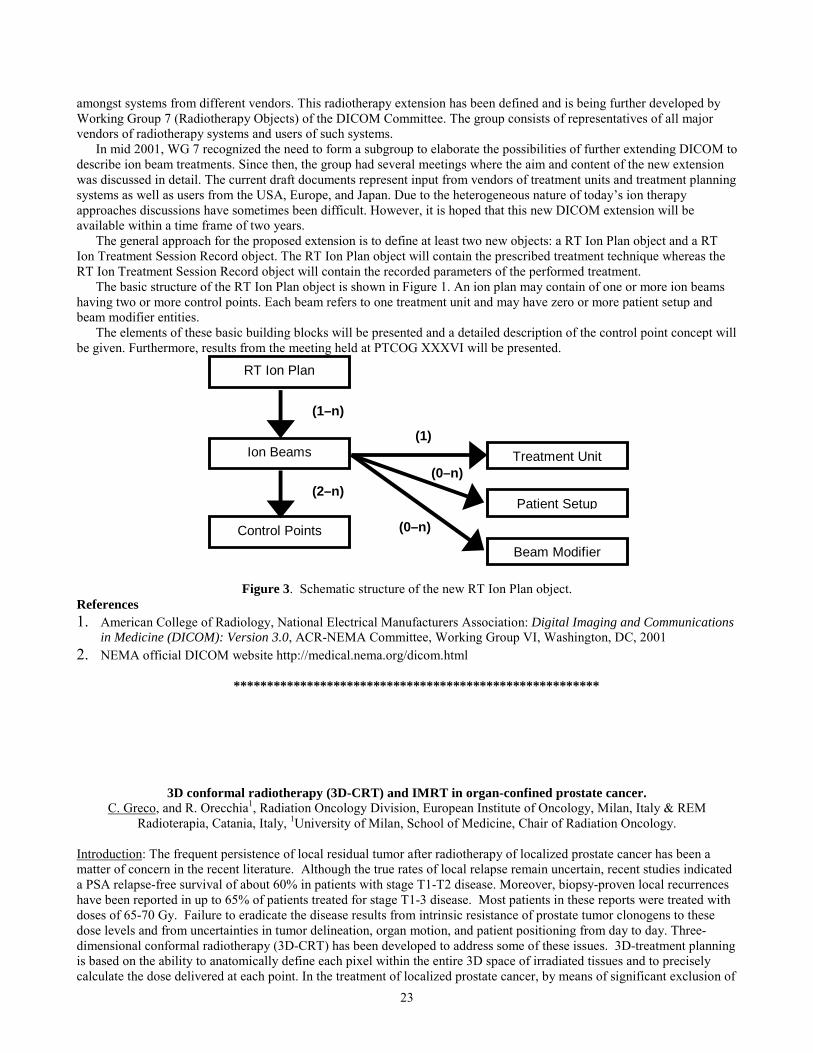
23
amongst systems from different vendors. This radiotherapy extension has been defined and is being further developed byWorking Group 7 (Radiotherapy Objects) of the DICOM Committee. The group consists of representatives of all majorvendors of radiotherapy systems and users of such systems.
In mid 2001, WG 7 recognized the need to form a subgroup to elaborate the possibilities of further extending DICOM todescribe ion beam treatments. Since then, the group had several meetings where the aim and content of the new extensionwas discussed in detail. The current draft documents represent input from vendors of treatment units and treatment planningsystems as well as users from the USA, Europe, and Japan. Due to the heterogeneous nature of today�s ion therapyapproaches discussions have sometimes been difficult. However, it is hoped that this new DICOM extension will beavailable within a time frame of two years.
The general approach for the proposed extension is to define at least two new objects: a RT Ion Plan object and a RTIon Treatment Session Record object. The RT Ion Plan object will contain the prescribed treatment technique whereas theRT Ion Treatment Session Record object will contain the recorded parameters of the performed treatment.
The basic structure of the RT Ion Plan object is shown in Figure 1. An ion plan may contain of one or more ion beamshaving two or more control points. Each beam refers to one treatment unit and may have zero or more patient setup andbeam modifier entities.
The elements of these basic building blocks will be presented and a detailed description of the control point concept willbe given. Furthermore, results from the meeting held at PTCOG XXXVI will be presented.
RT Ion Plan
Ion Beams
Control Points
(1–n)
(2–n)
Treatment Unit
Patient Setup
Beam Modifier
(1)
(0–n)
(0–n)
Figure 3. Schematic structure of the new RT Ion Plan object.References1. American College of Radiology, National Electrical Manufacturers Association: Digital Imaging and Communications
in Medicine (DICOM): Version 3.0, ACR-NEMA Committee, Working Group VI, Washington, DC, 20012. NEMA official DICOM website http://medical.nema.org/dicom.html
*******************************************************
3D conformal radiotherapy (3D-CRT) and IMRT in organ-confined prostate cancer.C. Greco, and R. Orecchia1, Radiation Oncology Division, European Institute of Oncology, Milan, Italy & REM
Radioterapia, Catania, Italy, 1University of Milan, School of Medicine, Chair of Radiation Oncology.
Introduction: The frequent persistence of local residual tumor after radiotherapy of localized prostate cancer has been amatter of concern in the recent literature. Although the true rates of local relapse remain uncertain, recent studies indicateda PSA relapse-free survival of about 60% in patients with stage T1-T2 disease. Moreover, biopsy-proven local recurrenceshave been reported in up to 65% of patients treated for stage T1-3 disease. Most patients in these reports were treated withdoses of 65-70 Gy. Failure to eradicate the disease results from intrinsic resistance of prostate tumor clonogens to thesedose levels and from uncertainties in tumor delineation, organ motion, and patient positioning from day to day. Three-dimensional conformal radiotherapy (3D-CRT) has been developed to address some of these issues. 3D-treatment planningis based on the ability to anatomically define each pixel within the entire 3D space of irradiated tissues and to preciselycalculate the dose delivered at each point. In the treatment of localized prostate cancer, by means of significant exclusion of

24
adjacent normal tissues from the high-dose region, 3D-CRT has been shown to reduce the risk of rectal and bladdertoxicities. Moreover, 3D-CRT can be effectively used to achieve tumor dose escalation with a significant improvement inlocal tumor control.Materials and Methods: Since 1995 we have treated over 700 patients with biopsy-proven prostatic adenocarcinoma withhigh dose 3D-CRT. All patients were treated in a prone position, with rectum and bladder empty, with a 6-field technique.To ensure reproducible positioning, a custom-made polyurethane-foam half-body cast was produced for each patient. Atotal dose of 76 Gy in 2 Gy fractions prescribed at ICRU point was administered. Recently, we have acquired dedicatedmicro-multileaf collimator at both sites and we have therefore gradually abandoned the static approach in favor of aconformal dynamic arc treatment delivery. This mode of delivery has both shown a higher degree of conformality as wellas ha better patient throughput. We are currently fine-tuning all quality-assurance procedures required for IMRT deliverywhich we expect to launch in the next few months.Results: Acute toxicity was graded according to the RTOG scoring system. Acute gastro-intestinal (GI) toxicity was asfollows: 34% G0, 48% G1, 18% G2 and no G3 and G4. Acute genito-urinary (GU) toxicity was as follows: 16% G0, 52%G1, 28% G2, 4% G3 and no G4. It is noteworthy that no interruption of treatment due to acute reactions was recorded. Latetoxicity was recorded according to the RTOG scale. Late GI toxicity was as follows: 18% G1, 5% G2, 2% G3 and 1% G4.Late GU toxicity was as follows: 22% G1, 3% G2 and 1% G3. Overall late treatment toxicity greater than or equal to G3was 3%. No definitive biochemical disease-free survival data can yet be drawn from our series.Conclusions: Similarly to the experience of several other Institutions we may conclude that conventional 3D-CRT oflocalized prostate cancer has an excellent acute toxicity profile and a relatively low late toxicity rate at the dose level wehave employed so far. While our biochemical disease-free survival data are not yet fully mature for evaluation, we are wellaware of the fact that some recent reports indicate that higher doses may be required to achieve better local control andconsequently improved biochemical relapse-free survival in localized prostate cancer, especially in high-risk patients. Tothis end we have embarked upon the challenging project of dose escalation with IMRT.
*******************************************************
Conformal proton beam therapy of prostate cancer-report of long-term PSA-based outcomes in over twelvehundred patients
C. J. Rossi JR., J. D. Slater and J. M. Slater, Loma Linda University Medical Center, Loma Linda, CA
Purpose: Conformal proton beam radiotherapy of prostate cancer has been performed at LLUMC since October 1991 Thefollowing is an update of our previously reported results.Materials and Methods: Between 10/91 and 12/97, 1255 patients with localized prostate cancer (defined as stages T1c-T3,PSA < 50 ng/ml) were treated with conformal proton beam radiotherapy to doses of 74-75 CGE (Cobalt-Gray Equivalent)at 1.8-2.0 CGE per fraction. None of these patients underwent prior surgery or received adjuvant hormonal therapy. Patientswere assessed at regular intervals during treatment for tolerance to therapy. Follow-up consisted of serum PSAdeterminations and digital rectal exams every six months until five years post treatment, than annually, with additionaltesting being performed as appropriate. Biochemical failure was defined as per the ASTRO criteria.Results: The median follow up is fifty-seven months (range 1-115 months). Overall seven year actuarial BiochemicalDisease-Free Survival (BNED) was 66%. BNED survival was strongly influenced by pre-treatment PSA levels (94 vs. 87vs 73 vs 55% for pre-treatment PSA's of < 4.1 ng/ml, 4.1-10.0 ng/ml, 10.1-20.0 ng/ml, 20.1-50 ng/ml, p<0.0001). PSAnadir also correlated with BNED survival (91 vs 76 vs 35% for nadirs of < .051 ng/ml, 0.51-10 ng/ml, > 10 ng/ml,p<0.0001). Treatment was extremely well tolerated with a 0.5% incidence of RTOG > Grade 3 late complications. BNEDsurvival rates compare favorably with other modalities (surgery, 3-D CRT), while treatment-related morbidity is less thanthat which has been reported in large conformal x-ray therapy seriesConclusions: Long-term follow up continues to demonstrate the safety and efficacy of conformal proton beam radiotherapyof prostate cancer.
*******************************************************
Physical and clinical aspects of prostate IMRT with photons.J. Bohsung, D. Böhmer, A. Moys, S. Marnitz, and V. Budach, Charité Berlin, Department of Radiotherapy, Schumannstr.
20/21, D-10117 Berlin
25
Introduction: Intensity modulated radiotherapy with high energy photon fields (IMRTX) can solve two problems ofconventional 3D conformal radiotherapy (3DCRT): (1) Conformal treatments of concavely shaped targets can beperformed. (2) It is possible to plan and treat concomitant boosts in an easy and elegant way.Today�s IMRTX systems typically consist of three parts: (1) an optimization system to calculate the photon field fluencies(inverse planning system), (2) a multileaf collimator (MLC) which is able to create modulated fields either statically (�stepand shoot�) or dynamically (�sliding window�) and (3) a MLC sequencer, which calculates the MLC steps and/ormovements for the intensity modulation.During the last years IMRTX was established as a routine option in a few hospitals, it is nevertheless still a new and notwidely used method. One problem is that the complex delivery technique requires new and time consuming QA procedures.IMRTX of prostate cancer allows the safe application of high doses possibly resulting in an increased relapse free survivalwithout increasing acute toxicity and late morbidity. IMRTX has firstly been introduced for clinical use in 1996 at theMSKCC in New York.Materials and methods: We are using the Varian IMRT system, which consists of the Cadplan/Helios inverse planningsystem with integrated MLC sequencer and a dynamic MLC.A quality assurance system was established which take into account both machine and patient related aspects includingsystematic dosimetric checks and daily patient set-up verification.Treatment planning was done according to the so-called SMART technique (Simultaneous Modulated AcceleratedRadioTherapy). This concomitant boost technique offers two advantages. Firstly, it allows an increased dose within aspecific target region (e. g. prostate) and a reduced dose to the peripheral region (e. g. periprostatic area). Secondly, it keepsthe overall treatment time constant while increasing the target dose. In our study the CTV was irradiated with a single doseof 2.0 Gy per fraction whereas the PTV was irradiated with 1.8 Gy simultaneously. With the SMART technique we wereable to increase the therapeutic dose significantly from 73.8 Gy to 82.0 Gy without increasing the doses to the organs atrisk, especially the rectum.Results: Treatment was completed safely and without interruption in all 48 IMRTX patients treated so far. Dosimetric pre-treatment results performed for all patients were always very precise. Results of the daily set-up verification revealed a verygood patient positioning. Acute radiation side effects were scored according to the Common Toxicity Criteria (Version2.0). All critical acute side effects showed no significant differences compared to conventionally treated patients. No lateradiation toxicity has been observed in our IMRTX patients so far.
*******************************************************
Real time tumor tracking (TULOC): first clinical measurements and plans for applications in prostate irradiation.H. Blattmann, R. K. Muench, P. G. Seiler, A. Sumova, J. Verwey, B. Kaser-Hotz1 and C. Rohrer Bley1, Department of lifesciences, Paul Scherrer Institut CH-5232 Villigen PSI Switzerland, 1Department for small animals, Vet. Hosp. University
Zurich SwitzerlandE-mail: [email protected], [email protected]
As a contribution to the solution of the organ motion problem we have developed a novel magnetic tracking methodwhich we call TULOC (acronym for TUmor LOCation). The demonstration of its proper functioning in the lab has beendescribed in previous PTCOG-meetings.
In this paper we report on the implementation of TULOC into the clinical environment. A first clinical measurementwas accomplished at the LINAC of the University of Zurich Veterinary school. During the irradiation of a dog patient themovements of a chondrosarcoma in the lumbosacral area of the spine were measured. Effects of eddy currents in theLINAC nozzle and in the patient table have been corrected for using special computational methods. The next step will bethe gating of the LINAC during dog treatments.
As a first application in human patients, the real time tracking of the position of the rectal wall and with it of theprostate during conformal radiotherapy is in preparation. For this, rectal balloon catheters equipped with TULOC sensorswill be used.
*******************************************************
Patient positioning for proton therapy of prostate tumors: First experience and outlook to the future.

26
E. Egger, A. Lomax and Team Radiation Medicine, Division of Radiation Medicine, Paul Scherrer Institute, CH � 5232Villigen-PSI, Switzerland
E-mail: [email protected]
At PSI, patients undergoing proton therapy at the spot scanning gantry are positioned outside the treatment room.Position verification is done on a remote CT by comparison of several CT slices with the slices of planning CT andmeasurement of several points on scouts view. Patients are brought inside the treatment room in their mould after positionverification and necessary corrections have been completed.
For prostate tumors, patients are positioned in an individual mould. A rectal balloon is used to immobilize the prostate.This can be a time consuming procedure to obtain high precision positioning. We will analyze the positioning precisionobtained with this system. We will also discuss the time necessary for precise positioning of the patient, the personalrequired for an optimal use of the irradiation facility and raise the question whether in a new facility, patient positioningshould also be done outside the treatment room or preferably inside the treatment room.
*******************************************************
A new superconducting cyclotron to produce 250 MeV light ion beams.L. Calabretta, M. Maggiore, and D. Rifuggiato, Laboratori Nazionali del Sud � I.N.F.N., Via S. Sofia 44, 95123 Catania,
Italy
A four sector compact superconducting cyclotron able to deliver light ion beams with a maximum energy of 250 AMeV has been studied. This cyclotron is mainly designed to accelerate H2
+ ions to be extracted by stripping. Moreover ionslike C, O or Ne can also be accelerated and extracted by stripping. Thanks to the extraction by stripping, two simultaneousbeams can be extracted, allowing to performs therapy treatment, interdisciplinary research and radioisotope productionusing low or medium intensity beam. The use of the extraction by stripping simplify greatly the maintenance of thecyclotron and reduce its activation. The preliminary design model of the magnet circuit has been accomplished with the3D electromagnetic code OPERA. The design of the main coils and of the cryostat has been investigated too. Theisochronous magnetic field, the flutter, and the focusing properties of the cyclotron for different ions will be presented.
*******************************************************
Status of development of installation for proton conformal therapy mass application.V. Balakin, Branch of Institute of Nuclear Physics, Post Box 113/1, Protvino, Moscow region, 142281, Russia.
E-mail: [email protected]
The proton therapy will become mass, if the proton installation will have cost of the equipment, size of the building,consumption of electric power and cost of operation comparable with the same cost for electron accelerator.
In paper the proton installation is presented, which completely, together with a treatment room, is placed in the radiationprotected hall with the sizes 7х13 sq.m, required about 50 KW of the electric power in therapy mode.
Energy of the proton accelerator reaches 330 MeV, that in the future will allow to have on the same installation protontomography. The experimental sample of the accelerator works, the first full sample of installation with the equipment fortherapy is planned to commissioning in 2002, and in 2003 to put two serial samples in clinics of Russia.
*******************************************************
Successful acceleration test of the first module of the proton linac booster “LIBO”.

27
P. Berra, E. Rosso, B. Szeless, M. Vretenar, U. Amaldi1, K. R. Crandall1, M. Mauri1, D. Toet1, M. Weiss1, R. Zennaro1, C.Cicardi2, C. De Martinis2, D. Giove3, L. Grilli2, D. Davino4, M. R. Masullo5, V. G. Vaccaro4, L.Calabretta6 and A. Rovelli6,
CERN, Geneva, Switzerland, 1TERA Foundation, Novara, Italy, 2University and 3INFN Milan, Italy, 4University and5INFN Naples, Italy, 6INFN � LNS Catania, Italy
The linac booster �LIBO� project aims at building a 3 GHz proton linear accelerator to boost the energy up to 200 MeVof the beam from 30 to 70 MeV cyclotrons, which exist in several laboratories and hospitals. A prototype of the first LIBOmodule was designed, constructed and tested by a collaboration formed by CERN, University and INFN of Milan,University and INFN of Naples, and the TERA Foundation.
Low power RF measurements have shown a good field uniformity and stability along the axis of the four tankscomposing the LIBO module. Full power RF measurements at a repetition rate of 100 Hz have been performed at CERN.After a very short conditioning period, an accelerating gradient approaching 30 MV/m has been easily achieved in thetanks, well above the nominal value of 15 MV/m.
In February 2002 in the LNS of INFN in Catania the module was powered with a 3.6 MW RF modulator produced byIBA/Scanditronix. As expected, protons have been accelerated over about 1 meter from 62 MeV to 73 MeV. Theparticularities of the design and the reasons for the successful performance are discussed.
*******************************************************
Status of TOP Linac Costruction.S. Frullani, L. Picardi1 and C. Ronsivalle1, Istituto Superiore di Sanità, Viale Regina Elena, 299, 00161 Roma, ITALY,
1ENEA � FIS-ACC � Via E. Fermi 45 00044 Frascati (Roma), ITALYE-mail: [email protected], [email protected]
The TOP LINAC (short for Terapia Oncologica con Protoni LINear ACcelerator), an innnovative linac forprotontherapy in the energy range 65 - 200 MeV is being constructed in the ENEA Frascati Research Centre by acollaboration between ISS and ENEA. The system is intended to be installed in the main Oncologycal Hospital in Rome,Istituto Regina Elena. Up to now funding allowed the construction of a 7 MeV injector and of the first 3 GHz module (up to13.4 MeV). The injector has been produced by AccSys Company (USA) and is under commissioning at Frascati. The lowenergy (7 MeV) beam lines for F-18 production and injection in the following accelerating sections are under construction.The first 3 GHz SCDTL module has been completely built. The characteristics of the various accelerator components arepresented.
*******************************************************
Recent Improvements on the Proteus 230 for a Flawless System Operation.G. Gevers, Ion Beam Applications s.a., Chemin du Cyclotron, 3, 1348 Louvain-la-Neuve, Belgium
E-mail: [email protected]
The recent developments accomplished on the Proton Therapy System that is installed and currently operational at theNortheast Proton Therapy Center at the Massachusetts general Hospital in Boston will be presented at the Catania PTCOGconference of May 2002. These developments reflect IBA�s wish and also ability to constantly adapt its products to itsclient�s needs, whilst using their experience to this purpose and integrating their suggestions. The new snout family designshall be presented along with the newly designed snout removal system. Examples of the new features that have beenintegrated into our control system will also be given, such as those that will improve the information readability and alsothe operation flexibility.
As system availability is a key factor of client and patient satisfaction, a process for the putting into place of acontinuous tuning preventive maintenance program and a system fault diagnostic is being reviewed.
*******************************************************
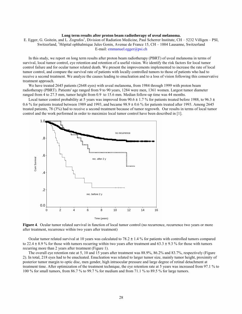
28
Long term results after proton beam radiotherapy of uveal melanoma.E. Egger, G. Goitein, and L. Zografos1, Division of Radiation Medicine, Paul Scherrer Institute, CH � 5232 Villigen � PSI,
Switzerland, 1Hôpital ophhalmique Jules Gonin, Avenue de France 15, CH � 1004 Lausanne, SwitzerlandE-mail: [email protected]
In this study, we report on long term results after proton beam radiotherapy (PBRT) of uveal melanoma in terms ofsurvival, local tumor control, eye retention and retention of a useful vision. We identify the risk factors for local tumorcontrol failure and for ocular tumor related death. We present the improvements implemented to increase the rate of localtumor control, and compare the survival rate of patients with locally controlled tumors to those of patients who had toreceive a second treatment. We analyze the causes leading to enucleation and to a loss of vision following this conservativetreatment approach.
We have treated 2645 patients (2648 eyes) with uveal melanoma, from 1984 through 1999 with proton beamradiotherapy (PBRT). Patients' age ranged from 9 to 90 years, 1284 were men, 1361 women. Largest tumor diameterranged from 4 to 27.5 mm, tumor height from 0.9 to 15.6 mm. Median follow-up time was 44 months.
Local tumor control probability at 5 years was improved from 90.6 ± 1.7 % for patients treated before 1988, to 96.3 ±0.6 % for patients treated between 1989 and 1993, and became 98.9 ± 0.6 % for patients treated after 1993. Among 2645treated patients, 78 (3%) had to receive a second treatment because of tumor regrowth. Our results in terms of local tumorcontrol and the work performed in order to maximize local tumor control have been described in [1].
Time (years)
1614121086420
Cum
Sur
viva
l
1.0
.8
.6
.4
.2
0.0
no recurrence
rec. after 2 y
rec. before 2 y
Figure 4. Ocular tumor related survival in function of local tumor control (no recurrence, recurrence two years or moreafter treatment, recurrence within two years after treatment)
Ocular tumor related survival at 10 years was calculated to 78.2 ± 1.4 % for patients with controlled tumors comparedto 22.4 ± 8.9 % for those with tumors recurring within two years after treatment and 63.3 ± 9.3 % for those with tumorsrecurring more than 2 years after treatment (Figure 1).
The overall eye retention rate at 5, 10 and 15 years after treatment was 88.9%, 86.2% and 83.7%, respectively (Figure2). In total, 218 eyes had to be enucleated. Enucleation was related to larger tumor size, mainly tumor height, proximity ofposterior tumor margin to optic disc, men gender, high intraocular pressure and large degree of retinal detachment attreatment time. After optimization of the treatment technique, the eye retention rate at 5 years was increased from 97.1 % to100 % for small tumors, from 86.7 % to 99.7 % for medium and from 71.1 % to 89.5 % for large tumors.

29
Time [years]
1614121086420
Cum
Eye
Ret
entio
n
1,0
,8
,6
,4
,2
,0
1
2
3
Figure 2. Eye retention rate in function of tumor height (1: 401 eyes with a height smaller or equal to 3.0 mm, 7 of themhad to be enucleated, 2: 1570 eyes with a height ranging from 3 to 8 mm, of which 94 were enucleated, 3: 677 eyes with aheight larger than 8 mm, of which 117 were enucleated)
Larger tumor height, proximity of tumor to optic disc and macula and large amount of retinal detachment at baselinewere the parameters identified to lead to poor functional outcome. Ten years after treatment 35 % of the patients with atumor height smaller or equal 8 mm and 12 % of patients with a tumor height larger than 8 mm still had a useful vision(Figure 3).
Conclusion: Reduced safety margins, large ciliary body tumors, eyelids within the treatment field, inadequatepositioning of tantalum clips, and male gender were identified to be the main parameters to impair local tumor control. Theimprovement of local tumor control rate after 1993 is attributed to changes implemented in the treatment procedure. Ourdata strongly support that the rate of death by metastases is influenced by local tumor control failure: improvement of localtumor control rate allowed for better survival rate. The treatment technique as used today results in excellent eye retentionrates, even in less favorable cases like large tumors and tumors located close to the optic disc. The continuous qualitycontrol program allowed to increase the 5-year eye retention rate for all tumor sizes. These findings demonstrate thepositive impact of experience and quality control based efforts for treatment technique optimization. We conclude thatfunctional results are strongly influenced by tumor size and location. Even for less favorable cases it is possible to save auseful visual acuity to a small number of patients. Nevertheless, in most of the cases, proton therapy is the only possibleconservative treatment approach.
Time [months]
168144120967248240
Cum
rete
ntio
n of
use
ful v
isio
n
1.0
.8
.6
.4
.2
0.0

30
Figure 3. Retention of a useful vision in function of tumor height (1: eyes with a height smaller or equal to 8.0 mm, 2:eyes with a height larger than 8 mm)ReferencesE Egger, A Schalenbourg, L Zografos, L Bercher, T Boehringer, L Chamot, G Goitein, Maximizing local tumor control andsurvival after proton beam radiotherapy of uveal melanoma. Int. J. Radiat. Oncol. Biol. Phys. 51(1), 2001, 138-47
*******************************************************
Ten years of technical evolutions in the preparation of ocular diseases for protontherapy.P. Chauvel, N. Iborra, J. Herault, A.Courdi, C. Mosci1, S. Squarcia2, and W. Sauerwein3, Centre Antoine-Lacassagne,
Cyclotron Médical, 227 avenue de la Lanterne, F 06200 Nice, 1Divisione Oculistica OspedaleCelesia ASL3, Genova, Italy,2Dipartimento di Fisica Università degli studi di Genova, Italy, 3Strahlenklinik, Universitatsklinikum Essen, Germany
E-mail: [email protected]
Since june 1991, a total number of 2040 patients have been treated in Nice using the 65 MeV proton beam. The Eyeplanprogram was used for all patients. Along these years, we decided to modify some classical approaches in order to be moreand more accurate and sure to deliver the dose where it was planned. We first decided to develop the use of bolus in orderto flatten the entrance of the beam and therefore be certain that the distal shape is reliable, mainly when we have to use awedge filter. Bolus are now made of ultrasonographic gel and a thin sheet of plexiglas or transparency. The measurement ofthe thickness of the bolus has been facilitated by a special apparatus developped in Nice and used in routine. The wedgefilters design was also modified to be easy to produce and use. These developments will be presented.
But the main problems is the eye modelization using the Eyeplan spherical representation. The inaccuracies of themodelization were first suspected on wrong clips-limbus measurements, elevated chi-squared values and wrong light fieldsprojections. Changes in eye length sometimes ameliorate the values as well as changes in initial fixation. But thesemodifications were empiric. Some CT scans were initially performed to check the axial length measured byultrasonography. But they also demonstrated obviously that the eye is often not at all comparable to a sphere and that theclips position may depend on the eye shape irregularities.
CT scans are now performed systematically for each patient after clips insertion and contoured slice by slice in astandard radiotherapy treatment planning. The set-up of virtual fields, anterior, lateral and cranio-podal allows acomparison with beam�s eye view, lateral view and overview issued from Eyeplan. The eye position during CT scan isestimated from anterior and lateral fields rescaled to the magnification of the X-rays of protontherapy simulation, using theEyeplan function "Determine a true position". This position is given to the model and by superimposing the clips system ofCT scan and model at the same scale in the three views, we have the possibility to evidence the discrepancies between thetwo systems. Some modifications may be rationally introduced on eye length, center of the eye, initial angle of fixation.Practical examples will be given demonstrating the problems encountered and the solutions given using the Eyeplanmodelization. The influence of a wrong modelization on dose distribution will also be adressed.
*******************************************************
Statistical analysis on melanoma treatment with proton beam in Nice.C. Mosci, S. Donadio, D. Grosso1, B. Mascialino1, M. A. Penco1, S. Squarcia1, P. Chauvel2, N. Iborra2, Divisione Oculistica
Ospedale Celesia ASL 3, Genova, Italy, 1Dipartimento di Fisica - Università degli Studi di Genova e Sezione INFN,Genova, Italy, 2Centre Antoine Lacassagne Cyclotron Biomedical, Nice, France
e-mail: [email protected]
The Genoa Ocular Oncology Group presents the statistical results of the first ten years of activity with proton beamtherapy for ocular melanoma treated in Nice Antoine Lacassagne Cyclotron Biomedical Centre. The sample is composed by187 patients (48.1% men and 51.9% women) coming from all Italy and treated from 1991 onwards. We consider 7 caseshaving a tumour stadiation T1, 52 cases T2 and 128 cases T3, with an overall mean age of (60.5 ± 0.4) years. The follow-upmean time is (27 ± 2) months, only a few patients being followed till 96 months. With this therapy we obtain a local tumourcontrol in 96.8% of cases and eye retention in 94.1%. We were able to calculate the tumour regression for the T1+T2sample as a whole:
Thickness = (3.25 ± 0.11) - (0.049 ± 0.008) · Time (months)and for the T3 sample alone:Thickness = (7.07 ± 0.18) - (0.118 ± 0.014) · Time (months).Eleven eyes were enucleated: five eyes in consequence of tumour relapse (45%) and six because of the appearance of
neovascular glaucoma (55%), this pathology being highly brought about by some protons� treatment plannings. In any case,
31
the most frequent complication was vasculopathy (62.1%), while the 25.9% of cases showed the presence of cataract andabout the 13.8% of neovascular glaucoma. Papillopathy appeared in 15.5% of the cases with an average time of appearanceof (25 ± 3) months for the sample T1+T2 and of (23 ± 3) months for T3.
Eleven patients (5.9%) died because of metastases� appearance. In all these patients the main metastatic localization wasthe liver. Only one patient was already suffering from metastases� diseases at the time of the uveal melanoma diagnosis. Inour sample, the global survival expectation for protontherapy, estimated by means of a Kaplan-Meier survivorship function,is equal to (0.81 ± 0.08) from 96 months on. The global survival rate after 42 months is calculated to be 91%.
This statistics, the only one available up to now in Italy, will be here onwards extended to the totality of the patientstreated by SERAG (South Europe Radiotherapy Group) by means of a specialized clinical folder. The preliminary results ofthis study allow comparisons among different centre strategies.
*******************************************************
Proton beam radiotherapy of iris melanoma: long term follow-up study in UK.B. Damato1, A Kacperek, M Sheen, and R. D Errington, 1St Paul�s Eye Unit, Royal Liverpool University Hospital, PrescotStreet, Liverpool L7 8XP UK, and the Douglas Cyclotron Unit, Clatterbridge Centre for Oncology, Bebington Wirral, CH
63 4JY, UK
INTRODUCTION: The standard form of treatment for iris melanoma is iridectomy, with iridocyclectomy being performedif the tumour extends to angle or ciliary body. Both procedures are technically straightforward but the surgical iris defecttends to cause photophobia, which requires further treatment.Proton beam radiotherapy has been used for the treatment of ciliary body and choroidal melanomas for several decades,with excellent rates of local tumour control. However, to our knowledge this modality had not previously been used for thetreatment of iris melanomas. In view of the limitations of surgical resection and plaque radiotherapy of iris melanomasthere was strong motivation for exploiting the physical advantages of protons in providing precise, superficial beams forirradiating melanomas of the iris. In 1994, the first patients were treated at the CCO, using the standard melanoma dose andfractionation (53 Gy in 4 daily fractions). At present, 95 iris patients have been treated. This presentation will describe theearly results (longest follow-up) of proton radiotherapy of a treated study group, showing outcomes in terms of visualacuity, ocular complications and symptoms. The present results have not been described in detail previously as radiation-induced complications usually take time to appear.METHOD: Proton treatment plans were prepared according information on a special form, which included: (1) the shapeand extent of the tumour; (2) the target area comprising the tumour and safety margins; (3) echographic measurements ofdistances from cornea to back of lens, cornea to retina, retina to outer sclera, and transverse ocular diameter. Tantalummarkers were not necessary. Iris treatment planning was performed on the EYEPLAN (1.6/3.xx) eye therapy program,which had the possibility of modelling the tumour base on the iris. Treatment was delivered with the pupil fully dilated toreduce the tumour area. The eyelids were fully retracted to improve positioning accuracy with field-light and to minimisecosmetic effects. The superficial proton beam depths (4.5 to 7 mm) required the use of a flat, thin walled Markus IC.CONCLUSION: Proton beam radiotherapy of iris melanoma has achieved excellent local tumour control with minimalcomplications for patients in the study group, with 5 to 8 years follow-up.
*******************************************************
Results of proton and plaque therapy of choroidal melanoma treated in Vancouver.E. W. Blackmore, K. Paton1, C. Duzenli2, W. Kwa2, R. Ma2, and T. Pickles2, TRIUMF, 4004 Wesbrook Mall, Vancouver,
BC, Canada V6TT 2A3, 1Dept. of Ophthalmology, University of British Columbia, Vancouver, BC, Canada V6K 2E12British Columbia Cancer Agency, 600 West 10th Avenue, Vancouver, BC, Canada V5Z 3N9
E-mail: [email protected]
Treatment of choroidal melanoma using 70-74 MeV protons from the TRIUMF cyclotron started in 1995 with 68patients treated by the end of 2001. Patients presently receive 45 Gy (reduced from 50 Gy in 1998) in four daily fractions.The physicians and medical physicists responsible for proton therapy also carry out 198Au plaque therapy for smaller oculartumors with very favourable results reported (ref). Patients are referred for proton therapy based on tumor size and/orlocation close to the optic nerve.
This paper will describe the beam line and proton treatment system, some aspects of dosimetry and treatment planning,statistics on the patients treated, and a preliminary report on treatment outcomes.
32
References: A. Karvat, C. Duzenli, R. Ma, K. Paton, and T. Pickles. The treatment of choroidal melanoma with 198Auplaque brachytherapy. Radiation and Oncology 59 (2001) pp. 153-156.
*******************************************************
Proton beam therapy for wet macular degeneration: the rationale for adding photodynamic therapy.L. T. Yonemoto, Loma Linda University Medical Center, 11234 Anderson St. B121, Loma Linda, CA, 92354, USA
E-mail: [email protected]
Proton Beam Therapy (PBT) is a safe and effective therapy for the wet type of age-related macular degeneration (wetAMD). Over 400 patients have been treated since 1995, with an anterior chamber sparing and lens sparing proton therapytechnique. The leaky blood vessel of the wet AMD has closed (stopped bleeding) at a rate of 90% using 14 GyE in a singlefraction. The response time for blood vessel closure was 3 to 6 months post proton therapy. The recurrence rate is lessthan 5 % at 1 year.
The bleeding of this leaky vessel exposes the macula to the caustic substances found in blood. This will eventuallycause permanent damage and visual decline. Thus, reducing the macula�s exposure time to blood should reduce thisdamage. There is concern that the bleeding during the response time of PBT might allow additional macular damage andvisual decline.
The FDA has approved Photodynamic Therapy (PDT) for the classic type of wet AMD. PDT consists of aphotosensitizer that has a higher uptake in rapidly proliferating cells, such as the leaky blood vessel of the wet AMD. Anon-thermal laser is utilized to cause cellular death of the blood vessel cells. A randomized study has demonstrated itsefficacy, but it has required re-treatment an average of 3.4 times in the first year and 2.1 times in the second year.
The hypothesis is that the combination of PBT and PDT would be synergistic by reducing the exposure time of themacula to blood. PBT has a low recurrence rate but has a 3 to 6 month response time. PDT has a fast response time buthigh recurrence rate. Using PDT to stop the bleeding during the response time of PBT should reduce the exposure time ofthe blood to the macula and hopefully improve visual outcome.
ReferencesYonemoto LT, Slater JD, Friedrichsen EJ, et al. Phase I/II Study of Proton Beam Irradiation for the Treatment of
Subfoveal Choroidal neovascularization in Age-Related Macular Degeneration: Treatment Techniques and PreliminaryResults. . Int. J. Radiation Oncology Biol. Phys. Vol. 36, No. 4 pp. 867-871, 1996.
Archambeau JO, Mao XW, Yonemoto LT et al. What is the Role of Radiation in the Treatment o SubfovealMembranes: Review of the Radiobiologic, Pathologic, and Other Considerations to Initiate a Multimodality Discussion. Int.J. Radiation Oncology Biol. Phys. Vol. 40, No. 5 pp. 1125-1136, 1998.
M.F. Moyers, R.A Galindo, L.T.Yonemoto, et al. Treatment of SNVM with Proton Beams. Presented at the 38th
annual meeting of the American Association of Physicist in Medicine, Med. Phys. 26 (5), 1999Yonemoto LT, Slater JD, Blacharski P, et al. Dose Response in the treatment of Subfoveal Choroidal
Neovascularization in Age-related Macular Degeneration: Result of a Phase I/II dose escalation study using conformalproton radiation therapy. J Radiosurg 3(2000) 47-54, 2000
Archambeau JO, Mao XW, McMillan Dose Response of Rat Retinal Microvessels to Proton Dose Schedules UsedClinically: A Pilot Study. Int. J. Radiation Oncology Biol. Phys. Vol. 48, No. 4 pp. 1155-1166, Nov.1, 2000.
Miller JW, Schmidt-Erfurth U, Sickenberg M, et al. Photodynamic therapy with verteporfin for choroidalneovascularization caused by age-related macular degeneration: results of a single treatment in a phase 1 and 2 study. ArchOphthalmol. 1999;117:1161-1173.
*******************************************************
Proton therapy of ocular tumors in Berlin. The experience of the first four years.N. E. Bechrakis1, M. Nausner2, H. Fuchs3, J. Heese3, H. Kluge3, W. Hinkelbein2, M. H. Foerster1, Augenklinik1 und Klinikfür Strahlentherapie und Radioonkologie2, Universitätsklinikum Benjamin Franklin, Freie Universität Berlin. Hahn Meitner
Institut, Berlin3
From June 1998 226 pts. have been treated with protons at the Hahn Meitner Institute in cooperation with the UniversityHospital Benjamin Franklin in Berlin.
This includes 182 uveal melanomas, 14 iris-melanomas, 28 hemangiomas of the choroidea and the retina and 2 patientswith other indications. The most cases of uveal melanomas have been treated with the standard fractionation scheme, four
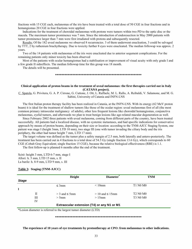
33
fractions with 15 CGE each, melanomas of the iris have been treated with a total dose of 50 CGE in four fractions and inhemangiomas 20 CGE in four fractions were applied.
Indications for the treatment of choroidal melanomas with protons were tumors within two PD to the optic disc or themacula. The maximum tumor prominence was 7 mm. Since the introduction of endoresection in May 2000 patients withtumor prominence larger than 7 mm were first irradiated with protons and subsequently resected.
Results: Of the 182 uveal melanoms we observed 8 recurrencies. 3 of them underwent enucleation, 3 could be salvagedby TTT, 2 by ruthenium brachytherapy. Due to toxicity further 8 eyes were enucleated. The median followup was approx. 2years.
Two of the 14 patients with melanomas of the iris were enucleated due to anterior sequment complications. For theremaining patients only minor toxicity has been observed
Most of the patients with ocular hemangiomas had a stabilisation or improvement of visual acuity with only grade I anda few grade II sideeffects. The median followup time for this group was 18 month.
The details will be presented.
*******************************************************
Clinical application of proton beams in the treatment of uveal melanomas: the first therapies carried out in Italy(CATANA project).
C. Spatola, G. Privitera, G. A. P. Cirrone, G. Cuttone, J. Ott, L. Raffaele, M. L. Rallo, A. Reibaldi, V. Salamone, and M. G.Sabini, University of Catania and INFN-LNS
The first Italian proton therapy facility has been realized in Catania, at the INFN-LNS. With its energy (62 MeV protonbeam) it is ideal for the treatment of shallow tumors like those of the ocular region: uveal melanomas first of all (the mostcommon primary intraocular malignancy of adults), other less frequent lesions like choroidal hemangiomas, conjunctivamelanomas, eyelid tumors, and afterwards we plan to treat benign lesions like age-related macular degeneration as well.
Since February 2002 three patients with uveal melanoma, coming from different parts of the country, have been treatedsuccessfully. All patients had a localized disease, with no systemic metastases, and had specific indications for conservativeapproach by means of proton beams, depending on their size or location: according to the TNM-AJCC Staging System, onepatient was stage I (height 3mm, LTD 10 mm), two stage III (one with tumor invading the ciliary body and the irisperiphery, the other had tumor height 7 mm, LTD 17 mm).
The target volume was defined as the tumor plus a safety margin of 2.5 mm, both laterally and antero-posteriorly. Thetreatment has been carried out in 4 fractions to a total dose of 54.5 Gy (single fraction 13.6 Gy), which corresponds to 60CGE (Cobalt Gray Equivalent; single fraction 15 CGE), because the relative biological effectiveness (RBE) is 1.1.
The first follow-up is planned 6 months after the end of the treatment.
Scirè: height 3 mm, LTD 6-7 mm, stage IAllori: h. 5 mm, LTD 15 mm, s. IILa barile: h. 8-9 mm, LTD 9 mm, s. III
Table 3: Staging (TNM-AJCC)
StageHeight Diameter* TNM
I≤ 3mm < 10mm T1 N0 M0
II > 3 and ≤ 5mm > 10 and ≤ 15mm T2 N0 M0III > 5mm > 15mm T3 N0 M0IV
Extraocular extension (T4) or any N1 or M1*Lesion diameter is referred to the largest tumor diameter (LTD)
*******************************************************
The experience of 10 years of eye treatments by protontherapy at CPO: from melanomas to other indications.
34
C. Desblancs, Centre de Protonthérapie d�Orsay, B.P. 65, 91 402 ORSAY Cédex, FranceE-mail: [email protected]
Since 1991, 2000 patients have been treated at CPO for ocular diseases.Based on a well established procedure set up for melanomas, new indications have been treated: iris melanomas (1,4 %
patients treated) and haemangiomas (5.7% patients treated).Clinical requirements (dose, margins) and technical aspects (patient positioning, proton beam line, treatment planning
system) adapted to each case will be presented.The results (1) of a 1062 patients study between 1991 and 1998 with a median follow-up of 38 months are presented :
local control was obtained for 97.1 % of patients, the survival rate was 92 % at 2 years and 78 % at 5 years.A correlation between the different steps of the preparation and treatment and the low rate of local relapse (2.9 %) will
be discussed.References1. L. LUMBROSO et al : Résultats du traitement du mélanome de la choroïde par faisceau de protons. J. Fr. Ophtalmol,2002 ; 25,3,3,290-297 � Masson, Paris, 2002
*******************************************************
The Italian experimental radiobiological research related to hadrontherapy.P. Scampoli, Dipartimento di Scienze Fisiche, Università di Napoli Federico II, Complesso Universitario di Monte S.
Angelo, 80125 Napoli, ItalyE-mail: [email protected]
In the present review, which of course cannot be exhaustive, I�ll focus principally on the Italian activities related tohadrontherapy carried out in the framework of Istituto Nazionale di Fisica Nucleare (INFN).
The radio-biological section of the ATER experiment of INFN involved research groups from the University of Milano,the Laboratori Nazionali di Legnaro (LNL) of INFN, the Istituto Superiore di Sanità (ISS), and the Università di NapoliFederico II. It was devoted to the study of the cell inactivation frequency in human cells, normal and tumoural, withdifferent radio-sensitivity to photon irradiation, following low energy proton irradiation. The use of monoenergetic protonscould in fact provide useful information on the energy dependence of the biological effectiveness especially at lowenergies. Results showed that the relative biological effectiveness (RBE) for inactivation with protons of high linear energytransfer (LET) increased with the cellular radioresistance to γ-rays. The cell line with the greatest resistance to γ-rays wasthe most responsive to the highest LET protons [1].
This study was then continued with the same proton beams and biological samples with the realisation of split-doseexperiments, aiming at determining the relationship between the cell recovery from radiation induced damage and theradiation quality [2]. A higher maximum recovery was observed for radiosensitive cell lines in comparison to radioresistantcells, and the recovery potential after split doses was smaller for slow protons in comparison to low-LET radiation.
All proton irradiations were performed at the 7 MV CN accelerator at the LNL, and at the 3 MV TTT-3 Tandemaccelerator at the Università di Napoli Federico II. The incident LET on cells was between 7 and 33 keV/µm.
An analogous research was then extended to carbon ions, as carbon ion radiotherapy was started in Chiba (Japan) andDarmstadt (Germany). A systematic study of RBE dependence on the LET of carbons was accomplished which could giveuseful information for a realistic evaluation of the effective biological dose. RBE values for cell inactivation were foundhigher then RBE corresponding to proton beams and preliminary results showed that radioresistant tumour to γ-rays couldbe better treated with high LET particles.
The irradiation facilities used for carbon radiobiology were the Tandem-XTU accelerator at LNL, which deliveredcarbon beam with energies of 6 MeV/amu and 8 MeV/amu, and the Tandem-ALPI facility, always at LNL, to achieve theenergy of 20 MeV/amu. Successively these experiments took advantage from the superconducting cyclotron of theLaboratori Nazionali del Sud (LNS) of INFN, where carbon ions were accelerated up to 62 MeV/amu. Finally, also thetherapeutic beams were used for irradiations at the HIMAC facility in Chiba to understand the effects of the secondary ionscoming from the projectile and/or target fragmentation.
The studies on cellular clonogenity were enriched by the different research groups investigating other biological end-points, such as DNA fragmentation [3], and chromosome aberrations [4] to assess useful parameters for predictive test inradiotherapy treatment.References1. M. Belli, et al. Inactivation of human normal and tumour cell irradiated with low energy protons. In Int. J. Radiat.
Biol., 76 (2000) 831- 839.
35
2. F. Antonelli et al. Inactivation of human cells exposed to fractionated doses of low energy protons: relationshipbetween cell sensitivity and recovery efficiency. In J. Radiat: Res. 42 (2001) 347 � 359.
3. M. Belli, et al., DNA DSB induction and rejoining in V79 cells irradiated with light ions: a constant field gelelectrophoresis study. In Int. J. Radiat. Biol., 76 (2000) 1095- 1104.
4. V. d�Alesio, et al., Biological dosimetry by interphase chromosome painting in breast cancer patients treated withradiotherapy . In Proceedings of 31st Annual Meeting of the European Society for Radiation Biology, Dresden,September 1-5, 2001, 184
*******************************************************
Potential reduction of the incidence of radiation-induced second cancers by using proton beams in the treatment ofpediatric tumors.
R. Miralbell, L. Cella, A. Lomax1, and U. Schneider2, University Hospital, 1211 Geneva 14, Switzerland, 1Paul ScherrerInstitut, 5232 Villigen-PSI, Switzerland, 2Triemli Hospital, 8063 Zurich, Switzerland
E-mail: [email protected]: [email protected], [email protected]
We aimed to assess the potential influence of improved dose distribution with proton beams compared to conventionalor intensity modulated (IM) X-ray beams on the incidence of treatment-induced secondary cancers in pediatric oncology.
Two children, one with a parameningeal rhabdomyosarcoma (RMS) and a second with a medulloblastoma (MDB) wereused as models for the purpose of this study. After defining the target and critical structures, treatment plans werecalculated and optimized, four for the RMS case (conventional X-ray, IM X-rays, protons, and IM protons), and three forthe irradiation of the spinal axis in MDB (conventional X-ray, IM X-rays, protons). Secondary cancer incidence wasestimated using a model based on Publication #60 of the International Commission on Radiological Protection. This modelallowed computation of absolute life-time risks of secondary cancer for each treatment plan, based on dose-volumedistributions for the non-target organs.
Proton beams reduced the expected incidence of radiation-induced secondary cancers for the RMS patient by a factor of2 and 2.4 and for the MDB case by a factor of 1.7 and 15 when compared with either IM X-ray or conventional treatmentplans, respectively.
The potential for a significant reduction in secondary cancers in pediatric cancers after using proton beams (forwardplanned or IM) in the treatment of RMS and MBD in children and adolescents represents an additional argument supportingthe development of proton therapy for most radiotherapy indications in pediatric oncology.
*******************************************************Present status of the proton therapy system in PMRC.
M. Umezawa and collaboration with PMRC1 and KEK Accelerator Lab.2, Hitachi,Ltd. 7-2-1 Omika-cho, Hitachi-shi,Ibaraki-ken, 319-1221 Japan, 1PMRC (Proton Medical Research Center), 1-1-1 Tennoh-dai, Tsukuba-shi, Ibaraki-ken 305-8575, Japan, 2KEK(High Energy Accelerator Research Organization) Accelerator Lab., 1-1 Oho, Tsukuba-shi, Ibaraki-ken,
305-0801, Japan E-mail:[email protected]
We had constructed �PROBEAT-Tsukuba�, that is, the proton therapy system in Proton Medical Research Center,University of Tsukuba. The present system employs two treatment rooms with rotating gantries and a synchrotron with amaximum energy of 250MeV. Clinical trial to generate data for Governments' approval had started in September 2001, andended before previous PTCOG Meeting held at Tsukuba, November 2001.
Before starting the above clinical trial, we had confirmed several characteristics and functions needed in dailytreatments, one of which was to provide the highly stable and reproducible proton beam with simple operation. The beamposition measured at the entrance of the irradiation nozzle was kept in 0.5mm without feedback control. This characteristicwas realized by Hitachi�s RF driven slow extraction scheme and stable power supplies, magnet and so on. Another was therespiratory synchronized operation of synchrotron, which was essential for proton irradiation to the tumor moving with therespiration. High irradiation efficiency was realized by the variable repetition period of our slow-cycling synchrotron,which was synchronized to the patient�s respiration signal.
After the previous PTCOG meeting, the present system has been improved significantly. First of all, the intensity ofprotons extracted from synchrotron and transported to the nozzles has been increased up to the 20nC per pulse, which isrise of 50% from last November. The value of 20nC/p corresponds to 2Gy/min at 0.33Hz operation for the maximumirradiation field (20cm-diameter x 12cmSOBP). The second is the improvement of the respiratory synchronized irradiationsystem. The respiration signal generating and accelerator control systems have been modified to keep the high irradiationefficiency even in the various and irregular respiration patterns of different patients. The time for beam irradiation to thepatient are significantly shortened by these two key advances. There are many advances other than those above, for

36
example, shortening of the time for changing the treatment rooms, some improvements of treatment planning system, user-interface of accelerator control system and so on. All of them improve the patient throughput of this proton therapy facility.
*******************************************************
Proton beam therapy at the Svedberg Laboratory. a status report 2002.E. Blomquist, A. Montelius, E. Grusell, U. Isaksson, O. Gudjonsson, G. Nyberg, P. Enblad, E. Ronne-Engström, H.
Bolander, B. Glimelius, L. Pellettieri1, and H. C:Son Silander1, Departments of Oncology, Hospital Physics andNeurosurgery. Uppsala University, Akademiska sjukhuset, Uppsala, Sweden, 1Department of Neurosurgery, Sahlgrenska
University Hospital, Gothenburg, Sweden,E-mail: [email protected]
The synchrocyclotron at the The Svedberg Laboratory in Uppsala has been utilised for radiotherapy with a maximumenergy of 180 MeV. Since 1994 we have been offered 10 treatment weeks per year evenly distributed in time with oneweek per month, with the exception of July and August. Patients with benign targets have generally been irradiated withprotons only. Those with malignant targets have been given a combination of photons and proton beam therapy utilising theprotons as a �boost� to a smaller target.
Between 1989 and 2001 we have treated 311 patients with the following distribution:
Meningeomas at the base of the skull 83 patientsArterio-venous malformations 69Malignant gliomas 79Uveal melanomas 20Other targets 60
Twelve patients with metastasis or tumor growth in the vertebral cord or pelvis have been irradiated in a supine positionwith one field. All other patients were treated in a seated position, generally with two or three isocentric fields
The incidence of late side reactions has been low. The results of irradiation for each category of patients will be reportedand commented.
*******************************************************
CNA, the Italian national centre for proton and carbon ions and related projects.U. Amaldi and S. Rossi1, 1TERA Foundation and University of Milano Bicocca. Italy
E-mail: [email protected]
The first meeting of the board of the CNAO Foundation was held in November 2001. This Foundation was created bythe Italian government with an initial allocation of 10.4 million Euros �to realize the CNA designed by the TERAFoundation�. The TERA design is based on the Proton Ion Medical Machine Study (PIMMS) that was lead by Phil Bryantat CERN in the years 1996-2000. TERA and Med-Austron personnel contributed to PIMMS, while GSI gave expert advice.More recently TERA has introduced some modifications in the initial project. In particular protons and ions will be injectedon many turns by a single short injector, that is placed inside the synchrotron ring and was designed by GSI for theHeidlberg project. These modifications have been adopted also by the ETOILE project in Lyon and by the Karolinskaproject in Stockholm.
Phase 1 of CNA is based on three treatment rooms with three horizontal proton or ion beams. In one of them a fixedvertical beam will be also available. The present design foresees that the beam is bent vertically by a conventional magnetthat is very heavy and power consuming. A project is under way (under the name TESI) aimed at designing asuperconducting magnet in collaboration with the University and INFN Section of Genoa and Ansaldo Superconduttori.The coils of this magnet have a special form and are cooled with cryocoolers instead of standard liquid Heliumrefrigerators. A model of one of the coil is under construction.
For beam diagnostics a novel detector is under development in the framework of the European funded projectSUCIMA. The secondary electrons extracted by protons and ions from a very thin aluminum foil will be imaged by a pixelsolid state detector, giving continuous online measurements of the intensity, profile and position of the beam in theextraction lines.
A project named PRASSI (Piani Radioterapici con Adroni e Sistema di Scansione Integrati) has been submittedtogether with six hospitals and universities to a financing committee, for the development of an integrated planning, active

37
scanning and dosimetry system for hadrontherapy. An intermediate milestone consists in producing an atlas of the clinicalhadrontherapy indications.
The workshop held at CERN 12-13 February launched the ENLIGHT programme, a European Network for research inLIGht ion Hadron Therapy. It has been signed by ESTRO, EORTC, ETOILE, Karolinska, GHIP (German Heavy IonProject), Med-Austron, Macarena, TERA and CERN to coordinate the efforts that aim at the construction of nationalCentres for light ion therapy.
*******************************************************
The Italian project for the development of a proton accelerator for oncological therapy (TOP project).The TOP Collaboration, ISS/ENEA/IRE, Rome - Italy
In 1993, following a deep analysis of reports issued by TERA (short for Terapia con Adroni) Foundation, the PhysicsLaboratory of the Istituto Superiore di Sanità (ISS, the Italian National Institute of Health) decided to take a primaryresponsibility in fostering protontherapy in Italy, and enforced the project "Development of the use of protons inoncological therapy".
The idea underlying this project, referred to as TOP (short for Terapia Oncologica con Protoni), was that a designshould have been developed for a compact proton accelerator that could be housed in already existing hospitals, and whichrequired a limited space and limited shielding. Soon after, the Rome Oncological Institute Regina Elena (IRE) accepted tocooperate with ISS to demonstrate the operational validity of the Project and to assume the future responsibility of theclinical activity.
After a 2-year comparative study among different types of accelerators, the construction of a high-frequency protonlinear accelerator was approved, and ENEA decided to collaborate with ISS and IRE for the realization of the prototype.
Such a large project, to be developed over several years, required a deep insight into basic research topics in the fieldsof biophysics, of oncological radiobiology and of dosimetry and microdosimetry, together with a thorough study of theclinical effectiveness and of the treatment planning techniques and the development of information technologies.
For these reasons, the project has benefitted from the collaboration with several institutions, and INFN (IstitutoNazionale di Fisica Nucleare) funded part of the research activity.
A status report of the development of the TOP project will be presented.
*******************************************************
The CATANA (Centro di AdroTerapia ed Applicazioni Nucleari Avanzate) proton therapy beam delivery system.G. Cuttone, G. A. P. Cirrone, N. Romeo, P. A. Lojacono1, S. Lo Nigro1, M. G. Sabini1, L. M. Valastro1, L. Raffaele2, and
V. Salamone2, Laboratori Nazionali del Sud (INFN), Via S. Sofia 44, 95123 Catania, Italy, 1Dipartimento di FisicaUniversità di Catania, Corso Italia 57, 95100 Catania, Italy, 2Policlinico Universitario, Università di Catania, Cittadella
Universitaria, 95100 Catania, Italy
The 62 MeV horizontal beam proton therapy facility for the treatment of eye melanoma at the Laboratori Nazionali delSud (INFN) of Catania, Italy was commissioned in January 2002 and the first three patients were treated in March 2002.
The proton beam exits in air through 50 µm Kapton window placed at about 3 meters from isocenter. Before thewindow, under vacuum, is placed the first scattering foil made by a 15 µm tantalum.
The first element in air of the beam delivery system is a second tantalum foil 25 µm thick provided with a central brassstopper of 4 mm in diameter.
The double foil scattering system in optimized to obtain an acceptable homogeneity, in terms of lateral dosedistribution, minimizing the lateral penumbra and the energy lose.
Range shifter and range modulator are placed downstream the scattering system and mounted in a box. Two diodelasers, placed orthogonally, provide a system for the isocenter identification and for patient centering during the treatment.The emission light of a third laser is spread out to obtain the simulation field.
A key element of the treatment line is represented by the two transmission monitor chambers and by a four sectorchamber, implemented to have an on-line control of the dose furnished to the patients and an information on beamsymmetry respectively.
Two orthogonal X-Ray images are taken to establish relationship of the tantalum clips, surgically placed on the eye toindicate tumor margins, to the treatment system landmarks.

38
The patient is treated in a seated position with the head immobilized with a face mask and bite block. The treatmentchair has six motorized degree of freedom including the rotation about a vertical axis and it is fully computer controlled.
The main features of the proton beam line will be extensively reported.
*******************************************************
The control system of the CATANA proton therapy facility.A. Rovelli, A. Amato, L. Raffaele, P. Nicotra1, E. Pollara1, and C. Santoro1, INFN-LNS, Laboratori Nazionali del Sud,
Catania, 1Si.a.tel, CataniaE-mail: [email protected], [email protected]
A very important activity in the framework of the CATANA project, the first hadrotherapy application in Italy, is thedevelopment of a fully automated hardware/software interface of the facility as a strategic element designed to guaranteethe maximum efficiency and security level both during beam characterization and treatment. Our choices were madeaccording to intrinsic security and redundancy guidelines.
The control system consists of two main components: the operator interface and the interlocks management. The role ofthe operator interface is the supervisor of the correct functioning of the whole facility. In this sense, we developed acomplete set of tools, hardware and software, allowing the operator to achieve reliable results in the most simple andefficient way. All the informations coming from the beam instrumentation are reported in the operator interface togetherwith simple commands to start and stop the beam and to manage the patient file and database. The interlock management isa fully hardware system with the most important aim to guarantee the correct functioning of all the instrumentation usedduring both beam calibration and patient treatment. The system is able to accept, for security check, three kinds of inputs:analog signals, TTL signals and electric contacts. Furthermore, the same system manages the beam start/stop devices (twovalves and one faraday cup) and performs the control of all the voltage level used to supply the instrumentation and theelectronics itself. A suited electronic board (the interlock board) was realized in order to accept up to three different inputs;the board must be hardware configured in order to define the kind of input and the logic of the security check. The controllogic is cabled in the circuit and a microcontroller manage through a serial port the data exchange with the operator console.
*******************************************************
What are the questions we should be asking about scanning?J. B. Flanz, MGH Northeast Proton Therapy Center, 30 Fruit Street, Boston MA 02114, USA
E-mail: [email protected]
There has clearly been much good work done in the field of scanning and we should acknowledge the excellent resultsof the groups at PSI, and GSI for example. There have been scanning workshops and Marco Schippers has produced a nicereport from one of them. However I would like to use the PTCOG as a forum for a bit of brainstorming about what weshould do next. New facilities recently commissioned and under construction are ready to implement scanning. Howshould this implementation proceed? I am concerned that future scanning designs may be taking too much from theexisting implementations. For example the PSI and GSI facilities have particular issues regarding beam time uniformity,dose rate and methods of beam to target motion. The implementation that has been chosen in those cases while very clever,is possibly somewhat specific to those parameters.
It could be useful to identify what is meant by the ideal scanning system. What features are interesting, useful,important, or vital to the success of a flexible clinical program? What are the prerequisites to be able to implement thesefeatures? It would be useful then to compare existing and planned facilities with respect to how or indeed which of thefeatures can be implemented. The corollary question to that is: what features are required in the beam delivery system of aProton Therapy System to implement the most general scanning system?
The next step is to evaluate the necessity of the features of an ideal system. For each facility these features may have adifferent importance. Finally, it is useful to understand how the design of a facility is affected by the decision of thescanning requirements.

39
The intention is not to answer all the questions, but to try to ask them, and through a straw-person model, to start theprocess of evaluation. We have a particular interest in doing this at the NPTC since we hope working together with IBA toimplement scanning in the next year or two.
*******************************************************
Studies of optimization methods for dose delivery with A beam scanning system.A. V. Trofimov and T. R. Bortfeld, Northeast Proton Therapy Center, 30 Fruit Street, Boston MA 02214, USA
E-mail: [email protected]
Intensity Modulated Proton Therapy (IMPT) can be implemented at the Northeast Proton Therapy Center (NPTC) usingthe pencil beam scanning system developed by IBA and the treatment planning system KonRad. The IBA system isdesigned to perform a continuous scanning, on a raster pattern, of the pencil beam throughout equal-range layers within thetarget volume. The delivered dose along the scanning lines is modulated by simultaneously varying the scanning speed andthe beam intensity. Transition between the target layers is accomplished by varying the proton beam energy. The pencilbeam parameters (energy, width, intensity etc.) are calculated by the treatment planning system (TPS) for a discretespectrum of instantaneous beam positions, or �spots�, along the scanning lines. The spacing between the adjacent spots istypically of the order of the width of the pencil beam (σ).
For the purposes of continuous raster scanning, one needs to convert the discrete intensity map generated by the TPSinto a continuous one. Such conversion may produce a significant discrepancy between the prescribed dose, as planned bythe TPS, and the delivered dose. This discrepancy may be minimized by using a much finer spot spacing (e.g. σ/10) in thefast scanning direction at the planning stage. However, reducing the spot spacing would increase the time necessary for thecalculation and, eventually, for treatment. Alternatively, the intensity map calculated by the TPS may be adjusted(optimized) for the use by the delivery system to achieve the desired conformity of the delivered dose.
We have investigated various methods of conversion of a discrete spectrum into a continuous one. The results of thecalculation will be presented for the intensity map optimization techniques using, respectively, deconvolution of theplanned dose distribution with a pseudo-gaussian distribution, a triangular approximation, and bicubic spline. It will bedemonstrated that the desired reduction in the discrepancy between the planned and delivered doses may be achievedwithout decreasing the TPS spot spacing.
*******************************************************
Beam Scanning Research at LLUMC.G. B. Coutrakon, P. Koss, J. Hubbard, A. Ghebremedhin, M. F. Moyers, Loma Linda University Medical Center,
Department of Radiation Medicine, 11234 Anderson Street, Loma Linda, CA 92354, USAE-mail: [email protected]
A dedicated beam line in the research room of the LLUMC proton facility has been used for scanning beam research atLoma Linda since last year. Two scanning magnets followed by a dose monitoring ion chamber are used to control thebeam. In this presentation, we discuss recent improvements in beam delivery including production of a 400 mm by 400mm field size at isocenter, a 5 mm FWHM beam spot, and irregular field shapes using a spot scanning technique. We willpresent 2-D images of the scanned beam at the entrance region of a patient and discuss where future progress can be made.
*******************************************************
Optimization in proton scanning beams.E. Matsinos, B. Schaffner and W. Kaissl, Varian Medical Systems International AG, Baden, Switzerland
E-mail:[email protected]

40
The intensity-modulated proton therapy (IMPT) is a new challenging dimension in tumor treatment. The superpositionof Bragg peaks, corresponding to different energies, enables the uniformity of the dose distribution in the target, whereasthe dose delivered in the neighboring organs is kept minimal. Compared to conventional techniques, active scanning offersa larger spare of healthy tissue.
Due to its complexity, IMPT treatment planning can only be performed with the help of special optimization algorithms.Due to the presence of the third dimension in the problem (modulation in depth) for each beam, IMPT is more complexthan conventional photon IMRT.
Our treatment planning comprises three steps:The calculation of the dose-deposition coefficients (dose delivered from each spot at predefined voxels inside the
irradiated volume) for all beams.The subsequent optimization of the spot weights (which are proportional to the integral beam current at each spot
position), given the prescribed dose(s) for the target(s) and the dose constraints for the organs.The final calculation of the dose distribution, given the optimal spot weights.In step b, four optimization methods have been implemented and put into test: the conjugate-gradient algorithm, a
method incorporating principles of simulated annealing, the simultaneous optimization used at the Paul-Scherrer Institute(PSI), as well as an algorithm attempting the minimization of the angle between the desired and the delivered dose vectors(generalized sampled pattern matching).
All optimization methods are implemented in such a way as to be independent of the details of the actual beam line; theonly input relates to the dose-deposition coefficients. Hence, we can deal with discrete spot scanning (on a regular orirregular grid) as well as with raster scanning (small step size in one direction, i.e., simulating continuous scanning, largestep size in another direction).
First results will be presented and discussed.
*******************************************************
Status of the development of a pencil beam scanning system at IBA, Belgium.C. Brusasco, J. F. de la Hoye, B. Marchand, and D.Prieels, Ion Beam Applications, Rue du Cyclotron 3, 1348 Louvain la
Neuve, BelgiumE-mail: [email protected], [email protected], [email protected], [email protected]
Ion Beam Application is a Belgian enterprise with a unique expertise in particle accelerators technology, withapplications spanning from sterilization to radioisotope production for nuclear medicine and to radiotherapy. The ProtonTherapy System developed at IBA is a turnkey solution based on a 230 MeV cyclotron, provided with a beam-line, rotatinggantries, multi-axis patient couches and a software of control. The actual proposed solution utilizes the passive techniquefor treatment delivery by means of an advanced system of double scatterers, but a new development based on the pencilbeam scanning technique is ongoing and already in an advanced stage [0]. This system allows delivering the correct dosedistribution to the target by irradiating it slice after slice with a pencil beam. The energy of the proton beam is selected inorder to have the Bragg peak located at the depth of the corresponding target slice. The transversal shape of the target isreproduced by steering the pencil beam in the x and y directions through two scanning magnets as shown in Figure 5. Thedose distribution as calculated by the treatment plan is deposited by a contemporary modulation of the beam scanningvelocity and of the beam current, thus achieving a continuous irradiation in opposition to a voxel scanning technique. Thebeam spot size at isocenter can be modified by a pair of quadrupoles and may therefore be an optimization parameter in thetreatment planning system.
The status of the ongoing development at IBA will be presented.
Tumor dividedin iso-energyslices
2-D dosedistribution onthe actuallyscanned slice
Scanningmagnets in x
and y
ZX
Y
Bragg peak
On-linemonitorsystem

41
Figure 5. The principle of functioning of the IBA Pencil Beam Scanning System.ReferencesB. Marchand et al., IBA proton pencil beam scanning: an innovative solution for cancer treatment, Proceedings of the 7thEuropean Particle Accelerator Conference, Vienna (2000), 2539
*******************************************************
Performances of a pixel ionization chamber to monitor a voxelscan hadron beamA. Boriano, F. Bourhaleb, R. Cirio, M. Donetti, F. Marchetto, and C. Peroni, Istituto i Fisica dell'Universita' e sezione
INFN di Torino, Via Giuria 1, 10125 Torino, Italy
A planar ionization chamber with the anode segmented in (32x32) pixels to cover an area of (24x24) cm2 has beentested with an Carbon ion beam at the therapeutical beam line of GSI, Darmstadt (GE).
The chamber has been fluxed with nitrogen and operated with an electric field of 1500 kV/cm.The read out of each individual channel has been performed with a current-to-frequency converter implemented in
VLSI 0.8 µm technology. The whole chamber is served by 16 chips mounted on the frame and close to the pixels to controlthe noise. The data acquisition is based on a Motorola CPU hosted in a VME crate. The architecture is capable of the readout of a square of (7x7) pixels and to compute the beam center of gravity in about 50 µs. In a voxelscan beam environmentthe read out time is sufficiently fast to monitor the fluence delivery and the position of the beam several times during eachvoxel irradiation.
The reconstruction of the center of gravity has been compared to the expected beam position and furthermore thefluence as measured by the chamber has been compared to the expected fluence.
With a beam dimension of ~8.5 mm (FWHM), the position resolution is approximately 0.2 mm.
*******************************************************
Treatment Planning for Broad-Beam 3D Irradiation Heavy-Ion Radiotherapy.N. Kanematsu, M. Endo, T. Kanai, H. Asakura1, Y. Futami2, H. Oka3, and K. Yusa4, Department of Medical Physics,
National Institute of Radiological Sciences, 4-9-1 Anagawa, Inage-ku, Chiba 263-8555, Japan, 1Accelerator EngineeringCo., 2-13-1 Konakadai, Inage-ku, Chiba 263-0043, Japan, 2Administrative Office for Establish of Cancer Center, Shizuoka
Prefecture, 9-6 Ottecho, Shizuoka, Shizuoka 420-8601, Japan, 3Asahi-Kasei Jyoho System Co., Ltd., 2-10-14 Ryogoku,Sumida-ku, Tokyo 130-0026, Japan, 4Japan Science and Technology Corporation, 4-1-8 Honcho, Kawaguchi, Saitama 332-
0012, JapanE-mail: [email protected]
Since 1994, medical accelerator complex HIMAC at NIRS has been delivering therapeutic carbon beams to over onethousand patients. [1] In order to further improve dose-tumor conformity in heavy-ion radiotherapy, the idea of broad-beam3D irradiation was proposed, where the range shifter and the MLC system are dynamically controlled in accordance withthe delivered dose to longitudinally scan the minimally spread-out Bragg peak through the treatment target while keepingthe optimum irradiation field. [2,3,4] The treatment control system of HIMAC has been modified to deal with this newdelivery technique and here we summarize the modification to the treatment planning system. [5]
In broad-beam 3D irradiation, a treatment target is divided into slices along depth and a sequence of slice-wiseirradiations forms an optimum dose distribution by prescribing appropriate dose weights to each slice. The weights areoptimized to maximally flatten the effective dose over the target volume. The conversion from physical dose to theeffective dose is 1.43 times RBE value at 10% survival fraction for HSG cells to retain the clinical dose scale at HIMAC.[6,7] In comparisons with the conventional 2D irradiation, the consistency of the biophysical model was verified and theimprovement of dose distribution has been observed in simulations for actual cases.
As to the employed dose conversion, we apparently ignore the dose dependence of RBE and therefore the effective doseshould not be considered as the photon-equivalent dose. However, we decided to keep this historical dose scale concludingthat there would be little benefit in improving the photon-dose equivalency while sacrificing the dose consistency withinthe same treatment system and losing direct applicability of the established clinical protocols for the conventional 2Dirradiation.

42
An enhancement to the HIMAC treatment planning system has been made to consistently deal with the broad-beam 3Dirradiation in addition to the conventional 2D irradiation. The achieved high integrity of the system enables seamlesstransfer to the new irradiation technique for the regular clinical practices.References1. NIRS: Radiological Sciences 44, 282-288 (2001).2. Kanai T et al.: Med. Phys. 10, 344-346 (1983).3. Futami Y et al.: Nucl. Instr. Meth. A 430, 143-153 (1999).4. Schaffner B et al.: Med. Phys. 27, 716-724 (2000)5. Endo M et al.: Jpn. Soc. Ther. Radiol. Oncol. 8, 231-238 (1996).6. Kanai T et al.: Radiat. Res. 147, 78-85 (1997)7. Kanai T et al.: Int. J. Radiat. Oncol. Biol. Phys. 44, 201-210 (1999).
*******************************************************
Treatment Planning for Carbon ion Therapy.O. Jäkel, P. Heeg, C.P. Karger, P. Heeg, D. Schulz-Ertner, J. Debus M. Krämer1, and G. Kraft1, German Cancer Research
Center, INF 280, 69120 Heidelberg, Germany, 1Gesellschaft für Schwerionenforschung, Darmstadt, GermanyE-mail: [email protected]
For the GSI carbon ion therapy unit a treatment planning system (TPS) has been developed that accounts for thedynamic beam application using active energy variation in combination with beam scanning. Moreover, the scanner controlparameters are optimised to yield a homogeneous biological effective dose in the target volume. The underlying biologicalmodel takes into account the fragmentation of the carbon ions and the biological efficiency for the resulting mixed particlefield in tissue as a function of beam energy, depth in tissue and tumour type.
The TPS for carbon ions thus allows to optimise treatment plans that exhibit a very high degree of dose conformation tothe target volume in addition to the enhanced biological efficiency of carbon ions. With this treatment planning program thetreatment of 130 patients at GSI with carbon ions has been performed. The majority of these patients was treated fortumours of the skull base (clivus chordoma and chondrosarcoma) but also 7 patients with sacral chordoma were treated.The results for the progression free survival after 2 years are very promising (83% for the clivus chordoma and 100% forthe chondrosarcoma).
Currently several planning studies for the planned Heidelberg facility are ongoing that investigate the application ofcarbon ions for different tumor types and locations in comparison to other advanced techniques like IMRT with photons. Inaddition the limitations in the accuracy of the applied dose distributions due to uncertainties (e.g. in the range calculation)are assessed.
*******************************************************
Ammonium tartrate and alanine ESR detectorsfor proton beam dosimetry.A. Bartolotta, M. C. D�Oca, M. Brai1, and A. Kacpereck2, Dipartimento Farmacochimico, Tossicologico e Biologico, viaArchirafi 32 90123 Palermo and INFN Sezione di Catania Italy, 1Dipartimento di Fisica e Tecnologie Relative, Università
di Palermo e Sezione INFN Catania Italy, 2Clatterbridge Centre for Oncology, Bebington, UKEmail:− [email protected], [email protected]
In this work we discuss the results of an experimental study aimed to evaluate the dosimetric properties of ESRdosimeters, 1 mm thick, realized at the Università di Palermo using alanine or ammonium-tartrate as sensitive substance;the dosimeters were irradiated with the 60 MeV clinical proton beam at the Clatterbridge Centre for Oncology at differentdepths inside a water equivalent phantom, to study the dependence of their response on dose and on proton energy. Therelative effectiveness was significantly different from unity only at the lowest tested proton energy (13 MeV). This result,
43
together with dose dependence and time stability of the response, indicates that ammonium tartrate ESR dosimeters couldfind application in proton therapy, as well as alanine ones.
*******************************************************
Investigation on LET dependence of glow curve characteristics of thermoluminescent materials irradiated withproton beams.
M. Brai, S.Basile, G.Bruno, A. Bartolotta1, and A. Kacpereck2, Dipartimento di Fisica e Tecnologie Relative, Università diPalermo e Sezione INFN Catania Italy, 1Dipartimento Farmacochimico, Tossicologico e Biologico, Università di Palermo
and INFN Sezione di Catania Italy, 2Clatterbridge Centre for Oncology, Bebington, UKE-mail: [email protected], [email protected]
Different types of LiF thermoluminescent detectors (TLD100 and GR200) were irradiated with the 60 MeV clinicalproton beam at the Clatterbridge Centre for Oncology, at different depths inside a water equivalent phantom. Glow curvedeconvolution and analysis were carried out to gain deep understanding on the relationship between characteristics andconcentration of the doping materials, and kinetic parameters of the glow curve, such as activation energy and peaktemperature. The results showed a decrease in sensitivity with increasing LET of proton beam for both type of dosimeters;moreover, no variation was evident in the glow curve of TLD100 irradiated with proton with various LET, whereas theglow curve of GR200 showed some dependence from LET, suggesting that new TL defects are induced.
*******************************************************
Physical characteristics of lucite compensator used for conjonctival melanoma treatment.N. Iborra, J. Herault, P. Chauvel, A. Courdi, C. Mosci1, S. Squarcia2, and W.Sauerwein3, Centre Antoine-Lacassagne,
Cyclotron Médical, 227 avenue de la Lanterne, F 06200 Nice, 1Divisione Oculistica OspedaleCelesia ASL3, Genova, Italy,2Dipartimento di Fisica Università degli studi di Genova, Italy, 3Strahlenklinik, Universitatsklinikum Essen, Germany
E-mail: [email protected]
The purpose of lucite compensator is to treat the whole conjonctiva while sparing the inner part of the eye.We fit the range of proton to the depth to be treated by using a suitable thickness of some material. The compensator is
roughly, a semi spherical accessory. The material used is lucite as it is easy to machine.The shape of these personnalized spherical compensator depend on the diameter of the eye, the thickness of sclera to be
treated, the thickness of lid and ultrasonographic gel used as bolus and the maximum range of proton beam needed to treatall the conjonctiva. The width of modulation does not influence the shape of the compensator but the dose distribution asthe homogeneity depends largely of this value.
A computerized programme has been made and the coordinates are sent to a milling machine, in our worshop, wherethese compensators are manufactured.
The compensator is positionned in a fixed and reproducible way at the end of the optic bench using a lucite supportwhearing also the personnalized collimator for the patient. The quality control of treatment includes the verification of thecompensator position.
The Eyeplan programme does not give the possibility to modalize this kind of accessory and it was essential to verifyexperimentally the feasability and reliability of such an accessory. We used a little water phantom with a (1x 0.5mm) diodewhich automatically scans the beam in the three directions. We verify the dose distribution in different perpandicular andparallel planes. We are able to superimpose the eye géometry with the measured dose distribution.
We will present details of this compensator and dose distributions and we will discuss the influence of modulation ondose distribution.
*******************************************************Technical requirements and indications of protontherapy for conjunctival melanomas.
P. Chauvel, N. Iborra, J. Herault, A. Courdi, C. Mosci1, S.Squarcia2 and W.Sauerwein3, Centre Antoine-Lacassagne,Cyclotron Médical, 227 avenue de la Lanterne, F 06200 Nice, 1Divisione Oculistica OspedaleCelesia ASL3, Genova, Italy,
2Dipartimento di Fisica Università degli studi di Genova, Italy, 3Strahlenklinik, Universitatsklinikum Essen, GermanyE-mail: [email protected]
Conjunctival melanomas mainly benefit from surgery and post operative low energy roentgentherapy orelectrontherapy. For some cases, after multiple relapses, a conservative surgical procedure is no more possible. Thereforemultirelapsing conjunctival melanoma often lead to a salvage enucleation or exenteration although the eye itself is notaffected by the melanoma. Radiotherapy (including low-energy X-rays, electrons, protons) has been used to treat some of
44
these melanomas but give a more or less important dose to the inner eye. A technique of protontherapy allowing anhomogeneous irradiation of the whole conjunctiva with a total protection of the inner eye is proposed as an alternative forthese patients.
The natural history of the disease implies to treat both bulbar and lid conjunctiva, because of the existence of kissinglesions, but generally allows to spare the inner part of the eye. The target volume to be considered is very large. It includesat least half of the total surface of the conjunctiva, in order to try to avoid further relapses at the margin of the treatedvolume.
The techniques developped for the treatment of uveal melanomas using the medical proton beam in Nice have beenextrapolated to the treatment of multi-relapsing conjunctival melanomas. The beam has to pass through the lid but mustrespect the internal structures of the eye, in order to preserve the visual outcome.
The technical principles of the treatment are as follows : - the clips are inserted on the eye for localisation of the targetvolume and repositionning purpose, - a bolus is set-up on the lid to give a homogeneous flat entrance for the proton beamcollimated to the size of the tumor, - the beam passes through a semi-spherical plexiglass compensator to adapt the range ofthe proton beam to the shape the inner sclera and let the protons irradiate the thickness of sclera and conjunctiva. Thecompensator is individually customized from a plexiglas block by a computerized milling machine. The dose distribution ishomogeneous and gives the whole dose to the lid and bolus as well as to the conjunctiva.
From 1992 to the end of 2001, 49 cases have been treated. The dose fractionation currently used is 6 fractions of 5.2Gy(36 Gy cobalt equivalent) followed by a boost of 2 fractions of 7 to 9.1 Gy, depending on the clinical status.
*******************************************************
Raman spectroscopy of irradiated tissue samples.J. de Boer, J. Besserer, S. Froschauer, R. Gerlach, M. Loewe, M. Moosburger, P. Quicken, M. Wuerkner, P. Alexa1, A.Synytsya1, and K. Volka1, Sektion Physik, University of Munich, Am Coulombwall 1, D-85748 Garching, Germany,
1Institute of Chemical Technology, Technicka 5, Prague 6, CZ--166 28, Czech RepublicE-mail: [email protected], [email protected]
Tissue samples No. I (skin of mice, normal and tumor) and tissue samples No. II (skin of a woman, normal and tumor)of about 1 mm thickness were irradiated by 24 MeV protons from the Munich Tandem Accelerator. The samples weresealed in thin wrappers of plastic foil. The plateau region of the protons traversing the samples delivered doses of 1, 7, and50 Gray (samples No. I) and 0.5, 5, and 50 Gray (samples No. II).
The samples were analysed using Raman spectroscopy at the Institute of Chemical Technology in Prague by measuringthe intensity of signals sensitive to radiation damage. Effects depending on the delivered dose for both tissues were found:A significant decrease with increasing dose occurs for the amide I line. Aromatics dominantly present in the non-irradiatedtumor tissue are destroyed during the irradiation and the Raman spectrum for the highest dose approaches that of the normalskin with less pronounced lines of aromatics.Acknowledgments: Work supported by the Bavarian Ministry of Environmental Affairs and the Ministry of Education ofthe Czech Republic.References: Synytsya A., Alexa P., de Boer J., Froschauer S., Gerlach R., Loewe M., Moosburger M., Quicken P., VolkaK., Wuerkner M., Raman spectroscopy of tissue samples after irradiation by 24 MeV protons. In Proceedings of theWorkshop on Radiation Therapy and Dosimetry (Munich, November 2001) pp. 5-10.
*******************************************************
Long term results of proton beam radiotherapy for small posterior choroidal melanoma.E. Egger, G. Goitein and L. Zografos1, Divivsion of Radiation Medicine, Paul Scherrer Institute, CH � 5232 Villigen � PSI,
Switzerland, 1Hôpital ophhalmique Jules Gonin, Avenue de France 15, CH � 1004 Lausanne SwitzerlandE-mail: [email protected]
In recent years several groups have used transpupillary thermotherapy for the treatment of small posterior choroidalmelanoma. Preliminary results have been presented [1]. We will review the outcome of proton radiotherapy in patientspresenting with small posterior choroidal melanoma in terms of survival, local tumor control, eye retention, and retention ofa useful degree of visual acuity.

45
This is an institutional case series. Data have been collected prospectively. 236 patients with unilateral choroidalmelanoma treated between March 1984 and December 2000 with proton radiotherapy. Patients were included in theanalysis when they met the following inclusion criteria: Largest tumor diameter smaller or equal to 12 mm, tumor thicknesssmaller or equal to 4 mm, anterior tumor border located posterior to equator, retina not infiltrated by tumor, optic disc notinfiltrated by tumor. No hemorrhage, no lens opacities, no effraction of Bruch�s membrane, no extrascleral extensionpresent at treatment. The following outcomes are reported: overall and cause specific survival, local tumor control, eyeretention, retention of useful vision
The results at 15 years after treatment are as follows: The overall survival rate was 85.0 ± 4.1 %. Ocular tumor relatedsurvival was 92.8 ± 2.4 %. The local tumor control rate was 96.8 ± 1.7 %, four eyes had to receive a second treatmentbecause of tumor recurrence. The eye retention rate was 96.1 ± 2.0 %, three eyes were enucleated because of treatmentcomplications. 51.1 ± 4.9 % of the patients still had useful vision.
We conclude that proton beam radiotherapy is a very effective treatment technique in terms of patient survival rate,long-term local tumor control, retention of the eye, and even retention of a useful degree of visual acuity.References: C L Shields, J A Shields, N Perez, A Singh, J Cater, Primary transpupillary thermotherapy for small choroidalmelanoma in 256 consecutive cases. Ophthalmology 109, 2002: 225-34
*******************************************************
Centre for accelerator science imaging and medicine at the Daresbury Laboratory in the north west of the UnitedKingdom.
P. Butler1, A. Kacperek2, J. E. Shaw2 and R. A. Lewis3, University of Liverpool1; CCO, Wirral UK2; CLRC Daresbury,Warrington UK3
This important proposal should be considered in two parts: -
4GLS (the 4th generation light source) is one of the major components of CASIM to be based at Daresbury Laboratory inthe U.K. It is essentially a suite of accelerator-based light sources designed to complement the ESRF and �Diamond �byproviding state-of-the-art radiation in the low energy photon regime � from the far infrared to the extreme ultraviolet. Thiswill provide a great boost for imaging of the structure and dynamics of biological tissue, materials and even molecules.
2. SIRIUS:The SIRIUS proposal centres on a radioactive beam facility to be situated at Daresbury Laboratory. A 230 MeV proton
cyclotron will produce a high intensity proton beam, which will act as the driver to produce exotic radioactive nuclei bybombardment of selected targets. These nuclei will be subjected to post-acceleration to energies of a few MeVs pernucleon. This facility will form the basic infrastructure for UK experimental nuclear science that currently takes place inlaboratories abroad, and will be used to study such diverse fields as materials, biomedical science and astrophysics.
The same accelerator will be used to provide the basis for proton therapy, with the therapy beam operating in parallelwith the nuclear physics beam. The medical research facility will be unique within the UK, and will extend the role ofproton therapy in the UK to higher energies than presently available. Proton therapy is presently restricted to the treatmentof ocular tumours on the 62 MeV therapy beamline of the Douglas Cyclotron at Clatterbridge Centre for Oncology (CCO).The higher energy protons from SIRIUS will be used to study the treatment of deep-seated tumours such as spinalchordoma and chondrosarcoma, and other tumours situated close to critical structures. Applications where a low integraldose is required will also be investigated to determine optimal treatment techniques.
SIRIUS will also produce clinical isotopes in support of clinical research and service programmes across the north-west. The proposal will allow for the production of limited quantities of a wide range of high purity isotopes for potentialclinical research use, as well as the production of semi-commercial quantities of isotopes for common clinical service use.
The proposed facility links fundamental science at Daresbury with clinical research throughout a network of leadingmedical institutes centred in the north-west, but extended across the UK.
SIRIUS and the proposed medical facility can be constructed within 5 years from the point of funding, at a cost ofaround £50M. The design incorporates flexibility for future developmental upgrade.
4GLS and SIRIUS have had great international support and are under consideration for peer review and ministerialapproval.
*******************************************************
46
Status report of proton treatment facility at NCC (Kashiwa).T. Tachikawa, S. Kataoka, I. Kohmura, H. Nonaka, T. Kimura, T. Satoh, T. Nishio1, M. Shimbo1, T. Ogino1, and H. Ikeda1,
Sumitomo Heavy Industries, Ltd., 1National Cancer Center Hospital East, Kashiwa
The proton therapy system at National Cancer Center Hospital East (Kashiwa) was approved as a Medical Device byJapanese government in early 2001, and the clinical treatment has been started as the advanced medical technology.Specialized in clinical use, the system is equipped with the double scattering system and the ridge filter system forirradiation field formation devices. Fixed energy cyclotron (C235) and the energy selection system (ESS) can provide verystable and optically identical beams. In order to provide the same doze distribution at different gantry angles, the beam atthe gantry entrance section is tuned to be X-Y symmetric and almost parallel , which enables us to omit quadrupolemagnets in this section. Automatic tuning of flat irradiation field is performed by lateral movement of 2nd scattereraccording to the flatness monitor signal, which will realize a sequential treatment at three irradiation ports. The number ofridge filters necessary for treatment is reduced by using a single ridge filter for one SOBP but different energies. The shapeof ridge is optimized for different stepwise energies from ESS and different fine degrader settings.
*******************************************************
The design and implementation of a heavy ion therapy database.H. Koyama-Ito, M. Endo, S. Minohara, J. Mizoe, H. Tsujii, A. Ito1 and H. Asai2, National Institute of Radiological
Sciences, Chiba-shi, Chiba 263-8555, JAPAN, 1Cancer Institute of Tokyo, Toshima-ku, Tokyo 170-8455 , JAPAN, 2AsahiKasei Information System Inc., Sumida-ku, Tokyo , JAPAN
E-mail: [email protected]
A database system of carbon ion therapy to be used for the analysis of planning data as well as treatment records hasbeen designed and implemented. This database will give investigators access to the imaging and character data since thestart of the clinical trials June 1994.
A schematic diagram of the system is shown in Fig.1. The database is created on an Oracle relational databasemanagement system running on a Sun workstation. We implemented a DICOM v3.0 (1) including radiotherapy extensions.The MergeCom Toolkit (Merge Technologies Inc., Milwaukee, U.S.A.) was used. Data were mapped to DICOM standardobjects; however, limited numbers of private attributes were introduced for ion therapy specific objects since ion therapywas not supported yet.
All the saved planning data and treatment records of 1200 patients have been loaded into the database with a softwaretool. In routine use, planning data and treatment records saved in storage disks are loaded into the database on-line everynight. Besides this, GUI tools used to load data manually as well as to edit loaded data are prepared.
User can access database with standard WWW browser to search and retrieve information. A DICOM RT viewer hasbeen developed to view and retrieve RT images, dose distributions and structure set. The system has been fully tested.References1. Digital Imaging and Communications in Medicine (DICOM). NEMA Publications PS 3.1-PS 3.14. The National ElectricManufactures Association. Rosslyn VA, 1999.
*******************************************************
Potential advantage of 99-TC-methoxy-isobutil-isonitrile (MIBI) - spect in association with CT and MR imaging indelineation of target volume for high grade gliomas: preliminary results.
M. Krengli1, G. Loi2, G. Sacchetti3, E. Inglese3, and A. Cotroneo4, 1Radiotherapy, 2Medical Physics, 3Nuclear Medicine,4Radiology, University of Piemonte Orientale, Novara, Italy
The MIBI is a substrate of P-glyocoprotein that is codified by the gene of multidrag resistence 1 and is an efflux pumpfor several cytotoxic drugs. Literature data show that MIBI uptake is correlated with cell metabolism and positivity to
47
99Tc-MIBI SPECT with vascularization and density of tumor cells. We studied functional imaging (MIBI-SPECT) inaddition to morphological studies (CT, MR) in order to assess the potential advantage in target delineation for conformalradiotherapy (CRT) of high-grade gliomas.
Five patients affected by high-grade glioma and candidate to conformal radiation therapy (CRT) have been enrolled inthis study to date. All patients were immobilized by head-rest and thermoplastic mask. Contiguous spiral CT (Highspeed,GE) and MR T1-weighted images with gadolinium (Contour, GE) were obtained with 3 mm thickness. The SPECT studywas done by a two headed gamma-camera (Varicam, Elshint). After 740 MBq of 99Tc-MIBI injection. FOV was set to 25cm and the matrix varied from 512x512 for CT to 128x128 pixels for SPECT. The 3 images set was matched using thesoftware of the treatment planning system Pinnacle 6.0i, ADAC. Preliminary phantom study showed that registration errorswere comparable with the SPECT pixel size, i.e. 0.2 cm. The extension of the lesion detectable by SPECT was assumed asbiological target volume (BTV). For each patient the BTV and GTV were drawn by the same physician on SPECT, CT, andMR. Then the outlined volumes and of the regions of mutual intersection were calculated and compared.
The average volumes of GTV obtained by CT and MR were 61.0 cm3 and 59.6 cm3 respectively and the averagevolume of the overlapping region only 45.3 cm3. The average BTV outlined on SPECT was 55.6 cm3 and the averagevolumes of the overlapping region of CT and MR were 39.9 cm3 and 49.5 cm3. Looking at the images, the BTV was wellcorrelated with the most part of GTV delineated on T1-weighted MR images and the difference between BTV and MR-GTV could be due to necrotic areas within the tumour (MIBI negative). In some cases, a marginal extension of BTVoutside the MR-GTV (less than 3 mm) was seen; this finding could be related to partial volume effect due to the low matrixof SPECT or to the high background of MIBI of some structures such as choroid plexus and skull base.
This preliminary experience showed a tight relation between BTV drawn on MIBI-SPECT and GTV on MR T1-weighted images. In particular, MIBI positive area was evident in the GTV volume meaning a region with higherclonogenic cell density suitable for possible biologically based boost by using CRT with IMRT techniques or particletherapy.
*******************************************************
Development of 3 Tesla Magnet-in-magnet for a compact synchrotron.M. Kumada, E. Antokhin1, M. Endo2, M. Aoki2, E. Sugiyama2, Y. Iwashita3, I. Bolshakoba4, and R. Holyaka4, NationalInstitute of Radiological Science, 4-9-1 Anagawa, Inage, Chiba, Japan,263-8555,JAPAN, 1Budker Institute of Nuclear
Physics, 630090, Novosibirsk, Lavrentyev,11, Russia, 2Sumitomo Special Metal Corporation, 2-15-17 Egawa,Shimamoto-cho,Mishima-gun, Osaka,618-0013,Japan, 3Accelerator laboratory, Gokanosho, NSRF,ICR,, Kyoto University, Uji, Kyoto
y611-001, Japan, 4Magnet Sensor Laboratory, Kotliarevsky St 1,Lviv, 79013,UkraineE-mail: [email protected], [email protected], [email protected], [email protected],
A strong magnetic field as high as 3 Tesla with little saturation effect in iron pole is possible in Magnet-in �Magnetconcept. This concept was originally invented at a Snowmass2001 with a motivation to increase the magnetic filed of 2Tesla in the Pipetron magnet which was limited by an iron saturation effect[1,2]. The magnet-in-Magnet concept isapplicable not only to superconducting magnet but also to a conventional electromagnet. We have fabricated and tested amagnet capable of generating a magnetic field from 0 Tesla to more than 3 Telsa by a small model magnet. Magnetic fieldmeasurement was performed and its result is presented. Prototype actual size MiM magnet may be constructed to make acompact synchrotron for a future dedicated medical synchrotron of small size.References1. M.Kumada, Snowmass2001 workshop, Cololado,Aspen,20012. M.Kumada, patent pending
*******************************************************
Presentation of the IBA Therapy Control Software.A. Choppin & D. Leyman, Ion Beam Applications s.a., Chemin du Cyclotron, 3, 1348 Louvain-la-Neuve, Belgium
E-mail: [email protected]
48
This presentation will reflect Ion Beam Applications (IBA) experience with the Therapy Control System (TCS)* atMassachusetts General Hospital (MGH). It will describe several aspects of the TCS software, concentrating on interfaces,infrastructure and technologies rather than the technical details.
During the first section the IBA Therapy Control System functional requirements will be presented as well as oursoftware characteristics such as safety, reliability, performance, portability and scalability.
The second section will highlight both system and software architectures and its evolution based on MGH experience.This section will also address the interface to third-party components (Patient Alignment System and Treatment Planning).
The final section will mention some perspectives for the future. An overview of current TCS developments (includingGraphical User Interface prototype) will then be presented.
*TCS product:The Therapy Control System (TCS) from IBA performs five main functions. The system is designed to 1) extract andmanage the beam up to a target point, 2) manage clinical prescriptions and translate them into equipment parameters, 3)bring patients to a treatment position with high level of safety concerns, 4) manage an irradiation (beam delivery andquality checks) and 5) monitor equipment (such as security of magnets) and human safety interlocks (such as beamauthorization).
The system design contains the various functions that will be completed for the system to be operational in Wobbling andPencil Beam Scanning.
*******************************************************
Preliminary test of 123I production with proton induced reaction.S. Lo Nigro3,4, G. A. P. Cirrone1,4, G. Cuttone2, P. A. Lojacono3, I. V. Patti1, L. Raffaele1,2, M. G. Sabini1,4, V. Salamone1,2,L. M. Valastro4, 1INFN-Laboratori nazionale del Sud � Via S. Sofia 44 Catania (Italy), 2Instituto di Radiologia, Universitadegli Studi di Catania � Via S. Sofia Catania (Italy), 3Centro Siciliano di Fisica Nucleare e Struttura della Materia � Corso
Italia 57, Catania (Italy), 4Dipartimento di Fisica, Universita degli Studi di Catania � Corso Italia 57, Catania (Italy)
In the framework of the CATANA project and R&D program is under development studying the optimal condition forthe production of radioisotopes of medical interest with nuclear reactions induced by high energy protons. Particularly 62MeV proton beam produced by the INFN-LNS Superconducting Cyclotron has been used. Our initial attention has beendevoted to the 123I production mainly looking at its high interest for nuclear medicine applications.
In this work has been realized a preliminary test of 123I production irradiating some targets of 124Te evaporated onaluminum backing.
The used reaction is 124Te(p,2n)123I, the targets were irradiated at different energies, obtaining the best activity for 123Iat 23 MeV. The preliminary results will be extensively reported.
*******************************************************
Effects of proton beam irradiation on uveal melanoma. The experience of the Oncology Eye Center of Genoa, Italy.C. Mosci, M. Nicolò
1, P. Chauvel
2, A. Polizzi
1, S. Squarcia
3, G. Nicolò
4, D. Ghiglione
1, G. Calabria
1, Celesia Hospital,
ASL 3 Genoa; 1University Eye Clinic of Genoa;
2Biomedical Cyclotron, Nice France,
3National Institute of Nuclear
Physics, University of Genoa; 4National Institute for Cancer Research of Genoa
A review of 187 patients with uveal melanomas, who were treated by proton beam irradiation between 1991 and 2001,showed that 11 (5,9 %) of these patients have thus far required enucleation of the affected eye. Most common clinicalreasons for enucleation were neovascular glaucoma (72 %), tumor regrowth (9 %) and total retina detachment (18 %).Enucleation was performed on average 25 months after irradiation (min. 3, max. 36). These irradiated melanomas were ofmixed cell type in 4 cases (40%), spindle in 3 cases (30%) and epithelioid in 3 cases (30%). Some degree of necrosis wasseen in 40 % of cases. Overall, the median number of mitoses per 40 high-power filed in these irradiated tumor was 0.These findings together with the immunophenotypic characteristics will be discussed.
*******************************************************The physical results for proton treatment at National Cancer Center, Kashiwa.

49
T. Nishio, M. Shimbo, T. Ogino, S. Katsuta, S. Kawasaki, and H. Ikeda, National Cancer Center, Kashiwa, JapanE-mail: [email protected]
In 1997, the proton-treatment facility that has the therapeutic AVF cyclotron accelerator (C235) is constructed atNational Cancer Center Kashiwa1-2. The facility has 3-irradiation ports (rooms) that are 2-rotationg gantry ports and 1-horizontal fixed port. The C235 can accelerate proton to 235MeV with the beam intensity of 300nA. The external diameteris a very compact with about 4m. The radio frequency is 106MHz, the accelerating voltage is about 60kV, and the harmonicnumber is 4.
The proton beam therapy began at the end of November 1998. It has been curing 109 patients by the present. Also, theproton therapy system at our hospital got an approval as medical equipment from the Japanese government in April 2001.And the proton therapy at our hospital was approved as a high advanced medical technology from the Japanese governmentin July 2001. The treatment expenses are 2883,000 yen (about $22,000) uniformly. However, by influence of a high protontreatment cost in Japan, the increase of patient number for the proton therapy is presently difficult and is concerned with apolitical problem.
A beam stability of the C235 has an important relation with the uniformity of an irradiation field and is a very difficulty.The measured result indicated that the incident beam position against the 2.5-% dose uniformity must be into the 0.5- and6.6-mm circles with the double-scattering and wobbler methods, respectively3. The treatment planning of proton therapy isdone with the gantry angles of 5-degrees step. Therefore, the beam transport parameters at proton energies of 110, 150, 190and 235MeV are beforehand adjusted by 30-degree step and the parameter of the 30-degrees interval is revised it.
Table 6. A summary of physical data for proton treatment of 109 patients by a treatment site.patientnumber plan number gate number non-coplanar av. gate
numbertarget
volume [ml]target size
[mmφ]range
[mm-wel]energy[MeV]
Head&Neck 53 62 124 18 2.0 160.1 107.1 114.9 125.3
Liver 26 31 61 0 2.0 182.5 106.9 130.1 134.6
Lung 13 14 38 0 2.7 95.6 87.6 101.0 116.3
Prostate 16 16 32 0 2.0 125.5 98.9 225.7 184.9
other 1 1 1 0 1.0 77.4 137.8 108.7 121.3
50
40
30
20
10
Num
ber
350300250200150100500
Gantry Angle [deg.]
Total Head & Neck Liver Lung Prostate other
Rotating Gantry angle used for Proton Treatment
Figure 1. A use rotating gantry angle for 109 patients by a treatment site.
References1. T. Nishio, J. At. Energy Soc., Vol.41, No.11, p1134-1138, 1999.2. T. Tachikawa, et al., Radiation and Inductries, Vol.84, p48-53, 1999.3. T. Nishio, et al., Proceedings of the Fourth Symposium on Accelerator and Related Technology for Application, p9-10,
2000.
*******************************************************

50
The study of a target autoactivation by using a proton beam for therapy - part 2.T. Nishio, M. Knazawa1, A. Kitagawa1, T. Murakami1, T. Kanai1, T. Tomitani1, M. Suda1, E. Urakabe1, M. Hirai1, M.
Knazawa1, K. Narushima2, T. Motobayashi2, and H. Mizuno3, National Cancer Center, Kashiwa, Japan, 1National Instituteof Radiological Sciences, Japan, 2Rikkyo University, Japan, 3Saitama Cancer Center, Japan
E-mail: [email protected]
Heavy charged particle therapy is excellent radiotherapy that has highly the local control of cancer. The beam control ofthe high precision is needed for the therapy. Recently, the research of the imaging irradiation in the patient body that usedpositron emitter production is done at some facilities of heavy charged particle therapy. There are a way of using a positronemitter beam and a way of using autoactivation by incident and target nucleus. Therefore, we did the study of theautoactivation by using a proton beam for therapy that is one of the heavy charged particle. The autoactivation with protonbeam is the reaction by target fragmentation. The distribution of positron emitter products shows the passage information ofthe incident beam in patient body.
We had done the first report about this study of a target autoactivation with a proton beam for therapy by the conference(XXXIV in Boston) of the last year. The viewer of the proton dose distribution (irradiated field) in patient body just afterthe proton irradiation was confirmed with a medical PET at NCC Kashiwa as the study results1-3. We must examine variouskinds of reaction mechanism for a development of a highly precise conversion tool from the dose distribution to theproduced positron emitter nucleus distribution, because this reaction has strong dependence of a target. Therefore, weconstruct the detection system for the research on SB1 course at HIMAC. The system is constructed with large positroncamera and each small NaI(Tl) detector array for measurement of the reaction cross section by a proton energy dependence.
In this work, 12C(p,X)11C,10C reaction cross sections were investigated by irradiating the proton beam to (CH2)n target,as the experimental data is lacking. The proton beam energy and intensity were respectively 230MeV and 5×10⁷pps. Pairannihilation gamma rays from the positron emitter nucleus produced by the reaction in the target were measured with thedetection system. The average 12C(p,X)11C,10C reaction cross sections at relative energy of 79.7MeV to 186.5MeV deducedfrom the experimental results were 53.3±3.3mb and 0.97±0.84mb, respectively.
120
100
80
60
40
20
0
Rea
ctio
n C
ross
Sec
tion
[mb]
4003002001000
Proton Energy [MeV]
12C(p,X)11C Reaction Cross Section by Proton Energy , : 12C(p,x)11C Reaction Data by A.B.Whitehead, J.S.Foster (1958) : 12C(p,pn)11C Reaction Data by D.F.Measday (1966) , : 12C(p,pn)11C Reaction Data by J.B.Cumming (1963) : 12C(p,pn)11C Reaction Data by M.M.Hintz, N.E.Ramsey (1951) , : 12C(p,pn)11C Reaction Data by R.L.Aamodt, et al. (1952) : 12C(p,pn)11C Reaction Data by V.Parikh (1960) : 12C(p,pn)11C Reaction Data by W.E.Crandall, et al. (1956) : this work
5
4
3
2
1
0
Rea
ctio
n C
ross
Sec
tion
[mb]
4003002001000
Proton Energy [MeV]
12C(p,X)10C Reaction Cross Section by Proton Energy
L.Valentin (1963) this work
Figure 7. These figures are the summary of the reaction cross section data by a proton energy. The left and right figuresshow the data by the 12C(p,X)11C and 12C(p,X)10C reactions, respectively.
References1. T. Nishio, et al., Jpn. J. Med. Phys. Proc. JSMP2001, Vol.21 Supple.2 p119 (2001).2. T. Nishio, et al., Abstracts of the XXXIV PTCOG MEETING in Boston, p15-16, June 2001.3. T. Nishio, et al., Proceedings of the Fourth Symposium on Accelerator and Related Technology for Application, p89-
92, 2001.
*******************************************************

51
Radiochromic film employed as a dosimeter to characterize a clinical proton beam.L. Raffaele1,2, G. A. P. Cirrone1,4, G. Cuttone2, P.A. Lojacono3, S. Lo Nigro3,4, I. V. Patti1, M. G. Sabini1,4,
V. Salamone1,2, and L. M. Valastro4, 1INFN-Laboratori Nazionali del Sud � Via S. Sofia 44, Catania (Italy), 2Istituto diRadiologia, Università degli Studi di Catania � Via S. Sofia, Catania (Italy), 3Centro Siciliano di Fisica Nucleare e Strutturadella Materia � Corso Italia 57, Catania (Italy), 4Dipartimento di Fisica, Università degli Studi di Catania - Corso Italia 57,
Catania (Italy)
The characterization of the 62 MeV proton beam, employed at INFN-LNS (Laboratori Nazionali del Sud) in thaCATANA (Centro di AdroTerapia ed Applicazioni Nucleari Avanzate) facility to treat some ocular tumors (uvealmelanomas and macular degeneration), needs the use of several type of dosimeters with different properties and qualities.Among these, radiochromic films are widely appreciated for their excellent spatial resolution (≈ 200 µm), their twodimensional response after a single exposure and because they are particularly suitable for the characterization of irregularand small field (output factor ≈ 1).
These films allow to study, with great precision, especially the transversal dose distribution of the proton beam anddon�t need chemical development (necessary for radiographic films); moreover, radiochromic films are made of aequivalent tissue material and their response is independent from proton energy.
Also radiographic films were used in comparison with radiochromic films.
*******************************************************
Relative dosimetry with ionization chambers for the CATANA project.N. Romeo, A. Amato, A. Rovelli, L. Raffaele, G. Cuttone, G.A.P. Cirrone, S. Lo Nigro, M.G. Sabini, and V. Salamone,
INFN-LNS and Dipartimento di Fisica ed Astronomia Università degli Studi di Catania
It is well known that in the radiotherapy techniques with accelarators machines a system of ionization chambers is usedin order to perform functions necessary to deliver the treatment. Here it is presented the solution adopted by the CATANAgroup with particular interest to the system architecture, design and behaviour which is in perfect agreement with standardmodels available in litterature. At LNS INFN in the CATANA project, the system for the on-line beam monitoring iscomposed by three modular ionization chambers: two monitor chambers and a quadrant chamber. All the chambers, locatedalong the beam line, are transmission ionization chambers and free in air. The acquisitions of the electrical signals producedinside the active volumes allow, by means of appropriate calibrations, the measures of the integral dose, the dose deliveredper unit of time and the shift of the center of the proton beam with respect to the geometrical axis of the treatment line.Saturation effetcs have been investigated at very high dose rates; results show that when the electric field is about 85V/mmthe lost of signal due to the recombination effects is 1% at 20Gy/min. (the treatment dose rate is 15Gy/min.) and less than5% if dose rate is increased up to 100Gy/min.
No feedback system has been activated, but if a measure moves from its programmed value more than a certainprogrammed threshold, an interlock signal is automatically generated in oreder to stop the beam.
In treatment condition the chambers are calibrated in terms of delivered dose (for a fixed dose rate); as the dose isstrongly dependent from the treatment situations the calibration is performed on each patient day by day.
*******************************************************
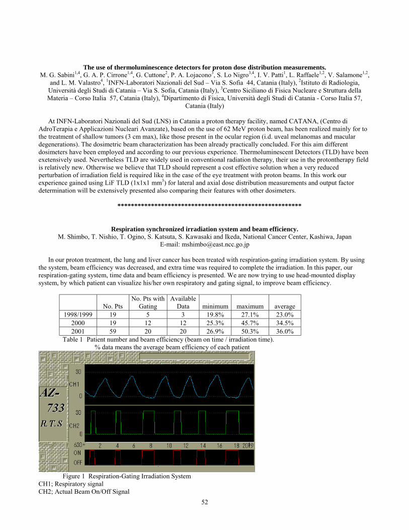
52
The use of thermoluminescence detectors for proton dose distribution measurements.M. G. Sabini1,4, G. A. P. Cirrone1,4, G. Cuttone2, P. A. Lojacono3, S. Lo Nigro3,4, I. V. Patti1, L. Raffaele1,2, V. Salamone1,2,
and L. M. Valastro4, 1INFN-Laboratori Nazionali del Sud � Via S. Sofia 44, Catania (Italy), 2Istituto di Radiologia,Università degli Studi di Catania � Via S. Sofia, Catania (Italy), 3Centro Siciliano di Fisica Nucleare e Struttura dellaMateria � Corso Italia 57, Catania (Italy), 4Dipartimento di Fisica, Università degli Studi di Catania - Corso Italia 57,
Catania (Italy)
At INFN-Laboratori Nazionali del Sud (LNS) in Catania a proton therapy facility, named CATANA, (Centro diAdroTerapia e Applicazioni Nucleari Avanzate), based on the use of 62 MeV proton beam, has been realized mainly for tothe treatment of shallow tumors (3 cm max), like those present in the ocular region (i.d. uveal melanomas and maculardegenerations). The dosimetric beam characterization has been already practically concluded. For this aim differentdosimeters have been employed and according to our previous experience. Thermoluminescent Detectors (TLD) have beenexetensively used. Nevertheless TLD are widely used in conventional radiation therapy, their use in the protontherapy fieldis relatively new. Otherwise we believe that TLD should represent a cost effective solution when a very reducedperturbation of irradiation field is required like in the case of the eye treatment with proton beams. In this work ourexperience gained using LiF TLD (1x1x1 mm3) for lateral and axial dose distribution measurements and output factordetermination will be extensively presented also comparing their features with other dosimeters.
*******************************************************
Respiration synchronized irradiation system and beam efficiency.M. Shimbo, T. Nishio, T. Ogino, S. Katsuta, S. Kawasaki and Ikeda, National Cancer Center, Kashiwa, Japan
E-mail: [email protected]
In our proton treatment, the lung and liver cancer has been treated with respiration-gating irradiation system. By usingthe system, beam efficiency was decreased, and extra time was required to complete the irradiation. In this paper, ourrespiration-gating system, time data and beam efficiency is presented. We are now trying to use head-mounted displaysystem, by which patient can visualize his/her own respiratory and gating signal, to improve beam efficiency.
� No. PtsNo. Pts with
GatingAvailable
Data minimum maximum average1998/1999 19 5 3 19.8% 27.1% 23.0%
2000 19 12 12 25.3% 45.7% 34.5%2001 59 20 20 26.9% 50.3% 36.0%
Table 1 Patient number and beam efficiency (beam on time / irradiation time).% data means the average beam efficiency of each patient
Figure 1 Respiration-Gating Irradiation SystemCH1; Respiratory signalCH2; Actual Beam On/Off Signal
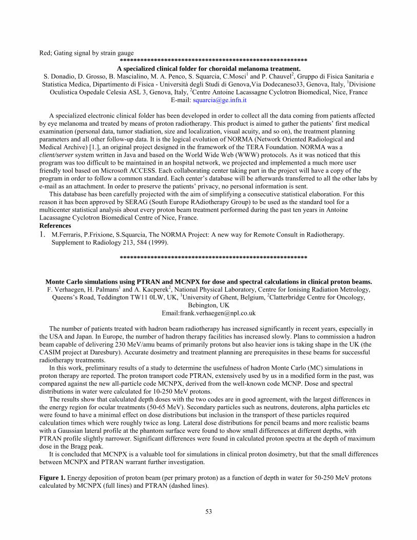
53
Red; Gating signal by strain gauge*******************************************************
A specialized clinical folder for choroidal melanoma treatment.S. Donadio, D. Grosso, B. Mascialino, M. A. Penco, S. Squarcia, C.Mosci1 and P. Chauvel2, Gruppo di Fisica Sanitaria eStatistica Medica, Dipartimento di Fisica - Università degli Studi di Genova,Via Dodecaneso33, Genova, Italy, 1Divisione
Oculistica Ospedale Celesia ASL 3, Genova, Italy, 2Centre Antoine Lacassagne Cyclotron Biomedical, Nice, FranceE-mail: [email protected]
A specialized electronic clinical folder has been developed in order to collect all the data coming from patients affectedby eye melanoma and treated by means of proton radiotherapy. This product is aimed to gather the patients� first medicalexamination (personal data, tumor stadiation, size and localization, visual acuity, and so on), the treatment planningparameters and all other follow-up data. It is the logical evolution of NORMA (Network Oriented Radiological andMedical Archive) [1.], an original project designed in the framework of the TERA Foundation. NORMA was aclient/server system written in Java and based on the World Wide Web (WWW) protocols. As it was noticed that thisprogram was too difficult to be maintained in an hospital network, we projected and implemented a much more userfriendly tool based on Microsoft ACCESS. Each collaborating center taking part in the project will have a copy of theprogram in order to follow a common standard. Each center�s database will be afterwards transferred to all the other labs bye-mail as an attachment. In order to preserve the patients� privacy, no personal information is sent.
This database has been carefully projected with the aim of simplifying a consecutive statistical elaboration. For thisreason it has been approved by SERAG (South Europe RAdiotherapy Group) to be used as the standard tool for amulticenter statistical analysis about every proton beam treatment performed during the past ten years in AntoineLacassagne Cyclotron Biomedical Centre of Nice, France.References1. M.Ferraris, P.Frixione, S.Squarcia, The NORMA Project: A new way for Remote Consult in Radiotherapy.
Supplement to Radiology 213, 584 (1999).
*******************************************************
Monte Carlo simulations using PTRAN and MCNPX for dose and spectral calculations in clinical proton beams.F. Verhaegen, H. Palmans1 and A. Kacperek2, National Physical Laboratory, Centre for Ionising Radiation Metrology,
Queens�s Road, Teddington TW11 0LW, UK, 1University of Ghent, Belgium, 2Clatterbridge Centre for Oncology,Bebington, UK
Email:[email protected]
The number of patients treated with hadron beam radiotherapy has increased significantly in recent years, especially inthe USA and Japan. In Europe, the number of hadron therapy facilities has increased slowly. Plans to commission a hadronbeam capable of delivering 230 MeV/amu beams of primarily protons but also heavier ions is taking shape in the UK (theCASIM project at Daresbury). Accurate dosimetry and treatment planning are prerequisites in these beams for successfulradiotherapy treatments.
In this work, preliminary results of a study to determine the usefulness of hadron Monte Carlo (MC) simulations inproton therapy are reported. The proton transport code PTRAN, extensively used by us in a modified form in the past, wascompared against the new all-particle code MCNPX, derived from the well-known code MCNP. Dose and spectraldistributions in water were calculated for 10-250 MeV protons.
The results show that calculated depth doses with the two codes are in good agreement, with the largest differences inthe energy region for ocular treatments (50-65 MeV). Secondary particles such as neutrons, deuterons, alpha particles etcwere found to have a minimal effect on dose distributions but inclusion in the transport of these particles requiredcalculation times which were roughly twice as long. Lateral dose distributions for pencil beams and more realistic beamswith a Gaussian lateral profile at the phantom surface were found to show small differences at different depths, withPTRAN profile slightly narrower. Significant differences were found in calculated proton spectra at the depth of maximumdose in the Bragg peak.
It is concluded that MCNPX is a valuable tool for simulations in clinical proton dosimetry, but that the small differencesbetween MCNPX and PTRAN warrant further investigation.
Figure 1. Energy deposition of proton beam (per primary proton) as a function of depth in water for 50-250 MeV protonscalculated by MCNPX (full lines) and PTRAN (dashed lines).

54
References1. M. Berger, Proton Monte Carlo transport program PTRAN, NIST Report NISTIR 5113 (Gaitersburg: NIST) (1993).2. L. Waters. MCNPX 2.1.5 User�s manual, LANL, 1999.
*******************************************************
Potential for a Radiotherapeutic Gain from an Anti-Proton Beam.S. Lee, W. McBride, T. Solberg, J. DeMarco, N. Agazaryan, J. B. Smathers, H. D. Suit1, H. R. Withers, A. J. Giorgio2, K.
A. Kubat-Martin2, C. J. Maggiore2, J. A. Martin2, and L. H. Welch2, UCLA Department of Radiation Oncology, LosAngeles, CA, USA, 1Massachusetts General Hospital, Boston, MA, USA, 2Pbar Medical, Inc., Newport Beach, CA, USA
Antiprotons have radiobiological characteristics which suggest they should be evaluated for a potential advantage overprotons in clinical practice. They may provide a steeper gradient of biological dose between a localized tumor and adjacentnormal tissues than is currently achieved with proton therapy, and with an even larger advantage over conventional externalbeam x-ray therapy.
Besides the physical advantage of a high Bragg-Gray peak, the potential exists for enhancement of biological dosedifferentials resulting from:
a. the high RBE of the high-LET charged nuclei which result from the antiproton annihilationb. the short distance (e.g. 30 microns) traveled by the charged particles, resulting in the high LET effects being
concentrated within the target volumec. a relative insensitivity to the effect of a low alpha-beta ratio for some tumors on the differential in response between
them and late-responding normal tissues. This may be relevant to such slowly proliferative tumors as prostate,meningiomas, chordomas, etc.
d. a relatively low oxygen enhancement ratio for the ionizing particlese. a high kinetics gain factor associated with high LET beams
Experiments are planned to test these principles.
0
20
40
60
80
100
0 10 20 30 40z (cm)
Dep
osite
d En
ergy
dE/
dz (M
eV/c
m)
100 MeV
150 MeV200 MeV
250 MeV
50 MeV