ABSTRACT Document: MECHANISM AND EVOLUTIONARY SIGNIFICANCE OF THE
50
ABSTRACT Title of Document: MECHANISM AND EVOLUTIONARY SIGNIFICANCE OF THE LOSS OF MELANIN PIGMENTATION IN THE CAVE FISH ASTYANAX MEXICANUS. Ernest R. Hixon, Master of Science, 2008. Directed By: Dr. William R Jeffery, Professor, Biology. The regressive evolution of traits such as eyes and pigmentation is common among cave organisms. As a model to study regressive evolution, I have used the teleost Astyanax mexicanus, which consists of eyed and pigmented epigean forms and many populations of cave-dwelling forms that have lost those traits. This study investigates the mechanism for the loss of melanin production, from the origin of chromatophores from the neural crest to the synthesis of melanin within the melanocyte. I show that cavefish retain a migratory population of neural crest derived cells that are tyrosinase positive and respond to exogenous signals as expected of a melanocyte. I then propose that the regressive evolution of melanin pigmentation is a selectively evolved trait that provides for an excess of dopamine, supported by the near two-fold increase in dopamine in cavefish brains, quantified via HPLC analysis. This study suggests that regressive evolution sometimes occurs via selection.
Transcript of ABSTRACT Document: MECHANISM AND EVOLUTIONARY SIGNIFICANCE OF THE

ABSTRACT
Title of Document: MECHANISM AND EVOLUTIONARY
SIGNIFICANCE OF THE LOSS OF MELANIN PIGMENTATION IN THE CAVE FISH ASTYANAX MEXICANUS.
Ernest R. Hixon, Master of Science, 2008. Directed By: Dr. William R Jeffery, Professor, Biology. The regressive evolution of traits such as eyes and pigmentation is common among
cave organisms. As a model to study regressive evolution, I have used the teleost
Astyanax mexicanus, which consists of eyed and pigmented epigean forms and many
populations of cave-dwelling forms that have lost those traits. This study investigates
the mechanism for the loss of melanin production, from the origin of chromatophores
from the neural crest to the synthesis of melanin within the melanocyte. I show that
cavefish retain a migratory population of neural crest derived cells that are tyrosinase
positive and respond to exogenous signals as expected of a melanocyte. I then
propose that the regressive evolution of melanin pigmentation is a selectively evolved
trait that provides for an excess of dopamine, supported by the near two-fold increase
in dopamine in cavefish brains, quantified via HPLC analysis. This study suggests
that regressive evolution sometimes occurs via selection.

MECHANISM AND EVOLUTIONARY SIGNIFICANCE OF THE LOSS OF MELANIN PIGMENTATION IN THE CAVE FISH ASTYANAX MEXICANUS.
By
Ernest R Hixon
Thesis submitted to the Faculty of the Graduate School of the University of Maryland, College Park, in partial fulfillment
of the requirements for the degree of Master of Science,
2008. Advisory Committee: Professor William R. Jeffery, Chair Professor Catherine Carr Professor Eric Haag

© Copyright by Ernest R. Hixon
2008

ii
Dedication
Dedicated to Nicole, without your support and inspiration this would not have been
possible.

iii
Acknowledgements
I would like to thank Dr. Allen Strickler, Dr. Daphne Soares, Dr. Yoshiyuki
Yamamoto, Dr. Masato Yoshizowa, and Dr. Glen Gebler for all of their advice,
support, and creative suggestions. I also extend thanks to Dr. Joanne Yew for her
expertise in HPLC technique, and Dr. Ed Kravitz for allowing the use of his HPLC
equipment. I thank Dr. Mardi Byerly for her assistance in performing the dissections
on Astyanax brains. I would also like to thank my committee- Dr. Catherine Carr and
Dr. Eric Haag, for their insights. Finally, I thank Dr. William Jeffery for all of his
guidance and patience.
iv
Table of Contents Dedication ..................................................................................................................... ii Acknowledgements...................................................................................................... iii Table of Contents......................................................................................................... iv
List of Figures ............................................................................................................... v
Introduction................................................................................................................... 1
Astyanax mexicanus as a system to study evolution and development.................... 1 Evolution of the Neural Crest and Pigment cells...................................................... 2
Melanophore development in Astyanax mexicanus ................................................. 3
Regressive evolution and pigmentation loss............................................................. 4
Materials and Methods.................................................................................................. 8
Biological Materials.................................................................................................. 8 DiI Injections ............................................................................................................ 8
Tyrosinase Assays..................................................................................................... 9
Fin Explant Preparation ............................................................................................ 9
Neural Keel Explantation and Culturing................................................................... 9
DiI-Tyrosinase Double Labeling ............................................................................ 11 Detection of Macrophages and Melanoblasts by Neutral Red Staining ................. 11
HPLC Analysis of Dopamine ................................................................................. 11
Results......................................................................................................................... 14
DiI Labeled Neural Crest Cell Migration in Pachón .............................................. 14
Tyrosinase Positive Cells in Cavefish Embryos ..................................................... 15 Tyrosinase Positive Cells are Neural Keel Derived Melanoblasts ......................... 18
HPLC Quantification shows Pachón brains contain more dopamine than epigean fish brains................................................................................................................ 19
Discussion ................................................................................................................... 24
Implications of a Functional Neural Crest in Cavefish........................................... 24 The Role of OCA2 in Cavefish Hypopigmentation................................................ 27
Regressive Evolution of Melanin Pigmentation in Astyanax mexicanus cavefish. 30
Conclusions............................................................................................................. 33
Bibliography ............................................................................................................... 35

v
List of Figures
Figure 1: Examples of epigean and cave forms of Astyanax mexicanus.............……..6
Figure 2: DiI injections used to trace neural crest cell migration................................14
Figure 3: Tyrosinase assays label melanoblasts……………………………………...16
Figure 4: Neutral red staining confirms tyrosinase assay is melanocyte specific……17
Figure 5: Neural crest cells in culture………………………………………………..21
Figure 6: Dopamine quantified in whole brain extracts of epigean and cave fish…...22
Figure 7: Dopamine quantified in specific brain regions of epigean and cave fish….23
Figure 8: Schematic of the melanin synthesis pathway……………………………...29
Figure 9: Model for a selective advantage for pigmentation loss……………………32
1
Introduction
Most cave organisms share highly convergent phenotypes including loss or
reduction of eyes and pigmentation. While pigmentation patterns are useful in
epigean environments for recognition (Parichy, 2001), they serve no purpose in a
lightless cave environment. Body pigment cells originate from the neural crest and
migrate throughout the body (DuShane, 1935). In cave populations of Astyanax
mexicanus, there is a reduction or complete loss of melanin pigmentation. It is
interesting that while all populations retain other pigment cell types, melanin
containing chromophores are consistently affected. This raises several questions
about the evolutionary mechanisms responsible for melanin pigmentation loss in
Astyanax which require laboratory experiments for their solution. The purpose of this
study is to first gain an understanding of the mechanism for melanin pigmentation
loss and then consider that mechanism in an evolutionary context.
Astyanax mexicanus as a system to study evolution and development
The teleost Astyanax mexicanus is common among the streams of the Sierra
de El Abra mountain range. These mountains are heavily cavernous, with many
limestone caves having been populated with Astyanax within the last ten thousand
years (Mitchell et al, 1977). Several populations of Astyanax trapped in the caves of
this region are believed to have independently adapted to cave life to varying degrees.
Some of these adaptations include the degeneration of the eye, changes in cranio-
facial structure, changes in jaw shape, a number of behavioral changes, and loss or

2
reduction of melanin pigmentation. These fish are considered a single species and are
interfertile. Additionally they are easily bred and raised in a laboratory setting.
Because of these traits, Astyanax provides for a unique system for the study of
development and evolution.
Evolution of the Neural Crest and Pigment cells
The neural crest is an ectodermally derived group of cells that originates from
the dorsal ridge of the neural tube. In most organisms, the neural tube forms by a
folding of the neural plate, and the neural crest is formed during this process of
neurulation by cell delamination along the “seam.” In teleosts, however, the neural
tube forms by hollowing of the neural keel, a solid mass of dorsal midline cells (Hall,
1999). Despite this difference in neurulation, the neural crest is largely unchanged,
still forming along the dorsal ridge of the neural tube. There are a number of
molecules, including Bone Morphogenic Protein (BMP) (Sela-Donnenfeld and
Kacheim, 1999), several Wnt family genes (Dunn et al., 2000), and Fibroblast
Growth Factor (FGF), which work to pattern the neural crest (Aybar and Mayor,
2002; Dorsky et al., 2000). It is believed that at this point or shortly after they have
delaminated and prepare to migrate, that neural crest cells undergo terminal
differentiation, though in some cell derivatives there remains a small amount of
plasticity (Hall, 1999).
A short time after trunk neural crest formation, cells begin to delaminate and
begin to migrate. This migratory process seems to be largely regulated by ephrin-B

3
ligands (Perris, 1997; Santiago and Erickson, 2002). Migrating cells of the trunk
neural crest move within one of two pathways (Weston, 1963). The first, the medial
pathway, proceeds from the dorsal neural tube and then ventrally into the body cavity
between the neural tube and the somites. These cells produce a wide variety of cell
types such as glia, smooth muscle, and neurons. The second pathway (the
dorsolateral pathway) proceeds ventro-laterally from the neural tube. These cells pass
between the somites and the epidermis. Cells in this pathway are far less diverse in
their fates, and they are thought to exclusively form pigment cells. In fact, it is
believed that all body pigment producing cells in vertebrates arise from the neural
crest (Weston, 1970; LeDourain, 1982). These cells are believed to migrate to their
destination, differentiate, and act as a stem cell population, producing the three
pigment cell types (xanthophores, iridophores and melanophores) as the body of the
animal increases in size.
Melanophore development in Astyanax mexicanus
A neural crest derived stem cell population is believed to produce pigment
forming cells throughout the life of the fish. The cells produced by these stem cells
(to which I will refer to as chromatoblasts) have been shown to have some degree of
plasticity (Hall, 1999). Though little is known about the genetic patterning
mechanisms of iridophores, recent studies have shed some light on the relationship
between xanthophores and melanophores. Melanoblast development is inhibited by
the absence of kit and mitf gene products. Mitf, a helix-loop-helix transcription
factor, has been shown to be crucial in the final stages of melanocyte maturation (Hou
4
et al., 2000; Tachibana, 2000). It is thought that these factors induce a somewhat
plastic chromatoblast to differentiate into melanoblasts. Further development is
triggered by dopachrome tautomerase (DCT) and up regulation of the tyrosinase gene
by α-MSH (Aroca et al., 1993; Prota, 1992). The chromatoblasts can be induced into
forming xanthoblasts by the presence of fms, a type-II receptor tyrosine kinase.
Further development into mature xanthophores is mediated by several growth factors
(Parichy and Turner, 2003). Astyanax cave forms have mature xanthophores and
iridophores while exhibiting a reduction or complete loss of melanophores.
Melanin is produced in specialized vesicles within melanophores called
melanosomes. These vesicles serve not only to sequester the toxic melanin from the
rest of the cell but also to regulate pH as many of the reactions in the melanin
metabolic pathway are highly sensitive to pH (Ancans et al, 2001). Once tyrosine is
transported into the melanosome, it is catalyzed by tyrosinase into L-dopa and again
by tyrosinase into dopaquinone (Prota, 1992). Because tyrosinase is exclusively
found in the melanosomes of melanocytes, addition of exogenous L-dopa can be used
to label and track melanoblasts in addition to assay for the presence of an active
enzyme (Laidlaw, 1932).
Regressive evolution and pigmentation loss
While the study of evolution is typically focused on the selection for new
traits, regressive evolutionary events are quite common. It has been proposed that for
every single instance of progressive evolution, there exist up to ten instances of
regressive evolution (Haldane, 1958). Regressive loss is typically either not thought

5
of as a major concern or considered to be the reduction of molecular “noise” (Regal,
1977). There are instances, though, of regressive traits that are viewed as
compensatory for other advantageous traits. Examples include the loss of lungs in
salamanders which ultimately allowed for a specialized projecting tongue (Lombard
and Wake, 1986), or potentially the loss of eyes in Astyanax allowing for an increase
in the number of taste buds (Yamamoto et al, 2004). Generally speaking, regressive
evolution is often easily explained as a simple accommodation to energy conservation
or accumulation of neutral mutations. While this is sometimes undoubtedly the case,
few studies have been done to support this notion. Estimates of energy conservation,
effects on fitness, and evolutionary rates are either rarely attempted or poorly
understood (Fong et al, 1995).
It has been theorized that in the absence of light the eyes and pigment of
Astyanax have no effect on fitness and are lost through the neutral accumulations of
mutations in genes that affect their development (Wilkens, 1988). However, cavefish
populations have all independently evolved a reduction or loss of melanin
pigmentation while retaining xanthophores and iridophores (Figure 1). These cells
should then be under the same evolutionary pressures that melanophores experience,
yet they persist while melanophores do not. That melanophores are singled for
regression provides a compelling argument that they could be under a unique set of
selective pressures- perhaps the loss of pigmentation in some way confers greater
fitness in the cave environment. Furthermore, because most cavefish populations
have been isolated for as little as 10,000 generations it is unlikely that neutral
mutations would have fixed in this relatively short time span (Barr, 1968).

6
Theories based on natural selection also exist to explain pigment loss,
typically centering on the concept of energy conservation (Barr, 1968). However,
given that cavefish retain a population of cells that
migrate and produce some of the tools for melanin
synthesis, it is hard to imagine where energy is actually
being conserved. I propose a novel hypothesis for the
selective advantage of pigmentation loss. Disuse of the
melanin metabolic precursor L-tyrosine could
potentially produce an excess of the essential amino
acid, allowing for other pathways that rely on L-
tyrosine to be upregulated. Most notably, L-tyrosine is
the metabolic precursor for the synthesis of dopamine,
an important neurotransmitter involved in circadian
rhythm (Andretic and Hirsh, 2000) and appetite
regulation via the reward circuit (Hernandez and
Hoebel, 1988). Astyanax maintains circadian rhythms,
although they are not as precisely regulated as in epigean fish (Espinasa and Jeffery,
2006). Observations of cavefish in the laboratory show that they feed more than
epigean fish, a trait that would be advantageous in a nutrient poor cave environment
(unpublished observations). It is therefore possible that dopamine is not regulated in
the same manner in cavefish as in epigean fish.
This study investigates the loss of melanin pigmentation, beginning at the
development of the neural crest through the migration and maturation of
Figure 1. The surface form of Astyanax mexicanus, as well as several independent cave populations.

7
chromatophores, and ultimately to the synthesis of melanin within the melanosome.
Based on the step in melanin production that I have found to be interrupted, a novel
theory is proposed for the regressive evolution of melanin pigmentation via a
selective mechanism that favors pigmentation loss to supply an increased demand for
dopamine. Finally, the results of some experiments are described that test my
hypothesis.

8
Materials and Methods
Biological Materials
Laboratory stocks of Astyanax mexicanus were founded by epigean specimens
collected from Balmorhea State Park, Texas, USA and hypogean forms collected
from La Cueva de El Pachón in Tamaulipas, Mexico. Stocks were maintained on a
12:12h light:dark cycle at 25oC. Embryos were collected after natural spawning and
raised to adults as described previously (Jeffery and Martasian, 1998; Jeffery et al,
2000). Methods were approved by University of Maryland animal care and use
committee and followed National Institutes of Health guidelines.
DiI Injections
Embryos in Ringer’s-1 solution (0.65% NaCl in double-distilled water;
Westerfield 2000) were manually dechorionated with watchmaker forceps. CM-DiI
(1,1´-dioctadecyl-3,3,3´,3´-tetramethyl-indocarbocyanine perchlorate, Cell Tracker,
Molecular Probes, Eugene, OR, USA) was first dissolved in ethanol (2µg/µl) and
then diluted 0.5µg/µl in 0.3M sucrose. Glass electrodes were backfilled with the
solution. At 24hpf, embyros were placed in 3% methyl cellulose (Sigma Chemical
Co, St. Louis, MO, USA) in Ringer’s-1 solution and pressure injected with a bolus of
DiI in the dorsal region of the neural keel at the 10th somite level. Embryos were
reared for 24-72h at room temperature in the 3% methyl cellulose solution, fixed in
4% paraformaldehyde, and viewed with fluorescence microscopy.

9
Tyrosinase Assays
Tyrosinase assays were performed according to Laidlaw, 1932. Samples
(embryos, hatched larvae, fin explants, or neural keel explants) were fixed for an hour
in 5% formalin in PBS then washed several times with PBS. The PBS was replaced
with 0.1% 3,4-dihydrophenylalanine (L-dopa, pH 7.4, Sigma) overnight at room
temperature. After washing with PBS, some embryos were embedded in polyester
wax and sectioned at 10µm. Sections were placed on a slide and viewed under a
compound microscope. Some epigean adults, embryos, hatched larvae and explants
were incubated in a tyrosinase inhibitor (1mM 1-phenyl-2-thiourea in aquarium
water, Sigma) (Bell, 1955).
Fin Explant Preparation
Adult fish were immobilized by brief immersion in iced aquarium water. Clips from
the tail fin were excised by sharp scissors. Tissue samples were immediately
immersed in 5% formalin in PBS and prepared for tyrosinase assay.
Neural Keel Explantation and Culturing
Embryos were raised to the 10 somite stage in Ringer’s-2 solution and manually
dechorionated with watchmaker forceps. Explantation was performed under a
dissection microscope using two sharpened tungsten needles- one straight and one
bent at a 90o angle. The explantation procedure was adapted from a similar study

10
(Sadaghani and Vielkind, 1990). First, the cranium and yolk mass were removed.
The epidermis was then peeled off the mass of the embryo using the straight needle as
an anchor and the bent needle to push. The mass of the embryo was transferred to a
clean dish containing Ringer’s-2 solution. Embryos were dissociated into parts by
brief incubation (90-120 seconds) in 0.25% pancreatin (Sigma), while being gently
agitated with a large bore plastic pipet. After incubation, the tissue was transferred to
Ringer’s-2 to stop enzyme activity and washed 4 times with further agitation.
Somites and notochord generally separate from the neural keel with washing and
agitation. Extraneous tissue still contaminating the neural keels was removed with
tungsten needles.
Explants were cultured in Lab-Tek-4 chamber slides coated in 10µg/ml
fibronectin (Roche, Indianapolis, IN, USA) in phosphate buffered saline (PBS).
Culture media consisted of L-15 (Cellgro, Mediatech, Inc., Heardon, Va, USA) with
20% fetal bovine serum (Sigma), 1x gentamicin (Roche; 50mg/ml), and 1x penicillin-
streptomycin (Roche; 1x is 100IU penicillin-0.1mg streptomycin-HCl). Some
cultures included 12-O-Tetradecanoylphorbol 13-acetate (TPA; Sigma). The TPA
was in DMSO and diluted to a concentration of 80nM in whole media. Other cultures
contained 0.2uM α-MSH (Sigma) as noted.
Tyrosinase assays were performed on some cultures by first washing culture
media with PBS, and then immersing in 5% formalin (pH 7.5) for 30min at RT. After
washing several times in PBS for 20min, cultures were exposed to buffered L-dopa
(pH 7.4) and incubated at 37oC for approximately 5 hours.
11
DiI-Tyrosinase Double Labeling
Embryos were injected with DiI as described above, then raised to 60hpf.
Embryos were fixed in 5% formalin and assayed for tyrosinase activity as described
above. Fixed embryos were viewed using both bright field and fluorescence
microscopy. In these preparations, melanin containing cells are black and DiI labeled
cells are red.
Detection of Macrophages and Melanoblasts by Neutral Red Staining
To distinguish between macrophages and melanoblasts, the neutral red vital
staining method was used (Herbomel et al., 2001). Adult tail fin clips were obtained
as described above. The clips were stained with 2.5ug/ml neutral red in the dark for
20hr at RT. Each clip was photographed before being fixed and processed for
tyrosinase assay. Although neutral red staining was washed away by the fixation
process, comparisons of double labeled cells were obtained by overlaying pre- and
post-tyrosinase assayed images in Adobe Photoshop.
HPLC Analysis of Dopamine
High pressure liquid chromatography (HPLC) was used to detect dopamine
levels in whole brains. Brain dissections were performed at the laboroatory of Ed
Kravitz, Department of Neurobiology, Harvard Medical School. The study consisted
of a total of 27 fish (14 Pachon, 13 epigean). Brains were either assayed whole or
dissected into 3 parts: optic tecta, hypothalamus, and the remainder.

12
Fish were anesthetized by exposure to MS-222 (pH 7.0, Sigma) until
unresponsive. Small scissors were used to first sever the spinal cord caudal to the
skull. The top of the skull was cut open and removed, the brain lifted with a blunt
probe, and the nerves connected to the brain were severed. Further dissection to
remove the optic tecta and hypothalamus were done using a fine scalpel while brains
were submerged in PBS. Samples were distributed evenly (alternating whole
brain/dissected brain samples as well as epigean/cave fish samples) over the course of
the day to eliminate variation caused by circadian rhythms.
Tissue was immersed in 100ul ice cold 0.1M perchloric acid and homogenized
using a plastic disposable pestle (Kontes Glass Company, Vineland, N.J., USA). The
homogenate was incubated on ice for 1hr, then centrifuged at 16,100g for 2 minutes.
Amine levels were measured with an ESA CoulArray Model 4600 HPLC with
electrochemical detection equipped with a C18 column (ESA; 150 mM, 120Å), a 20-
µl loop (Rheodyne, Rohnert Park, Ca., USA), and a column heater (Eppendorf,
Hamburg, Germany) held at 29°C. The flow rate was set at 500 µl/min. The mobile
phase was composed of 15% acetonitrile (Fisher, Pittsburgh, Pa., HPLC grade), 15%
methanol (Fisher, HPLC grade), 75 mM NaH2PO4, and 1.5 mM SDS, pH 5.6 (Shulz
and Robinson, 1999). The electrodes were set at 100, 425, 530, and 650 mV.
Dopamine was detected on the 100mV channel. Forty microliters of each diluted
homogenate was used for each injection. Retention times and concentrations of the
amines were determined by comparison to a standard composed of 25nM dopamine
in 0.1 M perchloric acid (Certel et al, 2007).

13
Results of HPLC were normalized by weight. Undiluted supernatant from
homogenate was assayed for protein by using the Quick Start Bradford Protein Assay
(Biorad, Hercules, Ca., USA) and a spectrophotometer. A protein standard was made
using serial dilutions of a epigean fish brain homogenate of known weight. Statistical
analysis was performed using a two-tailed Student’s T test for two samples of
unequal variance.

14
Results
DiI Labeled Neural Crest Cell Migration in Pachón
DiI labeling experiments were performed to determine if cavefish neural crest
cells are migratory. Cavefish from the Pachón population were used as they exhibit
nearly complete albinism (although rarely pigmentation in the retinal pigmented
epithelium is observed), typically exhibiting no body pigment. After injection of a
DiI bolus in the region of the dorsal neural keel, migration of labeled cells was
observed throughout the body in all directions (rostral, caudal and ventral) (Figure
2A). Labeled cells were also
observed in the yolk sack, one
of the areas where
melanophores are first
observed in epigean embryos.
In addition, sections were
made of DiI labeled embryos.
Neural crest cells migrate
through one of two pathways-
medial and dorsolateral
(Weston, 1963). Cells that
migrate through the
dorsolateral pathway
exclusively become
Figure 2. DiI marker used to trace migrating neural crest cells. DiI was injected into the dorsal neural tube at ~10 somites. After one day, whole mount embryos show a number of migrating cells (A). Cross sections show migrating cells to be primarily in the dorso-lateral pathway (B). DF, Dorsal fin. NT, Neural tube.

15
chromatophores (LeDourarin, 1982; Erickson, 1986; Newgreen and Ericson, 1986).
Sections of DiI labeled cells showed that many of the migrating cells were located
just under the epidermis, indicating that they were migrating dorsolaterally and likely
chromatophore precursors (Figure 2B). These experiments show that there is a
migratory population of trunk neural crest cells, and some of these cells migrate
through the dorso-lateral pathway, which is normally reserved for cells fated to
become chromatophores.
Tyrosinase Positive Cells in Cavefish Embryos
Tyrosinase assays were used to look for the presence of cells capable of
producing melanin. Embryos (epigean and Pachón) were assayed at 36hpf, the time
at which body pigmentation is typically appearing in the eye, trunk, and yolk sac of
epigean fish embryos (Figure 3A-B). Prior incubation of epigean embryos in 1-
phenyl-2-thiourea (PTU, a tyrosinase inhibitor) prevented production of melanin
(Bell, 1955). Tyrosinase assays after embryos were removed from PTU showed
similar patterns of tyrosinase positive labeled cells among both epigean and cave fish
embryos. Tyrosinase positive cells were first seen at about 24hpf in Pachón embryos,
the same time at which melanoblasts can first be detected via tyrosinase assays in
epigean embryos. Sections of assayed embryos show that labeled cells lie just below
the epidermis in the trunk and fins (Figure 3D). These are areas where melanin
producing cells typically are found in epigean embryos. Tyrosinase assays were also
done throughout development. Tyrosinase positive cells were found in 24, 36, 60hpf

16
Figure 3. Tyrosinase assays in Astyanax embryos. Staining patterns of epigean embryos raised in PTU (A) are similar to 30 hour cavefish embryos (B). Double staining of DiI and tyrosinase assays show some cells to be double labeled (arrow), showing these cells to be of neural crest origin (C). Tyrosinase positive cells are located between the somite and epidermis in cavefish, typical of melanoblasts or melanocytes (D). Bar, 100µm.

17
embryos and adult cave fish. Double labeling experiments using both DiI and the
tyrosinase assay were performed to determine if neural crest derived migratory cells
were fated to become pigment cells (Figure 3C). Some cells that migrated away from
the injection site were found to be both tyrosinase and DiI positive, showing that
Pachón possess a tyrosinase positive, migratory population of neural crest derived
cells.
It has been reported that the tyrosinase assay may be non-specific in that
macrophages, as well as pigment cells, are labeled (Laidlaw, 1932; Tucker and
Erickson, 1986). In order to investigate that the tyrosinase assay was specific to
melanoblasts, a double labeling experiment was performed (Figure 4). Fin clips from
adult Pachón fish were first stained with Neutral Red, a macrophage specific marker,
photographed, and then assayed for tyrosinase. Although the processing for the
tyrosinase assay washed away the
neutral red staining,
photomicrographs of the same
region of fin clip after each assay
were overlaid to determine if any
cells were labeled by both
techniques. Analysis of the
overlaid images showed no cells
labeled by both techniques,
indicating that in Astyanax mexicanus, the tyrosinase assay is specific for
melanoblasts.
18
These experiments show that although cave fish embryos do not have mature
pigment producing melanophores, tyrosinase positive melanoblasts- cells that are able
to produce melanin if provided L-DOPA as a substrate- still develop in a normal
pattern similar to the epigean embryos. These tyrosinase positive cells make up a
portion of the migratory cell population that originates at the neural crest.
Furthermore, tyrosinase positive melanoblasts are present into adulthood. Finally,
double labeling experiments showed that they tyrosinase assay is specific to
melanoblasts.
Tyrosinase Positive Cells are Neural Keel Derived Melanoblasts
Several approaches were used to confirm that the tyrosinase positive cells
were of neural crest origin. First, double labeling with DiI and tyrosinase showed that
cells migrating from the dorsal neural keel were tyrosinase positive. Second, I
confirmed the ability for cells to migrate from explanted neural keel in culture as has
been previously shown in other teleosts (Sadaghani and Vielkind, 1990) (Figure 5).
In basic media epigean and Pachón neural keel cultures did not develop melanoblasts,
although a number of cells migrated away from the neural keel (Figure 5 A, D). Two
different culture additives were used. Cultures were also given either αMSH, a
hormone that induces melanin synthesis and a stellate morphology in melanoblasts
(Aroca et al, 1993), or phorbol ester (TPA), which upregulates the MitF pathway
(Prince et al, 2003). Mature melanocytes were visible in epigean explant cultures
containing both αMSH and TPA, though no melanin synthesis was induced in the
Pachón explant cultures (Figure 5 B-C, E-F). Tyrosinase assays of Pachón explants

19
in normal and αMSH containing cultures showed that Pachón melanoblasts with
αMSH were dendritic as opposed to punctuate, as those seen in cultures without
αMSH (Figure 5 G-H). This result indicates that tyrosinase positive cells that migrate
from the neural keel also respond to αMSH, exhibiting multiple melanoblast
characteristics.
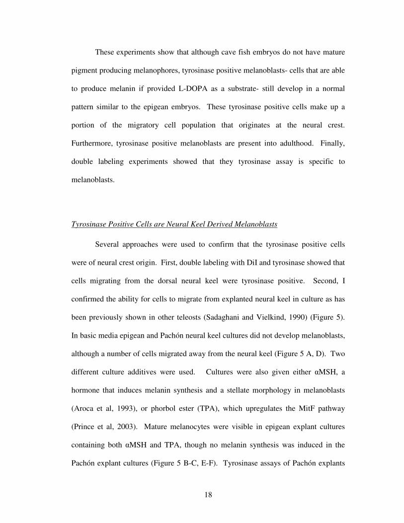
HPLC Quantification shows Pachón brains contain more dopamine than epigean fish
brains
In addition to being a metabolic precursor to melanin, L-tyrosine is also the
precursor to dopamine, epinephrine, and norepinephrine. In order to test whether an
interruption of the melanin synthesis pathway had created a surplus of dopamine
elsewhere, namely in the brain, HPLC was used to quantify the amount of dopamine
present in the brains of both epigean fish and Pachón cave populations. Analysis of
dopamine levels in whole brain homogenate showed nearly a two-fold increase in the
amount of dopamine per tissue weight found in Pachón over epigean fish (40.24nM/g
of tissue and 22.90nM/g of tissue respectively, p<0.05) (Figure 6). Further analysis
was done on individual portions of the brain to determine if there was a localized
change in dopamine levels in two areas of the brain previously reported to have
undergone morphological changes- specifically the optic tectum and the
hypothalamus (Menuet et al., 2007) (Figure 7). Results from both specific regions
showed higher levels of dopamine in Pachón brains, although probably due to the
difficulty of the dissection, the variation was too high to achieve a suitable level of
statistical significance. The remaining portion of the brains following dissection was

20
also assayed. Again, Pachón brains contained significantly more dopamine than seen
in epigean forms (19.151nM/g tissue and 12.44nM/g tissue respectively, p<0.05).
While a specific brain region was not identified as being responsible for the increased
concentration of dopamine in Pachón brains, it was shown that there is a significant
concentration disparity in an important neurotransmitter.

21
Figure 5.Cultured explants of Astyanax neural tubes. Surface fish cultures with normal media show no melanin (A) although addition of αMSH (B) or TPA (C) was sufficient to induce melanogenesis. Cavefish cultures in normal media (D), or with αMSH (E) or TPA (F) did not produced melanin. However, tyrosinase assays on cavefish normal media cultures and αMSH cultures showed tyrosinase positive cells with different morphologies, indicating cavefish melanoblasts respond to αMSH. Bar, 100µm.
22
23

24
Discussion
Implications of a Functional Neural Crest in Cavefish
This study had two goals. First, to identify the step at which the maturation of
melanin producing melanophores was interrupted, I used a number of techniques to
compare the development of melanophores in cave and epigean embryos of Astyanax
mexicanus. Second, I proposed and tested a model for an evolutionary mechanism by
which melanin pigmentation is lost due to selective forces. I determined that melanin
synthesis is interrupted in the late stages of melanocyte development. Further, I
provided evidence for a possible selective advantage to pigment loss, although it must
be investigated more thoroughly.
In order to determine the point at which melanin pigmentation developed, I
performed experiments to test each step of melanocyte development, from the origin
of the neural crest, to migration, differentiation and ultimately maturation into a
pigment producing melanocyte. Several experiments provide strong evidence that
cavefish maintain a population of neural crest derived chromatophores that
differentiate into chromatophores. DiI injections into the dorsal neural keel showed
that cells migrate through both the medial and the dorsolateral pathway. Previous
studies have shown that cells migrating through the dorsolateral pathway develop
solely into chromatophores (LeDourarin, 1982; Erickson, 1986; Newgreen and
Ericson, 1986). Cross-sections of DiI injected embryos also showed labeled cells
migrating through the dorsolateral pathway, between the epidermis and lateral surface
of the somites, suggesting these cells are fated to become chromatophores.
25
Histochemical assays using L-dopa as a substrate confirmed that the
tyrosinase enzyme was functional. The assay also provided an additional label for
unpigmented, migratory melanoblasts. Both epigean and cave embryotic
melanoblasts begin to produce tyrosinase early in their migration, soon after departing
the neural crest (approximately 25-30hpf). Early observation of tyrosinase in
unpigmented melanoblasts is not uncommon, having already been reported in several
other organisms such as zebrafish (Camp and Lardelli, 2001) and ascidians
(Whittaker, 1973). Tyrosinase assays confirmed that a subpopulation of
chromatophores was beginning to differentiate into melanoblasts. Additionally,
double labeling experiments using DiI along with tyrosinase assays demonstrated that
the double labeled cells originated at the neural crest and were migrating. Finally, all
tyrosinase positive cells were shown to be present just below the epidermis in the
yolk, in the dorsal fin, or between the epidermis and somite in the trunk of the
embryos- the expected locations for melanoblasts. Together, these experiments show
conclusively that cavefish embryos maintain a population of neural crest derived
melanoblasts which migrate dorsolaterally, and have differentiated to the degree that
they have begun to produce an active tyrosinase enzyme. While this study focused on
the Pachón population, tyrosinase positive cells have been observed in adult fin clips
of four other populations (Chica, Los Sabinos, Tinaja, and Curva) in numbers
substantially greater than those seen in epigean fish (McCauley et al, 2004),
indicating that this loss of mature melanocytes has evolved several times in separate
populations.
26
Cell culture assays provided further evidence as to the origin and maturation
of cavefish melanoblasts. First, cultures of isolated cavefish neural keels contained
tyrosinase positive cells that had migrated away from the neural keel. This supports
the conclusion that Pachón embryos maintain a migratory cell population fated to
become melanoblasts. Culture additives provided insight into the degree of
differentiation of these cells. While neither αMSH nor TPA proved to be sufficient to
induce melanin synthesis in Pachón cultures, the cells did respond to αMSH by
adopting a stellate morphology (Schwahn et al, 2001). These cells have therefore
taken on a number of melanoblast characteristics- they arise from the neural crest,
migrate through the dorsolateral pathway, produce tyrosinase, and change
morphology in response to MSH as expected.
I conclude that while Pachón embryos and adults do not develop melanized
body pigmentation, they do retain a population of melanoblasts, and therefore the
interruption in melanin synthesis in melanogenesis must be at a late step. Tyrosinase
assays exploit the presence of an endogenous, active enzyme proving that the enzyme
function is unaffected. Tyrosinase catalyzes three concurrent reactions in the melanin
synthesis pathway: the conversion of tyrosine to L-dopa, L-dopa to dopaquinone, and
after several spontaneous reactions, 5,6-dihydroxyindole to indole-5,6 quinone
(Korner and Pawelek, 1982). The assays demonstrate that all reactions beginning
with the conversion of L-dopa to dopaquinone and all subsequent steps are intact and
unaffected. Furthermore, it is unlikely that steps prior to the tyrosinase catalyzed
reactions are affected, as an inability to convert L-phenylalanine into tyrosine would
be lethal. Finally, a study showed tyrosinase assays on adult fin clips with tyrosine as

27
a substrate did not yield melanin in both Pachón and albino catfish (McCauley et al,
2004). It is therefore apparent that the interruption of the melanin synthesis pathway
must lie in the conversion of tyrosine to L-dopa. In the cell cytoplasm, phenylalanine
is converted to tyrosine. Tyrosine is then transported into melanosomes- membrane
bound organelles that contain tyrosinase- where the rest of the melanin synthesis
pathway occurs. It is likely that tyrosine and/or tyrosinase are sequestered in some
manner, preventing the cavefish melanosomes from producing pigment.
The Role of OCA2 in Cavefish Hypopigmentation
Shortly after this research was completed, a study using a Quantitative Trait
Loci (QTL) analysis revealed a strong correlation between pigmentation loss and
mutations in a gene known to be responsible for tyrosinase-positive albinism (Protas
et al, 2006). Oculocutaneous albinism, caused by mutations in the Oca2 gene
(referred to as the p gene in mice), is the most common form of albinism in humans.
The Oca2 gene codes for a large transmembrane protein that intergrates into the
melanosomal membrane. The exact function of OCA2 is not well understood. Early
studies suggested that the protein played a role in the transport of tyrosine across the
melanosome membrane after observing that high concentrations of tyrosine could
rescue pigmentation in p-deficient mouse retina cultures (Sidman and Pearlstein,
1965). However, recent experiments have shown the kinetics of tyrosine transport in
p-gene mice melanocytes to be normal (Gahl et al, 1995; Potterf et al, 1998).
Currently, it is believed that the OCA2 protein is involved in anion transport,

28
maintaining proper pH inside the melanosome (Brilliant et al, 1994; Brilliant, 2001)
(Figure 8).
Sequencing analyses of Pachón and Molino, another population of cave fish,
showed that Pachón OCA2 contained a unique deletion in exon 24 and that Molino
OCA2 had a similar large deletion, though in exon 21 (Protas et al, 2006).
Complementation experiments using Pachón and a third cavefish population, Japones,
failed to rescue pigmentation, suggesting that this population also has a mutation in
OCA2 (Protas et al, 2006). Not only does this show the mutability of the OCA2
gene, but it also provides some evidence that the populations have evolved
independently, a point that has been debated (Avise and Selander, 1972; Mitchell et
al, 1977; Wilkens, 1988; Espinasa and Borowsky, 2001; Strecker et al, 2003).
Melanocytes are typically divided into two major categories: cutaneous and
extracutaneous (Prota, 1992). To this point, this study focused on cutaneous
melanocytes that make up the body pigmentation seen on epigean forms.
Extracutaneous pigmentation does not arise from the neural crest. One example of
extracutaneous pigmentation is the retinal pigmented epithelium, which is derived
from the wall of the optic cup (Mann, 1964). While OCA2 is typically considered to
be albinism of eye and cutaneous pigment, some populations produce individuals that
have no or drastically reduced body pigmentation but a pigmented RPE. In fact,
although Pachón typically has no melanin pigmentation at all, some individuals have
been observed to have pigmented RPE (unpublished observation). While these
individuals are extremely rare, it does bring to light some conflict within the OCA2
model of melanin loss in Astyanax. The differentiation of cutaneous melanocytes
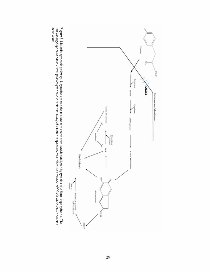
29

30
and RPE melanocytes is governed by slightly different mechanisms. RPE
melanocytes are predominantly regulated by MitF, Otx2, and Pax6 (Martinez-
Moralez et al, 2004). It is worth noting that embryonic expression of Pax6 in
cavefish is reduced, which may lead to smaller optic primordia (Strickler et al, 2001).
However, expression in the retinal ganglion and amacrine cells later in development
is unchanged (Strickler et al, 2001). In addition, there is evidence that growth factors
released by the underlying endoderm have a role in RPE cell development (Nguyen
and Arnheiter, 2000). It is therefore reasonable to conclude that there may be some
ability for extracellular factors within the surrounding environment that are sufficient
to override the mutations to OCA2, resulting in melanogenesis. This supports the
proposed presence of two genes that regulate melanin pigmentation loss in cavefish
(Sadogu and McKee, 1969, Wilkens, 1988).
Regressive Evolution of Melanin Pigmentation in Astyanax mexicanus cavefish
There is, historically, much debate as to the nature the regressive evolution of
cave organism pigmentation. Pigment loss is seen in many cave organisms from
arthropods and millipedes to salamanders and fish. Early theories about cave
organism (specifically the African cavefish Caecobarbus geertsii) pigment loss
centered on metabolic concerns, in which lower temperature or lower metabolic rates
were responsible for retarded development of non-essential systems (Heuts, 1951).
Today, many investigators subscribe to the neutral theory hypothesis- that mutations
simply pile up in genes that are under no selective pressure- to explain pigmentation
loss (Wilkens, 1988).
31
However, some flaws exist in the neutral theory explanation of Astyanax
melanin loss. While all Astyanax populations show some degree of melanin loss, the
other two chromatophores (xanthophores and iridophores) are retained. Secondly,
neutral theory predicts a longer span of time for mutations to fix within a population
than those mutations that are selected for (Kimura, 1983). Most cave populations of
Astyanax are predicted to be as little as 10,000 generations old, an age that does not
provide much opportunity for neutral mutations to fix within the population, though
some argue that the small populations allow for a more rapid fixation of mutations
(Barr, 1968; Mitchell et al, 1977; Avise and Selander, 1974; Kimura, 1983).
Due to the inability of the neutral theory to fully explain melanin pigment loss
in Astyanax, I propose a model for a novel selective advantage to pigment loss. A
mutation in OCA2 would result in quick saturation of L-tyrosine in melanosomes. I
propose that it is possible that L-tyrosine would therefore be more available
elsewhere in the body, namely the brain, where it could be converted into dopamine
(Figure 9).
High performance liquid chromatography (HPLC) analysis of epigean and
Pachón adult brains showed nearly a two fold increase of dopamine in Pachón brains
as compared to epigean forms. In two brain regions known to have altered
morphology in Pachón (optic tectum and hypothalamus), no change in dopamine
expression could be determined with a suitable degree of statistical significance.
Dissections of these regions proved difficult, and the experiment was further
complicated by constraints in the number of animals that could be used. This resulted
in high variation within the dissected samples. Despite this, I would expect that the

32
thalamic region of cavefish to be one of the regions of the brain that contains excess
dopamine.
Dopamine in the brain regulates a number of important processes, notably
circadian rhythm, appetite and locomotion. Cave forms of Astyanax retain circadian
rhythms, a process which regulates dopamine production in the hypothalamus, but
they are not as tightly regulated as epigean forms (Wilkens et al, 1993; Espinasa and
Jeffery, 2006). In addition, experiments in Drosophila showed that dopamine
receptors are more receptive to dopamine during night cycles (Andretic and Hirsch,
2000). Experiments on appetite regulation in humans showed dopamine agonists
suppressed appetite while antagonists enhanced it (Comings et al, 1993). However, it
is difficult to predict exactly what effect this excess of dopamine could have on the

33
behavior of cavefish without knowing its location in the brain. It should be noted,
however, that concurrent with my HPLC analysis of dopamine, serotonin levels were
also quantified. While cavefish had nearly twice the dopamine seen in epigean forms,
they had nearly half the serotonin than that of epigean fish (unpublished). This is
notable because one study has shown the ratio of dopamine and serotonin to be
significant in the determination of food intake in humans (Meguid et al, 2000).
Conclusions
I have shown cavefish retain a population of neural crest-derrived, migratory
melanoblasts that exhibit typical melanocyte characteristics. These cells migrate
through the dorsolateral pathway, an exclusive property of chromatophore precursors.
Further, these cells produce an active tyrosinase enzyme and respond to αMSH and
TPA in culture as one would expect a melanoblast to respond. Therefore, the
mechanisms that oversee melanocyte development remain unchanged in cavefish. I
showed the melanogenic processes is interrupted at a late step, prior to the conversion
of L-dopa to dopaquinone, but after the synthesis of L-tyrosine. This model was
consistent with the discovery of mutations in the OCA2 gene.
I believe that the neutral theory explanation of melanin pigmentation loss is
unable to explain the speed with which the mutations were fixed in the population and
the fact that other pigment cell types are conserved in all cave populations. I
proposed a model to explain pigmentation loss by selection, hypothesizing that an
interruption in pigmentation would result in excess tyrosine which could then be used
for other metabolic pathways, namely the synthesis of dopamine. Cavefish brains

34
contained almost twice as much dopamine as epigean fish, which undoubtedly
manifests in changed behaviors. Although this data is not conclusive, I believe it
provides some justification for a selective advantage for melanin pigmentation loss.
.

35
Bibliography
Ancans, J., Tobin, D. J., Hoogduijin, M. J., Smit, N. P., Wakamatsu, K., Thody, A. J.
(2001). Melanosomal pH controls rate of melanogenesis,
eumelanin/phaeomelanin ratio and melanosome maturation in melanocytes
and melanoma cells. Experimental Cell Research. 268: 26-35.
Andretic, R., Hirsh, J. (2000). Circadian modulation of dopamine receptor
responsiveness in Drosophila melanogaster. Neurobiology. 97(4): 1873-
1878.
Aroca, P., Urabe, K., Kobayashi, T., Tsukamoto, K., Hearing, V. J. (1993). Melanin
biosynthesis patterns following hormonal stimulation. Journal of Biological
Chemistry. 266(34): 25650-25655.
Avise, J. C., Selander, R. K. (1972). Genetics of cave-dwelling fishes of the genus
Astyanax. Evolution. 26: 1-19.
Aybar, M. J., Mayor, R. (2002). Early induction of neural crest cells: lessons learned
from frog, fish and chick. Current Opinion in Genetics and Development.
12: 452-458.
Barr, T. C. (1968). Cave ecology and the evolution of troglobites. Evolutionary
Biology. 2: 35-102.
Bell, L. G. E. (1955). Production of Abnormal Metamorphosis in a Tunicate.
Nature. 175: 1001.
Brilliant, M., King, R., Franke, U., Schuffenhauer, S., Meitinger, T., Gardner, J.,
Durham-Pierre, D., Nakatsu, Y. (1994). The mouse pink-eyed dilution gene:

36
Association with hypopigmentation in Prader-Willi and Angleman syndromes
and with human OCA2. Pigment Cell Research. 7: 398-402.
Brilliant, M. (2001). The mouse p (pink-eyed dilution) and human P genes,
oculocutaneous albinism type 2 (OCA2) and melanosomal pH. Pigment Cell
Research. 14: 86-93.
Camp, E., Lardelli, M. (2001). Tyrosinase gene expression in zebrafish embryos.
Development Genes and Evolution. 211:150-153.
Certel, S. J., Savella, M. G., Schlegel, D. C. F., Kravitz, E.A. (2007). Modulation of
Drosophila male behavior choice. Proceedings of the National Academy of
Science. 104(11): 4706-4711.
Comings, D. E., Flanagan, S. D., Dietz, G., Knell, E., Gysin, R. (1993). The
dopamine D2 receptor (DRD2) as a major gene in obesity and height.
Biochemical Medicine and Metabolic Biology. 50(2): 176-185.
Dorsky, R. I., Moon, R. T., Raible, D. W. (2000). Environmental signals and cell
fate specification in premigratory neural crest. BioEssays. 22: 708-716.
Dunn K. J., Williams, B. O., Li, Y., Pavan, W. J. (2000). Neural crest-directed gene
transfer demonstrates Wnt1 role in melanocyte expansion and differentiation
during mouse development. Proceedings of the National Academy of
Sscience. 97(18): 10050-10055.
DuShane, G. P. (1935). An experimental study of the origin of pigment cells in
amphibian. Journal of Experimental Zoology. 72: 1-31.

37
Erickson, C. A. (1986). Morphogenesis of the neural crest. In: Developmental
biology: A comprehensive synthesis. L Browder, ed. Plenum Press, New
York. pp. 481-543.
Espinasa, L., Borowsky, R. (2001). Origins and relationship of cave populations of
the blind Mexican tetra, Astyanax fasciatus, in the Sierra de El Abra.
Environmental Biology of Fishes. 62: 233-237.
Espinasa, L., Jeffery, W. R. (2006). Conservation of retinal circadian rhythms during
cavefish eye degeneration. Evolution and Development. 8(1): 16-22.
Fong, D. W., Kane, T. C., Culver, D. C. (1995). Vestigialization and loss of
nonfunctional characters. Annual Review of Ecology and Systematics. 26:
249-268.
Gahl, W. A., Potterf, B., Durham-Pierre, D., Brilliant, M. H., Hearing, V. J. (1995).
Melanosomal tyrosine transport in normal and pink-eyed dilution murine
melanocytes. Pigment Cell Research. 8: 229-233.
Haldane, J. B. S. (1958). The Unity and Diversity of Life. (Sardar Vallabhbhai Patel
Lectures). Delhi: Ministry of Information and Broadcasting, Government of
India.
Hall, B. K. (1999). The neural crest in development and evolution. Springer-Verlag,
New York.
Herbomel, P., Thisse, B., Thisse, C. (2001). Zebrafish early macrophages colonize
cephalic mesenchyme and developing brain, retina, and epidermis throughout
a M-CSF receptor-dependent invasive hypothesis. Developmental Biology.
238,

38
Hernandez, L., Hoebel, B. G. (1988). Food reward and cocaine increase
extracellular dopamine in the nucleus acumbens as measured by
microdialysis. Life Sciences. 42: 1705-1712.
Heuts, M. J. (1951). Ecology, variation and adaptation of the blind African cavefish
Caecobarbus geertsii. Annales de la Societe Royale Zoologique de Belgique.
82: 155-230.
Hou, L., Panthier, J. J., Arnheiter, A. (2000). Signaling and transcriptional
regulation in the neural crest-derived melanocyte lineage: Interactions
between KIT and MITF. Development. 127: 5379-5389.
Jeffery, W. R., Martasian, D. P. (1998). Evolution of eye regression in the cavefish
Astyanax: Apoptosis and the PAX-6 gene. American Zoologist. 38(4): 685-
696.
Jeffery, W. R., Strickler, A. G., Guiney, S., Heyser, D. G., Tomarev, S. I. (2000).
Prox 1 in eye degeneration and sensory organ compensation during
development and evolution of the cavefish, Astyanax. Development Genes
and Evolution. 210: 223-230.
Kimura, M. (1983). The neutral theory of molecular evolution. Cambridge
University Press, Cambridge. pp 195-252.
Korner, A., Pawelek, J. (1982). Mammalian tyrosinase catalyzes three reactions in
the biosynthesis of melanin. Science. 217: 1163-1166.
Laidlaw, G. F. (1932). The dopa reaction in normal histology. The Anatomical
Record. 53(3): 399-407.

39
LeDouarin, N. F. (1982). The Neural Crest. Cambridge University Press,
Cambridge, pp. 259.
Lombard, R. E., Wake, D. B. (1986). Tongue evolution in the lungless salamanders,
family Plethodontidae. IV. Phylogeny of plethodontid salamanders and the
evolution of feeding dynamics. Systematic zoology. 35: 532-551.
Mann, I. (1964). The Development of the Human Eye. pp 16-45. Grune and
Stratton, New York, New York.
Martinez-Morales, J. R., Rodrigo, I., Bovolenta, P. (2004). Eye development: A
view from the retina pigmented epithelium. Bioessays. 26: 766-777.
McCauley, D. W., Hixon, E., Jeffery, W. R. (2004). Evolution of pigment cell
regression in the cavefish Astyanax: A late step in melanogenesis. Evolution
& Development. 6(4): 209-218.
Meguid, M. M., Fetissov, S. O., Varma, M., Sato, T., Zhang, L., Laviano, A., Rossi-
Fanelli, F. (2000). Hypothalamic dopamine and serotonin in the regulation of
food intake. Nutrition. 16(10): 843-857.
Menuet, A., Alunni, A., Joly, J., Jeffery, W. R., Rétaux, S. (2007). Expanded
expression of Sonic Hedgehog in Astyanax cavefish: Multiple consequences
on forebrain development and evolution. Development. 134: 845-855.
Minganti, A. (1957). Inhibition of melanogenesis in Phallusia embryos (ascidians).
Acta Embryologie et Morphologie Experimentalis. 1: 33-47.
Mitchell, R. W., Russell, W. H., Elliot, W. R. (1977). Mexican eyeless characin
fishes, genus Astyanax: Environment, distribution, and evolution. Special
Publications of the Museum of Texas Technical University. 12: 1-89.

40
Newgreen, D. F., Erickson, C. A. (1986). The migration of neural crest cells.
International Review of Cytology. 103:89-145.
Nguyen, M., Arnheiter, H. (2000). Signaling and transcriptional regulation in early
mammalian eye development: A link between FGF and MITF. Development.
127: 3581-3591.
Parichy, D. M. (2001). Pigment patterns of ectothermic vertebrates: Heterochronic
vs nonheterochronic models for pigment pattern evolution. In Beyond
Heterochrony: The Evolution of Development. M. L. Zelditch ed. Wiley-
Liss. New York. pp 229-269
Parichy, D. M., Turner, J. M. (2003). Temporal and cellular requirements for Fms
signaling during zebrafish adult pigment pattern development. Development.
130: 817-833
Perris, R. (1997). The extracellular matrix in neural crest-cell migration. Trends in
Neuroscience. 20: 23-31.
Prince, S., Wiggins, T., Hulley, P. A., Kidson, S. H. (2003). Stimulation of
melanogenesis by tetradecanoylphorbol 13-acetate (TPA) in mouse
melanocytes and neural crest cells. Pigment Cell Research. 16: 26-34.
Potterf, S. B., Furumura, M., Sviderskaya, E. V., Santis, C., Bennet, D. C., Hearing,
V. J. (1998). Normal tyrosine transport and abnormal tyrosine routing in
pink-eyed dilution melanocytes. Experimental Cell Research. 244: 319-326.
Prota, G. (1992). Melanins and Melanogenesis. pp 20-22. Academic Press, Inc., San
Diego, California.

41
Protas, M. E., Hersey, C., Kochanek, D., Zhou, Y., Wilkins, H., Jeffery, W. R., Zon,
L. I., Borowsky, R., Tabin, C. J. (2006). Genetic analysis of cavefish reveals
molecular convergence in the evolution of albinism. Nature Genetics.
38(10): 107-111.
Puri, N., Gardner, J. M., Brillaint, M. H. (2000). Aberrant pH of melanosomes in
pink-eyed dilution (p) mutant melanocytes. Journal of Investigative
Dermatology. 115: 607-613.
Rebois, R. V., Patel, J. (1985). Phorbol ester causes desensitization of gonadotropin-
responsive adenylate cyclase in a murine Leydig tumor cell line. Journal of
Biological Chemistry. 260(13): 8026-8031.
Regal, P. J. (1977). Evolutionary loss of useless features: Is it molecular noise
suppression? The American Naturalist. 111: 123-133.
Sadaghiani, B., Vielkind, J. R. (1990). Explanted fish neural tubes give rise to
differentiating neural crest cells. Development, Growth, and Differentiation.
32(5): 513-520.
Sadoglu, P., McKee, A. (1969). A second gene affects eye and body color in
Mexican blind cave fish. Journal of Heredity. 60: 10-14.
Santiago, A., Erickson, C. A. (2002). Ephrin-B ligands play a dual role in the control
of neural crest cell migration. Development. 129: 3621-3632.
Schulz, D. J., Robinson, G. E. (1999). Biogenic amines and division of labor in
honey bee colonies: Behaviorally related changes in antennal lobes and age-
related changes in mushroom bodies. Journal of Comparative Physiology A.
184: 481-488

42
Sela-Donnenfeld, D., Kalcheim, C. (1999). Regulation of the onset of neural crest
migration by coordinated activity of BMP4 and Noggin in the dorsal neural
tube. Development. 126: 4749-4762.
Sidman, R. L., Pearlman, R. (1965). Pink-eyed dilution (p) gene in rodents: Increased
pigmentation in tissue culture. Developmental Biology. 12: 93-116.
Schwahn, D. J., Xu, W., Herrin, A. B., Bales, E. S., Medrano, E. E. (2001). Tyrosine
levels regulate the melanogenic response to [alpha]-melanocyte-stimulating
hormone in human melanocytes: Implications for pigmentation and
proliferation. Pigment Cell Research. 14(1): 32-39.
Strecker, U., Bernatchez, L., Wilkens, H. (2003). Genetic divergence between cave
and surface populations of Astyanax in Mexico (Characidae, Teleostei).
Molecular Ecology. 12: 699-710.
Strickler, A. G., Yamamoto, Y., Jeffery, W. R. (2001). Early and late changes in
Pax6 expression accompany eye degeneration during cavefish development.
Development Genes and Evolution. 211(3): 138-144.
Tachibana, M. (2000). MITF: A stream flowing for pigment cells. Pigment Cell
Research. 13: 230-240.
Tucker R. P., Erickson, C.A. (1986). Pigment pattern formation in amphibian
embryos: A reexamination of the Dopa technique. Journal of Experimental
Zoology. 240: 173-182.
Westerfield, M. (2000). The Zebrafish Book: A Guide for the Laboratory Use of
Zebrafish (Danio rerio). Eugene, OR: University of Oregon Press.

43
Weston, D. J. (1963). A radioautographic analysis of the migration and localization
of trunk neural cerest cells in the chick. Developmental Biology. 6: 279-310.
Weston, D. J. (1970). The migration and differentiation of neural crest cells.
Advances in Morphogenetics. 8:41:114.
Whittaker, J. R. (1973). Tyrosinase in the presumptive pigment cells of ascidian
embryos: Tyrosinase accessibility may initiate melanin synthesis.
Developmental Biology. 3:441-445.
Wilkens, H. (1988). Evolution and genetics of epigean and cave Astyanax fasciatus
(Characidae, Pisces). Support for the neutral mutation theory. In:
Evolutionary Biology, Vol 23. Hecht, M. K. and Wallace, B. ed, pp. 271-367.
Plenum Publishing Corporation, New York and London.
Wilkens, H., Langecker, T. G., Olcese, J. (1993). Circadian rhythms of melatonin
synthesis in the pineal organ of cave dwelling Astyanax fasciatus (Teleostei:
Characidae). Mem. Biospeleol. 20: 279-282.
Yamamoto, Y., Stock, D. W., Jeffery, W. R. (2004). Hedgehog signaling controls
eye degeneration in blind cavefish. Nature. 431: 844-847.