Abstract Book - MENDELUweb2.mendelu.cz/af_239_nanotech/data/up/Cadmium Symposium 2012... · Cadmium...
81
Cadmium Symposium 2012 Sassari Italy ICS 2012 International | Abstract Book |
Transcript of Abstract Book - MENDELUweb2.mendelu.cz/af_239_nanotech/data/up/Cadmium Symposium 2012... · Cadmium...
Cadmium Symposium 2012 Sassari Italy
ICS 2012
International
| Abstract Book |
Cadmium Symposium 2012 Sassari Italy 1
Table of contents
Letter of Chairman 2
Committees 3
Patronage/Sponsor 4
Program 5
Oral communications 8
Poster Session 44
Index of Authors 80
Cadmium Symposium 2012 Sassari Italy 2
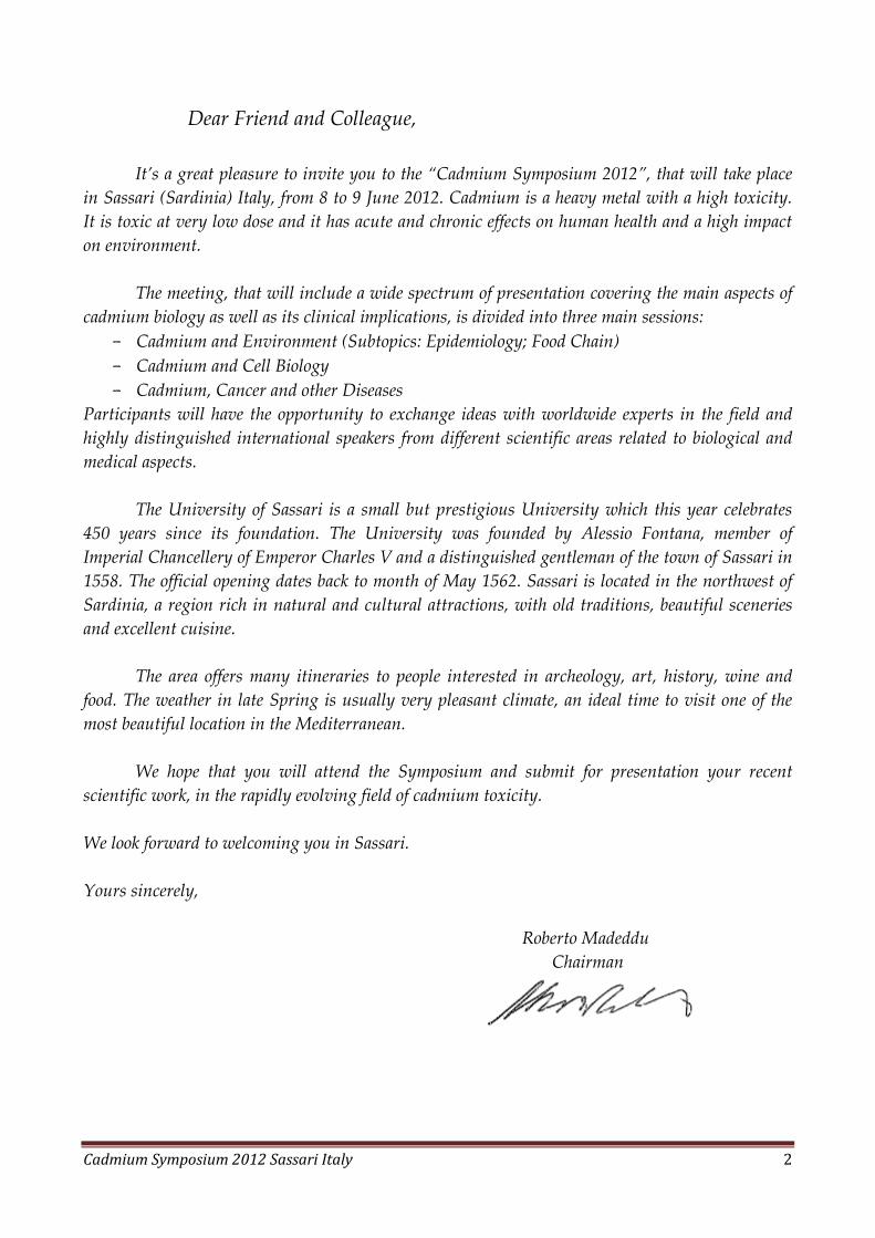
Dear Friend and Colleague,
It’s a great pleasure to invite you to the “Cadmium Symposium 2012”, that will take place
in Sassari (Sardinia) Italy, from 8 to 9 June 2012. Cadmium is a heavy metal with a high toxicity.
It is toxic at very low dose and it has acute and chronic effects on human health and a high impact
on environment.
The meeting, that will include a wide spectrum of presentation covering the main aspects of
cadmium biology as well as its clinical implications, is divided into three main sessions:
- Cadmium and Environment (Subtopics: Epidemiology; Food Chain)
- Cadmium and Cell Biology
- Cadmium, Cancer and other Diseases
Participants will have the opportunity to exchange ideas with worldwide experts in the field and
highly distinguished international speakers from different scientific areas related to biological and
medical aspects.
The University of Sassari is a small but prestigious University which this year celebrates
450 years since its foundation. The University was founded by Alessio Fontana, member of
Imperial Chancellery of Emperor Charles V and a distinguished gentleman of the town of Sassari in
1558. The official opening dates back to month of May 1562. Sassari is located in the northwest of
Sardinia, a region rich in natural and cultural attractions, with old traditions, beautiful sceneries
and excellent cuisine.
The area offers many itineraries to people interested in archeology, art, history, wine and
food. The weather in late Spring is usually very pleasant climate, an ideal time to visit one of the
most beautiful location in the Mediterranean.
We hope that you will attend the Symposium and submit for presentation your recent
scientific work, in the rapidly evolving field of cadmium toxicity.
We look forward to welcoming you in Sassari.
Yours sincerely,
Roberto Madeddu
Chairman
Cadmium Symposium 2012 Sassari Italy 3
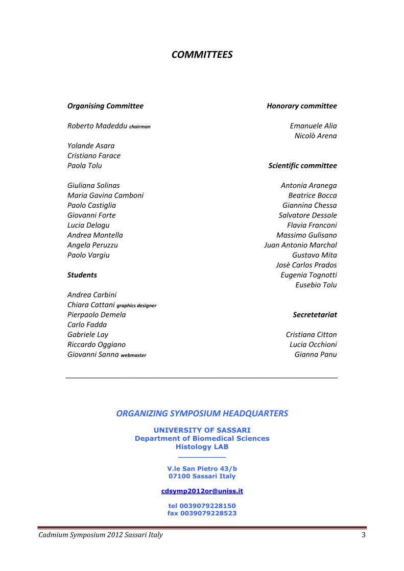
COMMITTEES
Organising Committee
Roberto Madeddu chairman Yolande Asara Cristiano Farace Paola Tolu Giuliana Solinas Maria Gavina Camboni Paolo Castiglia Giovanni Forte Lucia Delogu Andrea Montella Angela Peruzzu Paolo Vargiu Students
Andrea Carbini Chiara Cattani graphics designer Pierpaolo Demela Carlo Fadda Gabriele Lay Riccardo Oggiano Giovanni Sanna webmaster
Honorary committee
Emanuele Alia
Nicolò Arena
Scientific committee
Antonia Aranega
Beatrice Bocca Giannina Chessa
Salvatore Dessole Flavia Franconi
Massimo Gulisano Juan Antonio Marchal
Gustavo Mita Josè Carlos Prados
Eugenia Tognotti Eusebio Tolu
Secretetariat
Cristiana Citton
Lucia Occhioni Gianna Panu
___________________________________________________________________
ORGANIZING SYMPOSIUM HEADQUARTERS
UNIVERSITY OF SASSARI
Department of Biomedical Sciences
Histology LAB ___________
V.le San Pietro 43/b
07100 Sassari Italy
tel 0039079228150
fax 0039079228523
Cadmium Symposium 2012 Sassari Italy 4
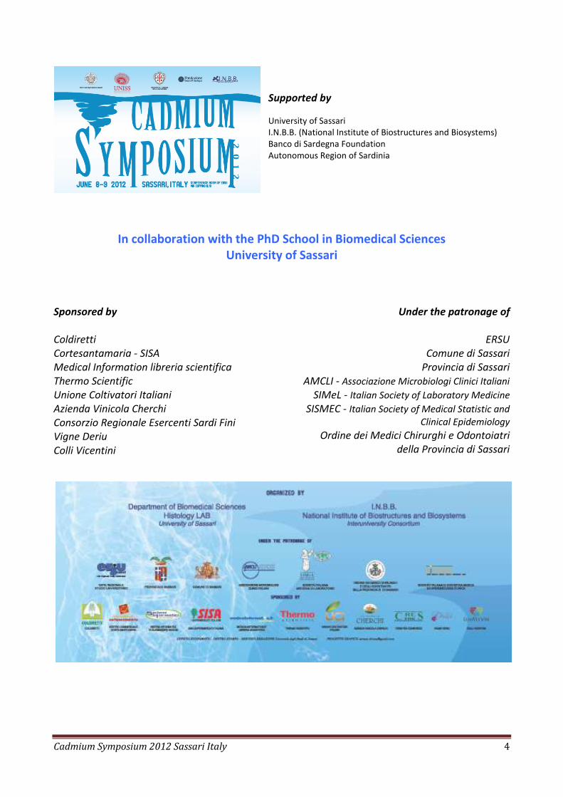
Supported by
University of Sassari I.N.B.B. (National Institute of Biostructures and Biosystems) Banco di Sardegna Foundation Autonomous Region of Sardinia
In collaboration with the PhD School in Biomedical Sciences
University of Sassari
Sponsored by
Coldiretti Cortesantamaria - SISA Medical Information libreria scientifica Thermo Scientific Unione Coltivatori Italiani Azienda Vinicola Cherchi Consorzio Regionale Esercenti Sardi Fini Vigne Deriu Colli Vicentini
Under the patronage of
ERSU
Comune di Sassari Provincia di Sassari
AMCLI - Associazione Microbiologi Clinici Italiani SIMeL - Italian Society of Laboratory Medicine
SISMEC - Italian Society of Medical Statistic and Clinical Epidemiology
Ordine dei Medici Chirurghi e Odontoiatri della Provincia di Sassari
Cadmium Symposium 2012 Sassari Italy 5
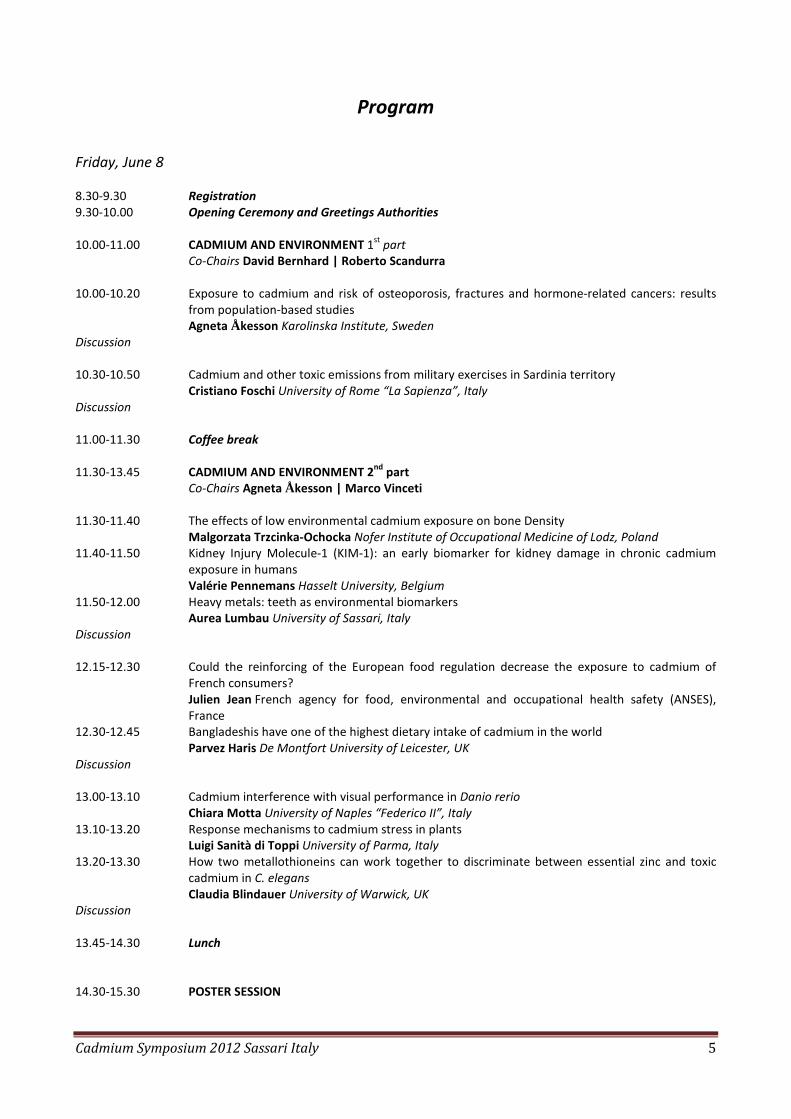
Program
Friday, June 8 8.30-9.30 Registration
9.30-10.00 Opening Ceremony and Greetings Authorities
10.00-11.00 CADMIUM AND ENVIRONMENT 1st part Co-Chairs David Bernhard | Roberto Scandurra
10.00-10.20 Exposure to cadmium and risk of osteoporosis, fractures and hormone-related cancers: results
from population-based studies Agneta Åkesson Karolinska Institute, Sweden
Discussion
10.30-10.50 Cadmium and other toxic emissions from military exercises in Sardinia territory Cristiano Foschi University of Rome “La Sapienza”, Italy
Discussion
11.00-11.30 Coffee break
11.30-13.45 CADMIUM AND ENVIRONMENT 2
nd part
Co-Chairs Agneta Åkesson | Marco Vinceti 11.30-11.40 The effects of low environmental cadmium exposure on bone Density
Malgorzata Trzcinka-Ochocka Nofer Institute of Occupational Medicine of Lodz, Poland 11.40-11.50 Kidney Injury Molecule-1 (KIM-1): an early biomarker for kidney damage in chronic cadmium
exposure in humans Valérie Pennemans Hasselt University, Belgium
11.50-12.00 Heavy metals: teeth as environmental biomarkers Aurea Lumbau University of Sassari, Italy
Discussion 12.15-12.30 Could the reinforcing of the European food regulation decrease the exposure to cadmium of
French consumers? Julien Jean French agency for food, environmental and occupational health safety (ANSES), France
12.30-12.45 Bangladeshis have one of the highest dietary intake of cadmium in the world Parvez Haris De Montfort University of Leicester, UK
Discussion 13.00-13.10 Cadmium interference with visual performance in Danio rerio
Chiara Motta University of Naples “Federico II”, Italy 13.10-13.20 Response mechanisms to cadmium stress in plants
Luigi Sanità di Toppi University of Parma, Italy 13.20-13.30 How two metallothioneins can work together to discriminate between essential zinc and toxic
cadmium in C. elegans Claudia Blindauer University of Warwick, UK
Discussion 13.45-14.30 Lunch
14.30-15.30 POSTER SESSION
Cadmium Symposium 2012 Sassari Italy 6
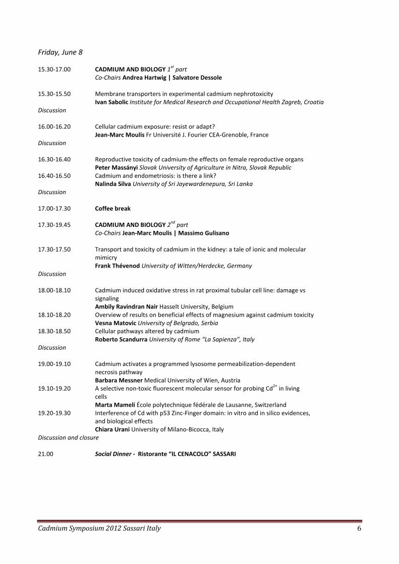
Friday, June 8
15.30-17.00 CADMIUM AND BIOLOGY 1st
part Co-Chairs Andrea Hartwig | Salvatore Dessole
15.30-15.50 Membrane transporters in experimental cadmium nephrotoxicity
Ivan Sabolic Institute for Medical Research and Occupational Health Zagreb, Croatia Discussion 16.00-16.20 Cellular cadmium exposure: resist or adapt?
Jean-Marc Moulis Fr Université J. Fourier CEA-Grenoble, France Discussion 16.30-16.40 Reproductive toxicity of cadmium-the effects on female reproductive organs
Peter Massányi Slovak University of Agriculture in Nitra, Slovak Republic 16.40-16.50 Cadmium and endometriosis: is there a link?
Nalinda Silva University of Sri Jayewardenepura, Sri Lanka Discussion 17.00-17.30 Coffee break
17.30-19.45 CADMIUM AND BIOLOGY 2nd
part Co-Chairs Jean-Marc Moulis | Massimo Gulisano
17.30-17.50 Transport and toxicity of cadmium in the kidney: a tale of ionic and molecular mimicry
Frank Thévenod University of Witten/Herdecke, Germany Discussion 18.00-18.10 Cadmium induced oxidative stress in rat proximal tubular cell line: damage vs
signaling Ambily Ravindran Nair Hasselt University, Belgium
18.10-18.20 Overview of results on beneficial effects of magnesium against cadmium toxicity Vesna Matovic University of Belgrado, Serbia
18.30-18.50 Cellular pathways altered by cadmium Roberto Scandurra University of Rome “La Sapienza”, Italy
Discussion 19.00-19.10 Cadmium activates a programmed lysosome permeabilization-dependent necrosis pathway Barbara Messner Medical University of Wien, Austria
19.10-19.20 A selective non-toxic fluorescent molecular sensor for probing Cd2+ in living cells
Marta Mameli École polytechnique fédérale de Lausanne, Switzerland
19.20-19.30 Interference of Cd with p53 Zinc-Finger domain: in vitro and in silico evidences, and biological effects
Chiara Urani University of Milano-Bicocca, Italy
Discussion and closure 21.00 Social Dinner - Ristorante “IL CENACOLO” SASSARI
Cadmium Symposium 2012 Sassari Italy 7
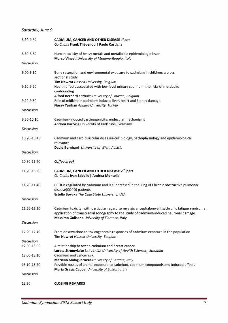
Saturday, June 9 8.30-9.30 CADMIUM, CANCER AND OTHER DISEASE 1st part
Co-Chairs Frank Thévenod | Paolo Castiglia 8.30-8.50 Human toxicity of heavy metals and metalloids: epidemiologic issue Marco Vinceti University of Modena-Reggio, Italy
Discussion 9.00-9.10 Bone resorption and environmental exposure to cadmium in children: a cross
sectional study Tim Nawrot Hasselt University, Belgium
9.10-9.20 Health effects associated with low-level urinary cadmium: the risks of metabolic confounding
Alfred Bernard Catholic University of Louvain, Belgium 9.20-9.30 Role of midkine in cadmium-induced liver, heart and kidney damage
Nuray Yazihan Ankara University, Turkey
Discussion 9.50-10.10 Cadmium-induced carcinogenicity: molecular mechanisms
Andrea Hartwig University of Karlsruhe, Germany
Discussion 10.20-10.45 Cadmium and cardiovascular diseases-cell biology, pathophysiology and epidemiological
relevance David Bernhard University of Wien, Austria
Discussion
10.50-11.20 Coffee break
11.20-13.20 CADMIUM, CANCER AND OTHER DISEASE 2
nd part
Co-Chairs Ivan Sabolic | Andrea Montella 11.20-11.40 CFTR is regulated by cadmium and is suppressed in the lung of Chronic obstructive pulmonar
disease(COPD) patients Estelle Boyaka The Ohio State University, USA
Discussion 11.50-12.10 Cadmium toxicity, with particular regard to myalgic encephalomyelitis/chronic fatigue syndrome;
application of transcranial sonography to the study of cadmium-induced neuronal damage Massimo Gulisano University of Florence, Italy
Discussion
12.20-12.40 From observations to toxicogenomic responses of cadmium exposure in the population Tim Nawrot Hasselt University, Belgium
Discussion 12.50-13.00 A relationship between cadmium and breast cancer
Loreta Strumylaite Lithuanian University of Health Sciences, Lithuania 13.00-13.10 Cadmium and cancer risk
Mariano Malaguarnera University of Catania, Italy
13.10-13.20 Possible routes of animal exposure to cadmium, cadmium compounds and induced effects Maria Grazia Cappai University of Sassari, Italy
Discussion 13.30 CLOSING REMARKS

Cadmium Symposium 2012 Sassari Italy 8
Oral
Communications
Cadmium Symposium 2012 Sassari Italy 9
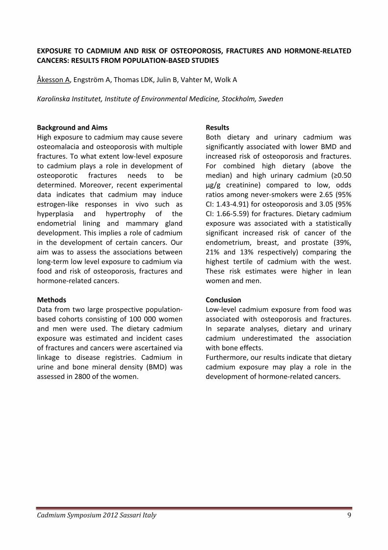
EXPOSURE TO CADMIUM AND RISK OF OSTEOPOROSIS, FRACTURES AND HORMONE-RELATED
CANCERS: RESULTS FROM POPULATION-BASED STUDIES
Åkesson A, Engström A, Thomas LDK, Julin B, Vahter M, Wolk A Karolinska Institutet, Institute of Environmental Medicine, Stockholm, Sweden Background and Aims
High exposure to cadmium may cause severe osteomalacia and osteoporosis with multiple fractures. To what extent low-level exposure to cadmium plays a role in development of osteoporotic fractures needs to be determined. Moreover, recent experimental data indicates that cadmium may induce estrogen-like responses in vivo such as hyperplasia and hypertrophy of the endometrial lining and mammary gland development. This implies a role of cadmium in the development of certain cancers. Our aim was to assess the associations between long-term low level exposure to cadmium via food and risk of osteoporosis, fractures and hormone-related cancers.
Methods
Data from two large prospective population-based cohorts consisting of 100 000 women and men were used. The dietary cadmium exposure was estimated and incident cases of fractures and cancers were ascertained via linkage to disease registries. Cadmium in urine and bone mineral density (BMD) was assessed in 2800 of the women.
Results
Both dietary and urinary cadmium was significantly associated with lower BMD and increased risk of osteoporosis and fractures. For combined high dietary (above the median) and high urinary cadmium (≥0.50 µg/g creatinine) compared to low, odds ratios among never-smokers were 2.65 (95% CI: 1.43-4.91) for osteoporosis and 3.05 (95% CI: 1.66-5.59) for fractures. Dietary cadmium exposure was associated with a statistically significant increased risk of cancer of the endometrium, breast, and prostate (39%, 21% and 13% respectively) comparing the highest tertile of cadmium with the west. These risk estimates were higher in lean women and men.
Conclusion
Low-level cadmium exposure from food was associated with osteoporosis and fractures. In separate analyses, dietary and urinary cadmium underestimated the association with bone effects. Furthermore, our results indicate that dietary cadmium exposure may play a role in the development of hormone-related cancers.

Cadmium Symposium 2012 Sassari Italy 10
CADMIUM AND OTHER TOXIC EMISSIONS FROM MILITARY EXERCISES IN SARDINIA TERRITORY
Carlo Brini1, Massimo Coraddu2, Mauro Cristaldi3, Cristiano Foschi3, M. Cristina Maltarello4, Fiorenzo Marinelli5, Germana Szpunar3and Lucio Triolo6 1ASL Biella, Veterinary Service, Italy 2DENERG, Department. Energetics, Politecnico di Torino, Italy 3Dipartimento di Biologia e Biotecnologie “C. Darwin”, Sapienza Università di Roma, Italy 4I.O. Rizzoli, Bologna, Italy 5Institute of Molecular Genetics CNR, I.O. Rizzoli, Bologna, Italy 6BAS-SIC (sez. Sicurezza Ambientale e Sanitaria), ENEA Casaccia, Roma, Italy Explosive testing, bullets impact on metallic armours and rockets launching cause atmospheric emissions of toxic elements (Al, As, Be, Cd, Co, Cr, Cu, Fe, Hg, Mn, Ni, Pb, Sb, Rb, Sr, Ti, 232Th, Tl, 238U, V, W, Zr), compounds (PAHs, CO, NOx, SO2, HCl, hydrocarbons, hydrazine, etc.), asbestos, PM10 and PM2,5
particulates. In particular, Cd is present in explosion emissions, in some solid propellants and in metallic armours. Final goal of this research is to define a monitoring project on toxic substances residues in the biotic and abiotic matrices in one of the Sardinia military areas (P.I.S.Q.=Poligono Interforze Salto di Quirra), including bioavailability studies on toxic substances. The biomonitoring should concern also the external areas of military territory. To this aim bioaccumulation analyses should be programmed starting from analytical data already available (ARPAS, 2010; Ist. Zooprofilattico Sperimentale per la Sardegna “G. Pegreffi”, 2011; Comm. Tec. Mista di Esperti, 2011). Therefore comparative analyses of these experimental data and epidemiological studies on population health could be carried out. Toxic elements, especially Cd, were found in soil samples collected in several internal sites (Perdasdefogu, Capo S. Lorenzo) and external areas to the PISQ.
Concentrations of these elements in many cases exceeded the environmental european limits (D. lgs. 152/06: G.U. Rep. Ital. 14/04/2006). These threshold concentrations were often exceeded by As and Tl residues, less frequently by Cd, Co, Cr and Sb soil contents. Ist. Zooprofilattico found higher concentrations of one or several toxic elements with respect to law threshold limits in 16,3% of livestock water samples collected in PISQ farms. Moreover, 52% and 64% liver samples of domestic Ruminants, living in PISQ farms and in control areas respectively, exceeded the law limits referred to Cd concentration. Finally, 90% and 100% of kidney samples of domestic Ruminants, living in PISQ farms and in “control” areas respectively, have shown Cd concentration higher than law thresholds. This result was probably due to a chemico-physical diffusion of contaminants in environmental matrices from PISQ boundaries. In order to characterize the environmental risk it would be important to study more territorial risk factors, which are relevant to determine the synergic effects of physical and chemical pollutants (i.e. ionizing radiations and electromagnetic fields interact with chemical pollutants enhancing the transgenerational and carcinogenetic risks: to see AIOM, Progetto Ambiente e Tumori, 2011).
Cadmium Symposium 2012 Sassari Italy 11
THE EFFECTS OF LOW ENVIRONMENTAL CADMIUM EXPOSURE ON BONE DENSITY
Malgorzata Trzcinka-Ochocka, Marek Jakubowski, Renata Brodzka Nofer Institute of Occupational Medicine, Laboratory of Biomonitoring, Lodz, Poland Background and Aims
Recent epidemiological data indicate that low environmental exposure to cadmium, as shown by cadmium body burden (Cd-U), is associated with renal dysfunction as well as an increased risk of cadmium-induced bone disorders. The present study was designed to assess the effects of low environmental cadmium exposure, at the level sufficient to induce kidney damage, on bone metabolism and bone mineral density (BMD).
Methods
The project was conducted in the area contaminated with cadmium, nearby a zinc smelter located in the region of Poland where heavy industry prevails. The study population comprised 170 women (mean age = 39.7) and 100 men (mean age = 31.9 years). Urinary and blood cadmium (Cd-U and Cd-B) and the markers of renal tubular dysfunction: urinary β2-microgobulin (β2M-U), retinol-binding protein (RBP-U), N-acetyl-b-d-glucosaminidase NAG, and markers of glomerular dysfunction: urinary albumin (Alb-U) and serum β2-microglobulin (β2M-S) as well as bone metabolism markers: bone alkaline phosphatase in serum (BAP-S) and C-terminale telopeptide (CTX-S) as well as forearm bone mineral density (BMD), were measured. Forearm BMD has been assessed by technique – dual energy X-ray absorptiometry (DXA).
Results
The results of simple dose-effect analysis showed the relationship between increasing cadmium concentrations and an increased excretion of renal dysfunction markers and decreasing bone density. However, the results of the multivariate analysis did not indicate the association between exposure to cadmium and decrease in bone density. They showed that the most important factors that have impact on bone density are body weight and age in the female subjects and body weight and calcium excretion in males. In the male population, the excretion of low molecular weight proteins occurred at a lower level of cadmium exposure than the possible loss of bone mass.
Conclusion
It seems that renal tubular markers are the most sensitive and significant indicators of early health effects of cadmium intoxication in the general population. The correlations Cd with markers of kidney dysfunction were observed in the absence of significant correlations with bone effects. Our findings did not indicate any effects of environmental cadmium exposure on bone density They are in contradiction to the results of many reported studies.
Cadmium Symposium 2012 Sassari Italy 12
KIDNEY INJURY MOLECULE 1 (KIM-1): AN EARLY BIOMARKER FOR KIDNEY DAMAGE IN CHRONIC
CADMIUM EXPOSURE IN HUMANS
Valérie Pennemans, Liesbeth M De Winter, Tim Nawrot, Elke Munters, Harrie De Witte, Emmy Van Kerkhove, Jean-Michel Rigo, Carmen Reynders, Joris Penders, Quirine Swennen Hasselt University, Belgium Background and Aims
Kidney injury molecule 1 (KIM-1) is a recently discovered biomarker for tubular kidney damage. Animal studies have shown that urinary KIM-1 levels are the first to rise after cadmium (Cd) exposure, suggesting that KIM-1 is a very early biomarker for Cd-induced tubular damage. It is also known that urinary Cd levels reflect lifelong exposure to Cd in humans. The goal of this study is to examine the correlation between KIM-1 and Cd in human urine samples. Methods
Urine samples were collected from 153 healthy, non-smoking 65-plus volunteers living in a region adjacent to a heavy-metal industrial zone. KIM-1 and Cd concentrations in urine were determined together with
other established renal biomarkers (α1-microglobulin, proteinuria, BUN). Results
A positive correlation between urinary KIM-1 and Cd after adjustment for sex, age, past smoking, BMI and socio –economic status was found (p=0.013). In contrast, no association was found between urinary Cd concentrations and urinary α1-microglobulin (p=0.202), proteinuria (p=0.571) or BUN (p=0.0621). Conclusion
In conclusion, these results show that urinary KIM-1 outperforms other renal biomarkers as an early biomarker for Cd-induced renal damage after lifelong Cd exposure in humans

Cadmium Symposium 2012 Sassari Italy 13
HEAVY METALS: TEETH AS ENVIRONMENTAL BIOMARKERS
Aurea Lumbau1, P.F. Lugliè1, Donatella Carboni2, Sergio Ginesu3, Simonetta Falchi2 and Laura Schinocca1 1Department of Surgery, Microsurgery and Medical Sciences, Dental Clinic, University of Sassari, Italy 2Department of Human and Social Sciences, University of Sassari, Italy 3Department of Nature and Territory, University of Sassari, Italy Background and Aims
Aim of this study was to measure the concentration of heavy metals in tooth matrix and to determine the factors that affect their presence. During tooth development and mineralization several metals can be absorbed in the tooth matrix, thus allowing us to use them as biological markers. Like in a bone, calcium can be partially substituted by a small amount of heavy metals (Boivin et al., 1996; Kwapulinski et al., 2003). This is rather a complex process that is affected by various factors including the chemical form of the metal and its binding sites, age, gender, environmental quality (Tvinnereim et al., 2000; Burguera et al., 2002). The way the metals are accumulated in a calcified tissue also reflects the interactions between elements (Lappalainen and Knuuttila, 1982). These elements cannot be eliminated and their toxicity results from their affinity to the sulfhydryl groups, which causes the formation of an insoluble complex by limiting cellular metabolism; abnormal enamel thus originates in the tooth by means of the competition with calcium. Cadmium alters the calcium/phosphorus turnover thus determining demineralization, osteomalacia and pathological fractures (Staessen, 1999). Methods
Using an inductively coupled plasma mass spectrometry we calculated the concentration of some heavy metals –
primarily uranyl ions (50 samples) – in the area of the military base of Escalaplano and then, using a Graphite Furnace Atomic Absorption Spectrometry (GFAA), we calculated the concentration of 4 heavy metals (Pb, Cd, Cu, Ni) in 91 caries-free teeth belonging to patients from three different Sardinian cities: Sassari, Ottana and Porto Torres. These cities were chosen with regard to their position and to the job opportunities they offered. Several dentists and patients took part in this research. Questionnaires were submitted to the patients in order to gather information such as personal data, qualification, residence, profession, diet, drunk water (spring, well or bottled up water), smoking habits and medication taken. Results
The mean concentration of Pb, Cu, Cd, Ni, was respectively 3,46±3,20 µg/g, 0,419±0,363 µg/g; 0,0257±0,0249 µg/g; <0,02 µg/g. Our results show correlations between different kinds of teeth, age and residence. The Pb e Cd concentration was higher for smokers (Pb 4,44±3,50µg/g, Cd 0,04±0,01 µg/g) than for no smokers (Pb 2,45±2,03 µg/g; Cd 0,028±0,015µg/g). Conclusions
Our work demonstrates that teeth are valuable markers of environmental pollution exposition and that teeth are permanent markers of exposition to polluting agents.
Cadmium Symposium 2012 Sassari Italy 14
COULD THE REINFORCING OF THE EUROPEAN FOOD REGULATION DECREASE THE EXPOSURE TO
CADMIUM OF FRENCH CONSUMERS?
Julien Jean1, Véronique Sirot2, Paule Vasseur3, Jean-François Narbonne4, Jean-Charles Leblanc1, Gilles Riviere1 1Contaminant Risk Assessment Unit - ANSES Maisons-Alfort, France 2Methodology and studies for physical and chemical risk assessment Unit - ANSES Maisons-Alfort, France 3Laboratoire de Physico-Toxico Chimie des Systèmes Naturels, Université Bordeaux 1, Talence, France 4Interactions Écotoxicologie Biodiversité Écosystèmes – Université Paul Verlaine de Metz – UMR CNRS Metz, France Background and Aims
Recently, several Food Safety Agency recommended to reduce the exposure to cadmium. A revision of the Maximum Levels for cadmium in foodstuffs (ML) is currently in discussion within the European Commission and the Member States. The 2nd French Total Diet Study (FTDS2) demonstrated that 0.6% of adults and 14.9 % of children exceed the tolerable weekly intake (TWI) of 2.5 µg/kg body weight per week. On this basis, the French agency for food, environmental and occupational health safety (ANSES) assessed whether a reinforcing of European ML in foodstuffs could significantly reduce the level of exposure of the French population. Method
The foodstuffs mainly contributing to the cadmium exposure in the general population and for the most exposed individuals were identified on the basis of the results of the FTDS2. For these foodstuffs, cut off limit scenarios have been applied on the distribution of contamination. These cut off levels was set at P90 and P95 as a ML and exposure were recalculated by combining data from the TDS2 and from the French Consumption Survey.
Results
The overexposure of several consumers can be partially explained by lower body weight and a variety of particular dietary patterns resulting in high food consumption of bread and dried bread products, of bivalve molluscs and potatoes. Excepted molluscs, these foods are the main contributors identified for the general population. The percentage of children exceeding the TWI should strongly decrease once the children reach adulthood, as it results from the children’s low body weight rather than any particular dietary pattern. Applying MLs set at P90 and P95 of the main contributors would neither significantly reduce exposure levels to cadmium for the general population, nor the percentage of subjects exceeding the TWI. Conclusions
To significantly reduce background consumer exposure to cadmium, a discussion should be initiated on the impact of acting on sources that are at the origin of the contamination levels of soil and foods. Furthermore, it could be of interest to assess the efficacy of consumption recommendations.
Cadmium Symposium 2012 Sassari Italy 15
BANGLADESHIS HAVE ONE OF THE HIGHEST DIETARY INTAKE OF CADMIUM IN THE WORLD
S.W. Al-Rmalli, R.O. Jenkins and P.I. Haris Faculty of Health & Life Sciences, De Montfort University, Leicester, UK The population in Bangladesh is exposed to high levels of arsenic through drinking water and consumption of rice. Our recent studies indicate that they may be also exposed to very high levels of Cadmium. We determined Cd levels in 327 and 94 samples of Bangladeshi food and non-food samples, respectively, using inductively coupled plasma mass spectrometry. This is the largest number of Bangladeshi food and nonfood samples investigated for their Cd content. High Cd levels were detected in leafy vegetables (mean 31 [SD 29]μg/kg). Of these vegetables, lal shak (Amaranthus tricolor) contained the highest Cd level (303 μg/kg [wet weight]; mean 100.5 [SD 95]μg/kg). Bangladeshi rice also showed significant concentration of Cd (mean 37.2 [SD 30]μg/kg). Of particular concern is the very high level of Cd detected in some puffed rice, which we attribute to the illegal practice of using urea for whitening the puffed rice. Tobacco leaves,
which are commonly consumed during betel quid chewing by Bangladeshis, contain significant levels of Cd (mean 95 [SD 87]μg/kg). The total daily intake (TDI) of Cd from foods for Bangladeshis was estimated to be 34.55 μg/d. This is rather high when compared to the TDI of Cd for other populations. Our analysis reveals that this is mainly due to the very high intake of rice and vegetables, and lower consumption of animal products (which are low in Cd), by the Bangladeshis. We also determined the provisional maximum tolerable daily intake and target hazard quotients values for Cd. Clearly a more balanced diet is necessary to reduce the Cd intake in the Bangladeshi population, especially by reducing the very high intake of rice and certain leafy vegetables. Food manufacturing and agricultural practices needs to be altered to reduce the entry of Cd into the food chain.
Cadmium Symposium 2012 Sassari Italy 16
CADMIUM INTERFERENCE WITH VISUAL PERFORMANCE IN Danio rerio
Chiara Motta, Raffaele Panzuto, Palma Simoniello, Roberta Crispino, Bice Avallone Department of Biological Sciences, University of Naples “Federico II”, Italy Sight is the sense mainly used by teleosts to hunt for food and escape predators. As a consequence, the eyes are large and a complex visual behavior has been developed. This consist in a series of ‘instinctive’ responses that the animal puts into action when exposed to a stimulation of visual nature. These mechanisms are potentially threatened by the fact that the eye is an easy target for many xenobiotics: in fact, it is directly exposed to the environment and, also, contains a large vitreal chamber that facilitates contaminants distribution to all its compartments. In the present work, we verified whether cadmium, at environmental concentrations, interferes with the visually mediated behaviors in adult Danio rerio, a model system for behavioral screens since 1970’s. Re-illumination tests were carried out with white and colored lights (red, yellow, green and blue), to test color sensitivity in fish contaminated for 30 days.
Results reveal that cadmium exposure induces a marked escape response to all light wavelengths. Parallel light and electron microscope investigations demonstrate that cadmium alters retinal organization: the ion, in particular, induces occasional retinal folding, a marked swelling, especially in the ganglion cell and, most important, induces degeneration among ganglion and inner nuclear layer cells. In conclusion, results indicate that cadmium has detrimental effects on visual behavior and that this probably depends on an altered signals transmission from the photoreceptors to the central nervous system. Further studies are required to fully understand the mechanisms underlying cadmium response in the retina; however, the evidences collected so far clearly indicate that animals living in contaminated sites have a reduced fitness. The implications at the ecological level are obvious.
Cadmium Symposium 2012 Sassari Italy 17
RESPONSE MECHANISMS TO CADMIUM STRESS IN PLANTS
Luigi Sanita' di Toppi Department of Evolutionary and Functional Biology, University of Parma, Italy As a general approach, the research performed in my lab is founded on the mechanistic hypothesis according to which plants (as well as algae, fungi and lichens) do not protect themselves against Cd stress with a sole, unique, response mechanism, but rather they put into action a number of stress-containment strategies, based on several parallel and/or consecutive events. This multi-component model, which I would call “fan-shaped” response, may agree with the Selyean “general adaptation syndrome” hypothesis. In this sense, the experimental work conducted by my research group, as far as Cd stress in plants is concerned, deals with the following studies: 1) the biosynthesis of phytochelatins, thiol-peptide compounds which have the following general structure: (gammaGlu-Cys)n-Gly, with n=number of repetition of the unit gammaGlu-Cys, normally variable from 2 to 11. Phytochelatins form various complexes with Cd, due to the presence of the thiolic groups of Cys, which chelate Cd and, as a result, prevent it from circulating inside the cytosol. An useful tool for such research is also the utilization of overexpressing plants for the phytochelatin synthase gene AtPCS1; 2) the Cd-induced synthesis of antioxidant metabolites (mainly glutathione, ascorbate and tocopherols) and the metal’s influence on the synthesis/activities of some
antioxidant enzymes, such as peroxidase, superoxide dismutase, catalase, (mono) dehydroascorbate reductase, ascorbate peroxidase and glutathione reductase, mostly involved in the Halliwell-Asada pathway; 3) the possible Cd-induced lipid membrane peroxidation; 4) the differential Cd distribution inside the plant cell, evaluated by cell wall immobilization, plasma membrane exclusion, intra cytosolic/organellar accumulation and vacuolar compartmentalization; 5) the structural and ultrastructural Cd-generated damage and the molecular sensing amongst Cd, nitric oxide and programmed cell death, supported by the expression of the marker senescence-associated gene12 (SAG12); 6) the proteome plasticity in the Cd response determined by a remodeling of protein synthesis in terms of differential expression of several putative proteins. An understanding of the mechanisms by which plants perceive, transduce and counterbalance Cd-induced stress signals to initiate acclimation or adaptive responses can be essential for obtaining plants with increased tolerance to metal stress, thus contributing to the improvement of so-called “phytoremediation”, an important tool for removing Cd and other environmental contaminants from soils and waters.
Cadmium Symposium 2012 Sassari Italy 18
HOW TWO METALLOTHIONEINS CAN WORK TOGETHER TO DISCRIMINATE BETWEEN ESSENTIAL
Zn AND TOXIC Cd IN C. elegans
Claudia A. Blindauer1, Oksana I. Leszczyszyn1, Sukaina Zeitoun-Ghandour2 and Stephen R. Stürzenbaum2 1Department of Chemistry, University of Warwick, CV4 7AL Coventry United Kingdom 2Analytical and Environmental Science Division, School of Biomedical Sciences, King’s College London, SE1 9NH London, United Kingdom Background and Aims
Organisms that are in direct contact with soil and water, such as plants, invertebrates and bacteria, need robust mechanisms to discriminate between essential zinc and toxic cadmium. Metallothioneins (MTs) may play unexpected roles in such mechanisms; this will be illustrated by recent findings on the two MTs (MTL-1 and MTL-2) of the nematode C. elegans.
Methods
Zn- and Cd-binding constants of both metallothionein isoforms were determined by competition with the metal chelator 1,2-bis(5-fluoro-o-aminophenoxy)ethane-N,N,N',N'-tetraacetic acid (5F-BAPTA) and 19F NMR spectroscopy. Speciation at different Zn:Cd ratios were studied by native Electrospray Ionisation MS.
Results
As dictated by basic coordination chemistry principles, both MTL-1 and MTL-2 bind soft Cd2+ more strongly than borderline Zn2+, as soft cysteine thiolates provide the majority of metal-binding capacity. However, whilst both MTs bind Zn2+ with the same overall affinity,
their Cd-binding constants differ by almost two orders of magnitude. As a consequence, in a mixture containing MTL-1, MTL-2, Cd2+, and Zn2+, Cd2+ will predominantly bind to MTL-2, whilst MTL-1 is left to bind the remaining Zn2+. The distributions calculated from measured affinity constants are reproduced closely by the results from ESI-MS.
Conclusions
The system demonstrates the importance of "relative affinities". Since basic coordination chemistry principles prohibit the construction of metallothioneins which bind Zn2+ more strongly than Cd2+, the affinities of MTL-1 and -2 are tuned in such a way that the combination Cd-MTL-2 is the overall strongest. Hence, providing sufficient amounts of proteins are expressed in the organism, even though MTL-1 still binds Cd2+ more strongly than Zn2+, most Cd2+ will end up bound to MTL-2, and will therefore not be available for binding to MTL-1. The biophysical data correlate well with physiological data, which indicated that the metabolic pathways for Cd and Zn differ.
Cadmium Symposium 2012 Sassari Italy 19
MEMBRANE TRANSPORTERS IN EXPERIMENTAL CADMIUM NEPHROTOXICITY
Ivan Sabolić1, Marija Ljubojević1, Davorka Breljak1, Carol M Herak-Kramberger1, Naohiko Anzai2, Hermann Koepsell3 1Molecular Toxicology, Institute for Medical Research and Occupational Health, Zagreb, Croatia 2Pharmacology & Toxicology, Dokkyo Medical University School of Medicine, Mibu, Tochigi, Japan 3Anatomy & Cell Biology, University of Würzburg, Würzburg, Germany Background and Aims
The symptoms of cadmium (Cd) nephrotoxicity (Cd-NTX) in humans and experimental animals manifest in the defects of reabsorptive and secretory functions of proximal tubules (PT), and include phosphaturia, aminoaciduria, glucosuria, proteinuria, increased excretion of organic anions and cations, and polyuria. These symptoms indicate that Cd targets various transporters in the PT brush-border (BBM) and basolateral (BLM) membrane. The aim of this study is to characterize the expression of these transporters in experimental subchronic and acute model of Cd-NTX in rats. Methods
Cd-NTX was induced by treating rats s.c. with CdCl2 (2 mg Cd/kg b.m./day for 14 days; subchronic model) or Cd-metallothionein (CdMT; a single dose of 0.4 mg Cd/kg b.m. 6 or 12 hours before sacrifice; acute model). Control animals were vehicle-treated. Various methods (immunocytochemistry, Western blotting, transmission and immunogold microscopy, end-point RT-PCR) were applied to study the expression of transporters localized in the PT BBM (V-ATPase, NaPi2, megalin, NHE3, SGLT1,
SGLT2), BLM (Na/K-ATPase, OAT1, OAT3, OCT1, OCT2), or in both membranes (AQP1). Results
In both models of Cd-NTX, PT exhibited loss of BBM and BLM. In subchronic model, the expression of specific transporters was strongly downregulated at the level of protein and mRNA. In acute model we observed: a) time-dependent loss of various BBM transporters and their accumulation in the randomly scattered intracellular vesicles, b) redistribution of NHE3 into the BLM, and c) time-dependent loss of various BLM transporters, and their redistribution in intracellular vesicles that accumulated in the cell subapical domain. Conclusions
The data indicate that the functional defects of PT in Cd-NTX result from: a) loss of absorptive and secretory surface, b) loss of transporting proteins in BBM and BLM, and c) loss of cell polarity. In subchronic Cd-NTX, loss of membrane transporters is largely mRNA-related, whereas in acute Cd-NTX, loss of membrane transporters due to derranged intracellular vesicle trafficking seems to be the major phenomenon.
Cadmium Symposium 2012 Sassari Italy 20
CELLULAR CADMIUM EXPOSURE: RESIST OR ADAPT?
Jean-Marc Moulis Institut de Recherches en Sciences et Technologies du Vivant CEA-GRENOBLE LCBM UMR CNRS 5249 Université J. Fourier, Grenoble, France Background and Aims
Although low-level and transient cadmium (Cd) exposure of mammalian cells in culture may enhance growth, long term cadmium challenge usually leads to cell death. Survival occurs upon molecular and cellular changes which allow cells to cope with the insult, but the underlying mechanisms are often unknown or incompletely described. Methods
Based on the chemical similarities between zinc and cadmium ions, a cell line was isolated which displayed lethal doses ca. 3 times higher for Zn2+, but more than one order of magnitude above that of control cells for Cd2+. The reasons for this spectacular insensitivity to Cd were sought by implementation of complementary methods, including measurement of metal concentrations and transport, of cellular damage and stress, microscopic metal imaging, proteomic and transcriptional analysis. Results
Whereas cellular resistance to toxic compounds, including cadmium, often relies
on their efficient export, impaired import was mainly responsible for the observations made with the present isolate. As a consequence, cells remained protected from cadmium induced damage and stress. Differential transcriptomic and proteomic analysis of Cd-insensitive and Cd-susceptible cells revealed the biological pathways which most effectively contributed to cellular Cd handling. Conclusions
The use of cellular models developed by sustained exposure to toxic species, including metals, are mimics of chronic poisoning, and they highlight behaviors contrasting with those observed with sudden acute insults to naïve cells. Comparison of the molecular pathways targeted by Cd in these contrasting situations defines the differences between the mechanisms supporting immediate resistance to Cd and long-term adaptation to the insult. Such analysis reveals the diversity of the Cd's mechanisms of action, hence illustrating the need to integrate extensive datasets for different cell types and conditions of exposure to deepen our understanding of Cd toxicology.
Cadmium Symposium 2012 Sassari Italy 21
REPRODUCTIVE TOXICITY OF CADMIUM – THE EFFECTS ON FEMALE REPRODUCTIVE ORGANS*
Peter Massanyi, Norbert Lukac, Robert Toman, Grzegorz Formicki, Robert Stawarz Slovak University of Agriculture, Nitra, Slovak Republic and Pedagogical University, Krakow, Poland In this study histological alteration of rabbit female reproductive organs after an experimental cadmium administration were analyzed. After a cadmium administration changes in the number of follicles with <2 layers, >2 layers of granulosa cells and antral follicles were detected. The number of atretic follicles was significantly higher in experimental groups with cadmium. The relative volume of growing follicles was significantly decreased and that of stroma significantly increased in experimental groups compared to control, directly suggesting the effect of cadmium on folliculogenesis. In uterus rapid edematization occurs caused by
the blood vessel dilatation, vessel wall disarrangement and diapedesis. In detail study the effect of cadmium on the rabbit ovarian cell ultrastructure was examined. Qualitative analysis determined undulation of nuclear membrane, dilated perinuclear cistern and endoplasmic reticulum. Qualitative analysis proved alterations in cell structures. Obtained data were confirmed also in in vitro conditions. The results proved negative effect of cadmium on the ovarian structure on the level of light as well as electron microscopy. *Support VEGA 1/0532/11
Cadmium Symposium 2012 Sassari Italy 22
CADMIUM AND ENDOMETRIOSIS: IS THERE A LINK?
N Silva, K H Tennekoon, H Senanayake, S Samarakoon, S Fernando, R Wickramasinghe, R Peiris-John Faculty of Medical Sciences, University of Sri Jayewardenepura, Sri Lanka Background and Aims
Metalloestrogen cadmium(Cd), is known to activate the oestrogen receptors to exert oestrogenic effects; thus implicated in the aetiology of endometriosis; an oestrogen dependent disease. The aim of this study was to elucidate the association between cadmium and endometriosis. Methods
A case-control study was conducted in a tertiary care hospital. Patients with endometriosis (patients) were compared with age matched normal women (controls) confirmed by laparoscopy or laparotomy. Blood samples(n=50 in each group) and ectopic endometrial tissue samples(n=50) were obtained and digested with supra pure 65% HNO3. Samples were analyzed for Cd by graphite furnace atomic absorption spectroscopy(GFASS). Eutopic endometrial samples were obtained in controls (n=5) and patients (n=5) to isolate endometrial stromal cells (ESC). Primary ESC cultures and subcultures were established in DMEM/F12 medium supplemented with 10% FCS and 1% antibiotic antimycotic. Cultures in the third passage were treated with Cd at a concentration of 10-6M. At 24 h and 48 h, following trypsinization, cell number was counted using the Neubauer haemocytometer. ESC were subjected to immunohistochemical staining with primary antibodies for oestrogen receptor alpha (ER) and progesterone receptor (PR).
Sulphorhodamine (SRB) cytotoxicity assay was used to test the effect of different concentrations of Cd on ESC cultures. After 24 h of treatment, caspase levels in ESC cultures were evaluated with a commercially available ELISA kit. Log transformed blood and tissue levels of cadmium were compared with t-tests while relative cell proliferation, SRB assay results and caspase levels were analyzed with ANOVA. Results
Cadmium levels [geometric mean (95% CI)] were significantly higher in the ectopic endometrial tissue than in blood [2.861 (2,126-3.596) vs 0.836(0.695-0.977) μg/L; p=0.001] in patients but the blood levels were similar in two groups [0.836 (0.695-0.977) vs 0.856 (0.658-1.055) μg/L]. At 48 h, cell proliferation was higher in patients (p=0.02) than in controls. Treatment with Cd reduced expression of ER and increased expression of PR in the ESC from patients which were most prominent at 48 h. SRB assay results and caspase levels were similar in the two groups. Conclusions
Metalloestrogen cadmium appears to accumulate in the ectopic endometrial tissue and was capable of inducing oestrogenic effects in cultured endometrial stromal cells. Cd induced the proliferation of ESC from women with endometriosis appears to be independent of reduced apoptosis.
Cadmium Symposium 2012 Sassari Italy 23
TRANSPORT AND TOXICITY OF CADMIUM IN THE KIDNEY: A TALE OF IONIC AND MOLECULAR
MIMICRY
Frank Thévenod Institute of Physiology and Pathophysiology, University of Witten/Herdecke, Faculty of Health, Department of Medicine, Stockumer, Germany As part of industrial developments increasing usage of Cd has led to widespread contamination of the environment that threatens human health, particularly today. Rather than acute, lethal exposures, the real challenge in the 21st century in a global setting seems to be chronic low Cd exposure (CLCE), mainly from dietary sources, which is associated with chronic organ toxicity, especially nephrotoxicity, and may lead to chronic organ fibrosis and failure as well as to cancer development. To enter the intracellular space, Cd ion (Cd2+) in extracellular fluids that is present as a free ion or complexed to proteins or peptides must permeate lipophilic cellular membranes. Free Cd2+ may be transported via ion channels and solute carriers and Cd2+ complexes may be taken up through receptor-mediated endocytosis. Cd2+ has similar chemical properties as essential metals (“ionic mimicry”) and Cd2+ complexes are analogous to endogenous biological molecules. Hence transport (and toxicity) of Cd2+ occurs through transport pathways for essential metals or biological molecules.
A variety of pathways have been suggested to allow Cd2+ entry in excitable and non-excitable cells. But it is important to know that free blood Cd2+ concentrations in the general population are in the range of 1-10 nM and may not exceed ~100-200 nM in occupationally exposed workers. The free Cd2+ concentrations in the extracellular fluid that cause tissue damage are prabably in the submicromolar range. Hence, most of the studies describing transport of Cd2+ may have only in vitro or mechanistic relevance and are not likely to significantly contribute to the in vivo toxicity of Cd2+ in tissues, including the kidney. Here I discuss several likely candidates for Cd2+ entry into cells, whose molecular structures have been identified and which have been characterized in kidney cells or heterologous expression systems. *Supported by DFG TH345/11-1 and ZBAF
Cadmium Symposium 2012 Sassari Italy 24
CADMIUM INDUCED OXIDATIVE STRESS IN RAT PROXIMAL TUBULAR CELL LINE: DAMAGE VS
SIGNALING
Ambily Ravindran Nair1, Karen Smeets1, Wing-Kee Lee2, Frank Thévenod2, Emmy Van Kerkhove1, Ann Cuypers1
1Centre for Environmental Sciences, Hasselt University, Diepenbeek, Belgium 2ZBAF, Institute of Physiology and Pathophysiology, University of Witten/Herdecke, Witten, Germany Background and Aims
Cadmium (Cd) is a toxic metal, omnipresent in the environment. Chronic exposure to Cd targets the proximal tubular cells of kidney and leads to damage via oxidative stress. Our study aims to understand the mechanisms behind signaling versus death scenario in WKPT 0293 Cl.2 cells (a rat-derived cell line from the S1 segment of proximal tubule), in the context of oxidative stress. Methods
A viability study was conducted to choose Cd concentrations that vary from no to significant damage. Markers of oxidative stress like hydrogen peroxide and lipid peroxidation were analyzed using cell-based assays. A screening for stable reference genes in our experimental set up was conducted to normalize gene expression. Eight candidate reference genes (Aip, Cxxc1, Ppia, Hprt1, Gapdh, ActB, Tuba1a, and Ywhaz) were chosen for this purpose. Genes involved in oxidative stress (Mt1a, Mt2a, Mt3, Sod1, Sod2, Gpx4, Prdx2, and Cat) were analyzed using qPCR. Estimation of the number of mitochondria at different doses was also done using mitochondrial encoded genes ND1, Cox1, Cox2 and Cox3. Results
The final Cd doses chosen were 1, 10 and 30µM and the LC50 value was 35µM.
Hydrogen peroxide levels were increased significantly at 30µM while lipid peroxidation showed a decreasing trend until 10µM, which subsequently got reversed and approached control concentrations at 30µM. The most stable reference genes in our experimental set up were Gapdh, Ywhaz and Actb. Mt1a and Mt2a transcripts were significantly up-regulated at 10µM and 30µM, while Mt3 showed a decreasing trend until 1µM that reversed and increased towards 30µM. Sod1 was up-regulated only at 30µM. The other antioxidant genes were not significantly altered, but Cat showed an increasing trend. Gene expression of mitochondrial genes was normalized against genomic DNA and showed an up-regulation at 1µM which decreased below the control values at 30µM. Conclusion
Our study suggests three multiple reference genes for accurate qPCR quantification in WKPT-0293 Cl.2 cell lines under Cd stress. Our results also suggest that cells survive lower Cd concentrations such as 1µM altering signaling pathways for adaptation, while 10µM is damaging, leading to cell death. Further investigations on signaling pathways are ongoing, and will reveal more clues to better understand what decides to ‘die’ or ‘not to die’.
Cadmium Symposium 2012 Sassari Italy 25
OVERVIEW OF OUR RESULTS ON BENEFICIAL EFFECTS OF MAGNESIUM AGAINST CADMIUM
TOXICITY*
Vesna Matović, Zorica Bulat, Danijela Đukić-Ćosić, Aleksandra Buha Department of Toxicology "Akademik Danilo Soldatović", Faculty of Pharmacy, University of Belgrade, Serbia Very important issue in toxicological practice is the therapy of Cd intoxication since the standard therapy with chelating agents does not produce satisfactory results. Our previous as well as other authors’ investigations on interactions between Cd and Mg encouraged us to start experimental studies with aim to determine whether Mg could be effective as a protective agent against Cd toxicity. The effect of excessive Mg intake on Cd toxicity was investigated in rabbits, rats and mice exposed to acute or prolonged Cd intoxication. Investigations carried out on rabbits given orally every day for 4 weeks 10 mg Cd/kg b.w. showed that co-treatment with 40 mg Mg/kg b.w. induced decrease of Cd content in blood, kidney, spleen and bone if compared with the group given only Cd. In our further investigations performed on mice we wanted to determine not only the effect of excessive oral intake of Mg on the content of Cd in different organs of mice exposed to acute or subacute Cd intoxication, but also to clarify, at least partly, the mechanisms of interactions between this toxic metal and Mg. The obtained results showed that in mice exposed to single oral dose of 20 mg Cd/kg b.w. pretreatment with 40 mg Mg/kg decreased renal uptake of Cd. Data acquired from subacute experiment provide evidence that Mg has a significant
ability not only to protect the kidney as the target organ of Cd toxicity, but also to decrease Cd content in spleen, testis and lungs for more than 30%. Furthermore, Mg pretreatment reduced changes of GSH content in liver and kidney which was elevated in acute Cd intoxication whereas in subacute intoxication Mg was efficient in restoring renal and testis GSH levels towards control levels. Beneficial effects of Mg were also observed, at least partly, on other parameters of oxidative stress induced by Cd. Since ours, as well as some other authors’ results implicated the relevance of interactions between Cd and Mg on the level of GIT, we performed the study on rats given Cd and Cd+Mg orally and intraperitoneally. The obtained results demonstrate that GIT is important place of Cd and Mg interactions since protective effects of Mg on O2 - levels, SOD activity, MDA and total SH group content were more profound when given orally. The observed results contribute to the possible use of Mg as protective agent against toxic effects caused by Cd. *This work was partly supported by the Ministry of Science and Environmental Protection, Republic of Serbia ( Project No. III 460009).
Cadmium Symposium 2012 Sassari Italy 26
CELLULAR PATHWAYS ALTERED BY CADMIUM
Roberto Scandurra1, Laura Politi1, Anna Scotto d'Abusco1, Vincenza Papa2, Paolo Pedone3, Luigi di Luigi2, Silvia Migliaccio2 1Department of Biochemistry Sciences, "Sapienza" University of Rome, Italy 2Department of Endocrines Researches, University of Rome, Foro Italico, Italy 3Department of Environmental Sciences, II University of Naples, Italy Background
Cadmium and Zinc behave similarly. Both belong to the same group, and their divalent cations have an external electron shell made by 10d electrons (3d for Zn and 4d for Cd), ionic radius of Zn2+and Cd2+are 75 and 97 pm, respectively, and the volume of Cd2+ is the double of that of Zn2+. The substitution of Zn2+ with Cd2+ may be critical if it takes place in the Zn fingers domains of transcription factors whose function is a fine interaction with DNA or in zinc finger proteins that control cellular pathways. METHODS
NMR technique was used to study the structural effects of zinc replacement by cadmium in a single Cys2His2 zinc finger of the SUPERMAN protein of Arabidopsis thaliana.To study the effect of cadmium on the cytoplasm/nucleous translocation of IKKα(one of the three subunits composing the inhibitor of kB kinase) and on the cytoskeleton in Saos-2 and MCF7 cells, fluorescence microscopy was used. To study the effect of cadmium on AkT pathway, phosphorylation of its serine 473 in Saos-2 cells was followed by Western blot.
RESULTS
SUP37-Cd2+ presents a dissociation constant higher than that measured for SUP37-Zn2+, retains the ββα fold but shows a global rearrangemnt affecting both the relative orientation of the secondary structure elements and the position of side chains involved in DNA recognition. Translocation of IKKα into the nucleous is reduced by a 10 μM Cd2+ treatment for 15h suggesting an interaction of cadmium with zinc finger nucleoporins Nup 358 and Nup 153. Saos-2 cells treated with a fluorescent peptide with high affinity for actin experienced a green fluorescence that was strongly reduced if the cells were previously treated with 10μM Cd2+ suggesting an interaction of Cd2+ with LIM proteins that control cytoskeleton. Actin structure in MCF7 cells was highly altered by 10 μM Cd2+ treatment. In Saos-2 cells, AkT pathway is late activated by 10 μM Cd2+ (at 15 h) through serine 473 phosphorylation with the consequent induction of apoptosis and necrosis:same results were obtained in MCF7 cells. CONCLUSIONS
The examples reported display that cadmium is a very dangerous toxicant for its ability to interact with many zinc finger proteins that control cellular pathways.
Cadmium Symposium 2012 Sassari Italy 27
CADMIUM ACTIVATES A PROGRAMMED, LYSOSOME PERMEABILIZATION-DEPENDENT NECROSIS
PATHWAY
Barbara Messner1, Christian Ploner2, Günther Laufer1 and David Bernhard1 1Surgical Research Laboratories - Cardiac Surgery, Department of Surgery, Medical University of Vienna, Austria 2Plastic, Reconstructive and Aesthetic Surgery Innsbruck, Department of Operative Medicine , Innsbruck Medical University, Austria Background and Aims
Cadmium is a highly toxic, carcinogenic, and atherogenic element. A central principle in many Cd-induced pathophysiologies is the induction of cell death. In past studies Cd was shown to cause apoptosis, necrosis, programmed necrosis, or autophagy. This study was conducted to precisely define the end stage processes and outcome of Cd-induced cell death in endothelial cells (ECs).
Methods
To analyse the signalling pathway of Cd-induced cell death, endothelial cells were incubated with various Cd concentrations and submitted to annexin/PI stainings, XTT-based analysis, LDH-release assays, immunoflourescence analysis, and lysosome specific FACS-analysis.
Results
In the present study we demonstrate that Cd leads to acidification and permeabilization of lysosomes, followed by the release of active
DNAse II from lysosomes. The absence of nuclear DNA due to DNAse II activity may have led to misinterpretations of the type of cell death outcome in previous studies. Further, Cd-induced cell death is characterized by a massive release of lactate dehydrogenase (LDH), a gold standard marker for the occurrence of plasma membrane rupture i.e. necrosis. Importantly, lentivirus-based over-expression of the anti-apoptotic protein BCL-XL abrogates lysosomal rupture, DNA degradation and LDH release, clearly indicating that Cd induces a programmed form of cell death with a necrotic endpoint.
Conclusion
In summary, the results suggest that Cd induces a form of programmed necrosis in endothelial cells through disintegration of lysosomes followed by proteolysis, lipidolysis and digestion of nucleic acids resulting in the deterioration of physiological functions.
Cadmium Symposium 2012 Sassari Italy 28
A SELECTIVE NON-TOXIC FLUORESCENT MOLECULAR SENSOR FOR PROBING Cd2+
IN LIVING CELLS
Marta Mameli1, Vito Lippolis1, Andrea Bencini2, Giovanna Farruggia3, Luca Prodi4, Nelsi Zaccheroni4 1Department of Analytic and Inorganic Chemistry, University of Cagliari, Italy 2Department of Chemistry, University of Florence, Italy. 3Department di Biochemistry “Giovanni Moruzzi”, University of Bologna, Italy 4Department di Biochemistry “Giacomo Ciamician”, University of Bologna, Italy Few metals in their ionic state are essential to plant and animal life. Four of these (Na, K, Mg, and Ca) are present in large quantities; the others, present in small quantities, are d-block elements and can be divided into two subgroups: trace metals (Fe, Cu, and Zn) and ultratrace metals (Co, Mo, Cr, V, Mn, and Ni). The pathological alteration of the optimal required quantity of these metals in living cells is the cause and/or effect of important metabolic disorders. Another crucial aspect is that living organisms can easily absorb and accumulate from the environment other metals that are not necessary for their survival (and therefore toxic, for example, Hg, Pb, Cd), thus causing dangerous conditions of intoxication and adverse effects upon human health. There is a great need for reliable, selective, and sensitive probes and methods for detecting and monitoring metal levels (including the highly toxic ones) in living cells and tissue samples. In particular, cadmium is currently used in many industrial processes and the resulting high level of contamination in soil, water, and food is raising great concern. In the last few years, fluorescent chemosensors featuring 8-
hydroxyquinoline (8-HDQ) derivatives as fluorogenic fragments have proved to be very effective in selectively discriminating Cd2+ over Zn2+ in solution, or Mg2+ over Ca2+ in solution and living cells. We synthesized and also investigated on the basic coordination properties, and optical response to a series of “borderline” and “soft” metal ions in MeCN/H2O mixtures, for a new class of fluorescent chemosensors based on the N2S2-donating 12-membered macrocycle 2,8-dithia-5-aza-2,6-pyridinophane appended with different fluorogenic groups. In particular, the derivative L, which bears a 5-chloro-8-hydroxyquinolinylmethyl pendant arm, demonstrated a selective chelation enhancement of fluorescence (CHEF)- type OFF–ON response to the presence of Cd2+ that was about four times higher than that to the presence of Zn2+ in MeCN/H2O (1:1 v/v) solutions We studied the structural and physical–chemical properties of the complex species [Cd(L)H2O]2+ responsible for the OFF–ON selective CHEF effect on L, and we investigate L as a fluorescent chemosensor for Cd2+ in aqueous solutions, SDS micelles, liposomes, and living cells.
Cadmium Symposium 2012 Sassari Italy 29
INTERFERENCE OF CADMIUM WITH p53 ZINC-FINGER DOMAIN: IN VITRO AND IN SILICO
EVIDENCES, AND BIOLOGICAL EFFECTS
Chiara Urani1, Maurizio Bruschi1, Marco Fabbri3, Matteo Lambrughi2, Pasquale Melchioretto1, Elena Papaleo2, Laura Gribaldo3 1Department of Environmental and Territory Sciences, University of Milano-Bicocca, Milan, Italy 2Department of Biotechnology and Biosciences, University of Milano -Bicocca, Milan, Italy 3Institute for Health and Consumer Protection, Molecular Biology and Genomics Unit, Joint Research Centre, Ispra (VA), Italy Background and Aims
Cd toxicity has been associated with the deregulation of cell homeostasis and interference with essential metals. However, elucidation of molecular mechanisms and pathways affected by this toxic element is still an open debate. In this regard, we have focused on the interference of Cd on the zinc-protein p53 structure, and on biological effects, in cells from a target organ (HepG2). Methods
Both in vitro and in silico approaches were used. In detail, the methods employed to get insight into the effects of Cd (0.1-10 µM) were: 1) whole genome analysis by Agilent microarray, and the microRNA modulation with a Low Density Array; 2) p53 expression and localization by biochemical technique and florescence microscopy; 3) Molecular Dynamics simulations to investigate the effects of the replacement of Zn with Cd on the conformation of p53, and on the interaction with DNA; 4) spectrofluorometer and fluorescence microscopy analysis were used to measure and visualize by the zinquin probe the intracellular free Zn2+ levels in Cd-treated cells.
Results
Different families of genes are up-regulated (536 genes), among which stress-related, and cancer-related pathways. In addition, the let-7 family, described as a tumour suppressor microRNA, is down-regulated. The p53 is not modulated at gene and protein level, even at Cd concentrations that trigger DNA damage. In addition, Molecular Dynamics simulations show that the replacement of Zn with Cd leads to conformational changes that affect the interaction between p53 and DNA, thus probably altering the transcriptional activity of the protein. Finally, the displacement of Zn by Cd in the zinc-finger region of p53, and possibly of other zinc-containing sites, causes an increase (+93±6.5%) of free zinc ions, previously reported as cellular signalling factors. Conclusions
Integrated in vitro and in silico methods, herewith used, stress the importance of a multidisciplinary approach in the comprehension of complex mechanisms.
Cadmium Symposium 2012 Sassari Italy 30
HUMAN TOXICITY OF HEAVY METALS AND METALLOIDS: EPIDEMIOLOGIC ISSUES
Marco Vinceti Department of Public Health Sciences, University of Modena and Reggio Emilia, Italy We review the epidemiologic issues concerning a problem of particular importance in environmental medicine, the relation between human health and low-dose overexposure to heavy metals such as cadmium or metalloids such as selenium. First, the issue of exposure assessment in the human is very complex for most trace elements; several indicators of exposure may be used, including dietary intake or blood, urine or toenail concentrations, with each indicator having different strengths and limitations. Moreover, several human studies have a major methodological limitation, selection bias, particularly when hospital-referred
individuals have been recruited in case-control studies. The statistical power of epidemiological investigations is often too limited to yield statistically stable risk estimates, a key limitation when inference is based on statistical significance testing. For health effects of chronic exposure to heavy metals and metalloids, of considerable importance are case reports, mainly from occupational medicine, in addition to the usual case-control, cross-sectional and cohort designs. Finally, most studies have issues regarding confounding or effect-modification by other environmental and lifestyle factors.
Cadmium Symposium 2012 Sassari Italy 31
BONE RESORPTION AND ENVIRONMENTAL EXPOSURE TO CADMIUM IN CHILDREN: A CROSS--
SECTIONAL STUDY
Sughis M, Penders J, Haufroid V, Nemery B, Nawrot TS Department of Public Health, Occupational and Environmental Medicine, Unit of Lung Toxicology, Katholieke Universiteit Leuven, Belgium Background
Exposure to cadmium has been associated with osteoporosis and fracture risk in women and elderly, but studies in children are lacking. In the present study we investigate the association between markers of bone demineralization [urinary calcium (Ca) and deoxypyridinoline (DPD) excretion] and urinary cadmium (Cd) excretion (as an index of lifetime body burden). Methods
155 schoolchildren from 2 elementary schools in Lahore, Pakistan were included. Urinary Cd was measured as an index of lifetime exposure. We assessed the multivariate-adjusted association of exposure with markers of bone resorption, urinary DPD as well as with Ca excretion.
Results
Urinary Cd averaged 0.50 nmol/mmol creatinine and was not influenced by age, height, weight and socio-economic status (SES). Independent of gender, age, height, weight and SES a doubling of urinary Cd was associated with a 1.72 times (p < 0.0001) increase in urinary DPD and, a 1.21 times (p = 0.02) increase in urinary Ca. Additional adjustment for urinary Ca revealed still significant associations between urinary Cd and urinary DPD. The shape of the association was linear without evidence of a threshold. Conclusions
Even in young children, low-level environmental exposure to cadmium is associated with evidence of bone resorption, suggesting a direct osteotoxic effect with increased calciuria. These findings might have clinical relevance at older age.
Cadmium Symposium 2012 Sassari Italy 32
ROLE OF MIDKINE IN CADMIUM-INDUCED LIVER, HEART AND KIDNEY DAMAGE
Yazihan N, Kocak MK, Akcil E, Erdem O, Sayal A Pathophysiology Department, Faculty of Medicine, Ankara University, Ankara, Turkey Background and Aims
Cadmium (Cd) is known as wide spread environmental toxin and it exerts toxic effects on multiple organs. Cd exposure induces inflammation in effected tissues. Midkine (MK) is a mitogenic, antiapoptotic, transforming growth factor. MK expression increases in inflammatory and toxic conditions but the relationship with Cd toxicity is stil unknown. The aim of this study was to determine the possibility of relationship between tissue MK expression levels, tumor necrosis factor α(TNF-α) levels and apoptosis in a chronic Cd toxicity model in rats. Methods
Male Wistar rats were exposed to Cd at the dose of 15 ppm for 8 weeks. MK levels were measured in kidney, heart and liver tissue by ELISA. MK mRNA expression was evaluated
by RT-PCR. Tissue apoptosis level was evaluated with tissue caspase-3 activity levels. Results
Accumulation of Cd in liver is higher than the kidney and heart. TNF-α and caspase-3 levels increased in Cd treated rats. MK mRNA and protein levels were higher in the Cd-treated group. Apoptosis was more prominent in the liver than kidney and heart. Conclusions
Our results showed that chronic Cd administration induces inflammation and apoptosis in liver, kidney and heart. MK involved in damage mechanisms of Cd-induced tissues. Further studies will show the underlying mechanism of increased MK expression in Cd toxicity.
Cadmium Symposium 2012 Sassari Italy 33
CADMIUM-INDUCED CARCINOGENICITY: MOLECULAR MECHANISMS
Andrea Hartwig, Claudia Keil and Sarah F. Risnes Institute of Applied Biosciences, Department of Food Chemistry and Toxicology, Karlsruhe Institute of Technology (KIT), 76131 Karlsruhe, Germany The carcinogenicity of cadmium has been long established, most evident for tumors in the lung and kidney, but with increasing evidence also for other tumor locations. While direct interactions with DNA appear to be of minor importance, the interference with the cellular response to DNA damage, the deregulation of cell growth as well as resistance to apoptosis have been demonstrated in diverse experimental systems. Thus, cadmium has been shown to disturb nucleotide excision repair, base excision repair and mismatch repair. For example, water soluble and particulate compounds inhibit the removal of bulky DNA adducts induced by benzo[a]pyrene diolepoxide, UVC-induced photoproducts as well as oxidative DNA base modifications recognized by the bacterial formamidopyrimidine DNA glycosylase (Fpg). Particularly sensitive targets appear to be proteins with zinc binding structures, present in DNA repair proteins such as XPA, PARP-1
as well as in the tumor suppressor protein p53. With respect to the latter, water soluble as well as particulate cadmium compounds provoke an unfolding of the “wild type” conformation into a so-called “mutant” form, leading to diminished expression of DNA repair proteins, which may – in addition to the inhibition of specific DNA repair proteins - explain for example the disturbance of NER. Cadmium also inhibits poly(ADP-ribosyl)ation, and detailed investigations suggest a direct interaction with PARP-1, presumably by inactivation of thiol groups. Finally, the unfolding of p53 diminishes apoptosis induced by sodium selenite and thus provokes resistance towards DNA-damaging agents. Particularly the combination of these multiple mechanisms may give rise to a high degree of genomic instability in cadmium-adapted cells, relevant not only for tumor initiation, but also for later steps in tumor development.
Cadmium Symposium 2012 Sassari Italy 34
CADMIUM AND CARDIOVASCULAR DISEASES– CELL BIOLOGY, PATHOPHYSIOLOGY, AND
EPIDEMIOLOGICAL RELEVANCE
David Bernhard Surgical Research Laboratories-Cardiac Surgery, Department of Surgery Medical, University of Vienna, Austria Background and Aims
Cadmium is well known to be a highly toxic, carcinogenic, and – at high concentrations - vascular endothelial and cardiomyocyte damaging element. However, only very recently, also chronic low dose Cd exposure of humans was found to constitute a significant risk factor for cardiovascular diseases.
Methods
Summary of literature and own work Results
We could show that Cadmium is a significant and independent risk factor for early atherosclerosis in healthy young adults. Large epidemiological studies support this finding and expand the relevance of Cd to also being a risk factor for cardiovascular endpoints i.e.
myocardial infarction, stroke, and peripheral arterial diseases. Mechanistically, Cd causes endothelial cell death – by programmed necrosis – which damages the function of the vascular endothelium, and favors lipid deposition and infiltration of the vascular wall by pro-inflammatory cells, the foundation stones of atherosclerosis. Further, Cd is a risk factor for cardiac hypertrophy, by causing cardiomyocyte hypertrophy and cell death, as well as by promoting fibrosis of the heart.
Conclusions
New data clearly show that chronic low dose exposure to Cd is an important risk factor for cardiovascular diseases. Current “tolerable levels of exposure” are far above the dosages that cause cardiovascular diseases.
Cadmium Symposium 2012 Sassari Italy 35
CFTR IS REGULATED BY CADMIUM AND IS SUPPRESSED IN THE LUNG OF COPD PATIENTS
Estelle Cormet-Boyaka1, Fatemat Hassan1, Gerard Nuovo2, David Killilea3, Phil Diaz1, Jun B. Jee4 and Prosper N Boyaka4 1Department of Internal Medicine, Division of Pulmonary, Critical Care and Sleep Medicine, The Dorothy M. Davis Heart and Lung Research Institute, USA 2Department of Pathology; The Ohio State University, Columbus, OH 43210, USA 3Children's Hospital Oakland Research Institute Nutrition and Metabolism Center Oakland, California 94609, USA 4Department of Veterinary Biosciences, The Ohio State University, Columbus, OH 43210, USA Background and Aims
The Cystic Fibrosis Transmembrane conductance Regulator (CFTR) is a chloride channel that primarily resides in the apical membrane of airway epithelial cells. Decreased CFTR expression/function leads to impaired regulation of the airway surface liquid resulting in altered clearance of bacteria, chronic infection and inflammation, and accumulation of mucus. The goal of this study was to investigate the role of cadmium present in cigarette smoke in suppression of CFTR. More specifically we addressed whether a correlation exists between cadmium accumulation in the lungs, and disease state of patients with chronic obstructive pulmonary disease (COPD). Methods
CFTR protein and mRNA were detected by immunohistochemistry and quantitative RT-PCR, respectively, in human lung samples from control (GOLD0) patients and patients with severe COPD (GOLD4). Both GOLD0 and GOLD4 patients had a history of smoking. Heavy metals present in human lung samples were quantified by ICP-AES. The role of toxic metals in regulation of the expression of the
CFTR protein was confirmed by immunoblotting using human bronchial epithelial cells Calu-3 in vitro Results
CFTR expression is suppressed in human lung samples from GOLD4 COPD patients when compared to control GOLD0, especially in bronchial epithelial cells. A comprehensive assessment of metals present in lung samples revealed that cadmium was the only non-physiologic metal that was significantly higher in COPD GOLD4 when compared to GOLD0. Human bronchial epithelial cells Calu-3 exposed to cigarette smoke results in suppression of CFTR protein and mRNA. The contribution of heavy metals, and more specifically cadmium, to suppression of CFTR was further confirmed by their removal and/or addition to cigarette smoke. Conclusions
These findings show that CFTR is suppressed in the lung of patients with severe COPD. This suppression is associated with accumulation of cadmium suggesting a role for this toxic metal to the development of COPD.
Cadmium Symposium 2012 Sassari Italy 36
CADMIUM TOXICITY, WITH PARTICULAR REGARD TO MYALGIC ENCEPHALOMYELITIS/CHRONIC
FATIGUE SYNDROME; APPLICATION OF TRANSCRANIAL SONOGRAPHY TO THE STUDY OF
CADMIUM-INDUCED NEURONAL DAMAGE
Massimo Gulisano, Gabriele Morucci, Stefania Pacini, Jacopo Branca and Marco Ruggiero Department of Anatomy, Histology and Forensic Medicine, University of Florence, Italy Cadmium (Cd) is one of the most toxic heavy metals to which man can be exposed. Human Cd poisoning results mainly from occupational and environmental exposures. Cd affects cell cycle progression, proliferation, differentiation, DNA replication and repair, as well as apoptotic pathways. Acute intoxication is responsible for injuries to the testes, liver and lungs. Chronic exposure leads to obstructive airway diseases, emphysema, end-stage renal failures, diabetic and renal complications, deregulated blood pressure, bone disorders and immune-suppression. Cd is strongly associated with lung, prostate, kidney, liver, pancreas and stomach cancers, and, due to its oestrogen- like activity, it also plays role in the onset of breast cancer. The toxic effects of Cd on the central nervous system are still inadequately understood. On human neuroblastoma, Cd stimulates neurite outgrowth; on mouse gangliar and cortical neurons it induces degeneration and apoptosis. Cd seems to be involved in the pathogenesis of neurodegenerative diseases, as well as malformations. Among the chronic disease, Myalgic encephalomyelitis/chronic fatigue syndrome
(ME/CFS) is a disabling disorder of unknown etiology. Exposure to pollutants and viral infections can act as triggers. The symptomatology of ME/CFS suggests that this disorder could be related to alterations at the level of the temporal lobe. A recent study evidenced significant reductions in global gray matter volume in ME/CFS patients, linked to the reduction in physical activity. We noticed that Cd, as ubiquitous environmental pollutant, induces apoptotic and necrotic death in cortical neurons in culture. Therefore, we reasoned that Cd could represent one of the chemical triggers affecting cortical neurons and contributing to the onset as well as the perpetuation of ME/CFS. In order to assess the effects of Cd on cortical neurons in vivo, we developed an ultrasound imaging technique that allows to visualize the temporal cortex in alert subjects. The level of definition allows the study of the cellular layers of the cortex and is instrumental in assessing whether Cd-exposed individuals show alterations of the layers of the temporal cortex as well as of the vascularisation of the meninges.
Cadmium Symposium 2012 Sassari Italy 37
FROM OBSERVATIONS TO TOXICOGENOMIC RESPONSES OF CADMIUM EXPOSURE IN THE
POPULATION
TS Nawrot Centre for Environmental Sciences, Universiteit Hasselt & Departement of Public Health, Hasselt University, Leuven, Belgium I focus on the recent evidence that elucidates our understanding about the effects of cadmium (Cd) on human health and their prevention. Recently, there has been substantial progress in the exploration of the shape of the Cd concentration-response function on mortality. Environmental exposure to Cd increases total mortality in a continuous fashion without evidence of a threshold, independently of kidney function and other classical factors associated with mortality including age, gender, smoking and social economic status. Pooled hazard rates of two recent environmental population based cohort studies revealed that for each doubling of urinary Cd concentration, the relative risk for mortality increases with 17% (95% CI 4.2-33.1%; P < 0.0001). Toxicogenomic technologies in a population setting may improve the understanding of observational epidemiological findings. A recent transcriptome analysis of 398 midle-aged participants of the Flemish Environmental health study1 showed that Superoxide Dismutase 2 (Mn) and mitogen-activated protein kinase 14 gene expression correlated significantly with urinary and/or blood cadmium. Acknowledging SOD2 for its
function in oxidant scavenging, this associaiton indicate elevated oxidative stress. In this regard many lines of evidence suggest that mitochondria are a target of oxidative stress and have a central role in ageing-related diseases. Therefore, we evaluated recently, in a cohort of elderly (non-smokers) whether longterm exposure to low environmental levels of cadmium was associated with increased mitochondrial DNA copy number (MtDNAcn), an established marker of mitochondrial function. Independent of sex, age, BMI, past smoking, blood glucose, atherosclerosis and statin use, a doubling of the urniary cadmium concentration was associated with 11% (95% CI: 3 to 19%; p=0.01) increase in mitochondrial DNA content. Cd-exposure is marked by increased oxidative DNA damage in concert with higher mitochondrial DNA content. The observed association of an increased mitochondrial DNA content in relation to urinary Cd may provide mechanistic plausibility to the hypothesis that carcinogenic metals like Cd induce ageing-related pathways.
Cadmium Symposium 2012 Sassari Italy 38
A RELATIONSHIP BETWEEN CADMIUM AND BREAST CANCER
Loreta Strumylaite, Dale Baranauskiene, Rima Kregzdyte, Olegas Abdrachmanovas Institute of Neuroscience, Lithuanian University of Health Sciences, Kaunas, Lithuania Background and Aims
Cadmium is regarded as a human carcinogen based on findings of lung cancer in population that had experienced long-term high level exposure to cadmium. A more controversial target site for cadmium is the human mammary gland, for which some studies indicate a link between cadmium exposure and cancer. We aimed to assess an association between cadmium exposure and risk of breast cancer. Methods
A hospital-based case-control study of 661 women, aged 23-90 years, with breast cancer and 642 controls without cancer diagnose was carried out in a Hospital of Lithuanian University of Health Sciences. Cadmium concentration in urine samples was determined by atomic absorption spectrometry (Perkin-Elmer, Zeeman 3030). A questionnaire was used to collect information on demographic characteristics, family history on breast cancer, factors
related to reproductive life of women and lifestyle. Odds ratios (OR) and corresponding 95% confidence intervals (CI) for breast cancer by cadmium levels were calculated by multivariable unconditional logistic regression analysis. Results
After adjustment for age, income, family history on breast cancer, hormone replacement therapy, and alcohol use, women in the highest tercile of cadmium concentration (>0.48 µg/l) had 1.7 the breast cancer risk of those in the lowest tercile (≤0.24 µg/l) (OR=1.67, 95% CI=1.17-2.40). There was a significant increase in risk with increasing cadmium level in urine (p for trend 0.011). Conclusion
The data obtained show a possible relationship between cadmium and breast cancer risk.
Cadmium Symposium 2012 Sassari Italy 39
CADMIUM AND CANCER RISK
Mariano Malaguarnera Research Centre “The Great Senescence”, University of Catania, Italy Cadmium is an extremely toxic heavy metal, commonly found in industrial workplaces. Occupational pollution with cadmium results mainly from metallurgy industry and manufactures of nickel–cadmium batteries, mining, pigments, coatings and plating, and as stabilizers for plastics. Exposures to cadmium are addressed in specific standards for the general industry, shipyard employment, construction industry, and the agricultural industry. Important sources of human intoxication are also cigarette smoke as well as food, water and air contaminations. In humans, cadmium exposures have been associated with cancers. Occupational cadmium exposure is associated expecially with lung cancer. Cadmium exposure has also been linked to human prostate, renal, testis, and breast cancer. Other target sites of cadmium carcinogenesis in humans are liver, pancreas and stomach. Studies reported in the literature indicate that cadmium may play a role in both the initiation of cancer, by activating oncogenes, and in the progression of cancer, by
increasing the metastatic potential of existing cancer cells. However, the mechanisms underlying these effects have yet to be elucidated. At the cellular level, cadmium affects proliferation, differentiation and causes apoptosis. It is non-genotoxic not mutagenic in tests on bacteria and weakly mutagenic in mammalian cells. But it is a co-mutagenic in tests on mammalian cells, when combined with genotoxic agents through inhibition DNA repair processes. Most studies indicate cadmium is poorly mutagenic and probably acts through indirect or epigenetic mechanisms, potentially including aberrant activation of oncogenes and suppression of apoptosis. The mechanisms of carcinogenesis are complex. They include modulation of gene expression and signal transduction, interference with enzymes of the cellular antioxidant system and generation of reactive oxygen species (ROS) and DNA damage, the inhibition of different types of DNA repair and the induction of apoptosis, and role in disruption of E-cadherin-mediated cell–cell adhesion.
Cadmium Symposium 2012 Sassari Italy 40
POSSIBLE ROUTES OF ANIMAL EXPOSURE TO CADMIUM AND CADMIUM COMPOUNDS AND
INDUCED EFFECTS: A REVIEW
Cappai M. G., Nieddu G., Pinna W. Department of Agriculture, University of Sassari, Italy Background and Aims
The complexity of animal exposure to cadmium and cadmium compounds is of concern for humans due to multiple reasons, leading to different outcomes. To date, the general veterinary approach to such a complex issue divides animals into four major categories, in agreement with the way they come into contact with humans: farm animals, involved in the early phases of food production chains; wild fauna, particularly hit after ecological disasters or existing in the nearby of intensely industrialized areas; companion animals which share daily life and habits with owners; laboratory animals, observed in experimental trials to develop our knowledge. As a consequence, they are necessarily involved into environmental conditions and lifestyle, both as potential routes of exposure by animal products consumption, and as animal models to explain etiology, pathologic pathways, clinical outcomes. Literature about cadmium and cadmium compounds in animals reports several studies on acute and chronic exposures, via of absorption, target organs, lethal doses (LD(50)), toxicity, teratogenicity and carcinogenicity. Due to different roles, anatomo-physiological diversity and dietary habits of farm, wild, companion and laboratory animals, latest update about possible routes of exposure to cadmium and cadmium compounds and induced effects and have been systematically reviewed.
Methods
A retrospective analysis about cadmium and cadmium compounds teratogenicity, toxicity and carcinogenicity was carried out on literature basis, up to date, reviewed according to animal species, routes of exposure, acute and chronic response to different dosages via gastrointestinal or inhalatory uptake, target organs and apparatus. Some recent updates from experimental trials were also comparatively considered. Results
The comparative review on cadmium-induced effects allowed to identify the genetic-type dependence to cadmium storage in organs and consequent excretion (urinary or fecal excretion). Emblematic results are reported about intraspecific differences in the aptitude to store cadmium in the liver. Carcinogenicity of cadmium and cadmium compounds appeared on the whole to confirm the involvement of different organs Conclusions
A comparative animal-human approach to a better evaluation on routes of exposure to cadmium and cadmium compounds appears to be a valid tool both to investigate on bio-pathological pathways and differentially plan strategies to biological diversity
Cadmium Symposium 2012 Sassari Italy 41
LOW ENVIRONMENTAL CADMIUM POLLUTION AND RENAL DISEASE IN PORTOSCUSO
Biggeri A, Catelan D, Mattana MG, Frau G, Dessì P, Loriga A, Monni GR, Farigu S, Rocca S, Carta G, Peluso M, Leoni A
Department of Statistics, University of Florence Biostatistics Unit, ISPO Institute for Cancer Research and Prevention, Florence
Background
In the epidemiological literature the association between low environmental exposure to lead and cadmium and renal diseases is well documented. In the Portoscuso area lead and cadmium soil pollution from industrial sources has been assessed since early eighties.The Environment and Health Report (Epidemiol Prev 2006) showed an excess hospital prevalence of renal failure of 107% among men and 99% among women.
Methods Exposure assessment was done using animals as pollution sentinels. All 14 sheep farms in the area were evaluated and metal concentration in lung, liver and kidney was measured. Farms were georeferenced and Bayesian kriging was used to produced predicted maps of metal concentrations in the area. Hospital discharge records were used to derive prevalence rates of renal diseases. Age and material deprivation adjusted Standardized Prevalence Ratios were calculated using regional reference rates.
Results In the exposure assessment study phase, we found a strong correlation between Cadmium
concentration in sheep liver and kidney and distance from putative sources. These results were consistent with the soil isoconcentation curves provided by the Environmental Protection Agency. We found an important excess of renal diseases in the resident population, overall we found 10 attributable cases per year for a population of five thousand people. The hospital prevalence were 806 patients for renal failure in the larger area, with 58 residents in the municipality in the 2001-2003 calendar period. Discussion and Conclusion The excess for renal disease in males may be explained also by the concomitant exposure to silica. Pneumoconiosis is a known risk factor for renal failure. It was also suggested that the risk of renal failure can be raised among diabetic patient chronically exposed to low level of metal pollution. We did not found evidence of interaction between pneumoconiosis or diabetes co-morbity and risk of renal failure among residents in the Portoscuso area. We also adjusted for socio-economic status. The major limitation of our study is the geographically aggregated nature of the data and the use of hospital discharge records. In fact we were not able to check the diagnosis of renal failure using the plasma creatinin threshold of 0,6 mg/ml.

Cadmium Symposium 2012 Sassari Italy 42
Poster Session
Cadmium Symposium 2012 Sassari Italy 43
P-1
INFLUENCE OF THE CIGARETTE SMOKING ON THE ACCUMULATION OF CADMIUM IN THE
PLACENTA AND FETAL MEMBRANES OF WOMEN FROM UPPER SILESIA (POLAND)
Robert Stawarz, Katarzyna Suprewicz, Iwona Kozikowska, Grzegorz Formicki, Peter Massanyi, Norbert Lukac Department of Vertebrate Zoology and Human Biology, Pedagogical University of Cracow The aim of this study was to determine the content of cadmium in three parts of the placenta and in fetal membranes of women exposed to cigarette smoke. Women were divided into two groups: smokers and nonsmokers. The research material was collected and prepared immediately for measurement after delivery from the Department of Obstetrics and Gynecology in Katowice (Poland). The tissues were taken form 40 women. Placental cadmium concentrations were determined by the flame atomic absorption spectrometry method (FAAS). The women's study groups: smokers and nonsmokers placentas and fetal
membranes showed the presence of cadmium. Women smoking cigarettes accumulate increased levels of cadmium in investigated tissues than non smokers. Statistically significant differences between the cadmium contents in placental tissues and this metal content in fetal membranes were detected. We noticed that there are varied amounts of the cadmium in investigated three parts of the placenta and fetal membranes. Placenta and fetal membranes can be a useful biomarkers for toxic metals (such as cadmium) exposure on the developing fetus.
Cadmium Symposium 2012 Sassari Italy 44
P-2
DIETARY EXPOSURE TO CADMIUM IN A NORTHERN ITALY POPULATION
Ilaria Bottecchi1, Elisa Arcolin1, Luciano Vescovi2, Carlotta Malagoli1, Marco Vinceti1
1Creagen, Department of Public Health Sciences, University of Modena and Reggio Emilia, Italy 2Laboratory of Environmental Chemistry, LIAG - IREN Reggio Emilia, Italy Background and Aims
Cadmium (Cd) is a heavy metal which can be very harmful to the human health, mainly introduced into the body through diet. Limited data are available on the amount of Cd contained in vegetable and animal foods.
Methods We selected foods characterizing the diet of Emilia-Romagna region population using the results of an EPIC survey, by selecting from a list of 233 foods foodstuffs and drinks having average consumption ≥3 g/day. Cd determinations of foods were performed at Iren Acqua Gas - IREN company, Reggio Emilia, using mass spectrometer inductively coupled plasma following mineralization of the food samples. From the results of the EPIC semi-quantitative food frequency questionnaire administered to 1099 residents in the Emilia-Romagna region, we assessed daily foodstuffs consumption and we calculated dietary Cd intake.
Results
The highest average Cd concentration (µg Cd/100 g food) was found in cereals (1.14), fish and mussels (5.10), chocolate (8,5),
vegetables (0.8) and particularly wild mushrooms (268,90). Average daily Cd intake in the study population resulted to be 11.11 µg, mainly due to intake of vegetables, wild mushrooms, cereals, fish and mussels. Considering that the European Food Safety Authority set in 2010 the tolerable Cd weekly intake as 2.5 µg/kg body weight (25 µg/day for a 70-kg individual) daily intake calculated in our population did not exceed such limit. According the EC Regulation 1881/2006, in our study only wild mushrooms (2,69 mg/kg) exceeded such limits.
Conclusions
The Cd weekly intake characterizing our study population did not exceed the EFSA limits. However, considering the high toxicity of this heavy metal particularly for chronic exposure and the relatively high intakes which may characterize consumers of large amounts of wild mushrooms, vegetables, cereals and fish, the issue of dietary Cd intake in the Italian population and its related health effects appears to be an important public health issue.
Cadmium Symposium 2012 Sassari Italy 45
P-3
CADMIUM CONTENT DETECTED IN FORAGE SPECIES GROWING IN A HEAVY METAL
CONTAMINATED SITE AND IMPLICATIONS FOR THE FOOD CHAIN
Simonetta Bullitta, Francesca Serralutzu, Giovanna Piluzza ISPAAM CNR, Sassari, Italy Background and Aims
Cadmium is a non-essential metal found as environmental contaminant both from natural occurrence and from industrial and agricultural sources. According to the European Food Safety Authority (EFSA), food is the dominating source of overall cadmium exposure for adult nonsmokers. Also, polluted areas may show higher cadmium concentrations in locally produced food and usage of cadmium-containing fertilizers in agriculture increases cadmium concentrations in the crops and derived products. In the frame of a project aimed at selecting plant-microbe associations useful for phytoremediation purposes in a heavy metal (HM) polluted area in the Sulcis-Iglesiente region, we screened the accumulation of Cd and other HMs in different plant parts of several spontaneous herbaceous species occurring in a HM polluted area and grazed by ruminants, with the aim to check the potential dangers that such plant species can represent for the food chain safety. Methods
A field experiment was performed in a HM polluted derelict mine site where local populations of plant species belonging to the botanic families Leguminosae, Graminaceae, Compositae and Brassicaceae were grown in 30 cm spaced rows, experimental design was
a randomized block with three replicates and 24 plants per plot. Plant development and biomass production and cadmium concentration in plant tissues were determined by means of an atomic absorption spectrophotometer, also other HMs and nutrient elements were determined. A pot experiment was also performed to determine Cd partition in roots and shoots of the plant species.
Results
In the field experiment, the average foliage Cd concentration ranged from 8 mg kg-1 in Pisum sativum to 2.5 mg kg-1 in Lolium rigidum. In the pot experiment Cichorium intybus had significantly higher Cd concentration in shoots, Lotus ornithopodioides and Pisum sativum in roots.
Conclusions
Considered the Cd content detected in the plant species under study, it is not likely that sheep grazing such plant species in the area ingest the critical dose that will produce chronic toxicity in sheep (approximately 2.5 mg kg-1 body weight per day); it should anyway be considered that a dose > 0.5 mg kg-1 body weight per d for 1 year is likely to produce subclinical effects in sheep according to Wilkinson et al. (2003) and Prankel (2002).
Cadmium Symposium 2012 Sassari Italy 46
P-4
CADMIUM LEVEL IN LEG SKELETAL MUSCLES OF SOME WILD BIRDS IN SLOVAKIA*
Marcela Capcarova1, Jozef Gasparik2, Denisa Vladarova2, Peter Smehyl2, Jaroslav Slamecka3, Peter Garaj4, Adriana Kolesarova1, Anna Kalafova1, Peter Massanyi1 1Department of Animal Physiology, Faculty of Biotechnology and Food Sciences, Slovak University of Agriculture in Nitra, Slovak Republic 2Department of Small Animal Husbandry, Faculty of Agrobiology and Food Resources, Slovak University of Agriculture in Nitra, Slovak Republic 3Animal Production Research Centre Nitra, Slovak Republic 4Faculty of Forestry, Technical University in Zvolen, Slovak Republic Cadmium (Cd) is a hazard pollutant presented in the environment. Wild animals are authentic indicators of environmental pollution. Except for known targets of accumulation of Cd such as kidney and liver, the skeletal muscle might also reflect pollution level. This study was designed to monitor accumulation of Cd in leg skeletal muscle of 3 species of wild birds: Eurasian coot (Fulica atra, n=24), mallard (Anas platyrhynchos, n=68), and pheasant (Phasianus colchicus, n=68) in selected areas of Slovakia. Concentration of Cd from
samples was measured using atomic absorption spectrophotometry (AAS). Metal concentration is expressed as mg/kg wet weight. The highest concentration of Cd in the muscle of Eurasian coot was detected followed by pheasant and mallard, however differences were not significant (P>0.05). The research on the field of monitoring of cadmium in wild animals is worthy of further investigation. *This work was financially supported by VEGA scientific grants 1/0084/12 and 1/0074/08.
Cadmium Symposium 2012 Sassari Italy 47
P-5
THE ROLE OF IRRIGATION TECHNIQUES IN Cd BIOACCUMULATION IN RICE (Oryza sativa L.) Antonino Spanu1, Gavino Sanna2 1Department of Agronomy 1University of Sassari, Italy. 2Department of Chemistry and Pharmacy, University of Sassari, Italy The bioaccumulation of toxic elements in rice is of great concern worldwide because rice is the staple food for billions of people. The uptake of toxic elements (like As, Cd and Pb) in rice comes mainly from their interaction with system soil/water, and the reducing conditions typical of paddy fields play often a decisive role in the mobilization of specific chemical forms of these elements. Recently we have demonstrated that the use of sprinkler irrigation produces rice kernels with a concentration of total arsenic about fifty times lower when compared to rice grown under continuous flooding irrigation. On the other hand, recent studies suggests that the adoption of “aerobic irrigation forms” in pot experiments may be associated with an increase of the concentration of cadmium in rice. In order to verify this hypothesis, we have cultivated 26 different genotypes of rice in our experimental farm near Oristano, Sardinia. The chosen experimental design followed a randomized block design with four replications for each genotype, that has been
cultivated in the same soil/water system using three different irrigation forms: sprinkling, continuous flooding and saturation. The determination of total Cd in rice kernels has been performed using a validated GFAAS method. Data obtained allowed us to conclude that the average amount of total Cd in rice cultivated by sprinkling irrigation is roughly halved in comparison to the average Cd amount found in rice obtained by flooding irrigation. On the other hand, it is surprising to note that rice irrigated with saturation contains an average concentration of total Cd seven times higher than that measured in the rice samples grown in a traditional paddy. In addition, an evident effect of genotype in Cd bioaccumulation in rice grains has been clearly observed. Our results confirm that sprinkling irrigation may be an alternative and promising method in order to produce rice containing low amounts of toxic elements in kernels.
Cadmium Symposium 2012 Sassari Italy 48
P-6
ENVIRONMENTAL LEVELS OF CADMIUM IN BROWN HARES (Lepus europaeus) AND THEIR
RELATION TO BLOOD AND HORMONAL PARAMETERS*
Adriana Kolesarova, Jaroslav Slamecka, Rastislav Jurcik, Marcela Capcarova, Peter Massanyi Department of Animal Physiology, Faculty of Biotechnology and Food Sciences, Slovak University of Agriculture in Nitra, Slovak Republic Concentrations of toxic metals such as cadmium (Cd) have increased in the atmosphere as a consequence of extensive industrialization and environmental pollution. Cadmium is a highly toxic industrial pollutant which occurs in almost all soils, surface waters and plants, and is considered a potential threat to wildlife species, including game animals. It is toxic to a wide range of organs and tissues, but the primary targets are the liver and kidney. The aim of the study was to assess environmental levels of cadmium in the liver and kidney of free-range brown hares (Lepus europaeus) and determine the effect of the metal on the biochemical and hormonal profile of blood plasma. Liver, kidney and blood samples were collected from brown hares (Lepus europaeus), which were caught in the area surrounding Trnava, Western Slovakia. This area is used extensively for agriculture and industry, and provides a suitable habitat for this species. Blood and tissues were obtained from 36 males and 38 females (n = 74) at the site during a one-year period. Biochemical parameters of mineral profile (Ca, P, Mg, Na, and K) and of energy and enzymatic profile (glucose, total proteins, urea, total lipids, bilirubin, AST and ALT) were analyzed in blood plasma. These parameters were measured by semi-automated clinical chemistry analyzer Microlab 300 (Vilat Scientific, Dieren, The Netherlands). Concentrations of progesterone, estradiol, androstendiol, testosterone and oxytocin
were determined using radioimmunoassay (RIA). Results show a significantly higher accumulation of Cd in the kidney in comparison with the liver. Liver concentrations of Cd ranged from 0.003 to 1.004 mmol/l, whereas Cd levels in the kidney ranged from 0.004 to 4.719 mmol/l. The average concentrations of biochemical parameters in the plasma were the following Ca 3.16 mmol/l, P 2.19 mmol/l, Mg 1.40 mmol/l, Na 148.71 mmol/l, K 8.12 mmol/l, glucose 6.56 mmol/l, total proteins 56.49 g/l, urea 5.00 mmol/l, total lipids 1.40 g/l, bilirubin 3.97 μmol/l, cholesterol 1.53 mmol/l, AST 6.06 μkat/l and ALT1.94μkat/l. The profile of reproductive hormones in the plasma consisted of testosterone 2.94 ng/ml, androstendiol 0.13 ng/ml, estradiol 501.59 pg/ml, progesterone 6.63 ng/ml, oxytocin 328.60 pg/ml. In conclusion, tissue analysis showed an accumulation of Cd in the liver and kidney of brown hares. There were no significant associations between cadmium and biochemical and hormonal parameters in blood plasma of the brown hares. Our results may contribute to an evaluation of reference levels of analyzed parameters, to monitoring the health and nutritional status of brown hares, as well as, the environmental conditions of the region. *This study was supported by VEGA scientific grants 1/0084/12 and 1/0790/11.
Cadmium Symposium 2012 Sassari Italy 49
P-7
CADMIUM IN TISSUE OF GRAY MULLETS (Mugil sp. AND Liza spp. OF MUGILIDAE FAMILY) FROM
BOI CERBUS LAGOON, NEAR A MINING AND INDUSTRIAL AREA LOCATED IN SOUTHWESTERN
SARDINIA, ITALY
Pierluigi Piras, Gianfranco Mulas, Maurizio Cossu. Local Sanitary Agency ASL 7, Carbonia, Italy Fish of brackish lagoons living near mining and industrial area may be exposed to various kinds of contaminants such as heavy metals. The aim of this study was to investigate the cadmium levels in tissue of gray mullets (identified as Mugil cephalus or “flathead mullet”, Liza ramada or “thinlip mullet” and Liza aurata or “golden gray mullet”, of Mugildae Family) used as “biological indicators” for evaluating the pollution status of the Boi Cerbus lagoon, near mining and industrial area located in southwestern Sardinia (coastal zone of Sulcis-Iglesiente). The sampling was conducted seasonally during autumn 2008 to spring 2011. Cadmium levels were determined in lagoon saltwater, surface sediments (0-20 cm depth), liver and muscle of fishes sampled in the Boi Cerbus lagoon. Concentrations of cadmium were 0.304 ± 0.217 µg/l in lagoon saltwater, 20.68 ± 20.38 mg/kg DW in surface sediments, 1.278 ± 0.438 mg/kg FW in liver
and below of the detection limit (<0.025 mg/kg FW) in all samples of muscle. Results obtained show that the metal contents in muscles of specie analyzed are not of concern for human health. While the concentration of cadmium in liver, much higher (≈50 times) than the corresponding muscle, can be correlated to the level of the environmental contamination. High level found in the sediment produces a significant concentration of this heavy metal in liver of benthic feeding and limivorous species as Mugilidae Family. The results obtained also indicate that these kind of gray mullets species could be used as “biological indicators” in studies of environmental contamination. Study of cadmium monitoring should be encouraged for analyze the risk and impact of this substance on the environment and on consumer’s safety health.
Cadmium Symposium 2012 Sassari Italy 50
P-8
DETERMINATION OF BLOOD CD IN SUBJECTS LIVING NEAR DISMESSED MINES AND ACTIVE
INDUSTRIAL SITES
Roberto Madeddu1,2, Paola Tolu1, Yolande Asara1, Cristiano Farace1, Giovanni Forte3, Beatrice Bocca3 1Department of Biomedical Sciences-Histology, University of Sassari, Sassari, Italy 2National Institute of Biostructures and Biosystems, Rome, Italy 3Italian National Institute of Health, Rome, Italy The environmental exposure to Cd in 265 subjects living in a South-Western area of Sardinia (Sulcis-Iglesiente) with a great history of mining activities and large industrial settings was assessed. Individuals living near the industrial plants had geometric means (GM) of blood Cd (0.79 µg/l) significantly higher than controls (0.47 µg/l) and than residents of the mining sites (0.54 µg/l). Demographic and lifestyle variables were also investigated and data showed that smoke and age are important variables to consider. In particular,
individuals aged 18-40 yrs, 41-60 yrs, and >60 yrs showed the following blood Cd GMs: 0.38, 0.55 and 0.76 µg/l. Smokers had blood Cd level (GM, 1.58 µg/l) much higher than ex-smokers (GM, 0.69 µg/l) and non-smokers (GM, 0.43 µg/l). The population showed blood GM Cd (0.56 µg/l) similar to the GM (0.53 µg/l) calculated in the Middle-Eastern part of Sardinia (Nuoro province) and only slightly higher than that (0.32 µg/l) recorded in the North-Western part the island (Sassari province).
Cadmium Symposium 2012 Sassari Italy 51
P-9
CADMIUM LEVELS IN SAMPLES OF MEDITERRANEAN MUSSEL (Mytilus galloprovincialis) FROM
THE COASTS OF SARDINIA (ITALY): HEALTH SURVEILLANCE AND PRELIMINARY ASSESSMENT OF
EXPOSURE
Gianuario Fiori, Maurizio Cossu, Federica Rubattu, Pierluigi Piras, Giannina Chessa Local Sanitary Agency ASL 7, Carbonia, Italy The widespread growing importance assumed by heavy metals contamination of the marine environment has evidenced the necessity to constantly deepen the knowledge of pollutant concentration that can reach man through the food chain. The Mediterranean mussel (Mytilus galloprovincialis), which is long cultivated in Sardinia, may be a potential accumulator of cadmium, like other heavy metals. The “Istituto Zooprofilattico Sperimentale” with local health authorities, has conducted a survey of cadmium, lead and mercury in mussel. This survey, during a three-year period (2009-2011), sampled 565 samples of Mytilus galloprovincialis from commercial mussel beds around Sardinia. Results show that average concentration of cadmium in the edible tissue was 0.109 ± 0.101 mg/kg FW, with a range of values between <0.010 -
0.987 mg/kg FW. All sample values don’t exceed the regulatory limit for cadmium in bivalve molluscs (≤1.0 mg/kg FW). The highest concentrations were found in samples collected in industrialized areas with recognized pollution. Data obtained were analyzed to evaluate the food safety implications of cadmium in mussels. According to exposure assessment based on provisional “Tolerable Weekly Intake” (set to 2.5 μg/kg BW by the CONTAM Panel of EFSA), the average concentration of cadmium found in samples analyzed, contributes only small proportion of the adult TWI and intake estimates of this metal show that even people who eat well above average amounts of mussels would still be below the safety guidelines taking into account the rest of the diet.
Cadmium Symposium 2012 Sassari Italy 52
P-10
EFFECTS OF CADMIUM CONTAMINATION ON THE SENSE OF SMELL OF D. RERIO Raffaele Panzuto, Palma Simoniello, Chiara Motta
Department of Biological Sciences, University of Naples Federico II, Naples, Italy Cytotoxic effects of cadmium are well known in a large number of tissues. The action of this metal is exerted due to its ability to interfere at multiple levels on several structures and physiological mechanisms. Consequences may be lethal ending in a marked impairment of cell function and, often, in cell death. Olfactory lamellae, due to their position, are an easy target for several environmental toxicants, including heavy metals, and injury at this level may constitute a serious threaten to the animal survival. In this study, therefore, we evaluated the impact of an acute treatment with cadmium on the sense of smell in Danio rerio. Investigations were carried out on animals contaminated for 72 hours in water containing 50μM CdCl2 or exposed to the above mentioned contamination followed by
a recovery period of 7 days in clean water. The effects were studied in parallel at the cytological and behavioral levels, by exposing the animals to odor discrimination tests in a labyrinth thank. Cytological analyses demonstrate a partial destruction of the olfactory epithelium, with evident edema and apoptosis, followed, in animals subjected to the recovery period, by a progressive regeneration. The odor discrimination tests demonstrate that treated animals reach the food in a time that is twice that employed by control animals. A moderate improvement of the performance is observed after the recovery period. In conclusion, Cd is uptaken in the olfactory lamellae were induces marked but temporary structural alterations resulting in a temporary, partial anosmia.
Cadmium Symposium 2012 Sassari Italy 53
P-11
EFFECT OF CADMIUM(II) IONS ON ACTIVITIES OF AMINOTRANSFERASES IN PLANTS CULTIVATED
UNDER VARIOUS LIGHTING CONDITIONS* Jiri Sochor1,2,3, Olga Krystofová1, Daniel Vicha1, Vojtěch Adam1,2,3, Rene Kizek1,2,3 1Department of Chemistry and Biochemistry, Faculty of Agronomy, Mendel University in Brno, Zemedelska 1, CZ-613 00 Brno, Czech Republic 2Central European Institute of Technology, Brno University of Technology, Technicka 3058/10, CZ-616 00 Brno, Czech Republic 3Lead and Cadmium Initiatives, United Nations Environment Program, Faculty of Agronomy, Mendel University in Brno, Zemedelska 1, CZ-613 00 Brno, Czech Republic Background and Aims
Cadmium thanks to its toxicity belongs to one of the most dangerous pollutants, which have been numerous times shown, but many biochemical and metabolic pathways connected with action of this metal is still not clear. In this study, we aimed on studying of cadmium(II) ions influence (0, 5, 10, 25, 50 and 100 μM) on maize (Zea mays L.), which were also exposed to (A) 24 hours day or (B) 12 day/12 night. After seven days long treatment, plants were sampled and analysed.
Methods
Plants were homogenised with liquid nitrogen and extracted in phosphate buffer. Determination of activities (ALT, AST and GGT) carried out spectrometrically. Cadmium was quantified using differential pulse voltammetry.
Results
Length of aboveground part of plants called “A” ranged from 12 (treated with 100 µM) to 18 mm (treated with 0 µM), at “B” from 15 (treated with 100 µM) to 33 mm (treated
with 0 µM). The effect of continuous illumination significantly (p = 0.05) influenced the growth of plants in all studied variants. We found that with increasing concentrations of cadmium(II) ions aminotransferase activity declined, the most significant effect of cadmium was observed at plants treated with 100 µM (ALT (A) 1.5 – 3.5 μcat•l-1/ (B) 1.9 – 3.9 μcat•l-1; AST (A) 1.2 – 1.9 μcat•l-1 / (B) 0.9 – 1.5 μcat•l-1. Activities of GST were (A) 0.35 – 0.70 μcat•l-1 / (B) 0.25 – 0.60 μcat•l-1. Cadmium content in plants were from 0 (0 µM) to 9.7 µM (100 µM) in “A”; from 0 (0 µM) to 12.6 µM (100 µM) in “B”.
Conclusion
It is known that cadmium inhibits fixation of carbon dioxide in photosynthesis of dark stage. In determining ALT, AST and GGT, we confirmed the effect of cadmium in higher plants with the regime light/dark than in plants with a steady light. *The financial support from CEITEC CZ.1.05/1.1.00/02.0068 is highly acknowledged.
Cadmium Symposium 2012 Sassari Italy 54
P-12
DETERMINATION OF CADMIUM(II) IONS USING Staphylococcus aureus BASED BIOSENSOR*
Jiří Sochor1,2,3, Eva Jílková1, Ondřej Zítka1, David Hynek1,2,3, Branislav Ruttkay-Nedecký, Andrea Bezděková1, Vojtěch Adam1,2,3, René Kizek1,2,3 1Department of Chemistry and Biochemistry, Faculty of Agronomy, Mendel University in Brno, Zemedelska 1, CZ-613 00 Brno, Czech Republic 2Central European Institute of Technology, Brno University of Technology, Technicka 3058/10, CZ-616 00 Brno, Czech Republic 3Lead and Cadmium Initiatives, United Nations Environment Program, Faculty of Agronomy, Mendel University in Brno, Zemedelska 1, CZ-613 00 Brno, Czech Republic Background and Aims
In this study, Staphylococcus aureus was used as a biological part of a biosensor for determination of cadmium(II) ions. We focused on monitoring of effect of different cadmium(II) ions concentrations (0, 1.25, 2.5, 5, 10, 15, 25 and 50 µg/ml) on growth and selected biochemical markers of this microorganism.
Methods Experimental protocol was carried out according to Sochor et al. Activities of glutathione S-transferase and urease, and concentration of sulfhydryl groups and total protein content were determined spectrometrically. Differential pulse voltammetry was used for quantification of cadmium(II) ions and low molecular mass protein metallothionein. Ratio of reduced and oxidised glutathione was determined using high performance liquid chromatography.
Results
In all measured parameters there were detected highly significant changes in
bacteria treated with 50 µg/ml. Content of metallothionein was determined as 79 mmol/mg of protein in control variant and 27 mmol/mg of protein in variant treated with 50 µg/ml. In a control variant the activity of glutathione S-transferase was 190 µmol/min./mg of protein and 5800 µmol/min./mg of protein in bacteria treated with 50 µg/ml. Concentration of sulfhydryl groups was 9.6 µmol cysteine/mg of protein in control and 270 µmol cysteine/mg of protein in bacteria treated with 50 µg/ml. In addition, dramatic changes in urease activity were determined.
Conclusions
In this study, the obtained experimental data provides basic information on the possible use of a range of biomolecules produced by a bacterial cell as markers of metal pollution together with the fact that S. aureus can be employed as part of a biosensor.
*The financial support from CEITEC CZ.1.05/1.1.00/02.0068 and Research 4 Industry CZ.1.07/2.3/.00/20.148 is highly acknowledged.
Cadmium Symposium 2012 Sassari Italy 55
P-13
CADMIUM BASED QUANTUM DOTS AS A TOOL FOR DETECTION OF INFLUENZA VIRUS OF H5N1
SUBTYPE*
Jiri Sochor1,2,3, Ludmila Krejcova1, David Hynek1,2, Pavel Kopel1, Vojtech Adam1,2,3, Rene Kizek1,2,3 1Department of Chemistry and Biochemistry, Faculty of Agronomy, Mendel University in Brno, Czech Republic 2Central European Institute of Technology, Brno University of Technology, Czech Republic 3Lead and Cadmium Initiatives, United Nations Environment Program, Faculty of Agronomy, Mendel University in Brno, Czech Republic Background and Aims
In spite of the fact that cadmium is considered as a toxic metal can be used in numerous biotechnological applications. The aim of this study was isolate and detect of cadmium sulphide (CdS) quantum dots labelled influenza oligonucleotide-SH (ODN-SH) H5N1. We described and designed method based on paramagnetic particles (MPs) for isolation of viral nucleic acid labelled quantum dots (QDs) with further electrochemical quantification of cadmium in the label. Methods
Detection of Cd labelling influenza oligonucleotide (ODN-SH-Cd) For cadmium detection, differential pulse voltammetry was used. The opti-mized parameters were as follows initial potential -0.9 V; end potential -0.45 V; deposition potential -0.9 V; duration 240 s; modulation amplitude 0.025).
Results
We designed and described MPs assay based on electrochemical detection of QDs labelled
influenza oligonucleotide. The method for CdS QDs detec-tion (Cd peak) was optimized and part of oligonucleotide isolation was fully automated. The hybridization process was influenced by wide range of hy-bridization conditions such as temperature, time, pH and composition of hybridization buffer. The effect of hybridization temperature was observed. The increasing temperature enhanced amount of hybridized target (ODN-SH-Cd) and thus increase Cd peak height, but only to Tm of isolated DNA (Tm influenza derived ODN was 28 °C). Optimal temperature was 25 °C.
Conclusion
QDs labelling of target oligonucleotide in their combination with isolation by using of MPs represents a sensitive diagnostics tool with possibility to be miniaturized. *The financial support from the following project CEITEC CZ.1.05/1.1.00/02.0068 is acknowledged.
Cadmium Symposium 2012 Sassari Italy 56
P-14
EFFECTS OF CADMIUM CHLORIDE ON HUMAN FETAL CELLS IN VITRO
Francesca Cambosu1, Raimonda Sanna1,2, Giovanna Soro2, Paola Maria Campus2, Maria Antonietta Moro1, Giuseppina Fogu1,2, Andrea Montella1,2
1Department of Biomedical Sciences; Clinical Genetics; University of Sassari; Italy 2Azienda Ospedaliero-Universitaria-Sassari; Sassari; Italy Background and Aims
Cadmium is one of the ubiquitous heavy metal pollutants, classified as human carcinogen by the International Agency for Research on Cancer. It is employed in several production cycles, and therefore the fortuitous exposure of workers to high doses is frequent, as also the continuous exposure of large population groups to low environmental doses. Several models were so far used to test genotoxic or teratogenic effects of several chemicals, among these cultured cells from humans or from chinese hamster are the most used. The evaluation of effects of various substances on human cells is more frequently based on lymphocyte cultures from peripheral blood of healthy donors treated in vitro, or from people exposed in vivo to chemicals tested. The principal aim of our work was to demonstrate the feasibility of tests with substances known as teratogenic in vivo on cell types which are the real target of their teratogenic effects. To this purpose Cadmium chloride has been tested on human amniotic fluid cells using
the Chromosome aberrations (CA) and Sister chromatid exchanges (SCE) tests. Methods
The amniotic fluid cells were collected from 10 patients who underwent amniocentesis for advanced maternal age. The cells were exposed to different doses of CdCl2 for the last 24 h, then harvested according to routine methods. Chromosome aberrations were evaluated on QFQ banded chromosomes, while for sister chromatid exchanges the slides were stained according to the original method described by Perry and Wolff for differential staining of sister chromatids. Results and Conclusions
We have positively assessed that amniotic fluid cells grown in vitro are reliable for testing genotoxic and teratogenic effects of chemicals. With regard to the specific tests with cadmium a clastogenic effect of cadium chloride was demonstrated, while we did not find a significantly increased induction of SCE.
Cadmium Symposium 2012 Sassari Italy 57
P-15
ICP-MS AS A USEFUL TECHNIQUE FOR SIMULTANEOUS ANALYSES OF LEAD (Pb-B) AND
CADMIUM (Cd-B) IN BLOOD
Renata Brodzka, Malgorzata Trzcinka-Ochocka, Beata Janasik Nofer Institute of Occupational Medicine, Laboratory of Biological Monitoring, Lodz, Poland
Background and Aims
The graphite furnace atomic absorption spectrometry (GF-AAS) technique until recently was the best technique for Pb-B and Cd-B determination. Presently, the most useful technique is inductively coupled plasma mass spectrometry (ICP-MS) allowed us for parallel determination on two or many metals. In Pb and Cd exposure, biological monitoring is based on measurements of Pb-B and Cd-B concentration. The good quality of determination has a decisive influence on workers’ protection against adverse health effects. Accreditation certificate according to PN-EN ISO/IEC 17025:2005 granted our laboratory to perform Cd-B and Pb-B determinations, using GF-AAS (1997) and ICP-MS (2010) in occupational medicine and environmental health, obligate us to fulfil requirements for high quality of determination.
Methods Methods based on deproteionisation of blood samples (400µl) by the addition of 5% nitric acid, was applied routinely for many years and was checked by using references materials and by participation in external quality control UK NEQAS and G-EQUAS. The technique regularly used in our laboratory for Pb and Cd determination was GF-AAS (Perkin Elmer 4100ZL, AAnalyst 600). Recently for determining of Pb-B and Cd-B concentrations ICP-MS (Perkin Elmer Elan –
DRCe) technique was used. The same method based on the Stoeppler and Brandt (1978, 1980) has been developed, evaluated and compared with GF-AAS as references technique.
Results The results indicate that validation parameters for ICP-MS are better for Pb-B determination in contrast to Cd-B where results are less satisfactory. The detection limit (DL3s) for Pb-B determination for ICP-MS: 0.08 µg/l and 1.0 µg/l for GF-AAS, Cd-B DL3s: 0.09 µg/l and 0.02 µg/l. Repeatability between run series for ICP-MS compare to GF-AAS calculated for reference material Seronorm II for Pb-B: 96.7% and 99.2%, Cd-B: 101.6% and 100%. The both methods are comparable. Pearson correlation coefficients for Pb-B and Cd-B determinations show very good significant correlations: y = 1.0148x + 3.5786, r = 0.998, P < 0.05 (Pb-B), y = 1.0117x + 0.0761, r = 0.9949, P < 0.05 (Cd-B). Conclusion
The comparison of techniques show that ICP-MS method is faster than GF-AAS and allows simultaneous analyses of Pb and Cd with lower DL3s for Pb-B. Other validation parameters are comparable however a bit worse for Cd-B.
Cadmium Symposium 2012 Sassari Italy 58
P-16
DIFFERENTIAL IMMUNOMODULATORY EFFECTS OF CADMIUM FOLLOWING SYSTEMIC
ADMINISTRATION IN RATS
Jelena Djokic1, Ivana Mirkov1, Aleksandra Popov1, Djordje Miljkovic2, Lidija Zolotarevski3, Vesna Subota4, Dragan Kataranovki1, Milena Kataranovski1 1Department of Ecology, Institute for Biological Research “Siniša Stanković", Belgrade, Serbia 2Department of Immunology, Institute for Biological Research “Siniša Stanković", Belgrade, Serbia 3Institute for Pathology, Military Medical Academy, Belgrade, Serbia 4Institute for Medical Biochemistry, Military Medical Academy, Belgrade, Serbia Background and Aims
Liver and kidneys are the main targets of cadmium (Cd) toxicity. Our previous data showed systemic inflammation and indicated lungs as target organ for this metal’s proinflammatory effects in rats. The present study investigate systemic and pulmonary response to acute Cd intoxication in two rat strains, Dark Agouti (DA) and Albino Oxford (AO), known to differ in the quality and/or the intensity of immune responses to variety of stimuli.
Methods
Peripheral blood total and differential cell counts, plasma interleukin (IL)-6 and haptoglobin levels were measured as markers of systemic response to intraperitoneal cadmium administration (CdCl2 in saline, 1 mg Cd/body mass) in DA and AO rats. As indicators of pulmonary activity, lung cell (isolated by enzyme digestion) numbers and composition were analyzed (cytospin inspection and flow cytometry) and their capacity to produce proinflammatory (interferon (IFN)-γ and IL-17) and anti-inflammatory (IL-10) cytokines (ELISA).
Results
Increase (compared to controls) in plasma IL-6 and haptoglobin and in peripheral blood leukocytes (based on neutrophils) was noted in both strains, but the rise was greater in DA rats. Neutrophil lung infiltration was observed in both rat strains, but with higher magnitude in DA rats. Cadmium administration resulted in increase in CD11b+ cells in DA rats solely, mainly in granular cell population. A decrease (vs control rats) in spontaneous IFN-γ production by lung cells was noted in both strains (greater in DA rats), with lack of changes in IL-17 in both strains and unchanged and increased production of IL-10 in DA and AO rats, respectively. Concavalin A (Con A) stimulation resulted in similar pattern of IFN-γ production, increase in IL-17 in DA rats, and IL-10 production in AO rats.
Conclusions
Presented data demonstrated both pro- and anti-inflammatory effect of cadmium in lungs after systemic Cd intoxication, with indications of the importance of genetic background.
Cadmium Symposium 2012 Sassari Italy 59
P-17
ASSESSMENT OF EFFECT OF ELEUTHEROCOCCUS SENTICOSUS (Rupr. et Maxim.) EXTRACT ON
THE ACCUMULATION OF CADMIUM IN SPLEEN AND IN LIVER AND THE EXTENT OF
MACROFAGUS, T AND B LIMPHOCYTES AREA
Alina Smalinskiene, Nijole Savickiene, Virgilijus Zitkevicius, Vaiva Lesauskaite, Dalia Pangonyte, Ilona Sadauskiene, Leonid Ivanov
Lithuanian University of Health Sciences, Medical Academy, Kaunas, Lithuania
Background and Aims Cadmium has a diversity of toxic effects including nephrotoxicity, carcinogenicity, teratogenicity, and endocrine and reproductive toxicity. There is the evidence of therapeutic benefits of Eleutherococcus senticosus (ES). The aim of the study was to evaluate the effect of (ES) extract on the accumulation of cadmium in liver and spleen and the extent of the macrophages, T and B lymphocytes area after the chronic intoxication by cadmium. Materials and Methods
Experiments were carried out on the white laboratory mice. Mice (n=57) were periodically i.p. injected for 6 weeks with CdCl2 (0.05 LD50 Cd2+) and ES extract solutions of two different concentrations (0.05 LD50 and 0.1 LD50) and their combinations. The control group of mice were injected with 0.9% saline. Cadmium concentration in spleen specimens was determined by atomic absorption spectrophotometer Perkin-Elmer Zeeman 3030.
The extent of macrophages, T and B lymphocytes area estimated using the immunohistochemistry reaction by antibody CD68, CD3 and CD20. Preparation of extract from roots of ES was made in the factory “Valentis” (Lithuania).
Results
Cd2+ concentration in liver and in spleen of mice in group ES (0.05)+Cd was 21.06 µg/g and 3.378µg/g respectively, therefore in group ES (0.1)+Cd was 18.79 µg/g and 3.133 µg/g respectively and in Cd group – 13.11 µg/g and 1.904 µg/g. The extent of macrophages area was less in groups: ES (0.05)+Cd and ES (0.1)+Cd compare with Cd group. Conclusion
Long-term injections of extract of ES (0.1 LD50 and 0,05 LD50) combined with CdCl2 (0.05 LD50) leads to the significant increase of cadmium concentration in spleen of experimental mice. ES decreases the activity of macrophages induced by cadmium.
Cadmium Symposium 2012 Sassari Italy 60
P-18
INFLUENCE OF CADMIUM EXPOSURE ON THE DISTRIBUTION OF COPPER AND ZINC AND
OXIDATIVE STRESS PARAMETERS IN RATS
Ethem Akcil1, Onur Erdem2, Mehtap Kocak3, Nuray Yazihan1, Ahmet Sayal2
1Department of Pathophysiology, Faculty of Medicine, Ankara University, Ankara, Turkey 2Department of Pharmaceutical Toxicology, Gulhane Military Medical Academy, Ankara, Turkey 3Department of Pathophysiology, Faculty of Medicine, Yeditepe University, İstanbul, Turkey Cadmium (Cd) is well known environmental toxic metal and stimulates oxidative damages in organisms. The aim of our study was to investigate the role of chronic Cd administration in oxidative parameters and trace element levels in liver, kidney and heart. Wistar albino male rats were divided in experimental (n=30) and control (n=20) groups. Control rats were fed with normal food and tap water for 8 weeks. But, in experimental group water contained 15 ppm cadmium chloride (Cd). The tissue levels of cadmium (Cd), zinc (Zn) and copper (Cu) were determined by atomic absorption spectrophotometry. The activities of copper-zinc superoxide dismutase (CuZn-SOD) and
selenium glutation peroxide (SeGPx) were determined spectrophotometrically. Our results showed that Cd, Zn and Cu accumulated mainly in liver tissue. Cd toxicity increased SeGPx enzyme activity and decreased SOD enzyme levels (p<0.001). Cd toxicity changed the distribution of Zn and Cu in the body and Cu level is decreased mainly in kidney (p<0.001) whereas Zn level decreased more prominently in liver tissue (p<0.001). Cd/Zn and Cd/Cu ratios significantly increased in kidney, liver and heart respectively. In conclusion, chronic Cd intoxication in rats results in different antioxidant enzyme responses in tissues, and is accompanied by significant changes in tissue distribution of Cu and Zn.

Cadmium Symposium 2012 Sassari Italy 61
P-19
INTERACTION OF CADMIUM WITH ESSENTIAL TRACE ELEMENTS AFTER ORAL CADMIUM
EXPOSURE: COMPARING NON-PREGNANT AND PREGNANT RATS
Antonija Sulimanec, Anja Mikolic, Jasna Jurasovic, Martina Piasek Analytical Toxicology and Mineral Metabolism Unit, Institute for Medical Research and Occupational Health, Zagreb, Croatia
Background and Aims
Cadmium accumulates in mammalian internal organs, especially the liver, kidney, and placenta. It has strong affinity to biological structures with -SH groups, similarly to iron and zinc, which is the basis for their interaction at different stages of element biokinetics. Specific anatomical changes and increased nutritional requirements in women during childbearing age enhance gastrointestinal absorption of both micronutrients and toxic elements. The aim of our work was to evaluate and compare the effects of oral cadmium exposure on cadmium, iron and zinc distribution in non-pregnant and pregnant rats and in the fetuses.
Methods
Female (Wistar) rats were exposed to 50 ppm Cd (as chloride)in demineralised water(Cd-exposed). Non-pregnant rats (N=15) were exposed for 20 days and pregnant rats (N=10) from gestation day 1 through 20. Respective controls (non-pregnant N=5; pregnant N=14) were supplied with demineralised water. The rats were then anaesthetised and euthanized. Whole blood was sampled from
the heart and the liver, right kidney, placentas, and whole fetuses were dissected and prepared for analysis of cadmium, iron, and zinc by atomic absorption spectrometry.
Results
Cadmium exposure increased the concentration of blood, kidney, and liver cadmium whereas the concentration of liver and kidney iron decreased. These effects were significantly more pronounced in Cd-exposed pregnant than non-pregnant rats. Liver zinc increased in all rats and kidney zinc decreased only in pregnant Cd-exposed rats. Cadmium accumulation in the placentas was associated with decreases in placental zinc and fetal iron.
Conclusions
Cadmium exposure causes more perturbations in essential microelements in tissues during pregnancy and disrupts their placental transport to the fetus. Increased cadmium and zinc retention in maternal organs and reduced fetal zinc and iron availability may compromise fetal growth and development.
Cadmium Symposium 2012 Sassari Italy 62
P-20
THE COMPARISON OF THE EFFECTS OF CADMIUM EXPOSURE ON TRACE ELEMENT DISTRIBUTION
IN RATS: ORAL VERSUS PARENTERAL EXPOSURE DURING PREGNANCY
Anja Mikolic, Antonija Sulimanec, Maja Vihnanek Lazarus, Martina Piasek Analytical Toxicology and Mineral Metabolism Unit, Institute for Medical Research and Occupational Health, Zagreb, Croatia
Background and Aims
Main sources of cadmium exposure in general population are food, water and tobacco smoke. Cadmium absorption after respiratory (parenteral) exposure is on avg. 5-10 times higher than after gastrointestinal absorption; the latter increases 2-3 times during gestation in rats. Cadmium accumulates in internal organs, including the placenta, and interacts with essential elements. The aim of this investigation was to compare cadmium and essential element distribution in maternal tissues and fetuses in rats after oral vs. parenteral cadmium exposure during pregnancy.
Methods
In our previous investigations, we evaluated the effects of cadmium in rats parenterally exposed (by osmotic pumps sc.) from gestation day (GD) 1 through 19 at total dose 3 or 5 mg Cd/kg b.wt (BioMetals 2004;17:1-14). In a recent investigation, female rats (Wistar) were exposed orally to 50 ppm Cd (as chloride) in demineralized water (7.26±0.86 mg Cd/kg b.wt a day) from GD 1 to 20. The rats were then anaesthetized and
euthanatized. Blood was sampled from the heart. Internal organs (liver, kidney), and placentas and fetuses were dissected after exsanguination from abdominal aorta. Cadmium, zinc and iron were analysed in tissues by atomic absorption spectrometry.
Results
We found no effect of cadmium exposure on the weights of mother rats, fresh organs, and fetuses. Cadmium concentrations increased in all tissues of exposed rats. Both routes of exposure increased zinc in maternal liver and decreased it in the fetus; only oral exposure reduced placental zinc. Oral cadmium exposure decreased iron in maternal organs and fetuses. Parenteral exposure decreased iron only in maternal organs but increased it in fetal body and the liver.
Conclusions
Optimal zinc and iron concentrations are essential for fetal growth. Different iron perturbations in fetal compartments after oral vs. parenteral exposure indicate different cadmium-iron interaction that depends on the route of maternal exposure.
Cadmium Symposium 2012 Sassari Italy 63
P-21
THE CHANGE IN THE CONCENTRATION OF CADMIUM AND LIVER TISSUE MORPHOLOGY
FOLLOWING TREATMENT WITH THE COMBINATION OF Echinacea purpurea AND CADMIUM IN
LABORATORY MICE
Nijole Savickiene, Alina Smalinskiene, Virgilijus Zitkevicius, Vaiva Lesauskaite, Ilona Sadauskiene, Leonid Ivanov Lithuanian University of Health Sciences, Medical Academy, Kaunas, Lithuania
Background and Aims
Heavy metal cadmium (Cd), a well-known environmental hazard, exerts a number of toxic effects in organism. It disturbs the activity of biochemical systems of cells. Echinacea purpurea (L.) Moench (EP) can modify its influence. The aim of the study was to evaluate the effect of EP on the accumulation of Cd in liver, the morphological changes in liver tissues, mitotic and apoptotic activity of liver cells after intraperitoneal intoxication by Cd2+.
Methods
Experiments were carried out on the white laboratory mice. Mice (n=57) were periodically i.p. injected for 6 weeks with CdCl2 (0.05 LD50 Cd2+) and EP extract solutions of two different concentrations (0.05 LD50 and 0.1 LD50) and their combinations. Cd concentration in liver specimens was determined by atomic absorption spectrophotometer Perkin-Elmer/Zeeman 3030. The number of mitotic liver cells was counted in 10 randomly selected reference areas (0.04 mm2). Apoptosis of liver cells was histochemically detected by the TUNEL assay using AP (Roche) in situ cell death detection kit. Preparation of extract from herb of
Echinacea purpurea (L.) Moench was made according British herbal pharmacopoeia.
Results
Cd2+ concentration in liver of mice in group EP(0.05)+Cd was 1.78-fold higher, whereas in group EP(0.1)+Cd that was 2.11-fold higher comparing to Cd group. Periodical CdCl2 i.p. injections for 6 weeks leads to formation of foci of inflammatory cells in liver tissue and capsule. Higher concentration (0.1LD50) of EP extract and Cd+EP 0.1LD50 increase apoptotic activity of liver cells. The mitotic activity of liver cells induced by Cd2+ after injection of EP extract was the same as in control group.
Conclusions
Long-term injections of extract of EP (0.1 LD50) combined with CdCl2 (0.05 LD50) leads to the significant increase of cadmium concentration in liver of experimental mice. After the long-term multiplex intraperitoneal intoxication with ions of cadmium develop foci of inflammatory cells in liver tissues and inflammatory reactions in liver capsule. EP decreases the mitotic activity of liver cells induced by cadmium and increases apoptotic activity of the liver cells.
Cadmium Symposium 2012 Sassari Italy 64
P-22
REPRODUCTIVE TOXICITY OF CADMIUM – THE EFFECT ON SEMEN AND SPERMATOZOA*
Norbert Lukac, Shubhadeep Roychoudhury, Jozef Bulla, Peter Massanyi Slovak University of Agriculture in Nitra, Slovakia In the study the concentration of cadmium in semen or insemination doses of bulls, rams, boars, stallions and foxes were detected. Concentrations of cadmium were relatively low in all analyzed species. In stallions high correlations between cadmium and separated flagellum was observed. In foxes analysis showed significant high negative correlations between cadmium and separated flagellum and between cadmium and retention of cytoplasmic drop. In human semen correlation analysis determined middle correlations between cadmium and large heads. In vitro cadmium experiments detected significant dose and time dependent decrease of percentage of motile spermatozoa. At higher cadmium
concentrations significantly increased occurrence of acrosomal changes, separated flagellum and other abnormalities, and for all concentration significantly increased number of knob twisted flagellum and small head was discovered. In groups with cadmium addition increased number of spermatozoa with altered membrane (Annexin detection) was detected. Annexin–positive reactions were confirmed in anterior part of head (acrosome) and mitochondrial flagellum segment. Also a dose dependent alteration of spermatozoa structure and motility (function) were found. *Support VEGA 1/0532/11.
Cadmium Symposium 2012 Sassari Italy 65
P-23
THE EFFECT OF SMOKING ON METALLOTHIONEIN, METALS: Cu, Zn CONCENTRATIONS AND
COPPER-ZINC SUPEROXIDE DISMUTASE ACTIVITY IN BLOOD HEALTHY PERSONS AND PATIENTS
WITH PANCREATITIS
Stanisław Milnerowicz1, Halina Milnerowicz2, Mariola Śliwińska-Mossoń2 1Department of Gastrointestinal and General Surgery, Wrocław Medical University, Poland 2Department of Biomedical and Environmental Analysis, Wrocław Medical University, Poland
Background and Aims The increased risk associated with smoking also is understandable and is consistent with the cadmium content of cigarettes. Earlier studies consistently demonstrate that the pancreata of smokers contain approximately twice the amount of cadmium as the pancreata of age-matched nonsmokers. The aim of the present report was to assess the effect of smoking on pro/antioxidative status in blood healthy persons and patients’ with pancreatitis. Methods
The study was performed in blood of 57 subjects: 22 patients with chronic pancreatitis (CP), 15 patients with chronically exacerbated pancreatitis (CEP), 9 patients with acute pancreatitis (AP), 12 normal healthy subject. The cotinine, interleukin-6 (IL-6) and metallothionein (MT) concentrations in the plasma were estimated by ELISA. Metals Cu, Zn were determined in serum by a Flame Atomic Absorption Spectrometry, model SOLLAR M6. The Cu/Zn SOD activity and malondialdehyde (MDA) concentration in the plasma were assayed by the colorimetric method.
Results
In this study, lowered serum Zn concentration and higher Cu concentration were observed in smoking healthy person and patients with AP and CEP compared to non-smoking. Interesting is fact, that lowered serum Cu concentration and higher Zn concentration were observed in smoking compared to non-smoking patients with CP. In patients with CP and CEP an elevation of serum the Cu/Zn ratio were observed. The study demonstrated a significant increase of the level MDA in smoking control group and patients with pancreatitis as compared with the non smoking. The plasma MT and IL-6 concentrations was significantly higher in smoking control group and patients with pancreatitis as compared with the non smoking. In all smoking person and patients with pancreatitis a significant elevation of activity Cu/Zn SOD as compared with the non smoking was assessed. Conclusions In smoking patients with pancreatitis an elevation of serum Cu/Zn ratio were observed, which might be a useful index in significant destruction of pancreatic tissue and progression of this disease. These observations favour a role for oxidative stress in the induction of pancreatitis associated with chronic cigarette smoking.
Cadmium Symposium 2012 Sassari Italy 66
P-24
CHANGES OF METALLOTHIONEIN CONCENTRATION AND SUPEROXIDE DISMUTASE ACTIVITY IN
THE BLOOD OF SMOKING SMELTERS
Halina Milnerowicz1, Anna Bizoń1, Jolanta Antonowicz-Juchniewicz2, Ryszard Andrzejak2 1Department of Biomedical and Environmental Analysis, Wrocław Medical University, Poland 2Department and Clinic of Internal and Occupational Diseases and Hypertension, Wrocław Medical University, Poland Background and Aims
Metallothionein (MT) plays essential role in the detoxification of Cd and scavenges a wide range of reactive oxygen species including superoxide radical, which is also neutralize by zinc, copper containing superoxide dismutase (Cu/Zn SOD). The aim of study was to examine the influence of tobacco smoking on MT concentration and Cu/Zn SOD activity in blood of smelters.
Methods
The studies were performed in the blood of 300 smelters (aged 40.8 ± 10.4 years old) and 100 non-exposed male subjects (aged 41.3 ± 6.7 years old). Cd concentration was performed by ASA. MT concentration was measured by the ELISA assay. Activity of Cu/Zn SOD was determined using a commercial test.
Results
In the blood of non-smoking and smoking smelters we observed a disturbance in pro/antioxidant balance. The higher Cd concentration was found in whole blood of smelters and smoking control group in comparison to non-smoking control group.
We observed elevation in the concentration of MT in plasma of smoking and non-smoking smelters when compared with the control group, whereas the activity of Cu/Zn SOD in plasma of smoking smelters was almost 3-fold lower in comparison to control group. In erythrocyte lysate of smelters we have found higher Cu/Zn SOD activity and MT concentration in comparison to the control group. We have observed a positive correlation between concentration of MT in erythrocyte lysate (r=0.70; p<0.001) and Cd concentration in blood. A negative correlation was found between MT concentration and Cu/Zn SOD activity both in plasma (r=-0.38; p<0.013) and erythrocyte lysate (r=-0.34; p<0.025).
Conclusions
Tobacco smoking disorders the pro/antioxidant balance, which manifested by higher MT concentration in plasma and erythrocyte lysate and Cu/Zn SOD activity in erythrocyte lysate, however decrease of Cu/Zn SOD activity in plasma. *Supported by grant ST-424 from Wroclaw Medical University.
Cadmium Symposium 2012 Sassari Italy 67
P-25
THE EFFECT OF TOBACCO SMOKING ON INTRACELLULAR AND EXTRACELLULAR
METALLOTHIONEIN CONCENTRATION IN PREGNANT WOMEN WITH INTRAUTERINE GROWTH
RESTRICTION*
Halina Milnerowicz1, Anna Bizoń1, Ewa Milnerowicz-Nabzdyk2,Mariusz Zimmer2 1Department of Biomedical and Environmental Analysis, Wrocław Medical University, Poland 22nd Department and Clinic of Gynaecology, Obstetrics and Neonatology, Wrocław Medical University, Poland Background and Aims
Tobacco smoking induces oxidative stress and is the source of cadmium (Cd) and other heavy metals. The aim of the present study was to investigate the influence of cigarette smoking on metallothionein (MT) concentration as antioxidant and protective protein against Cd in the blood of non-smoking and smoking pregnant women with intrauterine growth restriction (IUGR).
Methods
The investigation involved the blood of 185 subjects. The blood was collected from maternal vein of non-smoking and smoking pregnant women with IUGR on the first day of admission to hospital in the third trimester and after labour on the day of birth. The control group were free from complications during pregnancy. Cotinine concentration was measured using commercial test. Cd concentration was determined by ASA. The concentration of MT was assayed by the enzyme-linked immunosorbent assay elaborated in our laboratory.
Results
Tobacco smoking resulted in a 6-fold higher concentration in the blood of smoking pregnant women with IUGR in comparison to the control group. In plasma and erythrocyte lysate of pregnant women with IUGR in the third trimester we have observed higher MT concentration in comparison to the control group. On the day of birth only in plasma of non-smoking and smoking pregnant women with IUGR were respectively 4-fold and 7-fold higher MT concentration in comparison to the control group. A positive correlation between the Cd concentration in the blood of smoking pregnant women and MT concentration in erythrocyte lysate (r=0.97; p<0.001) was demonstrated.
Conclusions
MT can be useful marker of disorders in pro/antioxidant balance in development of pregnancy. *Supported by the State Committee of Scientific Research, Poland (Grant No. 2P05E 061 30)
Cadmium Symposium 2012 Sassari Italy 68
P-26
OXIDATIVE STRESS AND APOPTOSIS CHANGES OF RAT CEREBRAL CORTICAL NEURONS EXPOSED
TO CADMIUM IN VITRO
JianHong Gu, Yan Yuan, JianChun Bian, XueZhong Liu, Yin Zhang, Ya Sun, ZongPing Liu College of Veterinary Medicine, Yang Zhou University, Yangzhou, People’s Republic of China
Background and Aims
Cadmium (Cd), a toxic environmental contaminant induces oxidative stress, leading to neurodegenerative disorders. Cd can enter into the brain parenchyma and neurons causing neurological alterations in humans and animal models, leading to lower attention, hypernociception, olfactory dysfunction and memory deficits. However, the cellular and molecular mechanisms by which cadmium causes neurotoxicity are not fully understood. In the past years, Cd has been shown to induce apoptosis in vivo and in vitro at various concentrations ranging from 1 to 300 µmol/L. Therefore, Cd-mediated toxicity is thought to involve, at least in part, the induction of apoptosis. However, the mechanisms of Cd-induced apoptosis have not been well characterized, furthermore, the information concerned the Cd-induced apoptosis in cerebral cortical neurons is very little. In the present study, the model of rat primary cerebral cortical neurons were cultured. This cell model was set up to investigate the relationship between oxidative stress and apoptosis in the Cd-induced neurotoxicity in vitro.
Methods
The model of rat primary cerebral cortical neurons were obtained from neonatal SD
rats. The neurons were treated with different concentrations of cadmium acetate (0, 5, 10 and 20 µmol/L), and then the cell viability, apoptosis rate, intracellular [Ca2+]i and reactive oxygen species (ROS) levels, mitochondrial membrane potential (Δψm), activities of catalase (CAT) and superoxide dismutase (SOD) were measured.
Results
A progressive loss in cell viability and an increased number of apoptotic cells were observed. Simultaneously, elevation of intracellular [Ca2+]i and ROS levels, depletion of Δψm were revealed in a dose-dependent manner during the exposure. Moreover, CAT and SOD activities in the living cells increased significantly.
Conclusions
Exposure of cortical neurons to different doses of Cd led to cellular death, mediated by an apoptotic mechanism, and the apoptotic death induced by oxidative stress may be a potential reason. And the disorder of intracellular homeostasis caused by oxidative stress and mitochondrial dysfunction may be a trigger for apoptosis in cortical neurons.
Cadmium Symposium 2012 Sassari Italy 69
P-27
EFFECT OF THE DISEQUILIBRIUM OF CALCIUM HOMEOSTASIS ON THE APOPTOSIS IN RAT’S
PRIMARY CULTURED CEREBRAL CORTICAL NEURONS INDUCED BY CADMIUM
JianChun Bian, Yan Yuan, XueZhong Liu, JianHong Gu, Ya Sun, ChenYang Jiang, ZongPing Liu College of Veterinary Medicine, Yang Zhou University, Yangzhou, People’s Republic of China Background and Aims Cadmium (Cd), is an extremely toxic metal. Cd can severely damage several organs, including the brain. Calcium is a ubiquitous intracellular signal responsible for controlling numerous cellular processes including cell proliferation, differentiation, and survival/death. Studies have shown that Cd disrupts intracellular free calcium ([Ca2+]i) homeostasis, leading to apoptosis in a variety of cells. However, little is known about the role of Ca2+ signaling in Cd-induced apoptosis in neuronal cells. In this study, effect of the disequilibrium of calcium homeostasis on the apoptosis in rat’s cerebral cortical neurons induced by cadmium was investigated.
Methods
Cerebral cortical neurons of rat were exposured to cadmium acetate of different concentrations (0, 5, 10, 20 μmol/L), in the absence or the presence of 10 μmol/L BAPTA-AM, a specific intracellular Ca2+ chelator and the inositol 1,4,5 tri-phosphate receptor (IP3R) inhibitor, 50 μmol/L 2-APB (2-aminoethoxydiphenyl borate) for 12 h. Then, the morphological changes of apoptosis, the intracellular [Ca2+]i, the activities of Na+-K+-ATPase as well as Ca2+-Mg2+-ATPase, and the transcriptional level of CaM mRNA were detected.
Results
In comparison with the control group, the results showed that typical morphological changes of apoptosis were observed with Hoechst staining after Cd treatment, intracellular [Ca2+]i was increased significantly (P<0.01), in contrast, the activities of Na+-K+-ATPase and Ca2+-Mg2+-ATPase were decreased significantly ( P<0.05 or P<0.01 ) in the Cd-treated groups, and the transcriptional level of CaM mRNA was decreased significantly (P<0.01 ) in 20 μmol/L Cd group. Compared to the poisoning groups, apoptosis induced by Cd can be efficiently prevented by BAPTA-AM, furthermore, the intracelluar [Ca2+]i were decreased significantly (P<0.05 or P<0.01) in the presence of BAPTA-AM and 2-APB groups.
Conclusions It was suggested that Cd could disturb intracellular Ca2+ homeostasis by affecting the transcriptional level of CaM mRNA as well as the activities of Na+-K+-ATPase and Ca2+-Mg2+-ATPase and ER-released calcium, the disequilibrium of calcium homeostasis played a vital role in the Cd-induced apoptosis of rat’s cerebral cortical neurons.
Cadmium Symposium 2012 Sassari Italy 70
P-28
EFFECT OF CHRONIC CADMIUM EXPOSURE ON OXIDATIVE STRESS IN NERVE TISSUE OF
OVARIECTOMIZED RATS
Serap Yalin, Rezan Hantugil, Ulku Comelekoglu, Pelin Eroglu Mersin University, Turkey Background and Aims
Cadmium is one of the trace elements and it is a highly toxic heavy metal. This study was undertaken to examine the effect of chronic cadmium application on oxidative stress in ovariectomized rat model.
Methods
In the experiments, 29 female Wistar rats were used. The rats were divided into four groups as control (group I), cadmium (group II), ovariectomized (group III), ovariectomized+cadmium (group IV). Cadmium chloride was injected as a dosage of 0.5 mg/kg three days in a week for 18 weeks to cadmium and ovariectomized+cadmium groups. The remainder of the rats was injected serum physiologic at the same time and same
amount. At the end of experimental period, the sciatic nerves were removed under anesthesia.
Results
The levels of manoldialdehyde (MDA), activities of catalase (CAT) and superoxide dismutase (SOD) were determined. In the present study, MDA concentration was increased however CAT and SOD activities were declined in ovariectomized rats. In cadmium group, MDA, CAT and SOD levels were elevated. Similar paterns were observed in ovariectomized+cadmium group.
Conclusions
Our results showed that ovariectomy and cadmium cause to oxidative stress.
Cadmium Symposium 2012 Sassari Italy 71
P-29
DOSE DEPENDENT IMMUNE SUPRESSOR EFFECT OF CADMIUM IN LUNG ALVEOLAR
MACROPHAGE CELLS*
Nuray Yazihan1,2, Mehtap Kocak3, Ethem Akcil1, Burcu Sirin2, Asli Belen2 1Ankara University, Faculty of Medicine, Pathophysiology Department, Ankara, Turkey 2Ankara University, Faculty of Medicine Molecular Biology R&D Unit Ankara, Turkey 3Yeditepe University, Faculty of Medicine, Pathophysiology Department Istanbul, Turkey Inhalation is an important route of Cadmium (Cd) exposure, and the lung is considered to be one of the main target organs of Cd toxicity, which is present in both air pollution and cigarette smoke. Pulmonary inflammation seems to be involved in development of many lung diseases also in Cd toxicity. In the present study we aimed to evaluate effect of different dosages of Cd (1-100 µM) in normoxic, hypoxic and inflammatory conditions in rat alveolar macrophage cell line NR8383. Total/phospho NF-kB, p38/MAPK levels measured in cell lysates, TNF-α and IL-6 levels evaluated by ELISA. K. Pneumoniae LPS (10 µg/ml) stimulation induced NF-kB activation,
TNF-α and IL-6 secretions. Our results showed that Cd at fairly low concentrations (1-2,5 µM) induced inflammation via p38/MAPK and NF-kB activation and results in an increased secretion of cytokines; whereas with higher dosages Cd inhibits immune responses in rat alveolar macrophage cell line (p<0.05). Cd causes cytotoxicity and supresses inflammatory responses in dose and time dependent manner. Cd exposure might be one of the causes of the immune supressor results of cigarette smoking. *This study was supported by TUBITAK-BMBF ( SBAG108S262)
Cadmium Symposium 2012 Sassari Italy 72
P-30
CADMIUM INDUCED HYPERMETHYLATION OF GENOMIC DNA IN PRIMARY CULTURES OF RAT
HEPATOCYTE
Jia-Qiao Zhu, Lin-Lin Wang, Rui-Long Song, Yang-Jun Si, Xue-Zhong Liu and Zong-Ping Liu College of Veterinary Medicine, Yangzhou University, Jiangsu 225009, China Cadmium is one of the most toxic pollutants of the natural and environment. Toxicity of it likely acts via epigenetic mechanisms. DNA methylation is an important component of epigenetic modifications, which influences the transcriptional machinery. To better understand the epigenetic mechanisms of cadmium toxicity, the present study investigate the effect of cadmium on DNA methylation in primary cultures of rat hepatocytes. The primary rat hepatocytes were exposed to cadmium at 4 μM for 3 h and 6 h that is prior to any significant increase in cytotoxic parameters. The global DNA methylation was analyzed by immunostaining with specific antibody and ELISA-based colourimetric assay. DNA methylation status of metallothionein-1, p53 and c-myc were
analyzed by bisulfite sequencing. The result showed that the primary rat hepatocytes were demethylation during in vitro culture and cadmium induced hypermethylation of global DNA methylation compared with no cadmium group. However, metallothionein-1, p53 and c-myc have similar methylation pattern and degree in Cd exposed hepatocytes and in control hepatocytes. The results suggest cadmium induced golbal DNA hypermethylation and the effects of cadmium on DNA methylation may be gene-specific. The primary rat hepatocytes appear to maintain the genomic stability through global DNA hypermethylation to against the low-dose cadmium. DNA methylation of metallothionein-1, p53 and c-myc may be not the target of cadmium.
Cadmium Symposium 2012 Sassari Italy 73
P-31
MITOCHONDRIAL PATHWAY IN CADMIUM-INDUCED APOPTOSIS OF RAT’S PRIMARY CULTURED
CEREBRAL CORTICAL NEURONS Yan Yuan, JianChun Bian, XueZhong Liu, JianHong Gu, ChenYang Jiang, Hui Xu, ZongPing Liu College of Veterinary Medicine, Yang Zhou University, People’s Republic of China
Background and Aims
Cadmium (Cd) is an environmental pollutant which is widely used in industry and is a constituent of tobacco smoke. There are in vitro studies showing the neurotoxicity of Cd at µM range on cell culture models like neurons and glial cells, and the Cd-mediated toxicity is thought to involve, at least in part, the induction of apoptosis. However, the mechanisms of Cd-induced apoptosis have not been well elucidated. Cd-induced apoptosis in vivo and in vitro was found to primarily involve the intrinsic mitochondrial-dependent pathway. In the current experiment, mitochondrial pathway in Cd-induced apoptosis of rat’s cerebral cortical neurons was explored.
Methods
The model of rat’s primary cerebral cortical neurons in vitro was established successfully, which were obtained from Sprague-Dawley rat embryos of 18-19 days gestation. The neurons were exposured to cadmium acetate of different concentrations (0, 5, 10, 20 μmol/L) for 12 h. Then, the ultrastructural changes of mitochondria, the morphological changes of apoptosis, the level of reactive oxygen species (ROS), mitochondrial membrane potential (Δψm), the transcriptional level of Caspase-3 mRNA and protein expression of Bcl-2 and Bax were
detected. In addition, the neurons were exposured to 10 μmol/L Cd for 0, 12, 24, 48 h, and the cleaved Caspase-9, 3 and cleaved PARP were measured.
Results
After Cd exposure, in comparison with the control group, the results showed that ultrastructural changes were distortion of mitochondrial cristae and unusual arrangements, typical morphological changes of apoptosis were nucleus crimpled and chromatin condensedation, even nucleus disintegratation in Cd-treated groups. In addition, ROS level and the transcriptional level of Caspase-3 mRNA increased significantly (P<0.05 or P<0.01), in contrast, Δψm decreased significantly (P<0.05 or P<0.01). Meanwhile, the results obtained by western blot indicated that protein expression levels of Bcl-2 decreased and Bax increased with the increasing concentration of Cd exposure. Furthermore, Caspase-9 and Caspase-3 were activated early at 12 h and PARP was cleaved subsequently after treatment of 10 μmol/L Cd.
Conclusions
It was suggested that Cd-induced apoptosis involves mitochondrial pathway in rat’s cerebral cortical neurons.
Cadmium Symposium 2012 Sassari Italy 74
P-32
GLOMERULAR AND TUBULAR CELLS OF THE RAT KIDNEY: MORPHOLOGICAL CHANGES INDUCED
BY CADMIUM.
Bandiera P, Pirino A, Sotgiu MA, Mazzarello V, Meloni R, Montella A Department of Biomedical Sciences, University of Sassari, Italy
Introduction
Because of its high rates of soil-to-plant-transfer, Cd is a contaminant found in most human foodstuffs, which renders diet a primary source of exposure among non-smoking, non-occupationally exposed populations. A safe intake limit of 7 μg cadmium/week/kg body weight was set based on the critical renal cadmium concentration of between 100 and 200 μg/g wet weight. The aim of our research was to study Cd effects on morphology of the glomerular and tubular cells of kidney in experimental animal.
Materials and Methods
Wistar rats were divided into two groups: a control (4 rats) and a treated group (16 rats): 60 mg/l of Cadmium Acetate in drinking water for six weeks was administered to the treated group under controlled environmental conditions: twelve hours of artificial light exposition; temperature of 24 +/- 1°C; humidity between 60% and 65%; free access to food and water. The rats, at the end of the experiments, were sacrificed under diethyl ether anesthesia, then samples kidney were processed for light and electron microscopy.
Results
It was possible to see endothelial and mesangial iperplasia in cadmium-exposed rats. TEM analysis showed anomalous shapes with high spherical elements, so-called “humps”, on epithelial-side of basal lamina and in glomerular of Bowman’s space. The podocytes morphology was normal, but they showed electrondense inclusions inside their cytoplasm and among foot processes. The Bowman's capsule has an outer parietal layer with a basal lamina thickened. The epithelial cells of proximal tubule had an irregular shape and a vacuolated cytoplasm. TEM analysis revealed spaces between adjacent cells.
Conclusions
Our findings provide strong support for consideration of this metal as kidney disease risk factor. Although additional prospective studies are needed to fully characterize the impact of level metal exposure on the development and progression of kidney disease, these data underscore the need to monitor and reduce cadmium exposure.
Cadmium Symposium 2012 Sassari Italy 75
P-33
STUDY ON RAT OFFSPRING AFTER CADMIUM ADMINISTRATION IN VIVO DURING PREGNANCY:
EFFECTS ON LIVER MORPHOLOGY
Pirino A, Bandiera P, Sotgiu MA, Mazzarello V, Meloni R, Montella A Department of Biomedical Sciences, University of Sassari, Italy
Introduction:
Cadmium (Cd) is a common environmental pollutant. Both natural and anthropic sources have increased cadmium level in the biosphere. Cd has a very long biological half-life of 15–30 years, primarily because of its low rate of excretion from the body, and accumulates over time in the blood, kidney, and liver, where it has numerous undesirable effects on health. There is a wide range of research projects carried out within our Department of Biomedical Sciences about the environmental consequences caused by toxic heavy metals: we have demonstrated by in vitro experiments that cadmium can induce ultrastructural alterations, with different degree, in the structure of human fetal liver. The goal of this study was to develop our research testing in vivo in experimental animal model the results obtained.
Material and methods
Nine female adults rats were treated with 0,5 mg/Kg daily dose per os of CdS04. The adrninistration has been carried out for a period of 30 days until the coupling, during pregnancy and suckling. Liver tissue samples
were taken 30 days after delivery. Light and electron microscopic examinations were performed on specimens. Morphological patterns were compared with morphological profiles of healthy controls.
Results
There was evidence of structural and ultrastructural alterations of the liver parenchymal caused by cadmium administration during ontogenesis: alterations in organules, ìnclusions of hepatocytes cytoplasm, cytolysìs and overthrow hepatic plates. These results allow to confirm the previous data obtained in vitro and demonstrate later on teratogenetic effects of the cadmium.
Conclusions
The results of the present study suggested that Cd ingested during the gestation period leaks from the placenta and is taken up by the fetus. Additional research concerning the role of metal transporters is necessary to obtain a better understanding of the mechanism whereby Cd is transported between mother and fetus.
Cadmium Symposium 2012 Sassari Italy 76
P-34
EFFECTS OF CADMIUM AND VITAMIN D BINDING PROTEIN-DERIVED MACROPHAGE ACTIVATING
FACTOR (DBP-MAF) IN HUMAN BREAST CANCER CELLS Massimo Gulisano, Tiziana Punzi, Gabriele Morucci, Marco Ruggiero, Stefania Pacini Department of Anatomy, histology and forensic medicine, University of Florence, Italy Non-cytotoxic concentrations of Cadmium reduced viability and angiogenic potential of human breast cancer cell line (MCF-7). After Cadmium exposure, cells were treated with a macrophage-activating factor derived from vitamin D binding protein (DBP-MAF) known to have both activities on macrophages and anti-tumor properties. DBP-MAF completely reverse the effects of Cadmium on MCF-7 viability; furthermore,
DBP-MAF is able to modulate angiogenesis, morphology and cytoskeleton structure of MCF-7 cell line. From these data we conclude that the effects of Cadmium on tumor cell viability may be reversed by DBP-MAF which might represent a molecule useful in controlling the progression of Cadmium-related breast cancers.
Cadmium Symposium 2012 Sassari Italy 77
P-35
CONCENTRATION OF CADMIUM IN POLISH WOMEN WITH SPORADIC BREAST CANCER
Hanna Romanowicz-Makowska1, Beata Smolarz1, Ewa Forma2, Magdalena Bryś2, Wanda Małgorzata Krajewska2 1Department of Pathology, Research Institute of Polish Mother’s Memorial Hospital, Rzgowska 281/289, 93-338 Lodz, Poland 2Department of Cytobiochemistry, University of Lodz, Banacha 12/16, 90-237 Lodz, Poland Aims
Breast cancer is one of the major killers worldwide. In the present work the cadmium (Cd) concentrations in women with sporadic breast cancer was investigated.
Materials and methods
The concentration of metals in 16 non-cancerous breast tissues and 67 breast cancer samples was measured by flame atomic absorption spectrometry.
Results
In the case of normal breast tissue the concentrations were 0.61 ± 0.24 µg Cd/g dry
tissue, whereas in breast cancer concentrations of the cadmium were 0.76 ± 0.38 µg/g dry tissue. The concentration of Cd in normal breast tissue was significantly lower than in breast cancer. There were no significant differences in concentration of the cadmium, in breast cancer, in the context of age, menopausal status, and cancer histological grading.
Conclusion
The results support the hypothesis that higher concentration of cadmium may be associated with the incidence of sporadic breast cancer in Polish women.
Cadmium Symposium 2012 Sassari Italy 78
Index of Authors Abdrachmanovas, O 39
Adam, V 53, 54, 55 Akcil, E 32, 60, 71 Åkesson, A 9 Al-Rmalli, SW 15
Andrzejak, R 66 Antonowicz-Juchniewicz, J 66 Anzai, N 19 Arcolin, E 44 Asara, Y 50 Avallone, B 16 Baranauskiene, D 38 Belen, A 71 Bencini, A 28 Bernhard, D 27, 34 Bezděková, A 54 Bian, JC 68, 69, 73 Biggeri, A 41 Bizoń, A 62, 63 Blindauer, CA 18 Bocca, B 50 Bottecchi, I 44 Boyaka, PN 39 Branca, J 40 Breljak, D 22 Brini, C 13 Brodzka, R 14, 49 Bruschi, M 32 Bryś, M 69 Buha, A 28 Bulat, Z 28 Bulla, J 60 Bullitta, S 50 Cambosu, F 51 Campus, PM 51 Capcarova, M 52, 58 Cappai, MG 44 Carboni , D 16 Carta, G 41 Catelan, D 41 Chaumont, A 35 Chessa, G 66 Comelekoglu, U 56 Coraddu, M 13
Cormet-Boyaka, E 35 Cossu, M 49, 51 Crispino, R 16 Cristaldi, M 10 Cuypers, Ann 24 Dessì, P 41 De Winter, LM 12 De Witte, H 12 Di Luigi, L 26 Diaz, P 35 Djokic, J 58 Đukić-Ćosić, D 25 Engström A 9 Erdem, O 32, 60 Eroglu, P 70 Fabbri, M 29 Falchi, S 13 Farace, C 50 Farigu, S 41 Farruggia, G 28 Fernando, S 22 Fiori, G 51 Fogu, G 56 Forma, E 77 Formicki, G 21, 43 Forte, G 50 Foschi, C 10 Frau, G 41 Garaj, P 46 Gasparik, J 46 Ginesu, S 13 Gribaldo, L 29 Gu, JH 68, 69, 73 Gulisano, M 36, 76 Hantugil, R 70 Haris, PI 15 Hartwig, A 33 Hassan, F 35 Haufroid, V 31 Herak-Kramberger, CM 19 Hynek, D 54, 55 Ivanov, L 59,63 Jakubowski, M 11 Janasik, B 57

Cadmium Symposium 2012 Sassari Italy 79
Jee, JB 35 Jenkins, RO 15 Jiang, CY 69, 73 Jean, J 14 Jílková, E 54 Julin, B 9 Jurasovic, J 61 Jurcik, R 48 Kalafova, A 46 Kataranovki, D 58 Kataranovski, M 58 Keil, C 33 Killilea, D 35 Kizek, R 53,54,55 Kocak, MK 32, 60, 71 Koepsell, H 19 Kolesarova, A 46, 48 Kopel, P 55 Kozikowska,I 43 Kregzdyte, R 38 Krejcova, L 55 Krystofová, O 53 Lambrughi, M 29 Laufer, G 27 Lazarus, MV 62 Leblanc, JC 14 Lee, WK 24 Leoni, A 41 Lesauskaite, V 59, 63 Leszczyszyn, OI 18 Lippolis, V 28 Liu, XZ 68, 69, 72, 73 Liu, ZP 68, 69, 72, 73 Ljubojević, M 19 Loriga, A 41 Luglié, PF 13 Lukac, N 21, 43, 64 Lumbau, A 13 Madeddu, R 50 Malagoli, C 44 Malaguarnera, M 39 Małgorzata Krajewska, W 77 Maltarello, MC 10 Mameli, M 28 Marinelli, F 10 Massanyi, P 21, 43, 46, 48, 64 Matović, V 25 Mattana, MG 41
Melchioretto, P 29 Messner, B 27 Migliaccio, S 26 Mikolic, A 61, 62 Miljkovic, D 58 Milnerowicz, H 65, 66, 67 Milnerowicz, S 65 Milnerowicz-Nabzdyk, E 67 Mirkov, I 58 Monni, GR 41 Montella, A 56, 74, 75 Moro, MA 56 Morucci, G 36,76 Motta, C 16, 52 Moulis, JM 20 Mulas, G 49 Munters, E 12 Nair, AR 24 Narbonne, JF 14 Nawrot, TS 12, 31, 37 Nemery, B 31 Nieddu G 40 Nuovo, G 35 Pacini, S 36, 76 Pangonyte, D 59 Panzuto, R 16, 52 Papa, V 26 Papaleo, E 29 Pedone, P 26 Peiris-John, R 22 Peluso, M 41 Penders, J 12, 31 Pennemans, V 12 Piasek,M 61, 62 Piluzza, G 45 Pinna W 40 Piras, PL 49, 51 Ploner, C 27 Politi, L 26 Popov, A 58 Prodi, L 28 Punzi, T 76 Reynders, C 12 Rigo, J-M 12 Risnes, SF 33 Riviere, G 14 Rocca, S 41 Romanowicz-Makowska, H 77
Cadmium Symposium 2012 Sassari Italy 80
Roychoudhury, S 64 Rubattu, F 51 Ruggiero, M 36,76 Ruttkay-Nedecký, B 54 Sabolić, I 19 Sadauskiene, I 59, 63 Samarakoon, S 22 Sanita' di Toppi, L 17 Sanna, R 56 Savickiene, N 59, 63 Sayal, A 32, 60 Scandurra, R 26 Schinocca, L 13 Scotto d'Abusco, A 26 Senanayake, H 22 Serralutzu, F 45 Si, Y-J 72 Silva, N 22 Simoniello, P 16 Sirin, B 71 Sirot, V 14 Slamecka, J 46, 48 Śliwińska-Mossoń, M 65 Smalinskiene, A 59, 63 Smeets, K 24 Smehyl, P 46 Smolarz, B 77 Sochor, J 53, 54, 55 Song, R-L 72 Soro, G 56 Stawarz, R 21, 43 Strumylaite, L 38 Stürzenbaum, SR 18 Subota, V 80 Sughis, M 31 Sulimanec, A 61, 62 Sun, Y 68, 69 Suprewicz, K 43 Swennen, Q 12 Szpunar, G 10 Tennekoon, KH 22 Thévenod, F 23, 24 Thomas LDK 9 Tolu, P 50 Toman, R 21 Triolo, L 10 Trzcinka-Ochocka, M 11, 57 Urani, C 29
Vahter, M 9 Van Kerkhove, E 12, 24 Vasseur, P 14 Vescovi, L 44 Vicha, D 53 Vinceti, M 30, 44 Vladarova, D 46 Wang, L-L 72 Wickramasinghe, R 22 Wolk, A 9 Xu, H 73 Yalin, S 70 Yazihan, N 32, 60, 71 Yuan, Y 69, 70, 73 Zaccheroni, N 28 Zeitoun-Ghandour, S 18 Zhang, Y 68 Zhu, J-Q 72 Zimmer, M 67 Zítka, O 54 Zitkevicius, V 59, 63 Zolotarevski, L 58