Abstract Book - CNR · 2018-06-06 · NANOMEDICINE ROME 2018 Program TUESDAY JUNE 19 Chairpersons:...
108
Transcript of Abstract Book - CNR · 2018-06-06 · NANOMEDICINE ROME 2018 Program TUESDAY JUNE 19 Chairpersons:...

Abstract Book
NANOMEDICINE
ROME 2018
Rome
Istituto Superiore di Sanità
June 18-20, 2018
1

ISBN 978-88-97987-19-2
© Editore VALMAR – Roma, 2018.
Stampa Centro Copie l’Istantanea, www.istantanea.com, Roma.
Digital Editing, Stefano Tardiola, CNR IMC.
Stampa Centro Copie l’Istantanea, www.istantanea.com, Roma.
, Stefano Tardiola, CNR IMC.

Congress Coordinators
Giovanna Mancini Institute of Chemical Methodologies – CNR
Tel. 06 90672900
E-mail: [email protected]
Agnese Molinari
National Centre for Drug Research and Evaluation - Istituto Superiore di Sanità
Tel. 06 4990 2228
E-mail: [email protected]
Scientific Committee
Annarica Calcabrini, Giuseppina Bozzuto
National Centre for Drug Research and Evaluation - Istituto Superiore di Sanità
Cecilia Bombelli Institute of Chemical Methodologies - CNR
Organizing Committee
Monica Brocco
Core facilities - Istituto Superiore di Sanità
Marisa Colone, Maria Condello, Giuseppe Formisano, Stefania Meschini, Annarita Stringaro, Laura Toccacieli
National Centre for Drug Research and Evaluation - Istituto Superiore di Sanità
Francesca Ceccacci, Angelo Ferrari, Luisa Giansanti, Giorgio Giardini, Massimo Quici, Stefano Tardiola,
Alessandro Tozzi
Institute of Chemical Methodologies - CNR
Stefano Borocci
University of Tuscia
Luisa Giansanti
University of L'Aquila, Department of Physical and Chemical Sciences

NANOMEDICINE ROME 2018
PROGRAM
5

NANOMEDICINE ROME 2018
6

NANOMEDICINE ROME 2018 Program
MONDAY JUNE 18
09.00 Registration
09.30 Opening Ceremony
Gualtiero Ricciardi President of Istituto Superiore di Sanità, Rome
Massimo Inguscio President of Consiglio Nazionale delle Ricerche, Rome
Patrizia Popoli Director of Centre for Drug Research and Evaluation, ISS, Rome
Giovanna Mancini Director of Institute of Chemical Methodologies, CNR, Rome
Agnese Molinari Centre for Drug Research and Evaluation, ISS, Rome
Chairpersons: G. Storm, F. Wurm
Invited lecture
10.00 P. Couvreur
“Nanomedicines for the treatment of severe diseases”
Selected lectures
10.30 D. Alberti
“A theranostic approach for Boron Neutron Capture Therapy (BNCT) treatment based on the use of
Gd/B multimodal probes”
10.50 A. Fahr
“Development of novel m-THPC-liposomes and layersomes for the oral treatment of
cholangiocarcinoma and gastrointestinal tumors”
11.10 Coffee break
Invited lecture
11.30 J. Grimm
“When particles meet - utilizing the power of Cerenkov light with nanotechnology”
Selected lectures
12.00 S. Sennato
“Temperature-dependent aggregation of PNIPAM microgels for controlled release of
macromolecules”
12.20 S. Murgia
“Bicontinuous cubic liquid crystalline dispersions as potential tools in nanomedicine”
12.40 N. Medard
“SEEC Microscopy: a live and label-free analysis technique in the fields of materials and life
sciences”
13.00 Lunch and poster session
7

Program NANOMEDICINE ROME 2018
MONDAY JUNE 18
Chairpersons: C. Marianecci, T.J. Webster
Invited lecture
14.30 I. Herrmann
“Magnetic blood purification: from concept to clinics”
Selected lectures
15.00 C. Ferroni
“Unprecedented behavior of (9R)-9-hydroxystearic acid loaded keratin nanoparticles on cancer cell
cycle”
15.20 F. Sansone
“Ammonium containing calixarenes as multivalent systems for the delivery of nucleic acids and
mimics”
15.40 F. Caselli
“High-throughput microfluidic impedance cytometer for label-free counting, localization and
characterization of single cells”
16.00 Coffee break and poster session
Invited lecture
16.20 F. Wurm
“Poly(phosphoester)-functionalized nanocarriers: degradable alternatives to poly(ethylene glycol)”
Selected lectures
16.50 G. D’Avenio
“Integrative cytotoxicity assessment of nanostructured medical devices”
17.10 C. Giordani
“The role of the monosialoganglioside-GM1 in the interaction between model membranes and
unstructured metastable amyloid oligomers of salmon calcitonin”
8

NANOMEDICINE ROME 2018 Program
TUESDAY JUNE 19
Chairpersons: P. Couvreur, A. Salvati
Invited lecture
9.00 T.J. Webster
“20 years of developing FDA approved nanomedicine”
Selected lectures
9.30 A. Kovačević
“Development of ursodeoxycholic acid loaded nanostructured lipid carriers (NLC) for the therapy of
liver diseases”
09.50 E. Markova
“Protein binding capacity of nanostructured lipid carriers loaded with Salvia off. extract”
10.10 S. Di Gioia
“Isolation of nanoparticles from Brassica oleracea L. (Broccoli) and study of their effect on the
metabolic activity of lung tumor cell lines”
10.30 Y. Soleimanian
“Propolis wax-nanostructured lipid carriers for improving oral delivery and cholesterol lowering
activity of β-sitosterol
10.50 Coffee break and poster session
Invited lecture
11.10 J.M. Oliveira
“A multiscale approach in tissue engineering: from nano to tissues”
Selected lectures
11.40 M. Renault‐Mahieux
“Development and stability of liposomes co-encapsulating fisetin and cisplatin”
12.00 A. Arcovito
“Drug delivery using protein nanocarriers: human or virus templates?”
12.20 C. Martini
“Intercalation of bioactive molecules into nanosized ZnAl hydrotalcites for combined chemio and
photo cancer treatment”
12.40 R. Santoliquido
“Nanovectors: how to characterize size and concentration in liquid matrices”
13.00 Lunch and poster session
9

Program NANOMEDICINE ROME 2018
TUESDAY JUNE 19
Chairpersons: F. Ceccacci, I. Herrmann
Invited lectures
14.30 C. Marianecci
"’Soft’ nanocarriers: a versatile strategy for brain delivery”
15.00 F. Gelain
“Nanomaterials for nervous regeneration”
Selected lectures
15.30 M.G. Raucci
“Eumelanin-based substrates as smart materials for neuronal regeneration”
15.50 L. Talamini
“The role of nanocarrier physicochemical properties on the biodistribution and the blood brain
barrier passage”
16.10 G. Erel
“Radiation-induced targeted nanoparticle based gene delivery for brain tumors”
16.30 F. Garello
“VCAM-1 targeted paramagnetic micelles for Magnetic Resonance Imaging of neuroinflammation”
20.00 Social Dinner and poster prizes
10

NANOMEDICINE ROME 2018 Program
WEDNESDAY JUNE 20
Chairpersons: J. Grimm, J.M. Oliveira
Invited lecture
9.00 G. Storm
“The debate on (targeted) nanomedicine”
9.30 Flash presentation for poster prizes
Selected lectures
10.00 S. Bertoni
“Reactive oxygen species-responsive nano-in-micro composite for targeted therapy of
inflammatory bowel disease”
10.20 C. Conte
“Redox responsive polymeric nanocarriers for the combined therapy of lung cancer”
10.40 A. Sarra
“Study of membrane phase transition in bacterial vesicles”
11.00 H. Akbaba
“Induction of apoptosis in the lung cancer cells using ultrasound-sensitive nanobubble formulations
containing combination of survivin-siRNA and paclitaxel”
11.20 I. Russo Krauss
“Functionalized superparamagnetic iron oxide nanoparticles as theranostic devices: from
development to interaction with proteins”
11.40 Coffee break and poster session
Invited lecture
12.00 A. Salvati
“Dissecting how cells internalize and process nano-sized drug carriers for nanomedicine
applications”
Selected lectures
12.30 D. Shalabalija
“Influence of the surface properties of nanoliposomes on protein corona formation”
12.50 A. Fracassi
“Preparation of synthetic Low Density Lipoprotein (sLDL) with an on-surface chemoselective
ligation”
13.10 G. Celenza
“Cerium oxide nanoparticles as potential antibiotic adjuvant. Effects of CeO2 nanoparticles on
bacterial outer membrane permeability”
13.30 L. Chronopoulou
“Innovative nanofabrication methodologies for the preparation of drug delivery systems”
13.50 Closing remarks and lunch
11

NANOMEDICINE ROME 2018
12
12
NANOMEDICINE ROME 2018
Invited Lectures
13

NANOMEDICINE ROME 2018
14

NANOMEDICINE ROME 2018
Nanomedicines for the treatment of
Patrick Couvreur
University of Paris-Sud, Université ParisChatenay-Malabry, France.
Email: [email protected]
Even if new molecules are discovered to treat severe diseases like cancers, the clinical use and efficacy of conventional
chemotherapeutics is hampered by the following limitations: (i) drug resistance at the tissue level due to physiological
barriers (non-cellular based mechanisms), (ii) drug resistance at the cellular level (cellular mechanisms), and (iii) non
specific distribution, biotransformation and rapid clearance of the drugs in the body. It is therefore of importance to
develop nanodevices able to overcome these limitations.
This will be illustrated by various nanomedicine platforms developed in the laboratory:
- The design of biodegradable doxorubicin
multidrug resistant hepatocarcinoma (a nanomedicine with phase III clinical trials ended) [1].
- The construction of nanoparticles made of metal oxide frameworks (NanoMOFs) [2,10], a highly hyperporous
material obtained by the complexation of iron oxide clusters with diacids.
according to the molecular dimension of the drug molecule to be encapsulated.
- The “squalenoylation” [3,4], a technology that takes advantage of the squalene's dynamically folded molecular
conformation, to link this natural and biocompatible lipid to anticancer drug molecules [5] to achieve the spontaneous
formation of nanoassemblies (100–300 nm) in water, without the aid of surfactants. Surprisingly, these squalene
nanoparticles are using the circulating LDL as “indirect” carriers for targeting cancer cells with high expression of LDL
receptors [6]. The application of the “squalenoylation” concept for the treatment of brain ischemia and spinal cord injury
will be discussed too (Figure 1). The possibil
nanoparticles for the treatment of cancer will be discussed, too [9].
Figure 1: Adenosine-Squalene bioconjugate (a) spontaneously self
When injected into mice subject to brain ischemia, nanoparticles induce reduction of ischemic zone (c).
The design of “multidrug” nanoparticles combining in the same nanodevice chemotherapy and imaging (ie.,
“nanotheranostics”) or various drugs with complementary biological targets will be also discussed [7]. Finally, it will be
shown that the construction of nanodevices sensitive to endogenous (ie. pH, ionic streng
(ie., magnetic or electric field, light, ultrasounds etc.) stimuli may allow the spatio
and overcome resistance to current treatments [8].
_____________________________________1. L. Barraud et al., J. Hepatology, 2005, 42, 736.
2. P. Horcajada et al., Nature Materials., 2010, 9, 172.
3. P. Couvreur et al., Nano Letters, 2006, 6, 2544.
4. A. Gaudin et al., Nature Nanotechnology, 2014, 9, 1054.
5. A. Maksimenko et al., Proceedings of the National Academy of Science,
6. D. Sobot et al., Nature Communications, 2017, 8, 15678. DOI: 10.1038/ncomms15678.
7. A. Maksimenko et al., ACS Nano, 2014, 8, 2018.
8. S. Mura et al., Nature Materials, 2013, 12, 991.
9. S. Harisson et al., Angewandte Chemie Int. Edition,
10. T. Simon-Yarza et al., Angewandte Chemie Int. Edition,
NANOMEDICINE ROME 2018
Nanomedicines for the treatment of severe diseases
Sud, Université Paris-Saclay Institut Galien, UMR CNRS 8612, 5 rue J
Even if new molecules are discovered to treat severe diseases like cancers, the clinical use and efficacy of conventional
chemotherapeutics is hampered by the following limitations: (i) drug resistance at the tissue level due to physiological
cellular based mechanisms), (ii) drug resistance at the cellular level (cellular mechanisms), and (iii) non
specific distribution, biotransformation and rapid clearance of the drugs in the body. It is therefore of importance to
overcome these limitations.
This will be illustrated by various nanomedicine platforms developed in the laboratory:
The design of biodegradable doxorubicin-loaded polyalkylcyanoacrylate nanoparticles for the treatment of the
hepatocarcinoma (a nanomedicine with phase III clinical trials ended) [1].
The construction of nanoparticles made of metal oxide frameworks (NanoMOFs) [2,10], a highly hyperporous
material obtained by the complexation of iron oxide clusters with diacids. The nanopores of this material may be designed
according to the molecular dimension of the drug molecule to be encapsulated.
The “squalenoylation” [3,4], a technology that takes advantage of the squalene's dynamically folded molecular
link this natural and biocompatible lipid to anticancer drug molecules [5] to achieve the spontaneous
300 nm) in water, without the aid of surfactants. Surprisingly, these squalene
ting LDL as “indirect” carriers for targeting cancer cells with high expression of LDL
receptors [6]. The application of the “squalenoylation” concept for the treatment of brain ischemia and spinal cord injury
will be discussed too (Figure 1). The possibility to use other terpenes (natural or synthetic) than squalene to design
nanoparticles for the treatment of cancer will be discussed, too [9].
Squalene bioconjugate (a) spontaneously self-assemble in water as nanoparticles (SQAd NPs)
When injected into mice subject to brain ischemia, nanoparticles induce reduction of ischemic zone (c).
The design of “multidrug” nanoparticles combining in the same nanodevice chemotherapy and imaging (ie.,
us drugs with complementary biological targets will be also discussed [7]. Finally, it will be
shown that the construction of nanodevices sensitive to endogenous (ie. pH, ionic strength, enzymes etc.) or exogenous
ltrasounds etc.) stimuli may allow the spatio-temporal controlled delivery of drugs
and overcome resistance to current treatments [8].
_____________________________________
9, 172.
, 9, 1054.
5. A. Maksimenko et al., Proceedings of the National Academy of Science, 2014, 111 (2), E217.
, 8, 15678. DOI: 10.1038/ncomms15678.
Edition, 2013, 52, 1678.
Yarza et al., Angewandte Chemie Int. Edition, 2017. DOI: 10.1002/anie.201707346.
15
IL 1
Saclay Institut Galien, UMR CNRS 8612, 5 rue J-B Clément F-92296
Even if new molecules are discovered to treat severe diseases like cancers, the clinical use and efficacy of conventional
chemotherapeutics is hampered by the following limitations: (i) drug resistance at the tissue level due to physiological
cellular based mechanisms), (ii) drug resistance at the cellular level (cellular mechanisms), and (iii) non-
specific distribution, biotransformation and rapid clearance of the drugs in the body. It is therefore of importance to
loaded polyalkylcyanoacrylate nanoparticles for the treatment of the
The construction of nanoparticles made of metal oxide frameworks (NanoMOFs) [2,10], a highly hyperporous
The nanopores of this material may be designed
The “squalenoylation” [3,4], a technology that takes advantage of the squalene's dynamically folded molecular
link this natural and biocompatible lipid to anticancer drug molecules [5] to achieve the spontaneous
300 nm) in water, without the aid of surfactants. Surprisingly, these squalene-based
ting LDL as “indirect” carriers for targeting cancer cells with high expression of LDL
receptors [6]. The application of the “squalenoylation” concept for the treatment of brain ischemia and spinal cord injury
ity to use other terpenes (natural or synthetic) than squalene to design
assemble in water as nanoparticles (SQAd NPs) of ca. 100 nm (b).
When injected into mice subject to brain ischemia, nanoparticles induce reduction of ischemic zone (c).
The design of “multidrug” nanoparticles combining in the same nanodevice chemotherapy and imaging (ie.,
us drugs with complementary biological targets will be also discussed [7]. Finally, it will be
, enzymes etc.) or exogenous
temporal controlled delivery of drugs
IL 2
When particles meet - utilizing the power of Cerenkov light with nanotechnology
Jan Grimm
Molecular Pharmacology Program & Department of Radiology, Memorial Sloan Kettering Cancer Center, New
York, NY, USA. Email: [email protected]
Nanotechnology has been used in cancer therapy and diagnosis for quite some time. Nanoparticles
several desirable features for use in imaging and therapy. They serve as platforms for loading therapeutics and
contrast agents while simultaneously anchoring targeting ligands or stealth polymer coatings. Their size and
surface chemistry can be tuned such that they exhibit attractive biological properties, such as passive
accumulation and retention in cancer, in contrast to the rapid washout often observed by small molecular
imaging agents. Among the many imaging modalities that have adopted
imaging techniques such as PET or SPECT. Consequently, radiolabeled
luminescence (CL) is the low level of blue
through a dielectric medium such as tissue.
significant hurdles for approval of the imaging agent. By reverting to PET of the very same agent an internal
standard is provided that allows for quant
label. Clinical trials using Cerenkov imaging in patients are undergoing both in Europe and the US, and first
commercial hardware is being developed and evaluated.
for some new imaging modalities due to the
discussed in more depth.
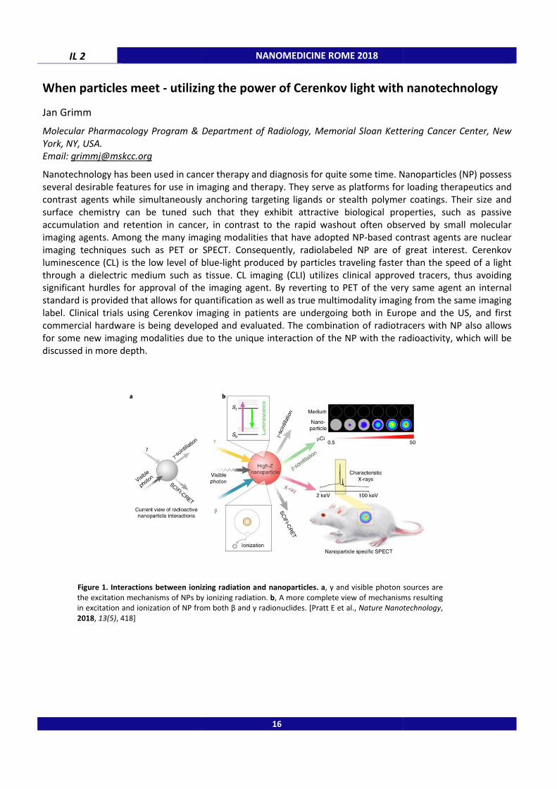
Figure 1. Interactions between ionizing radiation and nanoparticles. a
the excitation mechanisms of NPs by ionizing radiation.
in excitation and ionization of NP from both β and γ radionuclides. [Pratt E et al.,
2018, 13(5), 418]
NANOMEDICINE ROME 2018
utilizing the power of Cerenkov light with nanotechnology
Molecular Pharmacology Program & Department of Radiology, Memorial Sloan Kettering Cancer Center, New
Nanotechnology has been used in cancer therapy and diagnosis for quite some time. Nanoparticles
several desirable features for use in imaging and therapy. They serve as platforms for loading therapeutics and
contrast agents while simultaneously anchoring targeting ligands or stealth polymer coatings. Their size and
uned such that they exhibit attractive biological properties, such as passive
accumulation and retention in cancer, in contrast to the rapid washout often observed by small molecular
imaging agents. Among the many imaging modalities that have adopted NP-based contrast agents are nuclear
imaging techniques such as PET or SPECT. Consequently, radiolabeled NP are of great interest. Cerenkov
is the low level of blue-light produced by particles traveling faster than the speed of a light
a dielectric medium such as tissue. CL imaging (CLI) utilizes clinical approved tracers, thus avoiding
significant hurdles for approval of the imaging agent. By reverting to PET of the very same agent an internal
standard is provided that allows for quantification as well as true multimodality imaging from the same imaging
label. Clinical trials using Cerenkov imaging in patients are undergoing both in Europe and the US, and first
commercial hardware is being developed and evaluated. The combination of radiotracers with
for some new imaging modalities due to the unique interaction of the NP with the radioactivity
Interactions between ionizing radiation and nanoparticles. a, γ and visible photon sources are
the excitation mechanisms of NPs by ionizing radiation. b, A more complete view of mechanisms resulting
in excitation and ionization of NP from both β and γ radionuclides. [Pratt E et al., Nature Nanotechnology
16
utilizing the power of Cerenkov light with nanotechnology
Molecular Pharmacology Program & Department of Radiology, Memorial Sloan Kettering Cancer Center, New
Nanotechnology has been used in cancer therapy and diagnosis for quite some time. Nanoparticles (NP) possess
several desirable features for use in imaging and therapy. They serve as platforms for loading therapeutics and
contrast agents while simultaneously anchoring targeting ligands or stealth polymer coatings. Their size and
uned such that they exhibit attractive biological properties, such as passive
accumulation and retention in cancer, in contrast to the rapid washout often observed by small molecular
sed contrast agents are nuclear
are of great interest. Cerenkov
light produced by particles traveling faster than the speed of a light
utilizes clinical approved tracers, thus avoiding
significant hurdles for approval of the imaging agent. By reverting to PET of the very same agent an internal
ification as well as true multimodality imaging from the same imaging
label. Clinical trials using Cerenkov imaging in patients are undergoing both in Europe and the US, and first
iotracers with NP also allows
radioactivity, which will be
and visible photon sources are
, A more complete view of mechanisms resulting
Nature Nanotechnology,

NANOMEDICINE ROME 2018 IL 3
Magnetic blood purification: from concept to clinics
Inge K. Herrmann
Swiss Federal Laboratories for Materials Science and Technology (Empa).
Email: [email protected]
Sepsis is a potentially life-threatening condition that requires immediate medical attention. Early
administration of antibiotics has direct impact on patient outcome. However, sepsis is difficult to differentiate
from non-infectious systemic inflammation (SIRS), a condition that is very common in intensive care unit
patients. Treating all patients who show SIRS symptoms without proper diagnosis not only increases costs, but
leads to other complications and increased microbial resistance, which is equally undesirable. There is a major
unmet clinical need to better tailor antibiotic therapy to the patient’s individual needs.
Here, we report on the design of functional magnetic capturing agents for theranostic magnetic separation-
based blood purification. We designed iron oxide / polymer hybrid nanoclusters functionalized with a newly
developed antibody to capture and remove pathogens from body fluids. Bacteria quantification in the
supernatant and on the particle surface by plating and optical absorption revealed that bacteria were efficiently
captured by antibody-functionalized beads with capturing efficacies > 98%. Then, we demonstrate rapid and
sensitive detection of bacteria on magnetic beads recovered from the magnetic separator to allow speedy
pathogen identification and diagnosis based on the enrichment of bacteria out of large volumes.
The present theranostic approach could significantly help to reduce the overuse of antibiotics by allowing
speedy detection and identification of the causing pathogens and at the same time providing an effective
treatment modality by decreasing the bacterial load to bridge the time before appropriate antibiotics can be
administered. By rationally designing the magnetic particles (based on modelling of binding times and safety
considerations), we address safety risks at an early stage and ensure that the approach leverages the unique
benefits of nanoparticles while balancing associated risks.
17
IL 4 NANOMEDICINE ROME 2018
Poly(phosphoester)-functionalized nanocarriers: degradable alternatives to
poly(ethylene glycol)
Frederik R. Wurm
Max-Planck-Institut für Polymerfoschung, Ackermannweg 10, 55128 Mainz, Germany.
Email: [email protected]
For decades now polyethylene glycol (PEG) is the standard for shielding biomolecules as well as nanoparticles
from rapid clearance from the blood circulation [1]. Coating of surfaces and nanocarriers with PEG is supposed
to strongly reduce protein adsorption and this reduced protein absorption is thought to be the main course of
action, which is referred to as “stealth” effect. Nevertheless, it has been known that even on PEGylated surfaces
a lower but still detectable amount of proteins attach [2].
We have developed a series of biodegradable poly(phosphoester)s that can substitute PEG but prevents any
accumulation of nondegradable polymers [3]. The polyphosphoester synthesis platform allows further
controlling the polymers’ hydrophilicity on the nanocarrier surface. We proved that the stealth effect is caused
by a specific protein corona after contact with human blood, which depends on the hydrophilicity of the
polymer. Therefore the protein pattern is crucial to understand the stealth effect in general. In addition, the
same plasma protein is enriched on surfaces with hydrophilic polymers, which is clusterin that is a key player in
the stealth behavior. Increasing hydrophobicity does not increase the protein amount, but the type of
“recruited” protein from the blood plasma. In addition, targeting can be achieved by combining the stealth
properties with carbohydrates, which can be recognized by immune cells.
_____________________________________
1. Herzberger J., Niederer K., Pohlit H., Seiwert J., Worm M., Wurm F.R., Frey H., Chem. Rev., 2016, 116 (4), 2170. 2. Schöttler S., Becker G., Winzen S., Steinbach T., Mohr K., Landfester K., Mailänder V., Wurm F.R., Nat Nano, 2016, 11 (4), 372.
3. Bauer K.N., Tee H.T., Velencoso M.M., Wurm F.R., Prog. Polym. Sci., 2017, 73, (Supplement C), 61-122.
18
NANOMEDICINE ROME 2018 IL 5
20 years of developing FDA approved nanomedicine
Thomas J. Webster
Department of Chemical Engineering, Northeastern University, 313 Snell Engineering Center, 360 Huntington
Avenue, Boston, MA 02115 USA.
Email: [email protected]
Despite the fact that many researchers believe that nanomedicine is more hype than reality, there are
numerous FDA approved medical products that incorporate nanomaterials improving human health today. This
talk will highlight some of these FDA approved products with clinical data in the improvement of disease
prevention, diagnosis, and treatment. In particular, it will highlight medical devices that use nanoscale features
to improve tissue growth (such as bone, vascular, bladder, etc.) while limiting inflammation and inhibiting
infection, all without the use of drugs. It will also highlight novel FDA approved nanoparticles that can
simultaneously detect and treat diseases, such as infectious diseases and cancer. This talk will also review the
future of medicine in implantable sensors and how nanomaterials are being used to assess immune system
responses to an implant and then modify itself to ensure implant success. Lastly, this study will cover the
fundamental reason why nanomaterials are able to control cell responses without using drugs. For example,
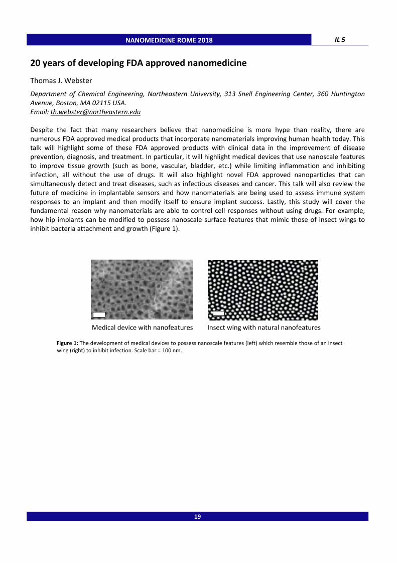
how hip implants can be modified to possess nanoscale surface features that mimic those of insect wings to
inhibit bacteria attachment and growth (Figure 1).
Medical device with nanofeatures Insect wing with natural nanofeatures
Figure 1: The development of medical devices to possess nanoscale features (left) which resemble those of an insect wing (right) to inhibit infection. Scale bar = 100 nm.
19

IL 6 NANOMEDICINE ROME 2018
A multiscale approach in tissue engineering: from nano to tissues
Joaquim Miguel Oliveira a,b,c
a 3B’s Research Group- Biomaterials, Biodegradable and Biomimetic, University of Minho, Headquarters of the
European Institute of Excellence on Tissue Engineering and Regenerative Medicine, Avepark, 4805-017 Barco,
Guimarães, Portugal; b ICVS 3Bs PT Government Associate Lab, Braga, Guimarães, Portugal;
c The Discoveries
Centre for Regenerative and Precision Medicine, Headquarters at University of Minho, Avepark, 4805-017 Barco,
Guimarães, Portugal.
Email: [email protected]
Nanotechnology will have, in a near future, a crucial role in advanced and personalized treatment solutions for
human diseases. The prospects for cell replacement and tissue regeneration in many musculoskeletal and
neurological diseases are impeded by inefficient stem cell/drugs delivery. Through the development of
nanoparticles, it is now possible to increase bioavailability and bioactivity of medical therapeutics and further
selective targeting to damaged tissues. In the last few years, we have been developing several dendrimer
nanoparticles [1] and gold nanoparticles that show great promise in tuning stem cells functions which open up
different application possibilities in the intracellular drugs delivery aiming to treat bone, spinal cord and brain-
related diseases/disorders. By its turn, tissue engineering (TE) aims combining engineering and biological
properties to create functional substitutes for damaged and diseased tissues. Importantly, nanotechnology can
open up a new era for TE, allowing the creation of nanostructures [2] that are comparable in size to those
appearing in natural tissues. Despite our important advances in these fields, the convergence of
nanotechnologies and TE strategies can allow us envisioning the development of safer and effective treatment
solutions in tissue/organ regeneration. On the other hand, recent evidences indicate that 3D and flow models
more closely resemble the in vivo function. In these contexts, our group has been exploiting several multi-scale
strategies combining the use of nanoparticles, scaffolds and stem cells, and emerging technologies such as
bioprinting and microfluidics towards developing both advanced treatment solutions and 3D in vitro models of
disease (e.g. cancer). In particular, a semi-automated microfluidic platform for real-time investigation of
nanoparticles’ cellular uptake and cancer cells’ tracking has been reported [3]. This microfluidic-based platform
composed of microfluidic chip together with fluorescence-labeled dendrimer NPs can allow the validation of
new chemotherapeutic agents and its potential use in the development of diagnostics platform and
personalized therapies. Herein, the challenges associated with the multiscale approaches will be discussed, and
examples of our most impactful achievements making use of nano- and micro-technologies combined with TE
strategies will be also presented.
_____________________________________
1. Oliveira J.M. et al., Progress in Polymer Science, 2012, 35 (9), 1163. 2. Pina S., Oliveira J.M. et al., Advanced Materials, 2015, 27 (7), 1143.
3. Carvalho M. and Oliveira J.M., Nanomedicine, 2017, 12 (6), 581.
Acknowledgments: This study was funded under the project FROnTHERA (NORTE-01-0145-FEDER-000023), supported by Norte Portugal Regional Operational Programme (NORTE 2020). The author is grateful for the FCT distinctions attributed to J. M. Oliveira
(IF/01285/2015).
20

NANOMEDICINE ROME 2018 IL 7
"Soft" nanocarriers: a versatile strategy for brain delivery
Carlotta Marianecci
Department of Chemistry and Technology of Drugs, University of Rome “Sapienza”, P.le A. Moro 5, 00185, Rome,
Italy.
Email: [email protected]
Diseases in the Central Nervous System (CNS) affect about the 20% of people worldwide and half of them are
adults expected to develop degenerative CNS pathologies, such as Alzheimer’s or Parkinson’s. However, the
greatest constraint in drug delivery to the brain is not the absence of drugs to treat CNS diseases, but rather the
mechanism to transport such drugs through the nearly impenetrable blood brain barrier (BBB). Developing
therapeutics for brain diseases is a major challenge; in particular, the most stimulating aspect of that challenge
is to pass through the blood–brain barrier. Currently, strategies to increase drug delivery to the brain use
invasive, non-invasive or alternative approaches to bypass the BBB. Nanomedicine has recently emerged as a
promising field for innovative and effective approaches to cross the BBB and target brain diseases. In this
presentation, the research activities performed in Nanomedicine_Lab of Rome Sapienza (M.Carafa, P.N. Hanieh,
A. Imbriano, F. Rinaldi) will be presented. In particular, the application of different “soft” nanocarriers by
different approaches to brain delivery will be illustrated (1-3).
Current approaches to CNS delivery
_____________________________________
1. C. Ingallina, F. Rinaldi, A. Bogni, J. Ponti, D. Passeri, M. Reggente, M. Rossi, A. Kinsner-Ovaskainen, D. Mehn, F. Rossi, B. Botta, M. Carafa, C. Marianecci, Int J Pharm, 2016, 511 (2), 969.
2. F. Rinaldi, P.N. Hanieh, L.K.N. Chan, L. Angeloni, D. Passeri, M. Rossi, J.T.W. Wang, A. Imbriano, M. Carafa, C.
Marianecci, Pharmaceutics, 2018, 10, 38.
3. M. Carafa, A. Bettucci, C. Marianecci, F. Rinaldi, A. Biagioni, “Nanobubbles and uses thereof” PCT/IB2017/052060.
21
Non-invasivetechniques
Invasivetechniques
Alternativeroutes for CNSdrug delivery
Chitosan Glutamate Coated
Tween20/Tween80
Nanobubble
Hydrophilic
region
Hydrophobic
region
Phospholipid
dublelayer
Water
Gas
IL 8 NANOMEDICINE ROME 2018
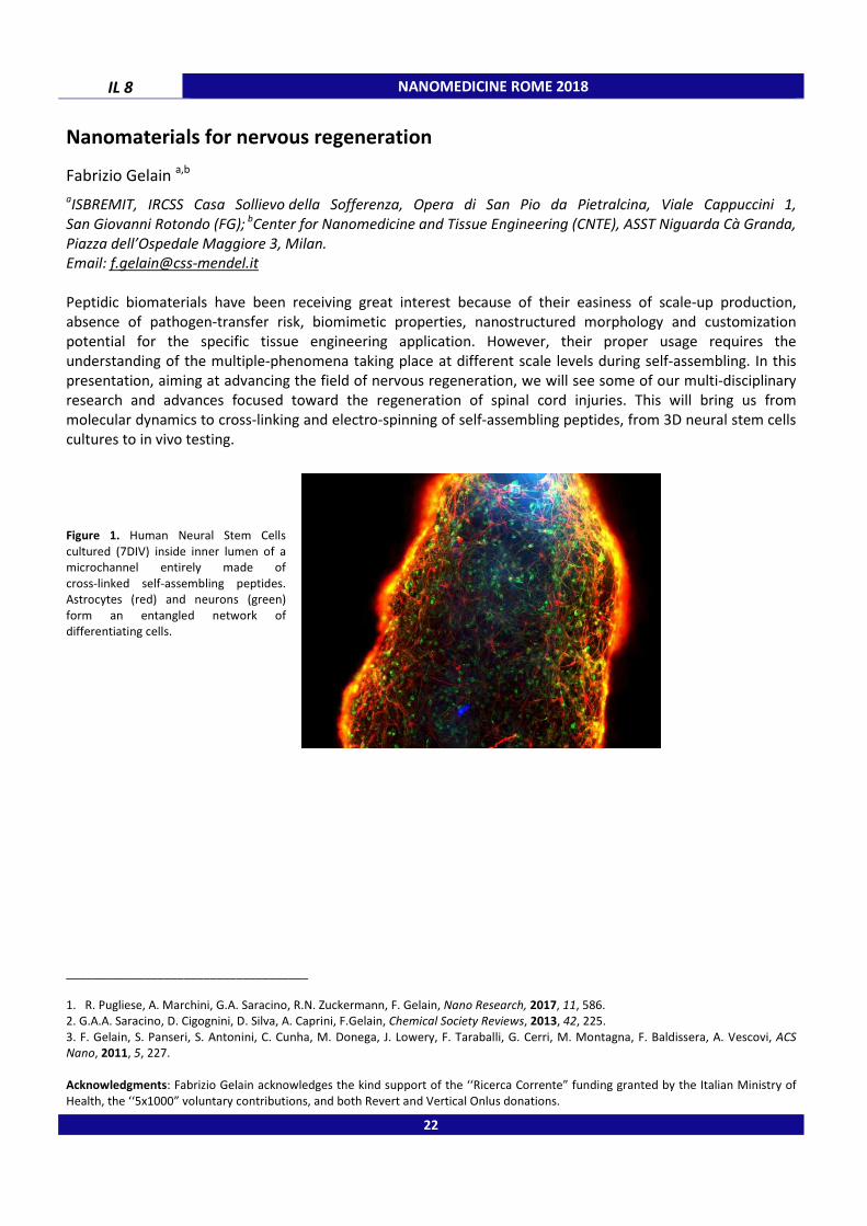
Nanomaterials for nervous regeneration
Fabrizio Gelain a,b
aISBREMIT, IRCSS Casa Sollievo della Sofferenza, Opera di San Pio da Pietralcina, Viale Cappuccini 1,
San Giovanni Rotondo (FG); b
Center for Nanomedicine and Tissue Engineering (CNTE), ASST Niguarda Cà Granda,
Piazza dell’Ospedale Maggiore 3, Milan.
Email: [email protected]
Peptidic biomaterials have been receiving great interest because of their easiness of scale-up production,
absence of pathogen-transfer risk, biomimetic properties, nanostructured morphology and customization
potential for the specific tissue engineering application. However, their proper usage requires the
understanding of the multiple-phenomena taking place at different scale levels during self-assembling. In this
presentation, aiming at advancing the field of nervous regeneration, we will see some of our multi-disciplinary
research and advances focused toward the regeneration of spinal cord injuries. This will bring us from
molecular dynamics to cross-linking and electro-spinning of self-assembling peptides, from 3D neural stem cells
cultures to in vivo testing.
Figure 1. Human Neural Stem Cells
cultured (7DIV) inside inner lumen of a microchannel entirely made of
cross-linked self-assembling peptides. Astrocytes (red) and neurons (green)
form an entangled network of differentiating cells.
_____________________________________
1. R. Pugliese, A. Marchini, G.A. Saracino, R.N. Zuckermann, F. Gelain, Nano Research, 2017, 11, 586. 2. G.A.A. Saracino, D. Cigognini, D. Silva, A. Caprini, F.Gelain, Chemical Society Reviews, 2013, 42, 225.
3. F. Gelain, S. Panseri, S. Antonini, C. Cunha, M. Donega, J. Lowery, F. Taraballi, G. Cerri, M. Montagna, F. Baldissera, A. Vescovi, ACS
Nano, 2011, 5, 227.
Acknowledgments: Fabrizio Gelain acknowledges the kind support of the ‘‘Ricerca Corrente” funding granted by the Italian Ministry of
Health, the ‘‘5x1000” voluntary contributions, and both Revert and Vertical Onlus donations.
22

NANOMEDICINE ROME 2018 IL 9
The debate on (targeted) nanomedicine
Gert Storm
Dept. Pharmaceutics, Utrecht Institute for Pharmaceutical Sciences (UIPS), Utrecht University, PO Box 80082,
3508 TB Utrecht, The Netherlands; University Medical Centre Utrecht (UMCU), Division Imaging, Utrecht, The
Netherlands; Dept. Biomaterials Science & Technology (BST), MIRA Institute for Biomedical Technology and
Technical Medicine, University of Twente, Enschede, The Netherlands.
Email: [email protected]
One most active sector of research within the field of nanomedicine has been the design of
nanopharmaceuticals for targeted drug delivery. In fact, novel nanomedicinal drug delivery systems continue to
flourish in the research laboratory. However, the number of such nanomedicines that have been approved for
the treatment of patients is still limited. Examples are Caelyx/Doxil (doxorubicin), Myocet (doxorubicin),
DaunoXome (daunorubicin), Marqibo (vincristine), Onyvide (irinotecan), Onco-TCS (vincristine), Vyxeos
(cytarabine and daunorubicin) and Abraxane (paclitaxel). While these examples illustrate that significant
advances have been made over the years in making nanomedicines a clinical reality, there is nevertheless
growing scepticism in the scientific literature regarding the future and clinical applicability of targeted
nanopharmaceuticals. In this presentation, I will discuss the arguments raised to justify this negative attitude as
well as my view on how targeted nanomedicine will face tomorrow.
23

IL 10 NANOMEDICINE ROME 2018
Dissecting how cells internalize and process nano-sized drug carriers for
nanomedicine applications
Anna Salvati
Groningen Research Institute of Pharmacy, University of Groningen, A. Deusinglaan 1, 9713Ave Groningen, The
Netherlands.
Email: [email protected]
Nano-sized materials have the unique capacity to distribute in organisms and enter cells easily using cellular
pathways. This has opened up tremendous opportunity in nanomedicine for using nano-sized carriers to deliver
drugs more efficiently to their site of action. However, the molecular details of the mechanisms of uptake and
intracellular trafficking of nano-sized drug carriers are in most cases still not clear. Such knowledge could allow
us to further improve the design of truly targeted nanomedicines.
To this aim, we have combined different cell biology methods including the use of transport inhibitors and RNA
interference to characterize the early steps of nanoparticle recognition by cells and the following uptake
mechanisms. Additional efforts have been focused on developing in vitro cell barriers more closely resembling
those nanomedicines encounter in vivo and in this way to elucidate whether the organization of cells into
polarized cell barriers affects nano-carrier uptake and behavior.
Our results show that the same cells process nanoparticles in different ways when they are developed into a
cell barrier rather than at different degrees of cell density, as commonly applied for in vitro studies.
Furthermore we show that the corona molecules adsorbing from the environment on the nanoparticle surface
not only can be recognized by cell receptors, as previously shown [1], but also can affect the details of the
following uptake mechanism. Thus, in other words, the same nanoparticles enter cells via different pathways
when different coronas are formed on their surface.
_____________________________________
1. S. Lara et al., ACS Nano, 2017, 11 (2), 1884.
Acknowledgments: This work was funded by the European Research Council (ERC) under the European Union's Horizon 2020 research
and innovation programme under grant agreement Nº637614 (NanoPaths).
24

NANOMEDICINE ROME 2018
Selected Lectures
25

NANOMEDICINE ROME 2018
26

NANOMEDICINE ROME 2018 SL 1
A theranostic approach for Boron Neutron Capture Therapy (BNCT) treatment
based on the use of Gd/B multimodal probes
Diego Alberti,a Nicoletta Protti,b Silva Bortolussi,b Saverio Altieri,b Annamaria Deagostino,c Silvio Aime,a Simonetta Geninatti−Crich a a
Department of Molecular Biotechology and Health Sciences, University of Torino, via Nizza 52, 10126, Torino,
Italy; b
Department of Physics, University of Pavia, via Bassi 6, 27100, Pavia, Italy; c
Department of Chemistry,
University of Torino, via P. Giuria 7, 10125, Torino, Italy.
Email of presenting author: [email protected]
This study aims at investigating a new theranostic approach for the treatment of primary tumours and
metastasis based on the use of BNCT that combines low energy neutron irradiation with the presence of boron-
containing compound at the targeted cells. This makes BNCT a promising option for the treatment of metastasis
disseminated, for example, in the thoracic cavity that cannot be treated by methods requiring a precise
localization, such as surgery or conventional radiotherapy. The innovation of this study lies on the development
of novel theranostic agents, able to maximize the selective uptake of boron atoms in tumour cells and, at the
same time, to quantify boron distribution in the tumour and in other tissues by Magnetic Resonance Imaging
(MRI). The measurement of local boron concentration is crucial to determine the optimal neutron irradiation
time, to calculate the delivered radiation dose and to evaluate the toxicity of the treatment by determining
differences in boron concentration between tumour and healthy tissues. To this purpose a new dual BNCT/MRI
agent has been synthesized and delivered to tumour cells using Low Density Lipoproteins as specific carriers. In
particular, this study has been focused on the treatment of lung metastases generated by intravenous injection
of a Her2 + breast cancer cell line (i.e. TUBO) in BALB/c mice, transgenic EML4-ALK mice used as primary lung
tumor model [1] and of a subcutaneous tumour mouse model of Malignant Mesothelioma (MM). The latter is
an aggressive tumour with a poor prognosis whose incidence and mortality is a function of past exposure to
asbestos, after a latency period of 30-50 years. MM is a disseminated tumour against which conventional
radiotherapy has limited effectiveness. Therefore, to improve both the clinical diagnostics and treatment, the
discovery of new MM potential target molecules is of great interest. BNCT has been performed after MRI
analysis at the TRIGA-Mark II reactor at the University of Pavia. With respect to controls, in boron treated
group, tumour growth was significantly reduced.
_____________________________________
1. D. Alberti, N. Protti , A. Toppino, A. Deagostino, S. Lanzardo, S. Bortolussi, S. Altieri , C. Voena, R. Chiarle, S. Geninatti Crich, S. Aime.
Nanomedicine, 2015, 11, 741.
27
SL 2 NANOMEDICINE ROME 2018
Development of novel m-THPC-liposomes and layersomes for the oral
treatment of cholangiocarcinoma and gastrointestinal tumors
Gerhard D. Wieland, Dietrich Scheglmann, Arno Wiehe, Alfred Fahr, Volker Albrecht
Biolitec research GmbH, Otto-Schott-Strasse 15, D-07745 Jena, Germany.
Email of presenting author: [email protected]
Introduction: Photodynamic Therapy (PDT) as a combination of photoactivable pharmaceutical active
ingredients (APIs, photosensitizers, PS) and PS-specific laser light is a well-proven method for local tumor
therapy. Following illumination of the tumors with laser light of accurately defined wavelengths, reactive
oxygen species built in situ can destroy the tumors. PDT therefore is a highly promising novel option addressing
cholangiocellular carcinoma (CCC) and tumors of the gastrointestinal tract (GIT). Physicochemical properties of
PDT-APIs like solubility and stability often hinder sufficient oral bioavailability and targeted accumulation at
tumor site. The goal of the projects summarized in this talk was the development of novel oral pharmaceutical
formulations of BCS class IV APIs, administered parentally (for example: Foscan® with Temoporfin (m-
tetrahydroxy-phenylchlorin (mTHPC)) as API) [1].
Liposomes have been often described as suitable drug carriers to increase the bioavailability of BCS Class IV
APIs. So these projects aimed to elucidate the possibility to use liposomes or novel drug delivery systems like
multilayered layersomes [2] amongst other nano-structured formulations, to deliver pharmaceutical APIs like
photosensitizers (PS) into cells and tissues by oral administration.
Materials and methods: The ratio of the layersomes and the construction of the standard liposomes and the
layersomes is documented in detail. The physicochemical and photochemical properties of the formulations are
presented, as well as the stability of these formulations in biological fluids. MTT cytotoxicity assays determined
the PDT efficacy of these novel liposomes on different cell lines. Life cell Fluorescence Microscopy shows the
uptake of the liposomes in cells and tissues.
Results and Conclusion: Liposomal formulations are a promising novel option as oral administered
pharmaceutical compositions for the treatment of cholangio carcinoma and GIT-tumours. Embedded in tablets
the formulations cross the intestinal barriers to target CCC in the liver, or regionally targeted in the GIT may
enter tumoral lesions in the intestine.
_____________________________________
1. Senge M.O, Brandt J.C., Photochem. Photobiol., 2011, 87, 1240. 2. Agrawal A.K., Harshad H., Thanki K., Jain, S., Biomacromolecules, 2014, 15, 350.
These projects were supported by the German Ministry for Education and Research (BMBF) (13N11386 BioTrap for CCC and 13N13422
GITCare).
28
NANOMEDICINE ROME 2018 SL 3
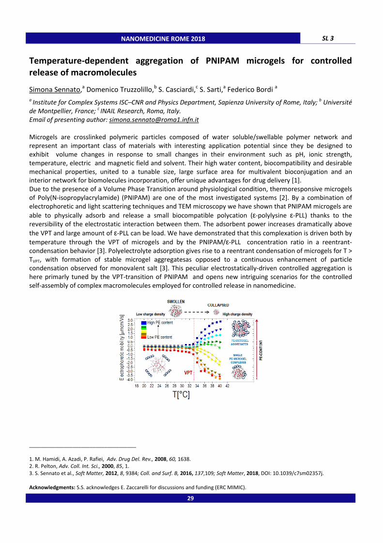
Temperature-dependent aggregation of PNIPAM microgels for controlled
release of macromolecules
Simona Sennato,a Domenico Truzzolillo,b S. Casciardi,c S. Sarti,a Federico Bordi a a
Institute for Complex Systems ISC–CNR and Physics Department, Sapienza University of Rome, Italy; b Université
de Montpellier, France; c INAIL Research, Roma, Italy.
Email of presenting author: [email protected]
Microgels are crosslinked polymeric particles composed of water soluble/swellable polymer network and
represent an important class of materials with interesting application potential since they be designed to
exhibit volume changes in response to small changes in their environment such as pH, ionic strength,
temperature, electric and magnetic field and solvent. Their high water content, biocompatibility and desirable
mechanical properties, united to a tunable size, large surface area for multivalent bioconjugation and an
interior network for biomolecules incorporation, offer unique advantages for drug delivery [1].
Due to the presence of a Volume Phase Transition around physiological condition, thermoresponsive microgels
of Poly(N-isopropylacrylamide) (PNIPAM) are one of the most investigated systems [2]. By a combination of
electrophoretic and light scattering techniques and TEM microscopy we have shown that PNIPAM microgels are
able to physically adsorb and release a small biocompatible polycation (ε-polylysine ε-PLL) thanks to the
reversibility of the electrostatic interaction between them. The adsorbent power increases dramatically above
the VPT and large amount of ε-PLL can be load. We have demonstrated that this complexation is driven both by
temperature through the VPT of microgels and by the PNIPAM/ε-PLL concentration ratio in a reentrant-
condensation behavior [3]. Polyelectrolyte adsorption gives rise to a reentrant condensation of microgels for T >
TVPT, with formation of stable microgel aggregatesas opposed to a continuous enhancement of particle
condensation observed for monovalent salt [3]. This peculiar electrostatically-driven controlled aggregation is
here primarly tuned by the VPT-transition of PNIPAM and opens new intriguing scenarios for the controlled
self-assembly of complex macromolecules employed for controlled release in nanomedicine.
_____________________________________
1. M. Hamidi, A. Azadi, P. Rafiei, Adv. Drug Del. Rev., 2008, 60, 1638. 2. R. Pelton, Adv. Coll. Int. Sci., 2000, 85, 1.
3. S. Sennato et al., Soft Matter, 2012, 8, 9384; Coll. and Surf. B, 2016, 137,109; Soft Matter, 2018, DOI: 10.1039/c7sm02357j.
Acknowledgments: S.S. acknowledges E. Zaccarelli for discussions and funding (ERC MIMIC).
29

SL 4 NANOMEDICINE ROME 2018
Bicontinuous cubic liquid crystalline dispersions as potential tools in
nanomedicine
Sergio Murgia
Department of Chemical and Geological Sciences, University of Cagliari, s.s. 554 bivio Sestu, 09042, Monserrato
(CA), Italy.
Email of presenting author: [email protected]
New formulation strategies were recently developed with the purpose of combining imaging probes, drugs, and
targeting agents in the same therapeutic/diagnostic (theranostic) nanocarrier to discover and treat diseases at
the initial stage and with limited side effects. In this context, lipid-based nanoparticles can be considered as
flexible platforms since they can be personalized depending on their application.
This presentation focuses on the possible use as theranostic tools of monoolein-based liquid crystalline
nanoparticles, known as cubosomes, showing an inner structure characterized by a reverse bicontinuous cubic
symmetry. Physicochemical and photophysical analysis demonstrated that cubosomes can effectively be loaded
with anticancer drugs, UV-visible or NIR emitting fluorophores, and decorated with folic acid as cancer cells-
targeting ligand. Their living cells imaging skills, cytotoxicity features, and biodistribution in vivo will be
discussed.
Figure 1. Cryo-TEM image of cubosomes (left), and whole body fluorescence intensity distribution of a healthy mouse that received an i.v. injection of fluorescent cubosomes
(right).
_____________________________________
1. V. Meli et al., Langmuir, 2015, 31, 9566. 2. S. Biffi et al., Nanotechnology, 2017, 28, 055102.
30
NANOMEDICINE ROME 2018
SEEC Microscopy: a live and label
Materials and Life Sciences
Nicolas Medard, Imed Ayadi
NANOLANE, Pole Novaxud, 57 Boulevard Demorieux, 72100 Le Mans, France.
Email of presenting author: [email protected]
SEEC Microscopy is a label-free analysis technique offering new characterization capabilities such as the live
nanoscale imaging, the multiplex molecular interaction analysis or the real
technique implements unique optical sensitive sensors (SEEC sensors) with specific contrast
properties enabling the live visualization of samples down to nanoscale (0.1nm). In addition, a proprietary
algorithm (Q-SEEC) enables quantitative analyses (surface in
accuracy of 0.3nm.
SEEC Microscopy is dedicated to the study of samples in the fields of Materials and Life Sciences. Thanks to its
high sensitivity, the technique can be applied for the analysis of nanofilms, nan
nanoparticles or DNA molecules…
Amongst successful analyses recently conducted, we will present
bacteria motion, the tracking of dendritic cells
examples will be also presented concerning
reactions [1] or also the label-free and real
environment.
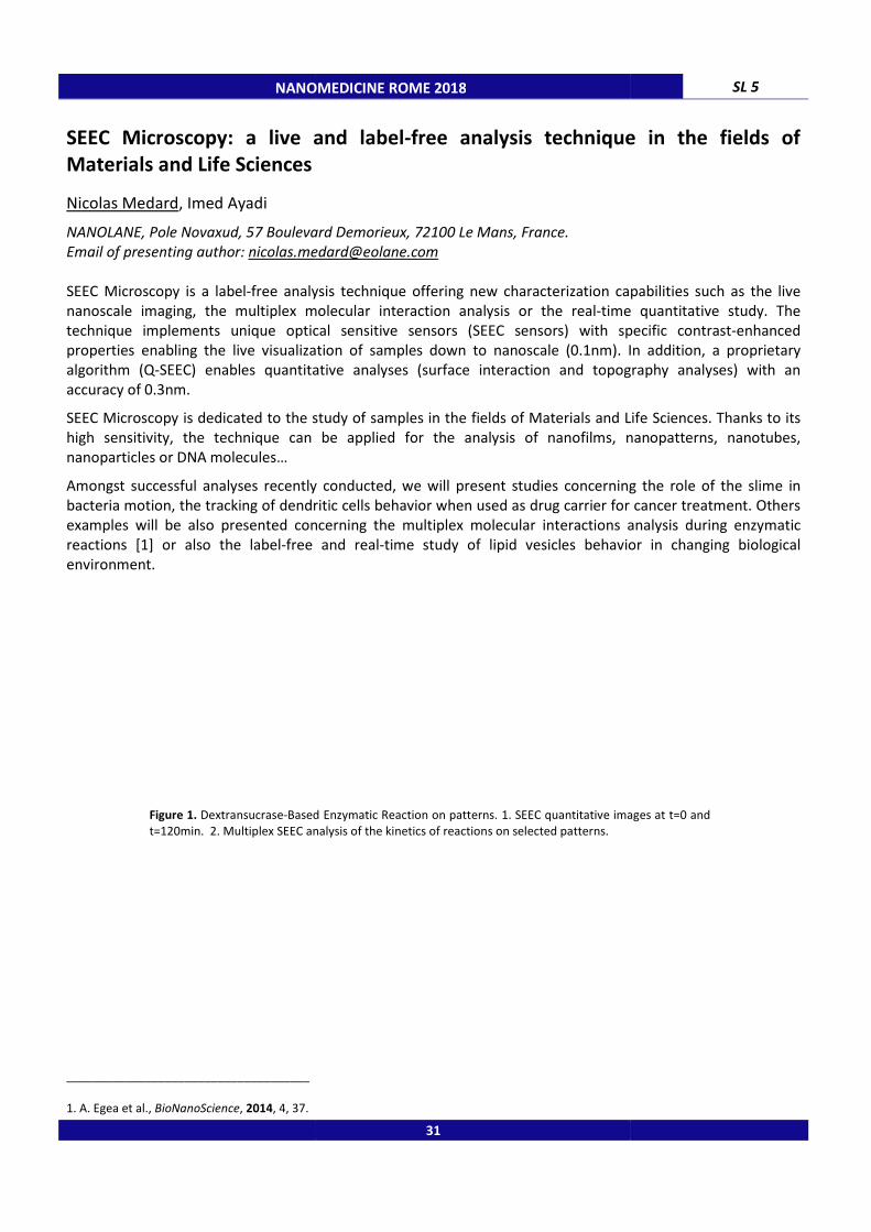
Figure 1. Dextransucrase-Based Enzymatic Reaction on patterns. 1. SEEC qua
t=120min. 2. Multiplex SEEC analysis of the kinetics of reactions on selected patterns.
_____________________________________
1. A. Egea et al., BioNanoScience, 2014, 4, 37.
NANOMEDICINE ROME 2018
SEEC Microscopy: a live and label-free analysis technique in the fields of
Materials and Life Sciences
NANOLANE, Pole Novaxud, 57 Boulevard Demorieux, 72100 Le Mans, France.
free analysis technique offering new characterization capabilities such as the live
nanoscale imaging, the multiplex molecular interaction analysis or the real-time quantitative study. The
lements unique optical sensitive sensors (SEEC sensors) with specific contrast
properties enabling the live visualization of samples down to nanoscale (0.1nm). In addition, a proprietary
SEEC) enables quantitative analyses (surface interaction and topography analyses) with an
SEEC Microscopy is dedicated to the study of samples in the fields of Materials and Life Sciences. Thanks to its
high sensitivity, the technique can be applied for the analysis of nanofilms, nan
Amongst successful analyses recently conducted, we will present studies concerning the role of
he tracking of dendritic cells behavior when used as drug carrier for cance
presented concerning the multiplex molecular interactions analysis
free and real-time study of lipid vesicles behavior
Based Enzymatic Reaction on patterns. 1. SEEC quantitative images at t=0 and
t=120min. 2. Multiplex SEEC analysis of the kinetics of reactions on selected patterns.
31
SL 5
free analysis technique in the fields of
free analysis technique offering new characterization capabilities such as the live
time quantitative study. The
lements unique optical sensitive sensors (SEEC sensors) with specific contrast-enhanced
properties enabling the live visualization of samples down to nanoscale (0.1nm). In addition, a proprietary
teraction and topography analyses) with an
SEEC Microscopy is dedicated to the study of samples in the fields of Materials and Life Sciences. Thanks to its
high sensitivity, the technique can be applied for the analysis of nanofilms, nanopatterns, nanotubes,
concerning the role of the slime in
used as drug carrier for cancer treatment. Others
multiplex molecular interactions analysis during enzymatic
of lipid vesicles behavior in changing biological
titative images at t=0 and

SL 6 NANOMEDICINE ROME 2018
Unprecedented behavior of (9R)-9-hydroxystearic acid loaded keratin
nanoparticles on cancer cell cycle
Claudia Ferroni,a Alberto Busi,b Annalisa Aluigi,a Carla Boga,b Natalia Calonghi,c Andrea Guerrini,a Giovanna Sotgiu,a Tamara Posati,a Franco Corticelli,d Jessica Fiori,e Greta Varchi a aISOF-CNR, Via Gobetti 101 - 40129 Bologna, Italy;
b Department of Industrial Chemistry, Viale Risorgimento 4,
40136 Bologna, Italy; c Department of Pharmacy and Biotechnology, Via Irnerio 48, 40126 Bologna, Italy;
d IMM-
CNR, Via Gobetti 101 - 40129 Bologna, Italy; e Department of Pharmacy and Biotechnology, Via Belmeloro 6,
40126, Bologna, Italy.
Email of presenting author: [email protected]
High level expression of histone deacetylase 1 (HDAC1) plays a pivotal role in the pathobiology of cancer [1].
The endogenous fatty acid (9R)-9-hydroxystearic acid, 9R, is a natural HDAC1 inhibitor, which exerts its anti-
proliferative activity by arresting cancer cells growth in G0/G1 phase [2]. However, one of its major limitations is
the unfavorable pharmacokinetic that hampers its therapeutic effectiveness. To overcome this constraint, 9R
keratin nanoparticles (9R@Ker) were prepared and described here for the first time. Keratin was selected as
carrier because possesses excellent biocompatibility and low toxicity to cells, thus resulting very promising
material for drug delivery applications [3]. The formation of 200 nm nanoparticles (NPs) was induced by
hydrophobic interactions between 9R and the hydrophobic protein domains, affording water-stable NPs with
no need of toxic cross-linking agents. NPs were characterized in terms of particles size distribution, zeta
potential, morphology, thermogravimetric behavior and drug release profile. Moreover, in vitro uptake and
cytotoxicity was evaluated using human colorectal adenocarcinoma cells (HT29). Our data revealed that
9R@Ker are efficiently internalized by tumor cells, altering membrane lipidic composition. In vitro results
demonstrate that the activity of 9R as free or loaded onto NPs is similar in terms of anti-proliferative effect.
Remarkably, some significant differences were observed in the cell cycle analysis, showing that while free 9R
caused a growth arrest in the G0/G1 phase, 9R@Ker induced a S phase arrest, thus promoting apoptosis (Fig.1).
Additional studies are ongoing to better elucidate the underpinning biochemical mechanism of action of our
newly developed delivery system.
Figure 1. Figure illustrating the In vitro biological activity of (R)-9HSA@Ker nanoparticles.
_____________________________________
1. B.M. Müller et al., BMC Cancer, 2013, 13, 215.
2. C. Parolin et al., Biochimica et Biophysica Acta, 2012, 1821, 1334.
3. A. Aluigi et al., RSC Adv., 2016, 6, 33910.
32
NANOMEDICINE ROME 2018 SL 7
Ammonium containing calixarenes as multivalent systems for the delivery of
nucleic acids and mimics
Francesco Sansone,a Jessica Gasparello,b Michela Lomazzi,a Alessia Finotti,b Alex Manicardi,a Alessandro Casnati,a Roberto Corradini,a Roberto Gambari b
aDipartimento di Scienze Chimiche, della Vita e della Sostenibilità Ambientale, Università di Parma, Parco Area
delle Scienze 17/a, 43124 Parma, Italy; b
Dipartimento di Scienze della vita e biotecnologie, Università di Ferrara,
Via Fossato di Mortara 74, 44121 Ferrara, Italy.
E-mail of presenting author: [email protected]
Gene therapy is based on the possibility of delivering proper nucleic acids or mimics into the cells in order to
block or restore processes and activities related with alterations in the genome of the patients. The first recent
successes in this field exploit suitably modified viruses as carriers. However, their use is related with some
possible drawbacks such as inflammation, toxicity, mutagenesis, limits in the cargo size, expensive procedures
for large scale preparation. Therefore, it is relevant the development of non viral vectors based on organic
molecules and polymers as safe and efficient delivery systems. In this context, some years ago we designed and
synthesized molecular vectors characterized by a multivalent exposition of guanidinium or arginine units linked
to a calixarene scaffold [1]. Some of them indeed showed a very high efficiency in the transfection of plasmid
DNA associated with a very low or negligible citotoxicity, resulting better than commercially available
formulations for transfection protocols. Very recently we interestingly demonstrated a remarkable activity of
these macrocyclic vectors in the transfection of cells also with RNAs, such as miRNA and pre-miRNA [2], and
nucleic acid mimics such as Peptide Nucleic Acids (PNAs) [3]. It is particularly relevant that for the latter ones no
other significant vectors are currently available despite the importance of these molecules as potential
therapeutics. These new findings make then our multivalent vectors promising non viral transfecting agents of
interest for researchers and companies working on gene therapy and development of drugs based on nucleic
acids and mimics.
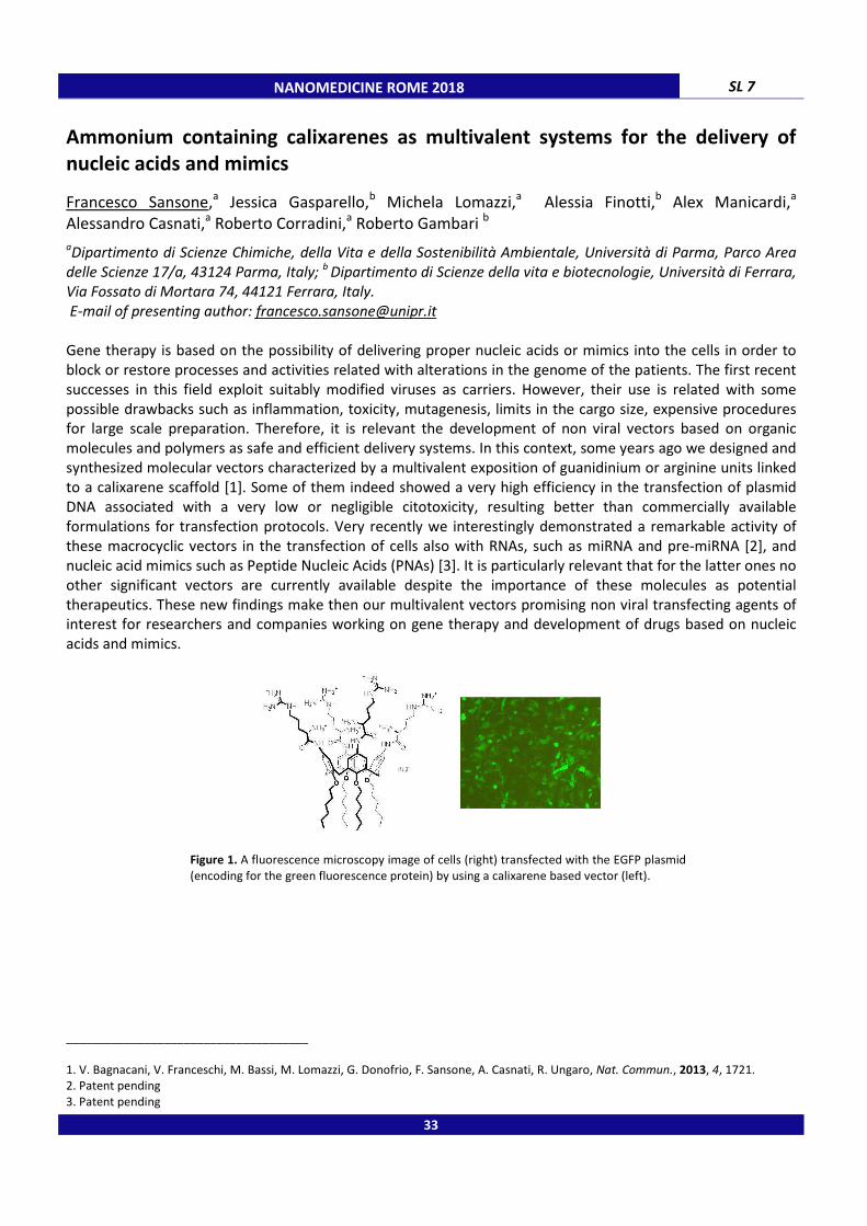
Figure 1. A fluorescence microscopy image of cells (right) transfected with the EGFP plasmid
(encoding for the green fluorescence protein) by using a calixarene based vector (left).
_____________________________________
1. V. Bagnacani, V. Franceschi, M. Bassi, M. Lomazzi, G. Donofrio, F. Sansone, A. Casnati, R. Ungaro, Nat. Commun., 2013, 4, 1721. 2. Patent pending
3. Patent pending
33

SL 8
High-throughput microfluidic impedance cytometer for
localization and characterization of single cells
Riccardo Reale,a Adele De Ninno,
a,b
aDepartment of Civil Engineering and Computer Science, University of Rome Tor Vergata, Via del Pol
00133, Rome, Italy; b Italian National Research Council, Institute for Photonics and Nanotechnologies, Via Cineto
Romano 42, 00156, Rome, Italy.
Email of presenting author: [email protected]
Microfluidic impedance spectroscopy is an
characterization of single particles and cells. It is used in different biological assays, including particle sizing and
counting, cell phenotyping, and disease diagnostics. An impedance
consists of a microchannel equipped with microelectrodes and filled with a conductive buffer. The current
change upon passage of a cell between the electrodes under an AC voltage is measured and then analyse
determine cell properties.
In this work, we present innovative chip layouts and relevant operation modes that increase the accuracy and
information content of the features embedded in the impedance signal traces
electrical metrics encoding particle cross
accurate sizing, overcoming the positional dependence issue, and to investigate particle inertial focusing
mechanisms (Figure 1).
Figure 1. Electrical measurement of particl
electrical position X (a, d), electrical velocity
position Y (c, f). At higher particle Reynolds number Re
_____________________________________
1. D. Spencer et al, Lab Chip, 2016, 16, 2467.
2. A. De Ninno et al, Lab Chip, 2017, 17, 1158.
3. R. Reale et al, Microfluid Nanofluid, 2018, 22
Acknowledgments: The research leading to this work was supported by the Mission Sustainability Programme of the University of Rome
Tor Vergata (SPY-Project).
NANOMEDICINE ROME 2018
throughput microfluidic impedance cytometer for label
localization and characterization of single cells
a,b Luca Businaro,
b Paolo Bisegna,
a Federica Caselli
Department of Civil Engineering and Computer Science, University of Rome Tor Vergata, Via del Pol
Italian National Research Council, Institute for Photonics and Nanotechnologies, Via Cineto
Microfluidic impedance spectroscopy is an attractive label-free technique for high
characterization of single particles and cells. It is used in different biological assays, including particle sizing and
counting, cell phenotyping, and disease diagnostics. An impedance-based flow microcytometer typically
consists of a microchannel equipped with microelectrodes and filled with a conductive buffer. The current
change upon passage of a cell between the electrodes under an AC voltage is measured and then analyse
In this work, we present innovative chip layouts and relevant operation modes that increase the accuracy and
information content of the features embedded in the impedance signal traces [1,2,3]
particle cross-sectional position are presented. They are exploited to achieve
accurate sizing, overcoming the positional dependence issue, and to investigate particle inertial focusing
measurement of particle localization and focusing. Density plots of electrical position
(a, d), electrical velocity V vs. electrical position X (b, e), electrical velocity
(c, f). At higher particle Reynolds number Rep (d-f), the hydrodynamic focusing is more pronounced.
, 1158.
22, 41.
The research leading to this work was supported by the Mission Sustainability Programme of the University of Rome
34
label-free counting,
Federica Caselli a
Department of Civil Engineering and Computer Science, University of Rome Tor Vergata, Via del Politecnico 1,
Italian National Research Council, Institute for Photonics and Nanotechnologies, Via Cineto
free technique for high-throughput electrical
characterization of single particles and cells. It is used in different biological assays, including particle sizing and
flow microcytometer typically
consists of a microchannel equipped with microelectrodes and filled with a conductive buffer. The current
change upon passage of a cell between the electrodes under an AC voltage is measured and then analysed to
In this work, we present innovative chip layouts and relevant operation modes that increase the accuracy and
[1,2,3]. In particular, novel
sectional position are presented. They are exploited to achieve
accurate sizing, overcoming the positional dependence issue, and to investigate particle inertial focusing
e localization and focusing. Density plots of electrical position Y vs.
(b, e), electrical velocity V vs. electrical
f), the hydrodynamic focusing is more pronounced.
The research leading to this work was supported by the Mission Sustainability Programme of the University of Rome

NANOMEDICINE ROME 2018 SL 9
Integrative cytotoxicity assessment of nanostructured medical devices
Giuseppe D’Avenio,a Giuseppina Bozzuto,b Maria Condello,b Simona Sennato,c Giuseppe Familiari,d Ezio Battaglione,d Stefania Meschini,b Carla Daniele,a Agnese Molinari,b Mauro Grigioni a aNational Center for Technological Innovation in Public Health, Istituto Superiore di Sanità Rome, Italy;
bNational Center for Drug Research and Evaluation, Istituto Superiore di Sanità Rome, Italy;
cInstitute for
Complex Systems ISC–CNR and Physics Department, Sapienza University of Rome; dDepartment of Anatomy,
Histology, Forensic Medicine and Orthopaedics, Sapienza University of Rome.
Email of presenting author: [email protected]
Nanostructured medical devices (MDs) are gaining a rapid diffusion. The recently introduced European Medical
Device Regulation, for the first time, explicitly mentions such objects in the European regulatory framework.
Nanostructured MDs can be fabricated using many different processes. In this study, we addressed those MDs
that present metallic nanoparticles (NPs) as a constituent, to be integrated in the final product upon proper
handling (such as, e.g., in dental cements). Traditional cytotoxicity tests (e.g., Neutral Red, MTT), as per the EN
ISO 10993-6 standard, together with studies of ultrastructural cellular pathology, were used in order to provide
established references, based on the traditional approach to the evaluation of cytotoxicity presented by
medical devices. Those tests were compared to Electrical Cell-substract impedance sensing (ECIS), a label-free,
real-time test capable of recording continuously the electrical parameters of a cell layer subject to an external
agent. In this case, different concentrations of ZnO NPs were delivered to cell layers of different type (A549,
gingival fibroblasts). The utility of the proposed comparison is demonstrated by the evidence about criticalities
in traditional cytotoxicity tests involving the use of dyes (e.g., MTT), as underlined by the recently issued ISO/TR
10993-22 Guidance on nanomaterials.
Electrical resistance of the cell-covered electrodes was found to decrease remarkably in the presence of ZnO
NPs, especially at the highest concentration (20 µg/cm2). The time course of the normalized resistance for the
20 µg/cm2 ZnO was observed to be in striking resemblance to that of the positive control (a known apoptotic
agent, STS), suggesting similar mechanisms of cell death, in the very first hours after treatment. ECIS test
highlighted an enhanced proliferative activity at lower ZnO NPs concentration (1 µg/cm2), not clearly evidenced
by traditional tests. The comparison of MTT, NR cytotoxicity and cloning efficiency tests together with studies of
ultrastructural cellular pathology allowed to clarify the interaction of NP and cell survival/death mechanisms.
ECIS test has the potential to recapitulate requirements needed for the evaluation of nanomaterials
cytotoxicity.
_____________________________________
Acknowledgments: Funded by Regione Lazio - RinnovaReNano - FILAS - RU-2014-1041
35

SL 10
The role of the monosialoganglioside
membranes and unstructured
calcitonin
Cristiano Giordani,a Marco Diociaiuti,
a Instituto de Física, Universidad de Antioquia, Calle 70 No. 52
Malattie Rare, Istituto Superiore di Sanità, I
Università dell’Aquila, via Vetoio (Coppito 1), 67010 L’Aquila, Italy;
Sezione Meccanismi di Reazione, c/o Dipartimento di Chimica, Università degli Studi di Roma “Sapienza”, I
00185 Rome, Italy. Email of presenting author: [email protected]
To investigate the molecular mechanisms of the interaction between amyloid aggregates and model
membranes containing GM1, we applied Circular Dichroism (CD) spectroscopy and Transmission Electron
Microscopy (TEM). In particular, we studied the interaction
proto- and mature-fibers with liposomes made of DPPC, with and without GM1 and cholesterol. All data
indicated that the presence of the negatively charged GM1 favored the interaction with all types of aggrega
accelerating the formation of beta-structures. TEM data clearly showed that only PFOs were able to modify the
bilayer structures by the formation of “amyloid channels” that were clearly visualized.
similar to that proposed by Molecular Dynamics simulations for A
hypothesis. We speculate that the electrostatic interaction occurring between positively charged, native,
flexible PFOs with the negatively charged GM1 localized in the outer part of
binding while the hydrophobic interaction could be responsible for the subsequent incorporation in the
membrane leading to the formation of the observed amyloid pores.
Figure 1. The typical aggregation curve of the
amyloid proteins characterized by three zones: Lag
Growth- and Saturation-phases. The relative
aggregation states of the protein are also depicted
(for the courtesy of Iannuzzi et al. [1]).
_____________________________________
1. C. Iannuzzi, G. Irace, I. Sirangelo, Molecules,
Acknowledgments: This work was supported by the Italian “Ministero della Salute” with the “Progetto Ordinario di Ricerca Finalizzata
(RF-2013-02355682)” and the research funds of Committee for Research Development from University of Antioquia (CODI, UdeA,
Medellin, Colombia) through grant #IN641CE (Act 8700
NANOMEDICINE ROME 2018
The role of the monosialoganglioside-GM1 in the interaction between model
membranes and unstructured metastable amyloid oligomers of salmon
Marco Diociaiuti,b Laura Zanetti-Polzi,
c Raoul Fioravanti,
b Cecilia Bombelli
Instituto de Física, Universidad de Antioquia, Calle 70 No. 52-21, Medellín, Colombia;
Malattie Rare, Istituto Superiore di Sanità, I-00161 Rome, Italy; c Dipartimento di Fisica e Scienze Chimiche,
Università dell’Aquila, via Vetoio (Coppito 1), 67010 L’Aquila, Italy; d CNR, Istituto di Metodologie Chimiche,
, c/o Dipartimento di Chimica, Università degli Studi di Roma “Sapienza”, I
To investigate the molecular mechanisms of the interaction between amyloid aggregates and model
membranes containing GM1, we applied Circular Dichroism (CD) spectroscopy and Transmission Electron
Microscopy (TEM). In particular, we studied the interaction of sCT monomers, prefibrillar oligomers (PFOs),
fibers with liposomes made of DPPC, with and without GM1 and cholesterol. All data
indicated that the presence of the negatively charged GM1 favored the interaction with all types of aggrega
structures. TEM data clearly showed that only PFOs were able to modify the
bilayer structures by the formation of “amyloid channels” that were clearly visualized.
olecular Dynamics simulations for Abeta. CD data are compatible with this
We speculate that the electrostatic interaction occurring between positively charged, native,
flexible PFOs with the negatively charged GM1 localized in the outer part of the lipid bilayer
binding while the hydrophobic interaction could be responsible for the subsequent incorporation in the
membrane leading to the formation of the observed amyloid pores.
The typical aggregation curve of the
amyloid proteins characterized by three zones: Lag-
phases. The relative
aggregation states of the protein are also depicted
2015, 20, 2510.
: This work was supported by the Italian “Ministero della Salute” with the “Progetto Ordinario di Ricerca Finalizzata
2)” and the research funds of Committee for Research Development from University of Antioquia (CODI, UdeA,
Medellin, Colombia) through grant #IN641CE (Act 8700-3278, May 28, 2013).
36
GM1 in the interaction between model
metastable amyloid oligomers of salmon
Cecilia Bombellid
21, Medellín, Colombia; b Centro Nazionale
Dipartimento di Fisica e Scienze Chimiche,
CNR, Istituto di Metodologie Chimiche,
, c/o Dipartimento di Chimica, Università degli Studi di Roma “Sapienza”, I-
To investigate the molecular mechanisms of the interaction between amyloid aggregates and model
membranes containing GM1, we applied Circular Dichroism (CD) spectroscopy and Transmission Electron
of sCT monomers, prefibrillar oligomers (PFOs),
fibers with liposomes made of DPPC, with and without GM1 and cholesterol. All data
indicated that the presence of the negatively charged GM1 favored the interaction with all types of aggregates
structures. TEM data clearly showed that only PFOs were able to modify the
bilayer structures by the formation of “amyloid channels” that were clearly visualized. Their structure was very
CD data are compatible with this
We speculate that the electrostatic interaction occurring between positively charged, native,
the lipid bilayer, drives the initial
binding while the hydrophobic interaction could be responsible for the subsequent incorporation in the
: This work was supported by the Italian “Ministero della Salute” with the “Progetto Ordinario di Ricerca Finalizzata
2)” and the research funds of Committee for Research Development from University of Antioquia (CODI, UdeA,

NANOMEDICINE ROME 2018 SL 11
Development of ursodeoxycholic acid loaded nanostructured lipid carriers (NLC)
for the therapy of liver diseases
Anđelka Kovačević, Paola Luciani
Institute of Pharmacy, Faculty of Biological Sciences, Friedrich-Schiller University Jena, Lessingstraβe 8, 07743,
Jena, Germany.
Email of presenting author: [email protected]
Poor solubility in water and poor dissolution in the gastrointestinal fluids are the limiting factors for the in vivo
bioavailability of numerous drug candidates administered orally. Ursodeoxycholic acid (UDCA) as a poorly
soluble bile acid increasingly used in therapy of cholestatic liver diseases is available on the market in the form
of solid dosage forms. However, commercial liquid formulations of UDCA for patients who cannot swallow
capsules or tablets, are still missing. The aim of this study is development and characterisation of liquid
formulation of UDCA in the form of NLC dispersions based on phospholipids. The potential benefits of
phospholipids for treating liver diseases and maintaining liver function have been already well documented in
the literature [1]. NLC, the second generation of the lipid nanoparticles have been marked as promising drug
delivery systems for poorly water soluble drugs, because they can enhance their apparent solubility and
dissolution in the GIT, and/or modulate the drug permeability and fate across the intestinal barrier [2]. In our
study, UDCA-loaded NLC dispersions containing phospholipids were prepared by hot high pressure
homogenization and physicochemical characterization of the developed carriers was performed. The lipid
screening study performed using solid lipids and liquid lipids suitable for oral application indicated poor drug
solubility. It was found that the highest amount of UDCA could be dissolved in the liquid lipid Transcutol® HP. In
order to obtain solid lipid matrix Transcutol® HP was mixed with Cutina® CP and NLC dispersions with 10% and
20% of the lipid content were prepared. According to the results of particle size analysis average particle size
from 182 nm at the day of production to 187 nm at day 7 was found for NLC dispersions with 10% solid lipid
content. Increasing solid lipid concentration to 20% resulted in an increase in the particle size up to almost 300
nm. No difference was found in the size distribution of these formulations. PI below 0.25 for both formulations
indicated a narrow size distribution favouring good physical stability. Zeta potential for all samples was above -
40 mV which is sufficient to provide physical stability over time.
In conclusion, preparation of UDCA loaded NLC dispersions based on phospholipids was feasible using hot high
pressure homogenization. Further study will be directed toward safety assessment of the developed carriers.
_____________________________________
1. A. Beloqui, A. del Pozo-Rodríguez, A. Isla, A. Rodríguez-Gascon, M.A. Solinís, J. Drug Deliv. Sci. Tech., 2017, 42, 144.
2. J.S. Cohn, E. Wat, A. Kamili, S. Tandy, Curr. Opin. Lipidol., 2008, 19, 257.
Acknowledgments: Lipoid GmbH is gratefully acknowledged for the endowment to FSU Jena.
37

SL 12 NANOMEDICINE ROME 2018
Protein binding capacity of nanostructured lipid carriers loaded with Salvia off.
extract
E. Markova, M. Kostovska, L. Taneska, Lj. Cambuleva, D. Shalabalija, M.G. Dodov, M.S. Crcarevska
Institute of pharmaceutical technology, Center of pharmaceutical nanotechnology, Faculty of pharmacy, Ss. Cyril
& Methodius University, Majka Tereza 47, 1000 Skopje, R. Macedonia.
Email of presenting author: [email protected]
Having in mind the current options for Alzheimer's disease (AD) treatment and its limitations on the one hand,
and advantages and possibilities offered by drug delivery carriers (DDS) on the other hand, nanostructured lipid
carriers (NLC) would be potentially efficient in AD treatment. One of the most important factors that influence
DDS in vivo faith is their plasma protein binding capacity, critical for formation of protein corona that largely
defines their biological identity [1]. Understanding the influence of formulation composition and DDS physico-
chemical properties upon affinity for protein corona formation might significantly contribute to development of
new innovative functional therapies in AD. NLC loaded with freeze-dried Salvia off. methanolic extract (FSE)
were formulated using solvent evaporation method previously described [2]. Phospolipon 90H (Phospholipid,
Germany) and oleic acid (Sigma-Aldrich, Germany) were used as lipid phase (1:0.43). Ratio of total lipid to
ethanol as organic solvent was 1:20, while total lipid to Tween 80 (Merck, Germany) ratio was 1:0.47, 1:0.84
and 1:1.16 for NLC-FSE5, NLC-FSE6 and NLC-FSE 7, respectively. Organic solvent with 0.58% FSE to water phase
(0.5% Poloxamer 407, BASF, Germany) ratio was 1:2. Prepared formulations were characterized in terms of
particle size and zeta potential (Zetasizer nano ZS, Malvern, UK). Protein binding capacity was determined by
protein adsorption studies [3]. Obtained results indicated that by increasing the amount of surfactant (Tween
80) the particle size decreased (from 236±4 to 132±3 nm) probably due to the lower surface tension thus
resulting with smaller NLC-FSE. Zeta potential values were in a range of -10±2.1 to -18±2.8 mV. At the same
time protein binding capacity increased (from 33.74±3.2 to 50.76±4.1%), most likely due to the larger surface
area exposed to the proteins as well as zeta potential values.
_____________________________________
1. D. Dutta, S.K. Sundaram, J.G. Teeguarden, B.J. Riley, L.S. Fifield, J.M. Jacobs, S.R. Addleman, G.A. Kaysen, B.M. Moudgil, T.J. Weber, Toxicol. Sci., 2007, 100,303.
2. L. Taneska, M. Kostovska, E. Markova, Lj. Cambuleva, D. Shalabalija, M.G. Dodov, I.C. Karanfilova, M. Petrushevska, R.S. Raicki, M.S. Crcarevska. Abstract book of 11th World Meeting on Pharmaceutics, Biopharmaceutics and Pharmaceutical Technology, Granada,
Spain, 2018. 3. M. Simonoska Crcarevska, N. Geskovski, S. Calis, S. Dimcevska, S. Georgievska, Gj. Petruševski, M. Kajdžanoska, Sonja Ugarkovic, K.
Goracinova, Eur. J. Pharm. Sci., 2013, 49(1), 65.
38

NANOMEDICINE ROME 2018 SL 13
Isolation of nanoparticles from Brassica oleracea L. (Broccoli) and study of
their effect on the metabolic activity of lung tumor cell lines
Lorena di Toma,a Stefano Castellani,a Rosanna Tamborra,a Vincenzo De Leo,b Simona de Nittis,a Lucia Catucci,b Massimo Conese,a Sante Di Gioia a a
Department of Medical and Surgical Sciences, University of Foggia, Viale Luigi Pinto 1, 71122, Foggia, Italy; b
Department of Chemistry, University of Bari “Aldo Moro”, Via Orabona, 4, 70126 Bari, Italy.
Email of presenting author: [email protected]
Objectives
The use of natural products in medicine is still elusive, partly due to the compounds’ low bioavailability.
Nanoparticles from plant cells can significantly increase the bioavailability of natural molecules with anti-
inflammatory or anti-proliferative properties [1], both in vitro and in vivo. In this study, we test our hypothesis
that Broccoli Derived Nanoparticles (BDNPs) can exert an effect on the metabolic activity, in vitro, on lung
cancer cell lines.
Methods
BDNPs were isolated from homogenized fresh broccoli (Brassica oleracea L.) using a sucrose gradient
ultracentrifugation method [2]. Briefly, broccoli extract was sequentially centrifuged and the pellet transferred
to a sucrose gradient (8%/30%/45%/60%). After centrifugation, three bands of BDNPs were obtained and the
particle size was measured by using Dynamic Light Scattering (DLS). The in vitro effect of BDNPs on the
metabolic activity of H441 (bronchiolar) and A549 (type II pneumocytes) cell lines, was evaluated by incubating
cells with five doses of nanoparticles (2.5, 5, 11, 22, 45 µg/ml). After 24h, MTT assay was performed to test the
metabolic activity. To study the cell uptake of BDNPs, H441 cells were incubated with Nile red-labelled BDNPs
(11 µg/ml) and evaluated by epifluorescence microscopy after 15, 60 and 180 min.
Results
After ultracentrifugation, BDNPs mainly accumulated at the 8/30% (band 1) and 30/45% (band 2) interfaces of
the sucrose gradient while a smaller band was also detected at the 45/60% interface (band 3). The average size
measured by DLS was about 191 nm (band 1, BDNP1), 440 nm (band 2, BDNP2), and 984 nm (band 3, BDNP3).
BDNP3 were excluded from subsequent study because of their dimensions. The treatment of A549 and H441
cells for 24h suggested that the effects of both BDNP1 and BDNP2 could differ in dependence on cell types.
Indeed, BDNP1 and BDNP2, in the range 2.5-22 μg/ml, resulted in an increase of A549 cell proliferation while
the higher concentration tested (45 µg/ml) inhibited the cell growth. On the other end, H441 cells were almost
insensitive to the treatment with BDNPs. Microscopy revealed that fluorescent punctate spots were observed
mainly in the cell cytosol, indicating that BDNPs were taken up by the cells. Moreover, the quantitative analysis
of the images, by ImageJ software [3], showed that the cell uptake at 180 min was ~ 3- fold higher than after 15
min of incubation.
Conclusions
In this preliminary in vitro study, we have found, for the first time that nanoparticles from broccoli can modify
the metabolic activity and hence the growth of two lung tumor cell lines upon their uptake. Further studies
should be performed with a broader range of BDNP concentrations and other tumor cell lines. Our findings
open to the possibility to find an alternative approach for cancer treatment, focused on using nanoparticles
from natural substances.
_____________________________________ 1. Raimondo et al., Oncotarget, 2015, 6, 19514.
2. Zhuang et al., Journal of Extracellular Vesicles, 2015, 4, 28713. 3. McCloy et al., Cell Cycle, 2014, 13, 1400.
39

SL 14 NANOMEDICINE ROME 2018
Propolis wax-nanostructured lipid carriers for improving oral delivery and
cholesterol lowering activity of β-sitosterol
Yasamin Soleimanian,a Sayed Amir Hossein Goli,a Francesca Maestrelli,b Lorenzo Di Cesare Mannelli c a Department of Food Science and Technology, Isfahan University of Technology, 84156-83111, Isfahan, Iran;
b Department of Chemistry, University of Florence, via Schiff 6, 50019 Sesto Fiorentino, Florence, Italy;
c Department of Neuroscience, Psychology, Drug Research and Child Health, Viale Pieraccini 6, 50139, Florence,
Italy.
E mail of presenting author: [email protected]
Nanostructured lipid carriers (NLCs) were produced for the delivery of β-sitosterol to increase its extremely low
water solubility and oral bioavailability. NLCs were prepared via melt emulsification-ultrasonication technique,
using functional pomegranate seed oil (as liquid lipid) and mixture of propolis wax and glyceryl behenate (1:1).
NLCs exhibited nanosized (105 nm) spherical morphologies with narrow size distribution and high drug
entrapment efficiency (97%), sustained drug release pattern, and negative surface charge (zeta potential of
-26 mV) that imparts sufficient electrostatic physical stability. Using three step simulated gastrointestinal
model, the bioavailability of β-sitosterol was evaluated around 73%. When tested in vivo, β-sitosterol NLCs
demonstrated improved reduction in the total and low density lipoprotein (LDL) cholesterol level (Figure 1). This
could be attributed to the improved solubility and dissolution of the drug associated with the NLC
administration. Overall, NLCs might provide efficient nanodevices for the management of hypercholesterolemia
and promising drug delivery systems to enhance β-sitosterol oral bioavailability.
Figure 1. Changes in cholesterol level in mice after administration of Normal diet (ND) and High cholesterol diet (HCD) (Normal diet supplemented with 2 % cholesterol) treated with either vehicle (1% carboxymethylcellulose)
or β-sitosterol formulations over 60 days. **P<0.01 vs normal diet + vehicle; ^P<0.05 and ^^P<0.01 vs HCD + vehicle; #P<0.05 vs HCD + β sitosterol.
_____________________________________
A.I. Katsarou , A.C. Kaliora, A. Chiou, N. Kalogeropoulos, A. Papalois, G. Agrogiannis, N.K. Andrikopoulos, European Journal of
Nutrition.,2016, 55, 1283.
40
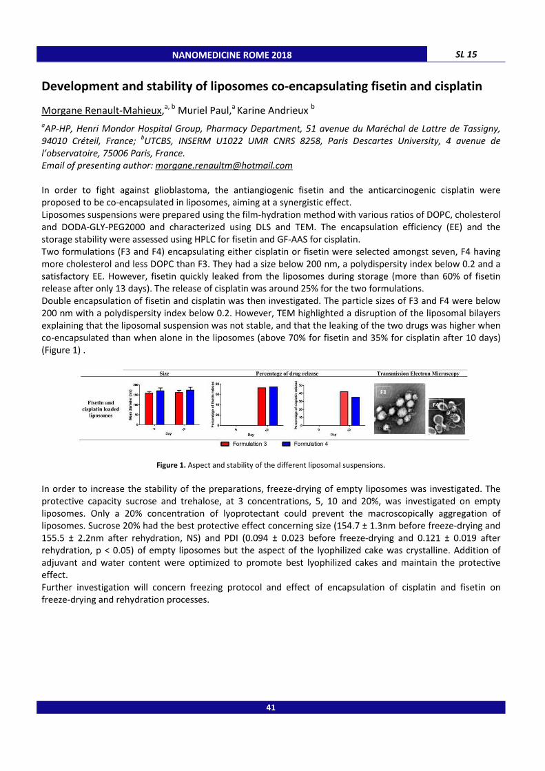
NANOMEDICINE ROME 2018 SL 15
Development and stability of liposomes co-encapsulating fisetin and cisplatin
Morgane Renault-Mahieux,a, b Muriel Paul,a Karine Andrieux b
aAP-HP, Henri Mondor Hospital Group, Pharmacy Department, 51 avenue du Maréchal de Lattre de Tassigny,
94010 Créteil, France; bUTCBS, INSERM U1022 UMR CNRS 8258, Paris Descartes University, 4 avenue de
l’observatoire, 75006 Paris, France.
Email of presenting author: [email protected]
In order to fight against glioblastoma, the antiangiogenic fisetin and the anticarcinogenic cisplatin were
proposed to be co-encapsulated in liposomes, aiming at a synergistic effect.
Liposomes suspensions were prepared using the film-hydration method with various ratios of DOPC, cholesterol
and DODA-GLY-PEG2000 and characterized using DLS and TEM. The encapsulation efficiency (EE) and the
storage stability were assessed using HPLC for fisetin and GF-AAS for cisplatin.
Two formulations (F3 and F4) encapsulating either cisplatin or fisetin were selected amongst seven, F4 having
more cholesterol and less DOPC than F3. They had a size below 200 nm, a polydispersity index below 0.2 and a
satisfactory EE. However, fisetin quickly leaked from the liposomes during storage (more than 60% of fisetin
release after only 13 days). The release of cisplatin was around 25% for the two formulations.
Double encapsulation of fisetin and cisplatin was then investigated. The particle sizes of F3 and F4 were below
200 nm with a polydispersity index below 0.2. However, TEM highlighted a disruption of the liposomal bilayers
explaining that the liposomal suspension was not stable, and that the leaking of the two drugs was higher when
co-encapsulated than when alone in the liposomes (above 70% for fisetin and 35% for cisplatin after 10 days)
(Figure 1) .
Figure 1. Aspect and stability of the different liposomal suspensions.
In order to increase the stability of the preparations, freeze-drying of empty liposomes was investigated. The
protective capacity sucrose and trehalose, at 3 concentrations, 5, 10 and 20%, was investigated on empty
liposomes. Only a 20% concentration of lyoprotectant could prevent the macroscopically aggregation of
liposomes. Sucrose 20% had the best protective effect concerning size (154.7 ± 1.3nm before freeze-drying and
155.5 ± 2.2nm after rehydration, NS) and PDI (0.094 ± 0.023 before freeze-drying and 0.121 ± 0.019 after
rehydration, p < 0.05) of empty liposomes but the aspect of the lyophilized cake was crystalline. Addition of
adjuvant and water content were optimized to promote best lyophilized cakes and maintain the protective
effect.
Further investigation will concern freezing protocol and effect of encapsulation of cisplatin and fisetin on
freeze-drying and rehydration processes.
41

SL 16 NANOMEDICINE ROME 2018
Drug delivery using protein nanocarriers: human or virus templates?
Alessandro Arcovito,a Adriana Amalfitano,a Francesca Bugli,b Pierpaolo Ceci,c Gianni Colotti,c Elisabetta Falvo,c Cecilia Martini,b Giuseppina Nocca,a Massimiliano Papi,d Alberto Vitali e a Institute of Biochemistry and Clinical Biochemistry, Catholic University of Sacred Heart, Largo F. Vito 1, 00168,
Rome, Italy; b Institute of Microbiology, Catholic University of Sacred Heart, Largo F. Vito 1, 00168, Rome, Italy;
c Institute of Molecular Biology and Pathology, CNR - National Research Council of Italy, Piazzale A. Moro 5,
00185 Rome, Italy; d Institute of Physics, Catholic University of Sacred Heart, Largo F. Vito 1, 00168, Rome, Italy;
e Institute of Chemistry of Molecular Recognition, CNR - National Research Council of Italy, Largo F. Vito 1,
00168, Rome, Italy.
Email of presenting author: [email protected]
In the modern view of selective drug delivery of bioactive molecules, the attention is moving onto the setup of
the perfect carrier more than in the optimization of the active compound. In this respect, nanoparticles (NPs)
are intensively studied for novel therapeutic use and are acquiring a special interest in drug-delivery and
regenerative medicine, due to the ability of release an active molecule in a more specific time and spatial
window, with respect to the systemic administration of a free drug. A subclass of NPs is made of self-assembling
protein subunits. A great advantage of using this last type of polymerizing agents, is the possibility to use at its
maximum extent the whole biotechnological support of protein engineering, in order to produce both wild-type
and designed mutants of the desired protein, as recombinant constructs of a specific expression vector. Two
different proteins have been deeply studied from our group to accomplish this task: VP6, a viral protein from
the viral capsid of human rotavirus A and human ferritin [1-5]. Results will be presented and discussed in light of
defining the characteristics that a modern drug nanocarrier should have to be effective and safe.
_____________________________________
1. Falvo E, Malagrinò F, Arcovito A, Fazi F, Colotti G, Tremante E, Di Micco P, Braca A, Opri R, Giuffrè A, Fracasso G, Ceci P, J Control
Release, 2018, 275, 177.
2. Fracasso G, Falvo E, Colotti G, Fazi F, Ingegnere T, Amalfitano A, Doglietto GB, Alfieri S, Boffi A, Morea V, Conti G, Tremante E, Giacomini P, Arcovito A, Ceci P, J Control Release, 2016, 239, 10.
3. Falvo E, Tremante E, Arcovito A, Papi M, Elad N, Boffi A, Morea V, Conti G, Toffoli G, Fracasso G, Giacomini P, Ceci P, Biomacromolecules, 2016, 17, 514.
4. Palmieri V, Bugli F, Papi M, Ciasca G, Maulucci G, Galgano S, Arcovito A, Sanguinetti M, De Spirito M, Journal of Nanomaterials, 2015, 1, 78786.
5. Bugli F, Caprettini V, Cacaci M, Martini C, Paroni Sterbini F, Torelli R, Della Longa S, Papi M, Palmieri V, Giardina B, Posteraro B,
Sanguinetti M, Arcovito A. Int J Nanomedicine, 2014, 9, 2727.
42
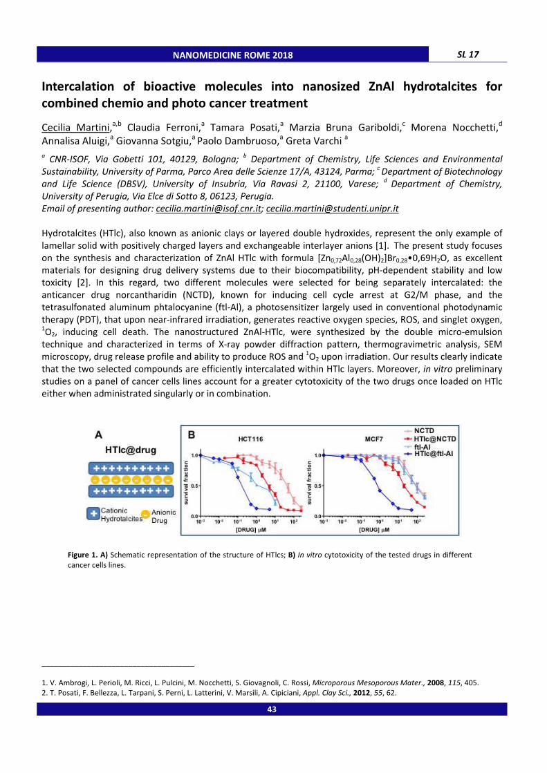
NANOMEDICINE ROME 2018 SL 17
Intercalation of bioactive molecules into nanosized ZnAl hydrotalcites for
combined chemio and photo cancer treatment
Cecilia Martini,a,b Claudia Ferroni,a Tamara Posati,a Marzia Bruna Gariboldi,c Morena Nocchetti,d Annalisa Aluigi,a Giovanna Sotgiu,a Paolo Dambruoso,a Greta Varchi a
a CNR-ISOF, Via Gobetti 101, 40129, Bologna;
b Department of Chemistry, Life Sciences and Environmental
Sustainability, University of Parma, Parco Area delle Scienze 17/A, 43124, Parma; c Department of Biotechnology
and Life Science (DBSV), University of Insubria, Via Ravasi 2, 21100, Varese; d
Department of Chemistry,
University of Perugia, Via Elce di Sotto 8, 06123, Perugia.
Email of presenting author: [email protected]; [email protected]
Hydrotalcites (HTlc), also known as anionic clays or layered double hydroxides, represent the only example of
lamellar solid with positively charged layers and exchangeable interlayer anions [1]. The present study focuses
on the synthesis and characterization of ZnAl HTlc with formula [Zn0,72Al0,28(OH)2]Br0,28•0,69H2O, as excellent
materials for designing drug delivery systems due to their biocompatibility, pH-dependent stability and low
toxicity [2]. In this regard, two different molecules were selected for being separately intercalated: the
anticancer drug norcantharidin (NCTD), known for inducing cell cycle arrest at G2/M phase, and the
tetrasulfonated aluminum phtalocyanine (ftl-Al), a photosensitizer largely used in conventional photodynamic
therapy (PDT), that upon near-infrared irradiation, generates reactive oxygen species, ROS, and singlet oxygen, 1O2, inducing cell death. The nanostructured ZnAl-HTlc, were synthesized by the double micro-emulsion
technique and characterized in terms of X-ray powder diffraction pattern, thermogravimetric analysis, SEM
microscopy, drug release profile and ability to produce ROS and 1O2 upon irradiation. Our results clearly indicate
that the two selected compounds are efficiently intercalated within HTlc layers. Moreover, in vitro preliminary
studies on a panel of cancer cells lines account for a greater cytotoxicity of the two drugs once loaded on HTlc
either when administrated singularly or in combination.
Figure 1. A) Schematic representation of the structure of HTlcs; B) In vitro cytotoxicity of the tested drugs in different
cancer cells lines.
_____________________________________
1. V. Ambrogi, L. Perioli, M. Ricci, L. Pulcini, M. Nocchetti, S. Giovagnoli, C. Rossi, Microporous Mesoporous Mater., 2008, 115, 405. 2. T. Posati, F. Bellezza, L. Tarpani, S. Perni, L. Latterini, V. Marsili, A. Cipiciani, Appl. Clay Sci., 2012, 55, 62.
43
SL 18 NANOMEDICINE ROME 2018
Nanovectors: how to characterize size and concentration in liquid matrices
Roberto Santoliquido
ALFATEST s.r.l., Via Giulio Pittarelli, 00166, Rome, Italy.
Email: [email protected]
Nanomedicine is daily evolving discovering new methods to deliver the drugs and control the bioavailability, the
size of the nanovector and its interaction with the biological fluids being key factors for successful drug delivery.
This presentation will show some of the latest technologies available to quantify by number the concentration
of different kind of nanovectors (viruses, SLN solid lipid nanoparticles, exosomes, polymer nanoparticles or
liposomes), monitor the protein corona formation and measure the size distribution to control the purification
steps. The technologies based on light scattering phenomena allow the physical characterization of size and
concentration while Field Flow Fractionation can increase dramatically the resolution to provide accurate
information about the physical state of the samples.
44

NANOMEDICINE ROME 2018 SL 19
Eumelanin-based substrates as smart materials for neuronal regeneration
Maria Grazia Raucci,a Ines Fasolino,a Irene Bonadies,a Alessandra Soriente,a Cosimo Carfagna,a Alessandro Pezzella,b Luigi Ambrosio a a Institute of Polymers, Composites and Biomaterials (IPCB) - CNR, Viale J.F. Kennendy 54 – Mostra d’Oltremare
Pad.20, 80125 Naples (Italy); b
Department of Chemical Sciences, University of Naples “Federico II”, Via Cintia 4,
I-80126 Naples (Italy).
Email of presenting author: [email protected]
The regeneration of neurite network constitutes a strategy for the treatment of neurodegenerative disorders
characterized by a loss and dysfunction of trophic factors such as BDNF and NGF. The fabrication of eumelanin-
coated microfibrous structures represents a novel strategy in order to realize tissue-engineering scaffolds for
neuronal cells growth and control by providing both mechanical support for growing cells and biological signals
to direct the axonal growth cone to the distal stump. Our results show that eumelanin coatings (random and
aligned fibres distribution), eliminating the need of differentiating factors in the media, expand the scope of
substrate driven cell culture growth and maturation. Indeed, biological results showed that both random and
aligned eumelanin microfibers support cell survival and adhesion. Furthermore, eumelanin random microfibers
induced the formation network of neuritic processes and stimulated GAP-43 expression over culture time, thus
confirming differentiation processes.
Figure 1. Scheme of Eumelanin-based substrates preparation for neuronal regeneration
_____________________________________
1. I. Fasolino et al., ACS Applied Materials & Interfaces, 2017, 9, 40070.
Acknowledgments: The study was supported by PNR Aging program 2012-2018 from the Ministero dell’Università e della Ricerca. The
authors also thank Mrs. Cristina Del Barone of LAMEST laboratory for SEM investigations and Mrs Stefania Zeppetelli for supporting biological investigations.
45

SL 20 NANOMEDICINE ROME 2018
The role of nanocarrier physicochemical properties on the biodistribution and
the blood brain barrier passage
Laura Talamini, Elisa Zanier, Mario Salmona, Paolo Bigini
IRCCS - Istituto di Ricerche Farmacologiche “Mario Negri”, Milano, Italy.
Email of presenting author: [email protected]
Nanocarriers (NC) are a big hope and, at the same time, a hard challenge for the medicine of the XXI century.
Ever since the discovery of liposomal formulation to reduce the anti-cancer compounds toxicity, many efforts
have been carried out, however the clinical relapses have been poor and controversial. One of the major
hurdles was the extreme heterogeneity of preclinical studies, both in terms of materials and biological tools. In
this context, the passage of NCs through the blood brain barrier (BBB) remains far from being elucidated.
Although different studies seem to correlate the NC physicochemical features and the rate of brain
accumulation, nowadays, reliable rules are strongly needed for successful clinical translation. Our study was
therefore aimed at understanding IF and HOW NC parameters (such as size, shape and surface
functionalization) may actually influence the BBB passage.
Through a step-by-step approach, we measured the amount of nano-material in the mouse brains after
systemic administration using a measurable and repeatable technique. To minimize biological variables,
healthy, immunocompetent mice were used and housed in specific pathogen free conditions.
Firstly, the role of the size was assessed. LPS-free spherical nanogolds (NGs) having two sizes (100 and 5 nm)
were intravenously injected. Mice were sacrificed at 1 hour, 1 day and 5 days after the treatment and the
percentage of injected dose (ID%) was calculated in the brain and other peripheral organs. In parallel, the role
of the shape was investigated following the same procedure described above.
Our work suggests that the tissue distribution of NGs is size- and shape- dependent but the brain tropism is not
affected by this.
Subsequently, the effect of Apoε coating and its specific architecture on 100 nm silica NCs was investigated.
Quite surprisingly, neither the presence of the peptide nor its orientation improved the BBB passage.
Next, we moved to consider the animal model of traumatic brain injury (TBI), and we treated them with Apoε
silica NCs. Expectedly, the data achieved pinpointed that the levels of silica in the traumatized hemisphere was
manifold higher than those measured in controlateral side. Quite interestingly, similarly to the results obtained
from healthy mice, no influence of the surface functionalization was observed.
The last experiment was carried out treating healthy mice with sugar-coated silica NCs (20 and 5 nm). Whilst,
similarly to NGs, we did not find a robust size-dependent effect, we demonstrated that the presence of sugar
significantly improved the BBB passage of 5 nm NCs exclusively.
Although preliminary, this study can be considered a reliable attempt to provide a correlation between the
geometry, the nature of the material, the type of ligand linked to NCs and the BBB passage in both undamaged
and injured brain. Ex vivo analyses on brain sections are in progress to evaluate the overall distribution of NCs in
brain parenchyma and their cellular tropism.
46
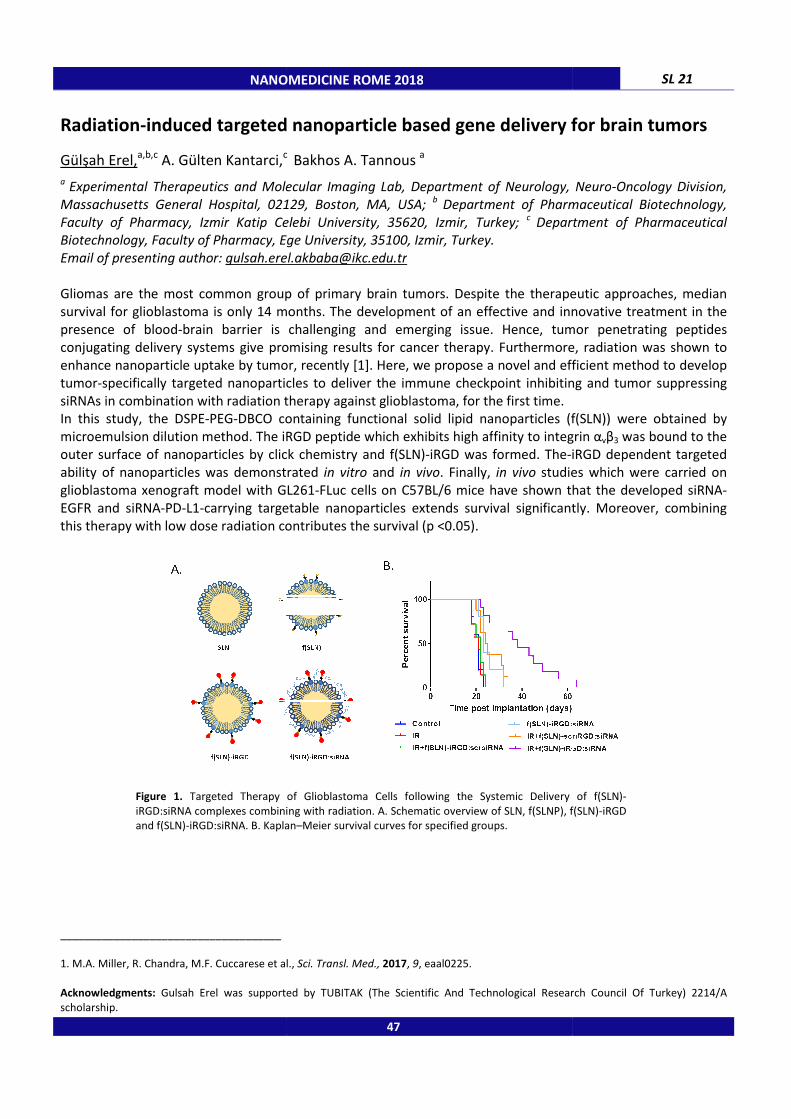
NANOMEDICINE ROME 2018
Radiation-induced targeted nanoparticle based gene delivery for brain tumors
Gülşah Erel,a,b,c
A. Gülten Kantarci,c
a Experimental Therapeutics and Molecular Imaging Lab, Department of Neurology, Neuro
Massachusetts General Hospital, 02129, Boston, MA, USA;
Faculty of Pharmacy, Izmir Katip Celebi University,Biotechnology, Faculty of Pharmacy, Ege University, 35100, Izmir, Turkey.
Email of presenting author: [email protected]
Gliomas are the most common group of primary brain tumors
survival for glioblastoma is only 14 months.
presence of blood-brain barrier is
conjugating delivery systems give promising results for cancer therapy. Furthermore, radiation was shown to
enhance nanoparticle uptake by tumor, recently
tumor-specifically targeted nanoparticles
siRNAs in combination with radiation therapy against
In this study, the DSPE-PEG-DBCO containing functional solid lipid nanoparticles (f(SLN)) were obtained by
microemulsion dilution method. The iRGD peptide which exhibits high af
outer surface of nanoparticles by click chemistry and f(SLN)
ability of nanoparticles was demonstrated
glioblastoma xenograft model with GL261
EGFR and siRNA-PD-L1-carrying targetable nanoparticles extends survival significantly. Moreover, combining
this therapy with low dose radiation contributes the survival (p <0.05).
Figure 1. Targeted Therapy of Glioblastoma Cells following the Systemic Delivery of f(SLN)
iRGD:siRNA complexes combining with radiation. A. Schematic overview of SLN, f(SLNP), f(SLN)
and f(SLN)-iRGD:siRNA. B. Kaplan
_____________________________________
1. M.A. Miller, R. Chandra, M.F. Cuccarese et al.,
Acknowledgments: Gulsah Erel was supported by TUBITAK (The Scientific And Technological Research Council Of Turkey) 2214/A
scholarship.
NANOMEDICINE ROME 2018
induced targeted nanoparticle based gene delivery for brain tumors
c Bakhos A. Tannous
a
Experimental Therapeutics and Molecular Imaging Lab, Department of Neurology, Neuro
Massachusetts General Hospital, 02129, Boston, MA, USA; b Department of Pharmaceutical Biotechnology,
Faculty of Pharmacy, Izmir Katip Celebi University, 35620, Izmir, Turkey; c Department of Pharmaceutical Biotechnology, Faculty of Pharmacy, Ege University, 35100, Izmir, Turkey.
the most common group of primary brain tumors. Despite the therapeutic approaches, median
survival for glioblastoma is only 14 months. The development of an effective and innovative
is challenging and emerging issue. Hence, tumor penetrating peptides
jugating delivery systems give promising results for cancer therapy. Furthermore, radiation was shown to
enhance nanoparticle uptake by tumor, recently [1]. Here, we propose a novel and efficient method to develop
nanoparticles to deliver the immune checkpoint inhibiting and tumor suppressing
n combination with radiation therapy against glioblastoma, for the first time.
DBCO containing functional solid lipid nanoparticles (f(SLN)) were obtained by
microemulsion dilution method. The iRGD peptide which exhibits high affinity to integrin
outer surface of nanoparticles by click chemistry and f(SLN)-iRGD was formed. The-iRGD dependent targeted
ability of nanoparticles was demonstrated in vitro and in vivo. Finally, in vivo studies which were carried on
glioblastoma xenograft model with GL261-FLuc cells on C57BL/6 mice have shown that the developed siRNA
carrying targetable nanoparticles extends survival significantly. Moreover, combining
th low dose radiation contributes the survival (p <0.05).
Targeted Therapy of Glioblastoma Cells following the Systemic Delivery of f(SLN)
iRGD:siRNA complexes combining with radiation. A. Schematic overview of SLN, f(SLNP), f(SLN)
iRGD:siRNA. B. Kaplan–Meier survival curves for specified groups.
1. M.A. Miller, R. Chandra, M.F. Cuccarese et al., Sci. Transl. Med., 2017, 9, eaal0225.
Gulsah Erel was supported by TUBITAK (The Scientific And Technological Research Council Of Turkey) 2214/A
47
SL 21
induced targeted nanoparticle based gene delivery for brain tumors
Experimental Therapeutics and Molecular Imaging Lab, Department of Neurology, Neuro-Oncology Division,
Department of Pharmaceutical Biotechnology,
Department of Pharmaceutical
Despite the therapeutic approaches, median
and innovative treatment in the
Hence, tumor penetrating peptides
jugating delivery systems give promising results for cancer therapy. Furthermore, radiation was shown to
and efficient method to develop
to deliver the immune checkpoint inhibiting and tumor suppressing
DBCO containing functional solid lipid nanoparticles (f(SLN)) were obtained by
finity to integrin αvβ3 was bound to the
iRGD dependent targeted
studies which were carried on
FLuc cells on C57BL/6 mice have shown that the developed siRNA-
carrying targetable nanoparticles extends survival significantly. Moreover, combining
Targeted Therapy of Glioblastoma Cells following the Systemic Delivery of f(SLN)-
iRGD:siRNA complexes combining with radiation. A. Schematic overview of SLN, f(SLNP), f(SLN)-iRGD
Gulsah Erel was supported by TUBITAK (The Scientific And Technological Research Council Of Turkey) 2214/A
SL 22 NANOMEDICINE ROME 2018
VCAM-1 targeted paramagnetic micelles for Magnetic Resonance Imaging of
neuroinflammation
Francesca Garello,a Amerigo Pagoto,a Francesca Arena,a Annalisa Buffo,b,c Francesco Blasi, a Diego Alberti, a Enzo Terreno a a Department of Molecular Biotechnology and Health Sciences, University of Torino, Via Nizza 52, 10126, Torino,
Italy; b Department of Neuroscience Rita Levi-Montalcini, University of Torino, 10126, Torino, Italy;
cNeuroscience Institute Cavalieri Ottolenghi, Regione Gonzole 10, 10043, Orbassano, Italy.
E-mail of presenting author: [email protected]
The need for specialized non invasive imaging techniques is increasing in order to better clarify the role and the
spatio-temporal correlation between neuroinflammation and the onset of neurodegenerative diseases [1].
Magnetic Resonance Imaging (MRI) with its remarkable spatial resolution and poor toxicity could be the
technique of choice. In the herein reported work paramagnetic micelles targeting Vascular Cell Adhesion
Molecule-1 (VCAM-1), over-expressed in case of inflammation, were designed and in vivo tested in a model of
neuroinflammation. The cyclic nonapeptide CNNSKSHTC [2], able to bind VCAM-1 with high specificity, was
synthesized and conjugated to DSPE-PEG2000 to formulate a lipid-based nanosystem including Gd-
DOTAMA(C18)2 and Rhodamine-DOPE. The scrambled version HSCNKNSCT was tested as control. VCAM-1
targeted micelles (size ca. 20 nm) showed a longitudinal relaxivity of 35 s-1
mMGd-1
at around 1T. Targeted
micelles were first tested in vitro, on brain endothelial cells, displaying higher affinity towards VCAM-1
expressing cells in comparison to the untargeted nanosystem. Subsequently targeted micelles were iv injected
in mice (n=20) bearing LPS induced neuroinflammation. The inflamed region was clearly identified and detected
only after targeted micelle administration, with a MRI T1 signal enhancement calculated over pre images of
39.3 ± 4.4 %, 24 h p.i. Whereas scrambled micelles and the clinical agent MultiHance® showed a comparable T1
SE in the inflamed region, probably due to passive extravasation (18.9 % and 13.4 % respectively). Histological
studies proved micelle extravasation at the lesion site. In conclusion, the developed probe allowed for the early
detection of the disease, with higher contrast and more precise localization in comparison to untargeted
micelles or to the clinical agent MultiHance®. Moreover, the relatively long blood half-life of the nanosystem
(ca. 6.3 h) guaranteed a good accumulation in the inflamed regions, paving the way to future
diagnostic/theranostic applications, implying the loading of neuroprotective or even anti-cancer drugs inside
the core of the micelles.
_____________________________________
1. Jacobs A.H., Tavitian B., INMiND consortium, J Cereb Blood Flow Metab., 2012, 32(7), 1393.
2. Burtea C. et al., J Med Chem. 2009, 52(15), 4725.
Acknowledgments: The research leading to these results has received funding from the European Union's Seventh Framework
Programme (FP7/2007-2013) under grant agreement n° HEALTH-F2-2011-278850 (INMiND)
48

NANOMEDICINE ROME 2018 SL 23
Reactive oxygen species-responsive nano-in-micro composite for targeted
therapy of inflammatory bowel disease
Serena Bertoni,a,b Wei Li,a Zehua Liu,a Alexandra Correia,a João Pedro Martins,a Antti Rahikkala,a Flavia Fontana,a Marianna Kemell,c Beatrice Albertini,b Nadia Passerini,b Hélder A. Santos a a Drug Research Program, Division of Pharmaceutical Chemistry and Technology, Faculty of Pharmacy,
University of Helsinki, 00014, Helsinki, Finland; b PharmTechLab, Department of Pharmacy and Biotechnology,
University of Bologna, via San Donato 19/2, 40127, Bologna, Italy; c
Laboratory of Inorganic Chemistry,
Department of Chemistry, University of Helsinki, 00014, Helsinki, Finland.
Email of presenting author: [email protected]
Oxidative stress and abnormally high levels of reactive oxygen species play an essential role in the pathogenesis
and progression of inflammatory bowel disease (IBD) [1]. In this study we designed a nanoplatform for the
encapsulation and “on demand” delivery of rifaximin (RIF) at sites of oxidative stress. Nanoparticles (NPs) were
formulated from a phenylboronic esters-modified dextran (OxiDEX), that degrades selectively in response to
hydrogen peroxide (H2O2) [2]. OxiDEX NPs with and without a chitosan (CS) coating were then encapsulated by
microfluidics in a pH-sensitive polymer to produce nano-in-micro composites called NPs@MF and CS-NPs@MF,
respectively (Figure 1b). The final composites were spherical with homogeneous particle size (53±3 μm) and
maintained integrity at acidic pH, preventing the premature release of the NPs in simulated gastric conditions.
The degradation of NPs was highly responsive to the level of H2O2, and the release of the loaded drug was
sustained in the presence of physiologically relevant H2O2 concentrations (Figure 1a). The presence of CS on the
particles surface significantly enhanced NPs stability in intestinal pH and the interactions between NPs and cells.
Compared to a traditional enteric formulation, the composites showed ten-fold decreased drug permeability
across intestinal cell monolayer, representing an advantage in terms of unspecific absorption and systemic side
effects. Based on the results, the developed nano-in-micro composite has great potential for selective drug
delivery in IBD treatment.
Figure 1. (a) Drug release profiles of NPs@MF and CS-NPs@MF and reference formulation consisted in pure drug encapsulated in the same enteric polymer (RIF@MF). (b) Schematic illustration of the
fabrication process of the nano-in-micro composites by microfluidics.
_____________________________________
1. T. Tian, Z. Wang, J. Zhang, Oxid. Med. Cell. Longev., 2017, 2017, 1.
2. K.E. Broaders, S. Grandhe, J.M. J. Fréchet, J. Am. Chem. Soc., 2011, 133, 756.
49

SL 24 NANOMEDICINE ROME 2018
Redox responsive polymeric nanocarriers for the combined therapy of lung
cancer
Claudia Conte,a,b Francesca Ungaro,a Fabiana Quaglia,a Snjezana Stolnik,b Maria Kavallaris,c Cameron Alexander b
a Department of Pharmacy, University of Napoli Federico II, Via D. Montesano 49, 80131, Napoli, Italy;
b MTF
Division, School of Pharmacy, University of Nottingham, NG7 2RD, Nottingham, UK; c Children's Cancer Institute,
C25/9 High St, Kensington, NSW 2750, Australia.
Email of presenting author: [email protected]
Redox-responsive nanoparticles (RR-NPs) are of interest for anticancer nanomedicines, owing to the possibility
to ‘design in’ selective modulation of drug release at target sites [1,2]. With this in mind, herein we have
developed new RR-NPs based on polyethylene glycol (PEG)-poly(lactic-co-glycolic acid) (PLGA) block copolymers
and novel poly (β-aminoesters) (PβAE) containing bioreducible disulfide units for the in situ delivery of
combination drugs. In particular, we have focused our attention on docetaxel (DTX) and TUBB3-siRNA, in order
to have a synergistic effect in the treatment of lung cancer. Spherical NPs of around 150 nm with negative zeta
potential and high loading efficiencies of both drugs were obtained. NPs showed high stability in the most
relevant simulated biological fluids whereas disassembly of NP structure was shown at a simulated intracellular
level of reducing agents, thus triggering drug release. Unloaded NPs were well-tolerated by lung cancer cells,
while in terms of intracellular siRNA delivery, demonstrated a fast and high gene-silencing efficiency. Uptake
and intracellular trafficking of NPs showed a high internalization of NPs and no co-localization of the siRNA with
the lysosomes, thus suggesting that NPs were able to escape to the reducing cytosolic regions. Finally, viability
of cells treated with combined DTX/TUBB3-siRNA-NPs remarkably decreased as compared to NPs loaded only
with DTX, thus showing an efficient synergistic anticancer effect. In fact, through immunofluorescence
microscopy, significant reduction of class III β-tubulin expression was found, thus potentiating DTX activity.
Taken together, these results demonstrate that PLGA/PβAE RR-NPs represent a promising new therapeutic
approach with great potential for the dual siRNA/drug therapy of lung cancer.
Figure 1. Schematic illustration of NP preparation.
_____________________________________
1. R. Cheng, F. Feng, F.H. Meng, C. Deng, J. Feijen, Z.Y. Zhong, Journal of Controlled Release, 2011, 152, 2.
2. C. Conte, F. Mastrotto, V. Taresco, A. Tchoryk, F. Quaglia, S. Stolnik, C. Alexander, Journal of Controlled Release, 2018, in press.
Acknowledgments: Financial support of AIRC/Marie Curie Actions is gratefully acknowledged.
50
Emulsion- solvent evaporation

NANOMEDICINE ROME 2018 SL 25
Study of membrane phase transition in bacterial vesicles
Angelo Sarra,a S. Sennato,b A. Celluzzi,c A. Masotti,c C. Ricci,d M.G. Ortore,d F. Bruni,a F. Bordi,b P. Postorino b a
Department of Science, University of Roma Tre, Via della Vasca Navale, 00146, Rome, Italy; b
Department of
Physics, Sapienza University of Rome, Pz.le A.Moro, 00185, Rome, Italy;c
Gene Expression Microarrays
Laboratory, Bambino Gesù Children's Hospital of Rome, Via di S.Paolo, 00146, Rome, Italy; d
SAIFET department,
Polytechnic University of Marche, Via B.Bianche, 60131, Ancona, Italy.
Email of presenting author: [email protected]
In the last few years, it has been discovered that the majority of eukaryotic and prokaryotic cells release small
lipid vesicles. These vesicles, generally called micro-vesicles or exosomes, are becoming the object of a great
scientific interest, because of their role as carrier of information about progenitor cells, through their cargo of
proteins and nucleic acids and the biochemical composition of their membrane [1]. Here we present a study of
the temperature-induced phase transitions in the membrane of bacterial Outer-Membrane-Vesicles (OMVs). In
particular, we study the OMVs produced by Escherichia Coli because they are considered a model system for
biological studies. Indeed, as known from the literature [2], Escherichia Coli membrane presents different
temperature phase transitions due to structural changes in the organization of the lipid bilayer. In our study, we
have studied the phase transition behaviour of the OMVs membrane by Light Scattering measurements at
varying temperature [3]. OMVs were characterized by the presence of phase transitions similar to those
revealed for E. Coli membrane but occurring at slightly different temperatures. In order to better understand
the membrane behaviour at the transition temperature, SAXS measurements have been made and will be
analysed in the next future.
Figure 1: Light scattered intensity as function of temperature. For both increasing (red) and decreasing (blue) temperature it is clear that a membrane phase transition occurs.
_____________________________________
1. A.V. Vlassov et al., Biochimica et Biophysica Acta, 2012, 1820, 940. 2. H. Trauble et al., Biochimica et Biophysica Acta, 1973, 307, 491.
3. N. Michel et al., Chemistry and Physics of lipids, 2006, 139, 11.
51
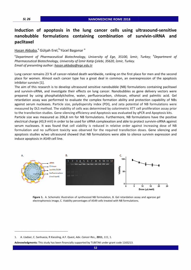
SL 26
Induction of apoptosis in the lung cancer cells using ultrasound
nanobubble formulations containing combination of survivin
paclitaxel
Hasan Akbaba,a Gülşah Erel,
b Yücel Başpınar
aDepartment of Pharmaceutical Biotechnology, University of Ege, 35100, İzmir, Turkey;
Pharmaceutical Biotechnology, University of İzmir Katip Çelebi, 35620, İzmir, Turkey.
Email of presenting author: [email protected]
Lung cancer remains 23 % of cancer-related death worldwide, ranking on the first place for men and the second
place for women. Almost each cancer type has a great deal in common, an overexpression of the apoptosis
inhibitor survivin [1].
The aim of this research is to develop ultras
and survivin-siRNA, and investigate their effects on lung cancer.
prepared by using phosphatidylcholine, water, perfluorocarbon, chitosan, ethanol a
retardation assay was performed to evaluate the complex formation ability and protection capability of NBs
against serum nucleases. Particle size, polydispersity index (PDI), and zeta potential of NB formulations were
measured by DLS method. The viability of cells was determined by colorimetric XTT cell proliferation assay prior
to the transfection studies. Gene silencing efficiency and Apoptosis
Particle size was measured as 206,8 nm for NB formulations.
electrical charge (43,9 mV) in order to be used for siRNA complexation and
serum nucleases. It was found that
formulation and no sufficient toxicity was observed for the required transfection doses.
apoptosis studies w/wo ultrasound showed that NB formulations were able to silence survivin expression a
induce apoptosis in A549 cell line.
Figure 1. A. Schematic illustration of synthesized NB formulation, B. Gel retardation assay and agarose gel
electrophoresis image, C. Viability percentages of A549 cells treated with NB formulations.
_____________________________________
1. A. Lladser, C. Sanhueza, R Kiessling, A.F. Quest,
Acknowledgments: This study has been financially supported by TUBITAK under grant code 116S213.
NANOMEDICINE ROME 2018
Induction of apoptosis in the lung cancer cells using ultrasound
nanobubble formulations containing combination of survivin
Yücel Başpınar a
Pharmaceutical Biotechnology, University of Ege, 35100, İzmir, Turkey;
Pharmaceutical Biotechnology, University of İzmir Katip Çelebi, 35620, İzmir, Turkey.
related death worldwide, ranking on the first place for men and the second
place for women. Almost each cancer type has a great deal in common, an overexpression of the apoptosis
The aim of this research is to develop ultrasound sensitive nanobubble (NB) formulations containing paclitaxel
siRNA, and investigate their effects on lung cancer. Nanobubbles as gene delivery vectors were
prepared by using phosphatidylcholine, water, perfluorocarbon, chitosan, ethanol a
retardation assay was performed to evaluate the complex formation ability and protection capability of NBs
article size, polydispersity index (PDI), and zeta potential of NB formulations were
method. The viability of cells was determined by colorimetric XTT cell proliferation assay prior
to the transfection studies. Gene silencing efficiency and Apoptosis was evaluated by qPCR and Apopto
206,8 nm for NB formulations. Furthermore, NB formulations have the positive
electrical charge (43,9 mV) in order to be used for siRNA complexation and able to protect
serum nucleases. It was found that cell viability is reduced in relative order against increasing dose of NB
formulation and no sufficient toxicity was observed for the required transfection doses.
apoptosis studies w/wo ultrasound showed that NB formulations were able to silence survivin expression a
. Schematic illustration of synthesized NB formulation, B. Gel retardation assay and agarose gel
electrophoresis image, C. Viability percentages of A549 cells treated with NB formulations.
A. Lladser, C. Sanhueza, R Kiessling, A.F. Quest, Adv. Cancer Res., 2011, 111, 1.
This study has been financially supported by TUBITAK under grant code 116S213.
52
Induction of apoptosis in the lung cancer cells using ultrasound-sensitive
nanobubble formulations containing combination of survivin-siRNA and
Pharmaceutical Biotechnology, University of Ege, 35100, İzmir, Turkey; bDepartment of
related death worldwide, ranking on the first place for men and the second
place for women. Almost each cancer type has a great deal in common, an overexpression of the apoptosis
ound sensitive nanobubble (NB) formulations containing paclitaxel
Nanobubbles as gene delivery vectors were
prepared by using phosphatidylcholine, water, perfluorocarbon, chitosan, ethanol and palmitic acid. Gel
retardation assay was performed to evaluate the complex formation ability and protection capability of NBs
article size, polydispersity index (PDI), and zeta potential of NB formulations were
method. The viability of cells was determined by colorimetric XTT cell proliferation assay prior
was evaluated by qPCR and Apoptosis kits.
Furthermore, NB formulations have the positive
able to protect survivin-siRNA against
elative order against increasing dose of NB
formulation and no sufficient toxicity was observed for the required transfection doses. Gene silencing and
apoptosis studies w/wo ultrasound showed that NB formulations were able to silence survivin expression and
. Schematic illustration of synthesized NB formulation, B. Gel retardation assay and agarose gel
electrophoresis image, C. Viability percentages of A549 cells treated with NB formulations.

NANOMEDICINE ROME 2018 SL 27
Functionalized superparamagnetic iron oxide nanoparticles as theranostic
devices: from development to interaction with proteins
Irene Russo Krauss,a,b Alessandra Picariello,a Alessandra Luchini,b Luigi Paduano a,b
a
Department of Chemical Science, University of Naples Federico II, Via Cinthia, I-80126, Naples, Italy; b
CSGI, Via
Lastruccia, I-50019, Florence, Italy.
Email of presenting author: [email protected]
Metal-based nanoparticles (NPs) have attracted great interest in the last years for different uses in biomedical
field, including diagnostic, drug delivery and therapeutic applications [1]. Among metal-based nanoparticles,
iron oxide nanoparticles with a radius less than 20 nm are very promising for the design of diagnostic and
theranostic devices, because they exhibit a strong magnetic response to an applied external magnetic field,
which allow their use as contrast agents, and are inherently poorly toxic [2]. However, in order to be safely
employed in vivo, NPs need to be water soluble, biocompatible and non toxic. The first two targets can be
achieved through a proper coating of NP surface. In this respect we have developed small Fe3O4 NPs coated
with a double layer of oleic acid/oleylamine and 1-octadecyl-2-hydroxy-sn-glycero-3-phosphocholine (18LPC)
[3], which can be further functionalized employing drugs [4] or different targeting or diagnostic agents bearing a
proper hydrophobic tail. The possible toxicity of the NPs is a far serious issue. Apart from organ/tissue
accumulation and removal of NPs, the first event to take in account is the interaction of the NP with proteins,
because interaction between NPs and proteins can cause toxic effects by altering, unfolding or inducing
aggregation of proteins [5], but also change NP properties and functionalities, through extensive covering of
nanoparticle surface by proteins. In this respect, NP behaviour strongly depends on their chemical nature and
geometrical properties (dimensions, curvature of the surface and so on) [6] as well as on their coating. With the
aim at fully developing our nanosystems for biomedical applications, we have investigated their interaction with
two abundant serum proteins, human apo transferrin (HTF) and human serum albumin (HSA), by means of
spectroscopic and scattering techniques. We found that interaction with NPs does not cause the unfolding of
the investigated proteins and has only limited effects on their tertiary structure. In the case of HSA a protein
layer on the NP surface forms. None of these effects is observed with control 18LPC solutions, suggesting that
they depend on the geometrical properties of the NP and on protein structural features. Our results confirm the
high biocompatibility of 18LPC as coating molecule for development of biomedical NPs.
_____________________________________
1. Nasrabadi H.T., Abbasi E., Davaran S., Kouhi M., Akbarzadeh A., Art cells nanomed biotech, 2016, 44, 376. 2. Ghazanfari M.R., Kashefi M., Shams S.F., Jaafari M.R., Bioch res int, 2016, 7840161.
3. Luchini A., Vitiello G., Rossi F., Ruiz De Ballesteros O., Radulescu A., D'Errico G., Montesarchio D., de Julian Fernandez C., Paduano L., PCCP, 2015, 17, 6087.
4. Luchini A., Irace C., Santamaria R., Montesarchio D., Heenan R.K., Szekely N., Flori A., Menichetti L., Paduano L., Nanoscale 2016, 8,
10078. 5. Neagu M., Piperigkou Z., Karamanou K., Engin A.B., Docea A.O., Constantin C., Negrei C., Nikitovic D., Tsatsakis A., Arch tox, 2017, 91,
1031. 6. Hu Z., Zhang H., Zhang Y., Wu R., Zou H., Coll surf B, 2014, 121, 354.
53

SL 28 NANOMEDICINE ROME 2018
Influence of the surface properties of nanoliposomes on protein corona
formation
Dushko Shalabalija,a Ljubica Cambuleva,a Ivana C. Karanfilova,b Maja S. Crcarevska,a Krtistina Mladenovska, a Vladimir Ivanovski, c Marija Glavas Dodova a
Institute of Pharmaceutical Technology, Faculty of Pharmacy, University Ss. Cyril and Methodius, Majka Tereza
47, 1000, Skopje, R. Macedonia; b Institute of Pharmacognosy, Faculty of Pharmacy, University Ss. Cyril and
Methodius, Majka Tereza 47, 1000, Skopje, R. Macedonia; c Department of Chemistry, Faculty of Natural Science
and Mathematics, University Ss. Cyril and Methodius, Arhimedova, 1000, Skopje, R. Macedonia.
Email of presenting author: [email protected]
One of the biggest problems upon nanoparticles’ administration in vivo is their interaction with blood proteins
and the formation of the protein corona (PC), followed by the rapid recognition and uptake of the particles by
the mononuclear phagocyte cells, thus leading to quick removal from the circulation. Therefore, understanding
of the particle–PC complex formation is a prerequisite for successful development of drug carrier system and
proper characterization is essential.
This problem could be partially overcome by the decoration of the particles’ surface with polyethylene glycol
(PEG) [1]. In order to investigate the effect of PEG on the PC formation, as well as to characterize the
interaction, two formulations of nanoliposomes (NLs) (lechitin:cholesterol:PEG = 8.7:1:1.7 and 9:1:0.17 for
sample 1 and 2, respectively) loaded with rosmarinic extract (RE) were prepared by the modified lipid film
hydration technique [2]. Blank samples were prepared for comparison. The mean size of the prepared NLs was
~120 nm, with unimodal narrow size distribution and high encapsulation efficiency (~90%). In vitro release
studies showed that the prepared vesicles had prolonged drug release properties (26 and 46% for 24h, for
sample 1 and 2, respectively). The slower release of RE from sample 1 could be probably due to the fast
hydration process of the higher amount of PEG present on the surface of the vesicles. NLs–PC complex
formation was confirmed using SDS-PAGE. Our previous investigations using Bratford assay confirmed that the
PEG increases the hydrophilicity of the NLs surface thus resulting in reduced PC formation. These results were
confirmed using FTIR and quantitative information of protein–NLs interactions was gained. From the obtained
spectra it could be concluded that NLS-protein interaction was mainly due to the hydrogen bonds formed
between C = O...N-H or C = O...H-OH. These interactions were stronger between the NLs prepared with lower
PEG amount and the protein. Incorporation of RE into the NLs did not affect the intensity of the interactions.
_____________________________________
1. M. Sangrà, J. Estelrich, R. Sabaté, A. Espargaró, M.A. Busquets, Nanomaterials (Basel), 2017, 7(2), 37.
2. Lj. Cambuleva, D. Shalabalija, I. Cvetkovikj, M. Simonoska Crcarevska, M. Glavas Dodov, Maced. Pharm. Bull., 2016 , 62(suppl), 641.
54

OA-KAT
NANOMEDICINE ROME 2018 SL 29
Preparation of Synthetic Low Density Lipoprotein (sLDL) with an on-Surface
Chemoseletive Ligation
Alessandro Fracassi,a Sean Orian,a Naoko Yoshizawa-Sugata,b Hisao Masai,b Yoko Yamakoshi a a
Laboratorium für Organische Chemie, ETH Zürich, Vladimir-Prelog-Weg 3, 8093 Zürich, Switzerland; b
Tokyo
Metropolitan Institute of Medical Sciences, Kamikitazawa 2-1-6, Setagaya, Tokyo 156-8506, Japan.
Email of presenting author: [email protected]
Low density lipoprotein (LDL) has been used as a powerful carrier for the targeted delivery of drugs and imaging
probes [1,2,3]. Since natural LDL (nLDL) suffers from disadvantages such as limited supply and deleterious side
effects, the development of synthetic LDL (sLDL) is an attractive alternative. In this study, we report a versatile
method for the chemical functionalization of sLDL using a chemoselective ligation reaction of potassium
acryltifluoroborate (KAT) with N-hydroxylamine (HA), which allows rapid amide bond formation under dilute
aqueous conditions with equimolar amounts of ligation partner, in the presence of unprotected functional
groups [4]. The sLDL was prepared by mixing a KAT derivative of oleic acid (OA-KAT, 5% mol) with the lipidic
contents used in the preparation of previously reported sLDL [5] (phosphatidyl choline (PC), triolein (TO), and
cholesterol oleate (CO) in 3:2:1 mol ratio). All starting materials were solubilized in a mixture of CHCl3 - MeOH -
acetone (2:1:1) and concentrated to provide a uniform lipid film, which was subsequently hydrated with Tris-
HCl buffer (pH 8.0), sonicated and extruded through membranes (with pore size of 50 and 100 nm) to give
lipidic nanoparticles (NP) with KAT moiety (NP-KAT). The obtained NP-KAT was well-dispersed, with particle
diameters of 60-75 nm by DLS. Separately, HA derivatives of apoB100-mimetic peptide 1 (HA-GTTRLTRKRGLKLA)
and fluorescein 2 were synthesized and purified by HPLC. The “on-particle” KAT ligation of NP-KAT with both 1
and 2 was carried out in NH4Cl saturated solution (pH 5.5) to provide fluorescent sLDL. Using a macrophage cell
line RAW 264.7, sLDL was shown to accumulate in cells, as observed by fluorescent microscopy, demonstrating
the potential of sLDL as a carrier.
Figure. NP-KAT preparation and subsequent surface modification by KAT ligation with HA derivatives of apoB100-mimetic peptide 1 and
fluorescein 2.
_____________________________________
1. X. Zhang and G. Huang, Drug Delivery, 2017, 24, 16. 2. Y. Yamakoshi et al., Chem. Commun. 2011, 47, 8835.
3. A.N. Lowell et al., Bioconjugate Chem., 2012, 23, 2313. 4. H. Noda et al., J. Am. Chem. Soc., 2014, 136, 5611.
5. G. Baillie et al., J. Lipid Res. 2002, 43, 69.
55
on-particle KAT ligation
1) sonication2) extrusion
KAT
NP-KAT
2
1

SL 30 NANOMEDICINE ROME 2018
Cerium oxide nanoparticles as potential antibiotic adjuvant. Effects of CeO2
nanoparticles on bacterial outer membrane permeability
Pierangelo Bellio,a Carla Luzi,a Alisia Mancini,a Salvatore Cracchiolo,a Maurizio Passacantando,b Letizia Di Pietro,a Mariagrazia Perilli,a Gianfranco Amicosante,a Sandro Santucci,b Giuseppe Celenza a aDepartment of Biotechnological and Applied Clinical Sciences, University of l’Aquila, via Vetoio, 67100, l’Aquila,
Italy; bDepartment of Physical and Chemical Sciences, University of L'Aquila, via Vetoio, 67100, l’Aquila, Italy.
Email of presenting author: [email protected]
Background: Therapeutic options against Multi Drug Resistant (MDR) pathogens are limited and the overall
strategy would be the development of adjuvants able to enhance the activity of available antibiotics. Unspecific
outer membrane permeabilizer, like metal-oxide nanoparticles, can be used to increase the activity of
antibiotics in drug-resistant pathogens. The study aims to investigate the effect of cerium oxide nanoparticles
(CeO2 NPs) on bacterial outer membrane permeability and their application in increasing the antibacterial
activity of antibiotics in MDR pathogens (Figure 1).
Methods: The ability of CeO2 NPs to permeabilize Gram-negative bacterial outer membrane was investigated by
calcein-loaded liposomes. The extent of the damage was evaluated using lipid vesicles loaded with FITC-dextran
probes. The effect on bacterial outer membrane was evaluated by measuring the coefficient of permeability at
increasing concentrations of CeO2 NPs. The interaction between CeO2 NPs and beta-lactams was evaluated by
chequerboard assay against a Klebsiella pneumoniae clinical isolate expressing high levels of resistance against
those antibiotics.
Results: Calcein leakage increases as NPs concentrations increase while no leakage was observed in FITC-
dextran loaded liposomes. In Escherichia coli the outer membrane permeability coefficient increases in
presence of CeO2 NPs. The antibacterial activity of beta-lactam antibiotics against K. pneumoniae was enhanced
when combined with NPs.
Conclusions: CeO2 NPs increases the effectiveness of antimicrobials which activity is compromised by drug
resistance mechanisms. The synergistic effect is the result of the interaction of NPs with the bacterial outer
membrane. The low toxicity of CeO2 NPs makes them attractive as antibiotic adjuvants against MDR pathogens.
Figure 1. CeO2 NPs increase the permeable area allowing the passive diffusion of antibiotics, but not disruption of the membrane. CeO2 NPs exert synergistic action enhancing the activity of β-lactam
antibiotics in Klebsiella pneumoniae.
56
NANOMEDICINE ROME 2018 SL 31
Innovative nanofabrication methodologies for the preparation of drug delivery
systems
Laura Chronopoulou,a Antonio DI Nitto,a Adriana Amalfitano,b Giuseppina Nocca,b Alessandro Arcovito,b Ida Silvestri,c Fabio Dominici,d Sabrina Giantulli,c Francesco Brasili,d Cleofe Palocci a
a Department of Chemistry, Sapienza University of Rome, P.le A. Moro 5, 00185, Rome, Italy;
b Biochemistry and
Clinical Biochemistry Institute, Catholic University Sacro Cuore, L.go F. Vito 1, 00168, Rome, Italy; c Department
of Molecular Medicine, Sapienza University of Rome, V.le Regina Elena 324, 00161, Rome, Italy; d Department of
Chemical Science and Technology, University of Rome Tor Vergata, V.le della ricerca scientifica 1, 00133, Rome,
Italy.
Email of presenting author: [email protected]
In recent years, novel nanofabrication approaches have attracted the growing interest of researchers in the
biomedical field. Nanomaterials are highly versatile tools that can interact with cells in general, including
bacteria, animal and plant cells. When used as drug carriers, NPs can afford improved circulation and
biodistribution, in addition to high drug loading and controlled release rates, as well as protection from
degradation that may occur both in vitro and in vivo. Biopolymers have been extensively explored in recent
years for biomedical applications thanks to their properties such as biocompatibility and biodegradability.
Our research group has been designing biopolymeric nanovectors for the controlled delivery of bioactive
molecules by using different nanoprecipitation approaches. Conventional methods such as ionotropic gelation
have been used for the development of positively charged chitosan-based NPs for the delivery of nucleic acids
(plasmid DNA, siRNA) or antibiotics for bacterial biofilm management [1]. Moreover, we have patented a
methodology based on the use of semipermeable membranes to fabricate polymeric NPs by exploiting their
solution properties at the interface in different media. Size modulation can be obtained by changing
experimental conditions such as temperature, polymer concentration and solvent polarity. The NPs obtained
are free of contaminants such as surfactants, initiator residues, and their decomposition products. Moreover,
this methodology allows to prepare in one step drug-loaded NPs. We used this methodology for the production
of dexamethasone (DXM)-loaded biopolymeric NPs that were able to induce human gingival fibroblasts to
acquire an osteogenic phenotype [2]. Also, we developed stable poly-(lactic-co-glycolic) acid (PLGA) NPs loaded
with doxorubicin (DOX). We tested these preparations on human breast cancer cells and we found that the
uptake efficiency of DOX was dramatically increased when loaded in PLGA NPs that improved the antitumor
efficacy with a reduced toxicity in cell cultures. More recently, we have assembled a novel and versatile
capillary-based microfluidic system for the reproducible production of stabilized polymeric NPs with low
polydispersion and diameters ranging between 35 and 350 nm. This system also allowed the synthesis of DXM-
loaded PLGA NPs in a one-step procedure [3].
_____________________________________
1. L. Chronopoulou, E.G. Di Domenico, F. Ascenzioni, C. Palocci, J. Nanopart. Res., 2016, 18, 308.
2. L. Chronopoulou, G. Nocca, A. Amalfitano, C. Callà, A. Arcovito, C. Palocci, Biotechnol. Prog., 2015, 31, 1381.
3. L. Chronopoulou, C. Sparago, C. Palocci, J. Nanopart. Res., 2014, 16, 2703.
57

NANOMEDICINE ROME 2018
58
NANOMEDICINE ROME 2018
Poster
59

NANOMEDICINE ROME 2018
60
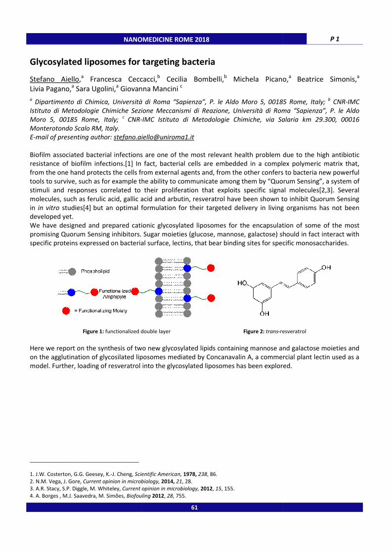
NANOMEDICINE ROME 2018
Glycosylated liposomes for targeting bacteria
Stefano Aiello,a Francesca Ceccacci,
Livia Pagano,a Sara Ugolini,
a Giovanna Mancini
a Dipartimento di Chimica, Università di Roma “Sapienza”, P. le Aldo Moro 5, 00185 Rome, Italy;
Istituto di Metodologie Chimiche Sezione Meccanismi di Reazione, Università di Roma “Sapienza”, P. le Aldo
Moro 5, 00185 Rome, Italy; c CNR-
Monterotondo Scalo RM, Italy. E-mail of presenting author: [email protected]
Biofilm associated bacterial infections are one of the most relevant health problem due to the high antibiotic
resistance of biofilm infections.[1] In fact, bacterial cells are embedded in a complex polymeric
from the one hand protects the cells from external agents and, from the other confers to bacteria new powerful
tools to survive, such as for example the ability to communicate among them by “Quorum Sensing”,
stimuli and responses correlated to their proliferation that exploits specific signal molecules
molecules, such as ferulic acid, gallic acid and arbutin, resveratrol have been shown to inhibit Quorum Sensing
in in vitro studies[4] but an optimal formulation for their targeted delivery in living organisms has not been
developed yet.
We have designed and prepared cationic glycosylated liposomes for the encapsulation of some of the most
promising Quorum Sensing inhibitors. Sugar moiet
specific proteins expressed on bacterial surface, lectins, that bear binding sites for specific monosaccharides.
Figure 1: functionalized double layer
Here we report on the synthesis of two new glycosylated lipids containing mannose and galactose moieties and
on the agglutination of glycosilated liposomes mediated by Concanavalin A, a commercial plant lectin
model. Further, loading of resveratrol into the glycosylated liposomes has been explored.
_____________________________________
1. J.W. Costerton, G.G. Geesey, K.-J. Cheng, Scientific American,
2. N.M. Vega, J. Gore, Current opinion in microbiology,
3. A.R. Stacy, S.P. Diggle, M. Whiteley, Current opinion in microbiology,
4. A. Borges , M.J. Saavedra, M. Simões, Biofouling
NANOMEDICINE ROME 2018
Glycosylated liposomes for targeting bacteria
Francesca Ceccacci,b Cecilia Bombelli,
b Michela Picano,
Giovanna Mancini c
Dipartimento di Chimica, Università di Roma “Sapienza”, P. le Aldo Moro 5, 00185 Rome, Italy;
Istituto di Metodologie Chimiche Sezione Meccanismi di Reazione, Università di Roma “Sapienza”, P. le Aldo
-IMC Istituto di Metodologie Chimiche, via Salaria km 29.300, 00016
Biofilm associated bacterial infections are one of the most relevant health problem due to the high antibiotic
In fact, bacterial cells are embedded in a complex polymeric
from the one hand protects the cells from external agents and, from the other confers to bacteria new powerful
tools to survive, such as for example the ability to communicate among them by “Quorum Sensing”,
correlated to their proliferation that exploits specific signal molecules
molecules, such as ferulic acid, gallic acid and arbutin, resveratrol have been shown to inhibit Quorum Sensing
but an optimal formulation for their targeted delivery in living organisms has not been
We have designed and prepared cationic glycosylated liposomes for the encapsulation of some of the most
promising Quorum Sensing inhibitors. Sugar moieties (glucose, mannose, galactose) should in fact interact with
specific proteins expressed on bacterial surface, lectins, that bear binding sites for specific monosaccharides.
functionalized double layer Figure 2: trans-resveratrol
Here we report on the synthesis of two new glycosylated lipids containing mannose and galactose moieties and
on the agglutination of glycosilated liposomes mediated by Concanavalin A, a commercial plant lectin
model. Further, loading of resveratrol into the glycosylated liposomes has been explored.
Scientific American, 1978, 238, 86.
Current opinion in microbiology, 2014, 21, 28.
Current opinion in microbiology, 2012, 15, 155.
Biofouling 2012, 28, 755.
61
P 1
Michela Picano,a
Beatrice Simonis,a
Dipartimento di Chimica, Università di Roma “Sapienza”, P. le Aldo Moro 5, 00185 Rome, Italy; b CNR-IMC
Istituto di Metodologie Chimiche Sezione Meccanismi di Reazione, Università di Roma “Sapienza”, P. le Aldo
IMC Istituto di Metodologie Chimiche, via Salaria km 29.300, 00016
Biofilm associated bacterial infections are one of the most relevant health problem due to the high antibiotic
In fact, bacterial cells are embedded in a complex polymeric matrix that,
from the one hand protects the cells from external agents and, from the other confers to bacteria new powerful
tools to survive, such as for example the ability to communicate among them by “Quorum Sensing”, a system of
correlated to their proliferation that exploits specific signal molecules[2,3]. Several
molecules, such as ferulic acid, gallic acid and arbutin, resveratrol have been shown to inhibit Quorum Sensing
but an optimal formulation for their targeted delivery in living organisms has not been
We have designed and prepared cationic glycosylated liposomes for the encapsulation of some of the most
ies (glucose, mannose, galactose) should in fact interact with
specific proteins expressed on bacterial surface, lectins, that bear binding sites for specific monosaccharides.
resveratrol
Here we report on the synthesis of two new glycosylated lipids containing mannose and galactose moieties and
on the agglutination of glycosilated liposomes mediated by Concanavalin A, a commercial plant lectin used as a
model. Further, loading of resveratrol into the glycosylated liposomes has been explored.

P 2 NANOMEDICINE ROME 2018
VP6-SUMO: a novel promising self assembling unit for drug-delivery
Adriana Amalfitano,a Francesca Bugli,b Cecilia Martini,b Giuseppina Nocca,a,c Alberto Vitali,c Marco De Spirito,d Massimiliano Papi,d Maurizio Sanguinetti,b Alessandro Arcovitoa
a Institute of Biochemistry and clinical biochemistry, Catholic University of Sacred Heart, Rome;
b Institute of
Microbiology, Catholic University of Sacred Heart, Rome, Italy; c
Centre of Molecular Recognition Chemistry,
CNR, Rome, Italy; d
Institute of Physics, Catholic University of Sacred Heart, Rome, Italy.
Email of presenting author: [email protected]
Introduction. Nanoparticles are acquiring even more interest in drug-delivery and are strongly studied to
develop novel therapeutic strategies. Among them, we decided to study those derived by self-assembling
protein subunits produced from viral capsid proteins, that are able to form the so called virus-like particles
(VLPs). The low expression and purification yield during the production in Escherichia coli is, sometimes, a limit
of this strategy. In order to overcome this problem, NPs formed by a highly conserved protein present in the
middle of the human rotavirus’ three-capsid structure called VP6 was fused with the small ubiquitin-like
modifier (SUMO) at the N-terminal. The final adduct VP6-SUMO, is a very stable and soluble protein, still able to
form trimers and to self-assemble in safe nanostructures such as nanospheres (NPs) and nanotubes (NTs),
depending on the inducing pH used to activate this polymerization reaction [1,2]. The aim of this study was to
entrap doxorubicin, an anticancer drug, into VP6-SUMO-NPs using them as carrier system to deliver the drug
directly inside human hepatocellular carcinoma cell line (HepG2) and verify if the carrier can alter the anti-
tumor capability of doxorubicin.
Experimental methods. After expression and production in Escherichia coli and purification by affinity
chromatography, VP6-SUMO was put in presence of an acid buffer to induce the formation of the
nanoparticle’s quaternary spherical structure desired. During this step, it is possible to upload, in the forming
NPs, drugs or bioactive molecules and then dialyze the final product in PBS before using it for in vitro and in vivo
test. To evaluate the cellular uptake of NPs, the latter were loaded with a fluorescent probe (Calcein), incubated
with HepG2 cell line and finally analyzed by confocal microscopy. The cytotoxicity on HepG2 cell line induced by
NPs loaded with the doxorubicin (Dox-NPs) was also compared with doxorubicin directly added to the medium
(Dox-free) by means of MTT. The cytotoxic effects caused by NPs empty were also evaluated.
Results and discussion. Transmission electron microscopy (TEM) results revealed that VP6-SUMO NPs obtained
with this protocol are arranged in spheroidal structures with diameters between 100 and 200 nm. When these
NPs were loaded with Calcein, they show a very fast uptake in HepG2 cell line yet after five minutes. The
Doxorubicin entrapment efficiency was around 40% and cytotoxicity test showed a similar effect when the drug
is administered as Dox-free and as Dox-NPs. Moreover, a small cytotoxic effect - provoked by carrier empty -
was observed. Conclusions. From these experiments, we demonstrated that VP6-SUMO virus-like particles are
able to entrap a bioactive molecule and deliver it into tumor cells. Future perspective will be to chemically
engineer and/or genetically modify the surface of these VP6-SUMO NPs to deliver bioactive molecules in a
more selective way.
_____________________________________
1. Bugli F. et al., International Journal of Nanomedicine, 2014, 9, 2727.
2. Palmieri V. et al., Journal of Nanomaterials, 2015: 78786.
62

NANOMEDICINE ROME 2018 P 3
Solid lipid nanoparticles-mediated delivery of grape seed-derived
proanthocyanidins to airway epithelial cells reduce oxidative stress
Stefano Castellani,a Adriana Trapani,b Anna Spagnoletta,c Lorena di Toma,a Sante Di Gioia,a Delia Mandracchia,b Thea Magrone,d Giuseppe Trapani,b Emilio Jirillo,d Massimo Conese a
a Department of Medical and Surgical Sciences, University of Foggia, Viale Luigi Pinto 1, 71122, Foggia, Italy;
b Department of Pharmacy-Drug Sciences, University of Bari “Aldo Moro”, Via E. Orabona 4, 70125 Bari, Italy;
c ENEA Research Centre Trisaia, Laboratory "BioProducts and BioProcesses", Rotondella (MT), Italy;
d Department
of Basic Medical Sciences, Neuroscience and Sensory Organs, University of Bari “Aldo Moro”, 70125 Bari, Italy.
Email of presenting author: [email protected]
Objectives: Chronic respiratory diseases, which are still incurable and need novel therapeutic tools, are
characterized by unsurmountable oxidative stress. Grape phenolic compounds, such as proanthocyanidins, are
endowed with well-recognized anti-oxidant and anti-inflammatory activities. Given that these natural anti-
oxidants have poor water solubility and oral bioavailability, we have developed a drug delivery system based on
solid lipid nanoparticles (SLN), in order to encapsulate these therapeutic molecules. Our aim was to test their
efficacy in reducing oxidative stress when used as carriers for molecules derived from grape seed extracts (GSE).
Methods: SLN containing GSE, formulated according to the melt-emulsification method (Trapani et al.,
Biointerfaces, 2015, 127, 79) by using Gelucire® 50/13 as lipid, were characterized for their physicochemical
parameters and the amount of incorporated GSE was determined by spectrophotometry. To evaluate the
efficiency of anti-oxidant activity, airway epithelial cells (H441 line) were incubated for 24-72 h with SLN-GSE (or
free GSE as control) at 2.5, 5 and 10 µM followed by treatment with 0.1 mM H2O2 for 24 h in order to induce
oxidative stress. ROS production was evaluated by fluorimetry by using the H2DCFDA probe, as previously
described (D'Apolito et al., Atherosclerosis, 2015, 239, 393), and flow-cytometry (Castellani et al., accepted in
Journal of Translational Medicine).
Results: SLN-GSE exhibited a mean diameter of 243 nm and a zeta-potential of -14.5 mV. SLN entrapped an
average of 0.059 mg proanthocyanidins/mg lipid, an amount which remained constant for up to 60 days. Pre-
treatment of H441 cells with SLN-GSE (or free GSE) 24 h prior to the H2O2 treatment resulted in a significant
reduction of ROS at the highest dose of SLN-GSE while free GSE was effective at the three doses used.
Pretreatment for 48 and 72 h resulted in a significant reduction of ROS at the three doses of SLN-GSE used,
while free GSE was no more effective at the lowest dose used.
Conclusions: GSE-loaded SLN exerted their effect as anti-oxidant agents for longer times than free GSE in airway
epithelial cells, suggesting the controlled release of their payload. Overall, our results are promising for the
development of novel natural substance-based pharmaceutics aimed at reducing the oxidative stress in chronic
respiratory diseases.
_____________________________________
Acknowledgments: Stefano Castellani is a researcher funded by Intervento Cofinanziato dal Fondo di Sviluppo e Coesione 2007-2013 –
APQ Ricerca Regione Puglia “Programma regionale a sostegno della specializzazione intelligente e delle sostenibilità sociale ed
ambientali – Future In Research”.
63

P 4 NANOMEDICINE ROME 2018
TiO2-coated pSi microparticles for nanomedicine
Elena Chistè,a Ali Ghafarinazari,a Marta Donini,b Veronique Cremers,c Jolien Dendooven,c Christophe Detavernier,c Donatella Benati,d Marina Scarpa,e Stefano Dusi,b Nicola Daldosso a a Department of Computer Science, Fluorescence Laboratory, University of Verona, Strada le Grazie 15, 37134,
Verona, Italy; b
Department of Medicine, Division of General Pathology, University of Verona, Strada le Grazie 8,
37134, Verona, Italy; c
Department of Solid State Sciences, CoCooN Group, Ghent University, Krijgslaan 281/S1,
9000, Gent, Belgium; d
Department of Neuroscience, Biomedicine and Movement, Anatomy and Histology
Section, University of Verona, Strada le Grazie 8, 37134, Verona, Italy; e
Department of Physics, Laboratory of
Nanoscience, University of Trento, Via Sommarive 14, 38123, Trento, Italy.
Email of presenting author: [email protected]
Porous silicon (pSi) is a sponge-like material that, due to quantum confinement effect, is photoluminescent
when excited by UV light. We developed a process to obtain mesoporous silicon microparticles by
electrochemical etching and we proved them to be optically stable in biological buffers once opportunely
coated. This material is a promising system for nanomedicine application, first, because it is biodegradable,
biocompatible and not activator of the immune response, then, since the porosity and the photoluminescence
make them suitable for drug delivery and bioimaging. The microparticles were coated by ALD (atomic layer
deposition) in a rotary reactor to avoid optical and structural degradation in aqueous media. A thin and uniform
TiO2 layer was deposited on the pSi microparticles (pSi-TiO2 microparticles), without occluding the pores. In-
vitro tests were performed to control the effect of pSi-TiO2 microparticles on human dendritic cells (DCs). We
observed no decrease of cell viability after the internalization of the microparticles that maintained their PL, as
can be seen in Figure 1, but an increase of the immune response (priming effect) in case of co-stimulation with
an immune response activator.
These preliminary results, recently published [1], are promising for the pSi-TiO2 microparticles application in
nanomedicine, in particular, for drug delivery and bioimaging.
Figure 1. TEM images of pSi-TiO2 microparticles ( left) and their PL preservation after
internalization in human DCs (right)
_____________________________________
1. E. Chistè, A. Ghafarinazari, M. Donini, V. Cremers, J. Dendooven, C. Detavernier, D. Benati, M. Scarpa, S. Dusi, N. Daldosso, J. Mat.
Chem. B, 2018, DOI: 10.1039/C7TB02614E.
64

NANOMEDICINE ROME 2018 P 5
Curcumin-loaded Poly (d,l-lactide-co-glycolide) nanovesicles induce
antinociceptive effects after local administration in mice
Laura Ciarlo, Francesca Marzoli, Paola Minosi, Amalia Di Giannuario and Stefano Pieretti
National Center for Drug Research and Evaluation, Istituto Superiore di Sanità, Viale Regina Elena 299, 00166,
Rome, Italy.
Email of presenting author: [email protected]
Both acute and chronic pain are the most widespread medical issue strongly affecting people in terms of health
and quality of life. Unlike acute pain, chronic pain is a pathophysiological state arising from the alteration of the
peripheral and/or central nervous systems. It is frequently accompanied by the onset of hyperalgesia (increased
sensitivity to pain) and allodynia (painful sensation in response to usually innocuous stimuli). Pain is currently
treated with two major groups of analgesic drugs, namely non-steroidal anti-inflammatory drugs (NSAIDs) and
opioids; their use is associated with important side effects, which include gastrointestinal lesions [1] and
nephrotoxicity [2]; in the case of NSAIDs, respiratory depression, tolerance and physical dependence for opioids
[3]. For this reason, there is growing interest for the identification of alternative therapeutic strategies.
Curcumin (1,7-Bis(4-hydroxy-3-methoxyphenyl)-1,6-heptadiene-3,5-dione) is a yellow polyphenol,
diferuloylmethane, extracted from the rhizomes of turmeric (Curcuma longa)[4]. The therapeutic properties of
curcumin are well known indeed possesses low intrinsic toxicity along with a wide range of pharmacological
activities that include antitumor, anti-amyloid, antioxidant and anti-inflammatory capacities [5]. Antinociceptive
properties of curcumin have also been reported in preclinical studies [6], but its poor bioavaibility limits clinical
use as analgesic. Polymeric nanoparticle-based drug delivery is being increasingly investigated as a delivery
route able to overcome many obstacles associated with the delivery of free drugs. Recently, we investigated
the effects of curcumin-loaded PLGA nanovesicles (PLGA-CUR) administered via intravenous (i.v.) or intrathecal
(i.t.) routes in several experimental models of pain [7]. We found that i.v. or i.t. routes of administration of
PLGA-CUR nanoformulations were effective in reducing the nociception induced by chemical stimuli or after the
ligation of the sciatic nerve in mice [7]. In the present study, putative antinociceptive effects induced by CUR
and PLGA-CUR after local subcutaneous administration was investigated in two animal models of pain i.e. the
formalin test and the hyperalgesia induced by zymosan. We found PLGA-CUR vesicles able to reduce
nociception induced by chemical stimuli, whereas CUR alone induced only a transient but not significant
antinociceptive effects. These results obtained after acute subcutaneous local PLGA-CUR vesicles
administration, further suggest that PLGA-CUR formulation should be developed as a new potential drug in the
treatment of pain in humans.
_____________________________________
1. M. Sinha, L. Gautam, P.K. Shukla et al., Mediators Inflamm., 2013, 258209.
2. M. Musu, G. Finco, R. Antonucci et al., Eur. Rev. Med. Pharmacol. Sci, 2011, 15, 1461.
3. R. Benyamin, A.M. Trescot, S. Datta et al., Pain Physician, 2018, S105. 4. S.C. Gupta, G. Kismali, B.B. Aggarwal, Biofactors, 2013, 39, 2.
5. S. Sharma, S.K. Kulkarni, J.N. Agrewala et al., Eur. J. Pharmacol., 2006, 536, 256. 6. L. Allegri, F. Rosignolo,C. Mio et al., J. Cancer Res. Clin. Oncol. 2018, 144, 285.
7. S. Pieretti, A.P. Ranjan, A. Di Giannuario et al., Colloids Surf B Biointerfaces, 2017, 158, 379.
65
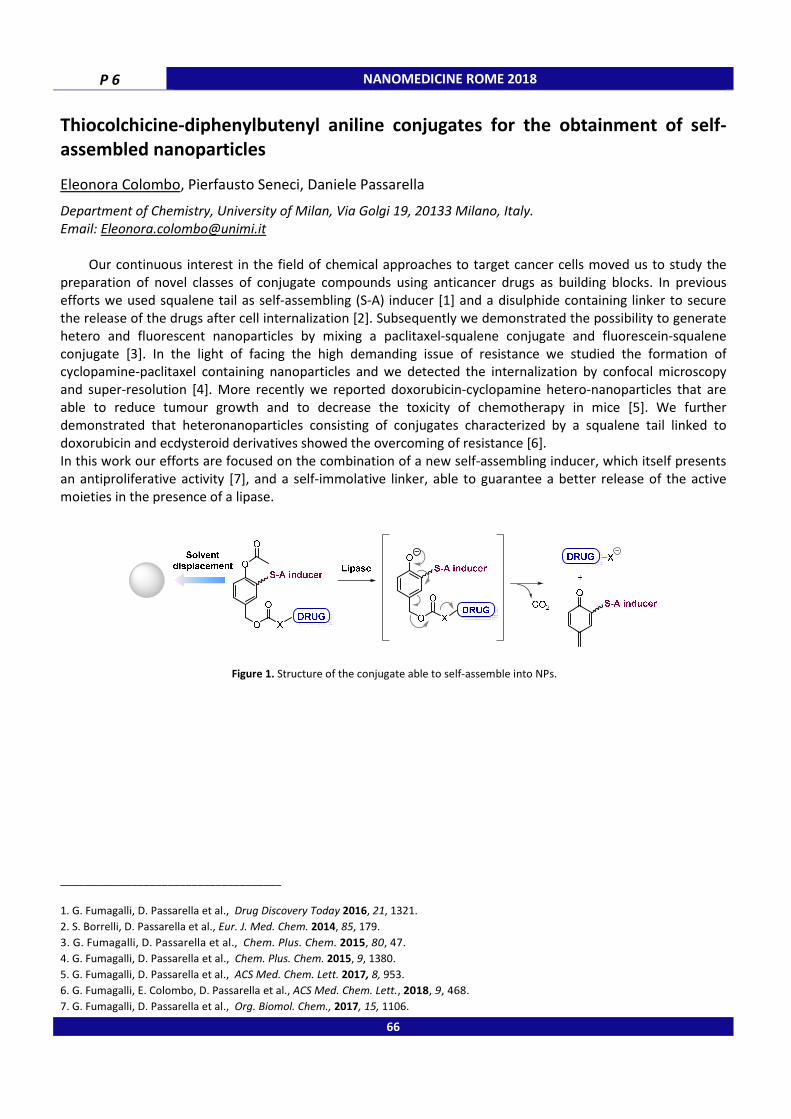
P 6 NANOMEDICINE ROME 2018
Thiocolchicine-diphenylbutenyl aniline conjugates for the obtainment of self-
assembled nanoparticles
Eleonora Colombo, Pierfausto Seneci, Daniele Passarella
Department of Chemistry, University of Milan, Via Golgi 19, 20133 Milano, Italy.
Email: [email protected]
Our continuous interest in the field of chemical approaches to target cancer cells moved us to study the
preparation of novel classes of conjugate compounds using anticancer drugs as building blocks. In previous
efforts we used squalene tail as self-assembling (S-A) inducer [1] and a disulphide containing linker to secure
the release of the drugs after cell internalization [2]. Subsequently we demonstrated the possibility to generate
hetero and fluorescent nanoparticles by mixing a paclitaxel-squalene conjugate and fluorescein-squalene
conjugate [3]. In the light of facing the high demanding issue of resistance we studied the formation of
cyclopamine-paclitaxel containing nanoparticles and we detected the internalization by confocal microscopy
and super-resolution [4]. More recently we reported doxorubicin-cyclopamine hetero-nanoparticles that are
able to reduce tumour growth and to decrease the toxicity of chemotherapy in mice [5]. We further
demonstrated that heteronanoparticles consisting of conjugates characterized by a squalene tail linked to
doxorubicin and ecdysteroid derivatives showed the overcoming of resistance [6].
In this work our efforts are focused on the combination of a new self-assembling inducer, which itself presents
an antiproliferative activity [7], and a self-immolative linker, able to guarantee a better release of the active
moieties in the presence of a lipase.
Figure 1. Structure of the conjugate able to self-assemble into NPs.
_____________________________________
1. G. Fumagalli, D. Passarella et al., Drug Discovery Today 2016, 21, 1321.
2. S. Borrelli, D. Passarella et al., Eur. J. Med. Chem. 2014, 85, 179.
3. G. Fumagalli, D. Passarella et al., Chem. Plus. Chem. 2015, 80, 47.
4. G. Fumagalli, D. Passarella et al., Chem. Plus. Chem. 2015, 9, 1380.
5. G. Fumagalli, D. Passarella et al., ACS Med. Chem. Lett. 2017, 8, 953.
6. G. Fumagalli, E. Colombo, D. Passarella et al., ACS Med. Chem. Lett., 2018, 9, 468.
7. G. Fumagalli, D. Passarella et al., Org. Biomol. Chem., 2017, 15, 1106.
66

NANOMEDICINE ROME 2018 P 7
Design and development of biosafety devices with therapeutic activities
Marisa Colone,a Letizia Angiolella,b Alberto Vitali,c Stefano Serra,d Alessandro Gori,d Annarica Calcabrini,a Annarita Stringaro a a
Centro Nazionale per la Ricerca e la Valutazione Pre-clinica e Clinica dei Farmaci,
Istituto Superiore di Sanità,
00161, Rome, Italy; b Dipartimento di Sanità Pubblica e Malattie Infettive, Sapienza Università di Roma, 00185,
Rome, Italy; c
ICRM-CNRc/o Istituto di Biochimica e Biochimica Clinica, Università Cattolica di Roma, 00168,
Rome, Italy, d ICRM-CNR, 20131, Milan, Italy.
Email of presenting author: [email protected]
The possibility to realize injectable reservoirs for controlled and specific release may provide a strategy for the
administration of chemical drugs with poor solubility. The low oral bioavailability of hydrophobic drugs, their
broad therapeutic windows, the necessity for a low daily dose and long-time treatment of disease are the items
that support the requirement of new micro/nanodevices. Numerous studies are in progress to evaluate
different devices (cationic liposomes, lysozyme-shelled hollow nano/microbubbles and nano/microcapsules) in
order to increase the antifungal and antitumor efficacy of different drugs [1].
At present, biocompatible and biodegradable nanoparticles (NPs) are widely studied as an effective drug
delivery device [2]. Among candidates for a drug carrier system, chitosan represents a kind of natural cationic
polymer showing nontoxic, biocompatible, biodegradable features. Moreover, thanks to the specificity of short
sequences recognized by cell receptors, recently many peptides have been used to improve the specific
targeting of biologically active substances such as iRNAs, DNA, peptides and organic molecules [3]. We are
designing peptide sequences able to recognize, transport and release in a highly specific manner biologically
active natural compounds to specifically kill cancer and/or microbial cells. The aim of our work is to design
peptide decorated-chitosan nanoparticles in which active elements, such as natural products with anticancer
properties and antifungal characteristics, are associated to them.
_____________________________________
1. M. Colone, S. Kaliappan et al., Journal of Molecular and Genetic Medicine, 2016, 10, 200. 2. P. Desai, R. Patlolla et al., Journal of Membrane Biology, 2010, 27, 247.
3. G. Radicioni, A. Stringaro et al., Biochimica and Biophysica Acta, 2015, 1848, 2868.
67
P 8 NANOMEDICINE ROME 2018
Alkaloid Voacamine encapsulated in cationic liposomes increases doxorubicin
effect on osteosarcoma resistant cells
Maria Condello,a Luisa Giansanti,b Evelin Pellegrini,a Giovanna Mancini,c Stefania Meschinia aNational Center for Drug Research and Evaluation, National Institute of Health, Viale Regina Elena 299, 00161,
Rome, Italy; bDepartment of Physical and Chemical Sciences, University of L'Aquila, Via Giovanni Falcone 25,
67100 L'Aquila, Italy; cInstitute of Chemical Methodologies, National Research Council, P. le A. Moro 5, 00185
Rome, Italy.
Email of presenting author: [email protected]
In vitro studies demonstrated that voacamine (VOA), a bisindolic alkaloid isolated from Peschiera fuchsifoliae
plant, enhanced cytotoxic effect of doxorubicin (DOX) on resistant osteosarcoma and melanoma cells [1,2].The
chemosensitizing effect of VOA on osteosarcoma cells was due to DOX accumulation increase by P-gp
inhibition, the main efflux pump responsible of drug resistance [3]. Moreover, the dose of VOA used to sensitize
tumor cells to DOX was not toxic to normal cells [2]. These properties and the insolubility of VOA in aqueous
medium suggested the development of targeting liposomal formulations for the delivery of VOA.
Flow cytometric analyses of DOX intracellular uptake showed that VOA included into DOPC (1,2-dioleoyl-sn-
glycero-3-phosphocholine) or DPPC (1,2-dipalmitoyl-phosphatidylcholine) liposomes did not increase DOX
uptake with respect to free VOA. The addition of cholesterol to the formulations (DOPC/CHOL or DPPC/CHOL
liposomes with VOA) did not improve DOX accumulation either. However, when VOA was included into
liposomes composed of DOPC or DPPC, a gemini surfactant (SS), and cholesterol (DOPC/SS/CHOL and
DPPC/SS/CHOL), DOX retention increased with respect to free VOA. MTT cell viability assay demonstrated that
the most efficient liposome formulation was DOPC/SS/CHOL. Optical microscopic evaluations, 24 h after
recovery in drug free medium, showed that osteosarcoma cells pre-treated with DOPC/SS/CHOL liposomes-VOA
before DOX administration were dead compared to cells treated with free-VOA plus DOX. UIC2 flow cytometry
shift assay showed that liposomes did not alter P-gp functionality, and that VOA loaded into liposomes exerted
the inhibitory effect against P-gp as well as free VOA.
These results showed that the encapsulation of the VOA into liposomes improved its delivery in resistant tumor
cells and its chemosensitizing efficacy. Further studies are aimed at clarifying if VOA loaded liposomes and free
VOA have different molecular targets.
_____________________________________
1. S. Meschini, M. Marra, A. Calcabrini, E. Federici, C. Galeffi, G. Arancia. Int. J. Oncol., 2003, 23, 1505. 2. M. Condello, D. Cosentino, S. Corinti, G. Di Felice, G. Multari, F. R. Gallo, G. Arancia, S. Meschini. J. Nat. Prod., 2014, 77, 855.
3. S. Meschini, M. Marra, M. Condello, A. Calcabrini, E. Federici, ML Dupuis, M Cianfriglia, G. Arancia. Int. J. Oncol., 2005, 27, 1597.
68

NANOMEDICINE ROME 2018 P 9
New strategies to evaluate the efficacy in vivo of a novel benzophenone-based
antibiotic against S. aureus lung infections in Cystic Fibrosis
Gabriella Costabile, Olivia Merkel
Department of Pharmacy, Ludwig-Maximilians-University, Butenandtstrasse 5, 81377, Munich, Germany.
Email of presenting author: [email protected]
Antimicrobial resistance (AMR) within a wide range of infectious agents is a growing public health threat of
broad concern to countries and multiple sectors. As the 2014 WHO report says “A post- antibiotic era—in which
common infections and minor injuries can kill” is “far from being an apocalyptic fantasy,” but “instead a very
real possibility for the 21st century”. The AMR issue becomes even more pressing in pathologies such as Cystic
Fibrosis (CF) [1]. CF is a complex multisystem disease caused by the defect in a single gene encoding an
epithelial ion channel known as transmembrane conductance regulator (CFTR); a defective CFTR, results in
thickened mucus secretions which cannot be easily cleared and predispose the lung to persistent and chronical
bacterial infections that, with the AMR phenomena, cannot be well controlled anymore [2]. The general aim of
this project is the design and the development of engineered nanoparticles for pulmonary delivery of a novel
benzophenone-based antibiotic (SV7) against S. aureus chronic infection in CF therapy [3, 4]. S. aureus, in the
treatment of CF, is an ideal target. In fact, it has been demonstrated that S. aureus is one of the first bacteria
able to colonize the airways while producing the conditions for later infections for instance by P. aeruginosa.
Thus, the eradication of S. aureus infection may be helpful in order to retard the chronicization of the infection
and then the worsening of the lung injury. However, the individualization of new antibiotics is necessary but it is
not enough against the induction of AMR phenomena. As a matter of fact, to exploit this important feature,
only the reduction of patients’ exposure to antibiotics may be really helpful in order to delay eventual AMR
phenomena. In this sense, the local administration of novel API via inhalation route is considered as a route of
choice for the treatment of pulmonary infections [5]. Lastly, since the evaluation of inhaled drug behavior
through most reliable in vivo experimental models appears crucial for the translation to the clinics, a new G.
mellonella model is involved as preliminary in vivo model in the attempt to solve the ethical issues related to
the use of mammalian models [6-8].
_____________________________________
1. O. Ciofu, T. Tolker-Nielsen, PØ Jensen, H. Wang, N. Høiby. Adv Drug Deliv Rev, 2015, 85, 7. 2. B.P. O'Sullivan and S.D. Freedman, Lancet, 2009, 373, 1891.
3. S.K. Vooturi, C.M. Cheung, M.J. Rybak, S.M. Firestine, J Med Chem, 2009, 52, 5020. 4. S.K. Vooturi, M.B. Dewal, S.M. Firestine, Org Biomol Chem, 2011, 9, 6367.
5. Q.T. Zhou, S.S. Leung, P. Tang, T. Parumasivam, et al., Adv Drug Deliv Rev, 2015, 85, 83. 6. R. Baldan, C. Cigana, F. Testa, I. Bianconi, M. De Simone, D. Pellin, C. Di Serio, A. Bragonzi, D.M. Cirillo, PLoS One, 2014, 9 (3), e89614.
7. A.P. Desbois and P.J. Coote, J Antimicrob Chemother, 2011, 66, 1785. 8. C.J. Tsai, J.M. Loh, T. Proft, Virulence, 2016, 7, 214.
Acknowledgments: The stay of G.C. was financially supported by UniNa and Compagnia San Paolo in the frame of Programme STAR (CALL 2016).
69

P 10 NANOMEDICINE ROME 2018
Polysaccharide-based nanohydrogels as drug carriers
Chiara Di Meo, Elita Montanari, Nicole Zoratto, Tommasina Coviello, Pietro Matricardi
Department of Drug Chemistry and Technologies, Sapienza University of Rome, P.le Aldo Moro 5, 00185, Rome,
Italy.
Email of presenting author: [email protected]
Natural and biocompatible polysaccharides, hyaluronic acid (HA) and gellan gum (Ge), were derivatized with
hydrophobic moieties (cholesterol or riboflavin) in order to obtain amphiphilic polymers able to self-assemble
in water leading to the formation of nanosized structures, named nanohydrogels [1-3] (NHs) (Fig. 1).
Figure 1. Self-assembling of derivatized polysaccharides leading to the formation of NHs (A);
Cryo-TEM images of HA-based NHs (B).
These systems couple the features of both nanoparticles, as nano-dimensions, and hydrogels, such as soft
consistency, high amount of water, and biocompatibility. Moreover, all developed systems showed a high
capability to load model drugs, both hydrophilic [4] and hydrophobic ones [5], and also proteins [2], without
losing their activity. In the case of hydrophobic molecules, NHs act as solubility enhancers, increasing the
apparent water solubility and allowing to obtain water-based formulations.
Several derivatives were synthesized and characterized, and a new method to obtain simultaneously the
formation, the sterilization and the drug loading into NHs was developed and patented [6].
All NHs were tested for their cytocompatibility on a series of cell lines, showing a complete safety up to 0.5
mg/ml. Several NHs formulations obtained with model drugs, such as antibiotics [4], anti-inflammatory [3] and
anticancer drugs [5], were prepared and optimized; their activities were tested in vitro and in vivo, and in all
cases an improvement of the therapeutic activity was observed.
_____________________________________
1. G. D'Arrigo, C. Di Meo, E. Gaucci et al., Soft Matter, 2012, 8, 11557.
2. E. Montanari, S. Capece, C. Di Meo et al., Macromol. Biosci., 2013, 13, 1185. 3. C. Di Meo, E. Montanari, L. Manzi et al., Carbohyd. Pol., 2015, 115, 502.
4. E. Montanari, G. D’Arrigo, C. Di Meo et al., Eur. J. Pharm. Biopharm., 2014, 87, 518. 5. G. D'Arrigo, G. Navarro, C. Di Meo et al., Eur. J. Pharm. Biopharm., 2014, 87, 208.
6. E. Montanari, M.C. De Rugeriis, C. Di Meo et al., J. Materi. Sci. Mat. Med, 2015, 26.
70

NANOMEDICINE ROME 2018 P 11
Levetiracetam loaded microemulsions for percutaneous drug delivery:
physicochemical considerations
Ljiljana Djekic, Marija Primorac
Department of Pharmaceutical Technology and Cosmetology, University of Belgrade-Faculty of Pharmacy,
Vojvode Stepe 450, 11221, Belgrade, Serbia.
Email of presenting author: [email protected]
Levetiracetam (LEV), an antiepileptic drug, has been shown to have local peripheral antihyperalgesic and
antiedematous effects in a rat model of localized inflammation. This significant observation suggests the
potential of LEV for treating inflammatory pain conditions with a lower incidence of systemic side effects and
drug interactions [1]. The delivery of highly hydrosoluble LEV (logP -0.6) requires a carrier having a high
potential for percutaneous penetration inhancement, such as microemulsion systems [2]. The purpose of the
study was formulation and physicochemical characterization of LEV-loaded microemulsions. The designed
carrier (M), comprising Labrasol®/polisorbate 20/Kolliphor® RH 40/isopropyl myristate/water, was loaded with
5% (MLEV5%) and 10% (MLEV10%) of LEV into the carrier (up to 100%). Characterization of the samples was
performed after storage at ambient conditions for 48 h and 1 month, and included: centrifugation test (3000
rpm for 30 min), measurement of pH and conductivity (δ), droplet size analysis by photon correlation
spectroscopy, and rheological behavior characterization. The obtained results indicated that MLEV5% and
MLEV10% were transparent, colourless (Fig. 1a), physically stable, oil-in-water (δ≥67.4 µS/cm) monodisperse
nanodispersions (microemulsions) with average droplet size (Zave) near 13,5 nm (PdI<0.25) (Fig. 1b). Although
acidic (pKa<-2), LEV did not significantly affect pH; the values were 4.19 (M), 4.29 (MLEV5%), and 4.36
(MLEV10%), thus acceptable for topical administration. The drug-free and LEV-loaded samples were Newtonian
liquids (Fig. 1c) of low viscosity (η) with the value decreased as the drug level increased (866.3 mPas (M); 744.2
(MLEV5%); 599.7 mPas (MLEV10%), likely due to relaxation of the hydrogen bonds at the oil-water interface
caused by drug dissolving in water phase. The formulated LEV-loaded microemulsions are prospective nanodrug
delivery systems.
Figure 1. Appearance (a), droplet size distribution (b), and flow curves (c) of the drug-free (M) and LEV-loaded
microemulsions (MLEV5% and MLEV10%).
_____________________________________
1. R. Stepanović-Petrović, A. Micov, M. Tomić, N. Ugrešić, Anesth. Analg., 2012, 115, 1457. 2. L. Djekic, M. Martinovic, R. Stepanović-Petrović, A. Micov, M. Tomić, M. Primorac, Eur. J. Pharm. Sci., 2016, 92, 255.
Acknowledgments: The study is supported by the Ministry of Education, Science and Technological Development of the Republic of
Serbia within the project III 46010.
71

P 12 NANOMEDICINE ROME 2018
Hydrogels with escin β-sitosterol phytosomes – formulation approach
Ljiljana Djekic, Aleksandra Krstić, Jovana Lazić, Marija Primorac
Department of Pharmaceutical Technology and Cosmetology, University of Belgrade-Faculty of Pharmacy,
Vojvode Stepe 450, 11221, Belgrade, Serbia.
Email of presenting author: [email protected]
Complexing escin, a natural mixture of triterpene saponins from Aesculus hippocastanum seed, with
phospholipids, produces escin β-sitosterol phytosome (ES) with enhanced selfassotiation ability and cell
membrane permeability. With dermal use, escine can be released from the complex and achieve
antiinflammatory and antiedematous effects. ES shows increased safety compared to escin and does not cause
skin irritation or allergic reactions [1, 2]. The study aimed to formulate ES-loaded hydrogels for cutaneous
application. The hydrogel prepared from Carbopol® 934 (1%), triethanolamine (1%), water (63%), propylene
glycol (10%), and isopropanol (25%), was loaded with commercially available Escin β-Sitosterol Phytosome®
(Indena, Italy) 1% (ES1%) and 5% (ES5%). Physicochemical properties (consistency, pH, flow behaviour, minimal
apparent viscosity (ηmin), maximal apparent viscosity (ηmax), hysteresis area (A)) of the ES-loaded samples were
compared with the escin-loaded samples prepared with the equivalent content of the active substance (E1%
and E5%). ES1% and ES5% were opalescent light yellow and yellow semisolids, respectively, in contrast to the
clear colorless soft semisolid gel E1% and the viscous liquid E5%. The more significant change in the escin-
samples consistency was related with the higer pH decrease (Tab. 1) and weakening of the hydrogel network.
For all samples was observed pseudoplastic flow behavior with thixotropy. The considered rheological
parameters (Tab. 1) were higher for ES-loaded samples, thus the hydrogel structure was significantly less
affected by ES in comparison with the pure escin. Additionally, ligh microscopic analysis of ES5% revealed
presence of oval aggregates (˂50 µm) likely formed bay ES selfaggregation. The results of the study indicate the
superior performances of ES over pure escin in concentration range 1-5%, for development of the carbomer
hydrogel formulations.
Sample pH ηmax (Pas) ηmin (Pas) A (Pa/s)
ES1 6.3 31.7 3.59 1315.83
ES5 5.78 13.9 1.56 928.67
E1 6.25 24.2 2.91 191.15
E5 4.95 0.98 0.20 136.46
Table 1. pH values, and rheological parameters (minimal apparent viscosity (ηmin), maximal
apparent viscosity (ηmax), hysteresis area (A)) of the investigated samples.
_____________________________________
1. C. Sitori, Pharm. Res., 2011, 44, 183.
2. L. Djekic, D. Krajisnik, Z. Micic, Tenside Surfact. Det., 2015, 52, 186.
Acknowledgments: The study is supported by the Ministry of Education, Science and Technological Development of the Republic of
Serbia within the project III 46010.
72
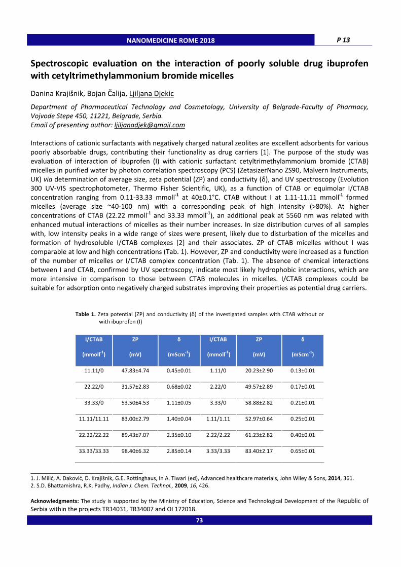
NANOMEDICINE ROME 2018 P 13
Spectroscopic evaluation on the interaction of poorly soluble drug ibuprofen
with cetyltrimethylammonium bromide micelles
Danina Krajišnik, Bojan Čalija, Ljiljana Djekic
Department of Pharmaceutical Technology and Cosmetology, University of Belgrade-Faculty of Pharmacy,
Vojvode Stepe 450, 11221, Belgrade, Serbia.
Email of presenting author: [email protected]
Interactions of cationic surfactants with negatively charged natural zeolites are excellent adsorbents for various
poorly absorbable drugs, contributing their functionality as drug carriers [1]. The purpose of the study was
evaluation of interaction of ibuprofen (I) with cationic surfactant cetyltrimethylammonium bromide (CTAB)
micelles in purified water by photon correlation spectroscopy (PCS) (ZetasizerNano ZS90, Malvern Instruments,
UK) via determination of average size, zeta potential (ZP) and conductivity (δ), and UV spectroscopy (Evolution
300 UV-VIS spectrophotometer, Thermo Fisher Scientific, UK), as a function of CTAB or equimolar I/CTAB
concentration ranging from 0.11-33.33 mmoll-1
at 40±0.1°C. CTAB without I at 1.11-11.11 mmoll-1
formed
micelles (average size ~40-100 nm) with a corresponding peak of high intensity (>80%). At higher
concentrations of CTAB (22.22 mmoll-1
and 33.33 mmoll-1
), an additional peak at 5560 nm was related with
enhanced mutual interactions of micelles as their number increases. In size distribution curves of all samples
with, low intensity peaks in a wide range of sizes were present, likely due to disturbation of the micelles and
formation of hydrosoluble I/CTAB complexes [2] and their associates. ZP of CTAB micelles without I was
comparable at low and high concentrations (Tab. 1). However, ZP and conductivity were increased as a function
of the number of micelles or I/CTAB complex concentration (Tab. 1). The absence of chemical interactions
between I and CTAB, confirmed by UV spectroscopy, indicate most likely hydrophobic interactions, which are
more intensive in comparison to those between CTAB molecules in micelles. I/CTAB complexes could be
suitable for adsorption onto negatively charged substrates improving their properties as potential drug carriers.
Table 1. Zeta potential (ZP) and conductivity (δ) of the investigated samples with CTAB without or
with ibuprofen (I)
I/CTAB
(mmoll-1
)
ZP
(mV)
δ
(mScm-1
)
I/CTAB
(mmoll-1
)
ZP
(mV)
δ
(mScm-1
)
11.11/0 47.83±4.74 0.45±0.01 1.11/0 20.23±2.90 0.13±0.01
22.22/0 31.57±2.83 0.68±0.02 2.22/0 49.57±2.89 0.17±0.01
33.33/0 53.50±4.53 1.11±0.05 3.33/0 58.88±2.82 0.21±0.01
11.11/11.11 83.00±2.79 1.40±0.04 1.11/1.11 52.97±0.64 0.25±0.01
22.22/22.22 89.43±7.07 2.35±0.10 2.22/2.22 61.23±2.82 0.40±0.01
33.33/33.33 98.40±6.32 2.85±0.14 3.33/3.33 83.40±2.17 0.65±0.01
_____________________________________
1. J. Milić, A. Daković, D. Krajišnik, G.E. Rottinghaus, In A. Tiwari (ed), Advanced healthcare materials, John Wiley & Sons, 2014, 361. 2. S.D. Bhattamishra, R.K. Padhy, Indian J. Chem. Technol., 2009, 16, 426.
Acknowledgments: The study is supported by the Ministry of Education, Science and Technological Development of the Republic of
Serbia within the projects TR34031, TR34007 and OI 172018.
73
P 14 NANOMEDICINE ROME 2018
The role of the monosialoganglioside-GM1 in the interaction between model
membranes and unstructured metastable amyloid oligomers of salmon
calcitonin
Marco Diociaiuti,a Laura Zanetti-Polzi,b Raoul Fioravanti,a,c Cecilia Bombelli,d Ilaria Fratoddi,c Cristiano Giordani e a
Centro Nazionale Malattie Rare, Istituto Superiore di Sanità, I-00161 Roma, Italy; b Dipartimento di Fisica e
Scienze Chimiche, Università dell’Aquila, via Vetoio (Coppito 1), 67010 L’Aquila, Italy; c Dipartimento di Chimica,
Università degli Studi di Roma “Sapienza”, I-00185 Roma, Italy; d
CNR, Istituto di Metodologie Chimiche, Sezione
Meccanismi di Reazione, c/o Dipartimento di Chimica, Università degli Studi di Roma “Sapienza”, I-00185 Roma,
Italy; e Instituto de Física, Universidad de Antioquia, Calle 70 No. 52-21, Medellín, Colombia.
Email of presenting author: [email protected]
In this work, the interaction of Dipalmitoyl Phosphatidylcholine (DPPC) liposomes and salmon calcitonin (sCT)
aggregates is examined. Specifically, we studied sCT monomers, prefibrillar oligomers (PFOs), proto- and
mature-fibers (PF, MF respectively) interacting with DPPC/Chol/GM1 and simple DPPC liposomes investigated
by Circular Dichroism (CD) spectroscopy and Transmission Electron Microscopy (TEM). Data clearly show the
interaction of GM1 with all the sCT species examined and its role as aggregating agent for amyloid proteins
favouring β-structure formation. Moreover, TEM data show that PFOs can modify the lipid bilayer by the
formation of pore-like structures. These structures are very similar to that proposed by Molecular Dynamic
simulations for Aβ based on experimental data obtained in model membranes supporting CD profiles of our
system. We suppose that the negative charge on the sialic head group of GM1 drives the initial interaction with
PFOs, exhibiting positive charges, whereas hydrophobic interactions could drive the amyloid insertion into the
lipid bilayer.
The next step is the study of the interaction of sCT aggregates with liposomes of increasing complexity and
biological similarity to better understand the evolution of amyloid diseases with focus on its beginning. The final
application would be the synthesis of functionalized metal nanoparticles as drug delivers for Alzheimer disease.
74

NANOMEDICINE ROME 2018 P 15
Synergistic effect of the combination of chemotherapy with DTX and PDT with
TPCS2a co-delivered by layer-by-layer nanoparticles
Elisa Gaio,a Claudia Conte,b Diletta Esposito,b Giovanni Miotto,c Fabiana Quaglia,b Francesca Moret,a Elena Reddi a a Cell Biology Unit, Department of Biology, University of Padova, Via Ugo Bassi 58/B, 35131, Padova, Italy;
b
Drug Delivery Laboratory, Department of Pharmacy, University of Napoli Federico II, Via Domenico Montesano
49, 80131, Napoli, Italy; c Department of Molecular Medicine, University of Padova, Via Ugo Bassi 58/B, 35131,
Padova, Italy.
Email of presenting author: [email protected]
Combinations of two or more drugs/treatment modalities are considered a valid tool to increase efficacy and
reduce side effects of anticancer therapies by exploiting synergic effects and the consequent possibility of drug
dose reduction while preserving therapeutic activity [1]. Nevertheless, the optimization of the drug ratio in
order to obtain the highest extent of synergism between the two treatment modalities remains difficult to
control when the drugs are administered in the standard formulations. To solve this issue, here, we propose
hyaluronic acid (HA)-targeted layer-by-layer NPs co-loaded with docetaxel (DTX) and meso-tetraphenyl chlorin
disulphonate (TPCS2a) at a fixed concentration ratio in order to obtain synergistic cytotoxic effect in DTX-
sensitive as well as in DTX-resistant cell lines. Thus, calculating the Combination Index (CI), using the dedicated
Compusyn software, we observed the highest synergism when drugs are co-delivered in the same NP
(DTX/TPCS2a NP) in comparison with the other formulations tested (drugs in standard solvent, drugs in
separate NPs) [2]. Interestingly, this result is maintained in the DTX-resistant cell line where, as indicated by the
calculation of the Dose Reduction Index (DRI), the dose of chemotherapeutic can be reduced more than 100
times when delivered by DTX/TPCS2a NP. Based on these impressive results, we demonstrated the advantage
of using HA-targeted layer-by-layer NPs as carriers of DTX and TPCS2a in order to finely control the drug ratio
inside the NPs and to precisely deliver the payloads in cancer cells.
Figure 1. Schematic representation of DTX/TPCS2a NPs.
_____________________________________
1. J.A. Kemp, M.S. Shim, C.Y. Heo, Y.J. Kwon, Advanced Drug Delivery Reviews, 2016, 98, 3.
2. T.C. Chou, Pharmacological Reviews, 2006, 58, 621.
Acknowledgments: We thank PCI Biotech AS for supplying TPCS2a.
75

P 16 NANOMEDICINE ROME 2018
Brij® niosomes and vegetal oil nanoemulsions for topical delivery: a comparative
study
Anna Imbriano,a Patrizia Nadia Hanieh,a Federica Rinaldi,b Daniele Passeri,c Livia Angeloni,c Marco Rossi,c,d Elena Del bavero,e Laura Cantù, e Carlotta Marianecci,a Maria Carafa a a Department of Drug Chemistry and Technologies, Sapienza University of Rome, P.zzle A. Moro 5, 00185, Rome,
Italy; b
Center for Life Nano Science@Sapienza, Istituto Italiano di Tecnologia (ITT), Viale R. Elena 291, 00185,
Rome, Italy; c
Department of Basic and Applied Sciences for Engineering, Sapienza University of Rome, Via A.
Scarpa 14, 00161 Rome, Italy; d
Center for Nanotechnology for Engineering (CNIS), Sapienza University of Rome,
P.le A. Moro 5, 00185, Rome, Italy; e Department of Medical Biotechnologies and Traslational Medicine,
University of Milan, Via F.lli Cervi 93, 20090, Segrate (MI), Italy.
Email of presenting author: [email protected]
The aim of the present work is the comparison between different nanocarriers, such as nanoemulsions (NEs)
and the niosomes by Brij®72 (Polyoxyethylene Stearyl Ether), for the topical delivery of active compounds useful
for the treatment of many skin diseases. Brij®72 seem to be able to enhance the skin permeation of drugs and
increase their residence time in the stratum corneum (SC), limiting transdermal permeation [1]. In order to
evaluate the drug loading capacity an hydrophobic model drug was used. All formulations were characterized in
terms of hydrodynamic diameter, ζ-potential and drug entrapment efficiency [2, 3]. Furtheremore stability
studies were carried out over time at two different storage temperatures (4°C and 25°C). The shape and the
surface morphology of all samples were also investigated using Atomic Force Microscopy (AFM) and Small-
Angle X-ray Scattering (SAXS). Differences in terms of fluidity, microviscosity and polarity between niosomes
and nanoemulsions were investigated. Finally, the release profile of drug was assessed using the synthetic
membrane Strat-M™. According to the obtained preliminary results, all formulations seem to be promising
delivery systems of therapeutic agents for the treatment of skin diseases.
Figure 1. Schematic representation of a) niosomes and b) nanoemulsions.
_____________________________________
1. M. Gupta, B. Vaidya et al., Artificial Cells, Blood Substitutes, and Biotechnology, 2011, 39 (6), 376. 2. F. Rinaldi, P.N. Hanieh, C. Longhi et al., Journal of enzyme inhibition and medicinal chemistry, 2017, 32 (1), 1265.
3. C. Marianecci, L. Di Marzio et al., Advances in Colloid and Interface Science, 2014, 205, 187.
76
Aqueous
compartment
Brij® 72
Hydrophobic drug
Oil
dropleta) b)Aqueous
compartment
Aqueous
compartment
Brij® 72
Hydrophobic drug
Brij® 72
Hydrophobic drug
Oil
droplet
Oil
dropleta) b)
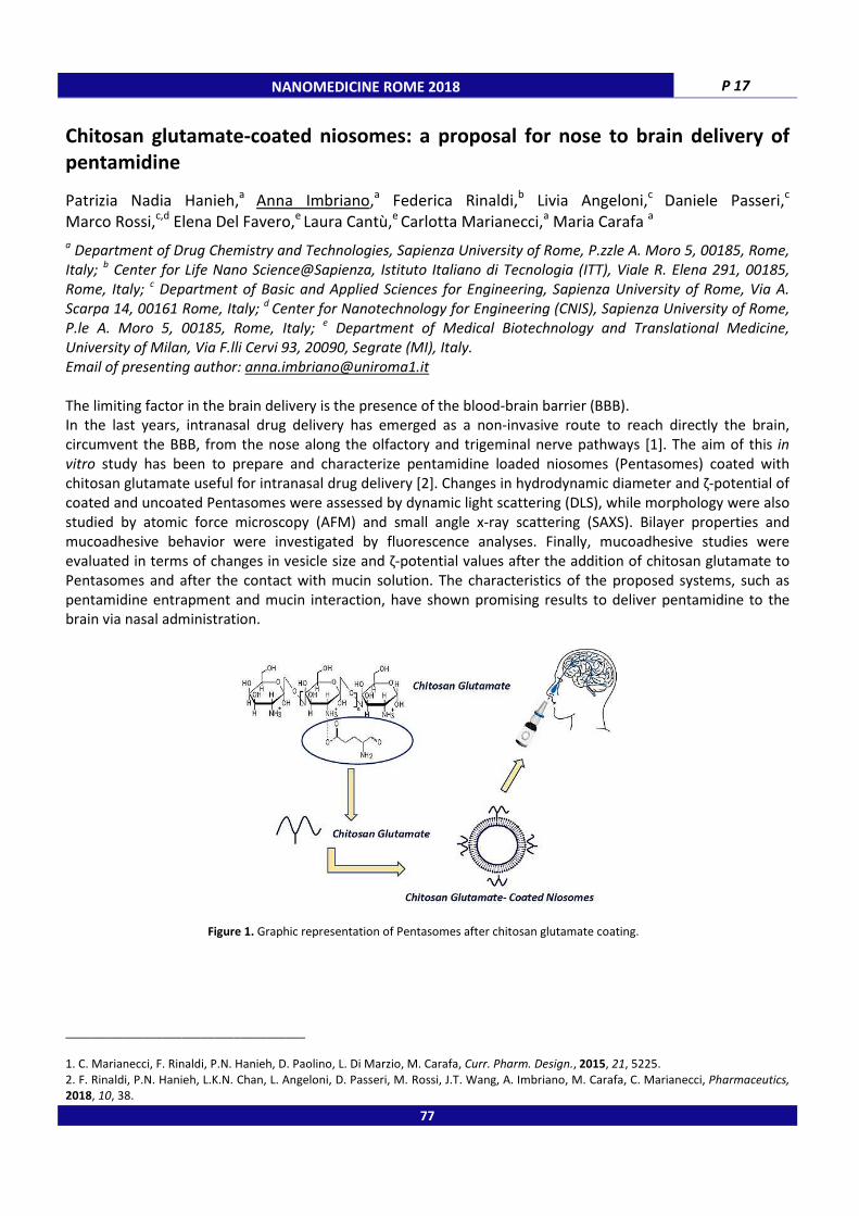
NANOMEDICINE ROME 2018 P 17
Chitosan glutamate-coated niosomes: a proposal for nose to brain delivery of
pentamidine
Patrizia Nadia Hanieh,a Anna Imbriano,a Federica Rinaldi,b Livia Angeloni,c Daniele Passeri,c
Marco Rossi,c,d Elena Del Favero,e Laura Cantù,e Carlotta Marianecci,a Maria Carafa a
a Department of Drug Chemistry and Technologies, Sapienza University of Rome, P.zzle A. Moro 5, 00185, Rome,
Italy; b Center for Life Nano Science@Sapienza, Istituto Italiano di Tecnologia (ITT), Viale R. Elena 291, 00185,
Rome, Italy; c Department of Basic and Applied Sciences for Engineering, Sapienza University of Rome, Via A.
Scarpa 14, 00161 Rome, Italy; d
Center for Nanotechnology for Engineering (CNIS), Sapienza University of Rome,
P.le A. Moro 5, 00185, Rome, Italy; e Department of Medical Biotechnology and Translational Medicine,
University of Milan, Via F.lli Cervi 93, 20090, Segrate (MI), Italy.
Email of presenting author: [email protected]
The limiting factor in the brain delivery is the presence of the blood-brain barrier (BBB).
In the last years, intranasal drug delivery has emerged as a non-invasive route to reach directly the brain,
circumvent the BBB, from the nose along the olfactory and trigeminal nerve pathways [1]. The aim of this in
vitro study has been to prepare and characterize pentamidine loaded niosomes (Pentasomes) coated with
chitosan glutamate useful for intranasal drug delivery [2]. Changes in hydrodynamic diameter and ζ-potential of
coated and uncoated Pentasomes were assessed by dynamic light scattering (DLS), while morphology were also
studied by atomic force microscopy (AFM) and small angle x-ray scattering (SAXS). Bilayer properties and
mucoadhesive behavior were investigated by fluorescence analyses. Finally, mucoadhesive studies were
evaluated in terms of changes in vesicle size and ζ-potential values after the addition of chitosan glutamate to
Pentasomes and after the contact with mucin solution. The characteristics of the proposed systems, such as
pentamidine entrapment and mucin interaction, have shown promising results to deliver pentamidine to the
brain via nasal administration.
Figure 1. Graphic representation of Pentasomes after chitosan glutamate coating.
_____________________________________
1. C. Marianecci, F. Rinaldi, P.N. Hanieh, D. Paolino, L. Di Marzio, M. Carafa, Curr. Pharm. Design., 2015, 21, 5225.
2. F. Rinaldi, P.N. Hanieh, L.K.N. Chan, L. Angeloni, D. Passeri, M. Rossi, J.T. Wang, A. Imbriano, M. Carafa, C. Marianecci, Pharmaceutics,
2018, 10, 38.
77
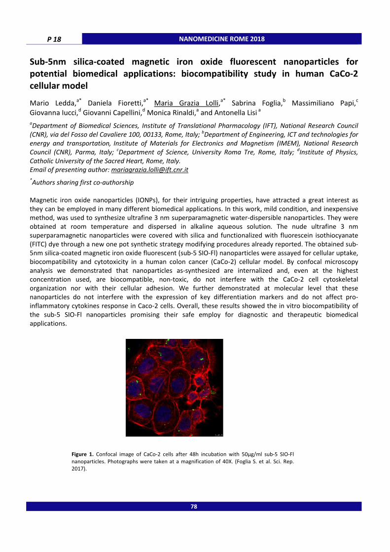
P 18 NANOMEDICINE ROME 2018
Sub-5nm silica-coated magnetic iron oxide fluorescent nanoparticles for
potential biomedical applications: biocompatibility study in human CaCo-2
cellular model
Mario Ledda,a* Daniela Fioretti,a* Maria Grazia Lolli,a* Sabrina Foglia,b Massimiliano Papi,c Giovanna Iucci,d Giovanni Capellini,d Monica Rinaldi,a and Antonella Lisi a aDepartment of Biomedical Sciences, Institute of Translational Pharmacology (IFT), National Research Council
(CNR), via del Fosso del Cavaliere 100, 00133, Rome, Italy; bDepartment of Engineering, ICT and technologies for
energy and transportation, Institute of Materials for Electronics and Magnetism (IMEM), National Research
Council (CNR), Parma, Italy; cDepartment of Science, University Roma Tre, Rome, Italy;
dInstitute of Physics,
Catholic University of the Sacred Heart, Rome, Italy.
Email of presenting author: [email protected]
*Authors sharing first co-authorship
Magnetic iron oxide nanoparticles (IONPs), for their intriguing properties, have attracted a great interest as
they can be employed in many different biomedical applications. In this work, mild condition, and inexpensive
method, was used to synthesize ultrafine 3 nm superparamagnetic water-dispersible nanoparticles. They were
obtained at room temperature and dispersed in alkaline aqueous solution. The nude ultrafine 3 nm
superparamagnetic nanoparticles were covered with silica and functionalized with fluorescein isothiocyanate
(FITC) dye through a new one pot synthetic strategy modifying procedures already reported. The obtained sub-
5nm silica-coated magnetic iron oxide fluorescent (sub-5 SIO-Fl) nanoparticles were assayed for cellular uptake,
biocompatibility and cytotoxicity in a human colon cancer (CaCo-2) cellular model. By confocal microscopy
analysis we demonstrated that nanoparticles as-synthesized are internalized and, even at the highest
concentration used, are biocompatible, non-toxic, do not interfere with the CaCo-2 cell cytoskeletal
organization nor with their cellular adhesion. We further demonstrated at molecular level that these
nanoparticles do not interfere with the expression of key differentiation markers and do not affect pro-
inflammatory cytokines response in Caco-2 cells. Overall, these results showed the in vitro biocompatibility of
the sub-5 SIO-Fl nanoparticles promising their safe employ for diagnostic and therapeutic biomedical
applications.
Figure 1. Confocal image of CaCo-2 cells after 48h incubation with 50µg/ml sub-5 SIO-Fl
nanoparticles. Photographs were taken at a magnification of 40X. (Foglia S. et al. Sci. Rep. 2017).
78

NANOMEDICINE ROME 2018 P 19
Enhanced antinociceptive and anti-inflammatory effects of ibuprofen
encapsulated in niosomal vesicles
Francesca Marzoli,a Paola Minosi,a Laura Ciarlo,a Amalia Di Giannuario,a Maria Carafa,b
Carlotta Marianecci,b Stefano Pieretti a
a National Centre for Drug Research and Evaluation, Italian National Institute of Health, Viale Regina Elena 299,
00161 Roma. Italy; b
Department of Drug Chemistry and Technology, University of Rome "Sapienza", Rome, Italy.
Email of presenting author: [email protected]
The non-steroidal anti-inflammatory (NSAID) drug Ibuprofen (α-methyl-4-(2-methylpropyl)-benzeneacetic acid),
is widely used in the treatment of pain, fever and inflammatory diseases. As far as its analgesic actions,
ibuprofen inhibits the production of prostanoids, which mediate peripheral and central pain sensation, reducing
the threshold to stimulation of nociceptors and increasing their terminal membrane excitability [1]. Niosomes,
are unilamellar or multilamellar non-ionic surfactant vesicles. The main characteristic of these vesicles is their
capability of encapsulating both lipophilic and hydrophilic drugs; hydrophilic drugs are encapsulated in the core
of vesicles, while lipophilic drugs can be encapsulated into the lipophilic domain of the lipid bilayer [2]. In this
work, we evaluated the analgesic activity of subcutaneous injection (s.c) of ibuprofen loaded TW20Gly
niosomes (N-IBU), in comparison with free ibuprofen (IBU) in acute and chronic pain animal models.
In vivo anti nociceptive activities of Ibuprofen-loaded vesicles were preliminarily tested by performing the
writing and capsaicin screening assays. In writhing test, acetic acid as peripheral pain inducer was utilized.
Analgesic activity was determined by entering the reduction of the number of writhes after acetic acid injection.
In the early tested mice, a statistically significant reduction of writhes was observed only in IBU branch, whereas
in the late tested mice the strongest reduction was observed in N[IBU] treated mice. In capsaicin experiments,
in which mice were injected under the hind paw with capsacin, nociceptive activity was evaluated by measuring
the time spent by mice in licking the injected paw. N-IBU significantly decreased capsaicin-induced paw licking,
while IBU was ineffective. N-IBU also induced the strongest antinociceptive effects in Zymosan-induced
hyperalgesia test. N-IBU increased pain threshold also in a model of neuropathic pain i.e. chronic sciatic nerve
ligation, reducing hypealgesia and allodyia 2h after the treatment up to 4h. In the same conditions IBU did not
gave any significant effect.
We can conclude that the encapsulation of the drug into the niosomes significantly increases IBU analgesic
activity, promoting a long lasting action of this drug. Thus, we propose TW20Gly niosomes as a new and more
effective strategy to vehicle IBU to treat acute and chronic pain conditions.
_____________________________________
1. Brunton LL, Chabner BA, Knollman BC. Goodman & Gilman’s the pharmacological basis of therapeutics. 12th ed. New York: McGraw-
Hill Medical; 2011.
2. Di Marzio L, Marianecci C, Petrone M, Rinaldi F, Carafa M., Colloids Surf B Biointerfaces 2011, 82(1), 18.
79

P 20 NANOMEDICINE ROME 2018
Prolongation of local pain insensitivity by anesthetic lidocaine loaded pH-TW20
Gly niosomes: effects on nociception in murine models of pain
Paola Minosi,a Francesca Marzoli,a Laura Ciarlo,a Amalia Di Giannuario,a Maria Carafa,b Ferderica Rinaldi,b Stefano Pieretti a a
National Center for Drug Research and Evaluation, Istituto Superiore di Sanità , Rome, Italy; b
Department of
Drug Chemistry and Technology , University of Rome "Sapienza", Rome, Italy.
Email of presenting author: [email protected]
Current drugs treating neuropathic pain fail in up to 40-50% of the patients, because they have limited efficacy
and are associated with dose related unwanted adverse effects [1]. One of the most extensively studied agents
for neuropathic pain in animals and humans is lidocaine, a local anesthetic with a short duration of action [2].
The great interest in lidocaine delivery systems is increased in the last years. The final purpose is to prolong the
effective time of lidocaine and to reduce the frequency of administration. Particularly, pH-sensitive molecules
to niosome formulation represents an effective and promising delivery strategy [3]. pH-sensitive nonionic
surfactant vesicles (niosomes) by polysorbate-20 derivatized by glycine (added as pH sensitive agent), were
developed to deliver Lidocaine (LID). Lidocaine (5%) were chosen into niosome (N[LID]) (TW20-GLY LIDO 5%)
[3]. Experiments to assess the in vivo efficacy of lidocaine loaded pH-TW20 GLY niosomes were carried out in
murine models to evaluate the potential advantages of stimuli responsive nanocarriers, loaded with lidocaine in
pain treatments. The data related to these tests and obtained from lidocaine loaded pH-TW20 Gly niosomes
were compared with those obtained from free lidocaine, in order to highlight the overlap with the data. The
following models of pain were used: formalin test, zymosan-induced hyperalgesia, Tail flick test and sciatic
nerve ligation inducing neuropathic allodynia and hyperalgesia. The subcutaneous administration of N[LID] in
the dorsal surface of mice paw 10 min or 180 min before formalin in a volume of 40 μL/paw and 1h after
zymosan A in a same volume was able to reduce the response to nociceptive stimuli in the formalin test and
hyperalgesia induced by zymosan. The already high effects of free lidocaine were improved in terms of higher
duration of its action over time. The results obtained by Tail flick test confirmed that N[LID] has a longer
analgesic effect than free lidocaine, especially in terms of longer duration of action. Experience to date suggests
that 40 μL/paw s.c. administration of N[LID] significantly reduced allodynia and hyperalgesia produced by sciatic
nerve ligation. Niosome represents an effective and promising delivery strategy, which may greatly increase the
utility of niosomes as a targeted delivery vehicle, which is degraded only in the target area, where the drug will
be released and accumulated. In our opinion, N[LID] should be developed as a new potential drug in the
treatment of pain in humans.
_____________________________________
1. Y.B. Martin, G. Herradón, L. Ezquerra, Curr Pharm Des., 2011, 17, 434.
2. C.P.N. Watson, Progress in Pain Research and Management, 2001, 21, 215.
3. F. Rinaldi, E. Del Favero, V. Rondelli, S. Pieretti, A. Bogni, J. Ponti, F. Rossi, L. Di Marzio, D. Paolino, J Enzyme Inhib Med Chem., 2017,
32, 538.
80
NANOMEDICINE ROME 2018 P 21
Development of size-controlled PLGA-PEG nanoparticles for targeted drug
delivery in inflammatory Bowel disease
Lauren J. Mohan,a,b Jacqueline S. Daly,a Zebunnissa Ramtoola b a
Division of Biology, Department of Anatomy, School of Medicine, b
School of Pharmacy, Royal College of
Surgeons in Ireland, Dublin 2, Ireland.
Email of presenting author: [email protected]
Nanoparticle (NP)-based drug delivery vehicles represent an innovative strategy for targeted drug delivery in
inflammatory bowel disease (IBD). Evidence in animal models of IBD has shown that orally administered NPs
exhibit preferential deposition in inflamed intestinal tissue over healthy, facilitating drug delivery to the site of
disease [1]. Size is a major determinant affecting NP transport across the intestinal barrier and therefore is a key
consideration in the design of delivery vehicles. To investigate the effect of NP size on transport it is essential to
develop NPs of reproducible and uniform sizes. In this study we aimed to generate and characterise poly(lactic-
co-glycolic acid)-polyethylene glycol (PLGA-PEG) NPs of three defined sizes, produced using a solvent dispersion
method [2]. Herein we describe the production of NPs of different sizes through variation of PLGA-PEG
concentration. Physicochemical characterisation of NPs was performed by dynamic light scattering, Laser
Doppler Electrophoresis and transmission electron microscopy. Size-distribution analysis was carried out by
differential centrifugation. The suitability of lyophilisation for the recovery of NPs was explored [3]. In addition
the stability of lyophilised NPs and NPs stored as aqueous dispersions, at 4°C, was determined over a 12 week
time period. Increasing PLGA-PEG concentration resulted in an increase in NP size, generating NPs of ~100,
~300 and ~600 nm in size. Statistical analysis of NP size for batches formulated at each polymer concentration
revealed the reproducibility of the solvent dispersion method. Differential centrifugation of NPs was found to
facilitate a reduction in NP sample polydispersity. All NPs displayed zeta potential values >-22 mV, indicating
colloidal stability. Storage of NPs at 4°C as an aqueous dispersion was superior for maintenance of physical
stability for up to 12 weeks for further applications. The results of this study suggest that solvent dispersion is
an attractive and reproducible method for the preparation of NPs of defined sizes. NP uniformity and stability
are critical to ensure consistent parameters for definitive in vitro analysis. Future work will focus on transport
studies of different NP sizes in in vitro disease models of the intestinal barrier. Understanding the impact of size
on the passive targeting ability of NPs is fundamental to the design and development of oral nanomedicines for
IBD.
_____________________________________
1. J. Youshia, A. Lamprecht, Expert Opin Drug Deliv, 2016, 13(2), 281.
2. Z. Ramtoola et al., J Pharm Pharmacol, 2011, 63(1), 26. 3. W. Abdelwahed et al., Advanced Drug Delivery Reviews, 2006, 58(15), 1688.
81

P 22 NANOMEDICINE ROME 2018
An insight to interface properties of low-energy nanoemulsions for curcumin
dermal delivery using electron paramagnetic resonance spectroscopy - a link
with release kinetics and biological activity
Ines Nikolić,a Evgenia Mitsou,b Bojan Markovic,c Vassiliki Papadimitriou,b Aristotelis Xenakis,b Snezana Savic a a Department of Pharmaceutical Technology and Cosmetology, University Belgrade, Vojvode Stepe 450, 11221, Belgrade,
Serbia; b
Institute for Biology, Medicinal Chemistry and Biotechnology, National Hellenic Research Foundation, 48 Vassileos
Constantinou Ave, 11635, Athens, Greece; Department of Pharmaceutical Chemistry, University of Belgrade, Vojvode Stepe
450, 11221 Belgrade, Serbia.
Email of presenting author: [email protected]
Introduction: Low-energy nanoemulsions (LE-NEs) represent promising, but poorly investigated nanocarriers
with several physicochemical and biological advantages compared to conventional vehicles, capable of meeting
highly imposed expectations for demanding drug molecules [1]. Curcumin (CU) is a polyphenol of natural origin,
famous not only for its powerful pleiotropic effects, but also for difficulties in terms of stability and
biopharmaceutical performances [2].
Aims: Characterization of interface properties of developed LE-NEs was performed using electron paramagnetic
resonance (EPR), with a view to capturing potential correlation between interface fluidity/rigidity with release
kinetics of CU (in 3 different concentrations: 0.1, 0.2 and 0.3 % m/m) and its reflection to antioxidant activity
(also assessed by EPR).
Results: Blank LE-NE (F), consisting of medium-chain triglycerides, as the oil phase, and a combination of
polysorbate 80 and soybean lecithin (9:1) as stabilizers, with surfactants-to-oil ratio 1, exhibited very small
droplet size (110 nm ± 0.21 nm) and narrow size
distribution. Release kinetics of CU showed that all
formulations followed Higuchi diffusion model. In
each case the total amount of released CU was ≈ 10 %
m/m. Antioxidant activity test (Fig. 1) revealed that CU
incorporation in the LE-NEs was followed by a
prolonged, but less intense activity compared to free
curcumin. This could be explained through EPR results
- even though CU interacted with the interface, its
rigidity made it difficult for CU to move and pass
through it, resulting in lower release amounts and
suppressed activity.
Conclusion: EPR appeared to be a multifunctional
tool, enabling to link physicochemical properties of
carrier’s inner structure with biopharmaceutical
performances, and valuable method in antioxidant
activity evaluation.
_____________________________________
1. T. Tadros, P. Izquierdo, J. Esquena, C. Solans, Adv Colloid Interface Sci., 2004, 108-109, 303.
2. M. Yallapu, K. Bhusetty Nagesh, M. Jaggi, C. Chauhan, APPSJ, 2015, 17, 1341.
Acknowledgments: This work was realized within the framework of the research project TR34031, supported by the Ministry of Education, Science and Technological Development of the Republic of Serbia.
82
Figure 1. Antioxidant activity obtained by EPR

NANOMEDICINE ROME 2018 P 23
Polymeric and Solid Lipid Nanoparticles for nose-to-brain delivery of geraniol-
ursodeoxycholic acid conjugate: development and characterization studies
Edilson Oliveira Junior,a Eleonora Truzzi,b Cecilia Rustichelli,b Eleonora Maretti,b Eliana Lima,a Marco Fogagnolo,c Alessandro Dalpiaz,c Eliana Leo b a Laboratory of Pharmaceutical Technology - FarmaTec, Faculty of Pharmacy, Federal University of Goiás,
University Square nº 1166, 74605 Goiânia, Brazil; b Department of Life Sciences, University of Modena and
Reggio Emilia, via G. Campi 103, 41125 Modena, Italy; cDepartment of Chemical and Pharmaceutical Sciences,
University of Ferrara, Via Fossato di Mortara 19, 44121 Ferrara, Italy.
Email of presenting author: [email protected]
Neurodegenerative disorder treatment is a challenge mainly due to the difficulty of drug transport across the
blood-brain barrier [1]. Intranasal administration of nanoparticles as carrier system may increase drug
concentration into the brain [2]. Geraniol (GER) has demonstrated antioxidant and neuroprotective activities in
Parkinson’s disease animal models [3]. However, due to its volatility, GER is hardly incorporated into freeze-
drying nanoparticles. On the other hand, GER-ursodeoxycholic acid conjugate (GER-UDCA) is a non-volatile
derivative with high potentiality to be incorporated into nanocarriers. Therefore, in this work GER-UDCA-loaded
Solid Lipid Nanoparticles (SLNs) and PLGA nanoparticles (NPs) intended for nose-to-brain delivery were
developed and characterized. SLNs were prepared by emulsion/solvent evaporation method and NPs by
nanoprecipitation method. Briefly, formulations were optimized considering various processing variables and
nanoparticle characterization was performed in terms of morphology, size, surface charge, drug loading (DL%),
encapsulation efficiency (EE%) and in vitro drug release. Finally, the stability of free and encapsulated GER-
UDCA was evaluated in enzymatic medium from rat liver homogenates. GER-UDCA-SLN and GER-UDCA-NPs
showed spherical shape, mean size of 120/180 nm with polydispersity index < 0.2, and zeta potential around
−22/-26 mV, respectively. After freeze-drying, the DL% was 6% for SLN and 12% for NPs with EE% values of
89.3% and 60.1%, respectively. Preliminary data regarding in vitro release of GER-UDCA from the nanoparticles
evidenced a higher dissolution rate than the free drug, probably due to the increase of surface contact. Results
in liver rat homogenate suggested a contribution of the nanoparticles in the stability of the prodrug in
physiologic environments. In conclusion, these GER-UDCA-loaded nanocarriers demonstrated a possible
application in further in vivo studies of nose-to-brain drug delivery.
_____________________________________
1. E. Garbayo et al., Maturitas, 2013, 76, 272. 2. L. Kozlovskaya et al., J Control Release, 2014, 189, 133.
3. K. Rekha et al., J Mol Neurosci, 2013, 51, 851.
Acknowledgments: FAPEG; CONFAP; UFG; UNIMORE.
83
P 24 NANOMEDICINE ROME 2018
DoE optimization of ketoconazole-loaded Nanostructured Lipid Carriers (NLC)
based on Amazon fat (Virola surinamensis) Ucuuba
Rayanne R. Pereira,a,b Matteo Testi,b Roseane M. R. Costa,a Josè O. C. Silva Junior,a Cristina Padula,b Fabio Sonvicob
a Pharmaceutical Sciences Faculty, Federal University of Para, Belem, Brazil;
b Food and Drug Department,
University Of Parma, Parma, PR, Italy.
E-mail of presenting author: [email protected]
Virola surinamensis, know like Ucuuba, is an oleaginous plant species found in Central and South America, especially in Amazonian region of Brazil. Ucuuba seeds are rich in fat (60-70%), already in use for the production of soaps, shampoos and moisturizers [1]. In the era of nanotechnology, nanostructured lipid carriers (NLC) present several advantages in relation to polymer nanoparticles, liposomes, nanoemulsions, microemulsions and traditional emulsions. In this context the aim of this work was the optimization of NLC produced with Ucuuba fat (NCL-ucuuba), for topical application, using ketoconazole as a model drug. The NLC-ucuuba were prepared by High Pressure Homogenization (HPH), (Panda Plus 2000, GEA Niro Soavi, Parma, Italy), using pegylated vitamin E (TPGS) as surfactant, and propylene glycol monocaprylate (Capryol ™ 90, Gattefossé) as liquid lipid. A Box-Behnken experimental design was performed, where the percentage of surfactant (3-6% w/v), that of liquid lipid (2-3% w/v) and of solid lipid (ucuuba fat 7 to 8% w/v) were the independent variables. In total 17 formulations were produced by varying factor concentration to minimize particle size, minimize polydispersity index (PDI) and maximize the entrapment efficiency of ketoconazole in NLC-ucuuba. Nanoparticles of 23.9 nm to 90.1 nm were obtained, the particle size was observed to decrease with increasing Vit E TPGS concentration (p<0.0001) while percentage of liquid lipid was not significant (p> 0.05) on particle size. Ucuuba fat concentration directly affected the size of NLC-ucuuba. The PDI ranged from 0.248 to 0.558. Counterintuitively, increasing concentrations of surfactant in the formulation contributed to an increase of the PDI (p-value 0.0041), while neither solid nor liquid lipid concentration showed significant effects on particle sized distribution. The entrapment efficiency ranged from 93.9 to 99.7, not presenting statistical significance. The manufactured optimized formulation produced according to the model obtained through the DoE showed particle size of 32.8 nm, encapsulation efficiency of 98.2 % and PDI of 0.207. In conclusion, NLC-ucuuba can be classified as having the necessary characteristics for topical application and studies are ongoing to explore their biopharmaceutical properties. 84

NANOMEDICINE ROME 2018
Folate-functionalized DNA nanocages for targeted delivery of Doxorubicin to
cancer cells
Sofia Raniolo,a Valeria Unida,
b Alessio Ottaviani,
a Department of Systems Medicine, University of Rome Tor Verga
Italy; b Department of Biology, University of Rome Tor Vergata, Via della Ricerca Scientifica 1, 00133,
Rome, Italy.
Email of presenting author: [email protected]
DNA possesses intrinsic properties of high stability and biocompatibility that make it an extremely suitable
polymer for the generation of nanoparticles. DNA nanostructures can be functionalized with different
molecules, such as small ligands or antibodies, in order to
wide variety of applications, including drug delivery. Here, we have assembled covalently bound truncated DNA
octahedral nanocages modified with a biotin molecule, which allows the detection through str
reaction, and folic acid for targeting cancer cells overexpressing the alpha isoform of the folate receptor (αFR).
HeLa cells, a human αFR overexpressing tumor cell line, bind and internalize folate
at least 40 times higher than that observed in cells not expressing this receptor. Folate
internalized in endosomes through αFR
way with high intracellular stability (>48 hours).
anticancer drug that intercalates the double helices of DNA. D
natural traffic and accumulate in the cytoplasm where D
degrades DNA cages, avoiding the problem of nanostructures accumulation in vivo
folate-DNA cages, shows a cytotoxic effect at a lower concentration compared to DOX delivered in the free
state that selectively kills αFR-positive ca
with folate can be pursued to selectively target αFR overexpressing cells in cancer therapy since the use of
nanostructures as drug vehicles can potentially
their dosage and minimizing unwanted side
Figure 1. Schematic representation of the uptake and intracellular fate of DOX
folate-DNA cages in αFR overexpressing cells.
NANOMEDICINE ROME 2018
functionalized DNA nanocages for targeted delivery of Doxorubicin to
Alessio Ottaviani, b
Alessandro Desideri, b
and Silvia Biocca
Department of Systems Medicine, University of Rome Tor Vergata, Via Montpellier 1, 00133, Rome,
Department of Biology, University of Rome Tor Vergata, Via della Ricerca Scientifica 1, 00133,
intrinsic properties of high stability and biocompatibility that make it an extremely suitable
polymer for the generation of nanoparticles. DNA nanostructures can be functionalized with different
molecules, such as small ligands or antibodies, in order to achieve specific cellular targeting to be used for a
wide variety of applications, including drug delivery. Here, we have assembled covalently bound truncated DNA
octahedral nanocages modified with a biotin molecule, which allows the detection through str
reaction, and folic acid for targeting cancer cells overexpressing the alpha isoform of the folate receptor (αFR).
HeLa cells, a human αFR overexpressing tumor cell line, bind and internalize folate-DNA cages with an efficiency
times higher than that observed in cells not expressing this receptor. Folate
internalized in endosomes through αFR-mediated pathway and accumulate inside cells in a time
way with high intracellular stability (>48 hours). We have loaded DNA cages with Doxorubicin (DOX), an
anticancer drug that intercalates the double helices of DNA. DOX-loaded folate-DNA
natural traffic and accumulate in the cytoplasm where DOX is released inducing a toxic mechanism tha
avoiding the problem of nanostructures accumulation in vivo
DNA cages, shows a cytotoxic effect at a lower concentration compared to DOX delivered in the free
positive cancer cells. Taken together these results indicate that functionalization
with folate can be pursued to selectively target αFR overexpressing cells in cancer therapy since the use of
potentially improve the in vivo pharmacological effects of drugs, reducing
unwanted side-effects.
Schematic representation of the uptake and intracellular fate of DOX
DNA cages in αFR overexpressing cells.
85
P 25
functionalized DNA nanocages for targeted delivery of Doxorubicin to
and Silvia Biocca a
ta, Via Montpellier 1, 00133, Rome,
Department of Biology, University of Rome Tor Vergata, Via della Ricerca Scientifica 1, 00133,
intrinsic properties of high stability and biocompatibility that make it an extremely suitable
polymer for the generation of nanoparticles. DNA nanostructures can be functionalized with different
achieve specific cellular targeting to be used for a
wide variety of applications, including drug delivery. Here, we have assembled covalently bound truncated DNA
octahedral nanocages modified with a biotin molecule, which allows the detection through streptavidin-biotin
reaction, and folic acid for targeting cancer cells overexpressing the alpha isoform of the folate receptor (αFR).
DNA cages with an efficiency
times higher than that observed in cells not expressing this receptor. Folate-modified DNA cages are
mediated pathway and accumulate inside cells in a time-dependent
We have loaded DNA cages with Doxorubicin (DOX), an
DNA cages divert the DOX
OX is released inducing a toxic mechanism that
avoiding the problem of nanostructures accumulation in vivo. DOX, delivered through
DNA cages, shows a cytotoxic effect at a lower concentration compared to DOX delivered in the free
e results indicate that functionalization
with folate can be pursued to selectively target αFR overexpressing cells in cancer therapy since the use of
cological effects of drugs, reducing
Schematic representation of the uptake and intracellular fate of DOX-loaded

P 26 NANOMEDICINE ROME 2018
In vitro characterization of drug loaded niosomes for brain delivery
Federica Rinaldi,a Maria Carafa,b Simone De Panfilis,a Laura Di Magno,a Carlotta Marianecci,b Giovanna Peruzzi a a
Center for LifeNano Science IIT@Sapienza, Istituto Italiano di Tecnologia, Viale Regina Elena, 00161, Rome,
Italy; b Department of Drug Chemistry and Technology, University of Rome “Sapienza”, Piazzale A. Moro, 00185,
Rome, Italy.
Email of presenting author: [email protected]
The complexity of human diseases may lead to poorly effective drug treatments. The development of
nanocarriers able to deliver active compounds to target specific organs and tissues recently acquired high
impact in the scientific community.
Indeed, properly designed nanocarriers are potentially able to reach the CNS (central nervous system) through
different strategies [1].
To this aim, we designed specific vesicular systems, niosomes (non-ionic surfactant vesicles), and we
characterized these nanovectors in terms of size, ζ-potential and stability in different media.
We used brain tumor cell lines as in vitro model systems (DAOY medulloblastoma cells and SKN neuroblastoma
cells) that were treated with lipophilic fluorescent molecules loaded niosomes (by Span or Tween surfactants).
As shown in Fig. 1, curcumin and Nile red where efficiently internalized in both cellular models, demonstrating
the interaction of non-ionic surfactant vesicles with brain tumor cells.
Figure 1. Fluorescence confocal images of tumoral cell lines after interaction with Nile red /
curcumin loaded niosomes.
_____________________________________
1. F. Rinaldi, P.N. Hanieh et al., Pharmaceutics, 2018, 10, 38.
86
NANOMEDICINE ROME 2018 P 27
Polyion-induced liposomal aggregates for antitubercular drug delivery
Francesca Sciolla,a Domenico Truzzolillo,b Federica Rinaldi,c Patrizia Nadia Hanieh,d Maria Carafa,d Federico Bordi,a Simona Sennato a aInstitute for Complex Systems (ISC)–CNR and Physics Department Sapienza University of Rome, Italy;
bLaboratoire Charles Coulomb CNRS-Université de Montpellier, Montpellier, France;
c Center for Life Nano
Science@Sapienza, Istituto Italiano di Tecnologia, Rome, Italy; dDepartment of Drug Chemistry and Technology,
Sapienza University of Rome, Italy.
Email of presenting author: [email protected]
The self-assembly of charged liposomal particles with oppositely charged polyions gives rise to long-lived finite-
size aggregates with intriguing properties [1,2].This new cluster phase is formed by intact vesicles "glued
together" by the oppositely charged polyions, the structure resembling a sort of blackberry formed by individual
compartments (vesicles) [3,4]. Stability and size of the aggregates can be properly tuned by varying the ratio of
the concentrations of liposomes and polyions (known as re-entrant condensation behavior). These nano-
structured clusters have a high potential for pharmaceutical applications, particularly when the primary
colloidal particles are bio-compatible lipid vesicles as phospholipid liposomes [5].
The present study aims to develop novel liposomal aggregates with an efficient loading of isoniazid (INH), a first
line anti-Mycobacterium tuberculosis-drugs. Considering the selective uptake the macrophages, due to the
large size of the aggregates, this system is particularly suitable for exploring therapeutic strategies against this
pathogen, intracellularly located in macrophages. In this investigation, anionic unilamellar liposomes were
prepared using different amounts of a charged lipid (DPPG) mixed with the zwitterionic HSPC (Hydrogenated
Soybean Phosphatidylcholine), the composition being adjusted to optimize stability and drug encapsulation
efficiency. The biocompatible short polyion, the ε-Polylisine (PLL), has been used. Both liposomal and aggregate
samples have been studied by dynamic light scattering, electrophoresis and transmission electron microscopy.
Differential Scanning Calorimetry coupled to Static Light Scattering were used to better understand the
parameters governing drug loading and the drug-lipid interaction. Stable and unilamellar ISH-liposomes and
PLL-liposomal aggregates have been obtained with size and effective charge tuned by the PLL content.
Moreover, we have proven that the interaction with ISH is able to modify the phase transition temperature, the
thickness and ordering of liposomal membrane and dependent on the liposomal composition.
_____________________________________
1. A.Y. Grosberg, T.T. Nguyen and B. Shklovskii, Rev. Mod. Phys., 2002 74, 329.
2. F. Bordi, S. Sennato and D. Truzzolillo, J. Phys. Condens. Matter, 2009, 21, 203102. 3. S. Sennato, D. Truzzolillo and F. Bordi, Soft Matter, 2012, 8, 9384.
4. S. Sennato, L. Carlini, D. Truzzolillo and F. Bordi, Coll Surf B, 2015, 137, 109. 5. C. Agrati, C. Marianecci, S. Sennato et al., Nanomedicine, 2011, 7,153.
Acknowledgments: S.S. acknowledges funding from the Phospholipid Research Center. Heidelberg, Germany. F. S. thanks support from
Torno Subito 2017 program of Regione Lazio.
87

P 28 NANOMEDICINE ROME 2018
Novel nanomicelles-loaded gelling ocular inserts for the delivery of
cyclosporine-A: technological characterization
Eleonora Terreni,a Daniela Monti,a Patrizia Chetoni,a Susi Burgalassi,a Silvia Tampucci,a Edwin Chipala,b Raid G. Alany,b,c Ali AT. Al-Kinani b a
Department of Pharmacy, University of Pisa, Via Bonanno 33, I-56126, Pisa, Italy; b
School of Life Sciences,
Pharmacy and Chemistry, Kingston University London, Penrhyn Road, Surrey KT1 2EE, Kingston upon a Themes,
London (UK); c School of Pharmacy, The University of Auckland, Auckland, New Zealand.
Email of presenting author: [email protected]
Cyclosporine A (CyA) is a cyclic undecapeptide with an immunosuppressive activity approved for the topical
treatment of several immune-mediated ocular surface disorders such as the sever Dry Eye Syndrome (DES). Its
high molecular weight, low water solubility and high lipophilicity makes the ocular bioavailability and the
development of an ophthalmic formulation of CyA a real challenge. The only approved European marketed
formulation for a “once a day” administration for treatment of DES is Ikervis®, a nanoemulsion containing
1mg/mL of CyA [1]. Nanoemulsions could present some disadvantages related to the stability, ocular irritation,
and pharmacokinetics issues [2]. To improve CyA solubility, nanomicelles consisted of non-ionic surfactants
mixture of Vitamin E-TPGS and Octoxynol-40 (Nano-CyA) was prepared to obtain an aqueous clear solution
suitable for ocular administration. CyA-loaded (0.1%w/v) nanomicelles have shown promising results in terms
of size, drug entrapment, loading capacity, and ocular tolerability. In addition, to improve CyA ocular resident
time and bioavailability Nano-CyA was incorporated into a solid insert constituted by different mucoadhesive
polymers (PVA, CMC, XG, ALG, CAR) able to form a gel system when in contact with the ocular tear fluid and to
increase Nano-CyA interaction with the ocular tissues. The inserts were prepared by casting of aqueous
polymeric dispersions containing Nano-CyA, previously analyzed for drug entrapment, loading capacity and size
distribution. After measurement of the weight, thickness, and diameter, the final inserts were analyzed by
physico-chemical point of view using FTIR and DSC analysis. Furthermore, the mechanical properties by tensile
strength measurement, folding endurance and surface morphology by SEM analysis were determined. Finally,
the CyA content in the solid inserts were evaluated by HPLC.
The more promising formulations will be subjected to further in vivo studies to evaluate ocular tolerability and
the pharmacokinetic behavior.
_____________________________________
1. F. Lallemand, M. Schmitt, J.L. Bourges, R. Gurny, S. Benita, J.S. Garrigue. Eur. J. Pharm. Biopharm., 2017, 117, 14.
2. F. Lallemand, P. Daull, S. Benita, R. Buggage, J.S. Garrigue. J. Drug Deliv., 2012 (doi:10.1155/2012/604204).
88
NANOMEDICINE ROME 2018 P 29
Small-angle neutron scattering characterization of liposomes for anti-
tuberculosis inhaled therapy
Eleonora Truzzi,a Angela Capocefalo,b Carlo Castellano,c Fabio Domenici,d Fiorella Meneghetti,e Eleonora Maretti,a Luca Costantino,a Valentina Iannuccelli,a Eliana Leo a a Department of Life Sciences, University of Modena and Reggio Emilia, via Campi 103, 41125, Modena, Italy;
b
Department of Physics, Sapienza University of Rome, P.le Aldo Moro 5, 00185, Roma, Italy; c
Department of
Chemistry, University of Milan, via Golgi 19, 20133, Milano, Italy; d
Department of Chemical Science and
Technology, University of Rome Tor Vergata, via della Ricerca Scientifica, 00133, Roma, Italy; e Department of
Pharmaceutical Science, University of Milan, Via L. Mangiagalli 25, 20133 Milano, Italy.
Email of presenting author: [email protected]
The present investigation studied the effects of two first-line anti-tuberculosis (TB) drugs, rifampicin (RIF) and
isoniazid (INH), on the structure of multilamellar liposomes. Liposomes have been shown to be a promising
system for inhaled therapy [1]. The study of liposome-drug interaction is essential, and small-angle neutron
scattering (SANS) technique provides valuable and unique data about steric bilayer thickness, particle
dispersion, number of lamellae and drug localization under physiological conditions [2]. Unloaded, single drug-
loaded and co-loaded liposomes were prepared using different amounts of drugs by reverse phase evaporation
method. Liposomal suspensions were prepared using D2O, in order to emphasize the contrast between the
aqueous and the lipid/drug phases. The samples were characterized by dynamic light scattering, atomic force
microscopy and finally by SANS technique (Rutherford Appleton Laboratory, U.K.). Neutron scattering curves
were analyzed using a multi-shell spherical model of the fitting routine SASView 2.2.0. Liposomes have been
shown to be physicochemically stable during the experiments, efficiently drug-loaded, and able to control drug
release. Dimensional analysis demonstrated that particle sizes are in the range of SANS dimensional detection.
SANS curves exhibited Bragg peaks for all samples, confirming the multilamellar liposome structure. By fitting
the data, significant differences among the samples have been highlighted. RIF-liposomes were less ordered
than unloaded liposomes. Indeed, a reduction of the lamellae number was observed and the periodicity of the
lipid bilayers slightly increased with the increment of the drug loading. This is probably due to RIF interaction
with phospholipid tails, which can destabilize liposome lamellarity, since RIF is a hydrophobic drug. In INH-
liposomes, the drug payloads did not change vesicle structure, because INH is a hydrophilic drug. However, INH
induced a change in the inter-bilayer periodical spacing, which could be compatible with the formation of drug-
liposome interactions at the water-lipid interface. Finally, the RIF-INH co-loaded liposomes exhibited the same
characteristics of unloaded liposomes, suggesting that INH and RIF together have a stabilizing effect on the
structure. In fact, no destabilization and no changing in inter-bilayer periodical spacing were observed. In
conclusion, SANS analysis provides fundamental information about drug-liposome interactions to comprehend
the relation between system structure behaviour and its biological activity. Moreover, data suggest that the co-
encapsulation of the two anti-TB drugs may have a synergic effect on liposome stability.
_____________________________________
1. A. Elhissi, Curr. Pharm. Des., 2017, 23, 362.
2. P.C. Lin, S. Lin, P.C. Wang, R. Sridhar, Biotechnol. Adv., 2014, 32, 711.
89
P 30 NANOMEDICINE ROME 2018
Study on anti-fibrotic effects of phospholipid-based formulations in human
hepatic stellate cells
Gina Valentino, Cristina Zivko, Lorine Brülisauer, Paola Luciani
Department of Pharmaceutical Technology, Institute of Pharmacy, Friedrich-Schiller-University Jena, Germany.
E mail of presenting author: [email protected]
During liver fibrogenesis, myofibroblastic transdifferentiation (or “activation”) of hepatic stellate cells (HSC) has
been identified as a key event [1]. Activated HSC are characterized by loss of lipid droplets and dysregulated
scar proteins production. Essential phospholipids have shown to exert antioxidative and antifibrotic effects due
to their polyenylphosphatidylcholine-rich (PPCs) fraction but mechanism of action and impact on HSC are still
unclear [2]. The aim of this project is to develop novel PPC-based formulations and identify potential
antifibrotic candidates by means of an in vitro model based on the differentiation of human immortalized HSC,
LX-2.
Methods: Liposomes of soy phosphatidylcholine (SPC), 1,2-dilinoleoyl-sn-glycero-3-phosphocholine (DLPC), 2-
dioleoyl-sn-glycero-3-phosphocholine (DOPC) and DOPC/DLPC (53/47 mol %) were produced by extrusion
method and characterized by dynamic light scattering (DLS). LX-2 cells, in their standard activated state, were
incubated with liposomal formulations (05-20 mM) for 24 h, or further activated for 24 h with TGF-β (10 ng/mL)
and then treated with liposomes (0.5-5 mM). Retinol and palmitic acid (PA) (10 mM retinol; 300 mM PA) were
used a control to inactivate cells. LX-2 lipid droplets were stained with Oil Red O (ORO) while the scar
production was followed by Sirius Red and Fast Green. Changes in membrane fluidity were studied by means of
fluorescence anisotropy of the probe diphenylhexatriene (DPH) (8 µM) on adherent cells.
Results and Discussion: All liposomes had a hydrodynamic diameter of < 200 nm (polydispersity index < 0.15).
LX-2 exposed to DLPC, SPC, DOPC/DLPC treatments revealed an increase in cytoplasmic lipid droplets coincident
with an intense ORO staining, indicating cell deactivation, visible also upon retinol and PA treatment. In
contrast, DOPC liposomes had only a minor effect on lipid droplets. Collagen appeared upon treatment with
DOPC liposomes. On the contrary, DLPC, SPC and DOPC/DLPC liposomes treatments showed a lower content of
collagen and more abundant non-collagenous proteins. Moreover, a decrease of LX-2 anisotropy upon
treatment with all the liposomes and especially with DLPC was observed.
Conclusions: The abundance of lipid droplets, the deficiency of collagen and the rise in membrane fluidity in LX-
2 exposed to SPC, DLPC and DOPC/DLPC suggest a reversion of HSC differentiation which occur in a minor
extent upon treatment with DOPC. In order to select the most efficient excipients for novel phospholipid-based
antifibrotic therapies and shed light on their mechanisms on liver fibrosis, an in-depth investigation of single
PPCs components is ongoing.
_____________________________________
1. Hihashi T. et al., Adv. Drug Deliv. Rev., 2017, 27.
2. Gundermann K-J. et al., Clin. Exp. Gastroenterol., 2011, 105.
Acknowledgments: Lipoid GmbH is gratefully acknowledged for the endowment to the University of Jena.
90
NANOMEDICINE ROME 2018 P 31
In vitro cytotoxicity evaluation of 5- Fluorouracil (5-FU) loaded PHB coated
magnetic nanoparticles on breast cancer SKBR-3 cell line
Serap Yalcin,a Ufuk Gündüzb
a Department of Molecular Biology and Genetics, Ahi Evran University,40100, Kırsehir, Turkey;
b Department of
Biology, Middle East Technical University,06800, Ankara, Turkey.
Email of presenting author: [email protected]
This study investigated the preparation, characterization and anti-proliferative effect of 5- Fluorouracil loaded
PHB coated magnetic nanoparticles for breast cancer therapy. In this study, we developed a new method that
enabled for the first time in situ and PHB coated MNPs, involving the coprecipitation of iron salts in the
presence of PHB. The 5- Fluorouracil loaded PHB-MNPs was confirmed by FTIR analysis. In vitro cytotoxicity
effects of 5- Fluorouracil-loaded PHB-MNPs on SKBR-3 cells were evaluated by means of the XTT Cell
Proliferation Kit (Biological Industries, Israel) according to manufacturer’s instructions. SKBR-3 cells were
seeded into 96-well microtiter plates (Greiner) at a concentration of 5.0X10⁴. The cytotoxic effect of 5-
Fluorouracil -loaded PHB-MNPs on SKBR-3 cells was investigated by XTT cell proliferation assay, and IC50 values
were calculated. In this study, empty PHB-MNPs were found not significantly cytotoxic up to 500 mg/mL. The
IC50 value of 600 mg/mL 5- Fluorouracil loaded PHB-MNPs were found as 12,5 μM on SKBR-3 cells. IC50 value of
free 5- Fluorouracil as 23,7 μM for SKBR-3 cells. From the IC50 values, it was seen that the loading of 5-
Fluorouracil onto PHB-MNPs increased its efficiency nearly up to 3- to 3.5-fold. The results indicated that the
synthesized magnetic nanoparticles have a high potential to be used as a 5- Fluorouracil delivery system, which
can be targeted to tumor cells under a magnetic field. The magnetic targeting will prevent the side effects of
chemotherapy, while the effective release from the nanoparticles may reduce the amount of 5- Fluorouracil
required, and the frequency of drug administration.
91

P 32 NANOMEDICINE ROME 2018
Synthesis and characterization of Annona Muricata extract loaded magnetic
nanoparticles for cancer therapy
Serap Yalcin,a Rana Kavurmacıb
a Department of Molecular Biology and Genetics, Ahi Evran University, 40100, Kırsehir, Turkey;
b Department of
Advanced Technologies, Ahi Evran University, 40100, Kırsehir, Turkey.
Email of presenting author: [email protected]
Annona muricata, also known as soursop, graviola, or guanabana, has traditionally been used to treat various
diseases. Plant extracts include effectively toxins, alkaloids and acetogenins. Several studies have been
investigated the in vitro effects of extracts on various cancer cell lines. However, plant extracts are also known
toxic on neurons and have contributed to Parkinson’s disease-like syndromes [1]. The magnetic nanoparticles
can be widely used for cancer treatment which increases anti-cancer agents accumulation in the targeted
tumor cells and healthy cells are not damaged. In this study, we investigated the preparation, characterization
and anti-proliferative effect of Annona muricata extract loaded magnetic nanoparticles on triple negative
cancer cell. we synthesed PHB coated MNPs, involving the coprecipitation of iron salts in the presence of PHB.
The structural properties, functional groups, size distribution and magnetic properties of the synthesized
nanoparticles were characterized by FT-IR, TEM, VSM, DLS analyses. The extract loaded PHB-MNPs was
confirmed by FTIR analysis. Antiproliferative effects of extract loaded PHB-MNPs on MDA-MB-231 cells were
evaluated by means of the XTT Cell Proliferation Kit (Biological Industries, Israel) according to manufacturer’s
instructions. As a results, IC50 value of free extract and extract-loaded magnetic nanoparticles were identified as
330μg/ml and 42 μM on MDA-MB-231 cells, respectively. In conclusion, the novel extract-loaded magnetic
nanoparticles may constitute a biodegradable and biocompatible delivery system for targeted delivery to
tumors without healthy cells damage.
_____________________________________
1. A. Lannuzel , G.U. Höglinge , P. Champy , P.P. Michel , E.C. Hirsch , M. Ruberg, J. Neural. Transm. Suppl., 2006, 70, 153.
Acknowledgments: This work was supported by the Ahi Evran University Scientific Research Projects Coordination Unit. Project
Number: FEF.A4.17.018
92

NANOMEDICINE ROME 2018 P 33
Overcoming drug resistance by cisplatin loaded magnetic nanoparticles on
cisplatin resistant model cell line
Serap Yalcin,a Ufuk Gunduzb
a Department of Molecular Biology and Genetics, Ahi Evran University,40100, Kırsehir, Turkey;
b Department of
Biology, Middle East Technical University,06800, Ankara, Turkey.
Email of presenting author: [email protected]
In this study, we investigated anti-proliferative effect of Cisplatin loaded PHB coated magnetic nanoparticles on
Cisplatin resistance model cell line for breast cancer therapy. In this study, Cisplatin loaded PHB-MNPs was
confirmed by FTIR analysis. Drug loading and release characteristics, and stability of the nanoparticles were
investigated. Anti-proliferative effects of Cisplatin-loaded PHB-MNPs on Cisplatin resistance MDA-MB231 cells
(1000nM) were evaluated by means of the XTT Cell Proliferation Kit (Biological Industries, Israel) according to
manufacturer’s instructions. Cisplatin was loaded onto PHB-MNPs, and the release efficiencies at different pHs
were studied under in vitro conditions. The most efficient drug loading concentration was found about 80% at
room temperature in phosphate-buffered saline (pH 7.4). The Cisplatin loaded MNPs were stable up to 2
months in neutral pH for mimicking physiological conditions. The cytotoxic effect of cisplatin-loaded PHB-MNPs
on Cisplatin resistance cells was investigated by XTT cell proliferation assay, and IC50 values were calculated. In
this study, empty PHB-MNPs were found not significantly cytotoxic up to 500 mg/ml. Cisplatin-loaded PHB-
MNPs were about 2.5-fold more cytotoxic as compared with free drug on cisplatin resistance MDA-MB-231 cell
line. The results indicated that the synthesized magnetic nanoparticles have a high potential to be used as a
Cisplatin delivery system, which can be targeted to Cisplatin resistance tumor cells. The magnetic targeting will
prevent the side effects of chemotherapy, while the effective release from the nanoparticles may reduce the
amount of Cisplatin required in tumor.
_____________________________________
Acknowledgments: This work was supported by the Ahi Evran University Scientific Research Projects Coordination Unit. Project Number: FEF.A3.17.009.
93
P 34 NANOMEDICINE ROME 2018
Delivery of miR-29 using nanoparticles suppress triple negative breast cancer
cell growth
Serap Yalcin
Department of Molecular Biology and Genetics, Ahi Evran University,40100, Kırsehir.
Email: [email protected]
Breast cancer is the most frequently diagnosed cancer in women worldwide. Molecular mechanisms play
important roles in cancer progression and aggressivity. In the last years, micro-RNAs have been associated in
molecular pathways of cancer and other diseases. Mir-29 has inhibitory role in tumorigenesis. Studies showed
that miR-29 is downregulated in breast cancer [1,2]. In this study, to restore normal miR-29 levels in triple
negative breast cancer cell line we developed an optimisation method. In this method,
dextran nanoparticles conjugated with miR-29 in a delivery system that specifically targets triple negative cell
line. Results indicated that the presence of miR-29 conjugated nanoparticles enhanced the selective delivery
of miR-29. Further, the upregulation of miR-29 by nanoparticles resulted in the inhibition of cells growth in
triple negative breast cancer cell line.
_____________________________________
1. Z. Wu , X. Huang , X. Huang , Q. Zou , Y. Guo, J. Exp. Clin. Cancer Res., 2013, 32, 98.
2. A. Starlard-Davenport, K. Kutanzi, V. Tryndyak, B. Word, B. Lyn-Cook, J. Carcinog. 2013, 12, 15.
94

NANOMEDICINE ROME 2018
Does length matter? Suitability of halloysite nanotubes in drug delivery
applications
F. Tardania, S. Casciardi
b, B. Ruzicka
a CNR-ISC, Department of Physics, La Sapienza, Piazzale Aldo Moro 5, 00185 Roma, Italy
Institute, Department of Occupational and Environmental Medicine, Epidemiology and Hygene, Roma, Italy
Email of presenting author: [email protected]
In last decades, a large number of different colloidal systems are under consideration as innovative carriers of
biologically active compounds as alternative to viral vectors. The ideal nanocarrier would
specificity, high loading capacity, good stability, slow and controlled cargo release. Liposomes, vesicles and
nanoparticles have been effectively shown as nanocarriers [1]. Even if quite promising, these systems are not
always suitable for large-scale applications. In this context, recently the halloysite nanotubes (HNT) have been
considered as an effective alternative for a number of reasons [2]. Firstly, HNTs are hydrophilic aluminium
silicate tubes with a nanometer-sized cavity (
reported that the loading process can be easily performed in water. Moreover, as other nanoclays, HNTs are
naturally abundant, cheap and biocompatible. Despite the numerous studies about HNTs
questions are still to be solved. The high density of HNTs (2g/cm
Sedimentation can produce severe accumulation of HNTs in biological tissues [3]. In addition, commercial
halloysite nanoclays are very polydisperse in length and diameter. As already observed for carbon nanotubes,
cytotoxic effects are expected also for very long HNTs.
In order to improve their actual usability, a deeper knowledge of their behaviour in water dispersions is
required. In this investigation, we used HNTs of different sources and purity. Aqueous dispersions were
prepared with or without addition of salt and additives to monitor stability against aggregation and
sedimentation. A very stable fraction was identified though Dynamic
measurements. This fraction is mainly composed of short tubes, freely diffusing in the solvent with low
aggregation degree. These short tubes could have potential application as nanocarriers with high
biocompatibility and low accumulation problems.
Figure 1. Effect of tube length and instability on settling and accumulation of circulating
nanotubes in blood vessels.
_____________________________________
1. M. Alavi, N. Karimi, M. Safaei Adv. Pharm Bull.
2. S. Leporatti Polym Int 2017
3. X. Wuang, J. Gong, R. Rong, Z. Gui, T. Hu, X. Xu
Acknowledgments: authors thank financial support from INAIL (BRIC02016 project ID 52).
NANOMEDICINE ROME 2018
atter? Suitability of halloysite nanotubes in drug delivery
, B. Ruzickaa, S. Sennato
a
ISC, Department of Physics, La Sapienza, Piazzale Aldo Moro 5, 00185 Roma, Italy
Institute, Department of Occupational and Environmental Medicine, Epidemiology and Hygene, Roma, Italy
In last decades, a large number of different colloidal systems are under consideration as innovative carriers of
biologically active compounds as alternative to viral vectors. The ideal nanocarrier would
specificity, high loading capacity, good stability, slow and controlled cargo release. Liposomes, vesicles and
nanoparticles have been effectively shown as nanocarriers [1]. Even if quite promising, these systems are not
scale applications. In this context, recently the halloysite nanotubes (HNT) have been
considered as an effective alternative for a number of reasons [2]. Firstly, HNTs are hydrophilic aluminium
sized cavity (lumen) which can easily filled with the desired drug. It is widely
reported that the loading process can be easily performed in water. Moreover, as other nanoclays, HNTs are
naturally abundant, cheap and biocompatible. Despite the numerous studies about HNTs
questions are still to be solved. The high density of HNTs (2g/cm3) produce annoying settling problems.
Sedimentation can produce severe accumulation of HNTs in biological tissues [3]. In addition, commercial
ydisperse in length and diameter. As already observed for carbon nanotubes,
cytotoxic effects are expected also for very long HNTs.
In order to improve their actual usability, a deeper knowledge of their behaviour in water dispersions is
s investigation, we used HNTs of different sources and purity. Aqueous dispersions were
prepared with or without addition of salt and additives to monitor stability against aggregation and
sedimentation. A very stable fraction was identified though Dynamic Light Scattering and microscopy
measurements. This fraction is mainly composed of short tubes, freely diffusing in the solvent with low
aggregation degree. These short tubes could have potential application as nanocarriers with high
ow accumulation problems.
. Effect of tube length and instability on settling and accumulation of circulating
nanotubes in blood vessels.
Pharm Bull. 2017, 7, 3
3. X. Wuang, J. Gong, R. Rong, Z. Gui, T. Hu, X. Xu J. Agric. Food Chem. 2018, 66, 2925
authors thank financial support from INAIL (BRIC02016 project ID 52).
95
P 35
atter? Suitability of halloysite nanotubes in drug delivery
ISC, Department of Physics, La Sapienza, Piazzale Aldo Moro 5, 00185 Roma, Italy; b2INAIL Research
Institute, Department of Occupational and Environmental Medicine, Epidemiology and Hygene, Roma, Italy.
In last decades, a large number of different colloidal systems are under consideration as innovative carriers of
biologically active compounds as alternative to viral vectors. The ideal nanocarrier would require high target
specificity, high loading capacity, good stability, slow and controlled cargo release. Liposomes, vesicles and
nanoparticles have been effectively shown as nanocarriers [1]. Even if quite promising, these systems are not
scale applications. In this context, recently the halloysite nanotubes (HNT) have been
considered as an effective alternative for a number of reasons [2]. Firstly, HNTs are hydrophilic aluminium-
) which can easily filled with the desired drug. It is widely
reported that the loading process can be easily performed in water. Moreover, as other nanoclays, HNTs are
naturally abundant, cheap and biocompatible. Despite the numerous studies about HNTs, some simple
) produce annoying settling problems.
Sedimentation can produce severe accumulation of HNTs in biological tissues [3]. In addition, commercial
ydisperse in length and diameter. As already observed for carbon nanotubes,
In order to improve their actual usability, a deeper knowledge of their behaviour in water dispersions is
s investigation, we used HNTs of different sources and purity. Aqueous dispersions were
prepared with or without addition of salt and additives to monitor stability against aggregation and
Light Scattering and microscopy
measurements. This fraction is mainly composed of short tubes, freely diffusing in the solvent with low
aggregation degree. These short tubes could have potential application as nanocarriers with high
. Effect of tube length and instability on settling and accumulation of circulating

NANOMEDICINE ROME 2018
96

NANOMEDICINE ROME 2018
List of Participants
97
NANOMEDICINE ROME 2018
98
NANOMEDICINE ROME 2018 List of Participants
1 Aiello Stefano Dipartimento di Chimica, Università di Roma “Sapienza”, Rome, Italy. P1
Email: [email protected]
2 Akbaba Hasan Department of Pharmaceutical Biotechnology, University of Ege, İzmir,
Turkey. SL26
Email: [email protected]
3 Alberti Diego Department of Molecular Biotechology and Health Sciences, University of Torino, Italy. SL1, SL22
Email: [email protected]
4 Albrecht Volker Biolitec research GmbH, Jena, Germany. SL2
Email: [email protected]
5 Amalfitano Adriana Catholic University of Sacred Heard. SL16, SL31,
P2 Email: [email protected]
6 Arancia Giuseppe Istituto Superiore di Sanità.
Email: [email protected]
7 Arcovito Alessandro Institute of Biochemistry and Clinical Biochemistry, Catholic University of
Sacred Heart,Rome, Italy. SL16, SL31,
P2 Email: [email protected]
8 Astone Chiara Università degli Studi di Roma "La Sapienza".
Email: [email protected]
9 Bertoni Serena
Drug Research Program, Division of Pharmaceutical Chemistry and Technology, Faculty of Pharmacy, University of Helsinki, Finland;
PharmTechLab, Department of Pharmacy and Biotechnology, University of Bologna, Italy.
SL23
Email: [email protected]
10 Bombelli Cecilia CNR-IMC Istituto di Metodologie Chimiche Sezione Meccanismi di
Reazione. SL10, P1, P14
Email: [email protected]
11 Borocci Stefano University of Tuscia.
Email: [email protected]
12 Bozzuto Giuseppina National Center for Drug Research and Evaluation, Istituto Superiore di
Sanità Rome, Italy. SL9
Email: [email protected]
13 Brocco Monica Core facilities - Istituto Superiore di Sanità.
Email: [email protected]
14 Calcabrini Annarica National Centre for Drug Research and Evaluation - ISS. P7
Email: [email protected]
15 Caselli Federica Department of Civil Engineering and Computer Science, University of Rome Tor Vergata, Rome, Italy. SL8
Email: [email protected]
16 Ceccacci Francesca CNR-IMC Istituto di Metodologie Chimiche Sezione Meccanismi di
Reazione. P1
Email: [email protected]
17 Ceci Pierpaolo Institute of Molecular Biology and Pathology, CNR , Rome, Italy. SL16
Email: [email protected]
18 Celenza Giuseppe Department of Biotechnological and Applied Clinical Sciences, University of
l’Aquila, Italy. SL30
Email: [email protected]
19 Chistè Elena Department of Computer Science, Fluorescence Laboratory, University of Verona, Italy. P4
Email: [email protected]
20 Chronopoulou Laura Department of Chemistry, Sapienza University of Rome, , Italy. SL31
Email: [email protected]
21 Ciarlo Laura National Center for Drug Research and Evaluation, Istituto Superiore di Sanità, Rome, Italy. P5, P19, P20
Email: [email protected]
22 Colombo Eleonora Department of Chemistry, University of Milan, Italy. P6
Email: [email protected]
99
List of Participants NANOMEDICINE ROME 2018
23 Colone Marisa National Centre for Drug Research and Evaluation - ISS. P7
Email: [email protected]
24 Condello Maria National Centre for Drug Research and Evaluation - ISS. SL9, P8
Email: [email protected]
25 Conte Claudia Drug Delivery Laboratory, Department of Pharmacy, University of Napoli
Federico II, Napoli, Italy. SL24, P15
Email: [email protected]
26 Costabile Gabriella Department of Pharmacy, Ludwig-Maximilians-University, Munich, Germany. P9
Email: [email protected]
27 Couvreur Patrick University of Paris-Sud, Université Paris-Saclay Institut Galien, UMR CNRS
8612, 5 rue J-B Clément F-92296 Chatenay-Malabry, France. IL1
Email: [email protected]
28 D’Avenio Giuseppe National Center for Technological Innovation in Public Health, Istituto
Superiore di Sanità Rome, Italy. SL9
Email: [email protected]
29 Decarolis Nadia nordtest srl.
Email: [email protected]
30 Di Gioia Sante Department of Medical and Surgical Sciences, University of Foggia, Italy. SL13, P3
Email: [email protected]
31 Di Meo Chiara Department of Drug Chemistry and Technologies, Sapienza University of Rome, Italy. P10
Email: [email protected]
32 Diociaiuti Marco Centro Nazionale Malattie Rare, Istituto Superiore di Sanità, Roma, Italy. SL10, P14
Email: [email protected]
33 Djekic Ljiljana Department of Pharmaceutical Technology and Cosmetology, University of
Belgrade-Faculty of Pharmacy, Belgrade, Serbia. P11, P12, P13
Email: [email protected]
34 Erel Akbaba Gülşah
Experimental Therapeutics and Molecular Imaging Lab, Department of
Neurology, Neuro-Oncology Division, Massachusetts General Hospital, Boston, MA, USA; Department of Pharmaceutical Biotechnology, Faculty of
Pharmacy, Izmir Katip Celebi University, Izmir, Turkey; Department of Pharmaceutical Biotechnology, Faculty of Pharmacy, Ege University, Izmir,
Turkey.
SL21
Email: [email protected]
35 Fahr Alfred Biolitec research GmbH, Jena, Germany. SL2
Email: [email protected]
36 Ferrari Angelo CNR - Institute of Chemical Methodologies.
Email: [email protected]
37 Ferroni Claudia CNR - Institute of Organic Syntesis and Photoreactivity. SL6, SL17
Email: [email protected]
38 Fioravanti Raoul Centro Nazionale Malattie Rare, Istituto Superiore di Sanità, Rome, Italy; Dipartimento di Chimica, Università degli Studi di Roma “Sapienza”, Italy. SL10, P14
Email: [email protected]
39 Formisano Giuseppe National Centre for Drug Research and Evaluation - ISS.
Email: [email protected]
40 Fracassi Alessandro Laboratorium für Organische Chemie, ETH Zürich, Switzerland. SL29
Email: [email protected]
41 Gaio Elisa Cell Biology Unit, Department of Biology, University of Padova. P15
Email: [email protected]
42 Galantini Luciano Università La Sapienza.
Email: [email protected]
43 Garello Francesca Department of Molecular Biotechnology and Health Sciences, University of
Torino, Italy. SL22
Email: [email protected]
100

NANOMEDICINE ROME 2018 List of Participants
44 Gelain Fabrizio
ISBREMIT, IRCSS Casa Sollievo della Sofferenza, Opera di San Pio da
Pietralcina, San Giovanni Rotondo, Foggia. Center for Nanomedicine and Tissue Engineering (CNTE), ASST Niguarda Ca Granda, Milan, Italy.
IL8
Email: [email protected]
45 Giansanti Luisa Department of Physical and Chemical Sciences, University of L'Aquila, Italy. P8
Email: [email protected]
46 Giardini Giorgio CNR - Institute of Chemical Methodologies.
Email: [email protected]
47 Giordani Cristiano Instituto de Física, Universidad de Antioquia, Medellín, Colombia. SL10, P14
Email: [email protected]
48 Grimm Jan
Molecular Pharmacology Program & Department of Radiology - Memorial Sloan Kettering Cancer Center, New York, USA. Gerstner Sloan-Kettering
Graduate School of Biomedical Sciences, New York, USA. Pharmacology,
Radiology - Weill Cornell Medical College, New York, USA. Integrated Cancer Center - King's College London, UK.
IL2
Email: [email protected]
49 Herrmann Inge Katrin Swiss Federal Laboratories for Materials Science and Technology (Empa), St. Gallen, Switzerland. IL3
Email: [email protected]
50 Imbriano Anna Department of Drug Chemistry and Technologies, Sapienza University of
Rome, Italy. P16, P17
Email: [email protected]
51 Kovačević Anđelka Institute of Pharmacy, Faculty of Biological Sciences, Friedrich Schiller
University Jena, Germany. SL11
Email: [email protected]
52 Lolli Maria Grazia National Research Council (CNR), Rome, Italy. P18
Email: [email protected]
53 Mancini Giovanna Institute of Chemical Methodologies - CNR. P1, P8
Email: [email protected]
54 Marianecci Carlotta Department of Chemistry and Technology of Drugs, Sapienza University, Rome, Italy.
IL7, P16,P17,
P19, P26 Email: [email protected]
55 Markova Elena
Institute of pharmaceutical technology, Center of pharmaceutical nanotechnology, Faculty of pharmacy, Ss. Cyril & Methodius University,
Skopje, R. Macedonia SL12
Email: [email protected]
56 Martini Cecilia CNR - Institute of Organic Syntesis and Photoreactivity SL16, SL17,
P2 Email: [email protected]
57 Marzoli Francesca National Center for Drug Research and Evaluation, Istituto Superiore di
Sanità, Rome, Italy. P5, P19, P20
Email: [email protected]
58 Medard Nicolas NANOLANE, Pole Novaxud, Le Mans, France. SL5
Email: [email protected]
59 Meschini Stefania National Centre for Drug Research and Evaluation - ISS. SL9, P8
Email: [email protected]
60 Minosi Paola National Center for Drug Research and Evaluation, Istituto Superiore di Sanità, Rome, Italy. P5, P19, P20
Email: [email protected]
61 Mohan Lauren Division of Biology, Department of Anatomy, School of Medicine; School of Pharmacy, Royal College of Surgeons in Ireland, Dublin 2, Ireland. P21
Email: [email protected]
62 Molinari Agnese National Centre for Drug Research and Evaluation - ISS. SL9
Email: [email protected]
63 Murgia Sergio Department of Chemical and Geological Sciences, University of Cagliari,
Monserrato (CA), Italy. SL4
Email: [email protected]
101
List of Participants NANOMEDICINE ROME 2018
64 Nicoletti Isabella CNR - Institute of Chemical Methodologies.
Email: [email protected]
65 Nikolić Ines Department of Pharmaceutical Technology and Cosmetology, University Belgrade, Belgrade, Serbia. P22
Email: [email protected]
66 Oliveira J. Miguel
3B’s Research Group- Biomaterials, Biodegradable and Biomimetic,
University of Minho, Headquarters of the European Institute of Excellence
on Tissue Engineering and Regenerative Medicine, Avepark, 4805-017 Barco, Guimarães, Portugal; ICVS 3Bs PT Government Associate Lab, Braga,
Guimarães, Portugal; The Discoveries Centre for Regenerative and Precision Medicine, Headquarters at University of Minho, Avepark, 4805-017 Barco,
Guimarães, Portugal.
IL6
Email: [email protected]
67 Oliveira Junior Edilson Laboratory of Pharmaceutical Technology - FarmaTec, Faculty of Pharmacy, Federal University of Goiás, Goiânia, Brazil. P23
Email: [email protected]
68 Pellegrini Evelin National Center for Drug Research and Evaluation, National Institute of Health, Rome, Italy. P8
Email: [email protected]
69 Pellegrino Chiara AcZon S.r.l.
Email: [email protected]
70 Pereira Rayanne Rocha Pharmaceutical Sciences Faculty, Federal University of Para, Belem, Brazil;
Food and Drug Department, University Of Parma, Parma, PR, Italy. P24 Email: [email protected]
71 Pieretti Stefano National Center for Drug Research and Evaluation, Istituto Superiore di
Sanità, Rome, Italy. P5, P19, P20
Email: [email protected]
72 Quici Massimo CNR - Institute of Chemical Methodologies.
Email: [email protected]
73 Raniolo Sofia Department of Systems Medicine, University of Rome Tor Vergata, Rome, Italy. P25
Email: [email protected]
74 Raucci Maria Grazia Institute of Polymers, Composites and Biomaterials (IPCB) - CNR, Naples (Italy). SL19
Email: [email protected]
75 Renault-Mahieux
Morgane
AP-HP, Henri Mondor Hospital Group, Pharmacy Department, Créteil, France; UTCBS, INSERM U1022 UMR CNRS 8258, Paris Descartes University,
Paris, France. SL15
Email: [email protected]
76 Rinaldi Federica Center for Life Nano Science@Sapienza, Istituto Italiano di Tecnologia (ITT),
Rome, Italy. P20, P26, P27
Email: [email protected]
77 Russo Krauss Irene Department of Chemical Science, University of Naples Federico II, Naples,
Italy; CSGI, Florence, Italy. SL27
Email: [email protected]
78 Salvati Anna Groningen Research Institute of Pharmacy, University of Groningen, The Netherlands. IL10
Email: [email protected]
79 Sansone Francesco Dipartimento di Scienze Chimiche, della Vita e della Sostenibilità Ambientale, Università di Parma, Italy. SL7
Email: [email protected]
80 Santoliquido Roberto ALFATEST s.r.l., Rome, Italy. SL18
Email: [email protected]
81 Sarra Angelo Department of Science, University of Roma Tre, Rome, Italy. SL25
Email: [email protected]
102
NANOMEDICINE ROME 2018 List of Participants
82 Sciolla Francesca Istitute for Complex Systems (ISC) – CNR and Sapienza University of Rome. P27
Email: [email protected]
83 Scipioni Anita Università La Sapienza.
Email: [email protected]
84 Sennato Simona Institute for Complex Systems ISC–CNR and Physics Department, Sapienza
University of Rome, Italy. SL3, SL9,SL25,
P27, P35 Email: [email protected]
85 Shalabalija Dushko
Institute of pharmaceutical technology, Center of pharmaceutical nanotechnology, Faculty of pharmacy, Ss. Cyril & Methodius University,
Skopje, R. Macedonia. SL28, SL12
Email: [email protected]
86 Soleimanian Yasamin Department of Food Science and Technology, Isfahan University of
Technology, Isfahan, Iran. SL14
Email: [email protected]
87 Storm Gert
Dept. Pharmaceutics, Utrecht Institute for Pharmaceutical Sciences (UIPS), Utrecht University. University Medical Centre Utrecht (UMCU), Division
Imaging, Utrecht. Dept. Biomaterials Science & Technology (BST), MIRA Institute for Biomedical Technology and Technical Medicine, University of
Twente, Enschede, The Netherlands.
IL9
Email: [email protected]
88 Stringaro Annarita National Centre for Drug Research and Evaluation - ISS. P7
Email: [email protected]
89 Talamini Laura IRCCS-Istituto di ricerche farmacologiche Mario Negri, Milano, Italy. SL20
Email: [email protected]
90 Tardiola Stefano CNR - Institute of Chemical Methodologies.
Email: [email protected]
91 Terreni Eleonora Department of Pharmacy, University of Pisa, Italy. P28
Email: [email protected]
92 Toccacieli Laura National Centre for Drug Research and Evaluation - ISS.
Email: [email protected]
93 Truzzi Eleonora Department of Life Sciences, University of Modena and Reggio Emilia, Modena, Italy. P23, P29
Email: [email protected]
94 Unida Valeria Department of Biology, University of Rome Tor Vergata, Rome, Italy. P25
Email: [email protected]
95 Valentino Gina Department of Pharmaceutical Technology, Institute of Pharmacy, Friedrich-Schiller-University Jena, Germany. P30
Email: [email protected]
96 Webster Thomas J. The Art Zafiropoulo Chair and Department Chair Department of Chemical
Engineering, Northeastern University, Boston, USA. IL5
Email: [email protected]
97 Wurm Frederik Max Planck Institute for Polymer Research, Mainz, Germany. IL4
Email: [email protected]
98 Yalcin Serap Department of Molecular Biology and Genetics, Ahi Evran University, Kırsehir, Turkey.
P31, P32,
P33, P34 Email: [email protected]
103
NANOMEDICINE ROME 2018
104
The NANOMEDICINE ROME 2018
Conference is organized
NANOMEDICINE ROME 2018
The NANOMEDICINE ROME 2018
onference is organized with the unconditional support
105
unconditional support of

NANOMEDICINE ROME 2018
106