Exercises for plantar fasciitis plantar fasciitis exercise, plantar fasciitis shoes women
The Plantar Soft Tissues
Sabrina Covert
February 28, 2008
Objectives
1) Review normal plantar anatomy of the foot
2) Describe optimal technique for MR Imaging of the foot
3) Review the most common abnormalities affecting the
plantar soft tissues
4) Infection

Anatomy - calcaneus
M
LM
L
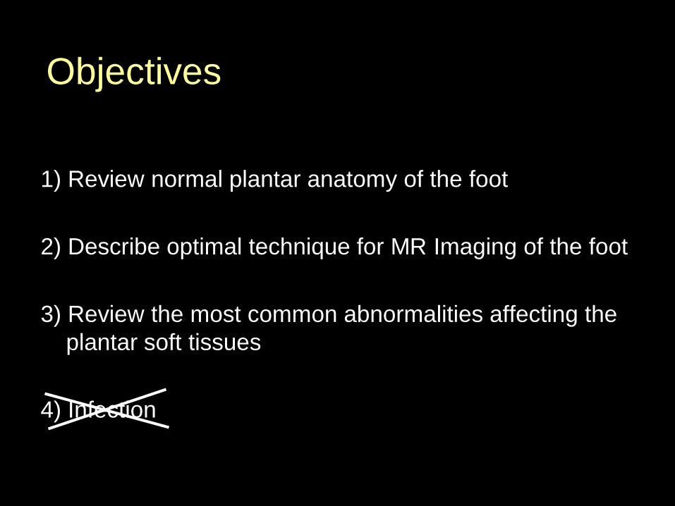
Supporting structures - longitundinal arch
• Plantar fascia
• Long and short plantar ligaments
• Spring ligament
• Tibialis posterior tendon
• Peroneus longus tendon
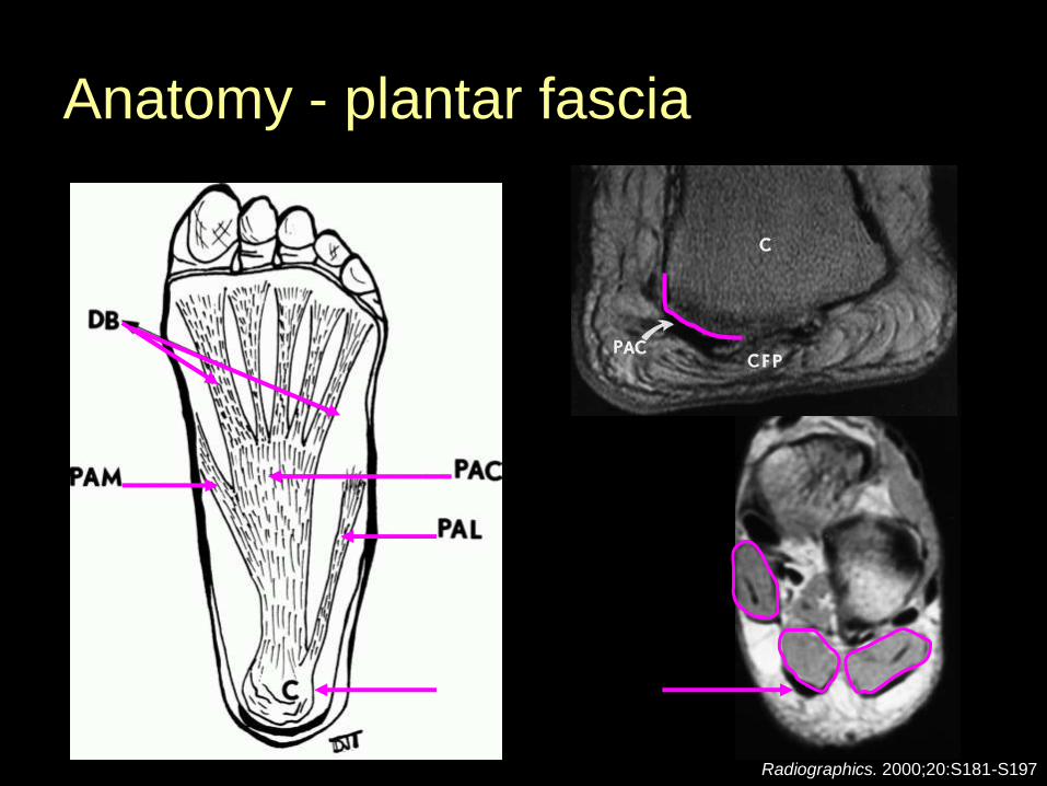
Anatomy - plantar fascia
Radiographics. 2000;20:S181-S197
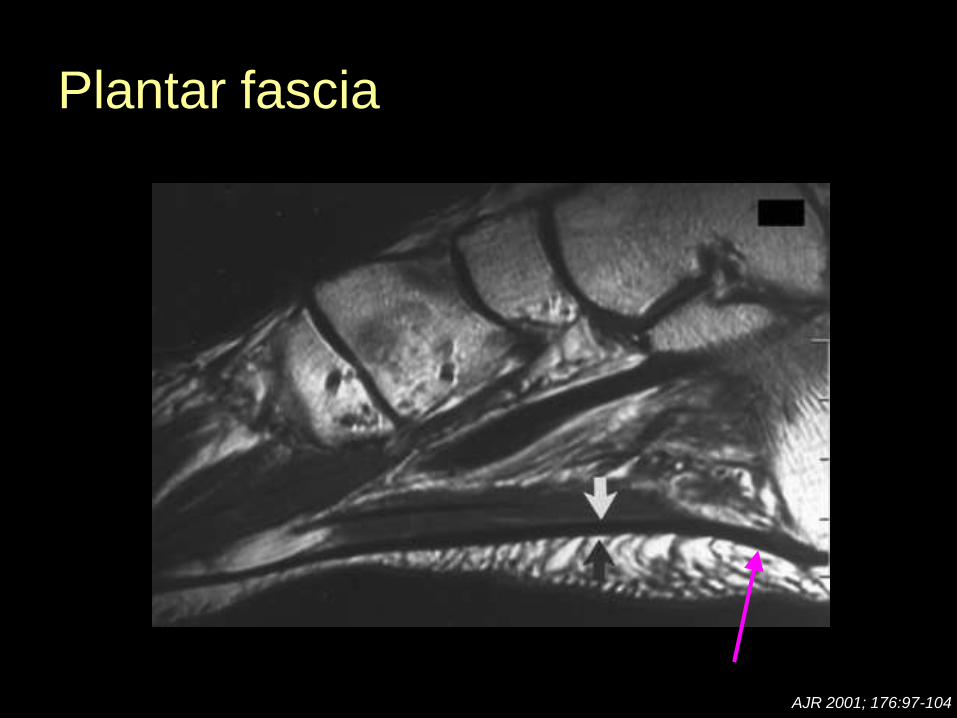
Plantar fascia
AJR 2001; 176:97-104
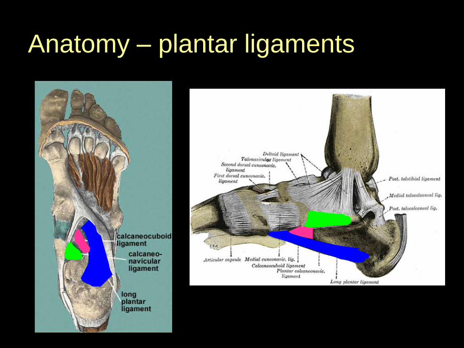
Anatomy – plantar ligaments

MRI Technique
• Extremity surface coil
• Small FOV (12 cm)
• Slight plantar flexion
• T1 sequence in 1 plane
• Fluid sensitive sequence in
all 3 planes
Radiographics. 2000;20:S153-S179
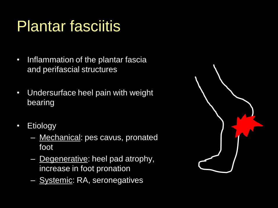
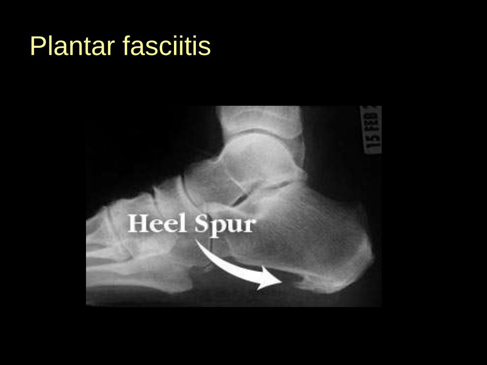
Plantar fasciitis
• Inflammation of the plantar fascia
and perifascial structures
• Undersurface heel pain with weight
bearing
• Etiology
– Mechanical: pes cavus, pronated
foot
– Degenerative: heel pad atrophy,
increase in foot pronation
– Systemic: RA, seronegatives
Plantar fasciitis
Plantar fasciitis
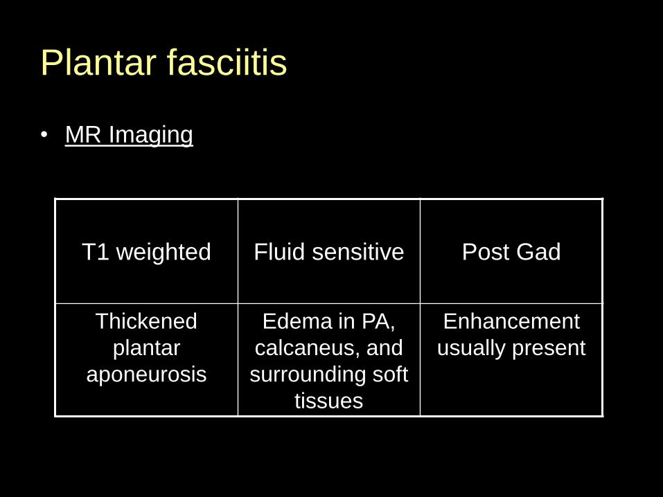
• MR Imaging
T1 weighted Fluid sensitive Post Gad
Thickened
plantar
aponeurosis
Edema in PA,
calcaneus, and
surrounding soft
tissues
Enhancement
usually present
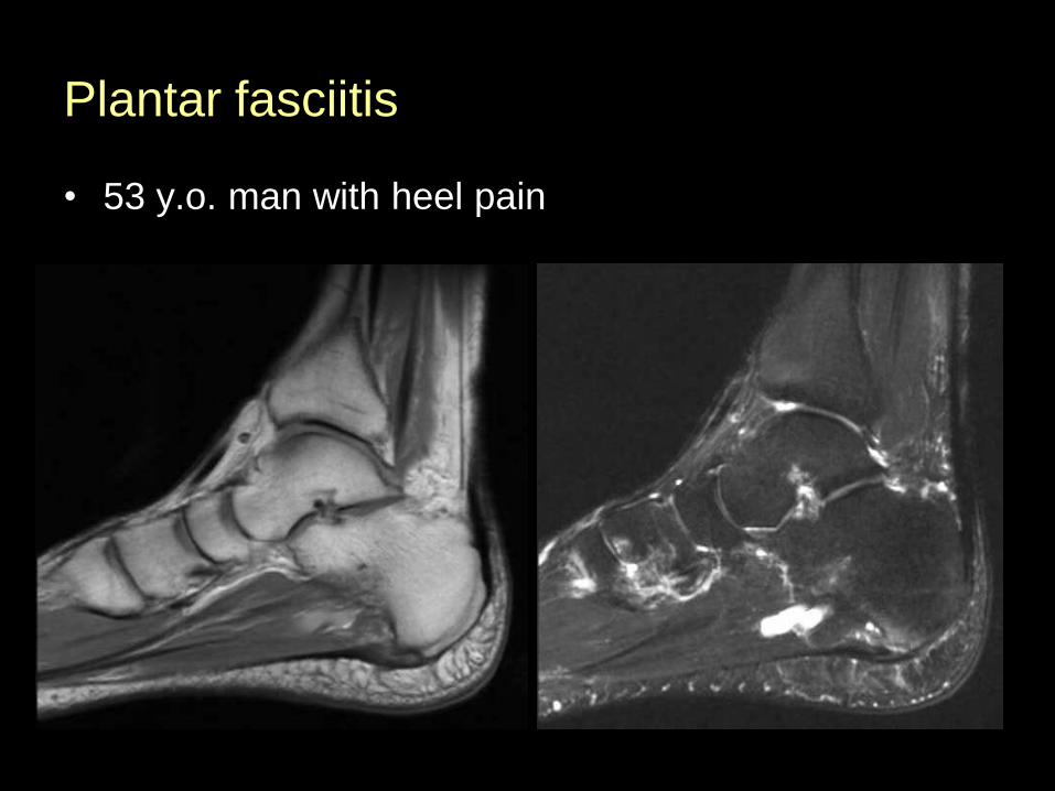
Plantar fasciitis
• 53 y.o. man with heel pain

Plantar fasciitis
• 40 y.o. man with heel pain
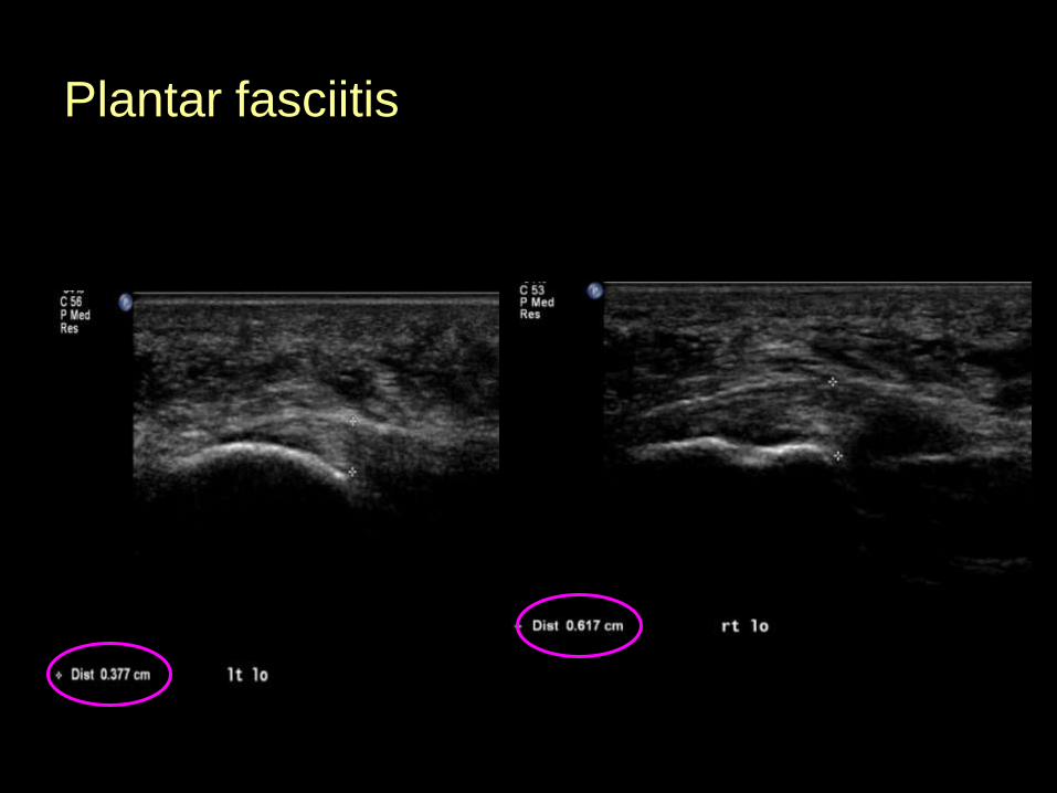
Plantar fasciitis
Plantar fasciitis
• Treatment
• Conservative: Most often successful
(rest, stretching & strengthening, orthotics,
anti-inflammatories)
• Local corticosteroid injections, ESWT
• Surgery: plantar fascial release, open or
endoscopic
- 50-80% of the plantar fascia transected medially
- successful in 70-80% of pts.
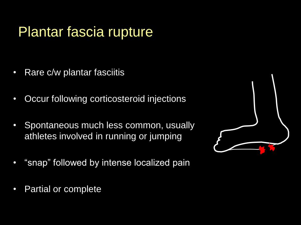
Plantar fascia rupture
• Rare c/w plantar fasciitis
• Occur following corticosteroid injections
• Spontaneous much less common, usually
athletes involved in running or jumping
• “snap” followed by intense localized pain
• Partial or complete

Plantar fascia rupture
• MR Imaging
• Gap in plantar fascia with edema/fraying of the torn
ends
• Edema in adjacent plantar musculature
• Partial rupture may be difficult to distinguish from
fasciitis on imaging. Clinical history helpful.
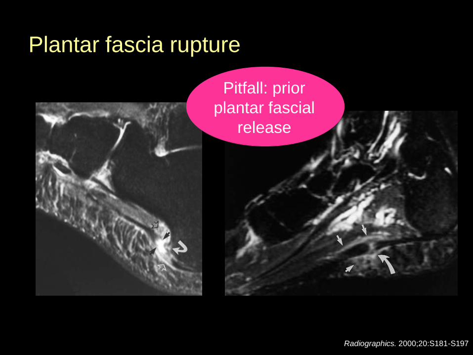
Plantar fascia rupture
Radiographics. 2000;20:S181-S197
Pitfall: prior
plantar fascial
release
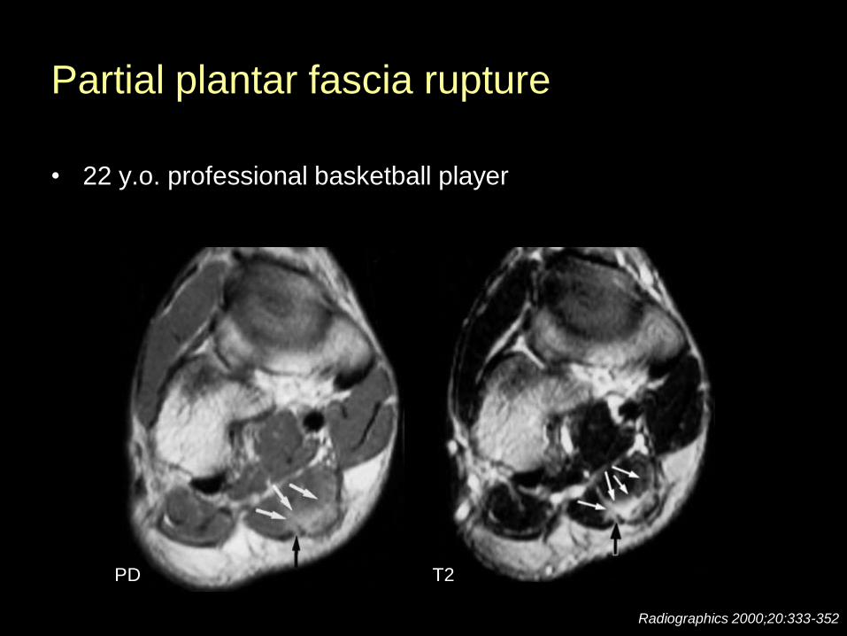
Partial plantar fascia rupture
• 22 y.o. professional basketball player
Radiographics 2000;20:333-352
PD T2

Plantar fascia rupture
Plantar fascia rupture
• Treatment
• Conservative: rest, boot brace followed by stiff sole
athletic shoe, physical therapy
• Surgery: plantar fascia release with resection of
scar tissue, calcaneal osteotomy, lengthening of the
lateral column of the foot
Plantar fibromatosis
• Originally described by Georg Ledderhose in 1897. “Ledderhose’s disease”
• Non-neoplastic process – fibrous proliferation and replacement of portions of the PA with abnormal fibrous tissue
• Typically involve the medial and central portions
• Solitary or multiple / unilateral or bilateral
• Possible association with Dupuytren’s contractures and Peyronie’s disease
Plantar fibromatosis
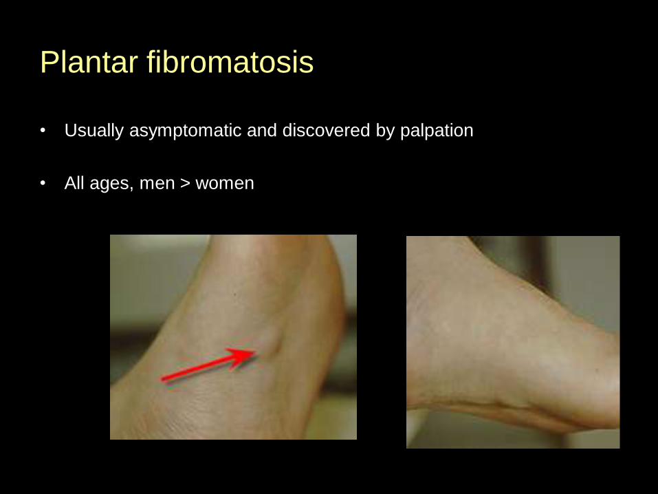
• Usually asymptomatic and discovered by palpation
• All ages, men > women
Plantar fibromatosis
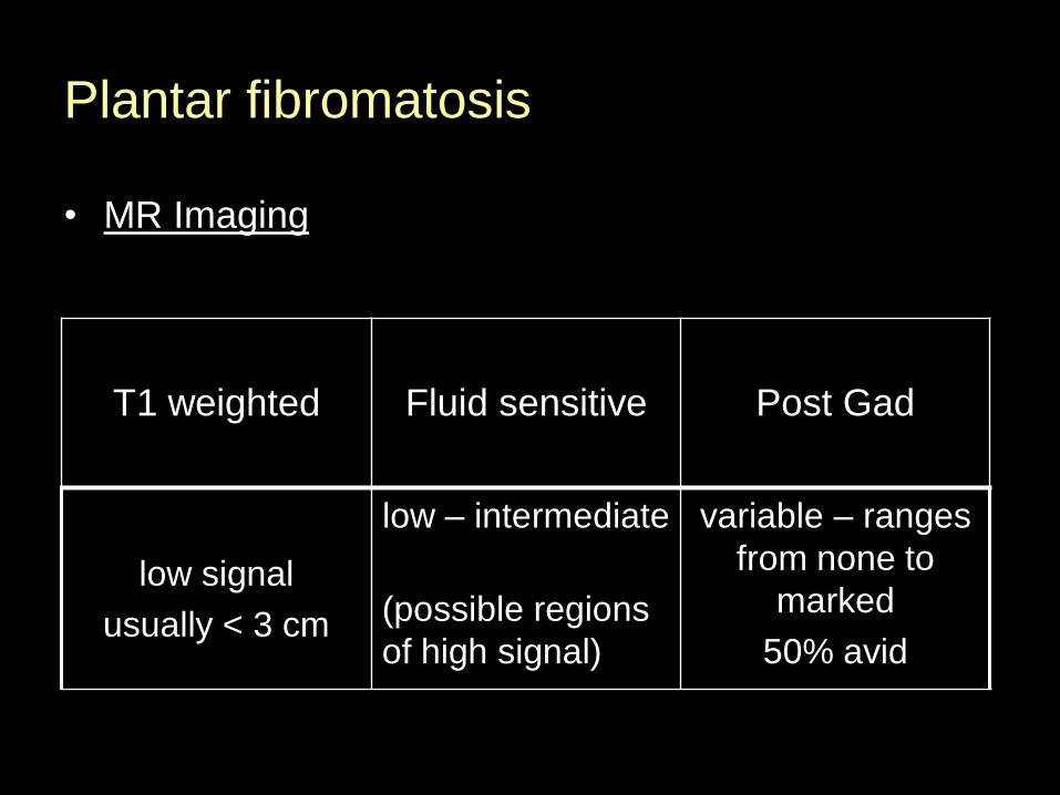
• MR Imaging
T1 weighted Fluid sensitive Post Gad
low signal
usually < 3 cm
low – intermediate
(possible regions
of high signal)
variable – ranges
from none to
marked
50% avid
Plantar fibromatosis
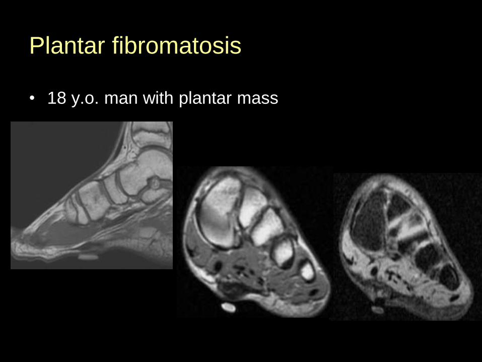
• 18 y.o. man with plantar mass
Plantar fibromatosis
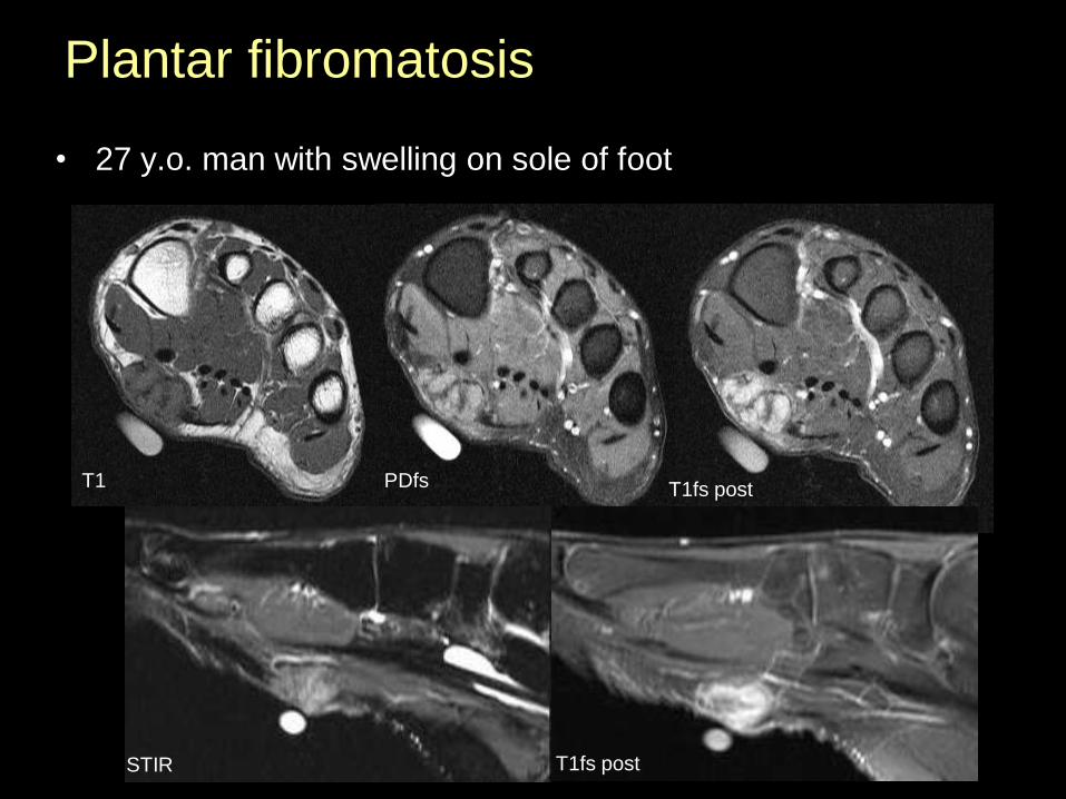
• 27 y.o. man with swelling on sole of foot
T1
T1fs postSTIR
T1fs postPDfs
Plantar fibromatosis
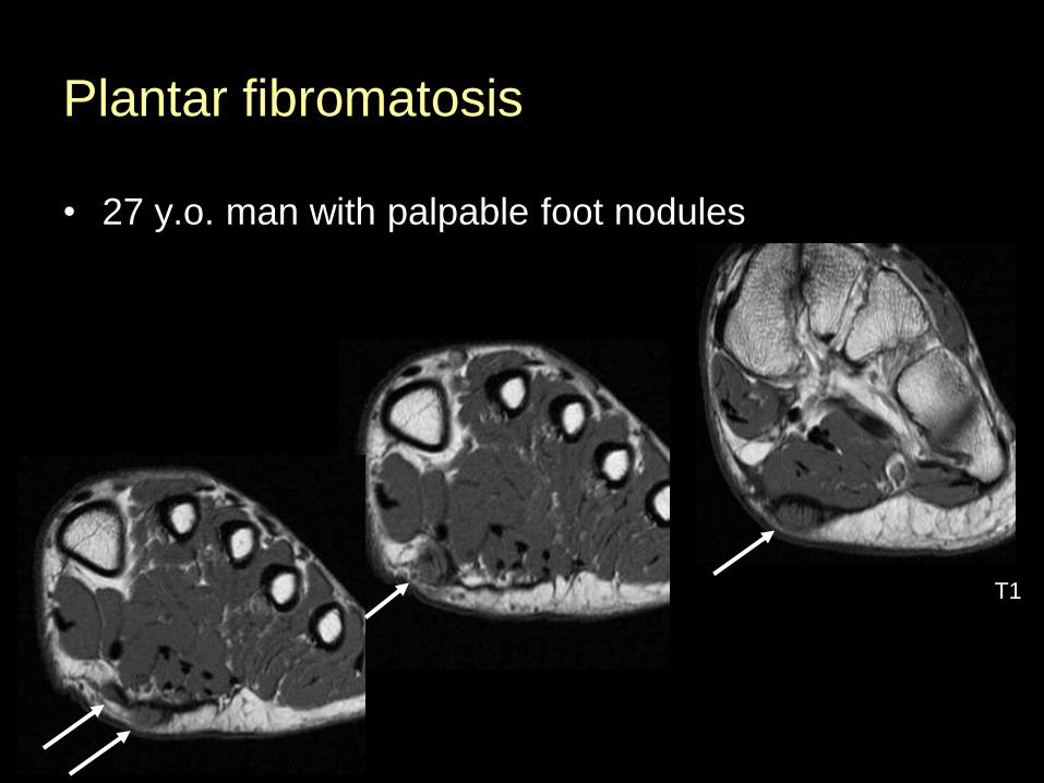
• 27 y.o. man with palpable foot nodules
T1
Plantar fibromatosis
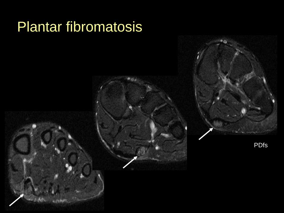
PDfs

Plantar fibromatosis
T1fs post
Plantar fibromatosis
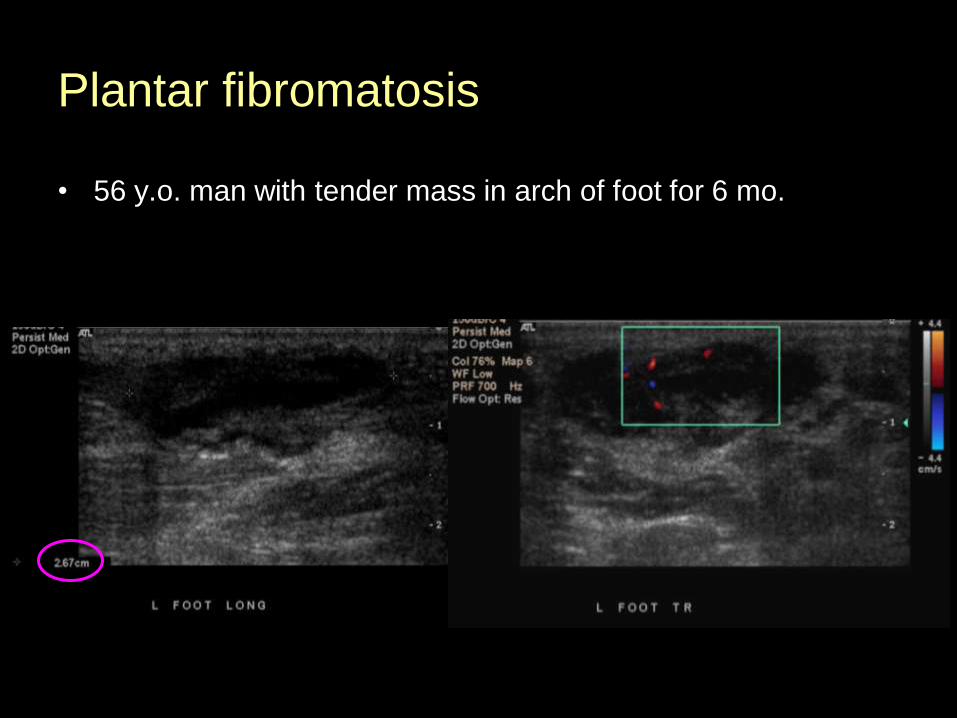
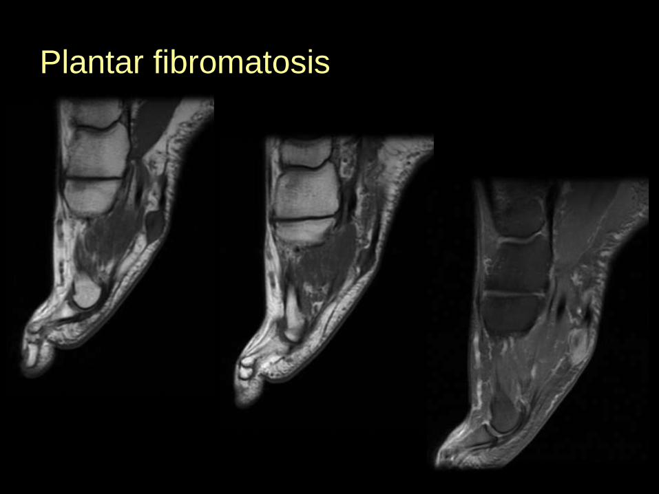
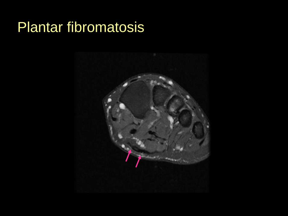
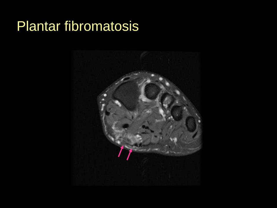
• 56 y.o. man with tender mass in arch of foot for 6 mo.
Plantar fibromatosis
Plantar fibromatosis
Plantar fibromatosis
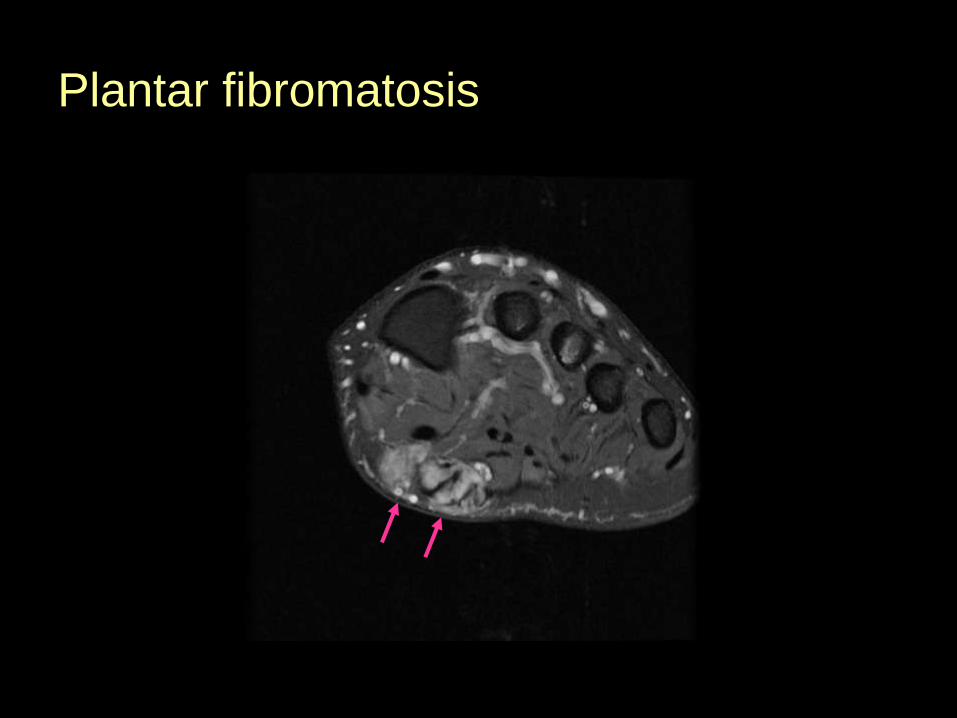
Plantar fibromatosis
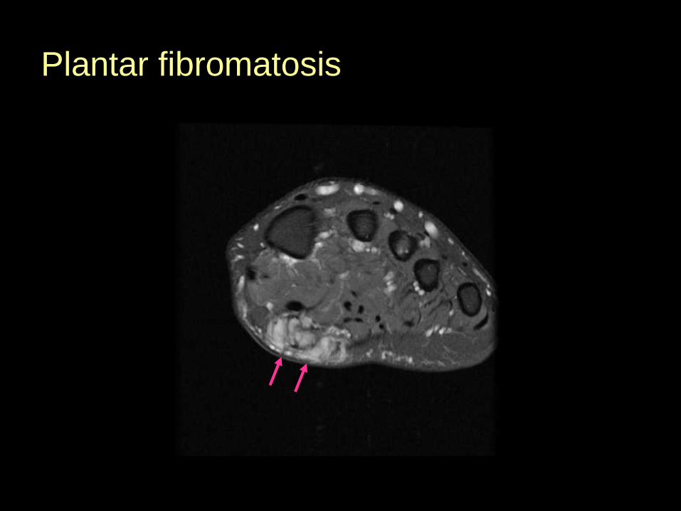
Plantar fibromatosis
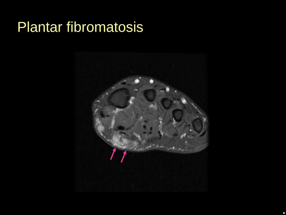
Plantar fibromatosis
.

Plantar fibromatosis
• 45 y.o. woman with pain and focal pea-sized bump
on bottom of foot
.
Plantar fibromatosis
• Treatment
• Conservative: orthopedic footwear
• Surgery: local excision
- high rate of post-surgical recurrence
- adjunctive XRT sometimes used to prevent local
recurrence

Is this a plantar fibroma?
T1 T2
Plantar
fascia
xanthoma
Radiographics 2000;20:333-352
Plantar fascia xanthoma
• Usually bilateral and symmetric
• Dorsum of hands > Achilles > Plantar fascia
• Focal aponeurotic enlargement with heterogeneous
signal intensity

Halifax, Nova Scotia, Canada

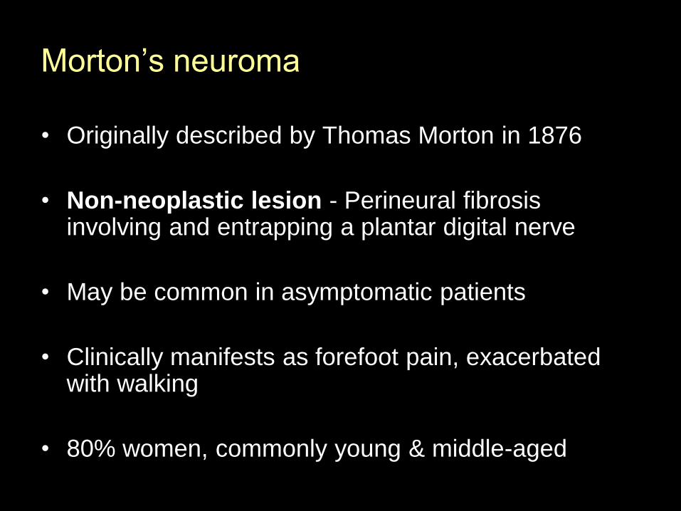
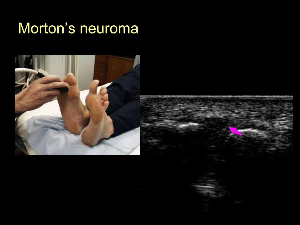
Morton’s neuroma
• Originally described by Thomas Morton in 1876
• Non-neoplastic lesion - Perineural fibrosis involving and entrapping a plantar digital nerve
• May be common in asymptomatic patients
• Clinically manifests as forefoot pain, exacerbated with walking
• 80% women, commonly young & middle-aged
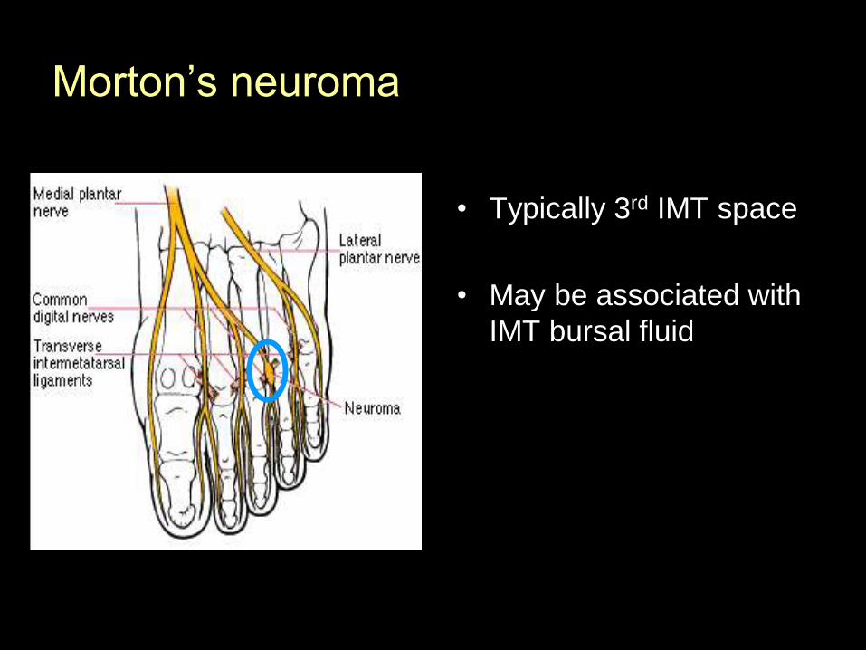
Morton’s neuroma
• Typically 3rd IMT space
• May be associated with
IMT bursal fluid
Morton’s neuroma
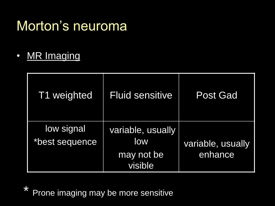
• MR Imaging
T1 weighted Fluid sensitive Post Gad
low signal
*best sequence
variable, usually
low
may not be
visible
variable, usually
enhance
* Prone imaging may be more sensitive
Morton’s neuroma
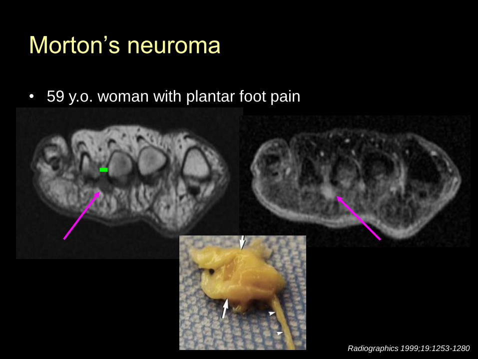
Radiographics 1999;19:1253-1280
• 59 y.o. woman with plantar foot pain
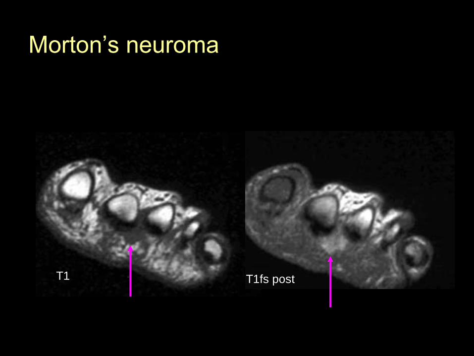
Morton’s neuroma
T1 T1fs post
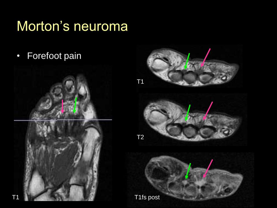
Morton’s neuroma
• Forefoot pain
T1
T2
T1fs postT1
Morton’s neuroma
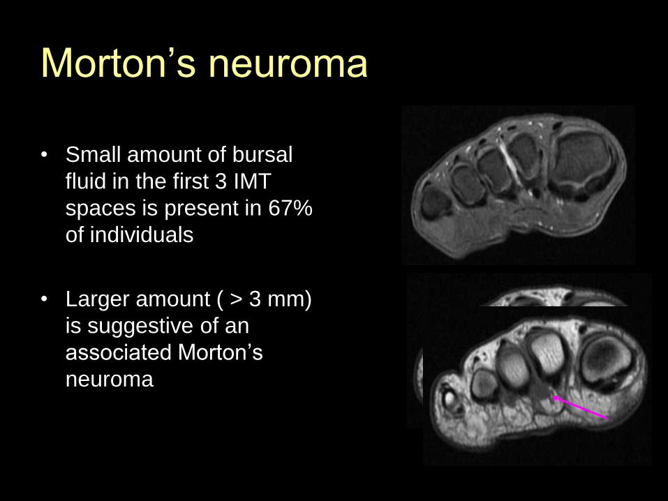
• Small amount of bursal
fluid in the first 3 IMT
spaces is present in 67%
of individuals
• Larger amount ( > 3 mm)
is suggestive of an
associated Morton’s
neuroma
Morton’s neuroma

Morton’s neuroma
• Treatment
• Conservative: footwear modification, neuroma pads
• Steroid injection, ultrasound therapy
• Surgery: release of the IMT ligament for decompression, surgical resection of the neuroma with the involved nerve segment
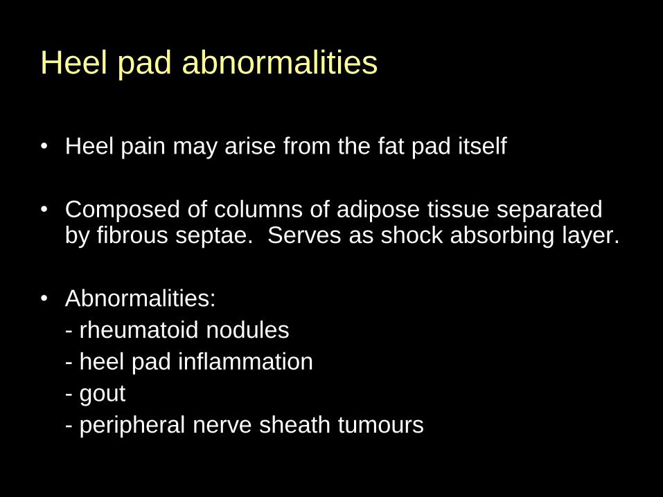
Heel pad abnormalities
• Heel pain may arise from the fat pad itself
• Composed of columns of adipose tissue separated by fibrous septae. Serves as shock absorbing layer.
• Abnormalities:
- rheumatoid nodules
- heel pad inflammation
- gout
- peripheral nerve sheath tumours

Rheumatoid nodules
• Affect 20-30% of patients with RA, rarely affect
seronegative pts.
• Occur in areas subjected to repetitive minor
trauma – areas overlying osseous prominences
• May be painful
• May break down and get infected
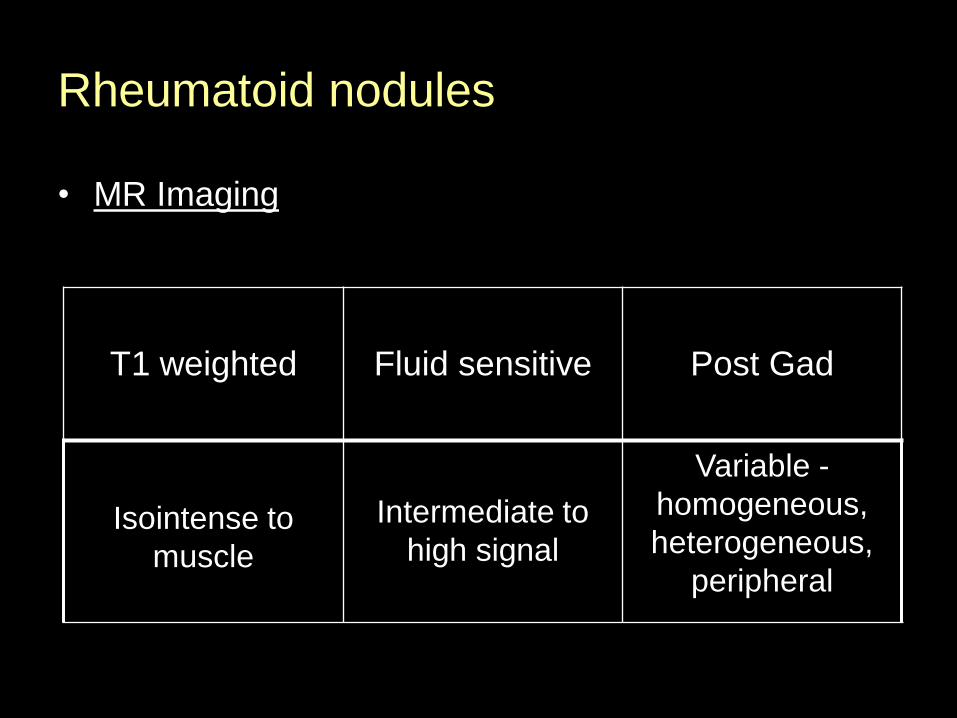
Rheumatoid nodules
• MR Imaging
T1 weighted Fluid sensitive Post Gad
Isointense to
muscle
Intermediate to
high signal
Variable -
homogeneous,
heterogeneous,
peripheral
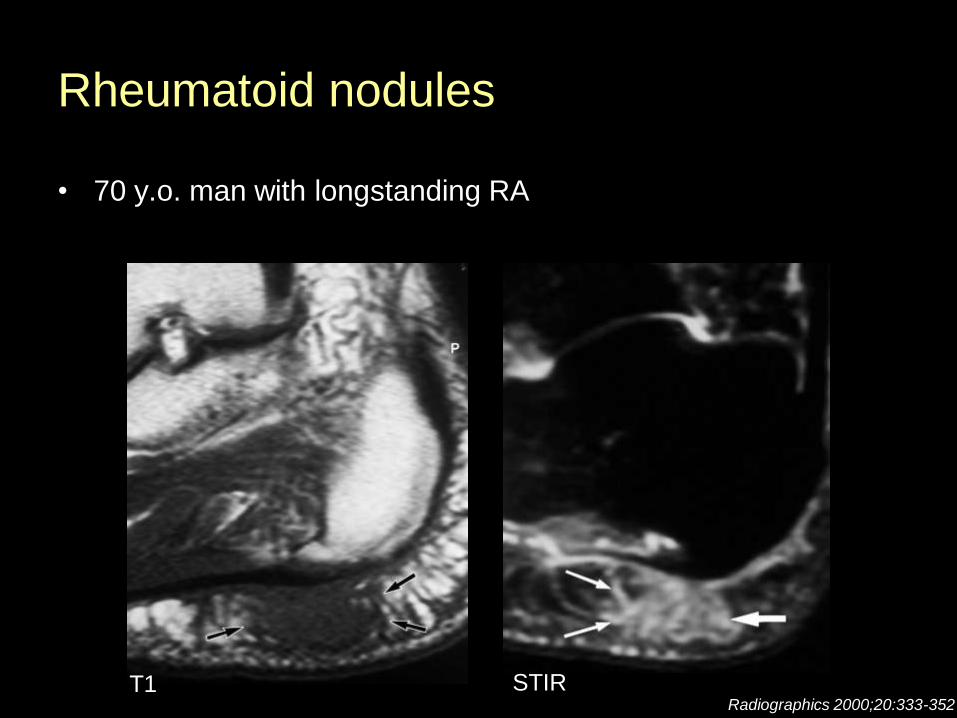
Rheumatoid nodules
• 70 y.o. man with longstanding RA
Radiographics 2000;20:333-352T1 STIR

Rheumatoid nodules
• 45 y.o. woman with RA and heel swelling
T1 T1fs post
AJR 2001; 176: 97-104
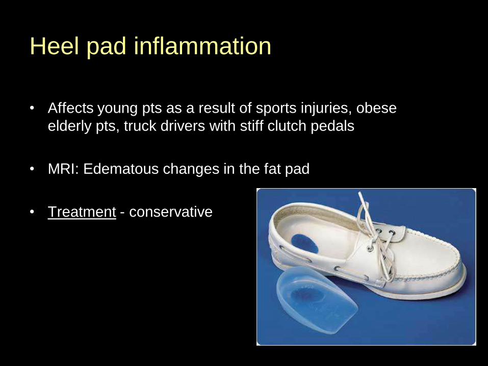
Heel pad inflammation
• Affects young pts as a result of sports injuries, obese
elderly pts, truck drivers with stiff clutch pedals
• MRI: Edematous changes in the fat pad
• Treatment - conservative
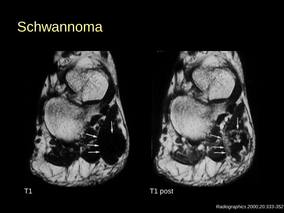
Schwannoma
Radiographics 2000;20:333-352
T1 T1 post
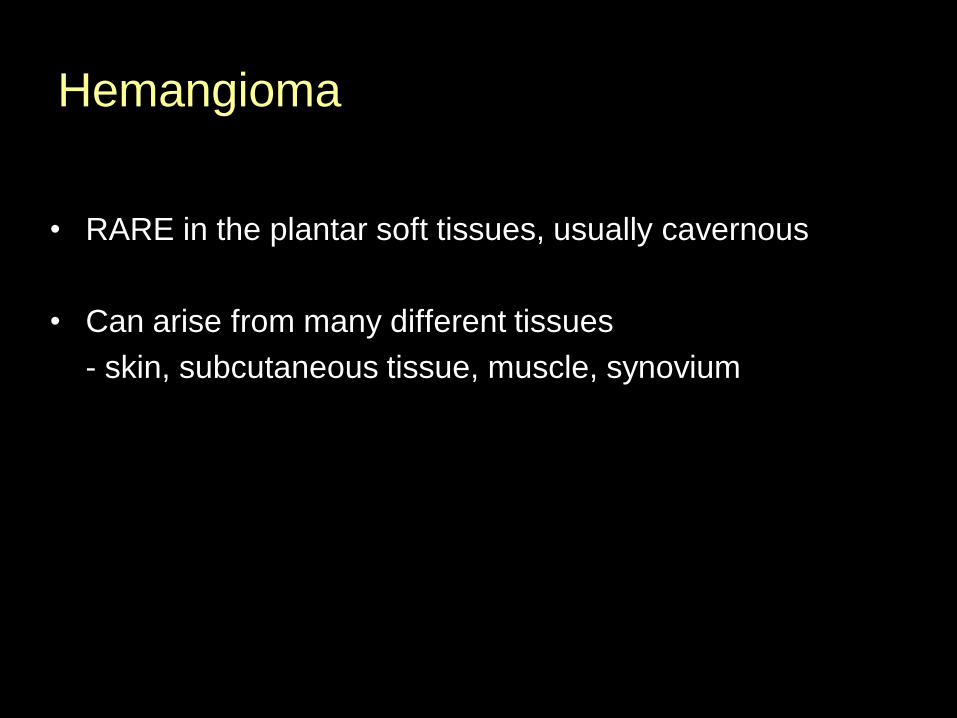
Hemangioma
• RARE in the plantar soft tissues, usually cavernous
• Can arise from many different tissues
- skin, subcutaneous tissue, muscle, synovium

Hemangioma
• MR Imaging
T1 weighted Fluid sensitive Post Gad
low to
intermediate
variable
amounts of high
signal fat
50% have
phleboliths
high signal
septations due
to vascular
channels or
fibrous septae
marked
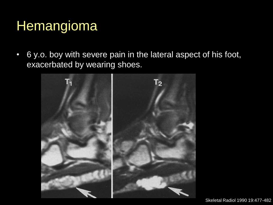
Hemangioma
• 6 y.o. boy with severe pain in the lateral aspect of his foot,
exacerbated by wearing shoes.
Skeletal Radiol 1990 19:477-482
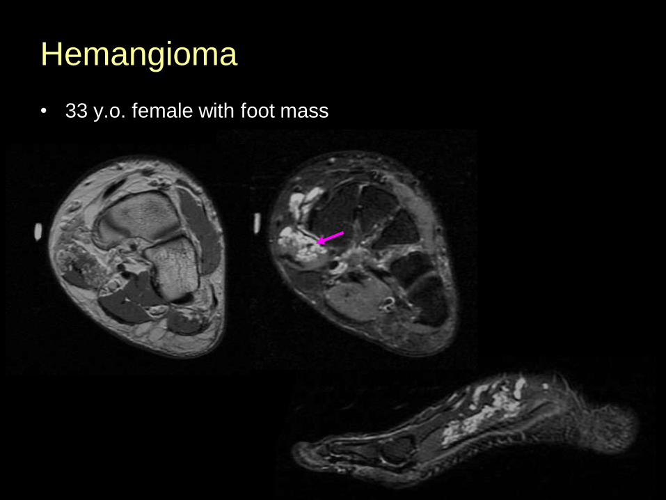
Hemangioma
• 33 y.o. female with foot mass
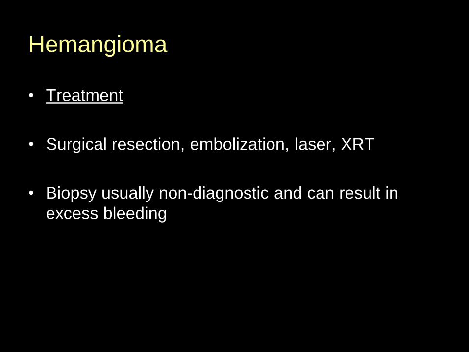
Hemangioma
• Treatment
• Surgical resection, embolization, laser, XRT
• Biopsy usually non-diagnostic and can result in
excess bleeding

Malignancies of the Plantar Soft Tissues
• RARE but they do occur
• Sarcomas – synovial sarcoma, dermatofibrosarcoma
• Synovial sarcomas
- can remain quiescent for long periods of time
- can be relatively small
- can have well-defined margins and homogeneous
appearance
J Bone Joint Surg Am. 1989; 71: 621-626
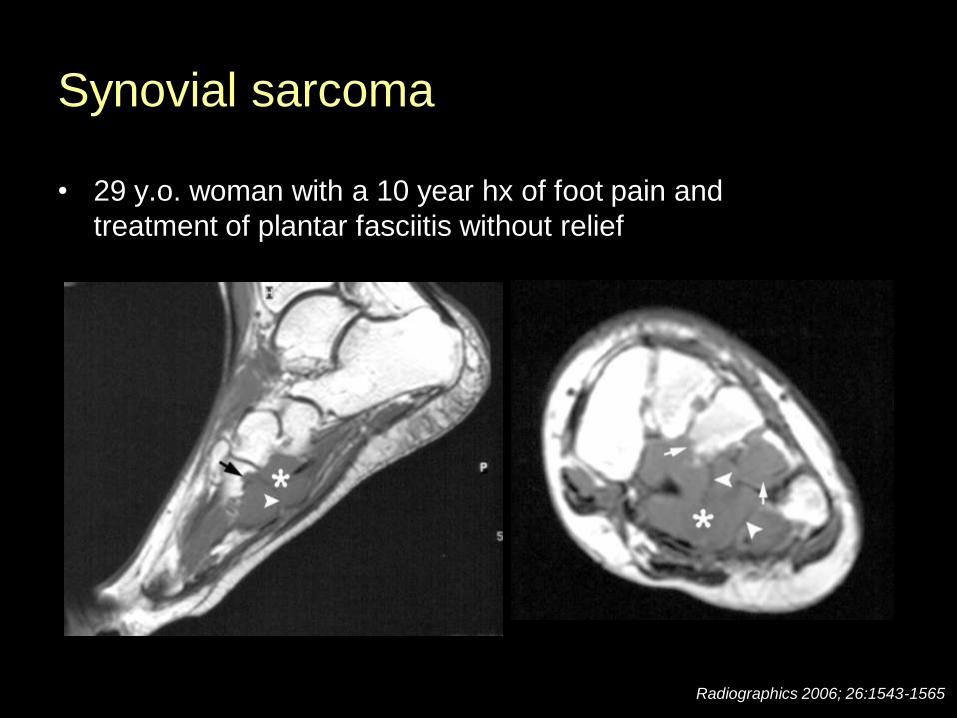
Synovial sarcoma
• 29 y.o. woman with a 10 year hx of foot pain and
treatment of plantar fasciitis without relief
Radiographics 2006; 26:1543-1565

TRIVIA
• What native of Halifax was nominated for Best
Actress at the 2008 Oscars?
Ellen Page
“Juno”

• Plantar ligament abnormalities
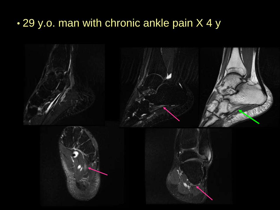
• 29 y.o. man with chronic ankle pain X 4 y
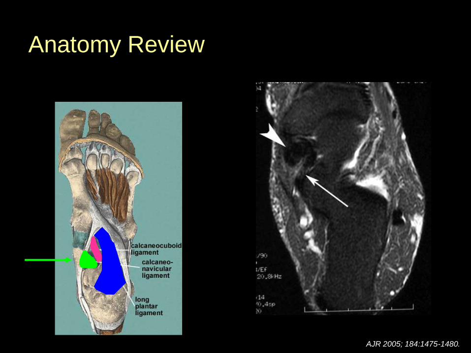
Anatomy Review
AJR 2005; 184:1475-1480.
Spring ligament tears
• Typically a chronic degenerative process that occurs with TPT insufficiency
• The larger superomedial component is the greater contributor to hindfoot stability
• MR findings
- abN calibre of the spring ligament
- increased signal on fluid sensitive sequences
- full thickness gap or wavy appearance
- abN TPT
• Tear of the spring ligament warrants surgical repair
AJR 2005; 184:1475-1480.

Spring ligament tear
AJR 2005; 184:1475-1480.
Take Home Points
• When performing MRI of the foot, use a small FOV centered over the region of interest with a skin marker
• Reactive non-tumoral lesions are the most common abnormalities
• Malignant tumours are very rare but they do occur
- Synovial sarcomas can remain quiescent for long periods of time and can have a non-aggressive appearance
• Evaluate the plantar ligaments
References• Plantar Fasciitis and Fascial Rupture: MR Imaging Findings in 26 Patients Supplemented with Anatomic Data in Cadavers.
DJ Theodorou, SJ Theodorou, Y Kakitsubata, N Lektrakul, GE Gold, B Roger and D Resnick. Radiographics 2000;20:S181-S197.
• MR Imaging of Benign Soft-Tissue Masses of the Foot and Ankle. J Llauger, J Palmer, JM Monill, T Franquet, S Bague, N Roson. Radiographics 1998;18(6): 1481.
• MRI of Plantar Fasciitis. B Roger, Ph Grenier. Eur. Radiol. 7, 1430-1435 (1997)
• Disorders of the Plantar Aponeurosis – A Spectrum of MR Imaging Findings. DJ Theodorou, SJ Theodorou, S Farooki, Y Kakitsubata, D Resnick. AJR 2001; 176:97-104
• Imaging of Musculoskeletal Neurogenic Tumors: Radiologic-Pathologic Correlation. MD Murphey, WS Smith, SE Smith, MJ Kransdorf, T Temple. Radiographics 1999;19:1253-1280
• MR Imaging of the Ankle and Foot. ZS Rosenbert, J Beltran, JT Bencardino. Radiographics 2000; 20:S153-S179
• Soft Tissue Tumors and Tumor-like Lesions of the Foot. An analysis of eighty-three cases. EJ Kirby, MJ Shereff, MM Lewis. J Bone Joint Surg Am 1989; 71: 621-626.
• Imaging of Synovial Sarcoma with Radiologic-Pathologic Correlation. MD Murphey, MS Gibson, BT Jennings, AM Crespo-Rodriguez, J Fanburg-Smith, DA Gajewski. Radiographics 2006; 26:1543-1565.
• Painful Heel: MR Imaging Findings. JA Narvaez, J Narvaez, R Ortega, C Aguilera, A Sanchez, E Andia. Radiographics 2000;20:333-352.
• Magnetic resonance imaging of peripheral soft tissue hemangiomas. MC Nelson, MA Stull, GP Teitelbaum, RH Patt, EE Lack, GP Bogumill, MT Freedman. Skeltal Radiol 1990; 19:477-482.
• MRI of Spring Ligament Tears. LR Toye, CA Helms, BD Hoffman, M Easley, JA Nunley. AJR 2005; 184:1475-1480.