Abdominal MR in Pregnancy - scbtmr.org MR... · 3 MR criteria for appendicitis C C 13 wks Mild...
5
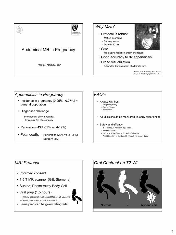
1 Abdominal MR in Pregnancy Neil M. Rofsky, MD A major teaching hospital of Harvard Medical School Why MRI? • Protocol is robust – Motion insensitive – Std sequences – Done in 20 min • Safe – No ionizing radiation (mom and fetus!) • Good accuracy to dx appendicitis • Broad visualization – Allows for demonstration of alternate dx’s Pedrosa, et al. Radiology 2009; 250:749 Oto, et al. Abd Imaging 2009: 34:243 Appendicitis in Pregnancy • Incidence in pregnancy (0.05% - 0.07%) = general population • Diagnostic challenge – displacement of the appendix – Physiologic ∆’s of pregnancy • Perforation (43%-55% vs. 4-19%) • Fetal death: - Perforation (20% vs 2 -3 %) - Surgery (3%) FAQ’s • Always US first! – Ectopic pregnancy – Ovarian Torsion – Appendicitis • All MR’s should be monitored (in early experience) • Safety and efficacy – 1.5 Tesla (Do not scan @ 3 Tesla) – NO Gadolinium – No harm to the fetus in 2 nd and 3 rd trimester – First trimester → risk-benefit (though no known risks) MRI Protocol • Informed consent • 1.5 T MR scanner (GE, Siemens) • Supine, Phase Array Body Coil • Oral prep (1.5 hours) – 300 mL Gastromark (Mallinckrodt Medical, St. Louis, MO) – 300 mL Readi-cat 2 (EZEM, Westbury, NY) • Same prep can be given retrograde Oral Contrast on T2-WI ? Appendicitis Normal
-
Upload
truongquynh -
Category
Documents
-
view
214 -
download
0
Transcript of Abdominal MR in Pregnancy - scbtmr.org MR... · 3 MR criteria for appendicitis C C 13 wks Mild...
1
Abdominal MR in Pregnancy
Neil M. Rofsky, MD
A major teaching hospital of Harvard Medical School
Why MRI?
• Protocol is robust– Motion insensitive– Std sequences– Done in 20 min
• Safe– No ionizing radiation (mom and fetus!)
• Good accuracy to dx appendicitis • Broad visualization
– Allows for demonstration of alternate dx’s
Pedrosa, et al. Radiology 2009; 250:749Oto, et al. Abd Imaging 2009: 34:243
Appendicitis in Pregnancy• Incidence in pregnancy (0.05% - 0.07%) =
general population
• Diagnostic challenge
– displacement of the appendix – Physiologic ∆’s of pregnancy
• Perforation (43%-55% vs. 4-19%)
• Fetal death: - Perforation (20% vs 2 -3 %)- Surgery (3%)
FAQ’s• Always US first!
– Ectopic pregnancy– Ovarian Torsion– Appendicitis
• All MR’s should be monitored (in early experience)
• Safety and efficacy– 1.5 Tesla (Do not scan @ 3 Tesla)– NO Gadolinium– No harm to the fetus in 2nd and 3rd trimester– First trimester → risk-benefit (though no known risks)
MRI Protocol
• Informed consent
• 1.5 T MR scanner (GE, Siemens)
• Supine, Phase Array Body Coil
• Oral prep (1.5 hours)– 300 mL Gastromark (Mallinckrodt Medical, St. Louis, MO)
– 300 mL Readi-cat 2 (EZEM, Westbury, NY)
• Same prep can be given retrograde
Oral Contrast on T2-WI
? AppendicitisNormal
2
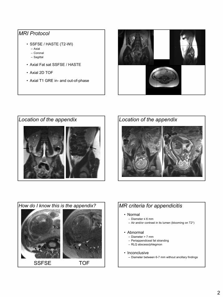
MRI Protocol
• SSFSE / HASTE (T2-WI)– Axial– Coronal– Sagittal
• Axial Fat sat SSFSE / HASTE
• Axial 2D TOF
• Axial T1 GRE in- and out-of-phase
Location of the appendix Location of the appendix
How do I know this is the appendix?
SSFSE TOF
MR criteria for appendicitis• Normal
– Diameter ≤ 6 mm– Air and/or contrast in its lumen (blooming on T2*)
• Abnormal– Diameter > 7 mm– Periappendiceal fat stranding– RLQ abscess/phlegmon
• Inconclusive– Diameter between 6-7 mm without ancillary findings

3
MR criteria for appendicitis
CC
13 wks
Mild Appendicitis (US “Negative”)
Acute appendicitis
C
C
“Ruling-out” Acute Appendicitis
•• MR MR → normal appendixnormal appendix seen:
89% (20/23) Oto et al. Radiology 2005
88% (105/119) Pedrosa I et al. RSNA 2006
• US → normal appendix typically not seenNegative Result = Ambiguous resultAmbiguous result
Alternative diagnoses
• Fibroid degeneration
• Ovarian torsion
• Right hydronephrosis
• Gas in urinary bladder 2ry to infection
• Right ovarian vein varices
U*
Fibroid “red” degeneration
4
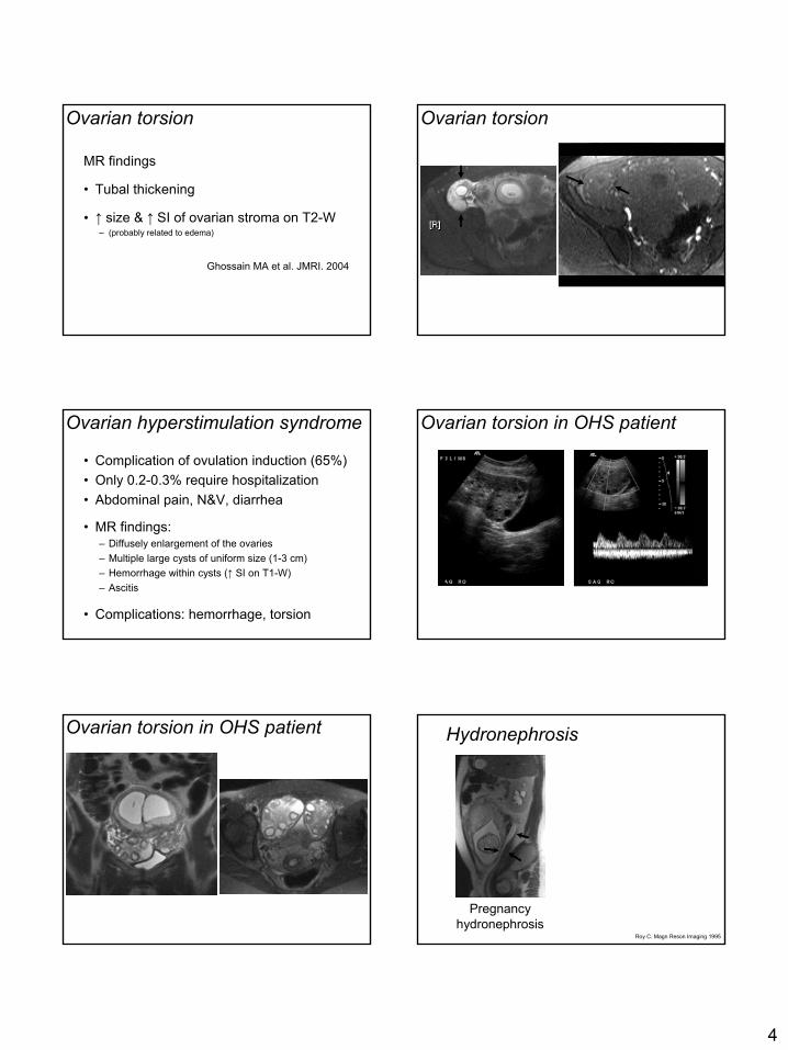
Ovarian torsion
MR findings
• Tubal thickening
• ↑ size & ↑ SI of ovarian stroma on T2-W– (probably related to edema)
Ghossain MA et al. JMRI. 2004
Ovarian torsion
Ovarian hyperstimulation syndrome
• Complication of ovulation induction (65%)• Only 0.2-0.3% require hospitalization • Abdominal pain, N&V, diarrhea
• MR findings:– Diffusely enlargement of the ovaries– Multiple large cysts of uniform size (1-3 cm)– Hemorrhage within cysts (↑ SI on T1-W)– Ascitis
• Complications: hemorrhage, torsion
Ovarian torsion in OHS patient
Ovarian torsion in OHS patient Hydronephrosis
Pregnancy hydronephrosis
Roy C. Magn Reson Imaging 1995

5
Ureteral Stone Pseudo-stone
Urinary bladder infection
32 wks; pain and ↑ WBCUS and urine dip stick neg
Right gonadal vein Right gonadal vein
Gastrointestinal Tract
Crohns Flare
Conclusion
• Prevalence of acute appendicitis among pregnant women with abdominal pain is extremely low.
• Identification of a normal appendix excludes acute appendicitis
• MR can avoid unnecessary radiation with an excellent NPV, diagnose acute appendicitis, and identify other sources of abdominal pain