Abdominal Aortic Aneurysm (AAA) - GIPHY · e incidence of abdominal aortic aneurysm (AAA) has been...
33
Abdominal Aortic Aneurysm (AAA) Introduction: e incidence of abdominal aortic aneurysm (AAA) has been reported as high as 36.2 per 100,000 and is increasing. 1 Up to 62% of patients with ruptured aneurysms die before reaching the hospital and the overall mortality rate aer rupture may exceed 90%. 2 An AAA should be suspected in any patient > 60 years with complaints of abdominal or back pain, particularly if they have a history of hypertension or smoking. Ruptured AAA may present as undifferentiated hypotension and rapid diagnosis is essential. Indications: • Clinical suspicion for AAA including abdominal pain, back pain, “renal colic”, syncope, hypotension, weakness or neurologic changes in the extremities, especially if > 60 years old. Probe selection: • Curved array general abdominal probe 2-5 MHz (preferable) • Phased array probe 1-5 MHz Relevant Anatomy: • Externally, the abdominal aorta enters through the diaphragm just inferior to the xiphoid process (T12) and bifurcates around the level of the umbilicus (L5). • Internally, the abdominal aorta has a predictable series of branches. Figure 1. • e 1 st branch off the abdominal aorta is the celiac trunk. o e celiac trunk divides into three vessels however typically only the hepatic artery and splenic artery are visualized, creating the “seagull sign”. • e 2 nd branch off the abdominal aorta is the superior mesenteric artery (SMA); typically 1cm caudal to the celiac trunk. o e SMA runs parallel with the abdominal aorta and can be tracked caudally with the abdominal aorta. • e branches of the renal, gonadal and inferior mesenteric arteries are not typically seen with ultrasound. • e abdominal aorta bifurcates into the common iliac vessels. Technique: • Start with probe in the 9 o’clock transverse (TRV) position immediately caudad to the xiphoid process. Note the external anatomy as you scan in transverse (proximal to distal) and longitudinal planes. Figures 2-3 and Figures 4-5. • Identify the vertebral body. o e vertebral body is horseshoe-shaped with an intense echogenic anterior surface and posterior shadowing. Figure 6. • Adjust depth if too deep or too shallow.
Transcript of Abdominal Aortic Aneurysm (AAA) - GIPHY · e incidence of abdominal aortic aneurysm (AAA) has been...

Abdominal Aortic Aneurysm (AAA)
Introduction:e incidence of abdominal aortic aneurysm (AAA) has been reported as high as 36.2 per 100,000 and is increasing.1 Up to 62% of patients with ruptured aneurysms die before reaching the hospital and the overall mortality rate aer rupture may exceed 90%.2 An AAA should be suspected in any patient > 60 years with complaints of abdominal or back pain, particularly if they have a history of hypertension or smoking. Ruptured AAA may present as undifferentiated hypotension and rapid diagnosis is essential.
Indications:• Clinical suspicion for AAA including abdominal pain, back pain, “renal colic”, syncope,
hypotension, weakness or neurologic changes in the extremities, especially if > 60 years old.
Probe selection:• Curved array general abdominal probe 2-5 MHz (preferable)• Phased array probe 1-5 MHz
Relevant Anatomy: • Externally, the abdominal aorta enters through the diaphragm just inferior to the xiphoid
process (T12) and bifurcates around the level of the umbilicus (L5).• Internally, the abdominal aorta has a predictable series of branches. Figure 1.• e 1st branch off the abdominal aorta is the celiac trunk.
o e celiac trunk divides into three vessels however typically only the hepatic artery and splenic artery are visualized, creating the “seagull sign”.
• e 2nd branch off the abdominal aorta is the superior mesenteric artery (SMA); typically 1cm caudal to the celiac trunk.
o e SMA runs parallel with the abdominal aorta and can be tracked caudally with the abdominal aorta.
• e branches of the renal, gonadal and inferior mesenteric arteries are not typically seen with ultrasound.
• e abdominal aorta bifurcates into the common iliac vessels.
Technique:• Start with probe in the 9 o’clock transverse (TRV) position immediately caudad to the
xiphoid process. Note the external anatomy as you scan in transverse (proximal to distal) and longitudinal planes. Figures 2-3 and Figures 4-5.
• Identify the vertebral body. o e vertebral body is horseshoe-shaped with an intense echogenic anterior surface
and posterior shadowing. Figure 6.• Adjust depth if too deep or too shallow.

• e aorta is anterior to the vertebral body and on the patient’s le. • e IVC is anterior to the vertebral body, and on the patient’s right. • Scan caudally to the aortic bifurcation with methodical real-time visualization, without
skipped sections. • While scanning cephalad to caudad (head to toe) identify the following:
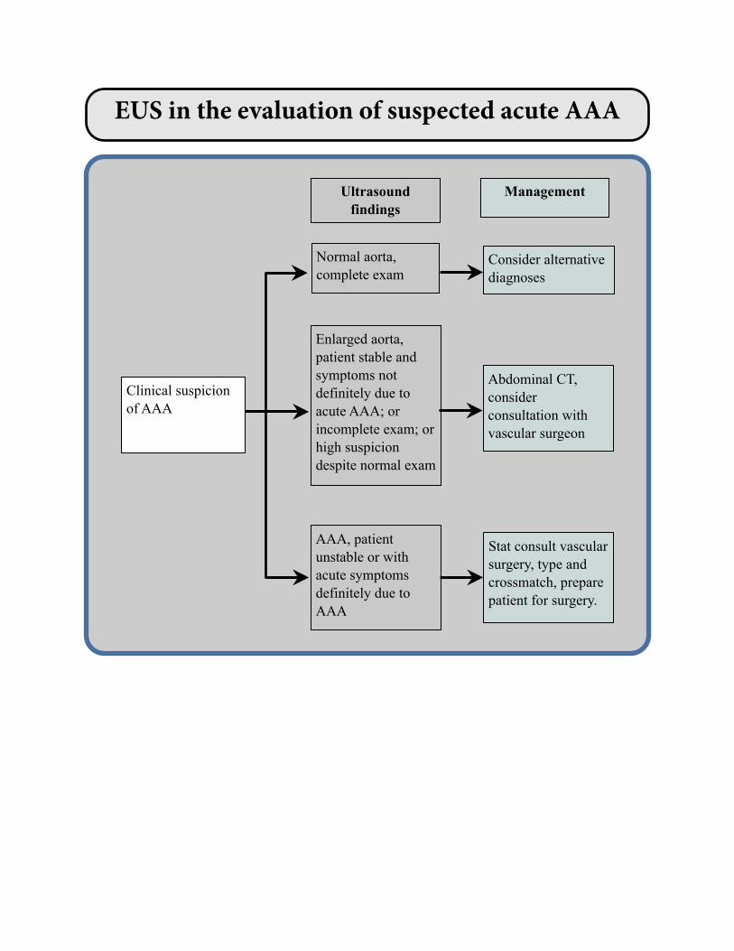
1. Celiac trunk. Figure 7.2. SMA. Figure 8.3. Aortic bifurcation. Figure 9.
• Turn the probe to the longitudinal plane (pointer to 12 o’clock) to obtain a long view of the aorta and the associated vessels (Celiac and SMA). Figure 10.
• e same proximal branches seen in TRV plane should be seen in Long plane. Again, move cephalad to caudad as you scan through the entire length of the vessel.
Impediments / solutions:• Bowel gas and obesity are the most common impediments. • Bowel:
o e transverse colon in the epigastrium is large and oen gas-filled. o If bowel is encountered, apply steady pressure with the probe to the area. e
bowel may be effectively compressed or undergo peristalsis.o “Jiggle” the probe to move bowel aside. o “Fan” through windows between loops of bowel. is is done by finding an open
window just cephalad to the area obscured by bowel and tilting the probe toward the feet; similarly, find a window just caudad to the obstructed area and angle the probe up toward the head.
• Obesity: o Lay the patient completely flat. o Have the patient flex their hips and knees to relax the abdominal muscles.o Lower the frequency to increase penetration.
Normal US Findings: • e normal aorta is <3cm and tapers distally. • Measurements: Figure 11.
o Take measurements at the proximal, mid and distal aorta and document the maximum diameter.
o Measurements should be taken in the transverse plane, to limit the tangential effect of underestimating diameter on a long axis image.
o ALWAYS MEASURE OUTER WALL TO OUTER WALL. A mural thrombus or plaque in the lumen may underestimate the size of the AAA.
Abnormal US Findings: • Any Aortic measurement >3cm is abnormal; any aorta that fails to taper appropriately as
it moves caudally is considered abnormal as well.

• e majority of AAAs are INFRARENAL.3• ere are two types of aneurysm: fusiform and saccular. Figure 12.
o Fusiform is much more common and involves dilation of the entire circumference of the affected segment of the aorta. Figures 13-17.
o A saccular aneurysm is an asymmetric outpouching (i.e. “sac-like”) of the aorta, and is much less common. Figure 12.
• Aneuryms may extend into the iliac arteries as well, so a comprehensive, methodical scan through the bifurcation is necessary. Figure 18.
• Emergency bedside ultrasound is highly sensitive for the presence of an AAA but has poor sensitivity for acute rupture.4
• AAA’s most commonly rupture into the retroperitoneal space and may tamponade before the patient becomes unstable.
• Ultrasound is insensitive for retroperitoneal blood.• AAA ruptures occasional produce intraperitoneal free fluid which, while readily identified
by ultrasound, is particularly ominous. Figure 19.• Calcifications in the vessel lumen are sometimes seen (particularly in elderly patients) but
rarely cause symptoms. Figure 20. NEED IMAGE!!!!• An Aortic dissection (floating intimal flap) may occasionally be found while performing
an US to rule out AAA. Figures 21-25.
Pearls and pitfalls: • rombus within an AAA can be mistaken for the aortic wall leading to underestimation
of AAA diameter and missed diagnosis. Figure 26.o Adjust the gain so that aortic lumen is black.o Decrease the dynamic range to improve the contrast between vessel wall and
lumen.• Retroperitoneal bleeding is NOT reliably detected by U/S. Figure 27 NEED IMAGE
o US can rule in or out the presence of an AAA, but unless there is intraperitoneal free fluid, it typically gives no information about rupture.
• Saccular aneurysms can be easily missed. o Systematic, continuous scanning in Long and TRV plane are essential to
prevent a false negative diagnosis. • An ectatic aorta may have irregular course. Dynamic scanning adjusting for changes in
vessel angle will allow for complete visualization.• Angled TRV cuts exaggerate the true aortic diameter. Obtain measurements at 90 degrees
to the vessel. • Off-axis (tangential) longitudinal cuts under estimate aortic diameter. Caliper
measurements should be made in TRV view only.• Inexperienced scanners can mistake the IVC for the Aorta, especially in long axis.
o Both the aorta and inferior vena cava (IVC) are pulsatile and cannot reliably differentiate the two. Figure 28.
o e aorta lies to the patient’s le of the IVC.

o e aorta has anterior branches caudad to the liver while the IVC does not. o e aorta is typically rounder, non-compressible, and has brighter, thicker
walls than the IVC. • Small aneurysms (<4.5cm) can rupture, although less frequently than a larger AAA. • If an AAA is identified by US, it still may not be the cause of the patient’s symptoms.
o Consider abdominal, renal or musculoskeletal pathology.• Dissection flaps are easily overlooked. • If there is a high clinical suspicion for AAA and US is equivocal, obtain CT (with contrast
if no renal impairment).
Clinical suspicion of AAA
Normal aorta, complete exam
Consider alternative diagnoses
Enlarged aorta, patient stable and symptoms not definitely due to acute AAA; or incomplete exam; or high suspicion despite normal exam
Abdominal CT, consider consultation with vascular surgeon
AAA, patient unstable or with acute symptoms definitely due to AAA
Stat consult vascular surgery, type and crossmatch, prepare patient for surgery.
ManagementUltrasound findings
EUS in the evaluation of suspected acute AAA

Bibliography
1. Bickerstaff LK, Hollier LH, Van Peenen HJ, Melton LJ, 3rd, Pairolero PC, Cherry KJ. Abdominal aortic aneurysms: the changing natural history. J Vasc Surg 1984;1:6-12.
2. Ernst CB. Abdominal aortic aneurysm. N Engl J Med 1993;328:1167-72.3. Golledge J, Muller J, Daugherty A, Norman P. Abdominal aortic aneurysm: pathogenesis
and implications for management. Arterioscler romb Vasc Biol 2006;26:2605-13.4. Kuhn M, Bonnin RL, Davey MJ, Rowland JL, Langlois SL. Emergency department
ultrasound scanning for abdominal aortic aneurysm: accessible, accurate, and advantageous. Ann Emerg Med 2000;36:219-23.

Figure 1. Abdominal aortic anatomy. Branches that are typically identified are highlighted in blue.

Figures 2 & 3. Transverse aorta HI and MID, with probe indicator to the le of the patient.
2
3

Figures 4 & 5. Transverse aorta LOW, with probe indicator to the le of the patient; longitudinal aorta with probe indicator to the patient’s head (cranial).
4
5

Figure 6. Transverse aorta demonstrating the vertebral body, with an echogenic anterior cortex and posterior shadowing. (Arrowheads=Vertebral body, Ao=Aorta, IVC=Inferior vena cava.)

Figure 7. Transverse aorta at the level of the celiac trunk, demonstrating the “seagull sign”. (*=Aorta, Hep A=Hepatic artery, Spl A=Splenic artery.)

Figure 8. Transverse aorta at the level of the superior mesenteric artery. (V=Vertebral body, Ao=Aorta, SMA=Superior mesenteric artery, Spl=Splenic vein, IVC=Inferior vena cava)
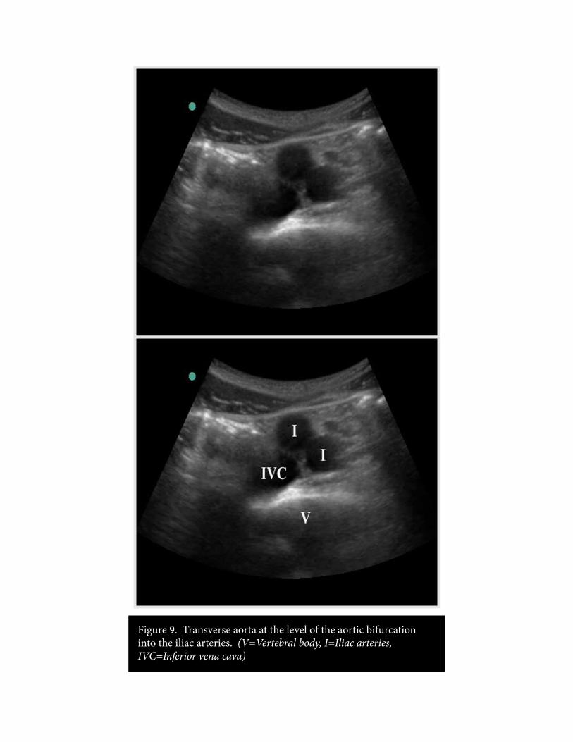
Figure 9. Transverse aorta at the level of the aortic bifurcation into the iliac arteries. (V=Vertebral body, I=Iliac arteries, IVC=Inferior vena cava)

Figure 10. Longitudinal aorta with Celiac trunk and SMA identified.

Figure 11. Transverse aorta with calipers in appropriate anterior/posterior measurement (outside wall to outside wall). Adjacent IVC.

Figure 12. Above: Schematic comparing saccular (le) versus fusiform (right) aneurysms of the abdominal aorta. Below: Ultrasound of the longitudinal aorta with a saccular aneursym (Ao=Aorta, *=Aneurysm)

Figure 13. Above: Transverse aorta with calipers, demonstrating a 3.5cm fusiform aneursym. Below: Longitudinal view of the same aortic aneurysm.

Figure 14. Above: Transverse aorta with arrows, demonstrating a 4 cm aneursym. Below: Longitudinal view of a 5.3 cm aneurysm (*).

Figure 15. Above: Transverse aorta with calipers, demonstrating a 6.7 cm aneursym. Below: Longitudinal view of a 6.7 cm aneurysm (arrows).

Figure 16. Transverse view of a 7cm aortic aneurysm. (V=Vertebral body, *=vessel lumen)

Figure 17. Longitudinal view of a 7cm aneurysm, with arrows defining extent of the aneurysm. (*=actual vessel lumen)
Figure 18. Transverse view of a 7cm common iliac artery aneurysm, with arrows defining extent. (L=vessel lumen)

Figure 19. Right upper quadrant ultrasound demonstrating intra-peritoneal free fluid. (Arrowheads=free fluid, Arrows=Rib shadow, K=Kidney)

Figure 20. Need picture of aortic calcification.

Figure 21. Transverse view of the abdominal aorta, with a dissection flap indicated by the arrowheads.

Figure 22. Longitudinal view of an aortic dissection, with a dissection flap indicated by the arrowheads.

Figure 23. Longitudinal view of an aortic dissection in an obese patient, with a less apparent dissection flap indicated by the arrowheads.

Figure 24. Longitudinal view of an aortic dissection, with the dissection flap indicated by the arrowheads. (Ao=Aorta)

Figure 25. Transverse view of an aortic dissection, with the dissection flap indicated by the arrowhead.
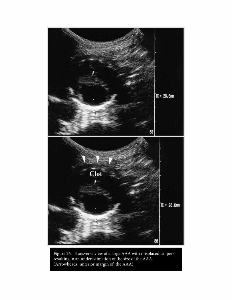
Figure 26. Transverse view of a large AAA with misplaced calipers, resulting in an underestimation of the size of the AAA. (Arrowheads=anterior margin of the AAA)

Figure 27. Need picture of retroperitoneal bleed.
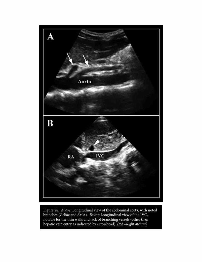
Figure 28. Above: Longitudinal view of the abdominal aorta, with noted branches (Celiac and SMA). Below: Longitudinal view of the IVC, notable for the thin walls and lack of branching vessels (other than hepatic vein entry as indicated by arrowhead). (RA=Right atrium)

Figure XX. Placeholder text.