Taiwan Safety Council CFSE and CFSP Training and Certification

www.abcam.com
ab113853
CFSE Fluorescent Cell
Labeling Kit
Instructions for Use For the durable fluorescent labeling of live cells for fluorescent microscopy and flow cytometrypopulation growth studies and within sample duplexing.
This product is for research use only and is not intended for diagnostic use.
Fluorescent Cell
For the durable fluorescent labeling of live cells for fluorescent microscopy and flow cytometry, population growth studies and within sample
This product is for research use only and is not intended

ab113853 CFSE Cell Labeling Kit
1
Table of Contents
1. Introduction 2
2. Assay Summary 4
3. Kit Contents 5
4. Storage and Handling 5
5. Additional Materials Required 5
6. Assay Procedure 6
7. Sample Data
i. Fluorescent cell labeling 9
ii. Tracking cell division 10
iii. Experimental duplexing using CFSE 12
ab113853 CFSE Cell Labeling Kit
2
1. Introduction
CFSE (5(6)-Carboxyfluorescein diacetate N-hydroxysuccinimidyl
ester) is a cell permeant, non-fluorescent pro-dye. Intracellular
esterases in live cells cleave the acetate groups which results in the
green fluorescent molecule carboxyfluorescein that is now
membrane impermeant. The succinimidyl ester group reacts
indiscriminately with intracellular free amines to generate covalent
dye-protein conjugates. The result is live cells with an intracellular
fluorescent label. At appropriate concentrations CFSE is non-toxic
to cells and the fluorescence is retained after formaldehyde and
alcohol fixation. CFSE labeled cells can be detected with any
instrument or filter set compatible with fluorescein detection:
Excitation(max)=492nm, Emission(max)=517nm.
CFSE is a versatile tool for the fluorescent intracellular labeling of
live cells. Labeled cells can be assayed using flow cytometry and
fluorescent microscopy. The dye is long lasting and well retained
within labeled cells. The provided CFSE is sufficient for ~1000
assays.
This product document provides a protocol for labeling cells with
CFSE and sample applications in which CFSE can be used. Sample
experiments include:
ab113853 CFSE Cell Labeling Kit
3
1. To label cells with CFSE for use in microscopy as a general
stain for cell morphology or as a counterstain for a red
fluorophore.
2. To quantify cell proliferation in vitro based on dilution of the
dye as parental labeled cells divide.
3. To allow the within sample duplexing by mixing differentially
treated CFSE labeled and unlabeled cells in the same
sample and subjecting to individual cell analysis (e.g. flow
cytometry).
Limitations:
• FOR RESEARCH USE ONLY. NOT FOR DIAGNOSTIC
PROCEDURES.
• Use this kit before expiration date.
• Do not mix or substitute reagents from other lots or sources.
• Any variation in operator, pipetting technique, washing
technique, incubation time or temperature, and kit age can
cause variation in binding.

ab113853 CFSE Cell Labeling Kit
4
2. Assay Summary
Label live cells with CFSE: Dilute CFSE in PBS or cell culture media
Cells can be in suspension or growing as an adherent monolayer
Incubate 10 minutes at 37C or room temperature
Wash cells with media to remove non-incorporated dye
Culture and treat labeled cells using standard methods
Harvest cells at desired timepoint
CFSE can be detected by flow cytometry or fluorescence microscopy
CFSE can be detected in live or fixed cells
(Emission(max): 495nm, Excitation(max): 517nm)
ab113853 CFSE Cell Labeling Kit
5
3. Kit Contents
• 10mM CFSE in DMSO (1000x) : 0.1 mL
4. Storage and Handling
Store CFSE at 4°C in the dark. The product is stable for 6 months
from receipt. For longer term storage, aliquot and store at -20°C or
-80C. Avoid multiple freeze-thaw cycles. Allow the product to
warm to room temperature before opening. Promptly use diluted
solutions of CFSE.
5. Additional Materials Required
• Flow cytometer and/or fluorescence microscope
(required excitation/emission wavelengths
495nm/517nm)
• General tissue culture supplies
• PBS (sterile)

ab113853 CFSE Cell Labeling Kit
6
6. Assay Procedure
1. Label live cells with 10nM—10M of CFSE
a. For uniform labeling, cells are best labeled in as a
single-cell suspension. Detach cells, by for example
trypsin which should then be quenched with media.
Cells that grow in suspension should be gently
pipetted to dissociate any aggregated cells. It is
also possible to label cells growing as a monolayer
by first removing the existing media and then
overlaying the CFSE staining solution.
b. CFSE can be diluted in PBS or culture media.
Labeling efficiency will be higher if CFSE is diluted in
PBS. If labeling in media, greater concentrations of
CFSE are required.
c. CFSE should be quickly mixed with cells for most
uniform labeling. For example, cells could be fully
resuspended in one volume of PBS and then an
additional volume of 2X CFSE concentration is
added and mixed. Alternatively, pelleted cells could
be resuspended in a 1X CFSE staining solution.
ab113853 CFSE Cell Labeling Kit
7
d. Concentrations of CFSE staining will need to be
determined individually based on the cell line used
and the goals of the experiment. For example, if
following cell proliferation by CFSE dilution, higher
levels of CFSE staining is desired. If using CFSE to
label cells as a counterstain for additional
fluorescent markers, lower levels of CFSE staining
would prevent fluorescent spillover between filter
sets.
2. Incubate cells in CFSE staining solution for 10-15
minutes at room temperature or at 37C.
3. Quench staining and wash cells with culture media to
remove unincorporated CFSE.
a. Add equal volume of culture media to the cells +
CFSE staining solution and allow to sit for 5
minutes.
b. Next, remove the CFSE-containing solution and
wash cells one time with an equal volume of culture
media.
c. Cells are now fluorescently labeled.
4. Return cells to standard culture conditions or
immediately assay.
ab113853 CFSE Cell Labeling Kit
8
IMPORTANT ADDITIONAL NOTES:
1. CFSE is amine group reactive. Do not use buffers that
contain primary amines during the CFSE labeling step. PBS
is the recommended CFSE dilution buffer.
2. The amount of cellular fluorescence drops substantially in
the first 24 hours after CFSE labeling. After that, the
fluorescence is stably incorporated and will decrease over
time as a function of dilution with cell division. If optimizing
CFSE labeling concentrations for long-term studies, it is best
to optimize to measurements taken at 24 hours post CFSE
labeling.

ab113853 CFSE Cell Labeling Kit
9
7. Sample Data
i. Fluorescent cell labeling for microscopy:
CFSE is useful as a tool to label live cells. Since the
fluorescence is dependent on esterase activity, only live
cells are labeled. Since the entire cell is labeled, CFSE can
be a useful counterstain when using red fluorophore markers
of specific subcellular structures. CFSE has also been used
to label cells that are subsequently injected into animals; in
this manner implanted cells’ migration, morphology and
division can be monitored in-vivo.
Figure 1. Jurkat cells labeled with CFSE. Jurkat cells were
labeled with 1mM CFSE in media for 15 minutes, washed once with
PBS and imaged on a flurorescence microscope. The cells in this
image are live but fixed cells give similar results.
ab113853 CFSE Cell Labeling Kit
10
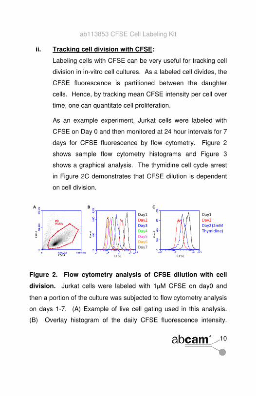
ii. Tracking cell division with CFSE:
Labeling cells with CFSE can be very useful for tracking cell
division in in-vitro cell cultures. As a labeled cell divides, the
CFSE fluorescence is partitioned between the daughter
cells. Hence, by tracking mean CFSE intensity per cell over
time, one can quantitate cell proliferation.
As an example experiment, Jurkat cells were labeled with
CFSE on Day 0 and then monitored at 24 hour intervals for 7
days for CFSE fluorescence by flow cytometry. Figure 2
shows sample flow cytometry histograms and Figure 3
shows a graphical analysis. The thymidine cell cycle arrest
in Figure 2C demonstrates that CFSE dilution is dependent
on cell division.
Figure 2. Flow cytometry analysis of CFSE dilution with cell
division. Jurkat cells were labeled with 1M CFSE on day0 and
then a portion of the culture was subjected to flow cytometry analysis
on days 1-7. (A) Example of live cell gating used in this analysis.
(B) Overlay histogram of the daily CFSE fluorescence intensity.

ab113853 CFSE Cell Labeling Kit
11
Mean CFSE intensity per cell decreases as the cells divide and the
dye is diluted amongst the daughter cells. (C) As a control, 2mM
Thymidine was added to a parallel culture of the day 0 CFSE labeled
cells at day1. 2mM thymidine causes an S-phase cell cycle arrest.
At day2, there is less dilution of CFSE in the thymide culture (blue)
relative to the untreated culture (red) due to decreased cell division.
Figure 3. Comparing CFSE dilution with cell count: 7 day
experiment. Graphical analysis of flow cytometry CFSE dilution
data from Figure 1 and comparison to cell concentration data. (A)
Plot of CFSE fluorescent intensity (green) and cell concentration
(blue) as a function of time; each y-axis axis is a log2 scale. Note
that the rate of decrease in CFSE intensity slows and the rate of
increase in cell concentration slows as the cell density increases.
(B) Re-plot of the data in (A) such that the inverse of CFSE intensity
increase is plotted. Note the excellent agreement between CFSE
dilution and cell concentration.

ab113853 CFSE Cell Labeling Kit
12
Note that the use of CFSE allows the specific tracking of labeled
cells in mixed cell populations. For example, proliferation of CFSE-
labeled cells could be monitored when co-cultured with other non-
labeled cell lines. This could be particularly useful in quantitative
evaluation of co-culture systems for isolated primary cells.
iii. Experimental duplexing using CFSE:
Immunostaining a mixture of CFSE-labeled and unlabeled
cells allows within-sample duplexing of treated and
untreated cells when single cell analysis is available (e.g.
flow cytometry, high content microscopy). In this way,
treated and untreated cells can be analyzed simultaneously.
This doubles throughput while simultaneously providing an
excellently controlled experimental setup by eliminating
potential for differences in reagent addition between control
and experimental samples.
To demonstrate this technique, CFSE labeling was
combined with deferoxamine (DFO) treatments. DFO is a
hypoxia mimetic and causes the stabilization of the hypoxia
induced transcription factor HIF1A. Figure 4 demonstrates
control of the cell population by manipulating CFSE labeling
and DFO exposure. Figure 5 shows an example experiment
ab113853 CFSE Cell Labeling Kit
13
with differential immunostaining between CFSE labeled and
unlabeled cells.
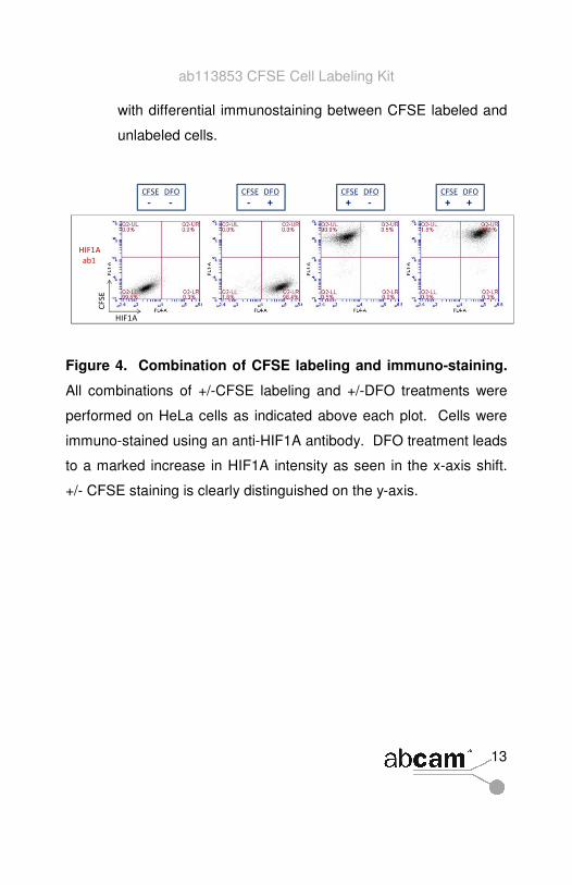
Figure 4. Combination of CFSE labeling and immuno-staining.
All combinations of +/-CFSE labeling and +/-DFO treatments were
performed on HeLa cells as indicated above each plot. Cells were
immuno-stained using an anti-HIF1A antibody. DFO treatment leads
to a marked increase in HIF1A intensity as seen in the x-axis shift.
+/- CFSE staining is clearly distinguished on the y-axis.
ab113853 CFSE Cell Labeling Kit
14
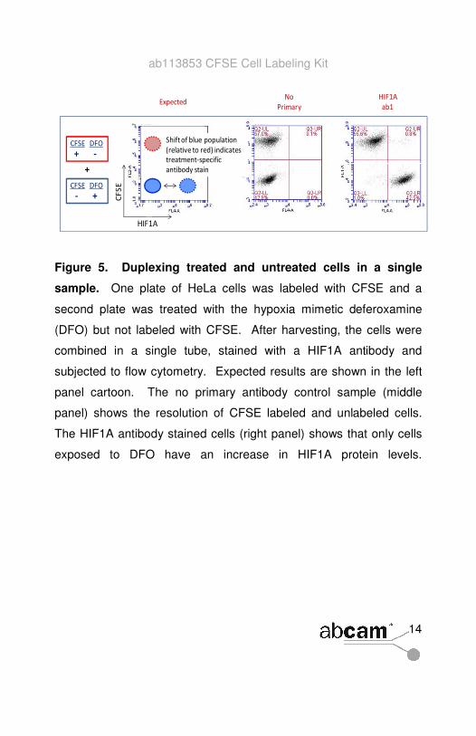
Figure 5. Duplexing treated and untreated cells in a single
sample. One plate of HeLa cells was labeled with CFSE and a
second plate was treated with the hypoxia mimetic deferoxamine
(DFO) but not labeled with CFSE. After harvesting, the cells were
combined in a single tube, stained with a HIF1A antibody and
subjected to flow cytometry. Expected results are shown in the left
panel cartoon. The no primary antibody control sample (middle
panel) shows the resolution of CFSE labeled and unlabeled cells.
The HIF1A antibody stained cells (right panel) shows that only cells
exposed to DFO have an increase in HIF1A protein levels.
ab113853 CFSE Cell Labeling Kit
15
Abcam in the USA Abcam in Japan
Abcam Inc Abcam KK
1 Kendall Square, Ste B2304 2-2-1 Nihonbashi
Cambridge, Horidome-cho,
MA 02139-1517 Chuo-ku, Tokyo
USA 103-0012
Japan
Toll free: 888-77-ABCAM (22226) Fax: 866-739-9884 Tel: +81-(0)3-6231-094 Fax: +81-(0)3-6231-0941
Abcam in Europe Abcam in Hong Kong
Abcam plc Abcam (Hong Kong) Ltd
330 Cambridge Science Park Unit 225A & 225B, 2/F
Cambridge Core Building 2
CB4 0FL 1 Science Park West Avenue
UK Hong Kong Science Park
Hong Kong
Tel: +44 (0)1223 696000
Fax: +44 (0)1223 771600 Tel: (852) 2603-682 Fax: (852) 3016-1888
Copyright © 2011 Abcam, All Rights Reserved. The Abcam logo is a registered trademark.
All information / detail is correct at time of going to print.