Aalborg Universitet The nociceptive withdrawal reflex in...
49
Aalborg Universitet The nociceptive withdrawal reflex in conscious dogs Bergadano, Alessandra Publication date: 2008 Document Version Publisher's PDF, also known as Version of record Link to publication from Aalborg University Citation for published version (APA): Bergadano, A. (2008). The nociceptive withdrawal reflex in conscious dogs: a new, non-invasive model of nociception. Aalborg: Center for Sensory-Motor Interaction (SMI), Department of Health Science and Technology, Aalborg University. General rights Copyright and moral rights for the publications made accessible in the public portal are retained by the authors and/or other copyright owners and it is a condition of accessing publications that users recognise and abide by the legal requirements associated with these rights. ? Users may download and print one copy of any publication from the public portal for the purpose of private study or research. ? You may not further distribute the material or use it for any profit-making activity or commercial gain ? You may freely distribute the URL identifying the publication in the public portal ? Take down policy If you believe that this document breaches copyright please contact us at [email protected] providing details, and we will remove access to the work immediately and investigate your claim. Downloaded from vbn.aau.dk on: maj 09, 2018
Transcript of Aalborg Universitet The nociceptive withdrawal reflex in...

Aalborg Universitet
The nociceptive withdrawal reflex in conscious dogs
Bergadano, Alessandra
Publication date:2008
Document VersionPublisher's PDF, also known as Version of record
Link to publication from Aalborg University
Citation for published version (APA):Bergadano, A. (2008). The nociceptive withdrawal reflex in conscious dogs: a new, non-invasive model ofnociception. Aalborg: Center for Sensory-Motor Interaction (SMI), Department of Health Science andTechnology, Aalborg University.
General rightsCopyright and moral rights for the publications made accessible in the public portal are retained by the authors and/or other copyright ownersand it is a condition of accessing publications that users recognise and abide by the legal requirements associated with these rights.
? Users may download and print one copy of any publication from the public portal for the purpose of private study or research. ? You may not further distribute the material or use it for any profit-making activity or commercial gain ? You may freely distribute the URL identifying the publication in the public portal ?
Take down policyIf you believe that this document breaches copyright please contact us at [email protected] providing details, and we will remove access tothe work immediately and investigate your claim.
Downloaded from vbn.aau.dk on: maj 09, 2018

1
The nociceptive withdrawal reflex in conscious dogs:
a new, non-invasive model of nociception
Ph.D. thesis
Alessandra Bergadano
2008
Department for Health Sciences and Technology
Aalborg University, Aalborg, Denmark
Department of Clinical Veterinary Medicine
Anaesthesiology division, Vetsuisse faculty,
University of Berne, Berne, Switzerland

2
ISBN: 978-87-90562-92-2

3
To my parents, with love.

4
Contents
1. Abbreviations ....................................................................................................................................... 6
2. Acknowledgments ................................................................................................................................ 7
3. List of original publications ................................................................................................................ 8
4. Abstract (English) ................................................................................................................................ 9
5. Abstract (Danish) ............................................................................................................................... 10
6. Introduction........................................................................................................................................ 11
6.1. Pain in dog and its diagnose ......................................................................................... 11
6.1.1. Nociceptive models in dogs ................................................................................................. 12
6.2. The Nociceptive Withdrawal Reflex (NWR) .............................................................. 13
7. Aim of the PhD project ...................................................................................................................... 14
8. Methods .............................................................................................................................................. 16
8.1. The experimental dogs .................................................................................................. 16
8.2. Eliciting and recording NWR in dogs .......................................................................... 16
8.2.1. Positioning of the dogs ........................................................................................................ 17
8.2.2. Stimulating and recording material ..................................................................................... 17
8.2.3. Stimulus parameters ............................................................................................................. 18
8.3. Behavioural reactions ................................................................................................... 20
8.4. Analysing the NWR ...................................................................................................... 20
8.4.1. Onset latency ....................................................................................................................... 20
8.4.2. Reflex magnitude ................................................................................................................. 20
8.4.3. Single NWR and temporal summation thresholds............................................................... 21
9. Physiology of the canine NWR ......................................................................................................... 21
9.1. Functional significance ................................................................................................. 21
9.2. Forelimb muscles .......................................................................................................... 22
9.3. Hind limb muscles ........................................................................................................ 23
9.4. The NWR threshold intensity (It) in dogs ..................................................................... 23
9.5. Reflex components ....................................................................................................... 24
9.5.1. The early reflex activity: 0 to 20 milliseconds epoch .......................................................... 24
9.5.2. The NWR: 20 to 100 milliseconds epoch ............................................................................ 24
9.5.3. The late reflex activity: 100 to 400 milliseconds epoch. Preliminary work ........................ 25
9.5.4. Eliciting the NWR in dogs: conclusions .............................................................................. 26
9.6. Facilitation of the NWR by repeated stimulations ....................................................... 26
9.6.1. Wind up and temporal summation ....................................................................................... 26
9.6.2. Temporal summation in conscious dogs (II-IV) .................................................................. 27

5
9.6.3. Temporal summation in conscious dogs: conclusions ......................................................... 28
10. Variations in the canine reflex ...................................................................................................... 29
10.1. Posture .......................................................................................................................... 29
10.2. Age, sex, circadian variations ....................................................................................... 29
10.3. Habituation ................................................................................................................... 29
11. Pharmacological modulation of the NWR and temporal summation ....................................... 30
11.1. Low-dose acepromazine (III). ...................................................................................... 30
11.1.1. Use of phenotiazine tranquillizers in human and veterinary medicine ................................ 30
11.1.2. Effects of acepromazine on NWR and temporal summation in dogs .................................. 31
11.1.3. Phenotiazine analgesia: mythos or reality? .......................................................................... 32
11.2. Low-dose ketamine constant-rate-infusion (IV) ........................................................... 32
11.2.1. Ketamine as an analgesic in dogs ........................................................................................ 33
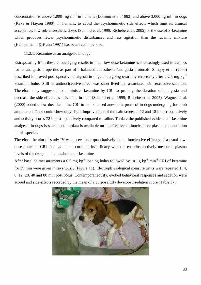
11.2.2. Plasma concentrations of ketamine in dogs ......................................................................... 34
11.2.3. Effects of ketamine on NWR and temporal summation in dogs ......................................... 34
12. Conclusions and Future applications ........................................................................................... 35
13. Tables .............................................................................................................................................. 38
14. References ....................................................................................................................................... 41

6
1. Abbreviations
NWR: nociceptive withdrawal reflex
It: individual NWR threshold intensity
TSt: temporal summation threshold intensity
RMS: root-mean-square
EMG: electromyography
ACP: acepromazine
SAL: saline
ms: milliseconds
NMDA : N-methyl-D-aspartate
WDR: wide dynamic range
CRI : constant rate infusion
IV: Intravenous
IQR : inter quartile range
AUC : area-under-the-curve

7
2. Acknowledgments
The present studies have been carried out at the Anaesthesiology division, Department of Clinical
veterinary medicine, Vetsuisse Faculty, University of Berne and at Novartis Pharma, Basel, Switzerland.
I wish to express my deepest and sincere gratitude to my friend Prof. Claudia Spadavecchia for the time
invested in discussing the scientific projects, in helping with the experiments and for her support as a
friend. Without her collaboration this PhD wouldn’t have been possible.
My warmest and sincere thanks go to my supervisors Prof. Ole Andersen and Prof. Lars Arendt-Nielsen
for their precious scientific guidance, critical reviewing and teaching despite the geographical distance
and for their constant and tactful encouragement.
Thousand thanks to Dr. Luciano Spadavecchia who developed the devices and softwares!
I am indebt to Prof. Markus Doherr for his most unselfish scientific contribution in advising and
reviewing my statistical work.
Thanks to Prof. Urs Schatzmann, to Dr. Irene Mueller and personnel of the animal facility in Novartis
Pharma. Thanks to my friends around the world for their ongoing positive support.
Finally my gratefulness and love to my partner Mathias, who worked in the familiar background to offer
me the most precious thing: time!
The studies of this PhD have been supported financially by a Vetsuisse grant and a Swiss National
Foundation grant (analytical part). The grants are gratefully acknowledged.
Berne, 06 08 2008

8
3. List of original publications
The present thesis is based on the following original publications, referred to in the text by their Roman
numerals (I- IV).
I. Alessandra Bergadano, Ole K. Andersen, Lars Arendt-Nielsen, Urs Schatzmann, Claudia Spadavecchia.
Quantitative assessment of nociceptive processes in conscious dogs by use of the nociceptive withdrawal
reflex. AJVR 2006; 67;5:882-9 doi: 10.2460/ajvr.67.5.882
II. Alessandra Bergadano, Ole K. Andersen, Lars Arendt-Nielsen, Claudia Spadavecchia. Non invasive
assessment of the facilitation of the nociceptive withdrawal reflex by repeated electrical stimulations in
conscious dogs. AJVR 2007; 68;8:899-907 doi: 10.2460/ajvr.68.8.899
III. Alessandra Bergadano, Ole K. Andersen, Lars Arendt-Nielsen, Claudia Spadavecchia. Modulation of
a low acepromazine dose on single and repeated nociceptive stimuli in conscious dogs. VAA 2008
(Accepted)
IV. Alessandra Bergadano, Ole K. Andersen, Lars Arendt-Nielsen, Regula Theurillat, Wolfgang
Thormann, Claudia Spadavecchia. The plasma concentrations of a low-dose constant-rate-infusion (CRI)
of ketamine and its effect on single and repeated nociceptive stimuli in conscious dogs. Vet J 2008 (In
press) doi:10.1016/j.tvjl.2008.06.003

9
4. Abstract (English)
In this thesis the nociceptive withdrawal reflex (NWR) and its facilitation by repeated electrical
stimulations in intact, conscious dogs were thoroughly investigated. This included methodological
development and pharmacological modulation studies. The pharmacological modulation aimed to
quantify objectively the efficacy of different drugs in dogs.
In paper I the feasibility of evoking and recording the NWR from the forelimb and hind limb of conscious
non-medicated dogs was first described. The stimulus-response curves and the evoked behavioral
responses were studied confirming the nociceptive origin of the reflex. In paper II, the facilitation of the
nociceptive withdrawal reflex by repeated electrical stimuli as a measure of neuronal temporal summation
and the associated behavioral response scores were investigated in conscious, non-medicated dogs.
Additionally the influence of stimulus intensity and stimulus frequency on temporal summation responses
were analyzed. In paper III, the within-session and intersession stability of the NWR thresholds could be
demonstrated, supporting that the model is reproducible and robust. Furthermore it was shown that
intravenous 0.01 mg kg-1
acepromazine can be used to ease data acquisition in anxious subjects without
altering the validity of the model. Based on these findings, the antinociceptive action of a low-dose
constant-rate-infusion of racemic ketamine (0.5 mg kg-1
loading bolus followed by 10 g kg-1
min-1
) in
conscious dogs was explored in paper IV. Temporal summation and the evoked behavioral responses
scores were inhibited compared to baseline, demonstrating the antinociceptive activity of ketamine in
correlated with peak plasma concentrations. This antinociceptive action was short lived owing to the
unexpectedly low plasma levels obtained at pseudo-steady-state, questioning the use of this low-dose
ketamine CRI as sole analgesic in dogs.
In conclusion the work presented in this PhD thesis has provided a new, non invasive, robust
experimental model of nociception in conscious dogs that may be used in clinical routine to study the
antinociceptive activity of drugs or to quantify the excitability of the nervous system in individual canine
patients.
10
5. Abstract (Danish)
I denne afhandling beskrives den nociceptive afværgerefleks (NWR) og dens facilitering ved hjælp
af gentagne elektriske stimulationer på intakte hunde, der er ved fuld bevidsthed (”intact” på engelsk
henviser typisk til ikke kastreret/steriliseret). Dette indebar metodeudvikling og farmakologiske
modulationsundersøgelser. Den farmakologiske modulation havde til formål objektivt at kvantificere
effekten af forskellig medicin i hunde.
I den første artikel beskrives anvendeligheden af en metode til at fremkalde og registrere NRW fra for-
og bagben på vågne, ikke medicinerede hunde. Stimulus-responskurven og den fremkaldte
adfærdsrespons bekræftede den nociceptive oprindelse af refleksen.
I den anden artikel beskrives undersøgelsen af faciliteringen af NWR ved hjælp af gentagne elektriske
stimuli som et mål for neuronal temporal summation og de tilhørende adfærdsrespons-scores i vågne,
ikke medicinerede hunde. Desuden blev indflydelsen af stimulus-intensiteten og stimulus-frekvensen på
temporal summations-responsen analyseret.
I tredje artikel kunne winhin-session og intersession stabiliteten af NWR’ens grænseværdier
demonstreres, hvilket understøtter modellens stabilitet og reproducerbarhed. Desuden blev det påvist, at
intravenøs acepromacin i en dosis på 0,01mg per kg kan bruges på meget nervøse hunde for at lette
erhvervelsen af data, uden at det har indflydelse på modellens validitet.
Baseret på de ovennævnte resultater blev den antinociceptive virkning af en konstant lav-dosis infusion
af racemic ketamin (0,5 mg kg-1
som start-bolus efterfulgt af 10 g kg-1
min-1
konstant infusion)
undersøgt i fjerde artikel. Temporal summation og de fremkaldte adfærdsrespons-scores blev hæmmet
sammenlignet med basislinien. Dette demonstrerede den antinociceptive virkning af ketamin i korrelation
med peak plasma-koncentrationer. Denne antinociceptive virkning var kortvarig på grund af de uventede
lave plasma-koncentrationer opnået på pseudo-steady state. Dette sætter spørgsmålstegn ved brugen af
denne lav-dosis ketamin-infusion som eneste analgetiske medicin hos hunde.
Afsluttende kan man sige, at det arbejde, der præsenteres i denne Ph.d.-afhandling har leveret en ny,
ikke-invasiv, solid eksperimentel model for nociception i hunde ved bevidsthed, der kan bruges i klinisk
rutine-arbejde til undersøgelse af den antinociceptive virkning af medikamenter eller til at kvantificere
excitabiliteten af nervesystemet i individuelle hunde.

11
6. Introduction
Understanding and treating pain in animals is one of the most challenging tasks in veterinary medicine. In
the last decade there has been a growing interest and research investigating the mechanisms underlying
animal pain and improving the therapeutic options (Hansen 2003). In 2002, experts in animal and human
pain developed a consensus statement indicating that animals feel pain and identified the key gaps in the
current knowledge of animal pain (Paul-Murphy et al. 2005). Because animals lack the ability to use
language to express emotions about pain, animal pain has been described in terms of behavioural
responses to damaging or potentially damaging noxious stimuli. The term “nociception” (i.e. perception
of a damaging or potentially damaging stimulus) is therefore used, also for the purpose of this thesis, as it
thought to represent more accurately the response to stimuli which would be associated with pain in man.
To date the most important gap in our knowledge of animal pain is related to the assessment of
nociception. Subjective assessment of abnormal demeanour or behaviours are extensively used and
multiple scales and scoring system have been developed in the attempt to better diagnose and quantify
pain. However, there is currently no gold standard to assess nociception in animals and no unit for pain.
And as stated by Lord Kelvin many years ago “when you can measure what you are speaking about and
express it in numbers you know something about it; but when you cannot measure it, when you cannot
express it in numbers, your knowledge is of a meagre and unsatisfactory kind”(Kelvin 1891). It is
difficult to say that pain have been effectively if it cannot be accurately assessed.
Another gap is related to a paucity of species-specific information concerning both basic nociceptive
mechanisms and efficacy of analgesics. Many current treatments are still extrapolated across species and
from experimental to clinical setting without any evidence of their efficacy or safety in a given animal
species.
To fill these gaps, there is a substantial need for a noninvasive, sensitive, specific, repeatable model to
investigate nociception for basic physiological studies, to objectively assess the degree of sensory
dysfunction and to quantitatively test pharmacological interventions. The final goal is to improve the
clinical treatment of pain in domestic animals.
6.1. Pain in dog and its diagnose
Dogs can experience physiological or pathological pain of inflammatory (somatic or visceral),
neuropathic or mixed origin. Many health conditions, medical and surgical procedures cause pain in dogs,
mainly of short duration (< 7 days) (Muir et al. 2004). The assessment of pain relies on the subjective
description of abnormal behavior and demeanor patterns, or on the use of Visual Analogue Scales after
direct or video-assisted (Hansen 2003) observation of the animal. To improve objective and quantitative
assessment of nociception, composite pain scales incorporating behavioral, physiological and interactive
parameters have been developed (Holton et al. 1998; Holton et al. 2001). Still few have been validated

12
and only for a specific noxious stimulus. These scales are not valid for assessing pain of another origin:
i.e. if a pain scale has been developed to evaluate acute postoperative pain after orthopedic surgery it will
not be sensitive for assessing abdominal pain.
The issue of pathological pain in dogs is even more complex. Only very recently there is increased
consciousness that dogs of any age can suffer of chronic pain. The most common medical conditions are
chronic musculoskeletal pathologies, i.e. hip dysplasia, cruciated ligament rupture, osteoarthritis (Jauernig
et al. 1999), and cancers (Lascelles & Main 2002). Chronic pain impairs the quality of life of the animals,
and represents a source of practical problems for the owners. Both veterinarians and owners are
convinced that those dogs should receive adequate pain-relieving treatment. However, accurate detection
of signs of pain and therefore adequate therapy is difficult. In dogs, few very scales are reported to be
valid for evaluating chronic osteoarthritis-associated pain (Bjorkman et al. 1993; Wiseman-Orr et al.
2004; Cimino Brown et al. 2007) and other types of chronic pain are actually not addressed. To date
pathological pain conditions in dogs are still under-recognized and thus under-treated.
6.1.1. Nociceptive models in dogs
Investigations involving animal models of nociception (Le Bars et al. 2001) are mainly used as
transitional studies to provide better understanding of pain mechanisms and the effectiveness of analgesic
drugs for subsequent administration to humans. Unlike cats (for which there is extensive literature), dogs
are seldom used as experimental animals in nociception studies. Some experimental and clinical studies
have been performed in dogs to provide objective ways of assessing antinociceptive activity of analgesics
for the benefit of the dogs. Mechanical, (Hamlin et al. 1988; Barnhart et al. 2000a; Barnhart et al. 2000b)
thermal (Andrews & Workman 1941; Ylisela & Vainio 1989; Barnhart et al. 2000b; Wegner et al. 2008),
and electrical stimulations (Hamlin et al. 1988; Vainio et al. 1989; Brown et al. 2002b; Brown et al.
2002a) have been applied to the skin to evoke nocifensive reactions and to evaluate their pharmacologic
modulation. The end point of these models of acute nociception in dogs is determined by monitoring the
evoked gross behavioral reaction or the thresholds at which the behavioral aversive response is elicited.
The prolongation of the latency of the withdrawal response or an increase in the response threshold is
interpreted as antinociception.
The major drawback of all these models is evident when the drugs used exert a contemporaneous sedative
effect that can clearly alter the pattern of the behavioral reaction observed and the interpretation of the
antinociceptive efficacy. Another drawback is that the stimulus intensities used are supramaximal with
obvious distress for the animals and potential risk of tissue damage. Additionally these models are
modestly sensitive as they do not allow analysing the stimulus-response curve.
A more refined model consists of recording the behavioral reflex response to a nociceptive (thermal or
electrical) stimulus by electromyography. Reflex-evoked muscle action potentials of the masseter muscles

13
after sensory dental pulp stimulation have been recorded in anesthetized dogs (Mitchell 1964; Brown et
al. 2002a; Brown et al. 2002b).
6.2. The Nociceptive Withdrawal Reflex (NWR)
In humans a reflex withdrawal reaction can be elicited by transcutaneous electrical stimulation of a
sensory peripheral nerve and the electromyographic response recorded from the flexor and extensor
muscles. This nociceptive withdrawal reflex (NWR) is a polysynaptic spinal nociceptive reflex, and
represents the mechanism for withdrawing an extremity from injury (Sherrington 1910). The NWR is
reproducible, stimulus-dependent and is closely correlated with the intensity of subjective pain perception
(Willer 1977; Willer 1984; Chan & Dallaire 1989). Therefore the NWR and its modulation have been
widely used in experimental (Hagbarth 1960; Kugelberg et al. 1960; Hugon 1973; Willer & Bathien
1977; DeBroucker et al. 1989; Arendt-Nielsen et al. 2000; Andersen 2007) and pharmacologic studies
(Willer & Bathien 1977; Willer 1985; Arendt-Nielsen et al. 1990; Petersen-Felix et al. 1995; Curatolo et
al. 1997; Petersen-Felix et al. 1998; Piguet et al. 1998; Escher et al. 2007) as a noninvasive
neurophysiologic tool to objectively assess spinal nociceptive processing. By applying appropriate
repetitive stimulation patterns, temporal summation can be evoked and quantified by a facilitation of the
reflex (Andersen et al. 1994; Arendt-Nielsen et al. 2000; Serrao et al. 2004). Temporal summation in
humans has been considered as a psychophysical correlate of the early phase of wind-up. This facilitation
of the nociceptive reflex response has been used as a tool to study and quantify aspects of central
integration and sensitisation in humans (Kugelberg et al. 1960; Shahani & Young 1971; Hugon 1973;
Akopian et al. 1996).
Electromyographic recordings of flexion reflexes of the limbs elicited by electrical stimuli have been
investigated in decerebrated or spinalized rats (Schouenborg & Dickenson 1985; Schouenborg &
Kalliomaki 1990; Schouenborg et al. 1995; You et al. 2003b; You et al. 2004), cats (Sherrington 1910;
Schomburg 1990a; Levinsson et al. 1999), and rabbits (Clarke & Harris 2001). Unfortunately these
models are of limited clinical interest because of their invasiveness and the influence of anesthetics on the
flexion reflexes. Aware of this drawback, Carstens and coauthors measured the limb flexion withdrawal
elicited by noxious thermal stimulation of the hindpaw in conscious rats (Carstens & Ansley 1993).
Recently, results of a series of studies demonstrated the feasibility of evoking and recording the NWR for
the fore- and hind limbs in standing, conscious horses (Spadavecchia et al. 2002; Spadavecchia et al.
2003; Spadavecchia et al. 2004; Spadavecchia et al. 2005), suggesting that the NWR could be used as a
non invasive, objective method to measure nociception in this species.
14
7. Aim of the PhD project
With a cross-species approach based on the capability to investigate objectively and non-invasively the
nociception-related responses in humans and in standing horses, it was assumed that a similar
investigation in conscious, non medicated dogs would be possible.
The aims of the PhD project presented here were:
1) To demonstrate the feasibility of evoking the NWR from the forelimb and hind limb in conscious,
non medicated dogs, and score the behavioral responses to the electrical stimuli.
2) To study the modulation of the reflex after repeated electrical stimulations (temporal summation)
3) To investigate the pharmacological modulation of the NWR and temporal summation in dogs.
To develop a new, non invasive model of nociception in dogs would allow to gain species-specific
knowledge about the nociceptive process and to obtain comparative physiologic data for a better
understanding of nociception in general. The pharmacological modulation of the reflex would provide
objective evidence on the efficacy of analgesic drugs in dogs.
The experimental work has been published in four papers dealing with the technical and physiological
aspects of the canine NWR and its pharmacological modulation (Figure 1). This thesis presents and
discusses the experimental work and the results obtained.
In the first paper (I) the feasibility of evoking the NWR by electrical stimulation of a sensory nerve and
recording of the electromyographic response in both the forelimb and the hind limb, in conscious non
medicated dogs is described. The recruitment of the NWR obtained with graded suprathreshold
stimulations as the correlation between reflex characteristics and evoked behavioral responses were
studied. The effect of the stimulus paradigm was analysed.
The second paper (II) investigated the facilitation of the NWR by repeated electrical stimuli and the
associated behavioral response scores in conscious, non-medicated dogs as a measure of temporal
summation. The influence of different stimulus intensities and frequencies on temporal summation was
evaluated.
In the third paper (III) the effects of a tranquillizing dose of acepromazine on the NWR and temporal
summation were analyzed. As a second objective the repeatability and stability of the NWR thresholds
were investigated.
In the fourth paper (IV) the NWR and its facilitation evoked by repeated stimulations were used for the
first time as a model to objectively and quantitatively analyze the antinociceptive properties of a usual
low-dose constant rate infusion of ketamine in conscious dogs. Low-dose ketamine CRI has gained
popularity in the management of post-operative pain in canine patients.
The conclusions outline the main findings and their clinical relevance and possible future
implementations.

15
Figure 1. Schematic representation of the content of the PhD thesis
16
8. Methods
In this chapter the methods used to evoke record and quantify the NWR and the reflex facilitation after
repeated stimulations in dogs will be described and discussed.
The experiments were approved by the committee for animal experimentation of the canton Basel city,
Switzerland (approval number 2090).
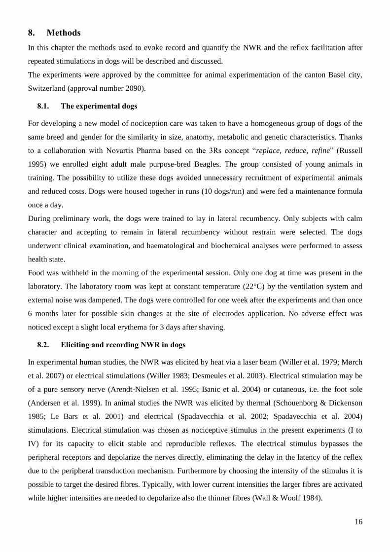
8.1. The experimental dogs
For developing a new model of nociception care was taken to have a homogeneous group of dogs of the
same breed and gender for the similarity in size, anatomy, metabolic and genetic characteristics. Thanks
to a collaboration with Novartis Pharma based on the 3Rs concept “replace, reduce, refine” (Russell
1995) we enrolled eight adult male purpose-bred Beagles. The group consisted of young animals in
training. The possibility to utilize these dogs avoided unnecessary recruitment of experimental animals
and reduced costs. Dogs were housed together in runs (10 dogs/run) and were fed a maintenance formula
once a day.
During preliminary work, the dogs were trained to lay in lateral recumbency. Only subjects with calm
character and accepting to remain in lateral recumbency without restrain were selected. The dogs
underwent clinical examination, and haematological and biochemical analyses were performed to assess
health state.
Food was withheld in the morning of the experimental session. Only one dog at time was present in the
laboratory. The laboratory room was kept at constant temperature (22°C) by the ventilation system and
external noise was dampened. The dogs were controlled for one week after the experiments and than once
6 months later for possible skin changes at the site of electrodes application. No adverse effect was
noticed except a slight local erythema for 3 days after shaving.
8.2. Eliciting and recording NWR in dogs
In experimental human studies, the NWR was elicited by heat via a laser beam (Willer et al. 1979; Mørch
et al. 2007) or electrical stimulations (Willer 1983; Desmeules et al. 2003). Electrical stimulation may be
of a pure sensory nerve (Arendt-Nielsen et al. 1995; Banic et al. 2004) or cutaneous, i.e. the foot sole
(Andersen et al. 1999). In animal studies the NWR was elicited by thermal (Schouenborg & Dickenson
1985; Le Bars et al. 2001) and electrical (Spadavecchia et al. 2002; Spadavecchia et al. 2004)
stimulations. Electrical stimulation was chosen as nociceptive stimulus in the present experiments (I to
IV) for its capacity to elicit stable and reproducible reflexes. The electrical stimulus bypasses the
peripheral receptors and depolarize the nerves directly, eliminating the delay in the latency of the reflex
due to the peripheral transduction mechanism. Furthermore by choosing the intensity of the stimulus it is
possible to target the desired fibres. Typically, with lower current intensities the larger fibres are activated
while higher intensities are needed to depolarize also the thinner fibres (Wall & Woolf 1984).

17
8.2.1. Positioning of the dogs
The dogs were placed in right lateral recumbency (I-IV), as it is a physiological, species-specific sleeping
position, in a comfortable, corncob-balls filled dog bed that took the shape of the body. The limbs were
extended laterally in a natural position but not supported, without weight bearing or movement restriction
of the nondependent limb. This position can be compared to the sitting position in humans (Willer 1977;
Willer 1983; Rossi & Decchi 1994; Andersen et al. 1995b), where the volunteers have the limbs
positioned so as to achieve complete muscle relaxation (Figure 2).
Figure 2. Dog laying without restrain in lateral recumbency, instrumented for stimulation and recording from the hind limb
8.2.2. Stimulating and recording material
Stimulation and recordings were performed by use of a specially designed, computerized system (I-IV).
The final stage of the electrical stimulator that received input from the computer was a battery-powered
optoisolated constant-current device with a maximum voltage of 100 V and a maximal current of 40 mA.
Electromyographic signals were amplified with an overall gain of 5,000 and bandpass of 7 to 200 Hz
(first-order active filters with 6 dB/octave slope). They were passed through a digital converter to a
computer for further processing and storage.
Electrical current was delivered via self adhesive electrodes (Spadavecchia et al. 2002; Spadavecchia et
al. 2004; Andersen 2007). The stimulation electrodes (Neuroline 700 05-j, Medikotest A/S, Olstykke,
Denmark) were placed over purely sensory nerves: the dorsal branch of the ulnar nerve at the level of the
left fifth metacarpal bone of the forelimb (Figure 3A) and over the lateral plantar digital nerve of the hind
limb at the level of the fourth metatarsal bone, just distal to the base and proximal to the head of each
bone (Figure 3B).

18
Figure 3A and B. Detail of the stimulation sites
The electrodes were placed parallel to the nerve with the anode in the distal position, with an
interelectrode distance of 0.8 cm. The distal portion of the limb was bandaged to prevent dislocation of
the electrodes. The ground electrode (Synapse 32 mm, Ambu A/S, Ballerup, Denmark) was placed over
the plantar side of the right foot and taped in place (Figure 1). Flexible leads were connected to the
electrodes. The resistance of each electrode pair was checked and confirmed to be less than 5 k before
starting and at the end of each experimental session. Typically the resistance was between 1 and 3 k .
This is necessary to ensure that the nerve stimulator can deliver enough current to elicit the reflex in a
stable and reproducible manner. To achieve low resistance, the skin was carefully clipped, shaved and
degreased before electrodes application.
The same electrode type was used to record the surface electromyograms from the forelimb and hind limb
muscles. Special care was taken to place the electrodes over the muscle bellies at a distance of 1 cm to
avoid multichannel cross-talk contamination from adjacent muscles and minimize common-mode noise
(Farina et al. 2002). Their position was marked with a pen, which allowed for exact repositioning in case
the electrodes were disconnected.
8.2.3. Stimulus parameters
Single stimulation. In the published literature (Spadavecchia et al. 2002; Andersen 2007) a train-of-five
pulses delivered at high frequency, which humans perceive as a single stimulus, is described as a standard
stimulus to elicit the NWR. Along with other factors, the number of pulses and stimulus duration can
influence the NWR (Tørring et al. 1981). The effect of stimulus configuration on the canine NWR was
evaluated by using a single 1 ms pulse stimulus compared to a train-of-five 1 ms pulses delivered at 200
Hz (total duration 25 ms) (I). The stimulus configuration did not influence the latency of the canine NWR
but the single 1 ms pulse stimulus resulted in a less reproducible reflex and of significantly lower
amplitude. The train-of-five pulses was used as a standard stimulus paradigm in dogs (I-IV).
Repeated stimulations. Several stimuli configurations have been used in experimental studies in humans
combining different numbers of pulses with a fixed frequency or different frequencies (ranging from 0.5
to 20 Hz) with a fixed number of pulses. Stimulus configuration is reported to affect the characteristics of
A

19
the reflex response (Arendt-Nielsen et al. 1994; Arendt-Nielsen et al. 2000; Bajaj et al. 2005). The effect
of three stimulation frequencies on the characteristics of the canine reflex was investigated in study II: 2
Hz with 4 pulse trains, 5 Hz with 10 pulse trains and 20 Hz with 40 pulse trains (II) while the total
duration of the stimulus (2 s) was kept constant (Arendt-Nielsen et al. 2000; Spadavecchia et al. 2004)
(Figure 4). The frequencies used are in the range of spontaneous firing of damaged A fibres (0.1–30 Hz)
(Devor 1994). Other study designs would have been possible: i) varying number of stimuli with fixed
frequency, ii) different frequencies with fixed number of pulses or iii) different frequencies and different
number of stimuli mixed in a way so that the duration of the train is constant. In study II option iii) was
selected with a fixed duration of 2 seconds, in accordance with previous studies (Arendt-Nielsen et al.
2000) as time is essential when integration over time is to be studied. Like in horses (Spadavecchia et al.
2004), the stimulus frequencies used did not influence the canine temporal summation thresholds TSt.
Still at 20 Hz, reflex facilitation effectively dissipated with a significant reduction in the root-mean-square
amplitude of the reflex activity during the final part of the stimulus series, compared with the other
frequencies. This can be explained by habituation or activation of descending inhibitory systems in
agreement with studies in men (Bajaj et al. 2005) and rats (You et al. 2003a; You et al. 2004). The highest
correlations between stimulus intensity, relative reflex amplitude, and behavioral reaction scores were
obtained at the 5 Hz frequency, which therefore is recommended as the standard for future studies in
dogs.
Figure 4. Electromyograms obtained form the biceps femoris (BF) and tibialis anterior (TA) muscles with a repeated stimulus
at 0.7 x It intensity for the 2, 5 and 20 Hz stimulus frequencies from one dog. The 500 to 2500 ms stimulation epoch is
indicated by the vertical lines (abscissa: time in milliseconds; ordinate: amplitude of the reflex in V).

20
8.3. Behavioural reactions
In humans, the value of the reflex amplitude is related to that of subjective pain intensity; therefore, the
NWR model is an interesting tool for correlation of an electrophysiologic measure with pain in
experimental studies (Willer 1983; Sandrini et al. 1993). To quantify the subjective pain sensation, a
visual analogue scale is generally used. Use of such a scale is obviously not possible in animals and
behavioral responses were used as a psychophysical correlate of the dogs’ perception of the electrical
nociceptive stimuli. A 6-point behavioral scoring system was developed and applied as an analogue of the
visual analogue scale (Table 1; I to IV). Each numerical score corresponded to a precise behavioral
pattern. The scoring system was adequate to describe the pattern of reactions and to detect changes related
to differences in stimulus intensity (I). With suprathreshold intensities typically the dogs looked at the
stimulated leg or stiffened or attempted to stand, revealing general awareness. This might indicate that the
evoked responses and thus the recorded reflex EMG activity, contain also a supraspinal (possibly cortical)
component.
For studies II to IV a new scoring system was developed as the behavioral responses were more complex
when repeated stimulations were used. The behavioral patterns were quite stereotyped and differed from
the behavioral reactions observed when a single stimulus was applied (I); for example, localized muscle
twitches with a repeated stimulus at sub-threshold intensity were never detected after application of a
single stimulus. At temporal summation threshold intensity i.e., the entire limb was flexed and flexion
maintained whereas only a weak localized joint flexion was induced with a single stimulus at the same
intensity. This can be interpreted as the nociceptive impulse being perceived more intensely and
prolonged in accordance with human reports (Price et al. 1978; Arendt-Nielsen et al. 1994; Andersen et
al. 1995a; Arendt-Nielsen et al. 2000).
8.4. Analysing the NWR
To quantify the electromyographic response two parameters were used: response delay as latency, and
magnitude as RMS amplitude (I-IV).
8.4.1. Onset latency
The onset latency of the NWR was defined as the time elapsed from the stimulus onset to the reflex onset
(EMG deflection). In the present work this was determined by visual inspection of the records using a
measurement cursor.
8.4.2. Reflex magnitude
In the literature different methods, such as peak amplitude of the rectified EMG (Willer et al. 1978), peak
to peak measures (Knobloch et al. 2006), root-mean-square (RMS) (Andersen 2007) and area-under-the
rectified curves (Chan & Dallaire 1989), have been used to quantify the EMG activity .

21
Single stimulation. The RMS amplitude of the reflex was calculated in fixed post-stimulation epochs (I-
IV). Considering that there was a variable degree of EMG activity at rest, the ratio of the RMS amplitude
of the reflex for each epoch to the RMS background EMG amplitude was calculated in order to minimize
the influence of variability among dogs (I to IV).
Repeated stimulations. To quantify the magnitude of the reflex response and reduce interindividual
variability, the relative amplitude was calculated as the ratio between the mean RMS reflex activity of
each 20 to 100 millisecond (50 milliseconds for 20 Hz) post stimulation interval in the stimulation epoch
and the RMS background activity (II). Thereafter (III-IV) the area-under-the relative reflex amplitude in
the 20 to 100 ms epoch following each repeated stimulus (temporal summation curve) was calculated.
8.4.3. Single NWR and temporal summation thresholds
In the present studies (I-IV) the individual NWR threshold intensity It was defined as the minimum
stimulus intensity that evoked EMG activity from the deltoideus muscle (forelimb) and the biceps femoris
muscle (hind limb) in the 20 to 100 millisecond epoch with an amplitude >10 times the EMG background
activity, and a duration > 10 milliseconds. To reduce intra- and interindividual variability it was
associated with an evoked behavioral reaction score between 1 or 2 (Table 1). The detected threshold
intensity was repeated 3 times to confirm the reproducibility of the response; if not reproducible, the
current intensity was increased by 0.2 mA and the threshold assessment repeated.
The temporal summation threshold (TSt) definition used for dogs was based on review of the human
literature, in which various definitions have been proposed (Andersen et al. 1994; Andersen et al. 1995a;
Petersen-Felix et al. 1995; Petersen-Felix et al. 1996; Arendt-Nielsen et al. 2000; Serrao et al. 2004;
Andersen 2007). Among those reports, the increase in amplitude of the last 1 or 2 reflexes above a certain
limit was considered indicative of facilitation. The canine temporal summation threshold TSt (II to IV)
was defined as the intensity at which the RMS amplitude of the EMG signal in the 20 to 100 millisecond
interval increased and exceeded 10 times the background activity from the third or fourth stimulus of the
pulse train and was associated with a clear behavioral reaction scored as ≥ 2. To assess the temporal
summation by the size of one reflex response only would have been too sensitive to the natural variation
in reflex amplitude and possible technical artifacts. The 3d and 4
th train were selected to be able to
compare consistently the three frequencies studied considering the lower number of stimuli (4) for the 2
Hz.
9. Physiology of the canine NWR
9.1. Functional significance
The “flexion reflex” is the mechanism for withdrawing a limb from a noxious stimulus (Sherrington
1910; Shahani & Young 1971; Schomburg 1990b) consisting of activation of flexor and inhibition of

22
extensors muscles from large receptive fields. Recently this “flexion reflex” concept has been refined by
the “modular organization” concept. Studies in rats (Schouenborg & Kalliomaki 1990), cats (Levinsson et
al. 1999) and humans (Andersen et al. 2001; Andersen 2007) showed that each muscle or group of
synergistic muscles involved in the withdrawal of the limb is activated by stimulating a specific skin area,
its “ receptive field”. The cutaneous receptive field corresponds closely to the skin area withdrawn upon
contraction of the associated muscle. This modular concept indicate that the nociceptive withdrawal
movement is not a trivial generalized flexion of the limb but a selective activation of the relevant muscles,
making the simple “sherringtonian flexion reflex” a sophisticated, highly functional and adaptable reflex
system.
For a thorough description of the NWR in dogs, the EMG activities of 2 flexor muscles for each limb
were studied (I, II): the deltoideus and cleidobrachialis muscles for the forelimb and the biceps femoris
caput pelvis and the tibialis anterior muscles for the hind limb (Figure 5 A and B). Those muscles are
relatively superficial and easy to localize. These anatomic characteristics allowed for standardized
positioning of the EMG electrodes, with minimal multichannel cross-talk contamination from adjacent
muscles, and common mode noise (Farina et al. 2002).
Figure 5 A). Recording electrodes over the deltoideus and cleidobrachialis muscles of the forelimb. B) Recording electrodes
over the biceps femoris and tibialis anterior muscles of the hind limb.
9.2. Forelimb muscles
It was assumed that the withdrawal response of the limb evoked by electrical stimulation could be
compared with the withdrawal movement to overcome an obstacle during deambulation. The initial
movement is a flexion of the shoulder joint together with a locking of the elbow joint and dorsiflexion of
the carpus, which activates the deltoideus and cleidobrachialis muscles. The principal functions are
flexion and protraction of the shoulder joint and flexion of the elbow joint, respectively These muscles
offer the largest and longest duration reflex responses in kinematic and EMG analysis of cutaneous
23
reflexes in cats (Drew & Rossignol 1987). Furthermore, results of a previous study (Kolb et al. 1997) in
cats have indicated that the cleidobrachialis muscle has a burst of EMG activity that coincides with the
evoked forelimb withdrawal response.
9.3. Hind limb muscles
The tibialis anterior muscle dorsiflexes and supinates the ankle joint. Correspondingly, its receptive fields
covers the distal and medial site of the paw in rats (Schouenborg & Kalliomaki 1990). The caput pelvis of
the biceps femoris muscle flexes the stifle joint and acts to withdraw the foot irrespectively of whether the
foot is in contact with the ground (Levinsson et al. 1999). Its receptive field is relatively large and covers
the entire paw and part of the anterior side of the lower hind limb in rats (Schouenborg & Kalliomaki
1990; Carstens & Ansley 1993). In humans, the biceps femoris muscle has the earliest reflex activity
(Hugon 1969) and the tibial muscle has been found to be most representative in the measurement of
responses of the NWR (Pedersen 1954; Shahani & Young 1971). Therefore, it seemed appropriate to
record the NWRs of the hind limb in dogs from these flexor muscles (Figure 5 B).
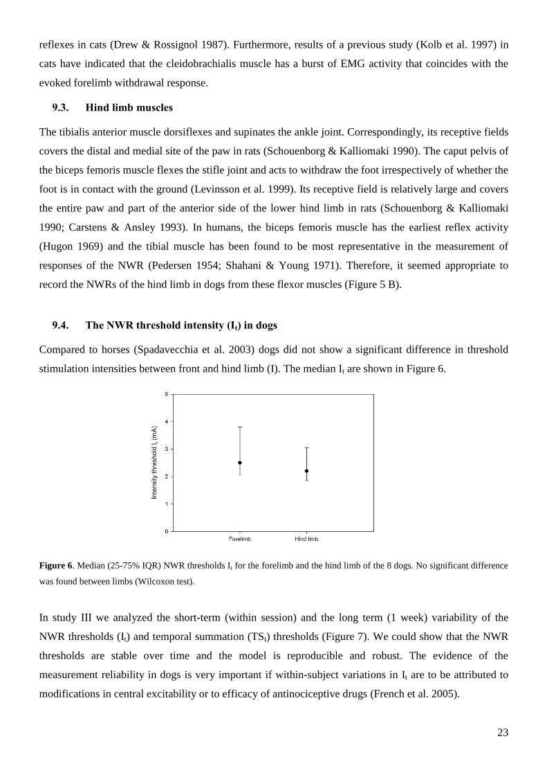
9.4. The NWR threshold intensity (It) in dogs
Compared to horses (Spadavecchia et al. 2003) dogs did not show a significant difference in threshold
stimulation intensities between front and hind limb (I). The median It are shown in Figure 6.
Figure 6. Median (25-75% IQR) NWR thresholds It for the forelimb and the hind limb of the 8 dogs. No significant difference
was found between limbs (Wilcoxon test).
In study III we analyzed the short-term (within session) and the long term (1 week) variability of the
NWR thresholds (It) and temporal summation (TSt) thresholds (Figure 7). We could show that the NWR
thresholds are stable over time and the model is reproducible and robust. The evidence of the
measurement reliability in dogs is very important if within-subject variations in It are to be attributed to
modifications in central excitability or to efficacy of antinociceptive drugs (French et al. 2005).

24
Figure 7. Median (25-75% IQR) forelimb reflex thresholds It of 8 dogs. No significant short term and long term
variability were found (Friedman repeated measures ANOVA).
9.5. Reflex components
To separate reflex components of various origins, the 400 milliseconds post stimulation interval
corresponding to the EMG recording time was divided into 3 epochs: 0 to 20 milliseconds, 20 to 100
milliseconds, and 100 to 400 milliseconds. These epochs were defined on the basis of the conduction
velocities of the canine nerve fibers (Gasser & Erlanger 1927; Burgess & Perl 1967) and the conduction
pathway lengths of the Beagles (Figure 8).
9.5.1. The early reflex activity: 0 to 20 milliseconds epoch
The first epoch is preferentially reflecting non-nociceptive components resulting from the activation of
A afferent nerve fibers. The short latency reflex component of tactile origin has been described for the
upper (Cambier et al. 1974) and lower limb (Hugon 1969; Willer 1977) in humans and in horses in the
(Spadavecchia et al. 2002; Spadavecchia et al. 2003). Its occurrence is highly variable. Based on the
conduction velocity of the sensory afferent fibers in dogs (ulnar nerve: 69.4 6.9 m/s; tibial nerve: 63.4
5.3m/s) (Redding et al. 1982), for a mean afferent distance of 38.5 cm, after adding a mean efferent time
of 2.5 milliseconds and an overall time of 5 milliseconds for spinal and motor endplate delay, the latency
of the canine early reflex should be approximately 14 milliseconds.
At It, none of the dogs showed a clear early reflex between 0 and 20 ms neither for the forelimb nor for
the hind limb muscles.
9.5.2. The NWR: 20 to 100 milliseconds epoch
In the experimental beagles (I to IV), taking into account the A fiber conduction velocity range (4 to 30
m/s) for the afferent component (Gasser & Erlanger 1927; Heinbecker et al. 1933; Burgess & Perl 1967)
and a mean afferent distance of 38.5 cm, after adding a mean efferent time of 2.5 milliseconds and an

25
overall time of 5 milliseconds for spinal and motor endplate delay, the NWR should occur in the 20 to
100 milliseconds post-stimulation epoch. These calculated latencies matched our experimental findings,
confirming the nociceptive origin of the reflex.
9.5.3. The late reflex activity: 100 to 400 milliseconds epoch. Preliminary work
The 100 to 400 milliseconds epoch most likely contains reflex components of mixed spinal and
supraspinal origin (Le-Bars et al. 1992; Andersen et al. 1999). In men, the late reflex activity have be
recorded episodically from the biceps femoris, rectus femoris and more consistently from the tibialis
anterior muscles (Shahani & Young 1971; Roby-Brami & Bussel 1987). In dogs, the late reflex activity
could be recorded from the deltoideus and cleidobrachialis muscles in 0/8 and 0/8 dogs respectively with
the one pulse stimulus paradigm, and in 3/8 and 0/8 dogs with the train-of-five pulses stimulus paradigm.
Late reflex activity was recorded from biceps femoris and tibialis anterior muscles respectively in 2/8 and
3/8 dogs with the one pulse stimulus paradigm, 6/8 and 8/8 dogs with with the train-of-five pulses
stimulus paradigm (I). This late reflex activity occurred 87.1 to 200 ms post-stimulation, being most
pronounced with the train-of-five pulses and almost not present for the single pulse paradigm unless
suprathreshold intensities were used. A tendency to increased reflex size with suprathreshold stimuli was
observed (Figure 8; Table 2).
On the basis of canine C fibres conduction velocity range (0.8 to 1.5 m/s) (Iriuchijima & Zotterman
1961) for the afferent component, after adding a mean efferent time of 2.5 milliseconds and an overall
time of 5 milliseconds for spinal and motor endplate delay, the late EMG response in dogs should occur
between 241 and 818 ms. This agrees with C fibres activity recorded in spinal cats (Le Bars et al. 1976).
Thus, it seems unlikely that the late EMG activity recorded in I is related to C fibres activation.
Additionally the stimuli intensities used here (up to 2 X It) are below intensities needed to activate C
fibres (Le Bars et al. 1976) since it is known that C fibres threshold is 4 –5 times higher than that of
A fibres. Hence, it would be necessary to stimulate the dogs with intensities of 4 to 5 X It to activate C
fibres.
According to recent work the limits for conduction velocities of A fibres are not so clearly demarcated,
with some A fibres having conduction velocities as low as 2.5 m/s associated with higher thresholds
(Kumazawa & Mizumura 1987; Djouhri & Lawson 2004). By taking into account this conduction
velocity, the reflex activity would occur 100 to 192.5 milliseconds after stimulation. The calculated
latency fits with the recorded late reflex activity assuming a direct spinal loop.
Furthermore it is important to remind that in this time frame it is not possible to exclude a supraspinal
loop but more invasive investigations are needed to confirm this hypothesis.
The significant higher incidence in the hind limb compared to forelimb could suggest different functional
adaptation of the limbs.

26
Figure 8. Records form the Biceps Femoris muscle. Stimulations at threshold (It) and suprathreshold (X It) intensities. The
dotted lines delimitate the epochs.
9.5.4. Eliciting the NWR in dogs: conclusions
The analysis of the recruitment curves showed a positive correlation between the intensity of stimulation,
the amplitude of the reflex and the behavioral reaction scores, confirming the nociceptive origin of the
NWR. In dogs the NWR is a complex reflex, whose nociceptive component is only a part of the flexion
reflex circuitry.
9.6. Facilitation of the NWR by repeated stimulations
9.6.1. Wind up and temporal summation
In neurophysiologic experimental settings, repetition of a fixed supramaximal stimulus at low frequency
activates afferent C fibres, which causes an augmented firing of the dorsal horn WDR neurons (Dubner
1991) followed by afterdischarge and increased sensitivity (Price 1972). This activity-dependent
facilitation was termed wind up (Mendell & Wall 1965). The voltage and ligand gated NMDA-receptors
are important for wind up in WDR-neurons. The ongoing afferent input from the C fibres depolarizes the
WDR neurons thus opening the channel (unplugging the Mg2+
ion in the ion channel). The intracellular
Ca2+
concentration further depolarise the cell which activates a protein kinase that contributes to keep the
NMDA channel open, increasing the sensitivity to glutamate (Dickenson 1995; Woolf 1996).
Wind up is only the initial step of the long-lasting state of neuronal hyperexcitability and plastic changes
that develop during central sensitization (You et al. 2004) which may lead to chronic pain states (Arendt-

27
Nielsen et al. 1994; Dickenson 1995; Guirimand et al. 2000). In between other causes, central
sensitisation can be initiated by surgery (Wilder-Smith & Arendt Nielsen 2006).
Studies in rats (Dickenson & Sullivan 1987; Schouenborg & Dickenson 1988), cats (Price 1972), horses
(Spadavecchia et al. 2004; Spadavecchia et al. 2005) and humans (Arendt-Nielsen et al. 1994; Arendt-
Nielsen et al. 2000) have revealed that application of repeated electrical stimulations results in facilitation
of the NWR, as a result of the temporal summation of action potentials at the level of the spinal dorsal
horn neurons. Clinically it is accompanied by an amplified sensation of pain (Hugon 1973; Andersen et
al. 1995a). Therefore in humans, the psychophysical and electrophysiologic responses to repetitive
nociceptive stimulations have been assessed as a noninvasive experimental surrogate of windup (Herrero
et al. 2000; Desmeules et al. 2003). The facilitation of the NWR by repeated stimulations has been used to
investigate the degree of sensorial dysfunction (Curatolo et al. 1995; Desmeules et al. 2003; Banic et al.
2004) and evaluate the analgesic efficacy of drugs in experimental and clinical setting in humans (Willer
& Bathien 1977; Price et al. 1994; Guirimand et al. 2000; Bossard et al. 2002; Escher et al. 2007) and
animals (You et al. 2003a; You et al. 2004; Spadavecchia et al. 2005; Knobloch et al. 2006; Spadavecchia
et al. 2007).
Summation of afferent activity seems to be more pronounced for C fibres mediating second pain
compared to A fibres mediating first pain (Price 1972; Sivilotti et al. 1993).
Many human studies (Andersen et al. 1994; Arendt-Nielsen et al. 1994; Arendt-Nielsen et al. 2000;
Serrao et al. 2004) on temporal summation concentrated on the facilitation of the NWR reflex mediated
by A fibres. Activation of A fibres causes a central discharge that lasts several hundred milliseconds
(Foreman et al. 1975) which can explain why repeated nociceptive electrical stimuli result in facilitated
polysynaptic reflexes.
9.6.2. Temporal summation in conscious dogs (II-IV)
In the studies II to IV, the analysis of the reflex activity focused on the A fibres evoked activity
expressed in the 20 to 100 ms post stimulus intervals. On the basis of canine C fibres conduction velocity
range (0.8 to 1.5 m/s) (Iriuchijima & Zotterman 1961), the reflex activity due to C fibres activation would
appear later, between 250 and 830 ms after each stimulus (Gasser & Erlanger 1927; Hallin & Torebjork
1973; Hugon 1973). Therefore it might be possible, at least with suprathreshold intensities, that C fibres
activity evoked by the first stimuli summates with A activity evoked by the last stimuli in the 2 s epoch.
In dogs (II) the facilitation of the NWR for the forelimb and hind limb occurred at intensities that were
significantly lower than It (Figure 9). At temporal summation intensity TSt, the entire limb was flexed,
whereas only localized joint flexion was induced with a single stimulus; indicating that the nociceptive
impulse was perceived more intensely despite the lower intensity, in accordance with findings in humans
(Price et al. 1978; Arendt-Nielsen et al. 1994; Andersen et al. 1995a; Arendt-Nielsen et al. 2000).

28
The intensities needed to facilitate the reflex where significantly higher for the forelimb, for all
frequencies. The reason for this remains unclear. In humans, few investigations (Bromm & Treede 1980;
Serrao et al. 2006) have analyzed the forearm NWR. In horses, the NWR and its facilitation have been
studied for both fore- and hind limbs (Spadavecchia et al. 2003; Spadavecchia et al. 2004) and only minor
differences in the characteristics of temporal summation between the limbs were noticeable with repeated
stimulations. Whether the spinal neuronal organization of the fore- and hind limb differs or the
supraspinal modulation for the forelimb is more pronounced than that for the hind limb in dogs will
require verification in future studies. It might be assumed on a functional, biomechanical basis that the
sustaining forelimb would be less sensitive to nociceptive stimuli compared to the propulsive hind limb.
Figure 9. Median (25-75% IQR) temporal summation thresholds (TSt) of the 8 dogs of the study (II) expressed as a fraction of
the NWR threshold intensity It. The It fractions needed to facilitate the reflex were significantly lower than It, except for the 5
Hz when corrected for multiple testing (§; Friedman ANOVA followed by a Tuckey test). * Between limbs differences (p<
0.05 Wilcoxon signed rank test).
9.6.3. Temporal summation in conscious dogs: conclusions
The intensity of stimulation affected the magnitude of the reflex response with a significant positive
correlation between the stimulus intensity-response curve and the reflex amplitude-response curve; on the
basis of neuronal recordings in other species, this is probably attributable to spatial summation of the
afferent information at spinal level (Arendt-Nielsen et al. 2000; You et al. 2003b). The behavioral
response scores increased with increasing stimulation intensities as an indication of increased nociception.
This positive correlation between intensity, relative amplitude, and behavioral response scores confirmed
the consistency of experimentally induced temporal summation in dogs. Temporal summation was more
easily elicited from the hind limb compared to the forelimb, the reason for this difference remaining to be
elucidated.
The temporal summation can be used as a model of wind-up in canines both for better understanding of
pathophysiology of chronic pain states and to prove specifically and objectively in pharmacodynamic
studies the efficacy of analgesic drugs in this species as in study IV.
29
10. Variations in the canine reflex
10.1. Posture
Numerous studies in humans have evaluated the NWR in the supine (Hugon 1973; Willer 1977;
DeBroucker et al. 1989) and standing (Hagbarth 1960; Rossi & Decchi 1994; Andersen et al. 2003)
positions. In horses, the NWR recordings were performed in the standing animal with full weight bearing
(Spadavecchia et al. 2002; Spadavecchia et al. 2003). The dogs (I to IV) were non-weight bearing. The
position and therefore the load to which the limb is submitted can modulate the NWR (Paquet et al. 1996;
Andersen et al. 2003) with a significant inverse correlation between the load to which the limb is
subjected and the size of the reflex response (Rossi & Decchi 1994). Thus, care should be taken when
comparing results from different studies.
10.2. Age, sex, circadian variations
In the present studies (I to IV) describing the NWR in conscious dogs, attention was paid to standardize
and control for possible cofactors that could have influenced the results. The NWR threshold and reflex
characteristics are influenced by the extreme of age (Sandrini et al. 1989; Edwards et al. 2003), therefore
adult dogs were retained for the study. Results of clinical studies in humans (Desmeules et al. 2003;
Banic et al. 2004) have indicated that NWR thresholds are often lower in individuals with pain disorders,
compared with healthy persons. None of the dogs in the present study had a painful condition (as assessed
by physical examination). Gender differences in the NWR thresholds are reported in humans (Serrao et al.
2006) with lower thresholds in females. This could be reconducted to the differences between sexes in the
perception and modulation of pain reported for humans (Berkley 1997) and animals (Aloisi et al. 1994;
Cook & Nickerson 2005) or to the differences in motor units of the constituent muscle fibres, thus
influencing the onset latency and the peak-to-peak amplitude of the reflex (van Selms et al. 2005).
Therefore only male dogs were studied (I to IV). All the experiments were performed at the same time of
the day to minimize interindividual circadian variations in NWR thresholds (Sandrini et al. 1986). After
instrumentation, the dogs received 4 test stimuli at different intensities to make them familiar with the
experimental method prior to formal threshold measurement. It was noticed during pilot work in dogs that
the reflex thresholds increased and then stabilized over time; this can be explained by high levels of
anxiety, which may increase central excitability as indexed by lowering of NWR thresholds (Willer 1980;
Willer & Albe-Fessard 1980; Willer 1983).
10.3. Habituation
By repeating electrical stimulations one can assist to a gradual decrease in the NWR amplitude, a purely
spinal phenomenon defined as “habituation”. Habituation is intensity and frequency dependent, occurring
more frequently at low intensities and at high stimulation frequencies (Shahani & Young 1971;

30
Dimitrijevic et al. 1972). In all studies (I to IV), at least 60 s between successive single stimuli were
allowed in order to avoid habituation which could have reduced reflex amplitude (Shahani & Young
1971).
When repeated stimulation were given (II) a decrease in the reflex facilitation was noticeable at all
intensities with the 20 Hz frequency and only at suprathreshold intensities for the 2 and 5 Hz frequencies.
This could be related to habituation but also to supraspinal descending inhibitory processes (Gozariu et al.
1997).
11. Pharmacological modulation of the NWR and temporal summation
Pharmacological modulation of the NWR is considered to occur when a drug modifies the NWR
threshold intensity and reflex characteristics. Analgesic activity is generally attributed when an increase
in the It or a reduction in the reflex amplitude or magnitude of temporal summation occur after its
administration.
11.1. Low-dose acepromazine (III).
One of the future goals is to implement the NWR and temporal summation model in clinical practice (see
later), as tools to detect and quantify the degree of sensory dysfunction in dogs affected from chronic
malignant or non malignant pathologies. Therefore to augment the compliance of canine patients to the
measurement technique, well-being and reduce stress, the pre-emptive administration of a neuroleptic
drug would be indicated. The ideal drug should be anxyolitic, safe, and deprived of antinociceptive action
which could exert a modulatory effect on the test altering its validity.
Based on clinical experience and previous work (Dasgupta & Werner 1955; Krivoy 1957; Silvestrini &
Maffii 1959) it was hypothesized that 0.01 mg kg-1
IV acepromazine would provide sufficient
tranquillisation for the purpose of the recordings while having minimal impact on the model and side
effects.
11.1.1. Use of phenotiazine tranquillizers in human and veterinary medicine
In human medicine, the phenotiazine derivatives, i.e. chlorpromazine, were very popular in the 1950s as
major tranquillizers to treat psychosis and other major psychiatric disorders. They were also used as
preanesthetic sedatives and for neuroleptoanalgesia (Brown 1969). Because of the major undesirable side
effects as intraoperative hypotension and dysphoria, their use has been supplanted by the availability of
the less toxic benzodiazepines. Neuroleptanalgesia remains a mainstay of veterinary anaesthesia and
acepromazine, a member of the phenotiazine family, is the most widely used sedative (Barnhart et al.
2000b). Acepromazine effect is dose related. Doses below 0.03 mg kg-1
it acts as a tranquillizer, exerting
a calming effect on the behaviour of excitable animals with minimal cortical depression (Pugh 1964; Hall
et al. 2001; Plumb 2002). With increasing dose sedation occurs, up to a plateau with prolonged duration

31
and higher incidence of side-effects. The central nervous system effects of acepromazine are attributed to
its antagonistic action at the D1 and D2 dopamine receptors. The dopaminergic neurons are
predominantly located in the reticular formation and modulate complex functions as arousal, movement,
posture, pain, and autonomic function. They provide a major ascending input to the cerebral cortex and
the basal ganglia, that is important for initiation of behavioural responses (Sapper 2000). Because of the
depression of basal ganglia activity, care has to be taken in the interpretation of the antinociceptive
activity of acepromazine when using models with behavioural endpoints (Steagall et al. 2008)
11.1.2. Effects of acepromazine on NWR and temporal summation in dogs
Low-dose acepromazine exerted a mild tranquillization lasting 30 minutes without modifying the NWR
threshold nor the NWR characteristics recorded in the 20 to 100 ms interval as latency, amplitude and
stimulus-response curve at any time point after administration (Figure 10). This indicates that
acepromazine did not inhibit A fibre evoked reflex activity nor affected the motor outflow. Our findings
are consistent with previous work, where acepromazine did not alter the baseline nociceptive thresholds
in a canine thermal and pressure nociceptive model (Barnhart et al. 2000b). Low-dose acepromazine did
not affect the temporal summation threshold, nor the positive correlation between the magnitude of
temporal summation (as measured by the area under the temporal summation curve) and its perception (as
measured by the evoked behavioural response scores) confirming the consistency of this experimental
model.
In conclusion, acepromazine can be used to facilitate data recording in anxious subjects without
altering the validity of the NWR model.
Figure 10. Representative electromyograms evoked at It intensity recorded from the deltoid muscle of a dog before and 20, 60
and 100 min after drug administration. The arrow indicates the start of the electrical stimulus. The dotted to dash-dotted lines

32
represent the 20 to 100 ms epoch and the dash-dotted lines represent the 100 to 200 ms epoch. ACP: Acepromazine; SAL:
saline.
11.1.3. Phenotiazine analgesia: mythos or reality?
The analgesic activity of phenotiazines remains a controversial topic (McGee & Alexandre 1979). To date
only for methotrimeprazine there is evidence for reliable dose related analgesia in men (McGee &
Alexandre 1979; Patt et al. 1994). The exact mechanism of action is not clear (Roberts et al. 1982). Due
to its depressing action on the reticular formation (Preston 1956; Engberg et al. 1968), acepromazine
could modulate nociception by reducing the afferent information to the cortex or by enhancing the tonic
activity of the descending inhibitory pathways. As stated in paragraph 9.5.3, the late reflex activity
recorded in the 100 to 200 ms epoch should contain signals of mixed spinal and supraspinal origin.
Therefore to specifically analyse this interval could improve the understanding of the mechanism of
action of phenotiazine. Acepromazine at the dose used in study IV, didn’t alter the late reflex activity
when single or repeated stimulations were used, indicating that the supraspinal control of the reflex was
comparable between treatments.
Our results confirm that low-dose acepromazine is deprived of antinociceptive properties in dogs (Gross
2001; Plumb 2002).
11.2. Low-dose ketamine constant-rate-infusion (IV)
Ketamine is a phencyclidine congener, and the molecule exists as two optical isomers R (-) and S (+)
ketamine; the racemic mixture is currently used clinically. In veterinary medicine ketamine is commonly
used for induction and maintenance of anaesthesia in a wide variety of species (Wright 1982). In men, it
has found a niche for anaesthesia in emergency situations. Its usefulness however is limited by its
undesirable psychic emergence effects.
The neuropharmacology ketamine is complex: the drug interacts with multiple binding sites, including N-
methyl-D-aspartate (NMDA) and non-NMDA glutamate receptors, nicotinic and muscarinic cholinergic,
opioid and monoaminergic receptors. All of these interactions play a role in pharmacological and clinical
properties of ketamine. However, the NMDA receptor antagonism accounts for most of the analgesic,
amnesic, psychomimetic effects of the compound. From animal (Hao et al. 1998) and human (Woolf &
Thompson 1991; Kohrs & Durieux 1998) experimental research there is evidence that due to its
antagonistic action at NMDA receptors, ketamine can modulate spinal “wind-up” and central sensitisation
in contrast to volatile agents such as isoflurane (Petersen-Felix et al. 1996). With a mechanism-based
approach, ketamine has been used in humans to implement peri-operative pain management (Woolf &
Max 2001; McCartney et al. 2004) and treat traumatic, neuropathic and chronic pain (Stubhaug & Breivik
1997; Carr et al. 2004). The clinical effects of ketamine are dose-dependent ranging from sedation with
plasma levels close to 300 ng ml-1
(Schmid et al. 1999; Rogers et al. 2004) to anesthesia when the plasma
33
concentration is above 1,000 ng ml-1
in humans (Domino et al. 1982) and above 3,000 ng ml-1
in dogs
(Kaka & Hayton 1980). In humans, to avoid the psychomimetic side effects which limit its clinical
acceptance, low sub-anaesthetic doses (Schmid et al. 1999; Richebe et al. 2005) or the use of S-ketamine
which produces fewer psychomimetic disturbances and less agitation than the racemic mixture
(Hempelmann & Kuhn 1997 ) has been recommended.
11.2.1. Ketamine as an analgesic in dogs
Extrapolating from these encouraging results in man, low-dose ketamine is increasingly used in canines
for its analgesic properties as part of a balanced anaesthesia /analgesia protocols. Slingby et al. (2000)
described improved post-operative analgesia in dogs undergoing ovariohysterectomy after a 2.5 mg kg-1
ketamine bolus. Still its antinociceptive effect was short lived and associated with excessive sedation.
Therefore they suggested to administer ketamine by CRI to prolong the duration of analgesia and
decrease the side effects as it is done in man (Schmid et al. 1999; Richebe et al. 2005). Wagner et al.
(2000) added a low-dose ketamine CRI to the balanced anesthetic protocol in dogs undergoing forelimb
amputation. They could show only slight improvement of the pain scores at 12 and 18 h post-operatively
and activity scores 72 h post-operatively compared to saline. To date the published evidence of ketamine
analgesia in dogs is scarce and no data is available on its effective antinociceptive plasma concentration
in this species.
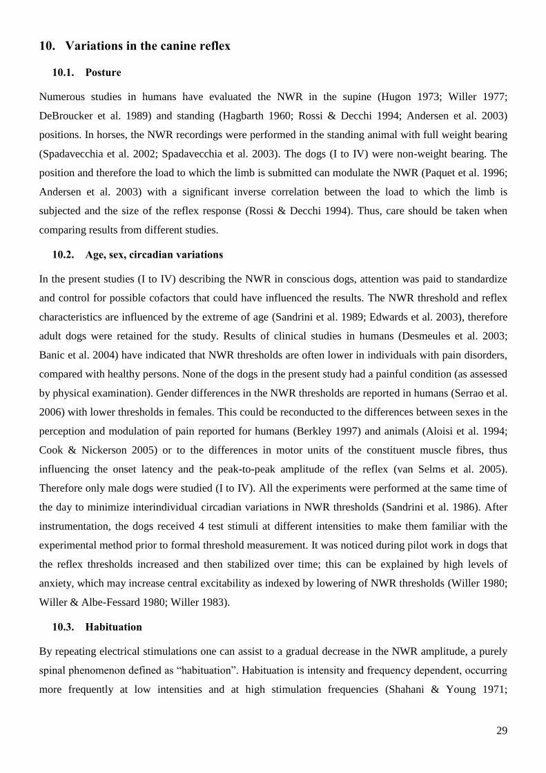
Therefore the aim of study IV was to evaluate quantitatively the antinocieptive efficacy of a usual low-
dose ketamine CRI in dogs and to correlate its efficacy with the enantioselectively measured plasma
levels of the drug and its metabolite norketamine.
After baseline measurements a 0.5 mg kg-1
loading bolus followed by 10 g kg-1
min-1
CRI of ketamine
for 59 min were given intravenously (Figure 11). Electrophysiological measurements were repeated 1, 4,
8, 12, 20, 40 and 80 min post bolus. Contemporaneously, evoked behavioral responses and sedation were
scored and side effects recorded by the mean of a purposefully developed sedation score (Table 3) .
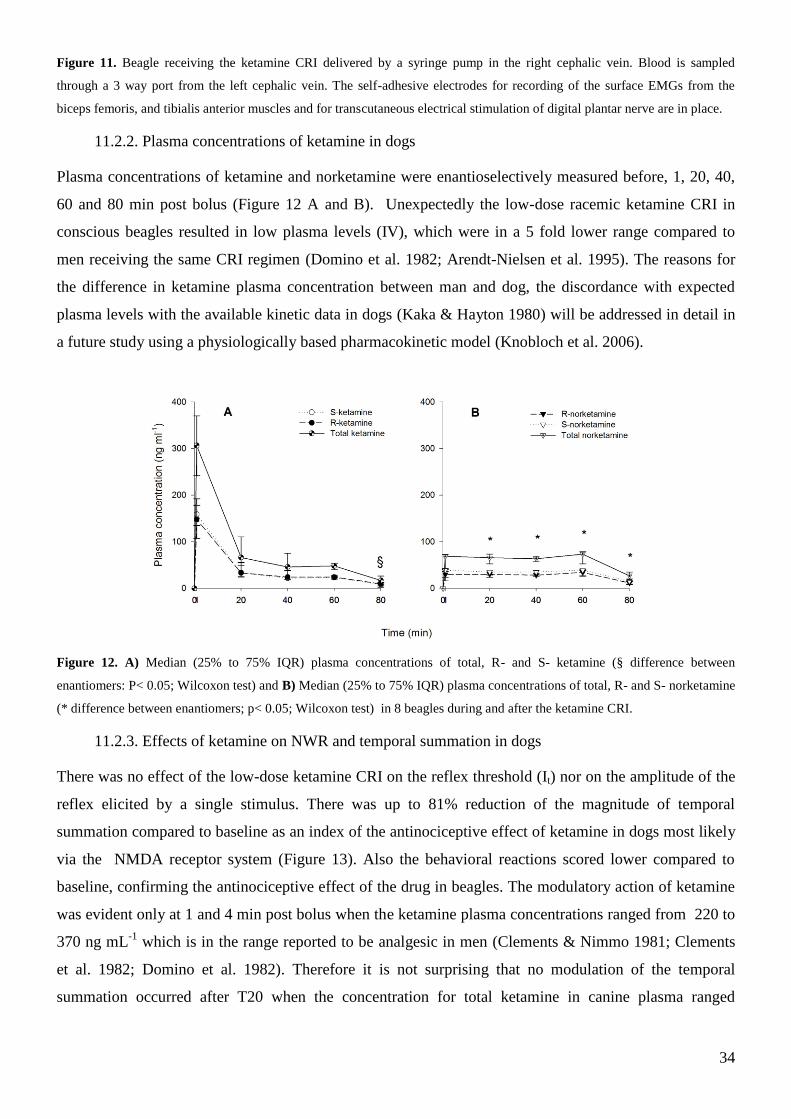
34
Figure 11. Beagle receiving the ketamine CRI delivered by a syringe pump in the right cephalic vein. Blood is sampled
through a 3 way port from the left cephalic vein. The self-adhesive electrodes for recording of the surface EMGs from the
biceps femoris, and tibialis anterior muscles and for transcutaneous electrical stimulation of digital plantar nerve are in place.
11.2.2. Plasma concentrations of ketamine in dogs
Plasma concentrations of ketamine and norketamine were enantioselectively measured before, 1, 20, 40,
60 and 80 min post bolus (Figure 12 A and B). Unexpectedly the low-dose racemic ketamine CRI in
conscious beagles resulted in low plasma levels (IV), which were in a 5 fold lower range compared to
men receiving the same CRI regimen (Domino et al. 1982; Arendt-Nielsen et al. 1995). The reasons for
the difference in ketamine plasma concentration between man and dog, the discordance with expected
plasma levels with the available kinetic data in dogs (Kaka & Hayton 1980) will be addressed in detail in
a future study using a physiologically based pharmacokinetic model (Knobloch et al. 2006).
Figure 12. A) Median (25% to 75% IQR) plasma concentrations of total, R- and S- ketamine (§ difference between
enantiomers: P< 0.05; Wilcoxon test) and B) Median (25% to 75% IQR) plasma concentrations of total, R- and S- norketamine
(* difference between enantiomers; p< 0.05; Wilcoxon test) in 8 beagles during and after the ketamine CRI.
11.2.3. Effects of ketamine on NWR and temporal summation in dogs
There was no effect of the low-dose ketamine CRI on the reflex threshold (It) nor on the amplitude of the
reflex elicited by a single stimulus. There was up to 81% reduction of the magnitude of temporal
summation compared to baseline as an index of the antinociceptive effect of ketamine in dogs most likely
via the NMDA receptor system (Figure 13). Also the behavioral reactions scored lower compared to
baseline, confirming the antinociceptive effect of the drug in beagles. The modulatory action of ketamine
was evident only at 1 and 4 min post bolus when the ketamine plasma concentrations ranged from 220 to
370 ng mL-1
which is in the range reported to be analgesic in men (Clements & Nimmo 1981; Clements
et al. 1982; Domino et al. 1982). Therefore it is not surprising that no modulation of the temporal
summation occurred after T20 when the concentration for total ketamine in canine plasma ranged

35
between 50 and 100 ng mL-1
, concentrations which are reported to be sub-analgesic in men (Clements &
Nimmo 1981; Grant et al. 1981; Clements et al. 1982). Transitory psychomimetic side effects were seen
after the loading bolus in all dogs as a moderate sedation (median score of 3.5 over 12) which unlikely
affected the results of the electrophysiological tests.
Figure 13. Median values showing the effect of ketamine on the temporal summation curves compared to baseline
(T0). The repeated stimulations (10 stimuli, 5 Hz during 2 s) were given at temporal summation threshold intensity.
* Values of p (significance set at p< 0.05) derived via a Dunn’s post hoc test after a significative Friedman
repeated-measure ANOVA (p< 0.01)
12. Conclusions and Future applications
In conclusion, the work presented in this PhD thesis has provided a new, non invasive, robust
experimental model of nociception in conscious dogs and has established a “baseline” condition for using
the model in clinical routine to study the antinociceptive activity of drugs or to quantify the excitability of
the nervous system in individual canine patients.
Study I showed that the NWR can be evoked from the fore- and hind limbs in dogs. The positive
correlation between the intensity of stimulation and amplitude of the reflex and between the intensity of
stimulation and behavioral reaction score confirms the nociceptive origin of the NWR. The train-of-five
stimulus paradigm can be used as a standard stimulus. Thus, assessment of the NWR is proposed as a
neurophysiologic tool for quantifying nociception in dogs
In study II by applying repeated stimulations, temporal summation was evoked. Temporal summation
appeared to be more easily elicited from the hind limb, compared with the forelimb, but the reason for
this difference remains to be elucidated. The 5-Hz frequency is recommended as the standard for future

36
studies in dogs. The evaluation of the temporal summation-evoked reflexes may be used to give
information about changes in nociceptive system gain when analgesics are administered.
In study III, the stability of the NWR thresholds was demonstrated for the first time. A low-dose
acepromazine exerted a mild tranquillization lasting 30 minutes without affecting NWR and TS
thresholds, reflex characteristics, behavioural responses and supraspinal control after single and repeated
stimulations. These findings suggest that acepromazine is deprived of antinociceptive action in dogs.
Intravenous 0.01 mg kg-1
acepromazine can facilitate the recordings in anxious dogs in clinical practice
without altering the validity of this model.
In study IV, temporal summation was used for the first time to evaluate quantitatively the analgesic
efficacy of a low-dose ketamine CRI in conscious dogs. Ketamine reduced considerably temporal
summation. However its antinociceptive action was short lived most likely due to the low plasma level
obtained, therefore we cannot recommend this low-dose ketamine CRI regimen as sole analgesic in dogs.
Further research to find a CRI regimen for dogs resulting in stable antinociceptive plasma levels with
minimal side effects should be undertaken.
There are many interesting future implementation possibilities based on the basic work presented in this
thesis, i.e. to confirm the modular organization of the withdrawal reflexes in dogs and to investigate the
effect of sex on the NWR and its characteristics.
By evaluating the modulating effects on NWR and temporal summation in an experimental setting, it will
be possible to bring evidence of species-specific antinociceptive efficacy of different drugs,. Based on the
results of study III and the upcoming pharmacokinetic study, we plan to find a ketamine CRI regimen
resulting in stable antinociceptive plasma levels in dogs with minimal psychomimetic side effects. The
modulatory effects on NWR and temporal summation of buprenorphine, a partial agonist, could be
studied too. Buprenorphine may represent a new therapeutic options for dogs affected of neuropathic and
neoplastic pain requiring long term analgesic treatment, as it appears to dampen central sensitization
(Kress; Penza et al. 2007). Another drug worthy investigating is tropisetron (5-hydroxytryptamine-3
receptor antagonist), which acts at spinal level and modulate central sensitization of dorsal horn neurons.
The final goal would be for the benefit of the canine patients by offering better treatment strategies for
central hypersensitivity especially for dogs not responding to conventional analgesics.
In humans, there is ongoing research to use the NWR and temporal summation as objective tools to detect
and quantify central hyperexcitability in individual patients (Desmeules et al. 2003; Banic et al. 2004;
Curatolo et al. 2004). In the same way, it is foreseen to implement the NWR and temporal summation
model after acepromazine sedation in clinical routine as tools to detect and quantify the degree of sensory
dysfunction in dogs affected from chronic malignant or non malignant pathologies (Hielm-Björkman et
al. 2003; Beckman 2006).
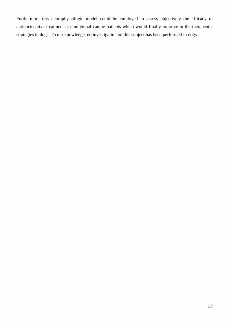
37
Furthermore this neurophysiologic model could be employed to assess objectively the efficacy of
antinociceptive treatments in individual canine patients which would finally improve in the therapeutic
strategies in dogs. To our knowledge, no investigation on this subject has been performed in dogs.

38
13. Tables
Table 1. Evoked behavioral responses score
Score Single stimulation Repeated stimulation
0 No movement No movement
1 Slight flexion of carpus/tarsus Muscle twitch
2 Flexion of elbow/stifle joint Flexion of elbow/stifle joint followed by
relaxation
3 Brisk flexion of elbow/stifle joint Flexion of entire limb followed by relaxation
4 Brisk flexion of the limb and flexion maintained Flexion of limb, and flexion maintained
5 Brisk flexion of the limb and general awareness (ie,
turning the head toward the stimulated limb or
attempts to stand from a lying position)
Sustained flexion of limb and general
awareness (ie, turning the head toward the
stimulated limb or attempts to stand from a
lying position)
6 Brisk flexion of the limb, general awareness and
vocalization
Sustained limb flexion, general awareness, and
vocalization.
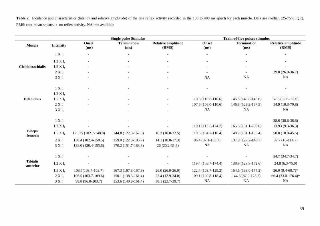
39
Table 2. Incidence and characteristics (latency and relative amplitude) of the late reflex activity recorded in the 100 to 400 ms epoch for each muscle. Data are median (25-75% IQR).
RMS: root-mean-square. -: no reflex activity. NA: not available
Single pulse Stimulus Train-of-five pulses stimulus
Muscle Intensity Onset
(ms)
Termination
(ms)
Relative amplitude
(RMS)
Onset
(ms)
Termination
(ms)
Relative amplitude
(RMS)
1 X It - - - - - -
1.2 X It - - - - - -
Cleidobrachialis 1.5 X It - - - - - -
2 X It - - - - - 29.8 (26.0-36.7)
3 X It - - - NA NA NA
1 X It - - - - - -
1.2 X It - - - - - -
Deltoideus 1.5 X It - - - 110.6 (110.6-110.6) 146.8 (146.8-146.8) 52.6 (52.6- 52.6)
2 X It - - - 107.6 (106.0-110.6) 146.8 (129.2-157.5) 14.9 (10.3-70.8)
3 X It - - - NA NA NA
1 X It - - - - - 38.6 (38.6-38.6)
1.2 X It - - - 119.1 (113.5-124.7) 165.5 (131.1-200.0) 13.93 (9.3-36.3)
Biceps
femoris 1.5 X It 125.75 (102.7-148.8) 144.8 (122.3-167.3) 16.3 (10.0-22.5) 110.5 (104.7-116.4) 148.2 (131.1-165.4) 50.0 (18.9-45.5)
2 X It 130.4 (102.4-158.5) 159.0 (122.3-195.7) 14.1 (10.8-17.3) 96.4 (87.1-105.7) 137.9 (127.2-148.7) 37.7 (10-114.7)
3 X It 138.0 (120.4-155.6) 170.2 (151.7-188.8) 26 (20.2-31.8) NA NA NA
1 X It - - - - - 34.7 (34.7-34.7)
Tibialis
anterior 1.2 X It - - - 119.4 (103.7-174.4) 138.9 (129.9-152.6) 24.8 (6.3-75.0)
1.5 X It 103.7(103.7-103.7) 167.3 (167.3-167.3) 26.0 (26.0-26.0) 122.4 (103.7-129.2) 154.6 (138.0-174.2) 26.0 (9.4-68.7)*
2 X It 106.5 (103.7-109.6) 150.1 (138.5-161.4) 23.4 (12.9-34.0) 109.1 (100.8-118.4) 144.3 (87.9-128.2) 66.4 (23.0-176.4)*
3 X It 98.8 (96.0-103.7) 153.6 (140.9-161.4) 38.1 (23.7-39.7) NA NA NA
40
Table 3. Composite sedation score. The sedation score (0 to 12; no sedation to deep sedation) was assigned by adding up the
ranking of different descriptors
Consciousness Eye Responsiveness Relaxation
0 Awake Not rotated Responds to voice Moves spontaneously
1 Aware Moderate rotation Responds to touch Relaxed, no shivering
2 Not aware but arousable Rotated Do not respond to touch Very relaxed
3 Not aware, not arousable Nystagmus Hyperexcitable Muscle tonus

41
14. References
Akopian AN, Sivilotti L, Wood JN (1996) A tetrodotoxin-resistant voltage-gated sodium channel
expressed by sensory neurons. Nature 379, 257-262.
Aloisi AM, Albonetti ME, Carli G (1994) Sex differences in the behavioral response to persistent pain in
rats. Neurosci Lett 179, 09-82.
Andersen OK (2007) Studies of the organization of the human nociceptive withdrawal reflex. Acta
Physiologica 189, 1-35.
Andersen OK, Gracely RH, Arendt-Nielsen L (1995a) Facilitation of the human nociceptive reflex by
stimulation of A beta-fibres in a secondary hyperalgesic area sustained by nociceptive input from
the primary hyperalgesic area. Acta Physiol Scand 155, 87-97.
Andersen OK, Jensen LM, Brennum J et al. (1994) Evidence for central summation of C and A delta
nociceptive activity in man. Pain 59, 273-280.
Andersen OK, Jensen LM, Brennum J et al. (1995b) Modulation of the human nociceptive reflex by
cyclic movements. Eur J Appl Physiol 70, 311-321.
Andersen OK, Sonnenborg F, Matjacic Z et al. (2003) Foot-sole reflex receptive fields for human
withdrawal reflexes in symmetrical standing position. Exp Brain Res 152, 434-443.
Andersen OK, Sonnenborg FA, Arendt-Nielsen L (1999) Modular organization of human leg withdrawal
reflexes elicited by electrical stimulation of the foot sole. Muscle Nerve 22, 1520-1530.
Andersen OK, Sonnenborg FA, Arendt-Nielsen L (2001) Reflex receptive fields for human withdrawal
reflexes elicited by non-painful and painful electrical stimulation of the foot sole. Clin
Neurophysiol 112, 641-649.
Andrews HL, Workman W (1941) Pain threshold measurements in the dog. Journal of Pharmacological
and experimental therapeutics 73, 99-103.
Arendt-Nielsen L, Anker-Møller E, Bjerring P et al. (1990) Onset phase of spinal bupivacaine analgesia
assessed quantitatively by laser stimulation. Br J Anaesth 65, 639-642.
Arendt-Nielsen L, Brennum J, Sindrup S et al. (1994) Electrophysiological and psychophysical
quantification of temporal summation in the human nociceptive system. Eur J Appl Physiol 68,
266-273.
Arendt-Nielsen L, Petersen-Felix S, Fischer M et al. (1995) The effect of N-methyl-D-aspartate
antagonist (ketamine) on single and repeated nociceptive stimuli: a placebo-controlled
experimental human study. Anesth Analg 81, 63-68.
Arendt-Nielsen L, Sonnenborg FA, Andersen OK (2000) Facilitation of the withdrawal reflex by repeated
transcutaneous electrical stimulation: an experimental study on central integration in humans. Eur
J Appl Physiol 81, 165-173.
Bajaj P, Arendt Nielsen L, Andersen O (2005) Facilitation and inhibition of withdrawal reflexes
following repetitive stimulation: electro- and psychophysiological evidence of activation of
noxious inhibitory control in humans. Eur J Pain 9, 25-31.
Banic B, Petersen-Felix S, Andersen OK et al. (2004) Evidence for spinal cord hypersensitivity in chronic
pain after whiplash injury and in fibromyalgia. Pain 107, 7-15.
Barnhart MD, Hubbell JA, Muir WW et al. (2000a) Pharmacokinetics, pharmacodynamics, and analgesic
effects of morphine after rectal, intramuscular, and intravenous administration in dogs. Am J Vet
Res 61, 24-28.
Barnhart MD, Hubbell JAE, Muir WW (2000b) Evaluation of the analgesic properties of acepromazine
maleate, oxymorphone, medetomidine and a combination of acepromazine-oxymorphone. Vet
Anesth Analg 27, 89-96.
Beckman B (2006) Pathophysiology and management of surgical and chronic oral pain in dogs and cats. J
Vet Dent 23, 50-60.
Berkley KJ (1997) Sex differences in pain. Behav Brain Sci 20.
Bjorkman R, Ullman A, Hedner J (1993) Morphine-sparing effect of diclofenac in cancer pain. Eur J Clin
Pharmacol 44, 1-5.
Bossard AE, Guirimand F, Fletcher D et al. (2002) Interaction of a combination of morphine and
ketamine on the nociceptive flexion reflex in human volunteers. Pain 98, 47-57.

42
Bromm B, Treede RD (1980) Withdrawal reflex, skin resistance reaction and pain ratings due to electrical
stimuli in man. Pain 9, 339-354.
Brown AS (1969) Neuroleptoanalgesia. Int Anesthesiol Clin 7, 159-175.
Brown DC, Bernier N, Shofer F et al. (2002a) Effect of intrathecal and intravenous administration of
oxytocin on amplitude of the reflex-evoked muscle action potential after electrical stimulation of
the tooth pulp in anesthetized dogs. Am J Vet Res 63, 1354-1358.
Brown DC, Bernier N, Shofer F et al. (2002b) Use of noninvasive dental dolorimetry to evaluate
analgesic effects of intravenous and intrathecal administration of morphine in anesthetized dogs.
Am J Vet Res 63, 1349-1353.
Burgess PR, Perl ER (1967) Myelinated afferent fibres responding specifically to noxious stimulation of
the skin. J Physiol (Lond) 190, 541-562.
Cambier J, Dehen H, Bathien N (1974) Upper limb cutaneous polysynaptic reflexes. J Neurol Sci 22, 39-
49.
Carr DB, Goudas LC, Denman WT et al. (2004) Safety and efficacy of intranasal ketamine for the
treatment of breakthrough pain in patients with chronic pain: a randomized, double-blind,
placebo-controlled, crossover study. Pain 108, 17-27.
Carstens E, Ansley D (1993) Hind limb flexion withdrawal evoked by noxious heat in conscious rats:
magnitude measurement of stimulus-response function, suppression by morphine and habituation.
J Neurophysiol 70, 621-629.
Chan CW, Dallaire M (1989) Subjective pain sensation is linearly correlated with the flexion reflex in
man. Brain Res 479, 145-150.
Cimino Brown D, Boston RC, Coyne JC et al. (2007) Development and psychometric testing of an
instrument to measure chronic pain in dogs with osteoarthritis. Am J Vet Res 68, 631-637.
Clarke RW, Harris J (2001) The spatial organization of central sensitization of hind limb flexor reflexes
in the decerebrated, spinalized rabbit. Eur J Pain 5, 175-185.
Clements J, Nimm W, Grant I (1982) Bioavailability, pharmacokinetics, and analgesic activity of
ketamine in humans. J Pharmacol Sci May;71, 539-542.
Clements JA, Nimmo WS (1981) Pharmacokinetics and analgesic effect of ketamine in man. Br J
Anaesth 53, 27-30.
Cook CD, Nickerson MD (2005) Nociceptive Sensitivity and Opioid Antinociception and
Antihyperalgesia in Freund's adjuvant-Induced Arthitic Male and female Rats. J Pharmacol Exp
Ther 313, 449-459.
Curatolo M, Arendt-Nielsen L, Petersen-Felix S (2004) Evidence, Mechanisms and Clinical Implications
of Central Hypersensitivity in Chronic Pain after Whiplash Injury. Clin J Pain 20, 469-476.
Curatolo M, Petersen-Felix S, Arendt-Nielsen L et al. (1995) Temporal summation during extradural
anaesthesia. Br J Anaesth 75, 634-635.
Curatolo M, Petersen-Felix S, Arendt-Nielsen L et al. (1997) Spinal anaesthesia inhibits central temporal
summation. Br J Anaesth 78, 88-89.
Dasgupta SR, Werner G (1955) Inhibitory action of chlorpromazine on motor activity. Arch int
pharmacodyn 3-4, 409-417.
DeBroucker T, Willer J.C., Bergeret S (1989) The nociceptive flexion reflex in humans: a specific and
objective correlate of experimental pain. In: Chapman CR & Loeser JD (eds). Issue in pain
measurement. Raven Press, Ltd., New York. pp. 337-352.
Desmeules JA, Cedraschi C, Rapiti E et al. (2003) Neurophysiologic evidence for a central sensitization
in patients with fibromyalgia. Arthritis Rheum 48, 1420-1429.
Devor M (1994) The pathophysiology of damaged peripheral nerves. In: Melzack R & Wall PD (eds).
Textbook of Pain. Churchill Livingstone, London. pp. 79-100.
Dickenson AH (1995) Spinal pharmacology of pain. Br J Anaesth 75, 193-200.
Dickenson AH, Sullivan AF (1987) Evidence for a role of the NMDA receptor in the frequency
dependent potentiation of deep rat dorsal horn nociceptive neurons following C fiber stimulation.
Neuropharmacology 26, 1235-1238.

43
Dimitrijevic M, Faganel J, Gregoric M et al. (1972) Habituation: effects of regular and stochastic
stimulation. J Neurol Neurosurg Psychiatry 35, 234-242.
Djouhri L, Lawson SN (2004) Abeta-fiber nociceptive primary afferent neurons: a review of incidence
and properties in relation to other afferent A-fiber neurons in mammals. Brain Research Brain
Research Reviews 46, 131-145.
Domino E, Zsigmond E, Domino L et al. (1982) Plasma levels of ketamine and two of its metabolites in
surgical patients using a gas chromatographic mass fragmentographic assay. Anesth Analg 61, 87-
92.
Drew T, Rossignol S (1987) A kinematic and electromyographic study of cutaneous reflexes evoked from
the forelimb of unrestrained walking cats. J Neurophysiol 57, 1160-1184.
Dubner R (1991) Neuronal plasticity in the spina and medullary dorsal horns: a possible role in central
pain mechanisms. In: Casey KL (ed). Pain and Central Nervous System Disease: The Central Pain
Syndromes. Raven Press, New York. pp. 143-155.
Edwards RR, Fillingim RB, Ness TJ (2003) Age-related differences in endogenous pain modulation: a
comparison of diffuse noxious inhibitory control in healthy older and younger adults. Pain 101,
155-165.
Engberg I, Lundberg A, Ryall RW (1968) Reticulospinal inhibition of transmission in reflex pathways. J
Physiol (Lond) 194, 201-223.
Escher M, Daali Y, Chabert J et al. (2007) Pharmacokinetic and Pharmacodynamic Properties of
Buprenorphine After a Single Intravenous Administration in Healthy Volunteers: A Randomized,
Double-Blind, Placebo-Controlled, Crossover Study. Clinical Therapeutics 29, 1620-1631.
Farina D, Cescon C, Merletti R (2002) Influence of anatomical, physical, and detection-system
parameters on surface EMG. Biol Cybern 86, 445-456.
Foreman RD, Applebaum AE, Beall JE et al. (1975) Responses of primate spinothalamic tract neurons to
electrical stimulation of hindlimb peripheral nerves. J Neurophysiol 38, 132-145.
French DJ, France CR, France JL et al. (2005) The influence of acute anxiety on assessment of
nociceptive flexion reflex thresholds in healthy young adults. Pain 114, 358-363.
Gasser H, Erlanger J (1927) The role played by the sizes of the constituent fibers of a nerve trunk in
determining the form of its action potential wave. Am J Physiol 80, 522-547.
Gozariu M, Bragard D, Willer JC et al. (1997) Temporal summation of C-fiber afferent inputs:
competition between facilitatory and inhibitory effects on C-fiber reflex in the rat. J Neurophysiol
78, 3165-3179.
Grant IS, Nimmo WS, Clements JA (1981) Pharmacokinetics and analgesic effects of i.m. and oral
ketamine. Br J Anaesth 53, 805-810.
Gross ME (2001) Tranquillizers, adrenergic agonists and related agents. In: Adams HR (ed).
Veterinary Pharmacology and Therapeutics. Iowa State University Press, Ames, Iowa. pp. 299-
342.
Guirimand F, Dupont X, Brasseur L et al. (2000) The effects of ketamine on the temporal summation
(wind-up) of the R(III) nociceptive flexion reflex and pain in humans. Anesth Analg 90, 408-414.
Hagbarth KE (1960) Spinal withdrawal reflexes in the human lower limbs. J Neurol Neurosurg
Psychiatry 23, 222-227.
Hall LW, Clarke KW, Trim CM (2001) Principles of sedation, analgesia and premedication. Veterinary
Anesthesia. W.B. Saunders, London, UK. pp. 75-112.
Hallin RG, Torebjork HE (1973) Electrically induced A and C fibre responses in intact human skin
nerves. Exp Brain Res 16, 309-320.
Hamlin RL, Bednarski LS, Schuler CJ et al. (1988) Method of objective assessment of analgesia in the
dog. J Vet Pharmacol Ther 11, 215-220.
Hansen BD (2003) Assessment of pain in dogs: veterinary clinical studies. Institute of Laboratory
Animals Research Journal 44, 197-205.
Hao JX, Sjolund BH, Wiesenfeld-Hallin Z (1998) Electrophysiological evidence for an antinociceptive
effect of ketamine in the rat spinal cord. Acta Anaesthesiol Scand 42, 435-441.

44
Heinbecker P, Bishop GH, O'Learly J (1933) Pain and touch fibres in peripheral nerves. Archives
Neurology and Psychiatry 29, 771-789.
Hempelmann G, Kuhn DFM (1997 ) Klinischer Stellenwert des S(+)-Ketamin. Anaesthesist 46, S3-S7.
Herrero JF, Laird JMA, Lopez-Garcia JA (2000) Wind-up of spinal cord neurones and pain sensation:
much ado about something? Prog Neurobiol 61, 169-203.
Hielm-Björkman A, Kuusela E, Liman A et al. (2003) Evaluation of methods for assessment of pain
associated with chronic osteoarthritis in dogs. J Am Vet Med Assoc 222, 1552-1558.
Holton L, Reid J, Scott EM et al. (2001) Development of a behaviour-based scale to measure acute pain
in dogs. The Veterinary Records 148, 525-531.
Holton LL, Scott EM, Nolan AM et al. (1998) Comparison of three methods used for assessment of pain
in dogs. Journal of the American Medical Association 212, 61-66.
Hugon M (1969) Réflexes polysynaptiques et réflexe monosynaptique évoqués dans le muscle biceps
femoris capitis brevis chez l'homme normal. Rev Neurol (Paris) 120, 492-494.
Hugon M (1973) Exteroceptive reflexes to stimulation of the sural nerve in normal man. In: Desmedt JE
(ed). New developments in electromyography and clinical neurophysiology, Vol. 3. Karger, Basel.
pp. 713-729.
Iriuchijima J, Zotterman Y (1961) Conduction rates of afferent fibres to the anterior tongue of the dog.
Acta Physiol Scand 51, 283-289.
Jauernig S, Spreng D, Schawalder P (1999) Excision arthroplasty as a therapy for recurring osteoarthritis
of the toe joint of dogs. Schweiz Arch Tierheilkd 141, 461-468.
Kaka JS, Hayton WL (1980) Pharmacokinetics of ketamine and two metabolites in the dog. J
pharmacokinet Biopharm 8, 193-202.
Kelvin W (1891) Popular lectures and addresses, nature series. Vol. 1; constitution of matter, London.
Knobloch M, Portier CJ, Levionnois OL et al. (2006) Antinociceptive effects, metabolism and disposition
of ketamine in ponies under target-controlled drug infusion. Toxicol Appl Pharmacol 216, 373-
386.
Kohrs R, Durieux M (1998) Ketamine: teaching an old drug new tricks. Anesth Analg 87, 1186-1193.
Kolb FP, Irwin KB, Bloedel JR et al. (1997) Conditioned and unconditioned forelimb reflex systems in
the cat: involvement of the intermediate cerebellum. Exp Brain Res 114, 255-270.
Kress HG Clinical update on the pharmacology, efficacy and safety of transdermal buprenorphine. Eur J
Pain In Press, Corrected Proof.
Krivoy WA (1957) Actions of Chlorpromazine and of Reserpine on Spinal Reflex Activity in the Cat.
Proc Soc Exp Biol Med, 18-20.
Kugelberg E, Eklund K, Grimby L (1960) An electromyographic study of the nociceptive reflexes of the
lower limb. Mechanism of the plantar responses. Brain 83, 394-410.
Kumazawa T, Mizumura K (1987) Response properties of polymodal receptors studied using invitro
testis superior spermatic nerve preparation in dogs. J Neurophysiol 57, 702-711.
Lascelles BDX, Main DJ (2002) Surgical trauma and chronically painful conditions-within our confort
level but beyond theirs? J Am Vet Med Assoc 221, 215-222.
Le-Bars D, Willer JC, De-Broucker T (1992) Morphine blocks descending pain inhibitory controls in
humans. Pain 48, 13-20.
Le Bars D, Gozariu M, Cadden SW (2001) Animal models of nociception. Pharmacol Rev 53, 597-652.
Le Bars D, Guilbaud G, Jurna I et al. (1976) Differential effects of morphine on responses of dorsal horn
lamina V type cells elicited by A and C fibres stimulation in the spinal cat. Brain Res 115, 518-
524.
Levinsson A, Garwicz M, Schouenborg J (1999) Sensorimotor transformation in cat nociceptive
withdrawal reflex system. Eur J Anaesthesiol 11, 4327-4332.
McCartney CJ, Sinha A, Katz J (2004) A qualitative systematic review of the role of N-methyl-D-
aspartate receptor antagonists in preventive analgesia. Anesth Analg 98, 1385-1400.
McGee JL, Alexandre ML (1979) Phenotiazine analgesia-fact or fantasy? Am J Hosp Pharm 36, 633-640.
Mendell LM, Wall PD (1965) Responses of single dorsal cord cells to peripheral cutaneous unmyelinated
fibers. Nature 206, 97-99.

45
Mitchell CL (1964) A Comparison of Drug Effects Upon the Jaw Jerk Response to Electrical Stimulation
of the Tooth Pulp in Dogs and Cats. J Pharmacol Exp Ther 146, 1-6.
Mørch CD, Andersen OK, Graven-Nielsen T et al. (2007) Nociceptive withdrawal reflexes evoked by
uniform-temperature laser heat stimulation of large skin areas in humans. J Neurosci Methods
160, 85-92.
Muir WW, Wiese AJ, Wittum TE (2004) Prevalence and characteristics of pain in dogs and cats
examined as outpatients at a veterinary teaching hospital. J Am Vet Med Assoc 224, 1459-1463.
Paquet N, Tam F, Hui-Chan CWY (1996) Functional modulation of the human flexion and crossed
extension reflexes by body position. Neurosci Lett 209, 215-217.
Patt RB, Proper G, Reddy S (1994) The Neuroleptics As Adjuvant Analgesics. J Pain Symptom Manage
9, 446-453.
Paul-Murphy J, Ludders JW, Robertson SA et al. (2005) The need for a cross-species approach to the
study of pain in animals. J Am Vet Med Assoc 224, 692-697.
Pedersen E (1954) Studies on the central pathway of the flexion reflex in man and animal. Acta Psychiatr
Neurol Scand 88, 1-81.
Penza P, Maggi L, Martini A et al. (2007) Analgesic effect of transdermal buprenorphine in patients with
uncontrolled painful neuropathy. J Neurol 254, 178-179.
Petersen-Felix S, Arendt-Nielsen L, Bak P et al. (1996) The effects of isoflurane on repeated nociceptive
stimuli (central temporal summation). Pain 64, 277-281.
Petersen-Felix S, Arendt-Nielsen L, Bak P et al. (1995) Analgesic effect in humans of subanaesthetic
isoflurane concentrations evaluated by experimentally induced pain. Br J Anaesth 75, 55-60.
Petersen-Felix S, Luginbuhl M, Schnider TW et al. (1998) Comparison of the analgesic potency of xenon
and nitrous oxide in humans evaluated by experimental pain. Br J Anaesth 81, 742-747.
Piguet V, Desmeules J, Dayer P (1998) Lack of acetominophen ceiling effect on RIII nociceptive reflex.
Eur J Clin Pharmacol 53, 321-324.
Plumb DC (2002) Acepromazine maleate. In: Plumb DC (ed). Veterinary Drug Handbook. Iowa State
Press, Ames, Iowa, USA. pp. 2-5.
Preston JB (1956) Effects of chlorpromazine on the central nervous system of the cat: a possible neural
basis for action. journal of Pharmacology 118, 100-115.
Price DD (1972) Characteristics of second pain and flexion reflexes indicative of prolonged central
summation. Exp Neurol 37, 371-387.
Price DD, Hayes RL, Ruda M et al. (1978) Spatial and temporal transformations of input to spinothalamic
tract neurons and their relation to somatic sensations. J Neurophysiol 41, 933-947.
Price DD, Mao J, Frenk H et al. (1994) The N-methyl-D-aspartate receptor antagonist dextromethorphan
selectively reduces temporal summation of second pain in man. Pain 59, 165-174.
Pugh DM (1964) Acepromazine in veterinary use. Vet Rec 76, 439-443.
Redding RW, Ingram JT, Colter SB (1982) Sensory nerve conduction velocity of cutaneous afferents of
the radial, ulnar, peroneal, and tibial nerves of the dog: reference values. Am J Vet Res 43, 517-
521.
Richebe P, Rivat C, Rivalan B et al. (2005) Ketamine a faibles doses: antihyperalgesique, non
analgesique. Ann Fr Anesth Reanim 24, 1349-1359.
Roberts TA, Hagardon AN, Daigneault EA (1982) Differential stereoselectivity of methotrimeprazine
enantiomers for selected central nervous system receptor types. Mol Pharmacol 21, 315-319.
Roby-Brami A, Bussel B (1987) Long-latency spinal reflex in man after flexor reflex afferent stimulation.
Brain 110, 707-725.
Rogers R, Wise R, Painter D et al. (2004) An investigation to dissociate the analgesic and anesthetic
properties of ketamine using functional magnetic resonance imaging. Anesthesiology 100, 292-
301.
Rossi A, Decchi B (1994) Flexibility of lower limb reflex responses to painful cutaneous stimulation in
standing humans: evidence of load-dependent modulation. J Physiol (Lond) 481, 521-532.
Russell WM (1995) The development of the three Rs concept. Alternative to laboratory animals. Lab
Anim 23, 298-304.

46
Sandrini G, Alfonsi E, Bono G et al. (1986) Circadian variations of human flexion reflex. Pain 25, 403-
410.
Sandrini G, Alfonsi E, Ruiz L et al. (1989) Age-related changes in excitability of nociceptive flexion
reflex. An electrophysiological study in school-age children and young adults. Funct Neurol 4, 53-
58.
Sandrini G, Arrigo A, Bono G et al. (1993) The nociceptive flexion reflex as a tool for exploring pain
control systems in headache and other pain syndromes. Cephalalgia 13, 21-27.
Sapper CB (2000) Brain stem, Reflexive Behavior, and the Cranial Nerves. In: Kandel ER, Schwartz JH
& Jessel TM (eds). Principles of neural science. McGraw-Hill. pp. 873-888.
Schmid RL, Sandler AN, Katz J (1999) Use and efficacy of low-dose ketamine in the management of
acute postoperative pain: a review of current techniques and outcomes. Pain 82, 111-125.
Schomburg ED (1990a) Spinal functions in sensorimotor control of movements. Neurosurg Rev 13, 179-
185.
Schomburg ED (1990b) Spinal sensorimotor systems and their supraspinal control. Neurosci Res 7, 265-
340.
Schouenborg J, Dickenson A (1985) Effects of a distant noxious stimulation on A and C fibre-evoked
flexion reflexes and neuronal activity in the dorsal horn of the rat. Brain Res 328, 23-32.
Schouenborg J, Dickenson A (1988) Long-lasting neuronal activity in rat dorsal horn evoked by impulses
in cutaneous C fibres during noxious mechanical stimulation. Brain Res 439, 56-63.
Schouenborg J, Kalliomaki J (1990) Functional organization of the nociceptive withdrawal reflexes. I.
Activation of hindlimb muscles in the rat. Exp Brain Res 83, 67-78.
Schouenborg J, Weng HR, Kalliomaki J et al. (1995) A survey of spinal dorsal horn neurones encoding
the spatial organization of withdrawal reflexes in the rat. Exp Brain Res 106, 19-27.
Serrao M, Pierelli F, Don R et al. (2006) Kinematic and Electromyographic Study of the Nociceptive
Withdrawal Reflex in the Upper Limbs during Rest and Movement. J Neurosci 26, 3505-3513.
Serrao M, Rossi P, Sandrini G et al. (2004) Effects of diffuse noxious inhibitory controls on temporal
summation of the RIII reflex in humans. Pain 112, 353-360.
Shahani BT, Young RR (1971) Human flexor reflexes. J Neurol Neurosurg Psychiatry 34, 616-627.
Sherrington CS (1910) Flexion-reflex of the limb, crossed extension-reflex and reflex stepping and
standing. J Physiol (Lond) 40, 28-121.
Silvestrini B, Maffii G (1959) Effects of chlorpromazine, promazine, diethazine, resperpine, hydroxyzine
and morphine upon some mono- and polysynaptic motor reflexes. J Pharm Pharmacol 11, 224-
233.
Sivilotti LG, Thompson SW, Woolf CJ (1993) Rate of rise of the cumulative depolarization evoked by
repetitive stimulation of small-caliber afferents is a predictor of action potential windup in rat
spinal neurons in vitro. J Neurophysiol 69, 1621-1631.
Spadavecchia C, Andersen OK, Arendt-Nielsen L et al. (2004) Investigation of the facilitation of the
nociceptive withdrawal reflex evoked by repeated transcutaneous electrical stimulations as a
measure of temporal summation in conscious horses. Am J Vet Res 65, 901-908.
Spadavecchia C, Arendt-Nielsen L, Andersen OK et al. (2003) Comparison of nociceptive withdrawal
reflexes and recruitment curves between the forelimbs and hind limbs in conscious horses. Am J
Vet Res 64, 700-707.
Spadavecchia C, Arendt-Nielsen L, Andersen OK et al. (2005) Effect of romifidine on the nociceptive
withdrawal reflex and temporal summation in conscious horses. Am J Vet Res 66, 1992-1998.
Spadavecchia C, Arendt-Nielsen L, Spadavecchia L et al. (2007) Effects of butorphanol on the
withdrawal reflex using threshold, suprathreshold and repeated subthreshold electrical stimuli in
conscious horses. Vet Anesth Analg 34, 48-58.
Spadavecchia C, Spadavecchia L, Andersen OK et al. (2002) Quantitative assessment of nociception in
horses by use of the nociceptive withdrawal reflex evoked by transcutaneous electrical
stimulation. Am J Vet Res 63, 1551-1556.
Steagall PVM, Taylor PM, Brondani JT et al. (2008) Antinociceptive effects of tramadol and
acepromazine in cats. J Feline Med Surg 10, 24-31.
47
Stubhaug A, Breivik H (1997) Long-term treatment of chronic neuropathic pain with the NMDA (N-
methyl-D-aspartate) receptor antagonist ketamine. Acta Anaesthesiol Scand 41, 329-331.
Tørring J, Pedersen E, Klemar B (1981) Standardization of the electrical elicitation of the human flexor
reflex. J Neurol Neurosurg Psychiatry 44, 129-132.
Vainio O, Vaha-Vahe T, Palmu L (1989) Sedative and analgesic effects of medetomidine in dogs. J Vet
Pharmacol Ther 12, 225-231.
van Selms MKA, Wang K, Lobbezoo F et al. (2005) Effects of masticatory muscle fatigue without and
with experimental pain on jaw-stretch reflexes in healthy men and women. Clin Neurophysiol
116, 1415-1423.
Wall PD, Woolf CJ (1984) Muscle but not cutaneous C-afferent input produces prolonged increases in the
excitability of the flexion reflex in the rat. J Physiol (Lond) 356, 443-458.
Wegner K, Horais KA, Tozier NA et al. (2008) Development of a Canine Nociceptive Thermal Escape
Model. J Neurosci Methods 168, 88-97.
Wilder-Smith OH, Arendt Nielsen L (2006) Postoperative hyperalgesia. Its clinical importance and
relevance. Anesthesiology 104, 601-607.
Willer JC (1977) Comparative study of perceived pain and nociceptive flexion reflex in man. Pain 3, 69-
80.
Willer JC (1980) Anticipation of pain-produced stress: electrophysiological study in man. Physiol Behav
25, 49-51.
Willer JC (1983) Nociceptive flexion reflexes as a tool for pain research in man. Adv Neurol 39, 809-827.
Willer JC (1984) Nociception flexion reflex as a physiological correlate of pain sensation in humans. In:
Bromm B (ed). Pain Measurements in Man. Neurophysiological Correlates of Pain. Elsevier,
Amsterdam. pp. 87-110.
Willer JC (1985) Studies on pain. Effects of morphine on a spinal nociceptive flexion reflex and related
pain sensation in man. Brain Res 331, 105-114.
Willer JC, Albe-Fessard D (1980) Electrophysiological evidence for a release of endogenous opiates in
stress-induced analgesia in man. Brain Res 198, 419-426.
Willer JC, Bathien N (1977) Pharmacological modulations on the nociceptive flexion reflex in man. Pain
3, 111-119.
Willer JC, Boureau F, Albe-Fessard D (1978) Role of large diameter cutaneous afferents in transmission
of nociceptive messages: electrophysiological study in man. Brain Res 152, 358-364.
Willer JC, Boureau F, Berny J (1979) Nociceptive flexion relfexes elicited by noxious laser radiant heat
in man. Pain 7, 15-20.
Wiseman-Orr M, Nolan A, Reid J et al. (2004) Development of a questionnaire to measure the effects of
chronic pain on health-related quality of life in dog. Am J Vet Res 65, 1077-1078.
Woolf CJ (1996) Windup and central sensitization are not equivalent. Pain 66, 105-108.
Woolf CJ, Max MB (2001) Mechanism-based pain diagnosis: issues for analgesic drug development.
Anesthesiology 95, 241-249.
Woolf CJ, Thompson SWN (1991) The induction and maintenance of central sensitization is dependent
on acid receptor activation; implications for the treatment of post-injury pain hypersensitivity
states. Pain 44, 293-299.
Wright M (1982) Pharmacologic effects of ketamine and its use in veterinary medicine. J Am Vet Med
Assoc 180, 1462-1471.
Ylisela E, Vainio O (1989) Effects of medetomidine on the experimental auricular pain in dogs. Acta Vet
Scand 85, 187-191.
You HJ, Morch CD, Arendt-Nielsen L (2004) Electrophysiological characterization of facilitated spinal
withdrawal reflex to repetitive electrical stimuli and its modulation by central glutamate receptor
in spinal anesthetized rats. Brain Res 1009, 110-119.
You HJ, Morch DC, Chen J et al. (2003a) Role of central NMDA vs. non-NMDA receptor on spinal
withdrawal reflex in spinal anesthetized rats under normal and hyperexcitable conditions. Brain
Res Bull 15, 12-22.

48
You HJ, Morch DC, Chen J et al. (2003b) Simultaneous recordings of wind-up of paired spinal dorsal
horn nociceptive neuron and nociceptive flexion reflex in rats. Brain Res 960, 235-245.


![IB Questionbank Testdougjorgensen.weebly.com/uploads/2/1/9/1/21915934/… · Web viewOption A 1a. [4 marks] Draw a labelled diagram of a reflex arc for a pain withdrawal reflex. Markscheme](https://static.fdocuments.us/doc/165x107/5f04fdd07e708231d410b7c6/ib-questionbank-web-view-option-a-1a-4-marks-draw-a-labelled-diagram-of-a-reflex.jpg)