Aalborg Universitet Brain plasticity Wang, Livbn.aau.dk/ws/files/14731571/PhDThesis_LiWang.pdf ·...
63
Aalborg Universitet Brain plasticity Wang, Li Publication date: 2007 Document Version Publisher's PDF, also known as Version of record Link to publication from Aalborg University Citation for published version (APA): Wang, L. (2007). Brain plasticity: dynamic changes of the cerebral activity as effect of pain modulation. Aalborg: Center for Sensory-Motor Interaction (SMI), Department of Health Science and Technology, Aalborg University. General rights Copyright and moral rights for the publications made accessible in the public portal are retained by the authors and/or other copyright owners and it is a condition of accessing publications that users recognise and abide by the legal requirements associated with these rights. ? Users may download and print one copy of any publication from the public portal for the purpose of private study or research. ? You may not further distribute the material or use it for any profit-making activity or commercial gain ? You may freely distribute the URL identifying the publication in the public portal ? Take down policy If you believe that this document breaches copyright please contact us at [email protected] providing details, and we will remove access to the work immediately and investigate your claim. Downloaded from vbn.aau.dk on: November 09, 2018
Transcript of Aalborg Universitet Brain plasticity Wang, Livbn.aau.dk/ws/files/14731571/PhDThesis_LiWang.pdf ·...

Aalborg Universitet
Brain plasticity
Wang, Li
Publication date:2007
Document VersionPublisher's PDF, also known as Version of record
Link to publication from Aalborg University
Citation for published version (APA):Wang, L. (2007). Brain plasticity: dynamic changes of the cerebral activity as effect of pain modulation. Aalborg:Center for Sensory-Motor Interaction (SMI), Department of Health Science and Technology, Aalborg University.
General rightsCopyright and moral rights for the publications made accessible in the public portal are retained by the authors and/or other copyright ownersand it is a condition of accessing publications that users recognise and abide by the legal requirements associated with these rights.
? Users may download and print one copy of any publication from the public portal for the purpose of private study or research. ? You may not further distribute the material or use it for any profit-making activity or commercial gain ? You may freely distribute the URL identifying the publication in the public portal ?
Take down policyIf you believe that this document breaches copyright please contact us at [email protected] providing details, and we will remove access tothe work immediately and investigate your claim.
Downloaded from vbn.aau.dk on: November 09, 2018

Brain Plasticity: Dynamic Changes of the Cerebral Activity as Effect of Pain Modulation
Ph.D. Thesis Li Wang
2007
Laboratory for Cortical Plasticity and Human Brain Mapping, Center for Sensory-Motor Interaction (SMI),
Department of Health Science and Technology, Aalborg University,
Fredrik Bajers Vej 7D3, DK-9220 Aalborg,
Denmark Phone: +45 9940 2415
Fax: +45 9815 4008 Email: [email protected]
1

ISBN 978-87-90562-76-2

Table of Contents Table of Contents ........................................................................................................2
List of Abbreviation .....................................................................................................4
Preface..........................................................................................................................5
Acknowledgements .....................................................................................................6
Abstract .........................................................................................................................7
1. Introduction ..............................................................................................................8
1.3. Aims of the Project ........................................................................................10
1.4. Structure of the Ph.D. project ......................................................................11
2. Pain mechanisms ..................................................................................................12
2.1. General sensory system for pain ................................................................12
2.2. General modulation system for pain processing ......................................13
2.3. Brain structures involved in pain processing.............................................15
2.4. EEG and pain .................................................................................................17
2.5. SEPs and pain ...............................................................................................18
2.5.1. SEP components and pain perception ...............................................18
2.5.2. Pain-related SEPs and attention .........................................................18
2.5.3. Pain-related SEPs and cortical reorganization..................................19
3. Experimental methods to investigate brain activity .........................................20
3.1. Fingers electrical stimulation (Study I, III, IV) ...........................................20
3.2. Electroacupuncture (EA, study II) ...............................................................21
3.3. Conditioning electrical stimulation (Study III) ............................................22
3.4. Experimental acute muscle pain stimulation (Study IV) ..........................23
4. EEG data acquisition and management ...........................................................24
4.1. EEG recording ...............................................................................................24
4.2. EEG data analysis .........................................................................................24
5. Integrated Discussions.........................................................................................26
5.1. Temporal-spatial dynamic changes in the SEP topography and compressed waveform profile (CWP) ................................................................26
5.2. Latency change in response to the stimulation ........................................27
5.3. Amplitude change..........................................................................................28
5.4. Source localization ........................................................................................30
2

5.5. Brain dynamic change by frequency-dependent modulation .................31
6. Summary and Conclusions .................................................................................33
Figures ........................................................................................................................35
References .................................................................................................................44
3
List of Abbreviation ACC Anterior cingulate cortex CES Conditioning electrical stimulation CWP Compressed waveform profile D1 Thumb, Digit-1 D5 Little finger, Digit-5 EEG Electroencephalography fMRI Functional magnetic resonance imaging MEG Magnetoencephalography PET Positron emission tomography SI Primary somatosensory cortex SII Secondary somatosensory cortex SEP Somatosensory evoked potential TENS Transcutaneous electrical nerve stimulation
4
Preface This PhD. thesis is in part based on the following four studies. These studies were carried out in the period of 2004-2006 at the Cortical Plasticity and Human Brain Mapping Laboratory, Center for Sensory-Motor Interaction (SMI), Aalborg University, Denmark. Study I Wang L, Arendt-Nielsen L, Chen AC. Scalp field potentials of human pain: spatial effects and temporal relation in finger stimulation. Brain Topogr. 2004;17:85-98. Study II Chen AC, Liu FJ, Wang L, Arendt-Nielsen L. Mode and site of acupuncture modulation in the human brain: 3D (124-ch) EEG power spectrum mapping and source imaging. Neuroimage. 2006;29:1080-91. Study III Wang L, Chen AC, Arendt-Nielsen L. Cortical plasticity: effect of high and low intensity conditioning electrical stimulations (100 Hz) on SEPs to painful finger stimulation. Clin Neurophysiol. 2006;117:1075-84. Study IV Wang L, Egsgaard LL, Petrini L, Arendt-Nielsen L. The use of SEPs to evaluate short-term cortical plasticity induced by experimental muscle pain. (Submitted)
5

Acknowledgements I would like to thank my supervisor Prof. Dr. Lars Arendt-Nielsen and Prof. Andrew CN Chen, for their invaluable help. I would like to express my sincerest gratitude to Prof. Lars Arendt-Nielsen restrict requirement and continuous support for my study. He gave me a lot of critical input for my study and paper writing. I greatly appreciate Prof. Andrew CN Chen for his supervision from the beginning of my study. His forceful push and supervision made me learn a lot of things! I would also like to thank Assistant Professor Laura Petrini for giving me a lot of valuable suggestions and critical comments related to my studies. Assistant Professor Hong-you Ge gave me extensive help and valuable suggestion during whole period for my study. He deserved my particular gratitude here too. Special thanks I would like to give to Shellie A Boudreau for her special dedication for linguistic correction on my papers. I would also thank my colleague Line Lindhardt Egsgaard for her assistance in the experiments. All the volunteers included in the experiments are also acknowledged. Administrator Peter Thonning Olesen, Secretaries Susanne Nielsen, Anne Schmidt gave me a lot of help during my study, and I would like to give them a special thanks here too. I would also like to thank all colleagues in SMI for their help and cooperation. Special thanks were given to Danish national technical research council for their financial support. Last but not least, I would like to express my sincere gratitude to my wife, Dan Yang and my daughter, Yaning Wang, thanks them for their patience, no-ending support and encouragement during my study. I would also thank my parents and my sister for their continuous support.
6

Abstract This thesis is to: (1) investigate the characteristics of the brain activity in response to the peripheral electrical stimulation in temporal, spatial, and frequency domain. (2) Present a model that describes short-term cortical reorganization. (3) Investigate the modulatory effects of the pain modulatory stimulation (transcutaneous electrical stimulation and acute experimental pain stimulation) on the pain processing system in brain evaluated by electroencephalography (EEG) or somatosensory evoked potentials (SEPs). Each study used SEPs or EEG to measure the brain dynamics before, during or after various modulatory effects. Study I used painful and non-painful finger stimulation applied to the thumb and little finger tips which as an experimental model to present the brain response to the peripheral stimulation. Study II explored the effect of acupuncture modulation on EEG power spectrum mapping and source imaging. Study III, and IV investigated the modulatory effects of conditioning electrical stimulation and glutamate induced acute experimental muscle pain on brain dynamic change/short-term cortical plasticity evaluated by SEPs in response to finger stimulation. The results of these four studies suggest that (1) brain activity can be reflected by EEG peak activities in response to the experimental stimulation, which have specific correlations in temporal and spatial aspects; (2) peripheral stimulation leads to EEG response changes not only in time domain but also in frequency domain; (3) SEPs can be influenced by the conditioning experimental pain (skin and muscle pain), and the changes in the pain sensory and pain affective aspects of SEPs are different and show site-dependent effects after conditioning stimulation; (4) the activities in cortex can be presented from dipole source localization, it may have the implication for exploring the brain plasticity in an early stage under the change of sensory system. Key words: Cortical plasticity, EEG, SEP, electrical stimulation, conditioning electrical stimulation, glutamate, dipole analysis.
7
1. Introduction Human brain activity is subjected of constant variation which is the product of external environment stimulations and human behaviour. Different structures of the brain are involved in specific functions during human daily life, e.g., sensory, motor, cognitive, etc. Their functions vary with the change in external environment, peripheral stimulation, pain, and psychophysical factor. Such changes could be of short-term or long-term or even become permanent. It may lead the human brain to change from functional activation to structure changes (cortical plasticity). The changes in brain dynamics are associated with the whole human body under the certain situations, which are affected by e.g. learning activities, behaviours, or nerve lesions. The electrical activities produced by certain neurons in a cerebral structure are directly related to the membrane potentials in cortex which can be measured by electroencephalography (EEG) and somatosensory evoked potentials (SEPs) (Garcia-Larrea et al., 2003; Valeriani et al., 2003, 2006). The electrophysiological scalp recordings and the topographic maps in EEG/SEP between experimental conditions can reflect brain dynamic short-term synaptic changes in response to the peripheral stimulation which are different from the structural long-term synaptic plasticity (Valeriani et al., 2003; Flor, 2003). 1.1. What’s cortical plasticity? Recent studies have shown that the brain is remarkably plastic not only in the young individuals but also in adults (Buonomano and Merzenich, 1998).Brain functional imaging studies have shown that the recovery after a stroke is associated with a discernible reorganization of the activation patterns of specific regions after moving the affected hand (Bütefisch, 2004) and modified constraint-induced movement therapy (mCIMT) (Szaflarski et al., 2006). This presents an adaptive response to the infarction of the brain. On the other hand, the peripheral part of the entire body has its own representative area in
8

the cortex corresponding to its sensory function. The representation area will shrink when the corresponding peripheral part is denervated or the related innervated nerve is deprived. Meanwhile, this inactive area can be taken over by the adjacent representation area, so called cortical plasticity (Rossini and Dal Forno, 2004). Cortical plasticity can occur not only in the early stages of development but also into adulthood. Local anesthesia of the thumb has been shown to cause a shift of the cortical representation areas of the neighbouring fingers (Waberski et al., 2003). A magnetic source imaging study in amputees revealed that tactile stimulation of the lip evoked responses not only in the representation area of the face in the somatosensory cortex but also within the cortical representation area that would normally correspond to the now absent hand (Elbert et al., 1997). 1.2. Cortical plasticity and pain Cortical reorganization, as a form of cortical plasticity, reflects the brain functional activation change of the underlying mechanisms in the cortex, and can be used for the investigation of physical rehabilitation (Szaflarski et al., 2006) and chronic pain treatment (Flor, 2002b, 2003). Cortical reorganization can be induced not only by amputation but also by pain (Tinazzi et al., 2004, Valeriani et al., 2003). The cortical reorganization is correlated with amount of phantom limb pain in amputees. The amount of the reorganization of the somatosensory cortex is correlated with the subjective pain ratings (Wiech et al., 2000) and increases with chronicity in patients with low back pain (Flor, 2002b, 2003). In this case, cortical plasticity has lost its meaning of original adaptive function. However, the basic mechanisms underlying cortical reorganization are not well understood. It has been shown that a number of structures of the brain are involved in pain processing and brain plasticity (Price et al., 2006; Staud et al., 2007) and the different functional activity changes in the cortex may reflect the status of these mechanisms (Staud et al., 2007). A magnetic resonance spectroscopy study showed that the concentrations of the
9

glutamate in anterior cingulate cortex (ACC) increased in response to painful stimulus, and that the concentration of glutamate is strongly related to the subjective pain perception (Mullins et al., 2005). Hence, activation change of the mechanisms involved in cortical reorganization can reflect the status of the underlying mechanisms in cortical or subcortical structures. In general, studies investigating cortical plasticity have focused on the reorganization of somatotopic mapping, e.g. the change of representation area, position over a long period. However, this representation area can also show short-term changes (dynamic change) before the long-term (structural change) changes. As an early stage for reorganization in cortex, the short-term cortical plasticity is important for the investigation of the long-term cortical plasticity. It implies the tendency of the functional change in brain structures. A number of studies on cortical plasticity have been limited by the temporal resolution of techniques, the dynamic change of the brain i.e., the early stage of the cortical plasticity are not fully understood. Hence, the study of brain dynamics may have significance for pain treatment in the early stage. 1.3. Aims of the Project It has been demonstrated that cortical reorganization occurs in the primary somatosensory cortex (SI) in patients with chronic pain. This cortical reorganization may reflect the functional changes in nociceptive information transmission and pain processing systems. Such changes can also be measured by modulated EEG and SEPs. In the past decades, most of clinical and experimental pain studies of EEG/SEPs focused on the change in EEG power spectrum and the change in amplitude and latency of SEPs. Today, the generators of these EEG/SEP brain activities in the cortical or subcortical level can be estimated by source localization. To date, cortical reorganization is usually investigated by using functional magnetic resonance imaging (fMRI) techniques. However, due to the limitation of the temporal resolution in fMRI technique, fMRI can not be used
10
to study brain dynamics change, i.e., short-term cortical reorganization. On the other hand, it may be possible to use EEG/SEPs recordings to measure the acute cortical reorganization. The characteristics of EEG/SEPs e.g., amplitude, latency, power, and source localization in responses to the peripheral stimulation are still need to be further investigated. The aims of this thesis are to: (1) Investigate the characteristics of the brain activity in response to the peripheral electrical stimulation in temporal, spatial, and frequency domain. (2) Present a model that describes short-term cortical reorganization. (3) Investigate the modulatory effects of the pain modulatory stimulation (transcutaneous electrical stimulation and acute experimental pain stimulation) on the pain processing system in brain evaluated by SEPs. 1.4. Structure of the Ph.D. project To fulfill the aims of the project, the project was divided into four studies (Fig. 1). Each study used SEPs or EEG to measure the brain dynamics before, during or after various modulatory effects. Study I used painful and non-painful finger stimulation applied to the thumb and little finger tips which as an experimental model to present the brain response to the peripheral stimulation. Study II explored the effect of acupuncture modulation on EEG power spectrum mapping and source imaging. Study III, and IV investigated the modulatory effect of conditioning electrical stimulation and glutamate induced acute experimental muscle pain on brain dynamic change/short-term cortical plasticity evaluated by SEPs in response to finger stimulation. The interaction between these studies is illustrated in Fig. 1.
11

Fig. 1 the outline of the project 2. Pain mechanisms 2.1. General sensory system for pain Peripheral noxious stimulation can excite the associated peripheral nociceptors of thinly myelinated A-δ and unmyelinated C-fibers (sensitive to noxious thermal and/or mechanical stimuli). Associated with these sensory nerve endings, the axon located in dorsal root ganglion and terminated in the dorsal horn of spinal cord carries the stimulus information and activates the associated nociceptive specific neurons and wide dynamic range neurons in dorsal horn. These nociceptive specific and wide dynamic range neurons are mainly distributed in the lamina I, II (out part), and deeper laminae V and VI, and X which are in charge of the transmitting and processing this nociceptive information (Millan, 1999). Each lamina in dorsal horn topographically encodes the body surface (Millan, 1999). Most of nociceptive specific neurons are distributed in the superficial lamina (I) in contrast wide dynamic neurons
12
are mainly distributed in the deeper laminae (IV-V) in dorsal horn. These nociceptive specific neurons have the corresponding area in skin and collect nociceptive information in response to the activation of the peripheral fibers. The neuronal receptive fields in the dorsal horn are somatotopically organized (Millan, 1999), and overlapping (Florence et al., 1988; Doubell et al., 1997). These neurons can release neuropeptides such as substance P, neurokinin A, calcitonin gene-related peptide (Duggan and Furmidge, 1994), and neurotransmitters such as glutamate in response to noxious stimulation. These neuropetptides and neurotransmitters can change the efficacy of the synaptic transmission and the excitability of the neurons via the related alpha-amino-3-hydroxy-5-methylisoxazole-4-propionic acid (AMPA) and N-methyl-D-aspartate (NMDA) receptors. Both interneurons and projection neurons are activated, and the interneurons are involved in the primary modulation processing (Millan, 1999). The noxious information mainly mediated through the projection neurons in the anterolateral system is project to the thalamus, periaqueductal grey and reticular structures (Millan, 1999). The anterolateral system includes the spinothalamic, spinoreticular, and spinomesencephalic tracts and the other parallel ascending pathways. In addition, the nociceptive information can also be transmitted to higher centres after the relay of the dorsal column nuclei and via median lemniscus through postsynaptic dorsal column to the ventroposterolateral and ventromedial posterior nuclei of the contralateral thalamus (Millan, 1999). The lateral and medial nuclei of the thalamus relay this nociceptive information to the cortical areas such as SI, secondary somatosensory cortex (SII), and the dorsal insula (Treede, 2003). Nociceptive information may be modulated at different supraspinal structures by descending modulatory system during transmission. 2.2. General modulation system for pain processing Merskey and Spear (1967) gave the definition of pain as “An unpleasant experience which we primarily associate with tissue damage or describe in terms of tissue damage or both” until the International Association for the Study of Pain (IASP) defines pain is “an unpleasant sensory and emotional
13

experience associated with actual or potential tissue damage, or described in terms of such damage”. Pain is described as a human experience, and a subjective complex feeling in response to the external stimulation or injury, or even no external stimulation. When nociceptive information is transmitted from peripheral sensory receptors to the higher nervous centre, the modulatory effects from the peripheral and central structures have been integrated to modulate physiological and pain processing (Willis and Westlund, 1997; Millan, 1999, 2002; Rainville, 2002) from the primary processing in the periphery, dorsal horn of the spinal cord, and the secondary processing in the thalamus, and the other structures in corticolimbic structures (Millan, 1999). The descending inhibition system can modulate the nociceptive information during the transmission. These modulatory effects originate from the somatic sensory cortex, the hypothalamus, the raphe nuclei, the periaqueductal gray (PAG) matter of the midbrain, and other nuclei of the rostral ventral medulla (RVM) (Goffaux et al., 2007) and act in these sites and the dorsal horn of the spinal cord by endogenous opioids, serotonin or other neurotransmitters (Willis and Westlund, 1997). For example, the PAG sends inhibitory projections to the spinal cord neurons directly, or the activities of the PAG indirectly project to the spinal cord through the nucleus raphe magnus, adjacent reticular formation and the other areas of the brain stem (Mantyh, 1983; Willis and Westlund, 1997) and produce diffuse analgesic responses (Fairhurst et al., 2007; Goffaux et al., 2007). The projection neurons in the dorsal horn of the spinal cord receive descending inhibition from the brainstem neurons and modulate the nociceptive information. Due to the key role of the PAG in the descending modulatory systems, the stimulation in PAG area can induce a modulatory effect on the nociceptive information (Carstens et al., 1979; Lin et al., 1996; Calejesan et al., 2000; McMullan and Lumb, 2006). Hence, the pain-related cortical areas which have inputs to the PAG area are involved to play a modulatory role such as anterior cingulate cortex, insular cortex, etc. (Calejesan et al., 2000; Rainville, 2002; Valet et al., 2004).
14
2.3. Brain structures involved in pain processing A number of the cerebral structures are involved with the transmission and processing of the nociceptive information (Davis, 2000; Peyron et al., 2000; Kakigi et al., 2004). The SI has been shown to play a main role in localization of the stimulus point, and the SII and insula are important sites for pain perception. An EEG and magnetoencephalography (MEG) study showed that only SI is activated contralateraly in response to peripheral stimulation and the others can be bilaterally activated (Kakigi et al., 2004). Positron emission tomography (PET) and fMRI studies demonstrated the activities of thalamus and cingulate cortex are most associated with the acute pain. The responses of the thalamus to the painful stimulation are bilateral. The thalamus plays a crucial role for receiving and relaying nociceptive information. It can receive nociceptive information from several spinal tracts and encode the discriminative-sensory and the motivational-affective aspects of the pain response (Schaible, 2007). The different parts of the thalamus are associated with the respective function for processing the nociceptive information, e.g., ventroposterolateral (VPL), ventroposteriomedial (VPM), and ventroposterior inferior nucleus (VPI), the posterior division of the ventromedial nucleus (VMpo). The VPL and VPM are targeted from the ventral spinothalamic tract (STT), and the dorsal STT is terminated in VPI. The VPL receives STT input mainly from the projection of the lamina V neurons in spinal cord, and the VPI can receive that from lamina I and V (Craig, 2006). The lateral thalamic nuclei projected the nociceptive information to the primary (through VPL, VPM) and secondary somatosensory cortices (through VPL, VPM, and VPI), insular cortex (through VMpo) (Brooks and Tracey, 2005), and parietal operculum. The medial thalamic nuclei of the thalamus project the nociceptive information to the various areas of the cortex, e.g., ACC (Arienzo et al., 2006), amygdala, hippocampus, and hypothalamus. These are so called lateral and medial pain systems based on the projection sites from medial or lateral thalamic structures to the cortex respectively
15

(Brooks and Tracey, 2005). Neuroimaging MEG, PET, fMRI studies showed that, the activities of the lateral pain system are associated with discriminative aspects of pain (Davis et al., 1995), whereas the activities of the medial pain system are associated with affective, attention and cognitive aspects of pain (Arienzo et al., 2006; Kupers and Kehlet, 2006). Lesions of the ventral posterior complex (VP) of the thalamus can lead to central pain (Montes et al., 2005). In addition, the VMpo of the thalamus also processes nociceptive information, which comes from the lamina I neurons (Craig, 2006), and projects nociceptive information to the insular cortex which are associated with both the sensory-discriminative and cognitive dimensions of pain processing (Schlereth et al., 2003, Brooks and Tracey, 2005). The cingulate cortex is mainly responsible for affective and cognitive or emotional aspects of pain perception (Kakigi et al., 2004). The ACC is modulated by analgesics (Bromm and Meier, 1989) and acupuncture analgesia (Zhang et al., 2003b). The cingulate cortex is part of the limbic system, widely connected with the descending modulation system for pain (Zhang et al., 2005). ACC is also important for the emotional component of pain and involved in pain processing in tonic persistent and acute pain (Derbyshire et al., 1997; Casey, 1999; Peyron et al., 1999; Buchel et al., 2002). The features of ACC in pain perception are reviewed in PET/fMRI neuroimaging research (van Veen and Carter, 2002; Barch et al., 2001; Allman et al., 2001; Peyron et al., 2000). The regions of the hypothalamus and periaqueductal grey are also involved in pain processing (Peyron et al., 2000). To summarize, the SI, SII, parietal cortex, insular cortex, anterior cingulate cortex, and thalamus are involved in pain processing. However they are also involved in other sensory processing.
16

2.4. EEG and pain When nociceptive information is transmitted to various cortical and the subcortical structures, it may lead to the change of bioelectrical potentials on the scalp. As a non-invasive recording method for neurophysiologic measurement of the electrical activity of the brain, EEG signals can precisely reflect the brain dynamics on a millisecond by millisecond basis. The activations of the underlying mechanisms of the brain in response to the pain can be explored by power spectrum, coherence, source, the correlation between the different structures in the cortical and subcortical level, and event-related potentials (ERP), which will be discussed in the following paragraphs. EEG recordings have been used to investigate brain activity in response to the clinical and experimental pain. Previously it has been shown that EEG power in delta, theta, and beta bands increases with increasing pain intensity ratings (Stevens et al., 2000). The power spectrum of the resting EEG in patients with chronic neurogenic pain showed higher spectral power over the frequency range of 2~25 Hz than that of the healthy subjects (control group). After a thalamic surgery, the excess theta EEG power decreased in the patients with pain, and 12 months post surgery approached normal values suggesting EEG power may be associated with the amount of neurogenic pain and the relief of the pain leads to EEG power decrease (Sarnthein et al., 2006). Tonic experimental pain induced by thermal stimulation causes change in EEG power (Huber et al., 2006). A decrease of power in the alpha 1 band (8~10 Hz) was observed during cutaneous painful stimulation (Drewes et al., 1999). Median nerve stimulation has shown that beta activities in cortex are associated with the alterations in sensory processing (Lalo et al., 2007). Attention, prediction, and anxiety may affect pain perception. The neuronal activity of the resting human brain is dominated by spontaneous oscillations in primary sensory and motor areas. A MEG study showed that the alpha band oscillatory activity has a negative correlation with the excitability of the human
17
somatosensory system (Ploner et al., 2006). Alpha rhythm prior to painful stimulation has been associated with the prediction of the subjective evaluation of pain intensity (Babiloni et al., 2006). Furthermore, EEG power showed that the oscillatory activity is associated with the change in brain structure, and might serve as a marker of cortical reorganization (Carmichael and Chesselet, 2002; Anderson and Horne, 2003; Luber et al., 2004; Kriegseis et al., 2006). 2.5. SEPs and pain 2.5.1. SEP components and pain perception SEPs are electrical potentials generated in sensory pathways at peripheral, spinal, or cortical levels in response to peripheral stimulation. According to the waveform of the SEPs in the brain cortex responding to an electrical stimulus applied to median nerve, the peak activities of SEPs are divided into early components (0~50 ms), middle components (50 ms~100 ms) and late components (100 ms~400 ms) (Niddam et al., 2000). EEG and MEG studies have shown that the sequence of activation in the cortex in response to peripheral stimulation is SI, SII, and insular cortex, and they are activated parallel, almost simultaneously. These cortical structures are related to pain discrimination, which can be reflected by the activation of the early and middle SEP components. The cingulate cortex and the medial temporal area around the amygdala and hippocampus were activated in response to painful stimulation. They are more associated with the affective and cognitive aspects of pain (Kakigi et al., 2004). The amplitude of N140 component of pain ERPs is modulated by anxiety, which is shown by an increase in the ERP amplitude (Warbrick et al., 2006). The late SEP component P320 is related to the cognitive aspect in pain perception (Kakigi et al., 1989) and is generated by the thalamus. Animal study showed the vertex potentials are associated with the pleasantness of the noxious stimuli (Stienen et al., 2006). 2.5.2. Pain-related SEPs and attention
18
SEPs are not only associated with external stimulation, but also affected by subjective psychophysical factors such as attention. Attention has been shown to influence both the negative and late positive components of the pain-evoked SEPs. In general, mechanisms of attention are associated with prefrontal, midfrontal, and posterior parietal cortices, anterior cingulate, and thalamus (Posner and Rothbart, 1992). Ohara et al., (2006) demonstrated that attention to painful stimuli enhances synchrony between cortical pain-related structures and the focuses of the attention may increase the synchrony activation between different structures (Babiloni et al., 2003, 2004; Del Percio et al., 2006). 2.5.3. Pain-related SEPs and cortical reorganization SEPs have a close relationship with pain perception and intensity, and have been used to investigate cortical reorganization. SEPs can provide information about the changes in the peripheral and central nervous systems. For example, chronic pain has been shown to be associated with the loss of tonic inhibition in tactile afferents (Treede, 2003). It may affect the SEPs to external stimulation in patients with chronic pain, and result in the cortical reorganization which has been shown to be occurred in phantom limb pain (Flor, 2002b, 2003). Hence, the modulation of SEPs can be used to investigate the cortical reorganization in nociception system following injury or lesions of the peripheral nervous system. The brain dynamic change to different modulation methods e.g. conditioning electrical stimulation (CES) and neurochemical injection into the muscle may be investigated by SEPs. The related spatial and temporal profiles of modulated SEPs can be used to explore the brain dynamics. The amplitude, latency, topography, and dipole analysis of SEPs may present this change from different dimensions.
19

3. Experimental methods to investigate brain activity 3.1. Fingers electrical stimulation (Study I, III, IV) The SI can be activated in response to the peripheral electrical stimulation (Buchner et al., 1994). In SI, the representation area of the 5 fingers is located in the postcentral gyrus corresponding to Brodmann area 3 (Kurth et al., 2000). The representation area of the thumb locates at most laterally, anteriorly and inferiorly and that of the little finger locates at most medially, posteriorly, and superiorly (van Westen et al., 2004). A number of studies also showed that the representation areas of fingers, from thumb to little finger, located in lateral to medial positions along SI (Okada et al., 1984, Sutherling et al., 1992). Multiple digit representations, observed in the SI, have been investigated with fMRI in response to finger stimulations (Kurth et al., 1998, Gelnar et al., 1998). The spatial distance of the cortical representations between thumb (digit 1, D1) and little finger (digit 5, D5) is the greatest in SI (Gelnar et al., 1998) around 12-18 mm (Maldjian et al., 1999, Kurth et al., 2000). The distance between the fingers’ representative areas reflects the hand-span (Tanosaki et al., 2004). The cutaneous innervation of the thumb and little fingertips are respectively located in the terminal branches of median nerve and ulnar nerve (Laroy et al., 1998). In this thesis, finger electrical stimulations were used to investigate the effects on different innvervation area (Study I) after conditioning electrical stimulation was applied at the He-Gu acupoint (Study III) and by glutamate injection into the muscle (Study IV). The effects of these modulation methods on the brain activity can be reflected by the dynamic change of representative area/position of these fingers in the cortex in response to the peripheral stimulation. In study I, III, and IV, the finger stimulations were applied at the right thumb and little finger fingertips with non-painful (intensity-2 according to verbal rating scaling system, study I) and painful intensity (Intensity-6 according to
20

verbal rating scaling system, study I, III, and IV), respectively by a pin-electrode fastened on a Velcro-ring device. The stimulation reference electrode was placed at the base of the first and the fifth metacarpophalangeal joints, respectively, corresponding to thumb and little finger stimulations. 3.2. Electroacupuncture (EA, study II) Essential mechanisms of the acupuncture physiologic effects have been hypothesized (Kaptchuk, 2002): (1) the short-term effect caused by frequency modulation of neuroplasticity and (2) the long-term effect caused by gene transformation of protein synthesis (Uchida et al., 2003) demonstrated by brain activities in anterior cingulus, insulae, cerebellum and frontal gyrus in neuroimaging studies (Biella et al., 2001). Central to the basic mechanisms of the acupuncture effect is the theory of frequency-dependent modulation of brain function (Han, 2003). The changes of sensorimotor oscillatory activity specifically reflect the modulations in excitability of the somatosensory system and thus provide direct evidence for the basic tenet of an association between oscillatory activity and cortical excitability (Ploner et al., 2006). Different electroacupuncture stimulation frequencies can induce different neurochemical effects. Stimulation at a frequency of 15– 30 Hz is more effective than a lower frequency of 2–3 Hz in triggering peptide releases of vasopressin and oxytocin into the incubation medium determined by specific radioimmunoassays (Racke et al., 1989). Burst stimulation was more effective in promoting hormone release than constant frequency stimulation on cortical excitation (Cazalis et al., 1985). Both low- and high-frequency stimulations have been shown to induce analgesia, but there were differential effects of low- and high-frequency acupuncture on the types of endorphins released (Shen, 2001). Low-frequency (2 Hz) and high-frequency (100 Hz) electroacupuncture stimulation selectively induced the release of enkephalins and dynorphins in animals and humans (Ulett et al., 1998). It remains to be
21

determined whether low- or high-frequency stimulation modulates human EEG responses. In study II, electroacupuncture stimulation (HANS: Han’s acupoint nerve stimulator, Model LH202H TEAS) was used. The selected acupoint stimulation performed at LI.4 (He-Gu point) i.e. the first interosseous muscle of the left hand. The control point was at the area overlaying the fourth interosseous muscle, as previously used in other fMRI studies (Hsieh et al., 2001; Wu et al., 2002). The frequency of electroacupuncture stimulation was 100 Hz vs. 2 Hz. The intensity was adjusted slightly below the pain or discomfort threshold. 3.3. Conditioning electrical stimulation (Study III) Previous studies have revealed the modulatory effect of transcutaneous electrical nerve stimulation (TENS) on SEPs during the conditioning electrical stimulation (Ashton et al., 1984; Disselhoff, 2000; Hoshiyama and Kakigi, 2000; Kaplan et al., 1997; Niddam et al., 2000; Turner et al., 2002). TENS studies have reported significant changes in SEPs and pain relief during TENS, which supports gate control theory (Garrison and Foreman, 1994; Melzack and Wall, 1965). In addition, Hoshiyama and Kakigi (2000) systematically explored the post-effect of TENS on SEPs, and revealed a long lasting effect on the SEP amplitude. However, little is known about the early stage of the SEPs after the conditioning stimulation as reported in the above studies (Hoshiyama and Kakigi, 2000). Similarly, acupuncture stimulation (Chen et al., 1996; Saletu et al., 1975; Xu et al., 1993) showed a significant decrease in the late SEP activities and increased pain sensitivity threshold. Further investigations of the effects of the different stimulation levels on SEPs are still required. High-frequency acupuncture stimulation can generate an effective analgesic effect via distinct central mechanisms (Zhang et al., 2003a). Although the results from Study II demonstrated the activity of the Theta power significantly decreased in
22
cingulate cortex compared with control during high-frequency electroacupuncture stimulation but not low-frequency electroacupuncture stimulation. However, it has not been investigated whether the stimulus intensity levels at the high frequency may induce cortical plasticity. In study III, the conditioning transcutaneous electrical stimulation (CES, 100 Hz) was applied to the acupoint LI.4 (He–Gu point) in the first interosseous muscle on the right hand (dorsum and palmar side) via a pair of self-adhesive skin electrodes by HANS device with low-level (intensity-1) and high-level (intensity-5). 3.4. Experimental acute muscle pain stimulation (Study IV) Glutamate has been shown to excite N-methyl-D-aspartate (NMDA) receptors on the muscle afferents (Cairns et al., 2003) and induce acute muscle pain and peripheral mechanical sensitization (Svensson et al., 2003; Ge et al., 2005). Rossi et al., (1998) have investigated the effect of muscle pain on the SEPs, they injected levo-ascorbic acid in the extensor digitorum brevis muscle of the foot, the result showed a depressed middle component, N60-P75 but no early component was affected in evoked SEPs. A number of studies have compared SEPs related to electrical stimulation of the muscles and skin (Chen et al., 2000; Niddam et al., 2000, 2001; Shimojo et al., 2000; Restuccia et al., 2002). Different topographies in the early phase of SEPs between muscle and skin stimulation have been found, in addition together with the prolonged peak latency in the late phase from SEPs for the muscle stimulation (Chen et al., 2000; Niddam et al., 2001). To date, the majority of these studies focused on the differential effects of painful stimulations between skin and muscle on the cerebral activities, and only a few of them investigated the effect of the acute muscle pain on the SEPs from cutaneous stimulation. Valeriani et al. (2003) examined the modulatory effects of topical capsaicin application on laser-evoked potentials. Rapid short-term plasticity, as reflected by amplitude, topography and dipole source in the
23
cingulate cortex was induced by acute cutaneous pain. However, little is known of the effect of the acute muscle pain on SEPs. In study IV, sterile solution of glutamate (0.2 ml, 1.0 M) or isotonic saline solution (0.2 ml, 0.9% as a control) was manually injected into the right thenar muscle in each session to induce the acute experimental muscle pain.
4. EEG data acquisition and management 4.1. EEG recording In studies I-IV, EEG recordings were carried out using 128-Channels high density A.N.T. System (0-550 Hz amplifier filter), EEG data was sampled at 2048 Hz (study I, III, and IV) or 512 Hz (study II), including two electro-oculogram (EOG) channels and two reference channels M1 and M2. All the EEG channels were recorded in reference to the left mastoid M1. The ground electrode was placed mid-way between the eyebrows. Reference electrodes M1 and M2 were placed on the left and right mastoid muscle respectively. Ag/AgCl electrodes (5mm diameter) were attached to the scalp using electrode cream (EC2, Grass), and the electrodes impedances were below 10 kΩ. The electrodes were mounted according to the montage 10-5 system (Oostenveld and Praamstra, 2001). 4.2. EEG data analysis In study I, III, IV, the 124-channel EEG signals were re-referenced versus the bilateral reference (averaged M1 and M2) in the offline analysis. Hereafter, the EEG signals were band–pass filtered at 0.5–100 Hz and notch filtered at 50 Hz (study I, and III, study IV no notch filtering). Fifty milliseconds of pre-stimulus and 450 ms of post-stimulus were recorded as one epoch. Then these epochs were conducted to linear-detrend and artefact rejection processing (e.g. an overview visual inspection of bad electrodes combining epoch by epoch visual inspection). After rejection of EOG contamination and non-specific artifacts (eye blinking, DC bias, Large amplitude, e.g. exceed ±
24
80 mV), each valid epoch was subjected to averaging and bad electrode correction. A grand average was obtained from the subject group. Data processing was carried out using the EEprobe program (ANT, The Netherlands). Further analysis was carried out using the Advanced Source Analysis program (ASA 3.0) where specific peaks of SEPs were extracted according to the compressed waveform profiles (CWPs). The related topography and potentials, dipole fit analysis (except study I) of distinct specific peak stage at different conditions was processed, and the 3-shell spherical head model and MRI slice were combined to perform dipole source localization. In the specific events which were extracted from CWPs, the dipole was superimposed on the MRI slices of the Montreal Neurological Institute (MNI) standard brain. The event for each peak stage was defined, and the duration of the event was defined as 4 ms for the early peak stages and 10 ms for the late peak stages according to their features. A moving dipole model was used to calculate the dipole for each peak stage. The related parameters of the dipole, e.g. position and magnitude, were also analyzed. The dipole analysis was carried out for each individual subject in order to investigate statistical significance and for group based on grand average file to present the dipole localization from group point of view. In study II, data processing procedures were similar to study I, III, and IV, except the length of epoch was set to 2 sec. Each epoch was subjected to Fast-Fourier Transform (FFT) analysis instead of dipole analysis to obtain the absolute EEG band power (μv2) at each electrode in the following 6 bands: Delta (0.5–3.5 Hz), Theta (4–7 Hz), Alpha-1 (7.5–9.5 Hz), Alpha-2 (10–12 Hz), Beta (13–30 Hz), and Gamma (35–45 Hz). These broad bands were defined by the conventional IFCN guideline (Nuwer et al., 1999). For each condition and stage, the six band powers of valid epochs were averaged. The group averages and FFT maps of different stages were performed by ASA 3.0 software (A.N.T. Enschede, Netherlands). Topographic maps of the mean amplitude of the surface EEG power were calculated on a 3D ‘‘quasi-realistic’’ cortical model by a spline interpolating function (Babiloni et al., 1996).
25

5. Integrated Discussions 5.1. Temporal-spatial dynamic changes in the SEP topography and compressed waveform profile (CWP) The spatial and temporal brain events may cause the subsequent activation of the groups of neurons through their afferent and efferent connections
(Horwitz, 2003), and lead to the activation of the different functional areas in cortex in response to the intramuscular electrostimulation of the abductor pollicis brevis muscle (Niddam et al., 2005). Some studies have focused on the very early SEP components (Niddam et al., 2000; Niddam et al., 2001). In study I, III, and IV (Fig.2), the current efflux activities of the early SEP component (see topography) were concentrated on the contralateral sites at the post central somatosensory cortex in response to the right D1 and D5 stimulation. In contrast, current influx activities mainly spread to the frontal area. These findings are consistent with the other studies (Kakigi et al., 2000). Different influx sites distributed in the frontal area between D1 and D5 may reflect different coding dynamics between D1 and D5. In addition, the activity of the focal maximum site and the short durations of the early SEP components may indicate that these activities are directly associated with the stimulus, and the generator of these activities is located in a shallow position. On the contrary, for the late SEP components, there was no correlation between the position of the activities in the topography and the stimulation site. Furthermore, the late activities lasted longer when compared to the early SEP components and the single flux current source spreads from the focal maximum site in parietal cortex (see CWPs and topography in fig. 2) similar to the study of Kakigi et al. (1989). Valeriani et al., (2006) showed that the temporal and spatial change of topography for the late SEP components elicited by nociceptive cutaneous CO2 laser stimulation in response to the tonic cutaneous pain, shifting from the central toward the parietal region. The change of topography may indicate certain brain activities (generators) are in a deeper position (cortex or subcortical level) in response to the stimulation, e.g. cingulate cortex (Chen et al., 1998; Bromm and Lorenz, 1998; Schnitzler
26
and Ploner, 2000; Garcia-Larrea et al., 2003). In addition, according to CWPs and topographies, the late SEP components were associated with the intensity of the stimulus (Study I), and their shapes were more distinct for painful electrical stimulation (Study I, III, IV) compared that to the non-painful electrical stimulation (Study I). These late components can be modulated by conditioning electrical stimulation (study III) and acute muscle pain (study IV). The changes of CWPs may reflect the brain activity change for the pain perception. Our results (Study I, III, and IV) indicate that the stimulation modalities, stimulated sites, recording modality, recording sites may affect topography and CWPs. 5.2. Latency change in response to the stimulation No change for the latency in the early SEP components indicates that there was no change in the sensitivity of the different fibers or sensory pathway from peripheral to central nervous systems to the stimulation. Different stimulus levels (Study I) or after conditioning stimulation (Study III) and acute muscle pain (Study IV) may not affect the ascending sensory pathway, hence, the latency for the early components are not affected. There was no change in peak latencies before and after high-level CES for both D1 and D5 (Study III). On the contrary, the SEP latency in the late phase for D1 was significantly reduced after acute muscle pain may arise from the injected site located in the same innervation area of D1, i.e. the median nerve (Fig.3, Study IV) but not for D5 (innervated by ulnar nerve). The reduction of the latency of the late component for D1 SEPs reflects the activation change of pain processing in the cortex (Kakigi et al., 1989; Lenz et al., 1998; Rios et al., 1999), and more underlying mechanisms in the sub-cortical or cingulate cortex become excitable and result in a change in generator location after glutamate induced muscle pain compared to that before pain. This effect is possibly dependent on the injection site, which located in the median nerve innervations areas and closed to D1. This sensitized local area results in the systematic effect on the occurrence of certain neurotransmitter or modulators
27
in ascending somatosensory pathway (Broman, 1994) and reduces the late SEP latency of D1, rather than D5. Hence, the functional change of the ascending somatosensory pathway might cause the activation change in the cingulate cortex, which may relate to emotion and cognitive processing (Price, 2000; Apkarian et al., 2005). The reduction of the latency of the late D1 SEP component may also reflect the functional activation of descending systems which modulates pain transmission by cognitive, attentional, and motivational aspects of the pain experience (Ren and Dubner, 1999). 5.3. Amplitude change Innocuous and noxious peripheral afferent information travels through different parallel pathways via different fibers and velocities to the cortex and forms the early and late SEP components as measured by EEG (Inui et al., 2003). The significant increase in amplitude of the positive activities for the early SEP components from the non-painful to painful stimulation (Fig. 4, study I), showed the positive focal amplitude of the early component reflects the intensity of the stimuli. This component is related to the site and the intensity of the stimulation (Study I, III, and IV). Since our electrical stimulation was applied to the fingertips, the response was more concentrated on the focal site in the contralateral SI. In contrast, the significantly increased area magnitude of the late SEP components from the non-painful to painful stimulation (study I) may reflect the response activation of the deeper structure compared to focal amplitude. In study III, the significant effect of the high-level CES on the positive activities of the early SEP components (Fig. 5) suggests that only high-level CES can affect the discriminative information transmission, and it can lead to the rapid changes in the underlying function of mechanisms in the cortex through Aβ
28

fiber. The increased amplitude (34 ms) and decreased amplitude (45 ms) of the early SEP components for D1 reflect the excitation of peripheral input to the cortex and the modulatory effect of the high-level CES respectively. This modulatory effect depends on the stimulation site and shows site-dependent effect. In contrast, the activations of the late components are enhanced after high-frequency CES but intensity-independent, and the enhanced activation of the mechanisms for pain processing is also showed a part of site-dependent. It may indicate that the long-lasting high frequency CES at He-Gu acupoint may enhance the activation of nerve endings and fibers innervated by median and radial nerve and cause the enhancement of the late SEP component. Meanwhile, since the stimulated sites of the CES and D1 stimulation belong to the same innervation area (median nerve), and may lead to the peripheral inhibition effect on D1 rather than on D5. In study IV, no change in amplitude for the late SEP components of D1 but not for D5 suggests that experimental muscle pain may contribute to the affective and unpleasant aspects of pain perception for D1. It indicates that acute muscle pain can cause the release of the neurotransmitters and lead to excitation of supraspinal processing (Flor, 2002a) and partly counteract the effect of the inhibition neuron activities. The activated underlying mechanisms in the supraspinal centers during acute muscle pain can also easily be activated by the painful electrical stimulus applied to the same median nerve innervation area (D1) after the experimental muscle pain. This could partly counteract the effect of the inhibition neuron activities and keep the activation of the cingulate cortex. Since the stimulated site of D1 stimulation was located in the same median nerve innervations area as the injection site, this phenomenon could be site-dependent. In contrast, the decrease of the late SEP amplitude before and after pain for D5 in the glutamate session, and for both D1 and D5 in the saline session indicates the adaptation function of the central nervous system to long-lasting painful electrical stimulation including descending inhibition.
29
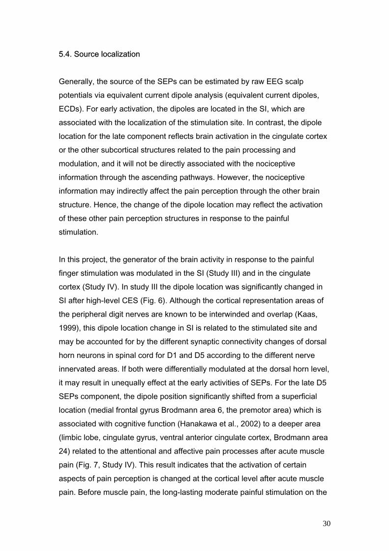
5.4. Source localization Generally, the source of the SEPs can be estimated by raw EEG scalp potentials via equivalent current dipole analysis (equivalent current dipoles, ECDs). For early activation, the dipoles are located in the SI, which are associated with the localization of the stimulation site. In contrast, the dipole location for the late component reflects brain activation in the cingulate cortex or the other subcortical structures related to the pain processing and modulation, and it will not be directly associated with the nociceptive information through the ascending pathways. However, the nociceptive information may indirectly affect the pain perception through the other brain structure. Hence, the change of the dipole location may reflect the activation of these other pain perception structures in response to the painful stimulation. In this project, the generator of the brain activity in response to the painful finger stimulation was modulated in the SI (Study III) and in the cingulate cortex (Study IV). In study III the dipole location was significantly changed in SI after high-level CES (Fig. 6). Although the cortical representation areas of the peripheral digit nerves are known to be interwinded and overlap (Kaas, 1999), this dipole location change in SI is related to the stimulated site and may be accounted for by the different synaptic connectivity changes of dorsal horn neurons in spinal cord for D1 and D5 according to the different nerve innervated areas. If both were differentially modulated at the dorsal horn level, it may result in unequally effect at the early activities of SEPs. For the late D5 SEPs component, the dipole position significantly shifted from a superficial location (medial frontal gyrus Brodmann area 6, the premotor area) which is associated with cognitive function (Hanakawa et al., 2002) to a deeper area (limbic lobe, cingulate gyrus, ventral anterior cingulate cortex, Brodmann area 24) related to the attentional and affective pain processes after acute muscle pain (Fig. 7, Study IV). This result indicates that the activation of certain aspects of pain perception is changed at the cortical level after acute muscle pain. Before muscle pain, the long-lasting moderate painful stimulation on the
30

little finger can increase the subject’s latent expectation to move the finger. Hence, the activation of the premotor area (BA 6) becomes dominant in response to the painful electrical stimulation to D5. After acute muscle pain, the activation of the anterior cingulate cortex (BA 24) becomes more responsive and dominant in the cortex when painful electrical stimulation is applied to the little finger (D5) and the dipole location is significantly shifted to the deeper area in the cingulate cortex. This result is consistent with a positron emission tomography study which showed that the premotor cortex (Brodmann area 6) intends to be more active during noxious cutaneous stimulations (Svensson et al., 1997), where the activation of the anterior cingulate cortex is more contributed by both noxious cutaneous stimulation and effect of muscle experimental pain. Furthermore, acute muscle pain may also affect other underlying pain mechanisms corresponding to the hand somatotopic position through the lateral spinothalamic tract (Kahle and Frotscher, 2003) in dorsal horn of spinal cord or to the thalamus and lead an overlap of the somatotopic projection of D5 to be sensitized (Dubner, 1991). Local anesthesia of the thumb caused the change in the neighbouring cortical representation areas (Waberski et al., 2003). Animal studies also support that the projections of the cutaneous afferent nerves in the spinal cord are somatotopically organized (Ygge, 1989). Furthermore, the excitation of the thenar muscle afferents may result in activation of wide-dynamic range neurons in lamina V in dorsal horn to receive cutaneous input. These neurons project to thalamic mechanisms (Price and Dubner, 1977; Dubner, 1991), e.g., ventral posterior inferior nucleus (Craig, 2003) and cause the activation of the cingulate cortex change (Petrovic et al., 1999; Price, 2002) compared to that before glutamate injection. Some mechanisms in the thalamus and the cingulate cortex may be involved and sensitized by painful electrical stimulation of D5, and finally lead to SEP dipole position changes at 230 and 350 ms (late SEP components for D5). 5.5. Brain dynamic change by frequency-dependent modulation
31
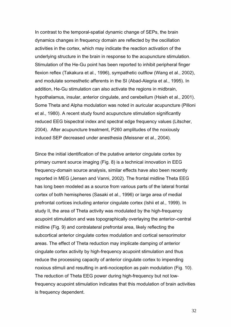
In contrast to the temporal-spatial dynamic change of SEPs, the brain dynamics changes in frequency domain are reflected by the oscillation activities in the cortex, which may indicate the reaction activation of the underlying structure in the brain in response to the acupuncture stimulation. Stimulation of the He-Gu point has been reported to inhibit peripheral finger flexion reflex (Takakura et al., 1996), sympathetic outflow (Wang et al., 2002), and modulate somesthetic afferents in the SI (Abad-Alegria et al., 1995). In addition, He-Gu stimulation can also activate the regions in midbrain, hypothalamus, insular, anterior cingulate, and cerebellum (Hsieh et al., 2001). Some Theta and Alpha modulation was noted in auricular acupuncture (Pilloni et al., 1980). A recent study found acupuncture stimulation significantly reduced EEG bispectral index and spectral edge frequency values (Litscher, 2004). After acupuncture treatment, P260 amplitudes of the noxiously induced SEP decreased under anesthesia (Meissner et al., 2004). Since the initial identification of the putative anterior cingulate cortex by primary current source imaging (Fig. 8) is a technical innovation in EEG frequency-domain source analysis, similar effects have also been recently reported in MEG (Jensen and Vanni, 2002). The frontal midline Theta EEG has long been modeled as a source from various parts of the lateral frontal cortex of both hemispheres (Sasaki et al., 1996) or large area of medial prefrontal cortices including anterior cingulate cortex (Ishii et al., 1999). In study II, the area of Theta activity was modulated by the high-frequency acupoint stimulation and was topographically overlaying the anterior–central midline (Fig. 9) and contralateral prefrontal area, likely reflecting the subcortical anterior cingulate cortex modulation and cortical sensorimotor areas. The effect of Theta reduction may implicate damping of anterior cingulate cortex activity by high-frequency acupoint stimulation and thus reduce the processing capacity of anterior cingulate cortex to impending noxious stimuli and resulting in anti-nociception as pain modulation (Fig. 10). The reduction of Theta EEG power during high-frequency but not low-frequency acupoint stimulation indicates that this modulation of brain activities is frequency dependent.
32
6. Summary and Conclusions
One main finding of this thesis is that painful electrical stimulation increased SEP amplitude for the early components and enlarged spatial area for the late components compared with non-painful electrical stimulation to thumb and little fingers. In frequency domain, EEG power in the theta band was significantly decreased during high-level transcutaneous acupoint electrical stimulation compared to control point electrical stimulation. In addition, another finding is that the amplitude of SEP component at 45 ms to painful stimulation of the thumb was significantly attenuated after high-level conditioning electrical stimulation. The late SEP amplitudes to painful stimulation of the little finger were significantly enhanced after both low- and high-level conditioning electrical stimulations. Lastly this thesis also observed that the latency of the late SEP component at 230 ms to painful stimulation of the thumb was significantly reduced after acute muscle pain induced by glutamate injection. In the mean time, the source localization of the late flux generators was significantly sunk in the cingulate cortex for little finger SEPs. These findings suggest that (1) Brain activities can be reflected by SEP peak activities in response to the experimental stimulation. (2) Nociceptive information transmission and pain processing to peripheral stimulation leads to brain response changes not only in the time domain such as SEPs but also in the frequency domain such as EEG broad bands. (3) Cortical activity can be affected after the conditioning experimental pain (skin and muscle pain), and the changes in SEP response for pain sensory and pain affective aspects are different and show strong site-dependent effects. (4) The activities of the underlying mechanism in cortex can be presented from dipole source localization. And the painful finger stimulation could be a valuable experimental model to present the cortical area change related to pain-discriminative and pain-affective aspects of pain processing.
33
The dynamics of SEPs and EEG may reflect the change of underlying sensory and pain processing mechanisms in the peripheral and central nervous systems. These dynamics may provide implication of the cortical reorganization at the beginning and have significance for pain treatment in the early stage.
34

Figures
Fig.2 Temporal-spatial SEP response of the grand average (n=15) to painful stimulation of the little finger (D5) (Study I). Compressed waveform (A), topography (B), the early phase (the electrode CCP3h located at the contralateral primary somatosensory cortex corresponding to the stimulation site, black; the electrode CCP4h located at the ipsilateral primary somatosensory cortex corresponding to the stimulation site, grey) (C), and the late phase, the isolated electrode FCC1h corresponding to the cingulate cortex (D) are respectively displayed. Fifty ms before and 450 ms after onset of stimulus, ten peak stages for painful were isolated and the peak stages with circle were analyzed for early and late phases.
35
2393
350
33 44240
15
180
375114
A
+
-
-6-4-20246
(ms)
(µV)
-6-4-20
246
(ms)
)
FCC1h
50ms
200 400
FCC1h
CCP4h
50ms
200 400
CCP3h C
D B
CCP3h
AFz
CCP3h
Fp2
FCC1h FCC1h
(µV
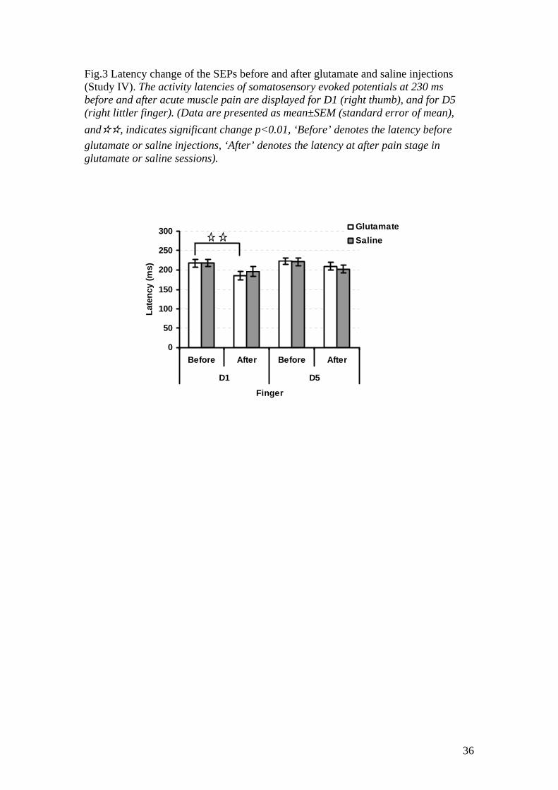
Fig.3 Latency change of the SEPs before and after glutamate and saline injections (Study IV). The activity latencies of somatosensory evoked potentials at 230 ms before and after acute muscle pain are displayed for D1 (right thumb), and for D5 (right littler finger). (Data are presented as mean±SEM (standard error of mean), and✫✫, indicates significant change p<0.01, ‘Before’ denotes the latency before glutamate or saline injections, ‘After’ denotes the latency at after pain stage in glutamate or saline sessions).
0
50
100
150
200
250
300
Before After Before After
D1 D5
Finger
Late
ncy
(ms)
GlutamateSaline
36
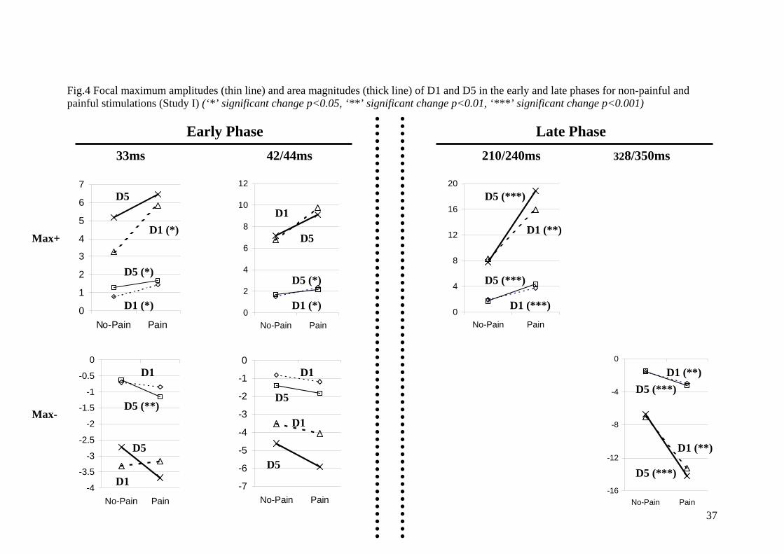
Fig.4 Focal maximum amplitudes (thin line) and area magnitudes (thick line) of D1 and D5 in the early and late phases for non-painful and painful stimulations (Study I) (‘*’ significant change p<0.05, ‘**’ significant change p<0.01, ‘***’ significant change p<0.001)
37
-7
-6
-5
-4
-3
-2
-1
0
No-Pain Pain
D1
D5
D1
D5
-16
-12
-8
-4
0
No-Pain Pain
0
4
8
12
16
20
No-Pain Pain
-4
-3.5
-3
-2.5
-2
-1.5
-1
-0.5
0
No-Pain Pain
0
2
4
6
8
10
12
No-Pain Pain0
1
2
3
4
5
6
7
No-Pain Pain
Early Phase Late Phase
Max+
Max-
33ms 42/44ms 210/240ms 328/350ms
D5
D1 (*)
D5 (*)
D1 (*)
D1
D5 (**)
D5
D1
D1
D5
D5 (*)
D1 (*)
D5 (***)
D1 (**)
D5 (***)
D1 (***)
D1 (**) D5 (***)
D1 (**)
D5 (***)

Fig.5 Focal maximum amplitudes of SEPs before and after conditioning electrical stimulation (Study III) The effect of conditioning electrical stimulation on the early SEP components for D1(thumb) stimulation is shown at (A), and the effect of conditioning electrical stimulation on the late SEP component (around 330 ms) for D1 and D5 (little finger) stimulations is shown at (B). (Low-level CES denotes low level conditioning electrical stimulation, High-level CES denotes high level conditioning electrical stimulation, BS and AS indicates before and after conditioning electrical stimulation respectively. The bar indicates the standard error of mean, and ‘☆’ indicates significant difference p<0.05, ‘☆☆’ indicates significant difference p<0.01, and ‘☆☆☆’ indicates significant difference p<0.001.)
(A)
0
-8-7-6-5-4-3-2-10
D1 D5 D1 D5
Low-level CES High-level CES
Finger
(µv)
BSAS
(B)
51525354
36ms 48ms 34ms 45ms
BSAS3,
2,
1,
0,
Low-level CES High-level CES
Peak Stage
(µV
)
38
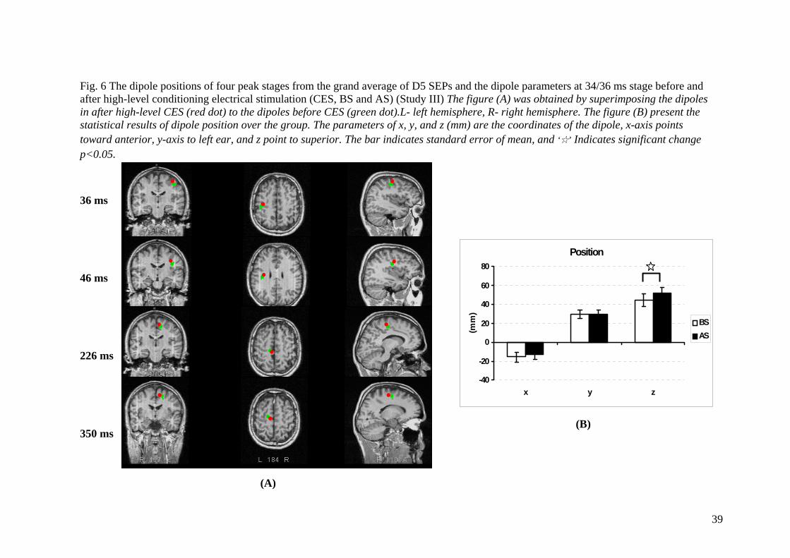
L L R F ions of four peak stages from the grand average of D5 SEPs and the dipole parameters at 34/36 ms stage before and a oning electrical stimulation (CES, BS and AS) (Study III) The figure (A) was obtained by superimposing the dipoles in (red dot) to the dipoles before CES (green dot).L- left hemisphere, R- right hemisphere. The figure (B) present the s ole position over the group. The parameters of x, y, and z (mm) are the coordinates of the dipole, x-axis points to to left ear, and z point to superior. The bar indicates standard error of mean, and ‘☆’ Indicates significant change p
3 4 2 3
Position
-40
-20
0
20
40
60
80
x y z
(mm
)
BSAS
(B)
(A)
39
R ig. 6 The dipole positfter high-level conditi after high-level CES
tatistical results of dipward anterior, y-axis
<0.05.
6 ms
6 ms
26 ms
50 ms

Fig.7 Dipole positions before and after pain in glutamate and saline sessions (Study IV) The statistical results of dipole position over the group at 230 and 350 ms are displayed respectively at (A) and (B) for D5. The parameters of x, y, and z (mm) are the coordinates of the dipole, x-axis points toward anterior, y-axis points to left ear, and z-axis points upwards. ‘Before’ is the dipole coordinates before pain, and ‘After’ is the dipole coordinates at after pain stage in glutamate or saline sessions. Data are presented as mean±SEM (standard error of mean), and ✫ indicates significant change P<0.05.
-40-20
0204060
Bef
ore
Aft
er
Bef
ore
Aft
er
Bef
ore
Aft
er
x y z
-40-20
0204060
Bef
ore
Aft
er
Bef
ore
Aft
er
Bef
ore
Aft
er
x y z
(mm) (mm) Glutamate Saline
(A) (B)
40

Fig.8 Topographic somatosensory field power (SFP) and tomographic current source images (CSI) of Theta EEG: high frequency (A/HF) and low frequency (A/LF) acupuncture stimulation in one subject (Study II).
A/LF
A/HF
41

Fig.9 The automatic quantification method (AQM) Theta-EEG mappings (n = 12, Study II) Baseline: before acupuncture stimulation, A/HF: acupuncture stimulation with high frequency, Post: after acupuncture stimulation, FM indicates the position of the focal maximum power.
e
F
A/H
Baselin
Post42
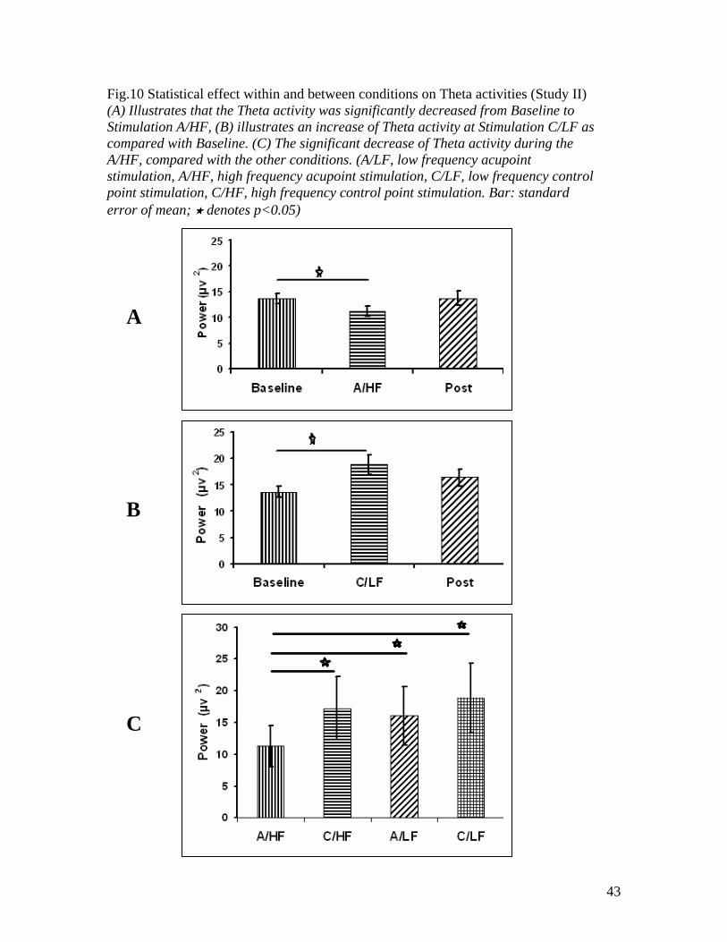
Fig.10 Statistical effect within and between conditions on Theta activities (Study II) (A) Illustrates that the Theta activity was significantly decreased from Baseline to Stimulation A/HF, (B) illustrates an increase of Theta activity at Stimulation C/LF as compared with Baseline. (C) The significant decrease of Theta activity during the A/HF, compared with the other conditions. (A/LF, low frequency acupoint stimulation, A/HF, high frequency acupoint stimulation, C/LF, low frequency control point stimulation, C/HF, high frequency control point stimulation. Bar: standard error of mean; denotes p<0.05)
A
B
C
43

References
Abad-Alegria F, Adelantado S, Martinez T. The role of the cerebral cortex in acupuncture modulation of the somesthetic afferent. Am J Chin Med. 1995;23:11–4. Allman JM, Hakeem A, Erwin JM, Nimchinsky E, Hof P. The anterior cingulate cortex. The evolution of an interface between emotion and cognition. Ann N Y Acad Sci. 2001;935:107–17. Anderson C, Horne JA. Electroencephalographic activities during wakefulness and sleep in the frontal cortex of healthy older people: links with “thinking”. Sleep. 2003;26:968-72. Apkarian AV, Bushnell MC, Treede RD, Zubieta JK. Human brain mechanisms of pain perception and regulation in health and disease. Eur J Pain. 2005;9:463-84. Arienzo D, Babiloni C, Ferretti A, Caulo M, Del Gratta C, Tartaro A, Rossini PM, Romani GL. Somatotopy of anterior cingulate cortex (ACC) and supplementary motor area (SMA) for electric stimulation of the median and tibial nerves: an fMRI study. Neuroimage. 2006;33:700-5. Ashton H, Golding JF, Marsh VR, Thompson JW. Effects of transcutaneous electrical nerve stimulation and aspirin on late somatosensory evoked potentials in normal subjects. Pain. 1984;18:377–86. Babiloni C, Brancucci A, Arendt-Nielsen L, Babiloni F, Capotosto P, Carducci F, Cincotti F, Del Percio C, Petrini L, Rossini PM, Chen AC. Attentional processes and cognitive performance during expectancy of painful galvanic stimulations: a high-resolution EEG study. Behav Brain Res. 2004;152:137-47.
44
Babiloni C, Brancucci A, Babiloni F, Capotosto P, Carducci F, Cincotti F, Arendt-Nielsen L, Chen AC, Rossini PM. Anticipatory cortical responses during the expectancy of a predictable painful stimulation. A high-resolution electroencephalography study. Eur J Neurosci. 2003;18:1692-700. Babiloni C, Brancucci A, Del Percio C, Capotosto P, Arendt-Nielsen L, Chen AC, Rossini PM. Anticipatory electroencephalography alpha rhythm predicts subjective perception of pain intensity. J Pain. 2006;7:709-17. Babiloni F, Babiloni C, Carducci F, Fattorini L, Onorati P, Urbano A. Spline Laplacian estimate of EEG potentials over a realistic magnetic resonance-constructed scalp surface model. Electroencephalogr. Clin Neurophysiol. 1996;98:363– 73. Barch DM, Braver TS, Akbudak E, Conturo T, Ollinger J, Snyder A. Anterior cingulate cortex and response conflict: effects of response modality and processing domain. Cereb Cortex. 2001;11:837–48. Biella G, Sotgiu ML, Pellegata G, Paulesu E, Castiglioni I, Fazio F. Acupuncture produces central activations in pain regions. NeuroImage. 2001;14:60–6. Broman J. Neurotransmitters in subcortical somatosensory pathways. Anat Embryol (Berl). 1994;189:181-214. Bromm B, Lorenz J. Neurophysiological evaluation of pain. Electroencephalogr Clin Neurophysiol. 1998;107:227-53. Bromm B, Meier W. Scharein pre-stimulus/post-stimulus relations in EEG spectra and their modulations by an opioid and an antidepressant. Electroencephalogr Clin Neurophysiol. 1989;73:188– 97.
45
Brooks J, Tracey I. From nociception to pain perception: imaging the spinal and supraspinal pathways. J Anat. 2005;207:19-33. Buchel C, Bornhovd K, Quante M, Glauche V, Bromm B, Weiller C. Dissociable neural responses related to pain intensity, stimulus intensity, and stimulus awareness within the anterior cingulate cortex: a parametric single-trial laser functional magnetic resonance imaging study. J Neurosci. 2002;22:970-6. Buchner H, Fuchs M, Wischmann HA, Dossel O, Ludwig I, Knepper A, Berg P. Source analysis of median nerve and finger stimulated somatosensory evoked potentials: multichannel simultaneous recording of electric and magnetic fields combined with 3D-MR tomography. Brain Topogr. 1994;6:299-310. Buonomano DV, Merzenich MM. Cortical plasticity: from synapses to maps. Annu Rev Neurosci. 1998;21:149-86. Bütefisch CM. Plasticity in the human cerebral cortex: lessons from the normal brain and from stroke. Neuroscientist. 2004;10:163-73. Cairns BE, Svensson P, Wang K, Hupfeld S, Graven-Nielsen T, Sessle BJ, Berde CB, Arendt-Nielsen L. Activation of peripheral NMDA receptors contributes to human pain and rat afferent discharges evoked by injection of glutamate into the masseter muscle. J Neurophysiol. 2003;90:2098-105. Calejesan AA, Kim SJ, Zhuo M. Descending facilitatory modulation of a behavioral nociceptive response by stimulation in the adult rat anterior cingulate cortex. Eur J Pain. 2000;4:83-96. Carmichael ST, Chesselet MF. Synchronous neuronal activity is a signal for axonal sprouting after cortical lesions in the adult. J Neurosci. 2002;22:6062-70.
46
Carstens E, Yokota T, Zimmermann M. Inhibition of spinal neuronal responses to noxious skin heating by stimulation of mesencephalic periaqueductal gray in the cat. J Neurophysiol. 1979;42:558-68. Casey KL. Forebrain mechanisms of nociception and pain: analysis through imaging. Proc Natl Acad Sci U S A. 1999;96:7668-74. Cazalis M, Dayanithi G, Nordmann JJ. The role of patterned burst and interburst interval on the excitation-coupling mechanisms in the isolated rat neural lobe. J Physiol. 1985;369:45– 60. Chen AC, Arendt-Nielsen L, Plaghki L. Laser-evoked potentials in human pain II. cerebral generators. Pain Forum. 1998;7:201-11. Chen AC, Shimojo M, Svensson P, Arendt-Nielsen L. Brain dynamics of scalp evoked potentials and current source densities to repetitive (5-pulse train) painful stimulation of skin and muscle: central correlate of temporal summation. Brain Topogr. 2000;13:59-72. Chen Z, Geng Z, Zhang J, Zhao Y, Gao Y, Zhang X. The effect of electric acupuncture on SEP of local cerebral ischemic-reperfusion rats. Zhen Ci Yan Jiu. 1996;21:34–40. Craig AD. Pain mechanisms: labeled lines versus convergence in central processing. Annu Rev Neurosci. 2003;26:1-30. Craig AD. Retrograde analyses of spinothalamic projections in the macaque monkey: input to ventral posterior nuclei. J Comp Neurol. 2006;499:965-78. Davis KD. The neural circuitry of pain as explored with functional MRI. Neurol Res. 2000;22:313-7.
47

Davis KD, Wood ML, Crawley AP, Mikulis DJ. fMRI of human somatosensory and cingulate cortex during painful electrical nerve stimulation. Neuroreport. 1995;7:321-5. Del Percio C, Le Pera D, Arendt-Nielsen L, Babiloni C, Brancucci A, Chen AC, De Armas L, Miliucci R, Restuccia D, Valeriani M, Rossini PM. Distraction affects frontal alpha rhythms related to expectancy of pain: an EEG study. Neuroimage. 2006;31:1268-77. Derbyshire SW, Jones AK, Gyulai F, Clark S, Townsend D, Firestone LL. Pain processing during three levels of noxious stimulation produces differential patterns of central activity. Pain. 1997;73:431-45. Disselhoff B. Transcutaneous electrical nerve stimulation (TENS) in neuropathic pain. Pain Clinic. 2000;12:145–6. Doubell TP, Mannion RJ, Woolf CJ. Intact sciatic myelinated primary afferent terminals collaterally sprout in the adult rat dorsal horn following section of a neighbouring peripheral nerve. J Comp Neurol. 1997;380:95-104. Drewes AM, Nielsen KD, Steengaard-Pedersen K, Birket-Smith L, Hansen LM, Arendt-Nielsen L. Electroencephalographic reactions during experimental superficial and deep pain stimuli in awake healthy subjects. Journal of Musculoskeletal Pain. 1999:7;29-44. Dubner R. Basic mechanisms of pain associated with deep tissues. Can J Physiol Pharmacol. 1991;69:607-9. Duggan AW, Furmidge LJ. Probing the brain and spinal cord with neuropeptides in pathways related to pain and other functions. Front Neuroendocrinol. 1994;15:275-300.
48

Elbert T, Sterr A, Flor H, Rockstroh B, Knecht S, Pantev C, Wienbruch C, Taub E. Input-increase and input-decrease types of cortical reorganization after upper extremity amputation in humans. Exp Brain Res. 1997;117:161-4. Fairhurst M, Wiech K, Dunckley P, Tracey I. Anticipatory brainstem activity predicts neural processing of pain in humans. Pain. 2007;128:101-10. Florence SL, Wall JT, Kaas JH. The somatotopic pattern of afferent projections from the digits to the spinal cord and cuneate nucleus in macaque monkeys. Brain Res. 1988;452:388-92. Flor H. Cortical reorganisation and chronic pain: implications for rehabilitation. J Rehabil Med. 2003;41:66–72. Flor H. Phantom-limb pain: characteristics, causes, and treatment. Lancet Neurol. 2002a;1:182-9. Flor H. The modification of cortical reorganization and chronic pain by sensory feedback. Appl Psychophysiol Biofeedback. 2002b;27:215-27. Garcia-Larrea L, Frot M, Valeriani M. Brain generators of laser-evoked potentials: from dipoles to functional significance. Neurophysiol Clin. 2003;33:279-92. Garrison DW, Foreman RD. Decreased activity of spontaneous and noxiously evoked dorsal horn cells during transcutaneous electrical nerve stimulation (TENS). Pain. 1994;58:309–15. Ge HY, Madeleine P, Arendt-Nielsen L. Gender differences in pain modulation evoked by repeated injections of glutamate into the human trapezius muscle. Pain. 2005;113:134-40.
49
Gelnar PA, Krauss BR, Szeverenyi NM, Apkarian AV. Fingertip representation in the human somatosensory cortex: an fMRI study. Neuroimage. 1998;7:261-83. Goffaux P, Redmond WJ, Rainville P, Marchand S. Descending analgesia - When the spine echoes what the brain expects. Pain. 2007;130:137-43. Hanakawa T, Honda M, Sawamoto N, Okada T, Yonekura Y, Fukuyama H, Shibasaki H. The role of rostral Brodmann area 6 in mental-operation tasks: an integrative neuroimaging approach. Cereb Cortex. 2002;12:1157-70. Han JS. Acupuncture: neuropeptide release produced by electrical stimulation of different frequencies. Trends Neurosci. 2003;26:17– 22. Horwitz B. The elusive concept of brain connectivity. Neuroimage. 2003;19:466-70. Hoshiyama M, Kakigi R. After-effect of transcutaneous electrical nerve stimulation (TENS) on pain-related evoked potentials and magnetic fields in normal subjects. Clin Neurophysiol. 2000;111:717–24. Hsieh JC, Tu CH, Chen FP, Chen MC, Yeh TC, Cheng HC, Wu YT, Liu RS, Ho LT. Activation of the hypothalamus characterizes the acupuncture stimulation at the analgesic point in human: a positron emission tomography study. Neurosci Lett. 2001;307:105–8. Huber MT, Bartling J, Pachur D, Woikowsky-Biedau S, Lautenbacher S. EEG responses to tonic heat pain. Exp Brain Res. 2006;173:14-24. Inui K, Tran TD, Qiu Y, Wang X, Hoshiyama M, Kakigi R. A comparative magnetoencephalographic study of cortical activations evoked by noxious and innocuous somatosensory stimulations. Neuroscience. 2003;120:235-48.
50
Ishii R, Shinosaki K, Ukai S, Inoue T, Ishihara T, Yoshimine T, Hirabuki N, Asada H, Kihara T, Robinson SE, Takeda M. Medial prefrontal cortex generates frontal midline theta rhythm. NeuroReport. 1999;10:675–9. Jensen O, Vanni S. A new method to identify multiple sources of oscillatory activity from magnetoencephalographic data. NeuroImage. 2002;15:568–74. Kaas JH. Is most of neural plasticity in the thalamus cortical? Proc Natl Acad Sci USA. 1999;96:7622–3. Kahle W, Frotscher M. Nervous system and sensory organs (5th revised edition). Georg Thieme Verlag. ISBN 3-13-533505-4 (GTV), ISBN 1-58890-064-9 (TNY). Spinal cord and spinal nerves. 2003;5:48-90. Kakigi R, Inui K, Tran DT, Qiu Y, Wang X, Watanabe S, Hoshiyama M. Human brain processing and central mechanisms of pain as observed by electro- and magneto-encephalography. J Chin Med Assoc. 2004;67:377-86. Kakigi R, Shibasaki H, Ikeda A. Pain-related somatosensory evoked potentials following CO2 laser stimulation in man. Electroencephalogr Clin Neurophysiol. 1989;74:139-46. Kakigi R, Watanabe S, Yamasaki H. Pain-Related somatosensory evoked potentials. J Clin Neurophysiol. 2000;17:295-308. Kaplan B, Rabinerson D, Pardo J, Krieser RU, Neri A. Transcutaneous electrical nerve stimulation (TENS) as a pain-relief device in obstetrics and gynecology. Clin Exp Obstet Gynecol. 1997;24:123–6. Kaptchuk TJ. Acupuncture: theory, efficacy, and practice. Ann Intern Med. 2002;136:374– 83.
51
Kriegseis A, Hennighausen E, Rosler F, Roder B. Reduced EEG alpha activity over parieto-occipital brain areas in congenitally blind adults. Clin Neurophysiol. 2006;117:1560-73. Kupers R, Kehlet H. Brain imaging of clinical pain states: a critical review and strategies for future studies. Lancet Neurol. 2006;5:1033-44. Kurth R, Villringer K, Curio G, Wolf KJ, Krause T, Repenthin J, Schwiemann J, Deuchert M, Villringer A. fMRI shows multiple somatotopic digit representations in human primary somatosensory cortex. Neuroreport. 2000;11:1487-91. Kurth R, Villringer K, Mackert BM, Schwiemann J, Braun J, Curio G, Villringer A, Wolf KJ. fMRI assessment of somatotopy in human Brodmann area 3b by electrical finger stimulation. Neuroreport. 1998;9:207-12. Lalo E, Gilbertson T, Doyle L, Lazzaro VD, Cioni B, Brown P. Phasic increases in cortical beta activity are associated with alterations in sensory processing in the human. Exp Brain Res. 2007;177:137-45. Laroy V, Spaans F, Reulen J. The sensory innervation pattern of the fingers. J Neurol. 1998;245:294-8. Lenz FA, Rios M, Zirh A, Chau D, Krauss G, Lesser RP. Painful stimuli evoke potentials recorded over the human anterior cingulate gyrus. J Neurophysiol. 1998;79:2231-4. Lin Q, Peng YB, Willis WD. Antinociception and inhibition from the periaqueductal gray are mediated in part by spinal 5-hydroxytryptamine(1A) receptors. J Pharmacol Exp Ther. 1996;276:958-67.
52
Litscher G. Effects of acupressure, manual acupuncture and laser needle acupuncture on EEG bispectral index and spectral edge frequency in healthy volunteers. Eur J Anaesthesiol. 2004;21:13–9. Luber B, Habeck C, Trott CT, Friedman D, Moeller JR. A ghost of retrieval past: a functional network of alpha EEG related to source memory in elderly humans. Brain Res Cogn Brain Res. 2004;20:144-55. Maldjian JA, Gottschalk A, Patel RS, Detre JA, Alsop DC. The sensory somatotopic map of the human hand demonstrated at 4 Tesla. Neuroimage. 1999;10:55-62. Mantyh PW. Connections of midbrain periaqueductal gray in the monkey. II. Descending efferent projections. J Neurophysiol. 1983;49:582-94. McMullan S, Lumb BM. Spinal dorsal horn neuronal responses to myelinated versus unmyelinated heat nociceptors and their modulation by activation of the periaqueductal grey in the rat. J Physiol. 2006;576:547-56. Meissner W, Weiss T, Trippe RH, Hecht H, Krapp C, Miltner WH. Acupuncture decreases somatosensory evoked potential amplitudes to noxious stimuli in anesthetized volunteers. Anesth Analg. 2004;98:141–7. Melzack R, Wall PD. Pain mechanisms: a new theory. Science. 1965;150: 971–9. Merskey H, Spear FG. The concept of pain. J Psychosom Res. 1967;11:59-67. Millan MJ. Descending control of pain. Prog Neurobiol. 2002;66:355-474. Millan MJ. The induction of pain: an integrative review. Prog Neurobiol. 1999;57:1-164.
53

Montes C, Magnin M, Maarrawi J, Frot M, Convers P, Mauguiere F, Garcia-Larrea L. Thalamic thermo-algesic transmission: ventral posterior (VP) complex versus VMpo in the light of a thalamic infarct with central pain. Pain. 2005;113:223-32. Mullins PG, Rowland LM, Jung RE, Sibbitt WL Jr. A novel technique to study the brain's response to pain: proton magnetic resonance spectroscopy. Neuroimage. 2005;26:642-6. Niddam DM, Arendt-Nielsen L, Chen AC. Cerebral dynamics of SEPS to non-painful and painful cutaneous electrical stimulation of the thenar and hypothenar. Brain Topogr. 2000;13:105-14. Niddam DM, Chen LF, Wu YT, Hsieh JC. Spatiotemporal brain dynamics in response to muscle stimulation. Neuroimage. 2005;25:942-51. Niddam DM, Graven-Nielsen T, Arendt-Nielsen L, Chen AC. Non-painful and painful surface and intramuscular electrical stimulation at the thenar and hypothenar sites: differential cerebral dynamics of early to late latency SEPs. Brain Topogr. 2001;13:283-92. Nuwer MR, Lehmann D, da Silva FL, Matsuoka S, Sutherling W, Vibert JF. IFCN guidelines for topographic and frequency analysis of EEGs and EPs. The International Federation of Clinical Neurophysiology. Electroencephalogr Clin Neurophysiol Suppl. 1999;52:15-20. Ohara S, Crone NE, Weiss N, Lenz FA. Analysis of synchrony demonstrates 'pain networks' defined by rapidly switching, task-specific, functional connectivity between pain-related cortical structures. Pain. 2006;123:244-53.
54

Okada YC, Tanenbaum R, Williamson SJ, Kaufman L. Somatotopic organization of the human somatosensory cortex revealed by neuromagnetic measurements. Exp Brain Res. 1984;56:197-205. Oostenveld R, Praamstra P. The five percent electrode system for high-resolution EEG and ERP measurements. Clin Neurophysiol. 2001;112:713-9. Petrovic P, Ingvar M, Stone-Elander S, Petersson KM, Hansson P. A PET activation study of dynamic mechanical allodynia in patients with mononeuropathy. Pain. 1999;83:459-70. Peyron R, Garcia-Larrea L, Gregoire MC, Costes N, Convers P, Lavenne , Mauguiere F, Michel D, Laurent B. Haemodynamic brain responses to acute pain in humans: sensory and attentional networks. Brain. 1999;122:1765-80. Peyron R, Laurent B, Garcia-Larrea L. Functional imaging of brain responses to pain. A review and meta-analysis. Neurophysiol Clin. 2000;30:263–88. Pilloni C, Caracausi RS, Tognali F, Sbarbaro V. EEG evaluation of patients undergoing extracorporeal circulation under analgesic and electrohypoalgesia with auricular acupuncture. Minerva Anestesiol. 1980;46:371–86. Ploner M, Gross J, Timmermann L, Pollok B, Schnitzler A. Oscillatory activity reflects the excitability of the human somatosensory system. Neuroimage. 2006;32:1231-6. Posner M, Rothbart MK. Attentional mechanisms and conscious experience. In: Milner, A.D., Rugg, M.D. (Eds.), The Neuropsychology of Consciousness. American Press, San Diego, CA, 1992; 91– 111. Price DD. Central Neural Mechanisms that Interrelate Sensory and Affective Dimensions of Pain. Molecular Interventions. 2002;2:392-403.
55

Price DD, Dubner R. Neurons that subserve the sensory-discriminative aspects of pain. Pain. 1977;3:307-38. Price DD. Psychological and neural mechanisms of the affective dimension of pain. Science. 2000;288:1769–72. Price DD, Verne GN, Schwartz JM. Chapter 20. Plasticity in brain processing and modulation of pain. Prog Brain Res. 2006;157:333-52. Racke K, Burns F, Haas B, Niebauer J, Pitzius E. Frequencydependent effects of activation and inhibition of protein kinase C on neurohypophysial release of oxytocin and vasopressin. Naunyn Schmiedebergs Arch Pharmacol. 1989;339:617–24. Rainville P. Brain mechanisms of pain affect and pain modulation. Curr Opin Neurobiol. 2002;12:195-204. Ren K, Dubner R. Central nervous system plasticity and persistent pain. J Orofac Pain. 1999;13:155-63. Restuccia D, Valeriani M, Insola A, Lo Monaco M, Grassi E, Barba C, Le Pera D, Mauguiere F. Modality-related scalp responses after electrical stimulation of cutaneous and muscular upper limb afferents in humans. Muscle Nerve. 2002;26:44-54. Rios M, Treede R, Lee J, Lenz FA. Direct Evidence of Nociceptive Input to Human Anterior Cingulate Gyrus and Parasylvian Cortex. Curr Rev Pain. 1999;3:256-64. Rossi A, Decchi B, Groccia V, Della Volpe R, Spidalieri R. Interactions between nociceptive and non-nociceptive afferent projections to cerebral cortex in humans. Neurosci Lett. 1998;248:155-8.
56
Rossini PM, Dal Forno G. Integrated technology for evaluation of brain function and neural plasticity. Phys Med Rehabil Clin N Am. 2004;15:263-306. Saletu B, Saletu M, Brown M, Stern J, Sletten I, Ulett G. Hypno-analgesia and acupuncture analgesia: a neurophysiological reality? Neuropsychobiology. 1975;1:218–42. Sarnthein J, Stern J, Aufenberg C, Rousson V, Jeanmonod D. Increased EEG power and slowed dominant frequency in patients with neurogenic pain. Brain. 2006;129:55-64. Sasaki K, Nambu A, Tsujimoto T, Matsuzaki R, Kyuhou S, Gemba H. Studies on integrative functions of the human frontal association cortex with MEG. Brain Res Cogn Brain Res. 1996;5:165–74. Schaible HG. Peripheral and central mechanisms of pain generation. Handb Exp Pharmacol. 2007;(177):3-28. Schlereth T, Baumgartner U, Magerl W, Stoeter P, Treede RD. Left-hemisphere dominance in early nociceptive processing in the human parasylvian cortex. Neuroimage. 2003;20:441-54. Schnitzler A, Ploner M. Neurophysiology and functional neuroanatomy of pain perception. J Clin Neurophysiol. 2000;17:592-603. Shen J. Research on the neurophysiological mechanisms of acupuncture: review of selected studies and methodological issues. J Altern Complement Med. 2001;7:121–7. Shimojo M, Svensson P, Arendt-Nielsen L, Chen AC. Dynamic brain topography of somatosensory evoked potentials and equivalent dipoles in
57

response to graded painful skin and muscle stimulation. Brain Topogr 2000;13:43–58. Staud R, Craggs JG, Robinson ME, Perlstein WM, Price DD. Brain activity related to temporal summation of C-fiber evoked pain. Pain. 2007;129:130-42. Stevens A, Batra A, Kotter I, Bartels M, Schwarz J. Both pain and EEG response to cold pressor stimulation occurs faster in fibromyalgia patients than in control subjects. Psychiatry Res. 2000;97:237-47. Stienen PJ, van Oostrom H, van den Bos R, de Groot HN, Hellebrekers LJ. Vertex-recorded, rather than primary somatosensory cortex-recorded, somatosensory-evoked potentials signal unpleasantness of noxious stimuli in the rat. Brain Res Bull. 2006;70:203-12. Sutherling WW, Levesque MF, Baumgartner C. Cortical sensory representation of the human hand: size of finger regions and nonoverlapping digit somatotopy. Neurology. 1992;42:1020-8. Svensson P, Cairns BE, Wang K, Hu JW, Graven-Nielsen T, Arendt-Nielsen L, Sessle BJ. Glutamate-evoked pain and mechanical allodynia in the human masseter muscle. Pain. 2003;101:221-7. Svensson P, Minoshima S, Beydoun A, Morrow TJ, Casey KL. Cerebral processing of acute skin and muscle pain in humans. J Neurophysiol. 1997;78:450-60. Szaflarski JP, Page SJ, Kissela BM, Lee JH, Levine P, Strakowski SM. Cortical reorganization following modified constraint-induced movement therapy: a study of 4 patients with chronic stroke. Arch Phys Med Rehabil. 2006;87:1052-8.
58

Takakura N, Iijima S, Kanamaru A, Shibuya M, Homma I, Ohashi M. Vibration-induced finger flexion reflex and inhibitory effect of acupuncture on this reflex in cervical spinal cord injury patients. Neurosci Res. 1996;26:391–4. Tanosaki M, Iguchi Y, Kimura T, Takino R, Hashimoto I. Functional connectivity between forearm and digits representations in human somatosensory area 3b. Clin Neurophysiol. 2004;115:2638-44. Tinazzi M, Valeriani M, Moretto G, Rosso T, Nicolato A, Fiaschi A, Aglioti SM. Plastic interactions between hand and face cortical representations in patients with trigeminal neuralgia: a somatosensory-evoked potentials study. Neuroscience. 2004;127:769-76. Treede RD. Neurophysiological studies of pain pathways in peripheral and central nervous system disorders. J Neurol. 2003;250:1152-61. Turner LC, Harrison LM, Stephens JA. Finger movement is associated with attenuated cutaneous reflexes recorded from human first dorsal interosseous muscle. J Physiol. 2002;542:559–66. Uchida Y, Nishigori A, Takeda D, Ohshiro M, Ueda Y, Ohshima M, Kashiba H. Electroacupuncture induces the expression of Fos in rat dorsal horn via capsaicin-insensitive afferents. Brain Res. 2003;978:136–40. Ulett GA, Han S, Han JS. Electroacupuncture: mechanisms and clinical application. Biol Psychiatry. 1998;44:129–38. Valeriani M, Arendt-Nielsen L, Le Pera D, Restuccia D, Rosso T, De Armas L, Maiese T, Fiaschi A, Tonali P, Tinazzi M. Short-term plastic changes of the human nociceptive system following acute pain induced by capsaicin. Clin Neurophysiol. 2003;114:1879-90.
59
Valeriani M, Tonali P, Le Pera D, Restuccia D, De Armas L, Del Vesco C, Miliucci R, Fiaschi A, Vigevano F, Arendt-Nielsen L, Tinazzi M. Modulation of laser-evoked potentials by experimental cutaneous tonic pain. Neuroscience. 2006;140:1301-10. Valet M, Sprenger T, Boecker H, Willoch F, Rummeny E, Conrad B, Erhard P, Tolle TR. Distraction modulates connectivity of the cingulo-frontal cortex and the midbrain during pain--an fMRI analysis. Pain. 2004;109:399-408. van Veen V, Carter CS. The anterior cingulate as a conflict monitor: fMRI and ERP studies. Physiol Behav. 2002;77:477–82. van Westen D, Fransson P, Olsrud J, Rosen B, Lundborg G, Larsson EM. Fingersomatotopy in area 3b: an fMRI-study. BMC Neurosci. 2004;5:28. Waberski TD, Gobbelé R, Kawohl W, Cordes C, Buchner H. Immediate cortical reorganization after local anesthetic block of the thumb: source localization of somatosensory evoked potentials in human subjects. Neurosci Lett. 2003;347:151-4. Wang JD, Kuo TB, Yang CC. An alternative method to enhance vagal activities and suppress sympathetic activities in humans. Auton Neurosci. 2002;100:90–5. Warbrick T, Sheffield D, Nouwen A. Effects of pain-related anxiety on components of the pain event-related potential. Psychophysiology. 2006;43:481-5. Wiech K, Preissl H, Birbaumer N. Neuroimaging of chronic pain: phantom limb and musculoskeletal pain. Scand J Rheumatol Suppl. 2000;113:13-8. Willis WD, Westlund KN. Neuroanatomy of the pain system and of the pathways that modulate pain. J Clin Neurophysiol. 1997;14:2-31.
60
Wu MT, Sheen JM, Chuang KH, Yang P, Chin SL, Tsai CY, Chen CJ, Liao JR, Lai PH, Chu KA, Pan HB, Yang CF. Neuronal specificity of acupuncture response: a fMRI study with electroacupuncture. NeuroImage. 2002;16:1028–37. Xu X, Shibasaki H, Shindo K. Effects of acupuncture on somatosensory evoked potentials: a review. J Clin Neurophysiol. 1993;10:370–7. Ygge J. Central projections of the rat radial nerve investigated with transganglionic degeneration and transganglionic transport of horseradish peroxidase. J Comp Neurol. 1989;279:199-211. Zhang L, Zhang Y, Zhao ZQ. Anterior cingulate cortex contributes to the descending facilitatory modulation of pain via dorsal reticular nucleus. Eur J Neurosci. 2005;22:1141-8. Zhang WT, Jin Z, Cui GH, Zhang KL, Zhang L, Zeng YW, Luo F, Chen AC, Han JS. Relations between brain network activation and analgesic effect induced by low vs. high frequency electrical acupoint stimulation in different subjects: a functional magnetic resonance imaging study. Brain Res. 2003a;982:168–78. Zhang WT, Jin Z, Huang J, Zhang L, Zeng YW, Luo F, Chen ACN, Han JS. Modulation of cold pain in human brain by electric acupoint stimulation: evidence from fMRI. NeuroReport. 2003b;14:1591– 6.
61