A Unique Polymeric Nano-carrier for Anti-tuberculosis … Unique Polymeric Nano-carrier for...
25
A Unique Polymeric Nano-carrier for Anti-tuberculosis Therapy Shivshankar R. Mane a , Vijayakameswara Rao N a , Koushik Chatterjee a , Himadri Dinda a , Soma Nag b , Abhinoy Kishore b , Jayasri Das Sarma* b , and Raja Shunmugam* a [a] Polymer Research Centre, Department of Chemical Sciences, and [b] Department of Biological Sciences Indian Institute of Science Education and Research Kolkata (IISER K), India. Contents: 1. Experimental Section…………………………………………………......S2 2. 1 H NMR spectrum of compound 1……………………………………….S9 3. 13 C NMR spectrum of compound 1………………………………………S9 4. 1 H NMR spectrum of compound 2……………………………………….S10 5. 13 C NMR spectrum of compound 2………………………………………S10 6. 13 C NMR spectrum of compound 3………………………………………S11 7. 13 C NMR spectrum of Mono 1……………………………………………S11 8. 1 H NMR spectrum of compound 4……………………………….……….S12 9. 13 C NMR spectrum of compound 4……………………………….………S12 10. 1 H NMR spectra of tertiary butyl hydrazone Nadic carboxylate…..……..S13 11. 1 H NMR spectrum of compound 5…………………………………..…….S13 12. 13 C NMR spectrum of compound 5…………………………………..……S14 13. 1 H NMR spectrum of Mono 2………………………………………......….S14 14. 13 C NMR spectrum of Mono 2………………………………..……………S15 15. 1 H NMR spectrum of CP1………………………………………………….S15 16. 1 H NMR spectrum of CP2 in DMSO-d 6 after cleavage……….…………..S16 17. 1 H NMR spectrum of CP2 in D 2 O after cleavage……………….………….S16 18. FT-IR spectra of compound 3 and Mono 1…………………….…………..S17 19. FT-IR spectra of compound 5………………………………………………S17 20. FT-IR spectra of retinal and Mono 2……..…………………….………….S18 21. FT-IR spectra of CP1…………………………………………….…………S18 22. UV of CP2……………………………………………………….………….S19 23. Fluorescence spectra of CP2…………………………………….………….S19 24. Fluorescence spectra of drug release from CP 2 at pH 4 - 6.5…………...S20 25. Fluorescence spectrum of CP2 from renal clearance (Control 1-4)...........S22 26. Cytotoxicity assay of isoniazid and retinal..............................................S24 Electronic Supplementary Material (ESI) for Journal of Materials Chemistry This journal is © The Royal Society of Chemistry 2012
Transcript of A Unique Polymeric Nano-carrier for Anti-tuberculosis … Unique Polymeric Nano-carrier for...

A Unique Polymeric Nano-carrier for Anti-tuberculosis Therapy
Shivshankar R. Manea, Vijayakameswara Rao Na, Koushik Chatterjeea, Himadri Dindaa, Soma Nagb,
Abhinoy Kishoreb, Jayasri Das Sarma*b, and Raja Shunmugam*
a
[a]Polymer Research Centre, Department of Chemical Sciences, and [b]Department of Biological Sciences Indian Institute of Science Education and Research Kolkata (IISER K),
India.
Contents: 1. Experimental Section…………………………………………………......S2
2. 1H NMR spectrum of compound 1……………………………………….S9
3. 13C NMR spectrum of compound 1………………………………………S9
4. 1H NMR spectrum of compound 2……………………………………….S10
5. 13C NMR spectrum of compound 2………………………………………S10
6. 13C NMR spectrum of compound 3………………………………………S11
7. 13C NMR spectrum of Mono 1……………………………………………S11
8. 1H NMR spectrum of compound 4……………………………….……….S12
9. 13C NMR spectrum of compound 4……………………………….………S12
10. 1H NMR spectra of tertiary butyl hydrazone Nadic carboxylate…..……..S13
11. 1H NMR spectrum of compound 5…………………………………..…….S13
12. 13C NMR spectrum of compound 5…………………………………..……S14
13. 1H NMR spectrum of Mono 2………………………………………......….S14
14. 13C NMR spectrum of Mono 2………………………………..……………S15
15. 1H NMR spectrum of CP1………………………………………………….S15
16. 1H NMR spectrum of CP2 in DMSO-d6 after cleavage……….…………..S16
17. 1H NMR spectrum of CP2 in D2O after cleavage……………….………….S16
18. FT-IR spectra of compound 3 and Mono 1…………………….…………..S17
19. FT-IR spectra of compound 5………………………………………………S17
20. FT-IR spectra of retinal and Mono 2……..…………………….………….S18
21. FT-IR spectra of CP1…………………………………………….…………S18
22. UV of CP2……………………………………………………….………….S19
23. Fluorescence spectra of CP2…………………………………….………….S19
24. Fluorescence spectra of drug release from CP 2 at pH 4 - 6.5…………...S20
25. Fluorescence spectrum of CP2 from renal clearance (Control 1-4)...........S22
26. Cytotoxicity assay of isoniazid and retinal..............................................S24
Electronic Supplementary Material (ESI) for Journal of Materials ChemistryThis journal is © The Royal Society of Chemistry 2012
Experimental section: Materials:
Exo-oxabicylo-[2.2.1]hept-5-ene-2,3-dicarboxylic anhydride were prepared following the reported
procedure. All reagents Furan, Maleic anhydride, dicyclohexyl carbodiimide (DCC), 4-dimethylamino-
pyridine(DMAP), N-(3-dimethylaminopropyl)-N′-ethylcarbodiimide hydrochloride, 1-
hydroxybenzotriazole (HObt), tri fluoroacetic acid (TFA), retinal, and tert-butyl carbazate were purchased
from Sigma Aldrich and used as received without further purification. Amino benzoic acid, 3-amino
benzaldehyde ethylene acetal, mono methyl ether poly (ethylene oxide) (PEO-OH, Mn = 1100) from Alfa
Aeser and Acros Organics and used without further purification. Dichloromethane (DCM) and dimethyl
formamide (DMF) were dried with calcium chloride (CaCl2) and calcium hydride CaH2 respectively and
then distilled prior to use. All deturated solvents CDCl3, D2O and DMSO-d6 were purchased from
Chembridge Isotope Laboratories. All other solvents and reagents of synthesis and analytical grade were
used as received unless otherwise mentioned.
For Cell Studies: Dulbecco’s modified Eagle’s medium (DMEM), minimal essential medium (MEM),
penicillin, streptomycin and fetal bovine serum (FBS) were purchased from Invitrogen. 3-(4, 5 dimethyl-
2-thiazolyl)-2, 5-diphenyl-2H- tetrazolium bromide (MTT) were purchased from USB (Cleveland, OH).
Vectashield mounting medium with DAPI (Vector Laboratories).
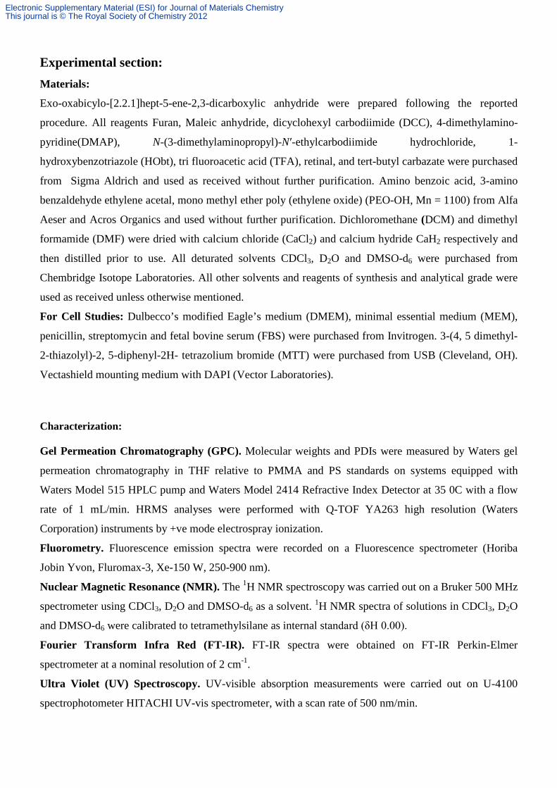
Characterization: Gel Permeation Chromatography (GPC). Molecular weights and PDIs were measured by Waters gel
permeation chromatography in THF relative to PMMA and PS standards on systems equipped with
Waters Model 515 HPLC pump and Waters Model 2414 Refractive Index Detector at 35 0C with a flow
rate of 1 mL/min. HRMS analyses were performed with Q-TOF YA263 high resolution (Waters
Corporation) instruments by +ve mode electrospray ionization.
Fluorometry. Fluorescence emission spectra were recorded on a Fluorescence spectrometer (Horiba
Jobin Yvon, Fluromax-3, Xe-150 W, 250-900 nm).
Nuclear Magnetic Resonance (NMR). The 1H NMR spectroscopy was carried out on a Bruker 500 MHz
spectrometer using CDCl3, D2O and DMSO-d6 as a solvent. 1H NMR spectra of solutions in CDCl3, D2O
and DMSO-d6 were calibrated to tetramethylsilane as internal standard (δH 0.00).
Fourier Transform Infra Red (FT-IR). FT-IR spectra were obtained on FT-IR Perkin-Elmer
spectrometer at a nominal resolution of 2 cm-1.
Ultra Violet (UV) Spectroscopy. UV-visible absorption measurements were carried out on U-4100
spectrophotometer HITACHI UV-vis spectrometer, with a scan rate of 500 nm/min.
Electronic Supplementary Material (ESI) for Journal of Materials ChemistryThis journal is © The Royal Society of Chemistry 2012

Dynamic Light Scattering (DLS). Particle size of QDs were measured by dynamic light scattering
(DLS), using a Malvern Zetasizer Nano equipped with a 4.0 mW He-Ne laser operating at λ = 633 nm.
All samples were measured in aqueous as well as methanol at room temperature and a scattering angle of
173°.
Transmission Electron Microscopy (TEM). Low resolution transmission electron microscopy (TEM)
was performed on a JEOL 200 CX microscope. TEM grids were purchased from Ted Pella, Inc. and
consisted of 3-4 nm amorphous carbon film supported on a 400-mesh copper grid.
Atomic Force Microscopy (AFM). The morphologies of the polymer was investigated from NT-MDT
micro-40 AFM instrument using a semicontact mode at a scan rate of 1 Hz.
Electronic Supplementary Material (ESI) for Journal of Materials ChemistryThis journal is © The Royal Society of Chemistry 2012

Synthesis Scheme 1: Monomers synthesis.
O O
O
OO
O
O
O
O N
O
O
MeOH, 56 °C, 72hrToluene
rt, 48 hr
1 O
O
2
Silica/H2SO4
O N
O
O
3
H2NNH
O
N
DMF/ 50 °C
O N
O
O CH
Mono 1
NNH
O
N
H2NOH
O
MeOH, 56°C, 72hr
O N
O
O
O
4
OH
NH2NH-Boc, DCM
(i) EDC.HCl, HObt, DCM
O
H
(ii) TFA, DCM
O N
O
O
O
5
NH
NH2
O N
O
O
O
NH
N CH
O
OOO
O25
HO
O 25
DMAP,Aceton
e,12hrs
O
OOO O
OO
6
25
25
HO
DCC/DMAPAcetone,12hrs
DMF/ 50 °C
H2NO
O
H
O
Mono 2
Mono 3
Electronic Supplementary Material (ESI) for Journal of Materials ChemistryThis journal is © The Royal Society of Chemistry 2012
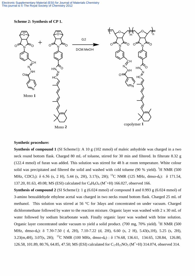
Scheme 2: Synthesis of CP 1.
O
N OO
CHN
NHO
N
O
N OO
OHNN
HC
m n
G2
N OO
CHN
NHO
N
N OO
OHNN
HC
O O
DCM:MeOH
Mono 1
Mono 2copolymer 1
Synthetic procedure:
Synthesis of compound 1 (SI Scheme1): A 10 g (102 mmol) of maleic anhydride was charged in a two
neck round bottom flask. Charged 80 mL of toluene, stirred for 30 min and filtered. In filterate 8.32 g
(122.4 mmol) of furan was added. This solution was stirred for 48 h at room temperature. White colour
solid was precipitated and filtered the solid and washed with cold toluene (90 % yield). 1H NMR (500
MHz, CDCl3): δ 6.56 (s, 2 H), 5.44 (s, 2H), 3.17(s, 2H); 13C NMR (125 MHz, dmso-d6): δ 171.54,
137.20, 81.63, 49.08; MS (ESI) calculated for C8H6O4 (M++H) 166.027, observed 166.
Synthesis of compound 2 (SI Scheme1): 1 g (6.024 mmol) of compound 1 and 0.993 g (6.024 mmol) of
3-amino benzaldehyde ethylene acetal was charged in two necks round bottom flask. Charged 25 mL of
methanol. This solution was stirred at 56 °C for 3days and concentrated on under vacuum. Charged
dichloromethane followed by water to the reaction mixture. Organic layer was washed with 2 x 30 mL of
water followed by sodium bicarbonate wash. Finally organic layer was washed with brine solution.
Organic layer concentrated under vacuum to yield a solid product. (700 mg, 70% yield). 1H NMR (500
MHz, dmso-d6): δ 7.30-7.50 ( d, 2H), 7.10-7.22 (d, 2H), 6.60 (s, 2 H), 5.43(s,1H), 5.25 (s, 2H),
3.23(m,4H), 3.07(s, 2H); 13C NMR (100 MHz, dmso-d6) : δ 176.68, 136.61, 134.65, 128.84, 126.80,
126.58, 101.89, 80.76, 64.85, 47.50; MS (ESI) calculated for C17H15NO5 (M++H) 314.074, observed 314.
Electronic Supplementary Material (ESI) for Journal of Materials ChemistryThis journal is © The Royal Society of Chemistry 2012

Synthesis of compound 3 (SI Scheme1): 1 g (3.19 mmol) of compound 2 was dissolved in 10 mL
dichloromethane. Charged 1 g of H2SO4-silica and stirred for 3h at 40 °C. The reaction mixture was
filtered. The filtrate was washed successively with Na2S2O3 (2x20mL), saturated NaHCO3 (2x20mL) and
brine (20mL). The organic layer was separated, dried over Na2SO4 and evaporated under vacuum to yield
greenish solid (80 % yield). 1H NMR (400 MHz, dmso-d6) : δ 10.08(s, 1H),7.98 ( d, 1H),7.76 (d, 2H),
7.58(d, 1H), 6.62 (s, 2 H), 5.27 (s, 2H), 3.12(s, 2H); 13C NMR (100 MHz, dmso-d6) : δ 192.54,175.58,
136.89, 132.61, 130.06, 129.69,127.00, 80.84, 47.65; MS (ESI) calculated for C15H11NO4 (M++H)
269.074, observed 269.61.
Synthesis of compound 4 (SI Scheme1): Exo - oxabicylo- [2.2.1] hept-5-ene-2, 3 - dicarboxylic
anhydride (compound 1), 1.914 g (11.5 mmol) was charged in two neck reaction flask. Charged 35 mL of
acetone and heated until it became clear solution. To this solution, charged 4- amino benzoic acid 1.605 g
(11.5 mmol) with stirring. After fifteen minutes heating was stopped and reaction mixture was allowed to
stir for about 30 minutes. The solid was filtered and dried under oven at 55 0C under vacuum. The dried
intermediate was then dissolved in 30 mL of dimethyl formamide and heated to 50 0C. Acetic anhydride
15 mL (158.97 mmol) and sodium acetate 0.635 g (7.743 mmol) were charged under stirring. The
reaction mixture was allowed to stir for three hours at 55 0C. After 3 h the reaction mixture was poured
into 500 mL of water acidified by addition of 5 mL concentrated HCl. White colour solid was precipitated
immediately and filtered the solid and washed with water and dried at 90 0C, under vacuum (80 % yield). 1H NMR (DMSO-d6, 400 MHZ): δ 13.03 (bs, 1H), 8.0 - 8.2 (m, 2H), 7.4 - 7.5 (m, 2H), 6.6 (s, 2H), 3.1 (s,
2H). 13C NMR (DMSO-d6, 400 MHz): δ 175.43, 166.59, 136.65, 135.78, 130.0, 126.79, 80.86, 47.58. IR
(KBr, cm-1): 3236, 2635,2073,1954,1826, 1780, 1729, 1698, 1607, 1515, 1418, 1218, 1144, 1125, 1020,
975, 950, 912, 883, 878, 804, 726, 672, 633, 598, 541, 521. MS (ESI) calculated for C8H10O2Na [M+ + H]
285.05, observed 284.95.
Synthesis of compound 5 (SI Scheme1): 1 g (6.92 mmol) of compound 4 was dissolved in 10 mL of
dimethyl formamide. 0.85 g (4.46 mmol) of N-(3-dimethylaminopropyl)-N’-ethylcarbodiimide
hydrochloride (EDC.HCl) and 1-hydroxybenzotriazole (HOBT) 0.62 g (4.46 mmol) in to the reaction
mixture. Reaction mixture allowed to stir for 15 h at room temperature. Reaction mixture was cooled to 0-
5 0C. Tertiary butyl carbazate was dissolved in dimethyl formamide and this solution was added to the
reaction mixture at 0-5 0C. Reaction mixture was stirred for another 30 minutes at 0-5 0C. Charged ethyl
acetate followed by water to the reaction mixture. Organic layer was washed with 2 x 30 mL of water
followed by sodium bicarbonate wash. Finally organic layer was washed with brine solution. Organic
layer concentrated under vacuum to yield a white colour solid. 500 mg (1.754 mmol) of this product was
dissolved in 5 mL of dichloromethane at room temperature. Trifluoroacetic acid 6 mL was charged in to
the reaction mixture. Reaction mixture stirred for 1 h at room temperature. Reaction mixture concentrated
to pasty mass, and charged diethyl ether resultant white product was collected by suction filtration,
Electronic Supplementary Material (ESI) for Journal of Materials ChemistryThis journal is © The Royal Society of Chemistry 2012

washed with 10 mL diethyl ether and dried at 40 0C under vacuum (420 mg, 84 % yield). 1H NMR (500
MHz, dmso-d6): δ10.92(s,1H), 7.97 ( d, 2H),7.42(d, 2H), 6.66 (s, 2 H), 5.30 (s, 2H), 3.7 (bs, 2H), 3.12(s,
2H); 13C NMR (125 MHz, dmso-d6) : δ 172.01, 169.20,137.31, 132.01, 128.01, 82.10, 49.53; MS (ESI)
calculated for C15H13N3O4 (M++H) 299.091, observed 299.
Synthesis of Mono 1(SI Scheme1): 0.2 g (0.743 mmol) of compound 3 was taken in two necks round
bottom flask. Charged 5mL of dry DMF followed by 0.102 g (0.743 mmol) of isoniazid and 0.1 mL
triethyl amine. The reaction mixture was stirred for 24 h at 45oC. Charged dichloromethane followed by
water to the reaction mixture. Organic layer was washed with 2 x 30 mL of water followed by sodium
bicarbonate and brine solution wash. Finally organic layer was dried over Sodium sulphate concentrated
under vacuum to yield brownish solid (70 % yield). 1H NMR (400 MHz, dmso-d6) : δ 12.29(s,
1H),8.90(s,2H), 8.48 (s, 1H), 7.83 ( d, 2H), 7.75(d,1H),7.68(s, 1H),7.60(t,1H), 7.31(d,1H), 6.62 (s, 2 H),
5.29 (s, 2H), 3.11(s, 2H); 13C NMR (125 MHz, dmso-d6) :δ175.75, 165.59, 150.41, 147.87,
140.39,136.70, 134.80, 132.74, 130.15, 129.74, 128.72, 121.62, 80.84, 47.66, MS (ESI) calculated for
C21H16N4O4(M++H) 389.121, observed 389.
Synthesis of Mono 2 (SI Scheme1): 0.1 g (0.3344 mmol) of compound 5 was taken in two necks round
bottom flask. Then 5 mL of dry DMF was charged, followed by 0.094 g (0.3344 mmol) of retinal and 0.1
mL triethyl amine was charged. Reaction mixture was stirred for 24 h at 45 oC. Dichloromethane was
added to reaction mixture and washed with water (3x20mL). Finally organic layer was dried over sodium
sulphate and concentrated under vacuum to yield dark brown solid (70 % yield). 1H NMR (500 MHz,
dmso-d6): δ10.35(s,1H), 7.97 ( d, 2H),7.42(d, 2H), 7.28(d, 1H), 6.66 (s, 2 H), 6-6.42(m, 4H), 5.9-6.2(m,
2H), 5.30 (s, 2H), 3.12(s, 2H), 1.91(m, 6H), 1.5-1.7(s, 3H), 1.3-1.5(m, 6H), 1.16 (s, 6H); 13C NMR (125
MHz, dmso-d6): δ 172.01, 159.20, 139.2, 137.31, 132.01, 128.01,124.08, 82.10, 49.53, 39.15, 33.02,
32.6,28.18, 21.56, 21.4,19.1, 13.3, 13.2; MS (ESI) calculated for C35H39N3O4 (M++H) 565.291, found
565.872.
Synthesis of Mono 3 (SI Scheme1): 8.7 g of (7.9 mmol) of PEO-OH (Mn=1100g/mol) was charged in a
round bottom flask and heated at 120 °C for 3h. Reaction mass cooled to room temperature. 10 mL of
acetone was added followed by 0.5 g (3.012 mmol) of 7-oxo-exo norbornene, 0.037 g of DMAP (10MOL
%, 0.3032 mmol) was added and the reaction was stirred for 12h at room temperature. To this reaction
mixture 1 g (4.854 mmol) of DCC was charged and stirred for another 12h. Finally reaction mixture was
filtered and purified by three time precipitations from diethyl ether (85 % yield). 1H NMR (500 MHz,
CDCl3): δ6.21(s, 2H), 4.27(m, 2H), 4.11 (m, 2H), 3.88 (m, 2H), 3.65 (s, 4H), 3.38 (s, 3H), 3.10 (s, 2H),
2.65 (s, 2H), 1.66 (d, J=1.2 Hz, 1H), 1.50(d, J=1.2 Hz, 1H). 13C NMR (125 MHz, CDCl3): δ 171.54,
137.20, 81.63, 68.29, 66.11, 59.20, 49.08.
Electronic Supplementary Material (ESI) for Journal of Materials ChemistryThis journal is © The Royal Society of Chemistry 2012

General Polymerization Procedure:
A known amount of monomers (Mono 1: Mono 2 : Mono 3 i.e. 2:2:1) was weighed into a two neck round
bottom flask, placed under an atmosphere of nitrogen, and dissolved in anhydrous dichloromethane
(1mL). Into a separate flask, a required amount of second generation Grubbs’ catalyst was added, flushed
with nitrogen, and dissolved in a minimum amount of anhydrous dichloromethane and methanol (9:1 v/v
%) solvent. The monomer was transferred to the flask containing the Grubbs‘ catalyst. The reaction was
allowed to stir at room temperature until the polymerization is complete, after which ethyl vinyl ether
(0.2mL) was added to quench the polymerization and An aliquot was taken for GPC analysis and the
remaining product was precipitated from pentane.
Cytotoxicity assay of CP2. Cytotoxicity of CP2 in Hela cell lines was quantitatively determined using
MTT enzymatic and colorimetric assay. All cell lines were seeded at 1×10⁴ cells /well in 96 well plates
and maintained in culture for 24 hrs at 37 °C in their respective medium. After 24 hrs medium was
removed, cells were washed with PBS and medium containing different concentration of CP2 (1µg - 1
mg) were added to the designated wells. The whole experimental plate was incubated upto 72 hrs. Fresh
20 µl of MTT from 5mg/ml stock solution were added to each well, followed by incubation for 4 hrs at 37
°C. After 4 hrs, medium from the wells were removed and 100 µl of DMSO were added to each well and
incubated for 15 mins to completely solubilize the cells. The absorbance of the resulting solution was
measured at 515 nm, and cell survivals were determined by comparison of optical density with untreated
respective control cell cultures.
Cell-growth inhibition assay by trypan blue exclusion method: Cells were seeded at 1.25 ×10⁴ cells in
24 well tissue culture plates and the inhibition activities of cell growth and division of CP2 polymer were
quantitatively determined by visual cell counting using a haemocytometer chamber. After 24 hrs of cell
plating different concentration of CP2 (1 µg - 1 mg) was added to the respective wells. Designated wells
for control were maintained with respective medium without adding any CP2. Whole plate was incubated
for additional 72 hrs. Cell counting was performed by trypan blue exclusion method at 24, 48 and 72 hrs.
The extent of cell inhibition was determined by viable cell population counts and compared with
untreated control cell culture for each time point.
Renal Clearance Experiment: 8 weeks old pathogen free C57BL/6 male mice were used from a
breeding colony originally obtained from Jackson laboratory (Bar harbour, USA). All animals were
maintained in accordance with protocols approved by the institutional animal ethical committee of IISER-
Kolkata. The authorization to use C57Bl/6 laboratory strain mice was approved by CPCSEA, India. One
mg of CP2 was dissolved into 1 ml of demonized autoclaved H2O-on the day of experiment. Working
Electronic Supplementary Material (ESI) for Journal of Materials ChemistryThis journal is © The Royal Society of Chemistry 2012

dilution was 14.2 µg/ml and from the working solution 0.5 ml was fed by stainless steel animal feeding
needle to each mice of a group of 4 mice. Urine was collected just before the feeding (control) and then
every one and half hrs interval of post feeding until 12 hrs continuously and then after 24, 48, and 72 hrs
for fluorescence measurement.
Spectral Details:
Fig S1: 1H NMR spectrum of compound 1 in CDCl3.
Fig S2: 13C NMR spectrum of compound 1 in DMSO-d6.
Electronic Supplementary Material (ESI) for Journal of Materials ChemistryThis journal is © The Royal Society of Chemistry 2012

Fig S3: 1H NMR spectrum of compound 2 in DMSO-d6.
Fig S4: 13C NMR spectrum of compound 2 in DMSO-d6.
Electronic Supplementary Material (ESI) for Journal of Materials ChemistryThis journal is © The Royal Society of Chemistry 2012

Fig S5: 13C NMR spectrum of compound 3 in DMS-d6.
Fig S6: 13C NMR spectrum of Mono 1 in DMSO-d6.
Electronic Supplementary Material (ESI) for Journal of Materials ChemistryThis journal is © The Royal Society of Chemistry 2012
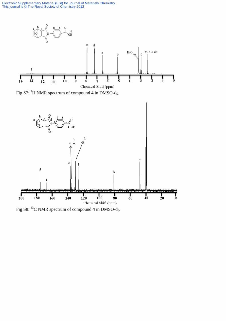
Fig S7: 1H NMR spectrum of compound 4 in DMSO-d6.
Fig S8: 13C NMR spectrum of compound 4 in DMSO-d6.
Electronic Supplementary Material (ESI) for Journal of Materials ChemistryThis journal is © The Royal Society of Chemistry 2012

Fig S9:
1H NMR spectra of tertiary butyl hydrazone Nadic carboxylate in DMSO-d6.
Fig S10: 1H NMR spectrum of compound 5 in DMSO-d6.
Electronic Supplementary Material (ESI) for Journal of Materials ChemistryThis journal is © The Royal Society of Chemistry 2012

Fig S11: 13C NMR spectrum of compound 5 in DMSO-d6.
Fig S12: 1H NMR spectrum of Mono 2 in DMSO-d6.
Electronic Supplementary Material (ESI) for Journal of Materials ChemistryThis journal is © The Royal Society of Chemistry 2012

Fig S13: 13C NMR spectrum of Mono 2 in CDCl3.
Fig S14: 1H NMR spectrum of CP 1 in CDCl3.
Electronic Supplementary Material (ESI) for Journal of Materials ChemistryThis journal is © The Royal Society of Chemistry 2012
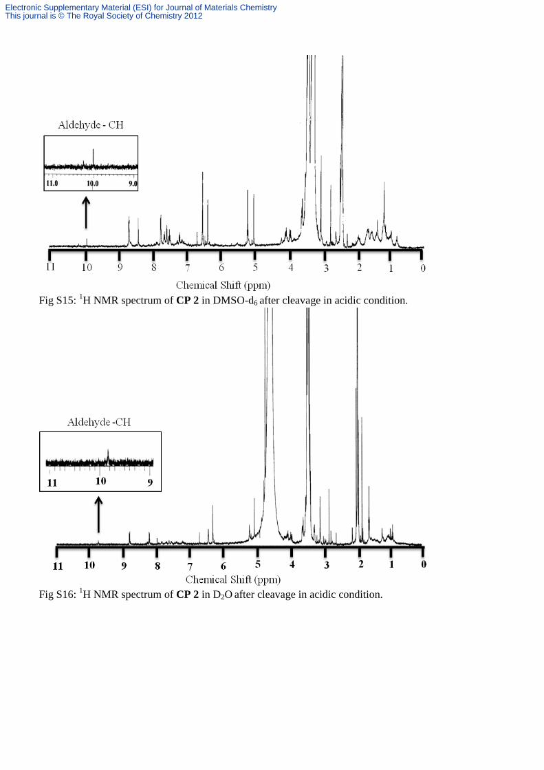
Fig S15: 1H NMR spectrum of CP 2 in DMSO-d6 after cleavage in acidic condition.
Fig S16: 1H NMR spectrum of CP 2 in D2O after cleavage in acidic condition.
Electronic Supplementary Material (ESI) for Journal of Materials ChemistryThis journal is © The Royal Society of Chemistry 2012

Fig S17. FT-IR spectrum of (a) compound 3 and (b) Mono 1.
Fig S18: FT-IR spectrum of compound 5.
Electronic Supplementary Material (ESI) for Journal of Materials ChemistryThis journal is © The Royal Society of Chemistry 2012

Fig S19: FT-IR spectrum of (a) retinal and (b) Mono 2.
Fig S20: FT-IR spectrum of CP 1.
Electronic Supplementary Material (ESI) for Journal of Materials ChemistryThis journal is © The Royal Society of Chemistry 2012
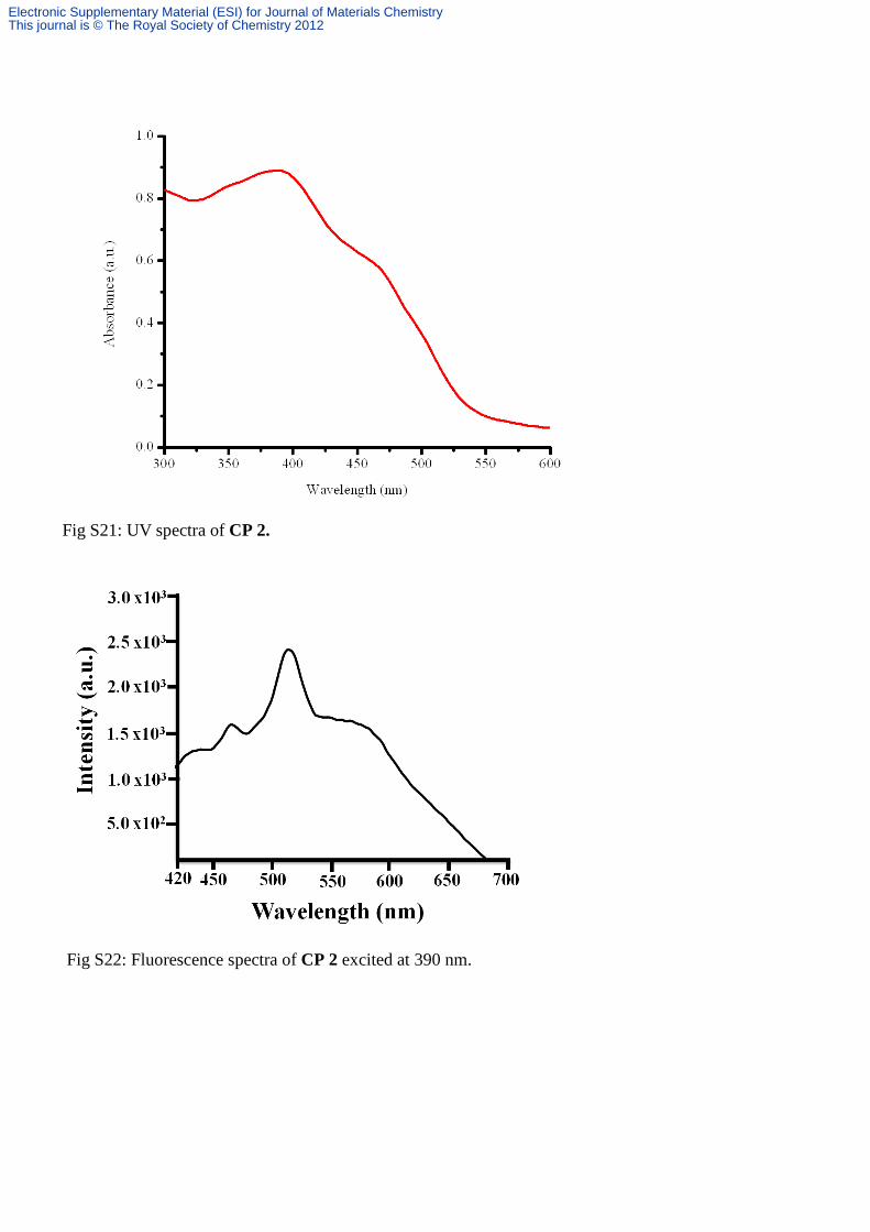
Fig S21: UV spectra of CP 2.
Fig S22: Fluorescence spectra of CP 2 excited at 390 nm.
Electronic Supplementary Material (ESI) for Journal of Materials ChemistryThis journal is © The Royal Society of Chemistry 2012

Fig S23: Fluorescence spectra for drug release from CP 2 at pH 4 by Dialysis method.
Fig S24: Fluorescence spectra for drug release from CP 2 at pH 5.5 by Dialysis method.
Electronic Supplementary Material (ESI) for Journal of Materials ChemistryThis journal is © The Royal Society of Chemistry 2012

Fig S25: Fluorescence spectra for drug release from CP 2 at pH 6.5 by Dialysis method.
Electronic Supplementary Material (ESI) for Journal of Materials ChemistryThis journal is © The Royal Society of Chemistry 2012

Fig S26: Fluorescence spectra of CP 2 in urine collected as renal clearance from mouse 1.The excitation wavelength is 390 nm.
Fig S27: Fluorescence spectra of CP 2 in urine collected as renal clearance from mouse 2.The excitation wavelength is 390 nm.
Electronic Supplementary Material (ESI) for Journal of Materials ChemistryThis journal is © The Royal Society of Chemistry 2012

Fig S28: : Fluorescence spectra of CP 2 in urine collected as renal clearance from mouse 3.The excitation wavelength is 390 nm.
Fig S29: : Fluorescence spectra of CP 2 in urine collected as renal clearance from mouse 4.The excitation wavelength is 390 nm.
Electronic Supplementary Material (ESI) for Journal of Materials ChemistryThis journal is © The Royal Society of Chemistry 2012

Fig. S30 : Cytotoxicity assay of Isoniazid.
Electronic Supplementary Material (ESI) for Journal of Materials ChemistryThis journal is © The Royal Society of Chemistry 2012

Fig. S31 : Cytotoxicity assay of Retinal.
Electronic Supplementary Material (ESI) for Journal of Materials ChemistryThis journal is © The Royal Society of Chemistry 2012