A to Z Book - Amanda's A to Z Medical Pocket Books · aka = also known as ... = joints =...
47
Dr A. L. Neill BSc MSc MBBS PhD FACBS The A to Z of Surface Anatomy The A to Z of Surface Anatomy
Transcript of A to Z Book - Amanda's A to Z Medical Pocket Books · aka = also known as ... = joints =...
Dr A. L. NeillBSc MSc MBBS PhD FACBS
The A to Z of Surface Anatomy
The A to Z of Surface Anatomy
A-Z Surface Anatomy_48:A to Z Book 20/11/12 7:05 AM Page A
The A to Z of Surface Anatomy
© A. L. Neill 1
IntroductionThis book describes the structures which lie beneath the skin whichinvolves a recognition of the surface curves of most skeletal muscles.It ties in well with the A to Z of Skeletal Muscles, and of course the A to Z of the Hair, Nails and Skin, but all the A to Zs are cross-referenced and together are forming a set covering the all structuralelements of the human body. Recently pathology as well as anatomyhas been tackled by the A to Zs with The A to Z of Bone & JointFailure the first book to cover the breakdown of the body’s structuresin this manner, expanding upon the knowledge of the A to Z of Bones,Joints, Ligaments and the Back.
Artists have used studied anatomy and surface anatomy to help intheir creations – paintings, sculpture etc and it is important inEmergency Medicine to be able to SEE inside the body in a 3dimensional manner.
If there is a structure/subject you want to see in the A to Zs let us [email protected]
We have 2 websites and there maybe others where you can view allimages of the A to Zs and any additional material please feel free toexamine the new books which may be placed here and to give anysuggestions after all it was due to the overwhelming number ofrequests for this title that this book was written placed:http://www.aspenpharma.com.au/atlas/student.htmwww.amandasatoz.com
AcknowledgementThank you Aspenpharmacare Australia for your support andassistance in this valuable project, particularly Mr. Greg Lan, and RobKoster. Thank you to all those who have helped when I have beenrushed to finish and have made time for this project, and have faith init, in particular Ante Mihaljevic and Phill Ryman. Thank you everyonewho has provided valuable feedback and help in many ways; Richard,Peter, Robbie, Jody, Quentin and there are others too, thank you.
DedicationTo those who read and use these books and find them helpful.
A-Z Surface Anatomy_48:A to Z Book 20/11/12 7:05 AM Page 1
© Dr Aman
da Neil
l

2
The A to Z of Surface Anatomy
© A. L. Neill
How to use this bookThe structure of the A to Zs grows and develops with each new bookwhile the principle of listing structures in an alphabetical ismaintained. Basic anatomical concepts are placed in the beginningof this book; then measurements and proportions of the body. Therole of the Common Terms section is enlarged, illustrated and colourcoded.
In this book the images are alphabetical whether they can be seen ornot – i.e. the heart cannot be seen but its projection are indicated onthe chest – but the tendons of the wrist can be visualized. Allstructures and regions are listed alphabetically. All entries are crossreferenced in the usual manner i.e. see for go to and also see foradditional images listed under that heading.
Thank you A.L. NeillBSc MSc MBBS PhD FACBSmedicalamanda@gmail ISBN 978-1-921930-17-1
A-Z Surface Anatomy_48:A to Z Book 20/11/12 7:05 AM Page 2
© Dr Aman
da Neil
l
The A to Z of Surface Anatomy
© A. L. Neill 3
Table of contents Introduction 1Acknowledgement 1Dedication 1How to use this Book 2Table of Contents 3Abbreviations 7Common Terms used in Surface Anatomy 9Anatomical Planes and Relations 17Movements – general
Upper limb & shoulder 19Head, Neck & Back 21Lower limb and Hip 23Foot and Hand 24
HandGrips 25Measurements 27
Proportions and forms of human measurement 29Human face proportions 31Human body proportions 33Vitruvian Man 35(Symbol of proportionality & derivative of measurements)
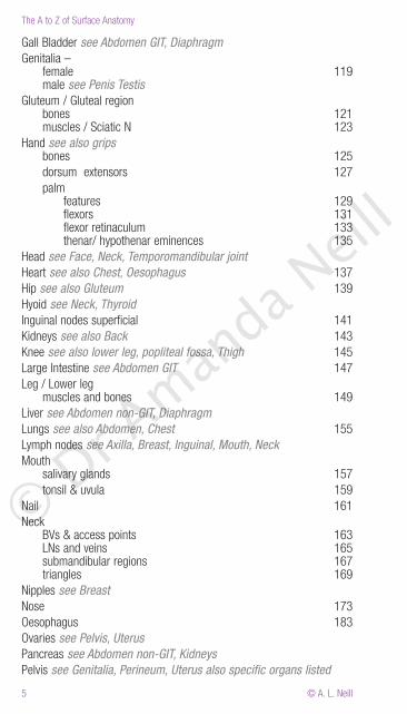
Index of surface anatomy Abdomen see also Aorta, Trunk
dermatomes 37 bones + muscles 39alimentary - GIT 41non- alimentary - / liver / pancreas / spleen 43regions 45scars - from incisions 47
Adam’s apple = Thyroid cartilage see Thyroid Adrenals see Back, Trunk Anatomical snuff box see Thumb Ankle see also Foot 49Anus see Perineum Aorta 51Appendix see Abdomen GIT Arm see also Axilla, Forearm, Shoulder 53Axilla see also Breast
bones , muscles boundaries 55lymph nodes 57
A-Z Surface Anatomy_48:A to Z Book 20/11/12 7:05 AM Page 3
© Dr Aman
da Neil
l

4
The A to Z of Surface Anatomy
© A. L. Neill
Back lower 59upper see Chest
Belly see Abdomen Belly button see AbdomenBladder see Kidneys, Pelvis, UterusBreast see also Axilla
arterial 61lymphatic & venous 62
Buttock see Gluteum Caecum see Abdomen GITCarpal tunnel see Hand Chest Wall see also Abdomen, Lungs
great vessels 63heart 64heart valves sounds 65incision or marks = scars 66lungs & pleura 67
Cubital Fossa 71Diaphragm + assoc structures see also Oesophagus 73Duodenum see Abdomen GIT Kidneys Ear 75Elbow see arm, cubital fossa, forearm Eye 77Face
arteries 81bones see also TMJ 87Facial N 89muscles 91veins 93
Femoral trianglecontents & borders 95muscles & bones 97
Finger see Hand Flexor Retinaculum see Hand Foot
dorsumbones 99tendons 101
solefascia / muscle layers 103bones / dermatomes 108
Forearmbones 111muscles 113
A-Z Surface Anatomy_48:A to Z Book 20/11/12 7:05 AM Page 4
© Dr Aman
da Neil
l
The A to Z of Surface Anatomy
© A. L. Neill 5
Gall Bladder see Abdomen GIT, DiaphragmGenitalia –
female 119male see Penis Testis
Gluteum / Gluteal regionbones 121muscles / Sciatic N 123
Hand see also gripsbones 125dorsum extensors 127palm
features 129flexors 131flexor retinaculum 133thenar/ hypothenar eminences 135
Head see Face, Neck, Temporomandibular joint Heart see also Chest, Oesophagus 137Hip see also Gluteum 139 Hyoid see Neck, Thyroid Inguinal nodes superficial 141Kidneys see also Back 143Knee see also lower leg, popliteal fossa, Thigh 145Large Intestine see Abdomen GIT 147Leg / Lower leg
muscles and bones 149 Liver see Abdomen non-GIT, Diaphragm Lungs see also Abdomen, Chest 155Lymph nodes see Axilla, Breast, Inguinal, Mouth, NeckMouth
salivary glands 157tonsil & uvula 159
Nail 161Neck
BVs & access points 163LNs and veins 165submandibular regions 167triangles 169
Nipples see Breast Nose 173Oesophagus 183Ovaries see Pelvis, Uterus Pancreas see Abdomen non-GIT, Kidneys Pelvis see Genitalia, Perineum, Uterus also specific organs listed
A-Z Surface Anatomy_48:A to Z Book 20/11/12 7:05 AM Page 5
© Dr Aman
da Neil
l
Penis 185Perineum see also Genitalia 187Phrenic N see Oesophagus Pleura see Chest, Lungs Popliteal fossa 191Rectum see Abdomen GIT, Pelvis, Perineum, Sigmoid colonSalivary Glands see Head, Mouth, Neck Sciatic N see also Gluteum 193 Scrotum see Femoral Triangle, Penis, TestisShoulder
bones 195BVs & Ns 197muscles 199
Sigmoid Colon see Abdomen GIT, Large IntestineSinuses 205 Small Intestine see Abdomen GIT Spleen see Abdomen, Back, Diaphragm Stomach see also Abdomen 207Teeth see Face, Mouth, TMJ
A to Z of the Head & Neck for complete map Temporomandibular Joint 209Testis/Testes see also Femoral Triangle, Penis, Scrotum Perineum 211 Thigh see also Femoral Triangle, Hip & Knee
muscle 213Throat see Mouth Thumb 215Thymus see also Oesophagus 217Thyroid see also Neck 217Tongue see also Mouth 219Tonsil see Mouth, Tongue Trachea see also Neck 223Trunk see Abdomen, Back Umbilicus see Abdomen Ureter see Kidneys Uterus see also Pelvis 225 Uvula see Mouth, TongueVagina see Pelvis, Perineum Vagus N see Oesophagus Womb see uterusWrist see also Hand & grips 227
6
The A to Z of Surface Anatomy
© A. L. Neill
A-Z Surface Anatomy_48:A to Z Book 20/11/12 7:05 AM Page 6
© Dr Aman
da Neil
l
The A to Z of Surface Anatomy
© A. L. Neill 7
A = atrium, (pl atria) / actions / movements of a jointa = arteryabdo = abdomen / abdominal ACF = anterior cranial fossa adj. = adjective AIIS = anterior inferior iliac spine aka = also known as alt. = alternativeAM = arachnoid mater ANS = autonomic nervous system ant = anteriorart. = artery AS = Alternative Spelling, generally referring to the diff. b/n British & American spellingASIS = anterior superior iliac spine assoc.= associated with AV = atrioventricular B = blood BBB = blood brain barrier bc = becauseBF = blood flow BM = basement membrane b/n = between BP = brachial plexus bpm = beats per minute br = branch (of a vessel) BS = blood supply / blood stream BV = blood vessel(s)cap. = capillary c.f. = compared to C = carpalC = cervicalCC = costal borderCC = costal cartilage CH = cerebral hemispheres cm = cell membrane CNS = central nervous system collat. = collateral CP = cervical plexus Cr = cranial CSF = Cerebrospinal fluid CT = connective tissue
CVA = cerebrovascular accident = stroke defn = definitiondiff. = difference(s)dist. = distal DM = dura mater DVT = deep vein thrombosis EAM = external auditory meatus e.g. = example EC = extracellular (outside the cell) ECG = electrocardiogramED = extensor digitorumER = Extensor RetinaculumFDP = Flexor digitorum porofundusFDS = Flexor digitorum superficialisFPB = Flexor pollicus brevisFPL = Flexor pollicus longusFR = Flexor Retinaculum Gk. = Greek H = hormone(s) H = hypochondriumHB = heart beat HF = heart failure HR = heart rate HS = heart sounds IC = intercostalIC = intercarpalICS = intercostal spaceIP = interphalangealIx = investigationIVC = inferior vena cava jt(s) = joints = articulations L = left L = lumbarLA = Left Atrium lat. = lateral LH = left hypochondrium LL = lower limb LIF = left iliac fossa lig = ligament Lt. = Latin m = muscleMC = metacarpal MCF = middle cranial fossa
Abbreviations
A-Z Surface Anatomy_48:A to Z Book 20/11/12 7:05 AM Page 7
© Dr Aman
da Neil
l
8
The A to Z of Surface Anatomy
© A. L. Neill
MCL = mid clavicular lineMCP = metacarpophalangealmed. = medial MI = myocardial infarction MIP = midinguinal point MT = metatarsal N = nerve NAD = normal (size, shape) NAD = no abnormality detected NR = nerve rootNS = nervous system/nerve supply NT = nervous tissuenv = neurovascular bundle P = pressure PAD = peripheral artery disease PaNS = parasympathetic nervous system Ph = phalangesPIIS = posterior inferior iliac spine pl. = pluralPM = pia mater PN = peripheral nerve post. = posteriorproc. = processprox. = proximalPS = pubic symphysis PSIS = posterior superior iliac spine R = right RA = right atriumRH = right hypochondrium RIF = Right Iliac Fossa S = sacral S1 = first heart sound S2 = second heart sound SA = sinoatrial SCM = sternocleidomastoid musclesing. = singular SC = spinal cord SN = spinal nerve SP = spinal process SR = sarcoplasmic reticulum subcut. = subcutaneous supf = superficial
SVC = superior vena cava SyNS = sympathetic nervous system T = thoracic TMJ = temporomandibular jointUL = upper limb, arm V = vertebra V = ventricle VC = vertebral column WM = white matter w/n = within w/o = without wrt = with respect to & = and ∩ = intersection with
A-Z Surface Anatomy_48:A to Z Book 20/11/12 7:05 AM Page 8
© Dr Aman
da Neil
l
The A to Z of Surface Anatomy
© A. L. Neill 9
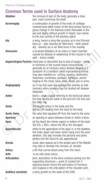
Common Terms used in Surface Anatomy Ablation the removal of part of the body, generally a bony part, most commonly the teeth Acromegaly a continuation of growth of the ends of cartilage covered bone (after fusion of the long bones) hence a gross change in the features (most noticeable in the jaw and digits) without growth in height, due mainly to the over activity of the pituitary gland Ala a wing, hence a wing-like process as in the Ethmoid bone pl. - alae. Alveolus Air filled bone - tooth socket adj - alveolar (as in air filled bone in the maxilla) Aneurysm a localized dilatation of an artery or heart chamber caused by disease or weakening of the muscle in the wall – tunica media. Angina/Angina Pectoris chest pain or discomfort due to lack of oxygen - anoxia or ischemia in the muscle tissue (myocardium) generally bc of coronary artery disease. Angina is a symptom of a condition called myocardial ischemia; may also manifest as : aching, burning, discomfort, heaviness, numbness, pressure, tightness, and/or tingling in the chest, back, neck, throat, jaw or arms.Angiography an X-ray that uses dye injected into arteries so that coronary artery anatomy can be studied wrt disease diagnosis.Ankle bend = angle usually referring to the bend just above the foot, hence the ankle is the joint b/n the foot and the lower leg Aorta the largest artery in the body and the primary BV leading from the heart to the body. Aortic Valve the valve that regulates BF from the heart to the aorta.Aperture an opening or space between bones or within a bone.Apex (of the Heart) the inferior aspect or bottom of the heart 5th ICS, L MCL, where the HB is the strongest Appendicular refers to the appendices of the axial i.e. in the skeleton, the limbs upper and lower which hang from the axial skeleton, this also includes the pectoral and pelvic girdles (not the Sacrum) adj. appendicularAreola small, open spaces as in the areolar part of the Maxilla may lead or develop into sinuses. pl. areolaeArtery a BV that carries blood away from the heart. Arth- to do with joints hence… Articulation joint, description of the bone surfaces joining w/o the supporting structures = point of contact b/n 2 opposing bones hence the articulation of Humerus and Scapula is the articulation of the shoulder joint. Auditory exostosis a bony growth on the walls of the EAM
A-Z Surface Anatomy_48:A to Z Book 20/11/12 7:05 AM Page 9
© Dr Aman
da Neil
l

10
The A to Z of Surface Anatomy
© A. L. Neill
Atrium Lt antrum = waiting room – top chambers R & L of the heart - 1/3 of the volume of the Ventricle or lower chamber. Blood flows from the atria to the Ventricles. Avulsion forceable tearing away of a structure or part of a structure as in an avulsed fracture where a fragment bone is torn away from the main bone Axis of the body - is the central part - the line through the head & spine, the axial skeleton as opposed to the appendicularBase - “of the Heart” top of the heart located in the 4th ICS Basilar relating to the base or bottom of structures Basiocranium bones of the base of the skull Boss a smooth round broad eminence - mainly in the frontal bone female > maleBregma refers to a junction of more than 2 bones in a joint as in the Bregma of the skull, junction between the coronal and sagittal sutures which in the infant is not closed and can be felt pulsating – site of the anterior fontanelle. Buccal pertaining to the cheek Calotte consists of the Calvaria from which the base has been removed.Calvaria refers to the Cranium without the facial bones attached.Canal tunnel / extended foramen as in the carotid canal at the base of the skull adj canular (canicular - small canal) Caput / Kaput the head or of a head, adj.- capitate = having a head (c.f. decapitate)Carotid to put to sleep; compression of the common or internal carotid artery causes coma.Carpo wrist Catheter a thin, flexible tubeCavity an open area or sinus within a bone or formed by two or more bones (adj. cavernous), may be used interchangeably with fossa. Cavity tends to be more enclosed fossa a shallower bowl like space (Orbital fossa-Orbital cavity). Cavum a cave. Cephalic pertaining to the head Cervico pertaining to the neck Concha a shell shaped bone as in the ear or nose (pl. conchae adj. chonchoid) old term for this turbinate. Condyle a rounded enlargement or process possessing an articulating surface.Cornu a horn (as in the Hyoid)
A-Z Surface Anatomy_48:A to Z Book 20/11/12 7:05 AM Page 10
© Dr Aman
da Neil
l

Corona a crown. adj.- coronary, coronoid or coronal; hence a coronal plane is parallel to the main arch of a crown which passes from ear to ear (c.f. coronal suture). Costo/Costa pertaining to the ribs Conductivity the ability to conduct an impulse to another region or another cell Congenital existing at birth. Congestive heart failure blood volume coming in is more than that pumped out - leading to fluid backup - backup from the LV results in fluid overload in the lungs - in the RV results in venous fluid retention and then swelling of dependant parts Coronary Arteries two arteries arising from the aorta that arch down over the top of the heart and branch out in additional arteries that provide B to the heart muscle – the main 4 being L main coronary artery, Circumflex coronary artery, L ant descending coronary artery, and R coronary artery. They join to form rings around the heart b/n the A & Vs and b/n the 2 Vs. These are the most commonly blocked arteries of the heart. Cranium comprises all of the bones of the skull except for the Mandible.Crest prominent sharp thin ridge of bone formed by the attachment of muscles particularly powerful ones eg Temporalis/Sagittal crest Cuneate /Cuneus a wedge / wedge-shaped Dens a tooth hence dentine and dental relating to teeth, denticulate having tooth-like projections adj dentate See odontoid Depression a concavity on a surface Diaphysis The shaft or body of a long bone. In the young this is the region between the growth plates & is composed of compact bone. pl.= diaphyses adj.= diaphyseal Diploë the cancellous bone between the inner and outer tables of the skull, adj.- diploic. Echocardiogram a study using high-frequency sound waves to picture or visualize the heart chambers, the thickness of the muscle wall, the heart valves and major BVs located near the heart. This is a non-invasive procedure.Edentulous without teeth Elbow any angular bend often in the arm, usually referring to the joint between the arm and the forearmEminence a smooth projection or elevation on a bone as in iliopubic eminence. Endocranium refers to the interior of the “braincase” divided into the 3 major fossae anterior (for the Frontal lobes) middle (containing Temporal lobes) & posterior (for the containment of the Cerebellum).
The A to Z of Surface Anatomy
© A. L. Neill 11
A-Z Surface Anatomy_48:A to Z Book 20/11/12 7:05 AM Page 11
© Dr Aman
da Neil
l

12
The A to Z of Surface Anatomy
© A. L. Neill
Epiphysis the end of a long bone beyond the growth plate or epiphyseal plate. Generally develops as a secondary ossification centre. There are 2 epiphyses to each long bone. Of a long bone the shafts are generally compact bone and the ends=epiphyses are trabecular bone adj.= epiphyseal
External Auditory Meatus ear hole Exostosis a bony outgrowth from a bony surface, often due to irritation (as in swimmer’s ear) and may involve ossification of surrounding tissues such as muscles or ligaments. Facet a face, a small bony surface (occlusal facet on the chewing surfaces of the teeth) seen in planar joints. Falciform relating to shapes that are in a sickle shape so falciform ligaments curve around and end in a sharp point Fissure a narrow slit or gap from cleft.Fontanelle a fountain, associated with the palpable pulsation of the brain as in the anterior fontanelle of an infant. these soft spots on the skull are cartilagenous connective tissue coverings “joints” which allow for skull cranial expansion and then become the mould for the bone development and shape joining long the sutural lines, later becoming the Bregma. Foramen a natural hole in a bone usually for the transmission of blood vessels and/or nerves. (pl. foramina).Fornix an archFossa a pit, depression, or concavity, on a bone, or formed from several bones as in temporomandibular fossa. Shallower and more like a “bowl” than a cavity Fovea a small pit (usually smaller than a fossa)- as in the fovea of the occlusal surface of the molar tooth. Genu / genio referring to the knee Groove long pit or furrow Hallux the big toe = the first toe Hamus a hook hence the term used for bones which “hook around other bones or where other structures are able to attach by hooking - hamulus = a small hook. Hyoid U-shaped Incisura a notch. Inter between Intra within Introitus an orifice or point of entry to a cavity or space. Joint = Articulation supporting structures Lacerum something lacerated, mangled or torn e.g. foramen lacerum small sharp hole at the base of the skull - often ripping tissue in trauma.
A-Z Surface Anatomy_48:A to Z Book 20/11/12 7:05 AM Page 12
© Dr Aman
da Neil
l

Lacrimal related to tears and tear drops. (noun lacrima) Lambda Greek letter a capital 'L' - written as an inverted\ V.(adj. lambdoid) - used to name the point of connection b/n 3 skull bones Occipital and L & R Temporal bones. Lamina a plate as in the lamina of the Vertebra a plate of bone connecting the vertical and transverse spines (pl. laminae) Ligament a band of tissue which connects bones (articular ligaments) or viscera - organs (visceral ligaments). Linea a line as in the Nuchal lines of the Occitipum Lingual pertaining to the tongue Locus a place (c.f. location, locate, dislocate). Lymphatic a vessel which carries fluid to the heart Magnum large pl magnaMalleus hammer (as in the ear ossicle) Mandible from the verb to chew, hence, the movable lower jaw; adj.- mandibular. Mastoid breast or teat shape - mastoid process of the Temporal bone. Maxilla the jaw-bone; now used only for the upper jaw; adj.- maxillary. Meatus a short passage; adj.- meatal as in external acoustic meatus connecting the outer ear with the middle ear. Mediastinum the region in the thorax b/n the lungs, ant. boundary- the sternum post. the VC, includes the heart, roots of the great vessels, oesophagus and trachea. Meniscus Gk. crescent as in the crescent shaped cartlages on the top of the TibiaMental relating to the chin (mentum = chin not mens = mind).Meta an extension of: cf. metacarpal = extension of the wristMetaphysis the slightly expanded end of the shaft of a bone.Microvasculature the network of small BVs arterioles [capillaries [venules in a tissue Minimally Invasive a variety of approaches using smallerHeart Surgery incisions to reduce the trauma of surgery and potentially speed recovery. Mitral Valve the valve that controls the BF b/n the LA & LV in the heart.Murmur a specific sound emanating from the chest in addition to the normal HS. Myocardial Infarct also called “heart attack”; the sudden interruption or insufficiency of the supply of B to the heart, typically resulting from occlusion or obstruction of a coronary artery and often characterized by severe chest pain Myocardial infarction death of myocardial tissue due to anoxia .
The A to Z of Surface Anatomy
© A. L. Neill 13
A-Z Surface Anatomy_48:A to Z Book 20/11/12 7:05 AM Page 13
© Dr Aman
da Neil
l
14
The A to Z of Surface Anatomy
© A. L. Neill
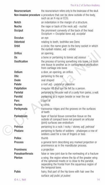
Neurocranium the neurocranium refers only to the braincase of the skull.Non-invasive procedure a procedure that can be done outside of the body, such as an X-ray or ECG.Notch an indentation in the margin of a structure. Nucha the nape or back of the neck adj.- nuchal. Occiput the prominent convexity of the back of the head Occipitum = Occipital bone adj. occipital Occulus an eye Odontoid relating to teeth, toothlike see Dens Orbit a circle; the name given to the bony socket in which the eyeball rotates; adj - orbital. Orifice an opening. Os a bone or pertaining to bones adj osseus Ossification the process of turning something into bone, i.e. from one tissue to another as in cartilagneous ossification from cartilage into bone Ostium a door, an opening, an orifice. Otic pertaining to the ear Ovale oval shaped Palate a roof adj.- palatal or platatine.Palpitation irregular HB that can be felt by a person. Parietal pertaining to the outer wall of a cavity from paries, a wall.Parotid pertaining to a region beside or near the ear Pars a part of Pecten a comb. Perikymata transverse ridges and the grooves on the surfaces of teethPeriosteum layer of fascial tissue connective tissue on the outside of compact bone not present on articular (joint) surfaces see endostium Petrous pertaining to a rock / rocky / stoney adj. petrosal Phalanx pertaining to flanks of soldiers - phalanges a row of soldiers used for a row of fingers or toes Pollex thumb Process a general term describing any marked projection or prominence as in the mandibular process.Prominens a projection Pseudoarthrosis false or new joint due to the nonhealing of a fracture Pterion a wing; the region where the tip of the greater wing of the sphenoid meets or is close to the parietal, separating the frontal from the squamous region of the temporal bone.Pubis hairy, that part of the hip bone with hair over the surface adj pubic pl pubes
A-Z Surface Anatomy_48:A to Z Book 20/11/12 7:05 AM Page 14
© Dr Aman
da Neil
l
The A to Z of Surface Anatomy
© A. L. Neill 15
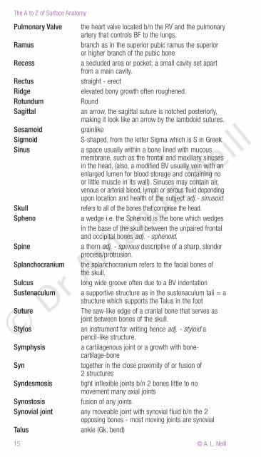
Pulmonary Valve the heart valve located b/n the RV and the pulmonary artery that controls BF to the lungs. Ramus branch as in the superior pubic ramus the superior or higher branch of the pubic boneRecess a secluded area or pocket; a small cavity set apart from a main cavity. Rectus straight - erect Ridge elevated bony growth often roughened. Rotundum Round Sagittal an arrow, the sagittal suture is notched posteriorly, making it look like an arrow by the lambdoid sutures. Sesamoid grainlike Sigmoid S-shaped, from the letter Sigma which is S in Greek. Sinus a space usually within a bone lined with mucous membrane, such as the frontal and maxillary sinuses in the head, (also, a modified BV usually vein with an enlarged lumen for blood storage and containing no or little muscle in its wall). Sinuses may contain air, venous or arterial blood, lymph or serous fluid depending upon location and health of the subject adj.- sinusoid.Skull refers to all of the bones that comprise the head.Spheno a wedge i.e. the Sphenoid is the bone which wedges in the base of the skull between the unpaired frontal and occipital bones adj. - sphenoid.Spine a thorn adj. - spinous descriptive of a sharp, slender process/protrusion.Splanchocranium the splanchocranium refers to the facial bones of the skull.Sulcus long wide groove often due to a BV indentationSustenaculum a supportive structure as in the sustenaculum tali = a structure which supports the Talus in the foot Suture The saw-like edge of a cranial bone that serves as joint between bones of the skull.Stylos an instrument for writing hence adj. - styloid a pencil-like structure. Symphysis a cartilagenous joint or a growth with bone- cartilage-bone Syn together in the close proximity of or fusion of 2 structures Syndesmosis tight inflexible joints b/n 2 bones little to no movement many axial joints Synostosis fusion of any joints Synovial joint any moveable joint with synovial fluid b/n the 2 opposing bones - most moving joints are synovial Talus ankle (Gk. bend)
A-Z Surface Anatomy_48:A to Z Book 20/11/12 7:05 AM Page 15
© Dr Aman
da Neil
l
16
The A to Z of Surface Anatomy
© A. L. Neill
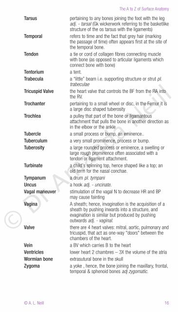
Tarsus pertaining to any bones joining the foot with the leg adj. - tarsal (Gk wickerwork referring to the basketlike structure of the os tarsus with the ligaments) Temporal refers to time and the fact that grey hair (marking the passage of time) often appears first at the site of the temporal bone. Tendon a tie or cord of collagen fibres connecting muscle with bone (as opposed to articular ligaments which connect bone with bone) Tentorium a tent. Trabecula a “little” beam i.e. supporting structure or strut pl. trabeculae Tricuspid Valve the heart valve that controls the BF from the RA into the RV. Trochanter pertaining to a small wheel or disc, in the Femur it is a large disc shaped tuberosity Trochlea a pulley that part of the bone or ligamantous attachment that pulls the bone in another direction as in the elbow or the ankle Tubercle a small process or bump, an eminence..Tuberculum a very small prominence, process or bump. Tuberosity a large rounded process or eminence, a swelling or large rough prominence often associated with a tendon or ligament attachment.Turbinate a child’s spinning top, hence shaped like a top; an old term for the nasal conchae. Tympanum a drum pl. tympaniUncus a hook adj. - uncinate. Vagal maneuver stimulation of the vagal N to decrease HR and BP may cause fainting Vagina A sheath; hence, invagination is the acquisition of a sheath by pushing inwards into a structure, and evagination is similar but produced by pushing outwards adj. - vaginal. Valve there are 4 heart valves: mitral, aortic, pulmonary and tricuspid, that act as one-way "doors" between the chambers of the heart. Vein a BV which carries B to the heart Ventricles lower heart 2 chambres – 3X the volume of the atria Wormian bone extrasutural bone in the skull Zygoma a yoke , hence, the bone joining the maxillary, frontal, temporal & sphenoid bones adj zygomatic.
A-Z Surface Anatomy_48:A to Z Book 20/11/12 7:05 AM Page 16
© Dr Aman
da Neil
l

The A to Z of Surface Anatomy
© A. L. Neill 17
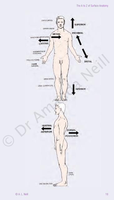
Anatomical Planes and RelationsThis is the anatomical position.
A = Anterior Aspect from the front = or / Posterior Aspect from the back. Used interchangeably with ventral and dorsal respectively B= Lateral Aspect from either side C= Transverse / Horizontal planeD= Midsagittal plane = Median plane; trunk moving away from this plane = lateral flexion or lateral movement.- plane medial movement; limbs moving away from this direction = abduction; limbs moving closer to this plane = adductionE = Coronal plane F = Median
A-Z Surface Anatomy_48:A to Z Book 20/11/12 7:05 AM Page 17
© Dr Aman
da Neil
l
18
The A to Z of Surface Anatomy
© A. L. Neill
A-Z Surface Anatomy_48:A to Z Book 20/11/12 7:05 AM Page 18
© Dr Aman
da Neil
l
The A to Z of Surface Anatomy
© A. L. Neill 19
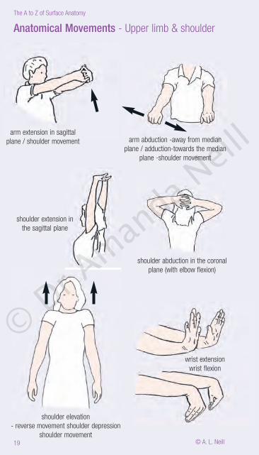
Anatomical Movements - Upper limb & shoulder
arm extension in sagittalplane / shoulder movement
shoulder extension inthe sagittal plane
shoulder abduction in the coronalplane (with elbow flexion)
shoulder elevation - reverse movement shoulder depression
shoulder movement
wrist extension wrist flexion
arm abduction -away from medianplane / adduction-towards the median
plane -shoulder movement
A-Z Surface Anatomy_48:A to Z Book 20/11/12 7:05 AM Page 19
© Dr Aman
da Neil
l
100
The A to Z of Surface Anatomy
© A. L. Neill
1d
1p
3h
3s 3b
4m
6t64i4L
5
1t
8
7
9L 2 9m
A-Z Surface Anatomy_48:A to Z Book 20/11/12 7:08 AM Page 100
© Dr Aman
da Neil
l

The A to Z of Surface Anatomy
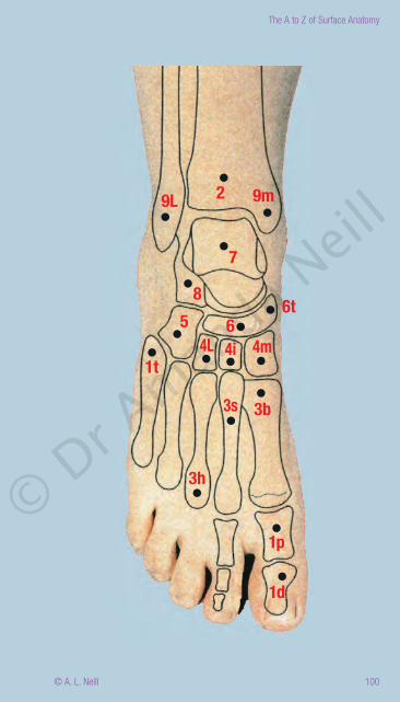
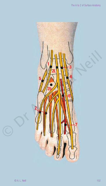
© A. L. Neill 101
Foot – dorsum Tendons Anterior view -showing the tendons of the foot.
1 Peroneus tertius2 Extensor digitorum b = brevis showing muscle and tendon L = longus 3 Extensor hallicus longus 4 Tibialis anterior 5 Peronius brevis 6 dorsalis pedis artery 7 1st dorsal MT artery
F
A-Z Surface Anatomy_48:A to Z Book 20/11/12 7:08 AM Page 101
© Dr Aman
da Neil
l
102
The A to Z of Surface Anatomy
© A. L. Neill
2
6
5
2b
4
1
2L3
7
A-Z Surface Anatomy_48:A to Z Book 20/11/12 7:08 AM Page 102
© Dr Aman
da Neil
l
The A to Z of Surface Anatomy
© A. L. Neill 103
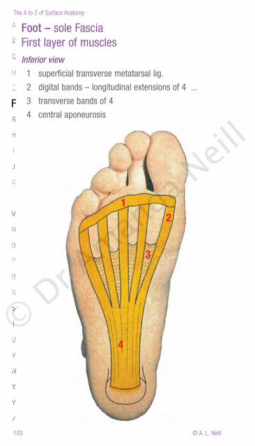
Foot – sole FasciaFirst layer of muscles Inferior view
1 superficial transverse metatarsal lig. 2 digital bands – longitudinal extensions of 4 ...3 transverse bands of 4 4 central aponeurosis
F
12
3
4
A-Z Surface Anatomy_48:A to Z Book 20/11/12 7:08 AM Page 103
© Dr Aman
da Neil
l

104
The A to Z of Surface Anatomy
© A. L. Neill
The foot has 4 muscle layers overlaid with a strong protective fascia.
the central aponeurosis (4) is similar to the palmar aponeurosiswith extensions (2) to accommodate the extended MTs. A bridgingfortified transverse ligament (1) joints all the heads of the MTs toreflect the weight bearing function of the foot
5 abductor digiti minimi 6 flexor digitorum brevis 7 abductor hallicus
765
A-Z Surface Anatomy_48:A to Z Book 20/11/12 7:08 AM Page 104
© Dr Aman
da Neil
l
The A to Z of Surface Anatomy
© A. L. Neill 105
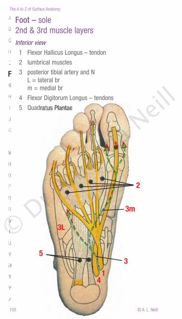
Foot – sole 2nd & 3rd muscle layers Inferior view
1 Flexor Hallicus Longus – tendon 2 lumbrical muscles 3 posterior tibial artery and N L = lateral br m = medial br 4 Flexor Digitorum Longus – tendons5 Quadratus Plantae
F
2
1
3m
3
14
5
3L
A-Z Surface Anatomy_48:A to Z Book 20/11/12 7:08 AM Page 105
© Dr Aman
da Neil
l

106
The A to Z of Surface Anatomy
© A. L. Neill
The 2nd layer consists of tendons to muscles which are found inthe leg – the long muscles + some of the short muscles (i.e. thosecompletely in the foot itself) – and the BVs and Ns
The 3rd layer contains the equivalent of the thenar (7) & hypothenar(8) muscles which insert into the long plantar lig (9) - technically inthe 4th layer.
6 Abductor hallicus 7 Flexor hallicus brevis 8 Flexor digiti minimi brevis 9 plantar lig = spring lig (maintains the medial long arch)
9
7
6
8
A-Z Surface Anatomy_48:A to Z Book 20/11/12 7:08 AM Page 106
© Dr Aman
da Neil
l

The A to Z of Surface Anatomy
© A. L. Neill 107
Foot – sole 4th muscle layerdermatomes Inferior view
1 peroneus lig b = brevis, L = longus 2 Tibialis posterior 3 plantar calcaneo - navicular lig & long plantar lig 4 Plantar interossei
F
3
3 2
44
4
1b
1L
1L
A-Z Surface Anatomy_48:A to Z Book 20/11/12 7:08 AM Page 107
© Dr Aman
da Neil
l

108
The A to Z of Surface Anatomy
© A. L. Neill
The 4th layer consists of tendons of muscles which are found in theleg and primarily act on the foot and ankle joint. - major lig arefound here and deep to this layer which support the arches of thefoot along with bony factors
Dermatome distribution of the sole of the foot can be used to testperipheral Ns
A-Z Surface Anatomy_48:A to Z Book 20/11/12 7:09 AM Page 108
© Dr Aman
da Neil
l
The A to Z of Surface Anatomy
© A. L. Neill 109
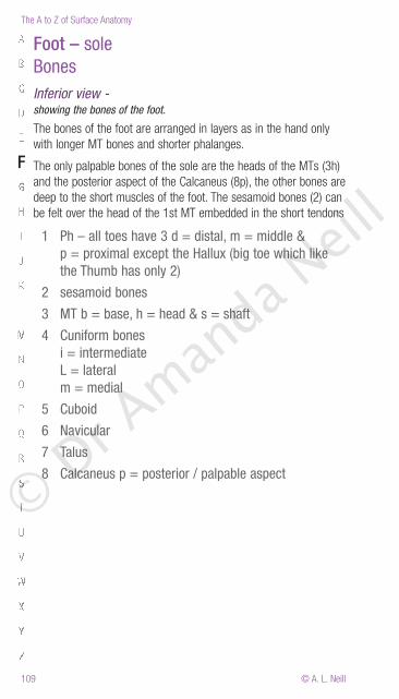
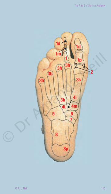
Foot – soleBones Inferior view - showing the bones of the foot.
The bones of the foot are arranged in layers as in the hand onlywith longer MT bones and shorter phalanges.
The only palpable bones of the sole are the heads of the MTs (3h)and the posterior aspect of the Calcaneus (8p), the other bones aredeep to the short muscles of the foot. The sesamoid bones (2) canbe felt over the head of the 1st MT embedded in the short tendons
1 Ph – all toes have 3 d = distal, m = middle & p = proximal except the Hallux (big toe which like the Thumb has only 2)2 sesamoid bones 3 MT b = base, h = head & s = shaft 4 Cuniform bones i = intermediate L = lateral m = medial5 Cuboid 6 Navicular 7 Talus 8 Calcaneus p = posterior / palpable aspect
F
A-Z Surface Anatomy_48:A to Z Book 20/11/12 7:09 AM Page 109
© Dr Aman
da Neil
l
110
The A to Z of Surface Anatomy
© A. L. Neill
1d
1m
1d
1 1p3h
3h3h
3h
3b
2
3s
4m
4i
67
8
8p
54L
A-Z Surface Anatomy_48:A to Z Book 20/11/12 7:09 AM Page 110
© Dr Aman
da Neil
l
The A to Z of Surface Anatomy
© A. L. Neill 111
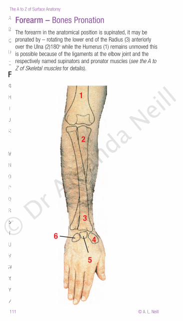
Forearm – Bones Pronation The forearm in the anatomical position is supinated, it may bepronated by – rotating the lower end of the Radius (3) anteriorlyover the Ulna (2)180o while the Humerus (1) remains unmoved thisis possible because of the ligaments at the elbow joint and therespectively named supinators and pronator muscles (see the A toZ of Skeletal muscles for details).
F
1
2
3
46
5
A-Z Surface Anatomy_48:A to Z Book 20/11/12 7:09 AM Page 111
© Dr Aman
da Neil
l

112
The A to Z of Surface Anatomy
© A. L. Neill
Forearm – Bones Supination
1
2
3
64
5
It articulates with the firat layer of carpal – wrist – bones, 4Scaphoid, 5 Lunate, 6 Triquetral.
A-Z Surface Anatomy_48:A to Z Book 20/11/12 7:09 AM Page 112
© Dr Aman
da Neil
l

The A to Z of Surface Anatomy
© A. L. Neill 113
Forearm – Muscles Anterior (Flexor surface) - Deep layer of muscles.
The forearm contains the muscle bellies of most finger flexors in 3layers: deep & 2 superficial layers on the flexor - anterior surface.The tendons of these muscles move under the Flexor Retinaculum(not shown - see Hand) to attach onto the digital phalanges (seethe A to Z of Skeletal muscles for details).
1 Biceps 2 Supinator3 Flexor Pollicus Longus 4 Pronator Quadratus 5 Flexor Digitorum Profundus
F
A-Z Surface Anatomy_48:A to Z Book 20/11/12 7:09 AM Page 113
© Dr Aman
da Neil
l
114
The A to Z of Surface Anatomy
© A. L. Neill
1
2
5
3
4
A-Z Surface Anatomy_48:A to Z Book 20/11/12 7:09 AM Page 114
© Dr Aman
da Neil
l

The A to Z of Surface Anatomy
© A. L. Neill 115
Forearm – Muscles Superficial layers of muscles The forearm contains the muscle bellies of most finger flexors. Thetendons of these muscles move under the Flexor Retinaculum (notshown - see Hand) to attach onto the digital phalanges (see Hand &the A to Z of Skeletal Muscles for details).
1 Brachioradialis2 Flexor Carpi Radialis 3 Pronator Teres 4 Flexor Carpi Ulnaris5 Palmaris Longus 6 Pisiform bone
F
13
45
2
6
4
A-Z Surface Anatomy_48:A to Z Book 20/11/12 7:09 AM Page 115
© Dr Aman
da Neil
l
116
The A to Z of Surface Anatomy
© A. L. Neill
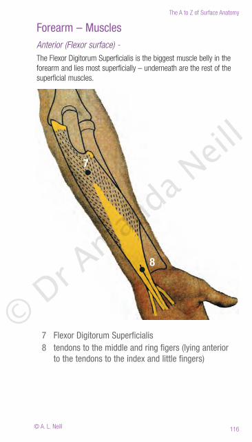
Forearm – Muscles Anterior (Flexor surface) - The Flexor Digitorum Superficialis is the biggest muscle belly in theforearm and lies most superficially – underneath are the rest of thesuperficial muscles.
7 Flexor Digitorum Superficialis 8 tendons to the middle and ring figers (lying anterior to the tendons to the index and little fingers)
7
8
A-Z Surface Anatomy_48:A to Z Book 20/11/12 7:09 AM Page 116
© Dr Aman
da Neil
l

The A to Z of Surface Anatomy
© A. L. Neill 117
Forearm – Muscles Posterior (Extensor surface) - deep layer The forearm contains the muscle bellies – tendons extend to thephalanges passing under the Extensor Retinaculum (not shown - see Hand).
1 Supinator 2 Abductor Pollicus Longus 3B Extensor Pollicus Brevis 3L Extensor Pollicus Longus 4 Extensor Indicis 5B Extensor Carpi Radialis Brevis 5L Extensor Carpi Radialis Longus 6 Flexor Carpi Ulnaris
F
1
6 2
4
5L
3L3L
5B
3B
A-Z Surface Anatomy_48:A to Z Book 20/11/12 7:09 AM Page 117
© Dr Aman
da Neil
l
118
The A to Z of Surface Anatomy
© A. L. Neill
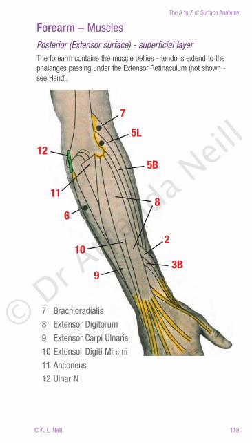
Forearm – Muscles Posterior (Extensor surface) - superficial layer The forearm contains the muscle bellies - tendons extend to thephalanges passing under the Extensor Retinaculum (not shown - see Hand).
7
5L
5B
8
2
3B9
10
6
11
12
7 Brachioradialis 8 Extensor Digitorum 9 Extensor Carpi Ulnaris 10 Extensor Digiti Minimi 11 Anconeus 12 Ulnar N
A-Z Surface Anatomy_48:A to Z Book 20/11/12 7:09 AM Page 118
© Dr Aman
da Neil
l

The A to Z of Surface Anatomy
© A. L. Neill 119
Genitalia – FemaleInferior view - showing the features of the female peroneal area in detail. Note the Peroneal body is inferior to this.
Medium level view 1 Mons Pubis 2 Labia Majora 3 area of hair & pigmentation 4 area of smooth delicate skin – less pigmentation
Detailed level view 5 Prepuce 6 Clitoris – enlarges on stimulation 7 Urethral sphincter & opening 8 Labia Minora – edge – engorged on stimulation 9 wall of the Labia Minora 10 vaginal opening 11 fourchette
G
A-Z Surface Anatomy_48:A to Z Book 20/11/12 7:09 AM Page 119
© Dr Aman
da Neil
l

120
The A to Z of Surface Anatomy
© A. L. Neill
1
7
6
5
8
9
11
10
2
3
4
A-Z Surface Anatomy_48:A to Z Book 20/11/12 7:09 AM Page 120
© Dr Aman
da Neil
l

The A to Z of Surface Anatomy
© A. L. Neill 121
Gluteal region = ButtocksPosterior thigh = back of the legBones
1 iliac crest 2 posterior iliac spine i = inferior s = superior 3 sciatic notch g = greater L = lesser 4 L4 spine 5 L5 spine 6 Sacrum 7 femur h = head g = greater trochanter L = lesser trochanter 8 gluteal tuberosity 9 linea aspera 10 femoral condyle L = lateral m = medial 11 adductor tubercle 12 ischeal tuberosity 13 ischeal spine 14 sacro-iliac joint15 Coccyx
G
A-Z Surface Anatomy_48:A to Z Book 20/11/12 7:09 AM Page 121
© Dr Aman
da Neil
l

122
The A to Z of Surface Anatomy
© A. L. Neill
15
14
7h 7g
87L
9
11
10L10m
12
153L
13
3g2i2s
6
4
A-Z Surface Anatomy_48:A to Z Book 20/11/12 7:09 AM Page 122
© Dr Aman
da Neil
l

© A. L. Neill 229
The A to Z of Hair, Nails & Skin ISBN 978-1-921930-027
The structure of the biggest & most visible organ in the body THE SKIN,is described in detail along with its associated structures. The book has3 distinct sections each listed in the A to Z way, with clear colourfuldiagrams. A large Common Terms section explains & illustratesterminology on the subject. With over 230 pages & 280 illustrations itstill fits in your pocket for convenience.
The A to Z of Peripheral Nerves ISBN 978-1-921930-05-8
The origins, pathways, branches and functions of all the Peripheralnerves are listed alphabetically and illustrated individually. The maincontent includes neurological testing techniques, basic structuralcomponents of the nervous system and overviews of the majornerve plexi. It begins with a comprehensive glossary of all terms,and illustrations of basic anatomical principles. With over 230 pagesand 290 illustrations this strong little book still fits in your pocket.
The A to Z of Skeletal Muscles ISBN 978-1-921930-188
The origins, insertions, blood & nerve supply for all muscles are listedalphabetically with separate illustrations. All the major muscle groups,their common names their functions, along with cross-referencing andregional tagging are included. Basic structural components of theskeletal muscle system are included with a comprehensive glossary ofall terms used in the field. With over 230 pages and 290 illustrationsthis strong little book still fits in your pocket
The A to Z of Bones, Joints, Ligaments & the BACKISBN 978-1-921930-19-5
All the bones, joints and ligaments of the body including teeth are listedalphabetically. At least 2 views of each bone and joint are illustrated. TheRange of movement and basic structure of all the skeletal componentsare categorized and illustrated. There is a separate section on the back –Vertebral Column where it is discussed as a functioning unit. Over 260pages and 300 illustrations make this little pocket book invaluable.
A-Z Surface Anatomy_48:A to Z Book 20/11/12 7:13 AM Page 229
© Dr Aman
da Neil
l
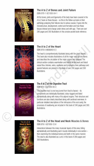
The A to Z of Bones and Joint Failure ISBN 978-1-921930-04-1
All the bones, joints and ligaments of the body have been covered in theA to Z book on these tissues – so this is the follow-up book on theirpathology analysing their failures due to various causes. It goes into themicrostructure, development, control and formation and how thesetissues interact and change under stress and with age. There are over280 pages and 350 illustrations in this concise pocket book reference.
The A to Z of the HeartISBN 978-0-9806840-6-3
The heart is comprehensively illustrated along with the great vessels.This book also includes illustrations of all the major vascular structuresand describes the circulation of the major organs and systems. Theclinical section contains examination and testing of the heart and bloodvessel flow. Arteries, veins, capillaries and lymphatics their pathways andspecial features are present in this book of over 240 pages and 300illustrations.
The A to Z of the Digestive Tract ISBN 978-1-921930-00-3
The Digestive tract is one long tunnel from food to faeces – itscomponents are individually illustrated, colour tagged and listedalphabetically along with many of its adjunct organs. Their structure andfunctions are also clearly described along with sectional overviews. Inparticular detailed descriptions of the intricacies of the oral cavity, theprocesses of swallowing are included in this book of 240 pages and 300illustrations.
The A to Z of the Head and Neck Muscles & BonesISBN 978-1-921930-12-6
interactions between the many muscular layers of this area, listingalphabetically and illustrating each muscle individually in one section –then examining the individual bones and teeth in the same manner.The skull is also illustrated as a unit, in this book of 280 pages and 300 illustrations.
A-Z Surface Anatomy_48:A to Z Book 20/11/12 7:13 AM Page 230
© Dr Aman
da Neil
l

The A to Z of Surface Anatomy ISBN 978-1-921930-17-1
The surface anatomy of all anatomical regions and structures areillustrated at several levels form superficial to deep. Methods of locatingstructures deep in the body using common landmarks are illustratedcross referenced and listed alphabetically. Proportions and relationsbetween limb and regional sizes are charted extensively. Photographs aswell as detailed graphics are used extensively, in this book of 240 pagesand 300 illustrations.
The A to Z of the Brain & Cranial Nerves ISBN 978-0-9806840-2-5
The brain as an entity and the individual structures within it areillustrated and then listed with their functions alphabetically – sectionson the testing and pathways and interactions of cranial nerves are alsoincluded in a separate clinical section in this book of 240 pages and 300illustrations.
The A to Z of Medical Terms ISBN 978-1-921930-01-0
This book is invaluable as a medical terminology reference – initiallydesigned for the derivation of the anatomical terms; it has expanded toinclude tables of medical, pathological and other specialist terms; tablesof prefixes and suffixes which allow interpretation of terms and lists ofabbreviations commonly in use. It also includes forms of address, titles,major medical associations and other useful material. These colour-coded illustrated sections are clear and concise.
Special rates for students and libraries.
A-Z Surface Anatomy_48:A to Z Book 20/11/12 7:13 AM Page 231
© Dr Aman
da Neil
l

97mm18mm
Please adjust SPINE to bookthicknessFold
Fold
BACK COVERSPINEFLAP
The A to Zs
Dr. A. L. NEILLBSc MSc MBBS PhD FACBS [email protected]
www.amandasatoz.com
The Ato Z
of Surface Anatomy
Back cover ‘outside view’
The Ato Z
of Surface Anatomy
Dr. A. L. NEILL
The A to Z of Surface Anatomy
The A to Z of Surface Anatomy
A-Z_SA_BC01:Layout 1 19/11/12 11:19 AM Page 1