A STUDY ON ROLE OF URINE TRYPSINOGEN – 2 IN DIAGNOSING ...
102
A STUDY ON ROLE OF URINE TRYPSINOGEN – 2 IN DIAGNOSING ACUTE PANCREATITIS A DISSERTATION SUBMITTED TO THE TAMILNADU DR.M.G.R MEDICAL UNIVERSITY In partial fulfillment of the regulations for the award of the M.S.DEGREE EXAMINATION BRANCH I GENERAL SURGERY DEPARTMENT OF GENERAL SURGERY STANLEY MEDICAL COLLEGE AND HOSPITAL THE TAMILNADU DR.M.G.R MEDICAL UNIVERSITY CHENNAI APRIL 2014
Transcript of A STUDY ON ROLE OF URINE TRYPSINOGEN – 2 IN DIAGNOSING ...

A STUDY ON ROLE OF URINE TRYPSINOGEN – 2 IN
DIAGNOSING ACUTE PANCREATITIS
A DISSERTATION SUBMITTED TO
THE TAMILNADU DR.M.G.R MEDICAL UNIVERSITY
In partial fulfillment of the regulations for the award of the
M.S.DEGREE EXAMINATION
BRANCH I GENERAL SURGERY
DEPARTMENT OF GENERAL SURGERY
STANLEY MEDICAL COLLEGE AND HOSPITAL
THE TAMILNADU DR.M.G.R MEDICAL UNIVERSITY
CHENNAI
APRIL 2014
CERTIFICATE
This is to certify that the dissertation titled
“A STUDY ON ROLE OF URINE TRYPSINOGEN – 2 IN
DIAGNOSING ACUTE PANCREATITIS” is the bonafide work done
by Dr. N.SANGARA NARAYANAN, Post Graduate student (2011 – 2014) in
the Department of General Surgery, Government Stanley Medical
College and Hospital, Chennai under my direct guidance and
supervision, in partial fulfillment of the regulations of The Tamil
Nadu Dr. M.G.R Medical University, Chennai for the award of
M.S., Degree (General Surgery) Branch - I, Examination to be held
in April 2014.
Prof. A. RAJENDRAN, M.S., Prof.K. KAMARAJ,M.S., Professor of Surgery, Professor and Head of the Department, Dept. of General Surgery, Dept. of General Surgery, Stanley Medical College, Stanley Medical College, Chennai-600001. Chennai-600001.
PROF. S. GEETHA LAKSHMI, M.D., PhD,
The Dean, Stanley Medical College,
Chennai-600001.

DECLARATION
I, DR.N.SANGARA NARAYANAN solemnly declare that
this dissertation titled “A STUDY ON ROLE OF URINE
TRYPSINOGEN – 2 IN DIAGNOSING ACUTE PANCREATITIS”
is a bonafide work done by me in the Department of General Surgery,
Government Stanley Medical College and Hospital, Chennai
under the guidance and supervision of my unit chief.
Prof. A. RAJENDRAN Professor of Surgery
This dissertation is submitted to The Tamilnadu Dr.M.G.R.
Medical University, Chennai in partial fulfillment of the university
regulations for the award of M.S., Degree (General Surgery) Branch - I,
Examination to be held in April 2014.
Place: Chennai. Date: December 2013 DR.N.SANGARA NARAYANAN

ACKNOWLEDGEMENT
My sincere thanks to Dr.S.GEETHALAKSHMI, MD., Ph.D.,
the Dean, Govt. Stanley Medical College for permitting me to conduct
the study and use the resources of the College.
I consider it a privilege to have done this study under the
supervision of my beloved Professor and Head of the Department
Prof.K.KAMARAJ, who has been a source of constant inspiration and
encouragement to accomplish this work.
I am highly indebted to my guide Prof. A. RAJENDRAN,
Professor of Surgery for his constant help, inspiration and valuable
advice in preparing this dissertation.
I express my deepest sense of thankfulness to my Assistant
Professors Dr. G. VENKATESH, Dr.JIM JEBAKUMAR, for their
valuable inputs and constant encouragement without which this
dissertation could not have been completed.
I express my sincere gratitude to my mentors
Prof.S.DEIVANAYAGAM, Prof.P.DARWIN and Prof.J.VIJAYAN
,former Heads of Department of General Surgery. I thank them for the
constant support, able guidance, inspiring words and valuable help they
rendered to me during my course.
I would like to thank my former assistant
professors DR.P.BALAJI, DR.G.V.MANOHARAN, and
DR.M.VIGNESH, for their valuable suggestions and help in
completing this dissertation.
I am particularly thankful to my fellow postgraduate colleagues
Dr.Gautham Krishnamurthy, and Dr.Soundarya.G and other fellow
postgraduates for their valuable support in the time of need throughout
the study.
I thank my Seniors Dr.S.Rakesh, Dr.O.k.Prakashen,
Dr. Shyam, Dr. Sudharsan, Dr. Naveen, Dr. Sivakumar, Dr. Saravana
Krushna Raja, Aravind R.M. and my friends Dr.Arshad Ali, Dr. Dinesh,
Dr. Kaushik Kumar, Dr. Aravind Menon, Dr.Prasanna, Dr. Sakthi Balan
who supported me in completing the dissertation.
It is my earnest duty to thank my parents and my wife, without
whom accomplishing this task would have been impossible.
I am extremely thankful to my patients who consented and
participated to make this study possible.

CONTENTS
S. NO. CHAPTER PAGE NO
1. INTRODUCTION 1
2. REVIEW OF LITERATURE 3
3. AIMS AND OBJECTIVES 60
4. MATERIALS & METHODS 60
5. OBSERVATION AND RESULTS 63
6. DISCUSSION 71
7. CONCLUSION & SUMMARY 74
8. BIBLIOGRAPHY 76
9. ANNEXURE
(i) PROFORMA 82
(ii) INSTITUTIONAL ETHICAL
COMMITTEE APPROVAL
CERTIFICATE
86
(iii) MASTER CHART 87
(iv) CONSENT FORM 90
(v) PATIENT INFORMATION SHEET 91

ABSTRACT
A STUDY ON ROLE OF URINE TRYPSINOGEN-2 IN DIAGNOSING
ACUTE PANCREATITIS
IntroductionAcute pancreatitis is a very common disorder, with substantial burden on the
healthcare system1. Acute pancreatitis includes wide spectrum of disease varying
from mild self-limiting symptoms to fulminant multi organ failure and high
mortality. Serum amylase and serum lipase which are used for the diagnosis of
acute pancreatitis are relatively less sensitive and specific and gives a lot of false
positive or false negative values. The urinary trypsinogen-2 dipstick test, proposed
to be a rapid method for the diagnosis of acute pancreatitis at the earliest, based
on the immune-chromatographic method.
Materials and Methodology100 Patients presenting with acute upper abdominal symptoms like pain,
vomiting, abdominal distention, admitted in the emergency department of
our hospital from January 2013 to November 2013 are enrolled in the study.
Urine sample were obtained from all the patients and tested with Spot Urine
trypsinogen-2 dipstick.

Serum amylase and serum lipase tests were also simultaneously done in
these patients. Patients are also evaluated with (USG) abdomen and (CECT)
abdomen ,if required.
Final diagnosis of acute pancreatitis is made on the basis of clinical picture,
serum amylase more than threefold rise and radiological findings.
Urine trypsinogen-2 dipstick test were compared with serum amylase,
serum lipase and imaging studies in patients with final diagnosis of acute
pancreatitis
ResultsSensitivity of amylase and lipase was found to be 73.77% and 59.02%
respectively, whereas as sensitivity of trypsinogen was found to be 78.69%.
Specificity of amylase and lipase was found to be 89.74% and 89.74%
respectively, whereas as specificity of trypsinogen was found to be
92.1%.Analysing the data ,it is found that sensitivity and specificity of trypsinogen
is higher than the routine investigations. Eventhough it has a low range of
sensitivity, its high specificity ensures that the test can be used as a screening test
to check the true negative cases.

Conclusion
1. Urine Trypsinogen-2 dip stick test is a simple, rapid, easy, and noninvasive
test which can diagnose or rule out, most of the cases of acute pancreatitis.
2. Urine Trypsinogen-2 estimation doesn't require laboratory facilities. It is
undertaken almost instantaneously (within 5 minutes) as opposed to serum
amylase and lipase, results for which may require an hour to get back to the
physician.
3. The urinary trypsinogen-2 test could be used as a screening test for acute
pancreatitis.
4. Modification of the cutoff point of this assay increases the specificity to the
point where it can be used for diagnosis.

Qualitative rapid urine trypsinogen-2 test strip is easy to perform. And
hence it has been shown to be a reliable and useful screening test for acute
pancreatitis in daily practice
[12-16], particularly in healthcare units lacking laboratory facilities.
Keywords
Acute Pancreatitis, Serum Amylase, Serum Lipase, Urine Trypsinogen-2.

1
INTRODUCTION
Acute pancreatitis is a very common disorder, with substantial
burden on the healthcare system1. Acute pancreatitis includes wide
spectrum of disease varying from mild self-limiting symptoms to
fulminant multi organ failure and high mortality. The overall mortality
rate is 3-10%, wherein 11-30% of cases are with severe disease
manifested as pancreatic necrosis.
Since 1974, several scoring systems have been developed
clinically and radiologically assessing the prognosis of the disease. The
rationale behind the assessment of severity is mainly for practical
purpose, where mild pancreatitis needs supportive care but severe
pancreatitis needs intensive monitoring and it has a guarded prognosis.
The key to reduce the mortality and morbidity of the disease is
early detection and appropriate management. An ideal diagnostic
method should be able to differentiate between patients with mild &
severe disease, easy usability, widely available and should be accurate,
and with low inter-observer variability. It should be able to detect early
disease ,so that patient before developing potential complications,could
be monitored and treated, if possible empirically.

2
Serum amylase and serum lipase which are used for the diagnosis
of acute pancreatitis are relatively less sensitive and specific and gives a
lot of false positive or false negative values. Various scoring systems are
being used in acute pancreatitis to predict the severity and outcome of
the disease. There is no single comprehensive test to aid in early and
accurate detection of acute pancreatitis.
The urinary trypsinogen-2 dipstick test, proposed to be a rapid
method for the diagnosis of acute pancreatitis at the earliest, based on
the immune-chromatographic method.

3
REVIEW OF LITERATURE
HISTORY OF THE PANCREAS
• Herophilus, a Greek anatomist cum surgeon, first discovered the
pancreas, in 336 BC ,on the Asiatic side of the Bosporus,
Chalcedon2.
• The word pancreas first mentioned in the writings of Eristratos
(310-250 B.C.). Then Four hundred years later, Rufus, an
anatomist cum surgeon of Ephesus, termed the name “pancreas”.
Written in Greek , the word quotes “pan: all, kreas: flesh”2.
• Galen (138-201 AD)a Romen physician, and “Physician to the
Gladiators”, said that the pancreas serves as a cushion, protecting
the large blood vessels, lying behind it2.
• In March 2, 1642, Johann Georg Wirsüng, a German,
discovered the pancreatic duct, at San Francisco Monastery,
Padua, Italy. But it was named by his colleague as “The Duct of
Wirsüng”2. The duct enlarges at the terminal point as the papilla,
which projects into the second part of duodenum, was first
4
described by Vater in 1720. In 1734,Santorini, described the
accessory duct,that bears his name.
• In 1869, Paul Langerhans (“Junior”), a student of -the famous
Berlin Institute of Pathology, headed by the eminent Professor,
Rudolph Virchow, described the pancreatic islets2,which was
the first histologic description of the pancreas.
• In 1893, Laguesse suggested that the islet cells produce a
hormone. In 1909 Jean de Meyer suggested the name 'insulin' for
this hormone.
• Eugene Lindsay Opie (1873-1971) was able to show the
association between diabetes and failure of the islet cells and in
1901, proposed his "common channel" hypothesis3.
• In 1908, Julius Wohlgemuth, Berlin, devised a method to
measure the serum amylase concentration (“diastase”), which was
found to be useful in diagnosing acute pancreatitis, prior to
laparotomy or autopsy2.
• Since 1898, many surgeons undertook various steps for the
resection of tumors of ampulla and head of the pancreas. Allen O.
Whipple (1881-1963), son of American missionaries, Persia, was

5
recognized as the “Father of Pancreatic Surgery” for his
successful single stage surgery in pancreatic head tumors2.
• In 1963, the first Marseilles Symposium favored the development
of classification system for pancreatitis. This was revised in 1984;
at the second Marseilles Symposium.
• Finally, at the Atlanta Symposium, in 1992, clinically oriented
classification system was established for acute pancreatitis.
• Although the disease now classified as acute pancreatitis has been
known from antiquity, it is only at mid-19th century, the
importance of pancreas and its severity became evident. In 1889,
Fitz presented the clinical and pathology of acute pancreatitis.
Moynihan in 1925 described "the most terrible of all the
calamities which occur in relation with the abdominal viscera" as
acute pancreatitis4, 5.

6
Gross Anatomy
The pancreas, a retroperitoneal organ , extends from the C-loop of
the duodenum to the splenic hilum in a oblique manner 7.
The pancreas lies behind the stomach, roughly in the Trans
pyloric plane. The gland weighs approximately 80gm, varying from
75 – 125gm and measures 15 to 22 cm length in adults7.
The pancreas has four parts7, 8:
• The head including the uncinate process,
• The neck,
• The body and
• The tail.

7
The head lies within the duodenal C- loop .It overlies the second
lumbar vertebral body and the inferior vena cava(IVC), with the aorta
lying beneath the neck of the gland. The right renal artery and the renal
veins lie posteriorly behind the head of the pancreas. Coming off ,the
side of the head of pancreas, and passing to the left , is the pancreatic
uncinate process.
The neck of pancreas lies anterior to the portal vein. Behind the
pancreatic neck , the superior mesenteric vein and the splenic vein join
to form the portal vein. The inferior mesenteric vein(IMV) forms the
tributary of the splenic vein. Sometimes, the IMV drains into the SMV
or with the superior mesenteric-portal venous junction, forming a
trifurcation. The common bile duct(CBD) lies within a groove in the
head of the pancreas, until joining the main pancreatic duct. Both the
ducts, join to form the ampulla of Vater, opening into the 2nd part of
the duodenum.
The pancreatic body and tail lies anterior to the splenic artery and
its vein. The splenic vein lies in a groove, draining multiple pancreatic
venous branches. These venous branches must be ligated to perform a
spleen-sparing distal pancreatectomy. Along the postero superior edge
of the pancreatic body and tail, lies the splenic artery , parallel to the

8
vein. The splenic artery is highly tortuous. The pancreatic body is
covered by the peritoneum, the gastrocolic omentum. On dividing the
gastrocolic omentum ,the body and tail of the pancreas can be visualized
at the floor of the lesser sac, just posterior to the stomach. It is in this
area pancreatic pseudocysts commonly develop , in relation to the
posterior aspect of the stomach , allowing drainage of the cyst to the
stomach. The transverse mesocolon base ,attaches to the inferior margin
of the body and tail of pancreas.
The body of pancreas overlies the aorta, near the origin of the
superior mesenteric artery. Blunt antero-posterior trauma can compress
the neck of the pancreas against the spine of L1 and L2, causing injury
to the pancreatic parenchyma and the duct.
Pancreatic Ductal Anatomy:
Pancreatic duct anatomy and its variations can be understood by
knowing the embryology of pancreas. It is formed by the fusion of a
ventral bud and a dorsal bud9.
• The duct from the smaller ventral bud, arises from the hepatic
diverticulum, connects directly to the CBD.
• The duct from the larger dorsal bud, arise from the duodenum.
9
The ventral anlage duct becomes the duct of Wirsung, and the
dorsal anlage duct becomes the duct of Santorini. The ducts from each
anlage fuse together, in the pancreatic head such that, most of the
pancreas drains through the main pancreatic duct (MPD) or the
Wirsung.
The common channel length is often variable. In one third of the
patients, the CBD and MPD remains separate till joining the papilla; in
another third the two ducts may merge at the papilla, and in the
remaining third few millimeters of true common channels persist.
The duct of Santorini, persist as the lesser pancreatic duct.It
drains into the duodenum through the lesser papilla,lying proximal to
the major papilla. In approximately 30% of patients, the Santorini duct
ends as a blind accessory duct . In 10% of patients, the ducts of Wirsung
and Santorini , fail to fuse with each other. This ends up with the
majority of drainage via the duct of Santorini and lesser papilla. The
pancreatic head and uncinate process, drains via the duct of Wirsung
and major papilla. This normal variant, occuring in 10% of patients, is
referred to as pancreas divisum. In a minority , the lesser papilla can’t
be able to handle the flow of pancreatic juices . This relative outflow

10
obstruction resulting in pancreatitis,is treated by sphincteroplasty of the
minor papilla.
The MPD is normally 2 - 3 mm in diameter. The MPD pressure
is about twice that of the CBD, which prevents the bile reflux into the
pancreatic duct.

11
The ampullary muscle fiber forms the sphincter of Oddi,
controling the biliary and pancreatic secretions into the duodenum.
Both hormonal and neural factors regulate the sphincter. When the
accessory pancreatic duct or lesser duct opens into the duodenum, a
lesser papilla is identified 2 cm proximal to the major papilla.
Arterial supply
The pancreas derives its arterial supply from the celiac axis and
the superior mesenteric artery7, 8.
The coeliac axis gives the common hepatic artery, giving rise to
the gastroduodenal artery ,continuing as the hepatic artery proper. The
gastroduodenal trunk continues as superior pancreatico-duodenal artery,
branching into the anterior and posterior divisions. The superior
mesenteric artery, behind the neck of pancreas, gives off the inferior
pancreatico-duodenal artery, branching into the anterior and posterior
divisions.

12
The superior and inferior pancreatico-duodenal arteries
anastomose within the pancreatic head, along the medial aspect of the
C-loop of duodenum, forming an arcade,giving off numerous branches
to the duodenum and the pancreatic head. Therefore, it is impossible in
resecting the pancreatic head, without devascularizing the duodenum.
The pancreatic body and tail are supplied by splenic artery
branches. They are the dorsal ( the transverse pancreatic artery), great,
and caudal pancreatic arteries. These arteries form an arcade within the
pancreatic body and tail, which accounts for the organs rich blood
supply.

13
Venous drainage
The venous drainage follows the pancreatic arterial supply7, 8.
The veins are usually superficial to the arteries within the parenchyma
of the pancreas, forming an anterior and posterior venous arcade. The
superior veins draining into the portal vein and the posterio inferior
vein draining into inferior mesenteric vein. The antero-inferior
pancreaticoduodenal vein, joins the right gastro-epiploic vein and the
middle colic vein, forming a common venous trunk, draining into the
superior mesenteric vein(SMV). Traction on the transverse colon,
during colectomy can tear these veins, making control tedious,as they
retract into the parenchyma. Numerous small venous branches, from the
pancreatic parenchyma drain directly into the lateral and posterior
aspect of the portal vein. The body and tail of the pancreas drains into
the splenic vein.

14
Lymphatic drainage
The pancreas has a rich lymphatic drainage and follows the
venous drainage7. It is due to the diffuse lymphatic drainage, pancreatic
cancer presents with positive lymph nodes and a high local recurrence
. Lymph nodes are palpated, along the posterior aspect of pancreatic
head, in the pancreaticoduodenal groove, along the inferior border of the
pancreas, along the hepatic artery ascending into the portahepatis, and
along the splenic artery and vein. The pancreatic lymphatics also
communicates with lymphatics in the transverse mesocolon and the
mesentery of the proximal jejunum. Tumors in the body and tail, often
metastasize to these nodes.

15
Nerve supply
The pancreas is innervated by both sympathetic via splanchnic
nerve & parasympathetic via vagus nerve7, 8. The acinar cells ,which are
responsible for exocrine secretion and the islet cells,which are
responsible for endocrine secretion are innervated by both the systems.
The parasympathetic system stimulates the endocrine and the exocrine
secretion and the sympathetic system inhibits the secretion. The
pancreas is also innervated by neurons that secrete amines and peptides,
such as somatostatin, calcitonin gene-related peptide (CGRP),
vasoactive intestinal peptide (VIP), and galanin.
The pancreas also has afferent sensory fibers, which are
responsible for the intense pain in advanced pancreatic cancer and
pancreatitis. Interrupting these somatic fibers travelling towards the
celiac ganglion can stop the transmission of pain sensation in the
pancreatic disease.

16
Histology
Pancreas has both exocrine and endocrine glandular tissues. The
exocrine pancreas consists of acinar glands, whereas the endocrine part
consists of islets of Langerhans10.
The pancreas contains 85% of exocrine gland, 10% of
extracellular matrix, and 4% of blood vessels & the major ducts, and
only 2% of endocrine tissue11. Thus the endocrine and exocrine pancreas
is thought to be functioning separately, but coordinated well for
regulating the feedback system of digestive enzyme and hormone
secretion.
The acinar cells, so named because they are clustered like grapes
on the stem. The main duct ramifies into intralobular and interlobular

17
ducts, ductules and finally acini, that secretes into a centrally located
acinar space that communicates with the main pancreatic duct.
Histologically, acinar cells have a high content of endoplasmic
reticulum with apically located eosinophilic zymogen granules. The
cells lining the main pancreatic duct are tall columnar cells, and many
contain mucin granules. With progression from the large ducts to the
smaller intralobular and interlobular ducts, the lining cells become
flatter, assuming a cuboidal configuration, and mucin granules are no
longer seen. Centroacinar cells, located at the junction between ducts
and acini, resemble acinar cells in size and shape but lack zymogen
granules7.

18
The islets of Langerhans are distributed throughout the pancreas.
Capillaries draining the islet cells drain into the portal vein forming a
pancreatic portal system.
Surgical physiology
In response to a meal, the duodenal mucosa releases the hormone
secretin ,which stimulates the pancreas to secrete an alkaline
bicarbonate-rich enzymes with a pH of 8.4. Cholecystokinin-
pancreozymin (CCK), released from the duodenal mucosa , produces no
increase in the volume of secretion, but is responsible for enzyme
secretion. Vagal stimulation increases the volume. Approximately
6 - 20 gm of digestive enzymes enters the duodenum each day7, 8.
Exocrine Pancreas
The pancreas secretes about 500 to 800 mL of odourless,
colourless, isosmotic, alkaline, pancreatic juice daily7. Pancreatic juice
is made up secretions from ductal and acinar cells, which are
responsible for digestion of carbohydrate, protein, and fatty foods.
Pancreatic amylase, the only enzyme secreted in the active form.
All other enzymes are secreted in the proenzymes form.. Trypsinogen
has several isoforms and a missense mutation on the cationic

19
trypsinogen, results in intrapancreatic activation of trypsinogen12
accounting for the hereditary pancreatitis.
Endocrine Pancreas
In adults,about 1 million pancreatic islet cells are present with
sizes from 40 – 900 µm. Larger cells lie close to major arterioles and
smaller cells are embedded deeply in the parenchyma.

20
Most islets contain five major types of cells:
1. α cells - secretes glucagon(20%)
2. β cells - secretes insulin(75%)
3. δ cells - secretes somatostatin
4. ε cells - secretes ghrelin and
5. PP cells - secretes pancreatic polypeptide.
21
ACUTE PANCREATITIS
Definition:
Acute pancreatitis is “an inflammatory disease, associated with,
little or no fibrosis of the pancreas”, several initiating factors including
gallstones, alcohol, trauma, and infections, and, rarely hereditary7.
Etiology of acute pancreatitis:
Acute Pancreatitis is multifactorial. On the basis of the
worldwide data, the most common cause is gallstones, accounting for
45 percent of cases. Alcoholism is the second common cause,
accounting for 35 percent of cases. In a study done at New Delhi, India,
gall stones and alcoholism were found to be the cause of pancreatitis in
49% and 23.6% of cases, respectively13.
The disease occurs at a higher rate in young men and old women.
Females are more prone to have gall stone pancreatitis and males are
more prone to have alcohol induced pancreatitis14.
22
CAUSES OF ACUTE PANCREATITIS7:
Alcohol
Biliary tract disease
Obstructive causes:
• Choledocholithiasis
• Ampullary carcinoma or pancreatic malignancy
• Papillary obstruction by worms/foreign bodies
• Pancreas divisum with minor duct obstruction
• Choledochocele
• Duodenal diverticula at periampullary region
• Spasm of sphincter of Oddi
Toxins or drugs:
• Toxins:- ethanol/methanol, scorpion sting, organo phosphorous
compounds
• Drugs:- Definite Cause
5-Aminosalicylate (ASA)
6-Mercaptopurine (6-MP)
Azathioprine
23
Cytosine arabinoside (cytarabine)
Didanosine
Diuretic agents
Estrogens, etc.
Trauma:
• External injury to the abdomen.
• Iatrogenic injury- postoperative trauma, post ERCP, post
endoscopic sphincterotomy and manometry of sphincter of
Oddi
Metabolic abnormalities:
• Hypercalcemia
• Hypertriglyceridemia
Inherited conditions
Infection:
• Parasitic:- ascariasis, Clonorchis sinensis
• Viral:- mumps,HIV,EBV,CMV rubella, hepatitis, coxsackie B,
echo virus, adenovirus varicella.

24
• Bacterial: - Camphylobacter jejuni ,mycoplasma pneumoniae, ,
Myco. tuberculosis, legionella pnemophila, MAC, leptospiral
infection
Vascular causes:
• hypo perfusion causing ischemia (e.g., after major cardio-vascular
surgery)
• Athero-embolism
• Vasculitis- SLE, PAN, malignant hypertension
Miscellaneous causes:
• Peptic ulcer penetration
• Cystic fibrosis
• Crohn’s disease
• Reye’s syndrome
• Hypothermia
Gall stones
Gall stone disease, the leading cause of acute pancreatitis
(30-60%). Women are more commonly affected , and incidence varies
between 50 to 60 yrs of age14.
25
In 1901, Opie, Johns Hopkins Hospital , Baltimore, reported a
case of impaction of gallstone at the ampulla of Vater ,following an
autopsy of a patient (operated on by Halsted) who died, due to gallstone
induced pancreatitis3.He suggested that the stone might have caused an
outflow obstruction of the common ‘biliopancreatic channel’, which led
him to propose the "common-channel hypothesis3",which states that a
blockage below the junction of the common ducts, would cause bile to
back flow into the pancreas to cause damage. Although this theory was
originally favored, most observers now believe that, it is the stone-
induced pancreatic duct obstruction and ductal hypertension, rather than
bile reflux that triggers acute pancreatitis.
Opie’s hypothesis dominated much of the twentieth century, but
it is regarded as a myth today. Lerch et al. demonstrated that pancreatic
duct obstruction alone causes necrotizing pancreatitis.
Another proposed mechanism is that, passage of a gallstone
through the sphincter of Oddi, renders it incompetent, allowing the
duodenal juice reflexing into the pancreatic ductal system.

26
Microlithiasis (occult gall stones/biliary sludge)is also a well-
known cause of acute pancreatitis. Microlithiasis is diagnosed by Biliary
microscopy & endosonography.
Alcohol
Alcohol, the second most common etiological agent,accounts for
30% of cases. The disease can recur with continuous abuse of alcohol.
Various theories have been put forward7, 8:
1. Alcohol consumption alters lipid metabolism. A transient
hyperlipidemic state, causing hypertriglyceridemia with

27
generation of fatty acids and their metabolites, that can injure the
pancreas.
2. Alcohol causes intra pancreatic generation of oxygen free
radicals, which can injure the pancreas.
3. It promotes secretion of pancreatic juice with high proteolytic
content and low enzyme inhibitor content. Enzyme activation
can theoretically occur in these conditions, causing pancreatic
injury.
4. "Secretion with blockage" mechanism: Ethanol produces
sphincter of Oddi spasm , leading to ductal hypertension. Ethanol
is a metabolic toxin to the acinar cells, interfering with enzyme
synthesis and secretion.
5. Secretion of enzyme-rich fluid with precipitation of protein and
calcium within this protein matrix, causes multiple ductal
obstructions. Continued secretion can cause pressure to buildup
with the formation of intra-ductal plugs, which cause ductal
obstruction and ductal hypertension.
6. Ethanol causes focal ischemic injury to the gland,with transient
decrease in the pancreatic blood flow.

28
Hyperlipidemia
It is responsible in 1.5-4 % of cases. Triglyceride level > 1000
mg/dl increases the likelihood of developing pancreatitis.
Hyperlipidemia type I, IV and V can cause pancreatitis. Lipase
liberates large amounts of toxic fatty acids into the pancreatic
microcirculation8 ,leading to endothelial injury and ischemic injury.
Hypercalcemia
Hypercalcemia, secondary to hyperparathyroidism or any other
cause can cause acute pancreatitis. The mechanism most likely is hyper
secretion and formation of calcified stones intra ductally.
Iatrogenic Pancreatitis
Acute pancreatitis is associated with a number of surgical
procedures and postoperatively with Billroth II gastrectomy and
jejunostomy, where increased intraduodenal pressure, can cause
backflow of the enzymes into the pancreas.It also occurs in association
with surgery with low systemic perfusion. Atheromatous emboli or
ischemia can cause pancreatic injury. Most commonly, endoscopic
retrograde cholangio pancreatography (ERCP) results in pancreatitis in

29
2 to 10% of patients, due to direct injury and/or intraductal
hypertension.
Tumours
About 1 to 2% of cases of acute pancreatitis, may harbour
pancreatic malignancy. Periampullary tumor may present as pancreatitis
,which would be the first clinical sign.
Drugs
For practical reasons, it is often difficult to implicate a drug as
the cause of pancreatitis. A drug is considered to be a cause, if the
pancreatitis-like illness resolves with its discontinuation.
Infections
Though mumps, coxsackie virus, and Mycoplasma pneumoniae
are believed to be capable of inducing acute pancreatitis, none of these
have been isolated, from a diseased pancreas. The antibody titres to
mumps and coxsackie virus are elevated in about 30% of cases with
acute pancreatitis . However, this elevation may be an anamnestic or
nonspecific response to pancreatitis.

30
Miscellaneous Causes
Infestations by Ascaris lumbricoides and Clonorchis sinensis,
cause Oriental cholangitis, associated with cholangiocarcinoma and
obstructing the pancreatic duct.
A dominant gene mutation with Mendelian inheritance, is seen in
hereditary pancreatitis. Whitcomb and associates, described mutations
in the cationic trypsinogen gene PRSS1, which results in acute
pancreatitis.
20 to 45% of patients with pancreas divisum (unfused ducts of
Wirsung and Santorini) develop pancreatitis. The failure of procedures
in improving the pancreatic drainage , and reducing attacks of
pancreatitis, contradicts pancreas divisum as an etiologic factor12.
Finally, no cause could be attributed to some episodes of
pancreatitis, and these groups are referred to as idiopathic pancreatits15.
Pathophysiology
Acute pancreatitis is triggered by digestive enzymes, which got
activated inside acinar cells. The ultimate severity depends upon the
event, that subsequently occurs following the acinar cell injury. The

31
events are activation and recruitment of inflammatory cell, synthesis and
release of cytokines and other inflammatory mediators. Large amounts
of liberated digestive enzymes however overwhelm the system as a
whole.
There are three reasons for this theory7, 15:
(a) Activated enzymes of the duodenum digest the pancreatic tissue.
(b) Activated enzymes are found within the pancreas, during acute
pancreatitis.
(c) The histology of pancreatitis is suggestive of a coagulative
necrosis.
According to “colocalization hypothesis” ,digestive enzymes are
localized in the cytoplasmic vacuoles. These vacuoles also contain the
lysosomal hydrolase Cathepsin B, known to activate trypsinogen7.
Recent studies suggest that, cathepsin B inhibition by specific
inhibitor, CA-074me, protects against the intra-acinar cell trypsinogen
activation, and hence pancreatitis. These findings suggest that, the
trypsinogen is activated because it erroneously colocalises in
cytoplasmic vacuoles with cathepsin B.

32
Recent studies suggest that, activated trypsin (appears similar to
autophagic vacuoles), mediates the permeability of these organelles
and release their contents into the cytosol.
Inside the cytosol, Cathepsin B initiates apoptotic cell death by
permeabilizing mitochondrial membranes.
FACTORS INFLUENCING THE PANCREATIC SEVERITY:
The severity of acute pancreatitis varies significantly. Some have
a mild form of disease, that is self-limiting, while others suffer a
severe form and sometimes lethal attack. Factors determining the
severity of pancreatitis are multifactorial. Their identification is of
considerable therapeutic significance, because their manipulation may
decrease the morbidity and mortality associated with the disease.

33
In addition to the neutrophils, the pancreatic acinar cells produce
inflammatory mediators . The factors compounding with pancreatitis
and lung injury include: tumor necrosis factor alpha, monocyte
chemotactic protein-1, Mob1, interleukin-1β (IL-1β), platelet activating
factor, substance P, adhesion molecules [intercellular adhesion
molecule-1 (ICAM-1) and selectins], IL-6, 8, 10, C5a, the CCR1
receptor, granulocyte-macrophage colony-stimulating factor,
macrophage migration inhibitory factor, COX-2, prostaglandin E1,
nitric oxide (NO) and reactive oxygen species. Heat shock proteins are
protective in pancreatitis The balance between the pro-inflammatory and
anti-inflammatory factors,decide the severity of pancreatitis and lung
injury7.
Several therapeutic regimens aimed at reducing the inflammatory
response. They include anti–tumor necrosis factor alpha antibody, IL-1
receptor antagonist, IL-10, anti-ICAM-1 and anti-CD3 Ab, rPAF acetyl
hydrolase, and the calcineurin antagonist FK5068.
Recent studies also indicate that , Toll-like receptor 4 (TLR4) is
significant in determining the severity of acute pancreatitis. The
TLR4,by initiating a complex signaling pathway, interacts with
lipopolysaccharides, resulting in a proinflammatory response.
However, this effect appears independently of lipopolysaccharides.
TLR4 antagonists would be a good therapy, against pancreatitis15.
34
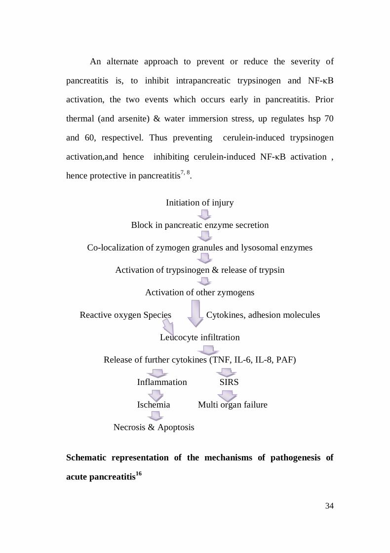
An alternate approach to prevent or reduce the severity of
pancreatitis is, to inhibit intrapancreatic trypsinogen and NF-κB
activation, the two events which occurs early in pancreatitis. Prior
thermal (and arsenite) & water immersion stress, up regulates hsp 70
and 60, respectivel. Thus preventing cerulein-induced trypsinogen
activation,and hence inhibiting cerulein-induced NF-κB activation ,
hence protective in pancreatitis7, 8.
Initiation of injury
Block in pancreatic enzyme secretion
Co-localization of zymogen granules and lysosomal enzymes
Activation of trypsinogen & release of trypsin
Activation of other zymogens
Reactive oxygen Species Cytokines, adhesion molecules
Leucocyte infiltration
Release of further cytokines (TNF, IL-6, IL-8, PAF)
Inflammation SIRS
Ischemia Multi organ failure
Necrosis & Apoptosis
Schematic representation of the mechanisms of pathogenesis of
acute pancreatitis16

35
Clinical presentation:
The clinical presentation, diagnosis, and management of an acute
attack of pancreatitis looks similar in both acute or chronic pancreatitis.
Acute pancreatitis can mimic like acute abdomen, and should never be
excluded in differential diagnosis8.
Abdominal pain, nausea, and vomiting present as the
predominant symptoms. Each episode begins with severe pain. The
cardinal symptom is usually epigastric pain. The pain was described as
"knifing" or "boring through" to the back, relieved by leaning
forward(Mohmadian prayer position). Pain starts 12-48 hours after a
bout of alcohol, or after a large meal in case of gall stone pancreatitis.
Pain became generalized once peritonitis has been sets in8, 15.
Painless pancreatitis is seen in Peritoneal dialysis, post-operative
situations, legionnaire’s disease.
If patient develops generalized paralytic ileus, abdominal
distension and vomiting can occur. The vomiting may lead to gastro
esophageal tears (i.e., Mallory-Weiss syndrome) and upper
gastrointestinal bleeding. Vomiting is more intense in necrotizing
pancreatitis than in edematous pancreatitis. Although vomiting and
36
retching may be relieved by passage of a nasogastric tube, the pain
usually persists even after gastric decompression.
Fever is an important sign. Fever presenting during first week ,is
due to acute inflammation, mediated by cytokines. Fever during the
second or third week, is due to infected pancreatic necrosis.
Physical Findings:
The patient may have tachycardia , tachypnoea , hypotension,
and hyper thermia7, 8.Abdomen findings include Voluntary and
involuntary guarding,with decreased or absent bowel sounds . Unless
pseudocyst develops ,pancreatitis does not present with mass. Abdomen
may be distended due to pancreatic acites. Acute pancreatitis may be
associated with left sided pleural effusion.
With increasing severity, there is sequestrations of fluid in the
retro peritoneum leading to life threatening intravascular fluid
loss,which leads to hemoconcentration. There might be bleeding into
the retro peritoneum or peritoneal cavity, which may dissect via the soft
tissues, appearing as a bluish discoloration around the umbilicus
(Cullen's sign) or in the flanks (Grey Turner's sign) and the inguinal
region (Fox's sign) 17. Neither sign is pathognomonic of AP.

37
The severe intravascular fluid loss may lead to acute renal
shutdown with elevated BUN and creatinine levels. And also there may
be hyperglycemia, hypoalbuminemia, and hypocalcemia that are
sufficient enough to produce tetany in few cases.
Diagnosis:
The clinical diagnosis is one of exclusion and diagnosis may be
difficult ,despite the multiple serological and radiological investigations
are available.
Serum pancreatic enzymes:
Serum pancreatic enzyme estimation,the gold standard for
diagnosis18. The reason is pancreatic acinar cells synthesize, store, and
secrete a large amount of digestive enzymes (e.g., amylase, lipase,
trypsinogen, and elastase), which gets elevated in acute pancreatitis.
Amylase, lipase, elastase and trypsin were secreted in the blood at
the same time, but their clearance differs due to the different
sensitivities.
Serum amylase concentration will increase immediately, reaches
the peak , within several hours after the onset of disease ,remains raised

38
for 3 to 5 days and then returns back to normal. No significant
correlation has been found, between the level of serum amylase and
severity of pancreatitis. Hyperamylasemia is also seen in biliary tract
disease, intestinal obstruction, mesenteric ischaemia, acute appendicitis,
mumps, parotitis, impaired amylase excretion etc…18. In contrast, a
patient with acute pancreatitis may even have a normal serum amylase
level, because of the interference by lipids, with chemical determination
of serum amylase. Urinary amylase clearance increases during
pancreatitis; predicting better results than serum amylase. For these
reasons, it is recommended to measure the urinary amylase , which
usually remain elevated for several days after serum amylase levels have
returned back to normal. In patients with severe necrotic pancreatitis ,
the pancreas may not release large amounts of enzymes .
The serum lipase has been found to be highly sensitive and
specific, in diagnosing acute pancreatitis, as there are no other sources
of lipase15, 17. Total amylase is having a sensitivity of 84%, the serum
P- amylase has 95% and lipase has 93%. Specificities for amylase,
P-amylase and lipase respectively are- 88%, 93% and 96%, respectively.
Thus P-amylase is the enzyme with the higher diagnostic value.

39
The rise of lipase: amylase has been found to differentiate
alcoholic from nonalcoholic pancreatitis. The serum (SGPT) alanine
aminotransferase level rise of three or more times above the base-line
value has great specificity in diagnosing gallstone pancreatitis.
Immunologic assay like serum trypsinogen or immune lipase are
generally less specific than the lipase assay. The increased urinary level
of activated peptides, released by trypsinogen, procarboxypeptidase, or
prophospholipase activation, may aid in predicting the severity of an
attack.
Leucocyte migration and its activation, is considered as a major
determining factor of local & systemic complications8, 15.
Although methemalbumin rise indicates severe pancreatits and a
poor prognosis, methemalbumin levels are usually not measured.
Circulating levels of several inflammatory mediators and acute phase
reactants
(e.g., IL-1, 6, TNF-alpha, and CRP) also increase during pancreatitis,
and the magnitude of those increases can be used to predict the severity
of an attack. C reactive protein is readily available in all centers and
vales > 120mg/L, after 72 hours are closely related to necrotising
pancreatitis.

40
Imaging:
In general, the plain chest and abdominal radiographs can be
useful in the management, by identifying other causes for the patient's
symptoms (e.g., pneumonia, perforated hollow viscous, mechanical
bowel obstruction). Plain abdominal X-ray findings are either
generalized or localised ileus ( sentinel loop), colon “cut-off” sign or
“renal halo” sign. A chest radiograph may show left pleural effusion,
elevated left hemi diaphragm or basal atelectasis17.
Ultrasonography:
Abdominal ultrasound (US) examination is the gold standard for
confirmation of gallstones pancreatitis. It is also helpful to detect extra
pancreatic ductal dilations & pancreatic edema, swelling, free peritoneal
fluid and peripancreatic acute fluid collections (PFCs).It may not be
sensitive in about 20% of cases, due to bowel gas interference with the
imaging.
CT scan:
The contrast-enhanced computed tomography (CECT), has
become gold standard for17

41
• Diagnosis
• Assessing the severity
• Identify the complications associated with acute pancreatitis.
The Balthazar scoring system and other similar grading systems
have incorporated various CT findings such as inflammation and fluid
collections in & around the pancreas to correlate radiographic
appearance with morbidity and mortality19.
Early CT scans often fail to detect evolving necrosis, which
would become well demarcated by 2 to 3 days after the onset of
symptoms. The CT scans are not useful in diagnosing necrosis or
predicting the severity in the first 24 hours of illness. The sensitivity for
identifying pancreatic necrosis using contrast-enhanced CT scan
approaches 100%, 4 days from diagnosis. CT scans is useful in the early
diagnosis of infected pancreatic necrosis and image guided aspiration of
necrosis, when patient not improving clinically or who experience
clinical decline. In the patient with moderate renal impairment or allergy
to intravenous contrast material, magnetic resonance imagining (MRI)
may be useful. MRI has been found to have sensitivity and specificity
similar to contrast-enhanced CT, for detecting severe acute pancreatitis.

42
ERCP should be done in patients with acute pancreatitis , whose
clinical course fails to improve despite full intensive care support, and in
whom ampullary or common bile duct stone impaction is suspected,
based on ultrasonography, or clinical/biochemical signs of cholangitis. It
may also be helpful in patients ,with recurrent attacks of acute
pancreatitis, without any obvious cause. It is useful in correcting
potentially correctable lesions such as CBD stones with impaction,
pancreas divisum, ampullary stenosis, pancreatic duct stenosis etc.
Assessment of Severity:
Assessment of mild and severe necrotizing pancreatitis, is the
most important thing for providing optimal care to the patient7. There
are so many predictors available for assessing the severity, which
includes early prognostication signs, serum markers, and CT scan15.
Scoring systems in acute pancreatitis:
The various prognostic scoring systems for assessing the severity
will be discussed in detail later.

43
UK guidelines for the management of AP20:
• Diagnosis should be made within 48 hrs. of admission.
• The etiology has to be determined in 80% of cases at least and
idiopathic cause should not exceed 20%.
• The serum lipase assay has been preferred over serum amylase
assay for diagnosis the acute pancreatitis.
• The contrast enhanced computed tomography has to be preferred
over USG for detection of the presence/absence of pancreatitis.
Treatment:
Evolution of an acute attack of pancreatitis occur in two phases,
overlapping on each other15, 17.
The initial phase, lasting for 1 to 2 weeks, involves an acute
inflammatory and autodigestive process. It may have systemic effects as
well.
The second phase, that may last for weeks or months, is primarily
characterized by the development of local complications that are,
themselves, the results of necrosis, infection and pancreatic duct rupture.

44
The initial management of patients with pancreatitis focuses on
early establishment the diagnosis, assessing the severity, treating the
major symptoms, and haltering the disease progression. The treatment
for acute pancreatitis is largely supportive. Since 15-30 % patients
develop severe pancreatitis, so each and every patient should be treated
aggressively. The main aim of the treatment is ‘allowing rest to the
gland’ by oral feed and fluids restriction21. The goal of initial
management consists of adequate fluid replacement,nutritional support,
correction of electrolyte imbalance, and prevention of local & systemic
complications.
Management of Pain
Good analgesics should be given to these patients as the pain can
be very severe in intensity. Most patients require narcotic analgesics.
Meperidine is preferred as morphine induces spasm of the sphincter of
Oddi, which can, at least theoretically, worsen biliary pancreatitis.
Fluid and Electrolyte Management
Aggressive fluid resuscitation is important to replenish
extravascular, or "third space," fluid loss, which may be considerable.
The fluid resuscitation is of utmost importance to prevent systemic
45
complications, mainly acute renal insufficiency, that may occur with
hypovolemia. Transudation of the fluid from intravascular space into the
areas of inflammation (i.e., peripancreatic, retroperitoneum and into the
pulmonary parenchyma and soft tissues elsewhere in the body) is the
principle cause of hypovolemia. Furthermore, studies have shown that
inadequate resuscitation may add upon as a significant risk that leads to
further pancreatic injury.
Banks and colleagues have showed that, aggressive fluid
resuscitation would not prevent the progression of developing
pancreatic necrosis. The degree and intensity of monitoring depends
upon the disease severity22.
During the first several days of a severe attack, circulating levels
of many proinflammatory factors, including cytokines and chemokines,
are elevated. This so-called “cytokine storm”, in many cases, triggers
the systemic immune response syndrome, and as a result, the
hemodynamic parameters of these patients may resemble those of sepsis
associated with other disease states23. Heart rate, cardiac output, and
cardiac index usually rise, and total peripheral resistance falls.
Hypoxemia can also occur as a result of the combined effects of
increased intrapulmonary shunting and a pancreatitis-associated lung

46
injury that closely resembles that seen in other forms of ARDS. Fluid
management, though critical, may be difficult when hypovolemia is
combined with respiratory failure of ARDS.
Measurement of central filling pressures, using a Swan-Ganz or
central venous pressure catheter, can be helpful in guiding fluid
management, particularly when hypovolemia is combined with lung
injury.
Nasogastric Decompression
The nausea and vomiting of pancreatitis can result in significant
fluid as well as electrolyte losses and retching can lead to gastro-
esophageal mucosal tears and result in upper gastrointestinal bleeding
(i.e., the Mallory-Weiss syndrome). For symptomatic relief and to
increase patient comfort, nasogastric decompression may be needed,
although the institution of nasogastric drainage does not shown to alter
the eventual outcome of an attack7, 8.
Prophylactic Antibiotics
Infection is a serious complication of acute pancreatitis and is the
most common cause of death17. It is mostly caused by the enteric
bacteria and was seen commonly in necrotizing pancreatitis. Local

47
infection occurs with pancreatic necrosis, and as time progresses for at
least the first 3 weeks,the disease progresses. Aerobic and anaerobic
gastrointestinal floras are the primary organisms involved, and
infections may be either mono or polymicrobial in nature. The
predominant microbes seen were E. coli (35%), Kleb. pneumoniae
(25%), Streptococcus (25%), Staphylococcus (15%), and Pseudomonas
(10%).The association of high mortality with pancreatic infection has
been the rationale behind the use of prophylactic antibiotics widely in
patients with pancreatic necrosis. In severe pancreatitis, beneficial
effects have been observed with regimens that included imipenem alone,
imipenem with cilastatin, metronidazole and third-generation
cephalosporin (cefuroxime). Because Candida species are common
inhabitants of the upper GI tract, Candida sepsis and secondary fungal
infection of pancreatic necrosis is a risk in severe disease, and many
surgeons advocate empirical therapy with fluconazole in severe acute
pancreatitis.
The duration of treatment has not defined clearly. A treatment
course of 1week to 4 weeks has been recommended commonly, but
many of them limit the treatment to 2 weeks17.

48
According to the current UK guidelines (Johnson 2005), the
duration of antibiotic prophylaxis is 1 to 2 weeks20.
Nutritional Support
Classically speaking, the enteral feeding should be limited,
thereby pancreatic stimulation and further pancreatic injury by the
release of proteolytic enzymes can be avoided. Recent data, suggests
that such strict limitations of enteral nutrition may have been
unnecessary. Most of the severe acute pancreatitis patients found to
have prolonged course of illness with hyper catabolic state and ileus that
have led to a generous use of parenteral nutrition in them.
The points favoring enteral nutrition are7, 15:
It might feasible, safe, and desirable in severe pancreatitis.
It has the advantage of avoiding the high cost of total parenteral
nutrition (TPN) as well as its associated catheter-related
complications.
The use of enteral nutrition may support intestinal mucosal
integrity by avoiding the alteration in intestinal permeability &
barrier function as seen with use of TPN.

49
Treatments of Limited or Unproven Value
In patients who develop severe disease, other treatment modalities
may be tried. The antiproteases like gabexate/aprotinin, antisecretory
agents like octreotide and anti-inflammatory drugs or PAF antagonists
like lexipafant were found to be less useful15, 17.
Treatment of Early Systemic Complications of Pancreatitis
The pathogenesis and management of the cardiovascular collapse,
respiratory failure, renal failure, metabolic encephalopathy,
gastrointestinal bleeding, and disseminated intravascular coagulation
that complicate severe pancreatitis appear to be identical to those
involved when these processes are superimposed on other disease states
that are characterized by peritonitis and hypovolemia8.
Cardiovascular collapse is largely caused by hypovolemia, and its
management requires aggressive fluid and electrolyte repletion.
The pulmonary manifestations of pancreatitis include atelectasis
and acute lung injury. The latter appears to be similar to the acute lung
injury caused by other systemic processes, including septic shock,
ischemia and reperfusion, and massive blood transfusion. Management
includes good pulmonary toilet combined with close monitoring of

50
pulmonary function. For many patients, intubation and respiratory
support may be required.
Renal failure in pancreatitis is usually prerenal and is associated
with a poor prognosis,sometimes requiring hemodialysis.
Stress-induced gastro duodenal erosions account for most of the
gastrointestinal bleeding, prophylaxis with antacids, H2-receptor
antagonists, or proton pump inhibitors may be appropriate.
Rarely, massive bleeding can result from injury to peripancreatic
vascular structures, leading to hemorrhage into the retroperitoneum. The
peripancreatic inflammatory process can also cause thrombosis of major
gastrointestinal vessels and result in ischemic lesions involving the
stomach, small intestine, or colon that can cause bleeding. Management
of these complications of pancreatitis is similar to that involved when
they occur in the absence of pancreatitis.
Some patients with severe pancreatitism, develop DIC, but it
rarely causes bleeding, and rarely needsprophylactic heparin.
Removal of precipitating factors, such as drugs or alcohol, is
appropriate. Once the acute phase has been survived, by the end of the

51
first week, then local complications become pre-eminent in the
management of these patients.
An indication for operative intervention in acute pancreatitis is
the drainage of an infected pancreatic necrosis. These patients require
removal of as much as possible of the infected necrosis and drainage for
the remaining viable exocrine tissue. Current opinion is against
debridement in sterile necrosis unless it is accompanied by life
threatening systemic complications17.
A pancreatic abscess usually occurs 2 to 6 weeks ,after an initial
attack of acute pancreatitis, in contrast to infected necrosis ,which
occurs in the first few hours or days. Treatment consists of external
drainage, either by surgical or percutaneous catheter based measures17.
Treatment of Biliary Pancreatitis
The presence of gallstones leading to choledocholithiasis is
recognized as a major etiological factor worldwide. Endoscopic
retrograde cholangio pancreatography (ERCP) has both diagnostic and
most therapeutic utility in patients with biliary obstruction or
cholangitis. By randomizing patients with AP to early ERCP versus no
ERCP, both Neoptolemos and colleagues, and Fan and colleagues have

52
showed a significant decrease in morbidity but there was no significant
improvement in mortality with routine use of ERCP. A metacentric
randomized control study in the ERCP group by Folsch and colleagues
recently, have demonstrated increased complication rate and mortality
rate, after excluding the patients with biliary sepsis or obstruction. It
therefore, found that early ERCP may be harmful even in the absence of
ongoing biliary obstruction. Magnetic resonance cholangio
pancreatography (MRCP) is an additional alternative to ERCP as a
diagnostic tool that avoids the risk of post procedure pancreatitis.
In general, cholecystectomy as an early intervention, within the
first 48 to 72 hours of admission, or delayed intervention (after 72
hours, but during the initial period of hospitalization) may be favored8,
15. Cholecystectomy with intra-operative CBD exploration is probably
the best option for obstructive pancreatitis. However, high risk patients
are best treated by endoscopic sphincterotomy, with clearance of stones
by ERCP.
Surgical Management: Indications and Timing
Limited indications prevail for surgical intervention.Intervention
is needed to address the etiology of pancreatitis or its complications.
Suspected choledocholithiasis needs open or laparoscopic intervension.
53
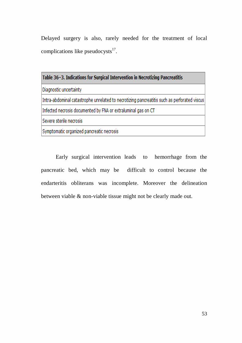
Delayed surgery is also, rarely needed for the treatment of local
complications like pseudocysts17.
Early surgical intervention leads to hemorrhage from the
pancreatic bed, which may be difficult to control because the
endarteritis obliterans was incomplete. Moreover the delineation
between viable & non-viable tissue might not be clearly made out.
54

55
Complications17:
Complications may be classified as15, 17:
I. LOCAL:
Fluid collections
Pancreatic ascites/pleural effusion
Pancreatic pseudocyst
Pancreatic necrosis
Infected pancreatic abscess
Hemorrhage/pseudo aneurysm

56
II. REGIONAL:
Venous thrombosis
Paralytic ileus
Intestinal obstruction
Intestinal ischemia/necrosis
Cholestasis
III. SYSTEMIC:
A. Pulmonary
1. Pneumonitis, basal atelectasis
2. ARDS
3. Pleural effusion (L)
B. Cardiovascular
1. Hypotension
2. Hypovolemia
3. Sudden arrest &death
4. Nonspecific ECG(ST-T wave) changes
5. Pericardial effusion

57
C. Hematologic
1. Hemoconcentration
2. Disseminated intravascular coagulopathy
D. GI hemorrhage
1. Acid peptic disease
2. Gastric erosion
3. Portal/splenic vein thrombosis with variceal bleed
E. Renal
1. Oliguria
2. Azotemia
3. Renal vessel thrombosis
F. Metabolic
1. Hyperglycemic state
2. Hypocalcemic state
3. Hyperlipidemia (triglyceridemia)
4. Metabolic encephalopathy
5. Sudden loss of vision (Purtscher's retinopathy)
58
G. Central nervous system
1. Acute psychosis
2. Fat embolism occlusion
3. Alcohol withdrawal syndrome (AWS)
H. Fat necrosis
1. Intra-abdominal saponification
2. Subcutaneous tissue necrosis
SCORING SYSTEMS IN ACUTE PANCREATITIS
Pancreatitis is a serious disease with high morbidity and mortality
rates. Some 80% were mild attack which recovers rapidly with
conservative management. The rest of 20% were severe, with protracted
course that needs intensive care and specialized management. Several
predictors of severity are commonly used for this purpose24.
Scoring systems can be used in predicting mortality, severity of
disease and intensity of its complications. Prognostic factor analysis
found to help in comparing the results, in-between the series of patients
under study.

59
These systems include25:
• Ranson’s criteria
• Balthazar computed tomography (CT) grading
• Imrie Glasgow coma score (GCS)
• Bank’s clinical Criteria
• Simplified Acute Physiology Score(SAPS)
• Marshall Multiple organ failure (MOF) score and
• Acute physiology and chronic health evaluation (APACHE) I, II,
III & O.
The GCS and Ranson’s multiple scoring systems, require 48
hours of data collection; however, APACHE can be calculated at any
time and shows prognostic correlation with acute pancreatitis, as
increasing scores are associated with poor prognosis.
Once the acute pancreatitis has been diagnosed, assessment of
severity is extremely important for execution of appropriate measures,
preferably in an ICU setup with close monitoring.

60
AIMS AND OBJECTIVES OF THE STUDY
1. To know the usefulness of urine trypsinogen-2 in accurately
diagnosing acute pancreatitis
2. To compare the diagnostic role of urine trypsinogen-2 with that of
serum amylase, serum lipase and imaging studies in acute
pancreatitis.
MATERIALS AND METHODS
Study design: Comparative Analytical study.
Setting: Department of General Surgery, Govt. Stanley Medical College
and Hospital, Chennai. The study was conducted after obtaining the
Institutional Ethical Committee approval (annexure 2).
Inclusion criteria:
1. Patients presenting with features suggestive of acute pancreatitis
2. Male and female subjects of age between 20 - 60 years shall be
selected.
3. Adult subjects willing to give informed consent.

61
• Exclusion criteria:
1. Subjects below the age of 20 years and above the age of 60 years
2. Subject who are not willing to participate in the study
•
3. Individuals who are cognitively impaired and/or who are unable
to give informed consent.
4. Proven cases of chronic pancreatitis and pancreatic cancer
5. Hereditary pancreatitis, cystic fibrosis
METHODOLOGY
• Patients presenting with acute upper abdominal symptoms like
pain, vomiting, abdominal distention, admitted in the emergency
department of our hospital from January 2013 to November 2013
are enrolled in the study.
• Urine sample were obtained from all the patients and tested with
Spot Urine trypsinogen-2 dipstick.
• Serum amylase and serum lipase tests were also simultaneously
done in these patients.

62
• Patients are also evaluated with (USG) abdomen and (CECT)
abdomen,if required.
• Final diagnosis of acute pancreatitis is made on the basis of
clinical picture, serum amylase more than threefold rise and
radiological findings.
• Urine trypsinogen-2 dipstick test were compared with serum
amylase, serum lipase and imaging studies in patients with final
diagnosis of acute pancreatitis
• Observations are tabulated according to the pre-designed
proforma.
• The results are analyzed using Microsoft Excel for tabular
transformation and graphical representation. For comparing the
parameters, Chi Square test or Fischer’s exact test are used. SPSS
software will be used for statistical analysis.

63
OBSERVATION &RESULTS
This study was conducted in the Department of General Surgery,
Govt. Stanley Medical College & Hospital, Chennai for a period of one
year. The 100 persons ,who fulfilled the inclusion criteria ,were enrolled
in this study, after obtaining an informed consent.
Table: 1 AGE DISTRIBUTION
The age group of patients enrolled in this study ranges from 20 to
80 yrs. The peak incidence was noted in the 4th decade of life.
Age Range (years) No. of patients Percentage (%)
21yrs - 30yrs 24 24
31yrs - 40yrs 25 25
41yrs - 50yrs 36 36
51yrs - 60yrs 13 13
>60yrs 2 2
Total 100 100 %

64
Figure : 1 AGE DISTRIBUTION

65
Table: 2 GENDER DISTRIBUTIONS
Sex No. of patients Percentage (%)
Male 84 84
Female 16 16
Total 100 100
Out of 100 patients enrolled in this study, there were 84 male and
16 female patients.
Male: Female ratio-5.25:1
Figure:2 GENDER DISTRIBUTION

66
Table:3 GENDER DISTRIBUTION IN ACUTE PANCREATITIS
Gender Acute pancreatitis(61) Percentage(%)
MALE 57 93.44
FEMALE 4 6.55
Of the total cases of Acute Pancreatitis,93% of patients were male
and the rest were female.
Figure:3 GENDER DISTRIBUTION IN ACUTE
PANCREATITIS(n=61)

67
Table: 4 CLINICAL FEATURES:
Symptoms No. of patients Percentage (%)
Pain abdomen 61 61
Fever 25 25
Vomiting 54 54
Jaundice 12 12
Abdominal distension 32 32
On clinical presentation,61% of patients presented with
abdominal pain as chief complain. Rest of the patients had vomiting,
abdomen distension and fever along with the presenting symptoms.
Figure:4 CLINICAL FEATURES

68
Table: 5 ETIOLOGY
Etiology No. of patients Percentage (%)
Alcohol 53 86.89
Gall stone disease 5 8.19
Post ERCP 1 1.64
Hypertriglyceridemia 2 3.28
Total 61 100
History of consumption of alcohol and the possibility of it being
the etiological factor were found in 53 patients. Gall stone disease was
attributed in 5 patients. Hyperlipidemia and Post ERCP, as a causative
factor in 2 & 1 patients, respectively.
Figure: 5 ETIOLOGY

69
Table:6 COMPARISON OF DIFFERENT PARAMETERS
USED IN THE DIAGNOSIS OF ACUTE PANCREATITIS
Parameters Acute
Pancreatitis(61)
Others(39) Total(100)
AMYLASE 45 4 49
LIPASE 36 4 40
USG 53 - 53
CECT 14 - 14
TRYPSINOGEN 48 3 51
Table:7 COMPARISON OF SENSITIVITY AND
SPECIFICITY OF DIFFERENT PARAMETERS
Parameters Sensitivity Specificity
AMYLASE 73.77 89.74
LIPASE 59.02 89.74
TRYPSINOGEN 78.69 92.31

70
Table 8: ESTIMATION OF SIGNIFICANCE OF THE TEST
Urine trypsinogen
Acute
Pancreatitis
Non pancreatic causes
Total
Positive 48 3 51
Negative 13 36 49
TOTAL 61 39 100
Sensitivity= a/(a+c) ;48/61=78.6%
Specificity=d/(b+d);36/39=92.3%
Using Kappa statistics the value was found to be 0.67
(Based on criteria originally proposed by Landis and Koch: kappa
values greater than about 0.75 are often taken as representing excellent
agreement; those between 0.4 and 0.75 as fair to good agreement; and
those less than 0.4 as moderate or poor agreement.)
FIGURE 6: ANALYSIS OF TRYPSINOGEN TEST

71
DISCUSSION
Trypsinogen, a precursor of trypsin is required for protein
digestion. Premature trypsin activation leads to pancreatic self-
digestion. Trypsinogen is a 25-kd pancreatic proteinase. In human
pancreatic juice, there are three trypsinogen (TPS) isoenzymes, namely,
cationic (TPS-1) and anionic TPS (TPS-2), and a minor isoenzyme
(TPS-3).
The inactive form of trypsinogen,stored in the cytoplasmic
zymogen granules of pancreatic acinar cells, are secreted into the
adjacent duct lumen and are subsequently delivered to the small
intestine.Within the intestine, they are activated by enterokinase.
Premature activation of trypsinogen to trypsin within the pancreas,is the
prime pathophysiologic event, in the development of acute pancreatitis.
Under normal conditions, trypsinogen are secreted into pancreatic fluid,
and only a small amount enters the circulation. For unknown reason,
the tubular reabsorption of trypsinogen-2 is lower than trypsinogen-1.
The urinary trypsinogen-2 dipstick test is proposed to be, a rapid
and simple method for the early diagnosis of acute pancreatitis. Urine
trypsinogen-2 concentrations were measured using a dipstick test

72
(Actim Pancreatitis, Medix Biochemica Oy AB,Kauniainen, Finland)
based on an immunochromatography assay. The detection limit of the
test is approximately 50 ng/mL. The test strip has a control line and two
lines indicate a positive result. The test results can be read after
5 minutes.
The test strip, dipped into the urine sample, contains
trypsinogen-2 bound to monoclonal antibody labelled blue latex
particles, which migrate across a nitrocellulose membrane with a zone
containing another antibody specific for epitope on trypsinogen-2.
Two retrospective studies[10, 27] carried out by a Finnish group
showed that Urine Trypsinogen-2 had better sensitivity and specificity
in predicting Acute Pancreatitis than amylase and lipase. A later larger
prospective cohort study[28] by the same group enrolling 53 patients
with AP and 447 patients with AAD (non-pancreatic) showed that Urine
Trypsinogen-2 has a sensitivity of 94% and a specificity of 95%, better
than serum amylase (85% and 91%) and urinary amylase (83% and
88%) in predicting Acute Pancreatitis.
Sensitivity of amylase and lipase was found to be 73.77% and
59.02% respectively, whereas as sensitivity of trypsinogen was found to
be 78.69%. Specificity of amylase and lipase was found to be 89.74%
73
and 89.74% respectively, whereas as specificity of trypsinogen was
found to be 92.1%.Analysing the data,it is found that sensitivity and
specificity of trypsinogen is higher than the routine investigations.
Eventhough it has a low range of sensitivity, its high specificity ensures
that the test can be used as a screening test to check the true negative
cases.
False positive results is seen in 3 of the 39 non-pancreatitis cases,
namely two cases of Gallstones and one case of renal failure. In case of
renal failure, defect in the excretion of trypsinogen makes the result
positive. In case of gallestone, studies with more sample size has to be
conducted to analyse the etiology of the positivity of the test.

74
CONCLUSION AND SUMMARY
1. Urine Trypsinogen-2 dip stick test is a simple, rapid, easy, and
noninvasive test which can diagnose or rule out, most of the
cases of acute pancreatitis.
2. Urine Trypsinogen-2 estimation doesn't require laboratory
facilities. It is undertaken almost instantaneously (within 5
minutes) as opposed to serum amylase and lipase, results for
which may require an hour to get back to the physician.
3. The urinary trypsinogen-2 test could be used as a screening test
for acute pancreatitis.
4. Modification of the cutoff point of this assay increases the
specificity to the point where it can be used for diagnosis.

75
5. Qualitative rapid urine trypsinogen-2 test strip is easy to perform.
And hence it has been shown to be a reliable and useful
screening test for acute pancreatitis in daily practice
[12-16], particularly in healthcare units lacking laboratory
facilities.

76
BIBLIOGRAPHY
1. Comparison of BISAP, Ranson’s, APACHE-II, and CTSI Scores
in Predicting Organ Failure, Complications, and Mortality in
Acute Pancreatitis Georgios I. Papachristou, MD, Venkata
Muddana, MD, Dhiraj Yadav, MD, et al. Am J Gastroenterology
2010; 105:435–441.
2. A brief history of pancreatitis. D A O’Reilly MRCS, A N
Kingsnorth MS FRCS. Journal of the royal society of medicine.
March 2001; volume 94; 130-132.
3. Opie EL. The etiology of acute hemorrhagic pancreatitis. Johns
Hopks Hosp Bull 1901; 12; 182-8.
4. Steinberg W, Tenner S, Acute Pancreatitis. The New England
Journal of Medicine 1994; 330(17): 1198-1210.
5. Bradley EL, A clinically based classification system for acute
pancreatitis, Arch Surg; 128: 586-590.
6. Muhmet Ihan et al., The etio-pathogenesis of acute biliary
pancreatitis, Dr. Sadikonuk training & research hospital, Istanbul,
Turkey. DOI: 10.5772/26272.
7. Reber HA, Schwartz’s Principles of Surgery (9th edition- 2010),
McGraw Hill, 1467-1500.
8. Steer Michael L, Sabiston Textbook of Surgery, The biological
basis of modern surgical practice (18th edition- 2007), Elsevier,
1643-60.
77
9. Skandalakis LJ, Rowe Jr JS, Gray SW, et al: Surgical embryology
and anatomy of the pancreas. Surg Clin North Am 1993; 73:
661-697.
10. Ellis Harold, Clinical anatomy, seventh edition-1983, Blackwell
scientific publications, 121-23.
11. Standring S, Ellis H, Gray’s Anatomy, Elsevier Churchill
Livingstone 2005, 1231-1238.
12. Whitcomb DC, Gorry MC, Preston RA, et al. hereditary
pancreatitis is caused by mutation in the cationic trypsinogen
gene. Nat genet 1996; 14; 141-5.
13. Thomson SR, Hendry WS, McFarlane GA, Davidson AI,
Epidemiology and outcome of acute pancreatitis, Br J Surg 1987;
74:398-401.
14. Russell R C G, Bailey and Love’s Short Practice of Surgery
(24th Edition-2004), Arnold Publishers, 1114-27.
15. Fisher WE, Andersen DK, Bell RH, Saluja AK, Brunicardi FC,
Schwartz’s Principles of Surgery(9th edition- 2010), McGraw
Hill, 1222-40.
16. Wig JD, The Pancreas, (1st Edition-2000), Azad offset printers,
1-327.
17. Michael J. Zinner, Stanley W. Ashley et al., Maingot’s abdominal
operations, Management of acute pancreatitis. 11th edition.
P 939-959.

78
18. Kazmierczak et al. Diagnostic accuracy of pancreatic enzymes
evaluated by use of multivariate data analysis. Clin Chem 993;
39: 1960–1965.
19. Emil J. Balthazar, MD Acute Pancreatitis: Assessment of Severity
with Clinical and CT Evaluation1 10.1148/radiol.2233010680
Radiology 2002; 223:603–613.
20. Glazer G, Mann DV, UK guidelines for the management of acute
pancreatitis Gut 2005; 54; 1-9.
21. Yeo CJ, Cameron JL, Sabiston Text book of Surgery, The
biological basis of modern surgical practice (18th edition-2008),
W.B. Saunders company, 1112-43.
22. Toouli J, Smith MB, Bassi C, Carr-locke D, et al. Guidelines for
the management of acute pancreatitis, Journal of
Gastroenterology and hepatology 2002; 17 : S15-S39.
23. Balakrishnan V, Philip M. Cytokines as predictors of severity in
acute pancreatitis. J Gastroenterology Hepatology 1998; 13:
1177-1179.
24. Bechien U. Wu MD MPH. Prognosis in acute pancreatitis. CMAJ
2011. DOI:10.1503/cmaj.101433.
25. Blamey S L, Imrie C W, O’Neill J, Prognostic factors in acute
pancreatitis, GUT 1984; 25: 1340-46.
26. Williams M, Simms H, Prognostic usefulness of scoring systems
in critically ill patients with severe acute pancreatitis. Crit Care
Med 1999; 27: 901-7.

79
27. Taylor SL et al. a comparison of the Ranson, Glasgow &
APACHE II scoring system to multi organ system score in
predicting outcome in pancreatitis. Amj surg., 2005; 219-222.
28. Koenraad J. Mortele, Walter Wiesner, Lisa Intriere, et al.
A Modified CT Severity Index for Evaluating Acute Pancreatitis:
Improved Correlation with Patient Outcome. AJR 2004; 183:
1261–1265.
29. Knaus WA, Zimmerman JE, and Wagner DP: APACHE -Acute
Physiological and Chronic Health Evaluation: A physiology
based classification system. Crit Care Med 1981:9: 591-7.
30. Knaus WA, Draper EA, Wagner et al: APACHE II A severity of
disease classification system. Crit Care Med-1985; 13(10):
818-29.
31. Ranson JHC, Acute Pancreatitis- Current problems in Surgery,
1979; 16: 1-84.
32. Roumen R.M.H., Schers T.J., de Boer H.H.M, Goris R.J.A,
Scoring systems for predicting outcome in acute hemorrhagic
necrotizing pancreatitis, Eur J Surg-1992; 158: 167-71.
33. Chatzicostas C, Roussomoustakaki M, Vardas E, et al, Balthazar
computed tomography severity index is superior to Ranson
criteria and APACHE II and APACHE III scoring systems in
predicting acute pancreatitis outcome, J Clin Gastroenterol-2003;
36(3):253-60.

80
34. Knaus WA, Wagner DP, Draper EA, et al. The APACIIE III
prognostic system: risk prediction of hospital mortality for
critically ill hospitalized patients. Chest 1991; 100; 1619-36.
35. Jack E Zimmerman, Douglous P Wagner, Elizabeth A. Draper:
Evaluation of Acute Physiological and Chronic Health Evaluation
III prediction in independent database. Crit Care Med 1998; 26:
1317-26.
36. Severity Scoring for Prognostication in Patients With Severe
Acute Pancreatitis Comparative Analysis of the Ranson Score and
the APACHE III Score Soumitra R. Eachempati, MD; Lynn J.
Hydo, et al. Arch Surg. 2002; 137:730-736.
37. R. Mounzer et al. Comparison of existing clinical scoring
systems to predict persistent organ failure in patients with acute
pancreatitis. Gastroenterology June 2012; 142 (2), 1476-1482.
38. V. K. Singh et al. A Prospective Evaluation of the Bedside Index
for Severity in Acute Pancreatitis Score in Assessing Mortality
and Intermediate Markers of Severity in Acute Pancreatitis.
The American Journal of Gastroenterology APR 2009; 104(4),
966-971.
39. J. Larino-Nola et al. assessing mortality in severe acute
pancreatitis. Evaluation of the bedside index for severity in acute
pancreatitis (BISAP). Pancreatology June 2010; 10(2-3), 344.

81
40. M. Bezmarevic et al. Procalcitonin and BISAP score versus
C-reactive protein and APACHE II score in early assessment of
severity and outcome of acute pancreatitis. Clinic for Abdominal
and Endocrine Surgery, University of Belgrade, Belgrade, Serbia.
Vojnosanit Pregl 2012; 69(5): 425–431.
41. B. Wu et al The early prediction of mortality in acute pancreatitis:
a large population-based study. Gut JUN 2008; 57, 1698-1703.
42. De Beaux A C, Palmer K R, Carter D C, Factors influencing
morbidity and mortality in acute pancreatitis; an analysis of 279
cases, GUT; 37: 121-126.
43. Bidarkundi GK et al. Clinical relevance of intracellular cytokines
IL-6 and IL-12 in acute pancreatitis, and correlation with
APACHE III score, Br J Biomedical Science 2002; 59(2): 85-89.
44. Larvin M et al. APACHE II scores for assessment and monitoring
of acute pancreatitis. Lancet 1989; 2: 201-205.

82
PROFORMA
SL. NO:
• NAME :
• AGE /SEX:
• IP NO:
• ADDRESS WITH CONTACT NUMBER:
• DATE OF ADMISSION:
• DATE OF SURGERY:
• DATE OF DISCHARGE:
HISTORY OF PRESENTING ILLNESS:
H/O abdomen pain
Duration
Nature of pain
onset
Progression
Radiating
Aggravating/relieving factors
H/O vomiting
Nature of vomitus
No. of episodes
Associated with hemetemesis

83
H/O Fever
Duration
Nature
Associated with chills & rigor:
H/O jaundice
H/O abdomen distension
H/O loss of appetite
H/O loss of weight
PAST HISTORY:
DM/hypertension/asthma/TB/epilepsy/cardiac illness
H/o similar episodes in the past, if any:
H/o major illness/ hospital admissions, if any
PERSONAL HISTORY:
Occupation
Socio-economic status
Alcohol
Smoking
Drug addiction
FAMILY HISTORY:
TREATMENT HISTORY:

84
CLINICAL EXAMINATION:
General examination: Pallor Icterus
Pulse
BP
Temperature
Respiratory Rate
Systemic examination:
CVS
RS
CNS
Abdomen
CLINICAL DIAGNOSIS:
INVESTIGATIONS:
CBC:Hemoglobin
TC/DC:
ESR
PLATELET
RFT:BLOOD UREA
SERUM CREATINE
SODIUM
POTASSIUM
RANDOM BLOOD SUGAR
CHLORIDE

85
LFT:TOTAL BILIRUBIN
DIRECT BILIRUBIN
INDIRECT BILIRUBIN
SGOT /SGPT
ALKALINE PHOSPATASE
TOTAL PROTEIN
ALBUMIN
X-RAY ABDOMEN
X-RAY CHEST
URINE TRYPSINOGEN-2 DIPSTICK:
SERUM AMYLASE
SERUM LIPASE
USG ABDOMEN
CECT ABDOMEN
FINAL DIAGNOSIS:

86

sl_no name ip_no age sexabd_pain fever vomiting jaundice
abd_distension alcohol gall_stone post_ercp hyper_tg amylase lipase usg ct
urine_trypsinogen diagnosis
1 Jegan 38392 45 Male 1 2 1 2 2 1 2 2 2 1 1 1 99 1 12 Anandhan 37332 42 Male 1 1 1 1 1 1 2 2 2 1 1 1 99 1 13 Dhanapal 34612 55 Male 1 1 1 2 1 1 2 2 2 1 1 1 99 1 14 Murugan 37377 40 Male 1 2 1 2 2 1 2 2 2 1 1 1 99 1 15 Premkumar 26628 29 Male 1 1 1 1 1 1 2 2 2 2 2 2 1 2 16 Chellakannu 26301 34 Male 1 1 1 2 1 2 1 2 2 2 2 1 1 1 17 Rajendran 46982 46 Male 1 1 1 2 1 1 2 2 2 1 1 3 3 2 58 Rafiq 18441 50 Male 1 1 1 2 1 1 2 2 2 2 2 2 1 2 19 Prakash 23987 29 Male 1 1 1 1 1 1 2 2 2 2 2 2 1 2 1
10 Anandhan 6918 47 Male 1 1 1 1 1 1 2 2 2 1 1 1 99 1 111 Gopinath 26635 25 Male 1 2 1 2 1 1 2 2 2 1 1 1 99 1 112 Arunprasath 39206 32 Male 1 1 1 2 1 2 1 2 2 1 1 1 1 1 113 Gopinath 22530 31 Male 1 2 1 2 1 1 2 2 2 1 1 1 99 1 114 Prabu 22444 31 Male 1 2 2 2 2 1 2 2 2 2 2 1 99 2 115 Senthil pandi 6923 32 Male 1 1 1 2 2 1 2 2 2 2 2 4 99 2 216 Prabu 17439 21 Male 1 1 1 2 2 1 2 2 2 2 2 2 1 2 117 Arivazhagan 19405 46 Male 1 1 1 1 1 1 2 2 2 1 1 1 99 1 118 Kannan 26665 32 Male 1 2 1 2 2 1 2 2 2 1 1 1 99 1 119 Pillayarsamy 37807 49 Male 1 2 2 2 2 1 2 2 2 2 2 1 99 2 120 Vijayakumar 36395 35 Male 1 2 1 2 2 2 2 2 2 2 2 99 99 2 321 Krishnan 37386 55 Male 1 2 1 2 2 1 2 2 2 1 1 1 99 1 122 Murugan 39214 35 Male 1 2 1 2 2 1 2 2 2 1 1 1 99 1 123 Rajam 39206 56 Male 1 1 1 2 2 1 2 2 2 1 1 4 3 2 724 Pushpavalli 39216 45 Female 1 1 1 2 1 2 2 2 2 2 2 3 3 1 525 Mariselvam 42570 24 Male 1 2 1 2 2 1 2 2 2 1 1 1 99 1 126 Mohan 37469 50 Male 1 2 1 2 2 1 2 2 2 1 1 1 99 1 127 Aruna 37419 30 Female 1 1 1 2 2 2 2 2 2 2 2 4 99 2 228 Munusamy 49947 42 Male 1 2 1 2 2 1 2 2 2 1 1 1 99 1 129 Prakash 50684 25 Male 1 1 1 1 1 1 2 2 2 2 2 2 1 2 130 Aruldas 38435 60 Male 1 1 1 2 1 1 2 2 2 1 1 1 99 1 131 Malleswari 42262 24 Female 1 1 1 2 2 2 2 2 2 2 2 3 99 2 232 Wilson 40677 39 Male 1 2 1 2 2 1 2 2 2 2 2 99 99 2 3
MASTER CHART
87

33 Srinivasan 42845 30 Male 1 1 1 2 2 1 2 2 2 2 2 4 99 2 234 Ramesh 42942 38 Male 1 2 1 2 2 1 2 2 2 2 2 99 99 2 335 Syed ibrahim 42947 32 Male 1 1 1 2 2 2 2 2 2 2 2 4 99 2 236 Vijayakumar 34459 40 Male 1 2 1 2 2 1 2 2 2 2 2 99 99 2 337 Elumalai 34520 47 Male 1 2 1 2 1 1 2 2 2 1 1 1 99 1 138 Manimegalai 34781 49 Female 1 1 1 1 2 2 1 2 2 2 2 3 3 2 639 Kathiresan 33291 42 Male 1 2 1 2 2 1 2 2 2 1 1 1 99 1 140 Raju 33413 40 Male 1 2 1 2 2 1 2 2 2 2 2 99 99 2 341 Pattamal 30611 53 Female 1 2 1 2 1 1 2 2 2 1 1 1 99 1 142 Jayaraman 33444 27 Male 1 2 1 2 2 1 2 2 2 1 1 1 99 1 143 Nagarajan 32075 57 Male 1 2 1 2 2 1 2 2 2 1 1 1 99 1 144 Palani 32520 32 Male 1 2 2 2 2 1 2 2 2 2 2 1 99 2 145 Baskar 28928 48 Male 1 1 1 2 1 1 2 2 2 1 1 1 99 1 146 Gayathri 30619 22 Female 1 1 1 2 2 2 2 2 2 2 2 3 99 2 247 Nagaboosanam 28937 38 Male 1 2 1 2 2 1 2 2 2 1 1 1 99 1 148 Ayyappan 29188 27 Male 1 1 1 2 1 1 2 2 2 1 1 1 99 1 149 Sivasankari 29136 34 Female 1 2 1 2 2 2 2 2 2 2 2 99 99 2 350 Premkumar 26628 29 Male 1 1 1 1 1 1 2 2 2 2 2 2 1 2 151 Vincent 26849 28 Male 1 1 1 2 2 2 2 2 2 2 2 3 99 2 252 Sivaranjani 26682 60 Female 1 1 2 1 1 2 2 2 2 2 2 3 3 2 853 Kumar 25379 46 Male 1 2 1 2 2 1 2 2 2 1 1 1 99 1 154 Babu 25328 30 Male 1 1 1 2 1 2 2 2 1 1 1 1 99 1 155 Santhi 24611 32 Female 1 1 1 2 1 2 2 2 2 1 1 4 3 1 756 Ramamoorthy 24700 48 Male 1 1 1 1 1 1 2 2 2 2 2 3 3 2 557 Selvakumar 22840 30 Male 1 1 1 2 2 1 2 2 2 2 2 3 99 2 258 Subramani 22619 56 Male 1 1 2 2 1 1 2 2 2 2 2 3 3 2 859 Govindhammal 40173 27 Female 1 1 1 2 2 2 2 2 2 2 2 3 99 2 260 Rajendran 22757 50 Male 1 1 2 1 1 1 2 2 2 1 1 3 3 2 661 anandhi 21681 38 Female 1 1 1 1 1 2 1 2 2 2 2 1 1 1 162 Ramalingam 21240 45 Male 1 1 1 2 2 1 2 2 2 2 2 99 99 2 463 Jayakumar 49951 80 Male 1 2 1 2 1 1 2 2 2 1 1 1 99 1 164 Vasudevan 7827 48 Male 1 1 1 1 2 2 1 2 2 2 2 3 3 2 665 Munusamy 34601 60 Male 1 2 1 2 2 1 2 2 2 1 1 1 99 1 166 Shanmugam 35947 47 Male 1 1 1 2 1 1 2 2 2 2 2 3 3 2 567 Udayakumar 34982 34 Male 1 2 1 2 2 2 2 2 2 2 2 99 99 2 368 Eswari 17445 37 Female 1 2 1 2 2 2 2 2 2 2 2 99 99 2 369 Kumar 17447 24 Male 1 2 1 2 2 1 2 2 2 1 1 1 99 1 1
88

70 Suganthi 28542 27 Female 1 1 1 2 1 2 2 2 1 2 2 2 1 1 171 Nandha 433112 55 Male 1 2 2 2 2 1 2 2 2 1 1 1 99 1 172 Vasanth 45186 23 Male 1 1 1 2 2 2 2 2 2 2 2 3 99 2 273 Veeraiah 38112 42 Male 1 1 1 2 2 2 2 2 2 2 2 99 99 2 474 Loganathan 43336 50 Male 1 2 1 2 2 1 2 2 2 1 1 1 99 1 175 Amsa 2461 52 Female 1 1 1 1 1 2 1 2 2 2 2 1 1 1 176 Ragaramya 3382 36 Female 1 2 1 2 2 2 2 2 2 2 2 99 99 2 377 Sengootuvan 33501 45 Male 1 1 2 2 2 1 2 2 2 2 2 99 99 2 478 Krishnamoorthy 51474 45 Male 1 2 1 2 2 1 2 2 2 1 1 1 99 1 179 Anandhan 26918 47 Male 1 1 1 1 1 2 1 2 2 1 1 1 1 1 180 Pillaiyarsamy 37807 49 Male 1 2 2 2 2 1 2 2 2 2 2 1 99 2 181 Arivazhagan 32076 46 Male 1 1 1 1 1 1 2 2 2 1 1 1 99 1 182 Suresh 24123 24 Male 1 1 1 2 2 2 2 2 2 2 2 3 99 2 283 Nageswara rao 64734 25 Male 1 2 1 2 2 1 2 2 2 1 1 1 99 1 184 Arun 31829 45 Male 1 1 1 2 1 2 2 1 2 1 1 1 1 1 185 Bagiyanathan 28043 26 Male 1 1 1 2 1 1 2 2 2 1 1 1 99 1 186 Abdulhameed 28086 72 Male 1 1 1 1 1 1 2 2 2 1 1 1 99 1 187 Gani 29630 45 Male 1 2 1 2 1 1 2 2 2 1 1 1 99 1 188 Sivaraman 30475 50 Male 1 1 1 2 2 1 2 2 2 2 2 4 3 1 789 Naseer hussain 29624 45 Male 1 2 2 2 2 1 2 2 2 1 1 1 99 2 190 Rajendran 34711 45 Male 1 1 1 2 2 2 2 2 2 2 2 99 99 2 491 Rangaraj 35579 40 Male 1 2 1 2 2 1 2 2 2 2 2 99 99 2 392 Murugammal 36497 30 Female 1 1 1 2 2 2 2 2 2 2 2 4 99 2 293 Palani 36509 37 Male 1 2 2 2 2 1 2 2 2 2 2 1 99 2 194 Dilly 35639 60 Male 1 2 1 2 1 1 2 2 2 1 1 1 99 1 195 Mabu basha 35685 45 Male 1 2 1 2 2 1 2 2 2 1 1 1 99 1 196 Ekambaram 38263 50 Male 1 2 1 2 1 1 2 2 2 1 1 1 99 1 197 Kamaleswaran 51612 32 Male 1 2 1 2 2 1 2 2 2 1 1 1 99 1 198 Radhakrishnan 51658 55 Male 1 1 1 2 1 1 2 2 2 2 2 2 1 2 199 Rajesh 33762 45 Male 1 1 1 2 2 2 2 2 2 2 2 99 99 2 4
100 Jeeva 34448 41 Male 1 2 1 2 2 1 2 2 2 1 1 1 99 1 1
89

91
wµ Pøn¯ AÇØ]°À ]Ö}º i›¨]@|õáß &2
‰»® ¯õv {ºn¯® öŒ#uÀ
”¯ J¨¦uÀ £iÁ®
ö£¯º: Á¯x: EÒ訦Gs:
C¢u ©¸zxÁ B#Âß ÂÁµ[PÒ GÚUS ÂÍUP¨£mhx.
GßÝøh¯ Œ¢@uP[PøÍU @PmPÄ®, AuØPõÚ uS¢u
ÂÍUP[Pøͨ ö£ÓÄ® Áõ#¨£ÎUP¨£mhx.
|õß CÆÁõ#ÂÀ ußÛaøŒ¯õPz uõß £[@PØQ@Óß. G¢u
PõµnzvÚõ¾®, G¢u Pmhzv¾®, G¢u ]UP¾US® Em£hõ©À
C¢u B#¼¸¢x »QU öPõÒÍ»õ® GßÖ AÔ¢x öPõs@hß.
|õß B#¼¸¢x »QU öPõshõ¾®, B#Áõͺ
GßÝøh¯ ©¸zxÁ AÔUøPPøÍ £õº¨£uØ@Põ AÀ»x E£@¯õQUP@Áõ Gß AÝ©v @uøÁ°Àø» GÚ AÔ¢x öPõÒQ@Óß. GßøÚ¨ £ØÔ¯ uPÁÀPÒ µP]¯©õP £õxPõUP¨£k® Gߣøu AÔ@Áß.
C¢u B#Âß ‰»® QøhUS® uPÁÀPøͲ®, £›@ŒõuøÚ
•iÄPøͲ®, B#Áõͺ AÁº ¸¨£zvØ@PØ£ GÆÂu©õP¨
£¯ß£kzvU öPõÒÍÄ®, Aøu ¤µ”›UPÄ® Gß •Ê©Úxhß Œ®©vUQ@Óß.
C¢u B#ÂÀ £[S öPõÒÍ J¨¦U öPõÒQ@Óß. GÚUSU öPõkUP¨£mh AÔÄøµPÎߣi |h¢x öPõÒÁxhß B#Áõ͸US Esø©²hß C¸¨@£ß GßÖ EÖv¯ÎUQ@Óß. Gß EhÀ |»® £õvUP¨£mhõ@»õ AÀ»x Gvº£õµõu ÁÇUPzvØS ©õÓõÚ @|õ#USÔ öuߣmhõ@»õ Eh@Ú Aøu öu›Â¨@£ß GÚ EÖv TÖQ@Óß.
C¢u B#ÂÀ £[S öPõÒÍÁuØS®, ©ØÖ® AøÚzx¨ £›@ŒõuøÚPøͲ® ]QaøŒPøͲ® @©ØöPõÒÍ •Ê©Úxhß Œ®©vUQ@Óß.
C¨£iUS,
B#Áõͺ øPö¯õ¨£® @|õ¯õ롧 øPö¯õ¨£® hõUhº.|õ.Œ[Pµ |õµõ¯nß (ö£¯º)

90
wµ Pøn¯ AÇØ]°À ]Ö}º i›¨]@|õáß &2 ‰»® ¯õv {ºn¯® öŒ#uÀ
B#Áõͺ : hõUhº.|õ.Œ[Pµ |õµõ¯nß •x{ø» £mh @©Ø£i¨¦ ©õnÁº. AÖøÁ ]QaøŒ £mh£i¨¦
£[@PØ£õÍ›ß uPÁÀ £iÁ®
}[PÒ C¢u B#ÂÀ £[@PØP AøÇUP¨£kQÕºPÒ, C¢u B#ÂÀ £[@PØS® •ßÚº, Cuß @|õUPzøu²®, •øÓPøͲ® CuÚõÀ HØ£hUTi¯ ¤ßÂøÍÄPÒ H@uøÚ²® }[PÒ AÔ¢xU öPõÒÍ B#Áõͺ AÎUS® uPÁÀ ¤ßÁ¸©õÖ.
wµ Pøn¯ AÇØ]°À, Pøn¯® £õv¨£øh¢x Pøn¯ GßøŒ® Cµzu® ©ØÖ® ]Ö}›À öÁίõS®. CvÀ i›¨]@|õáß&2 GßÝ® GßøŒ® EÒÍx.
wµ Pøn¯ AÇØ]°À Á°ÖÁ¼, Áõ¢v, Á°Ö Â›Ä ©ØÖ®
ªß£SÎ Œ©a^›ßø© EshõS®, @©¾® Pøn |]Ä HØ£mhõÀ, AuØS AÖøÁ ]QaøŒ @uøÁ £h Áõ#¨¦ EÒÍx.
@©¾® Pøn¯ AÇØ]°À ]Ö}µP® £õv¨£øh¯ Áõ#¨¦ÒÍx.
ªß£SÎ Œ®a^›ßø©¯õÀ ‰ø», Cu¯® ©ØÖ® ©ØÓ EÖ¨¦PÒ £õv¨£øh¯ Áõ#¨¦ÒÍx. @©¾® wµ Pøn¯ AÇØ]°À, wµ ]QaøŒ ¤›ÂÀ AÝ©vUP¨£mk øÁzv¯® öŒ#¯Ä® @uøÁ£k®.
wµ Pøn¯ AÇØ]°À i›¨]@|õáß &2 GßÝ® GßøŒ® Cµzu® ©ØÖ® ]Ö}›À P»US® Gߣøu²®, CuøÚ ÂøµÂÀ PshÔ¢x ]UPÀ CÀ»õ©À ]QaøŒ AÎUP•i²®.
uõ[PÒ C¢u B#ÂØS Œ®©u® öu›ÂUS® £mŒzvÀ C¢u £iÁzøu •Êø©¯õP £izx £õºzx •Ê©Úxhß øPö¯õ¨£ªk©õÖ
@PmkUöPõÒQ@Óß. @©¾® C¢u B#ÂÚõÀ u[PÒ Eh¾U@Põ, ©ÚxU@Põ G¢uÂu EøÍaŒÀ HØ£hõx GßÖ® EÖv¯ÎUQ@Óß.
C¢u B#Âß •iÄPÒ ©¸zxÁU Põµn[PÐUPõPÄ®, ©¸zxÁU PÀÂUPõPÄ® £¯ß£kzu¨£k®. C¢u B#Ä £ØÔ¯ Œ¢@uP[PÐUS E›¯ •øÓ°À ÂÍUP©ÎUP¨£k®. u[Pøͨ £ØÔ¯ uPÁÀPÒ µP]¯©õP £õxPõUP¨£k®.
C¢u B#¼¸¢x G¨ö£õÊx @Ásk©õÚõ¾®, uõ[PÒ GÆÂu •ßÚÔ¨¤ßÔ²®, GÆÂu ŒmhU ]UPÀ CßÔ²®, »QU öPõÒÍ»õ®.
C¢u B#ÂÀ £[@PØS©õÖ @PmkU öPõÒQ@Óß.
|ßÔ
B#Áõͺ øPö¯õ¨£® @|õ¯õ롧 øPö¯õ¨£®
hõUhº.|õ.Œ[Pµ |õµõ¯nß ö£¯º :