
A STUDY OF SMALL BOWEL PERFORATIONS
97
A STUDY OF SMALL BOWEL PERFORATIONS Dissertation submitted for BRANCH I - M.S., (General Surgery) SEPTEMBER 2006 THE TAMILNADU DR. M.G.R. MEDICAL UNIVERSITY CHENNAI – TAMILNADU
Transcript of A STUDY OF SMALL BOWEL PERFORATIONS

A STUDY OF SMALL BOWEL
PERFORATIONS
Dissertation submitted for
BRANCH I - M.S., (General Surgery)
SEPTEMBER 2006
THE TAMILNADU
DR. M.G.R. MEDICAL UNIVERSITY
CHENNAI – TAMILNADU
CERTIFICATE
This is to certify that this dissertation entitled “A STUDY OF SMALL
BOWEL PERFORATIONS” submitted by Dr. J. J. LANKARAM to The
Tamil Nadu Dr. M.G.R. Medical University, Chennai is in partial fulfillment of
the requirement for the award of M.S. degree Branch I (General Surgery) and is a
bonafide research work carried out by him under direct supervision and
guidance.
Dr. N.Sivaprahasam, M.S., Dr. M.Kalyana Sundaram M.S., FICS, Additional Professor, Professor and Head, Department of Surgery, Department of Surgery, Govt. Rajaji Hospital, Govt. Rajaji Hospital, Madurai Medical College, Madurai Medical College, Madurai. Madurai.
DECLARATION
I, Dr. J. J. Lankaram declare that I carried out this work on “A STUDY
OF SMALL BOWEL PERFORATIONS” at Department of General
Surgery, Government Rajaji Hospital during the period of November 2004 –
February 2006. I also declare that this bonafide work or a part of this work was
not submitted by me or any other for any award, degree, diploma to any
university, board either in India or abroad.
This is submitted to the Tamilnadu Dr.M.G.R. Medical University,
Chennai in partial fulfillment of the rules and regulation for the M.S. Degree
examination in General Surgery.
Govt. Rajaji Hospital Dr. J. J. LANKARAM
Madurai.
CONTENTS
PAGE.NO
1. INTRODUCTION 1
2. AIM OF THE STUDY 2
3. REVIEW OF EMBRYOLOGY 3
4. ANATOMY OF THE SMALL INTESTINE 6
5. ETIOLOGY OF SMALL BOWEL PERFORATIONS 9
6. PATHOPHYSIOLOGY 10
7. MANAGEMENT 31
8. INTRA ABDOMINAL HYPERTENSION AND THE
ABDOMINAL COMPARTMENT SYNDROME 48
9. MATERIALS AND METHODS 56
10. OBSERVATIONS AND RESULTS 58
11. DISCUSSION 69
12. CONCLUSION 74
13. BIBLIOGRAPHY
14. PROFORMA
15. MASTER CHART
ACKNOWLEDGEMENT
I wish to express my sincere gratitude to my chief. Prof.
Dr. N. Sivaprahasam, M.S., Addl. Professor of Surgery, Department of
Surgery, Govt. Rajaji Hospital and Madurai Medical College, Madurai for his
excellent guidance in making this study, inspiration and encouragement he
revealed at every stage of this study without which this dissertation would not
have been possible.
I have great pleasure in thanking to Prof. Dr. M. Kalyanasundaram
M.S., F.IC.S., Professor and Head of the Department , Department of General
Surgery, Govt. Rajaji Hospital and Madurai Medical College, Madurai for
granting me the permission for conducting this study.
I express my sincere gratitude and respect to our Honourable Dean, Govt.
Rajaji Hospital and Madurai Medical College, Madurai for permitting me to use
the facilities of the college and hospital for the purpose of this study.
I can never forget the constant encouragement and helpful advices at
every stage of this study by my unit Asst. Professors, Dr. Jebamani, M.S.,
Dr. Saravana Boopathy, M.S., Dr. Kalirathnam, M.S., Dr. Prabhakaran,
M.S., without which this study could not have been possible.
I also wish to record my deep sense of appreciation and gratitude to the
patients, who have co- operated for this study.
1
A STUDY OF SMALL BOWEL PERFORATIONS
INTRODUCTION
There are various causes for small bowel perforations. But among these,
Duodenal ulcer perforations and Typhoid ileal perforations are frequently
occurring in developing countries like ours.
Peptic ulcer perforation in duodenum is one of the commonest type of
gastro intestinal perforation. There has been a marked decrease in the elective
surgery for peptic ulceration following the introduction of medical treatment
including H2 receptor antagonists and Proton pump inhibitors . By contrast,
complications like perforation & bleeding requiring emergency surgery have
remained relatively constant, most of the etiological factors are modifiable like
smoking, alcohol, NSAIDS and Helicobacter pylori.
In tropical countries like India, enteric fever perforations are still
common. Typhoid fever is endemic in India especially among the poor.
Medical management is effective in controlling the typhoid fever before the
perforation occurs.
Hence prevention, good treatment and saving the people from the
complications not only protects the manpower of our country but also adds to
its economic growth.
2
AIM OF THE STUDY
To study the various causes for small bowel perforations.
To study the morbidity & mortality of intra abdominal hypertension in
small bowel perforations.
To analyse the various modalities of treatment offered to the patients.
To study the effect of immediate decompression of abdomen in patients
with perforative peritonitis.
3
REVIEW OF EMBRYOLOGY
As a result of the cephalo caudal and lateral folding of the embryo, the
endoderm – lined cavity is partially incorporated into the embryo to form the
primitive gut.
In the cephalic and caudal parts of the embryo, the primitive gut forms a
blind ending tube, the foregut and hindgut respectively. The middle part forms
the midgut. At an early stage of development the alimentary canal is
represented by tube suspended in the midline of the abdominal cavity by ventral
and dorsal mesentery.
The terminal part of the forgut and the cephalic part of the midgut form
Duodenal portion of the intestinal tract. As the stomach rotates, the duodenum
takes on the form of a c- shaped loop , rotates to the right & finally come to lie
retro peritoneally.
Midgut grows so rapidly that the intra- embryonic coelom is too small to
accommodate it, so that part of the loop is extended into the extra – embryonic
coelom.
Superior mesenteric artery sends off branches forwards to the anterior
segment of the midgut loop (pre – arterial segment) and backward to its
posterior segment (post arterial segment), the midgut loop and its mesentery
still lie in the sagittal plan.
4
ROTATION OF MIDGUT
FIRST STAGE :
The growth of the right lobe of the liver exerts pressure on the base of
the pre –arterial segment so that this segment is pushed down and to the right.
This movement forces the post arterial segment upwards and to the left . The
first stage of rotation is complete when the midgut loop has rotated through 90°
in an anticlockwise direction.
SECOND STAGE
The midgut loop returns to the abdominal cavity from the umbilical cord.
The pre- arterial portion returns first, commencing with its proximal portion.
The returning small gut enter the abdomen to the right of the superior
mesenteric artery, but the space here being too limited the coils first reduced
are pushed to the left behind the artery by those following on. By their passage
to the left, they displace the dorsal mesentery of the hindgut (which occupies
the midline) before them, so that the descending colon comes to occupy the left
flank.
The caecum still lies in the umbilical cord on a plane anterior to the
small intestine and its artery. The caecum and right half of the colon now
5
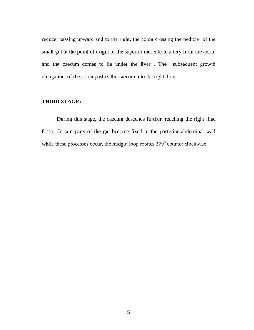
reduce, passing upward and to the right, the colon crossing the pedicle of the
small gut at the point of origin of the superior mesenteric artery from the aorta,
and the caecum comes to lie under the liver . The subsequent growth
elongation of the colon pushes the caecum into the right loin.
THIRD STAGE:
During this stage, the caecum descends further, reaching the right iliac
fossa. Certain parts of the gut become fixed to the posterior abdominal wall
while these processes occur, the midgut loop rotates 270o counter clockwise.
6
ANATOMY OF THE SMALL INTESTINE
Small intestine extends from the pyloroduodenal junction to the
ileocaecal valve. It is about 6 meters long.
Small intestine consists of
- Duodenum
- Jejunum and
- Ileum
DUODENUM
It is 20 –25 cms long and is the shortest, widest and most sessile part of
the small intestine . It has no mesentery and is thus only partially covered by
peritoneum . It encircles the head of the pancreas and consists of four parts.
- The superior (first) part, about 5cm long
- The descending (second) part, about 8-10 cm long
- The horizontal (inferior or third) part , about 10 cms long
- The ascending (Fourth part) about 2.5cms long
ARTERIAL SUPPLY
It is from the right gastric, right gastroepiploic, supra duodenal and
superior and inferior pancreatico- duodenal arteries.
VENOUS DRAINAGE
Veins end in the splenic, superior mesenteric and portal veins.
7
NERVE SUPPLY
Are from the coeliac plexus
LYMPHATIC DRAINAGE
Duodenal lymph drains by channels that accompany superior and
inferior pancreatico duodenal vessels to coeliac and superior mesenteric nodes.
JEJUNUM AND ILEUM
The small intestine except the duodenum is attached to the posterior
abdominal wall by the mesentery, the proximal two fifth being the jejunum, the
distal three – fifth , the ileum.
The jejunum is comparatively wider bored and thicker walled and
measures about 2.3 to 2.8 meters. It lies largely in the umbilical region.
The ileum is thinner than the jejunum. Its length varies from 3.6 to
4.2meters. It is mainly in the hypogastric and pelvic region.
The fan like mesenteric attachment of the jejunum and ileum to the
posterior abdominal wall allows free movement, each coil adapting to changes
in form and position.
ARTERIAL SUPPLY
Jejunal and ileal arteries from the left side of the superior mesenteric
artery .
These branches form arterial arcades
8
Jejunum has one or two arterial arcades and has high narrow windows
in the intestinal border of the mesentery.
Ileum has larger serious of arterial arcades – three to five and the straight
vessels branching off the arcades are shorter.
VENOUS DRAINAGE
The veins follow the arteries
LYMPHATIC DRAINAGE
The lymph vessels form an intricate plexus in mucosa & submucosa and
are joined by vessels from lymph spaces at the bases of solitary follicles and
drain to larger vessels at the mesenteric aspect of the gut. These drain to
superior mesenteric lymph nodes.
NERVE SUPPLY
Innervation is by vagus and thoracic splanchnic nerves through the
caeliac ganglia and superior mesenteric plexuses.
9
ETIOLOGY OF SMALL BOWEL
PERFORATIONS3
- Duodenal ulcer perforation
- Inflammatory diseases
Acute – salmonella typhi enteritis
Chronic – Tuberculosis common
- Vascular
Ischemic enterocolitis
Strangulated hernia
- Neoplastic
Rare
Seen in lymphomas
- Diverticulitis
Meckel’s diverticulitis
Jejunal diverticulitis
Duodenal diverticulitis
- Miscellaneous
Radiation enteritis
Necrotising enterocolitis
Meconium peritonitis
10
PATHOPHYSIOLOGY
PATHOPHYSIOLOGY OF PEPTIC ULCER
Gastric secretion aids in breakdown of food into smaller particles. About
2 litres of gastric juice is produced everyday.
In the stomach, oxyntic cells secrete Hcl & Intrinsic factor. Chief cells
secrete pepsinogen . Mucus cells secrete mucus and G cells secretes gastrin .
Hydrochloric acid is a major etiological factor in acid peptic disease.
Acetyl choline, histamine and gastrin stimulate Hcl secretion . Somatostatin
inhibits Hcl secretion.
GASTRIC MUCOSAL BARRIER
The gastric mucus layer is essential to the integrity of the gastric
mucosa. It is a viscid layer of mucopolysaccharides produced by the mucus-
producing cells of the stomach and pyloric glands. Gastric mucus is an
important physiological barrier to protect the gastric mucosa from mechanical
damage, and also the effects of acid and pepsin. Its considerable buffering
capacity is enhanced by the presence of bicarbonate ions within the mucus.
Many factors can lead to the breakdown of this gastric mucosal barrier.
Those include bile, NSAIDS, alcohol , trauma and shock.
11
DUODENAL ULCERATION
INCIDENCE
There have been marked changes in the last two decades in the
demography of patients presenting with duodenal ulceration.
First, even before the introduction of H2 receptor antagonists, the
incidence of duodenal ulceration and frequency of elective surgery for the
condition were falling. This may relate to the widespread use of gastric
antisecretory agents and eradication therapy for patients with dyspepsia .
Second , the peak incidence is now in a much older age group than
previously and although it is still more common in men, the difference is less
marked . These changes mirror the changes at least in part in the epidemiology
of H. Pylori infection.
The incidence of perforation and bleeding duodenal ulcers in young and
middle aged patients appear to be falling but in contrast, there is currently a
marked increase in the number of elderly suffering these complication. This
trend can be explained not only by the H. Pylori cohort effect but also by the
increased use of NSAIDS in elderly.
12
ETIOLOGY OF PERFORATION IN PEPTIC ULCER
a) NSAIDS
They interfere with cyclo oxygenase pathway which leads to production
of prostanoids which inturn affects mucosal protection by reducing the
effectiveness of mucus bicarbonate barrier.
They are not dependent on duration of usage.
The rate of recurrence of ulcer is very less after discontinuation of
drugs.
This can be prevented by additional therapy with prostaglandin
analogues.
b) HELICOBACTER PYLORI
It is a small curved gram negative micro aerophilic rod with multiple
polar flagellae. Stomach is its normal inhabitant.
One of the characteristics of the organism is its ability to hydrolyse urea,
resulting in production of ammonia, a strong alkali. This causes release of
gastrin from antral G cells. This is probably responsible for hypergastrinaemia
in peptic ulcer patients, which in turn may result in gastric acid hypersecretion.
Infection also leads to the disruption of the gastric mucosal barrier by the
enzymes produced by the organism.
Gastric metaplasia is the normal response of the duodenal mucosa to
excess acidity, an attempt by the mucosa to resist an injurious stimulus .
13
Although normal duodenal mucosa cannot be infected with H. Pylori, gastric
metaplasia in duodenum is commonly infected.
The incidence of infection within a population increases with age, and
infection rates of 80-90% are not unusual.
The possibility of infection is inversely related to socio economic group.
Eradication of H. Pylori reduces ulcer recurrence rate. Hence all patients
should be treated by H. pyloric eradication therapy as it speeds up healing and
decreases the rate of ulcer disease.
c) CIGARETTE SMOKING
It impairs the healing of ulcer
It promotes recurrence of ulcers and also increases the surgical risks.
d) MISCELLANEOUS
- Alcohol damages the gastric mucosal barrier
- Certain personality traits and psychological stress, or poor
tolerance to stress leads to ulcer formation.
- An association has been reported that patients with blood group O
have an increased risk of duodenal ulcer.
PATHOLOGY
Most of the peptic ulcers occur in the first part of the duodenum
A chronic ulcer penetrates the mucosa and into the muscle coat leading
to fibrosis. The fibrosis causes deformities such as pyloric stenosis.
14
Sometimes there may be more than one duodenal ulcer. The situation in
which there is both anterior and posterior duodenal ulcer is referred to as
“kissing ulcer”.
Anteriorly placed ulcers tend to perforate and posterior duodenal ulcer
tend to bleed, sometimes by eroding a large vessel such as the gastroduodenal
artery.
HISTOPATHOLOGY
MICROSCOPICALLY
Destruction of the muscular coat is observed and the base of the ulcer is
covered with granulation tissue, the arteries in this region showing the typical
changes of endarteritis obliterans.
STAGES IN ACUTE PERFORATION7
1. Stage of peritoneal irritation
2. Stage of peritoneal reaction
3. Stage of diffuse peritonitis
1) STAGE OF PERITONEAL IRRITATION
This is first stage known as peritonism.
Due to leakage of gastric juice into the peritoneal cavity – chemical
peritonitis.
Usually lasts for about six hours
There will be previous history of peptic ulcer.
15
There is acute burning pain over epigastrium. Pain may be referred to tip
of right shoulder due to irritation of undersurface of diaphragm.
Pain may gradually gravitate down along the paracolic gutter to the right
iliac fossa mimicking acute appendicitis.
Tenderness and muscle guard are constantly present.
There will be little change in the pulse, temperature and respiration.
2) STAGE OF PERITONEAL REACTION
This is second stage.
The irritant fluid becomes diluted with the peritoneal exudates.
The patient feels comfortable.
Muscular rigidity is present.
Obliteration of liver dullness is present.
Rectal examination may elicit tenderness in the recto – vesical or recto
uterine pouch.
3) STAGE OF DIFFUSE PERITONITIS
This is third or final stage.
There will be pinched and anxious face, sunken eyes and hollow cheeks,
so called facies Hippocratica.
Tachycardia with low in volume & tension, board like rigidity of
abdomen, increasing distension of the abdomen are present.
Septicemia and multi system organ failure frequently supervene.
16
SUBACUTE PERFORATION
An ulcer may perforate and seal rapidly before there is spillage of gastric
& duodenal contents into peritoneal cavity.
Sudden onset of acute abdominal pain.
Local tenderness and rigidity are present.
CHRONIC PERFORATION
When an ulcer perforates into an area that is walled of by adhesion or by
adjacent viscera like colon, greater omentum or when a gastric ulcer perforates
into a omental sac with sealing off the omental foramen, chronic abscess may
form.
Features of peritonitis may be less marked.
X ray abdomen may reveal air under diaphragm.
17
PATHOPHYSIOLOGY OF TYPHOID PERFORATIONS
Typhoid fever is caused by ingestion of salmonella typhi contaminated
with water and is a systemic infection. Contamination occurs due to infected
stools or urine. They invade the intestinal lymphatics and mesenteric nodes &
thus reach the blood stream. Once bacteremia is established, it leads to the
development of secondary areas of inflammation in the liver, gall bladder and
marrow. After one week, the bacteria are shed into the small bowel and
therefore appears in the stool.
The early change is the hyperplasia of the lymph follicles. The peyer’s
patches become swollen and ulcerated, which can progress to capillary
thrombosis and subsequent necrosis. In the second week, necrosis and
sloughing occurs and ulceration of the follicles leading on to perforation in the
third week of disease.
PATHOLOGY
The organisms cause enlargement of reticulo endothelial and lymphoid
tissue throughout the body. Proliferation of the phagocytes swells the lymphatic
submucosal nodules of the entire gut mainly peyer’s patches of the terminal
ileum. These become sharply delineated plateau like elevations upto 8mm in
diameter bulging into the intestinal lumen. During second week, the mucosa
over the swollen ileal lymphoid tissue is shed, resulting in oval ulcers with
their long axis in the direction of bowel flow.
18
Once passed the peak of the disease, the ulcers heal slowly and
lymphatic structures amazingly regenerate without scarring. Histologically
there is accumulation of mononuclear phagocytes which form nodular
aggregates filled with red cells and nuclear debris.
Perforation is due to the result of rupture of necrotic peyer’s patches
caused by distension of bowel or by excessive peristalsis. The spleen is
enlarged and soft. Microscopically, marked histocytosis and reticulo endothelial
proliferation are present. Sometimes spleen may rupture.
The liver shows scattered foci of parenchymal necrosis in which
hepatocyte is replaced by phagocytic mononuclear aggregate called as
“Typhoid nodule” which can also occur in bone marrow. Gall bladder
colonization produces a carrier state often requires cholecystectomy to
eliminate bacterial shedding.
CLINICAL FEATURES
The symptoms of head ache, fever, vomiting and abdominal pain and the
signs of abdominal tenderness, guarding and rigidity, distension, absent
intestinal sounds, presence of free fluid and obliteration of hepatic dullness
were considered most important.
The most prominent clinical features were the prolonged debilitating
feverishness with diarrhea and on examination, generalized abdominal

19
tenderness with rebound tenderness were the rule more marked on the right side
of abdomen
In typhoid perforations, the classical test like widal and blood culture are
little important value as the results are obtained only after few days. Therefore
history of fever and physical examination with signs and symptoms suggestive
of perforation assume importance.
SITE OF PERFORATION IN TYPHOID FEVER
Since the peyer’s patches are more in the terminal ileum, the incidence
of typhoid perforation is also more in the terminal ileum. Kim 197532 obtained
86% cases of perforation occurred in the last 60cm of ileum of which 72%
perforations were within the last 40cm . In kuruvilla series 197830 perforations
were confined to the last 30 cms of the terminal ileum. Purohit et al34, noted all
the perforations occurring within 40cms from the ileo caecal junction.
20
PATHOPHYSIOLOGY OF INTESTINAL TUBERCULOSIS
Tuberculosis can affect any part of the GIT from mouth to the anus.
Intestinal Tuberculosis can be of two varieties
1. Ulcerative tuberculosis
2. Hyperplasic tuberculosis
1. ULCERATIVE TUBERCULOSIS
This usually results from swallowing of tubercle bacilli (human type) in
sputum in a case of pulmonary tuberculosis
This condition is characterized by multiple ulcers at the terminal ileum
The long axis of the ulcer lies transversely.
The serous coat overlying the ulcer becomes thickened, so perforation is
unusual . But perforation occurs in ulcerative tuberculosis than in hyperplastic
tuberculosis.
Healing of the ulcers leads to stricture formation. Loss of weight and
diarrhea with fecal odour stools containing pus and occult blood are present.
There will be slight tenderness in right iliac fossa.
2. HYPERPLASTIC TUBERCULOSIS
Infection starts in the lymphoid follicles and then spreads to submucous
and subserous planes. The intestinal wall becomes thickened with narrowing of
its lumen.
21
There will be early involvement of regional lymph nodes which become
matted along with the involved terminal part of ileum and caecum to produce
the lump.
Recurrent attacks of abdominal pain with diarrhea and features of blind
loop syndrome are present.
There will be a lump in right iliac fossa on examination .
22
PATHOPHYSIOLOGY OF VASCULAR DISORDERS
ACUTE MESENTERIC ISCHAEMIA4
The mortality rate for acute mesenteric infarction varying between 60
and 85% because the condition is not common accounting for only 1-2 % of
patients with acute abdominal pain and it is usually diagnosed late.
This result in as many as 40% of patients receiving either no operation
or an open and close laparotomy .
PATHOLOGY
Acute arterial ischaemia may arise from an embolus or from formation
of in situ thrombus on an underlying stenosis.
It may also occur as a result of low cardiac output state.
Other rare causes include aortic dissection, fibromuscular dysplasia,
intimal hyperplasia associated with oral contraceptive pills, arteritis associated
with rheumatoid arthritis, systemic lupus erythematosus and polyarteritis
nodosa.
CLINICAL FEATURES
There will be acute colicky abdominal pain of abrupt onset in a patient
with atrial fibrillation or recent myocardial infarction with no antecedent history
of gastrointestinal upset or weight loss.
A more insidious onset suggests mesenteric artery thrombosis
23
As this is superimposed on chronic occlusive disease there may be a
prior history of post prandial abdominal pain (intestinal angina) , weight loss
and diarrhea .
History of minor gastrointestinal bleeding is a late symptom.
Abdominal pain may be mild or more classically out of proportion to
the physical findings.
Onset of signs of peritoneal irritation or frank peritonitis is usually a late
sign and indicative of irreversible bowel ischaemia .
24
PATHOPHYSIOLOGY OF STRANGULATED HERNIA1
A hernia is said to be strangulated when the contents are so constricted
as to interfere with their blood supply. Usually the small intestine is involved
in the strangulation .
Intestinal obstruction may not be present particularly in case of
omentocoele, Richter’s hernia and litter’s hernia.
There will be no impulse on cough, extremely tense and tender
These are followed by acute intestinal obstruction.
Gangrene may occur as early as 5-6 hours after the onset of first
symptoms.
Femoral hernia is more likely to strangulate because of the narrowness of
the neck and its rigid surrounds.
PATHOLOGY
Intestine is obstructed & its blood supply impaired. Initially only venous
return is impeded, the wall of the intestine becomes congested and bright red
with the transudation of serous fluid into the sac. As congestion increases, the
wall of the intestine becomes purple in colour. As venous stasis increases,
arterial supply becomes more and more impaired.
Blood is extravasated under serosa and is effused into the lumen. The
fluid in the sac becomes blood stained and shining serosa dull due to fibrinous ,

25
sticky exudates . At this stage, the walls of intestine have lost their tone and
become friable. Bacterial translocation occurs.
Gangrene appears at rings of constriction than at antimesentric border .
If the strangulation is unrelieved, perforation of the wall of the intestine
occurs.
Peritonitis spreads from the sac to peritoneal cavity.
26
PATHOPHYSIOLOGY OF SMALL BOWEL LYMPHOMAS4
The vast majority are Non- hodgkin’s lymphoma. They can be B- cell or
T- cell lymphoma. They are further subdivided into low grade and high grade.
The commonest types of intestinal Lymphomas are
- MALT lymphoma
- Centrocytic lymphoma
- Mediterranean lymphoma
- Burkitt type lymphoma
- Polymorphic T-cell lymphoma
They are more common in males
The clinical manifestation include malaise , abdominal pain, weight
loss, diarrhea and anaemia.
Some are presented with intestinal obstruction or a perforation leading to
peritonitis .
In patients with coeliac disease, enteropathy associated lymphoma tend
to occur in fifth to seventh decade . The symptoms of celiac disease are
abdominal pain, diarrhea & rapid weight loss. Perforation leading to peritonitis
is a common presentation in these patients.
27
PATHOPHYSIOLOGY OF DIVERTICULAR DISEASE1
MECKEL’S DIVERTICULITIS
Occur in 2% of patients, usually 2 inches in length and are situated 2
feet from the ileocoecal valve.
It is a congenital diverticulum having all three coats of the bowel.
It represents patent intestinal end of vitello intestinal tract.
Meckel’s diverticulitis with or without perforation, may result from
obstruction by food residue. The symptoms are those of acute appendicitis .
When the diverticulum perforates , the symptoms may stimulate those
of a perforated duodenal ulcer.
DUODENAL & JEJUNAL DIVERTICULA
Duodenal diverticula are of two types .
Primary - mostly occurring in older patients on inner wall of
second and third parts & usually do not cause symptoms.
Secondary – Diverticula of the duodenal cap result from long
standing duodenal ulceration.
Jejunal diverticulae are usually of variable size & multiple.
Clinically they may
- be symptomless
28
- give rise to abdominal pain, flatulence and borborygmi
- produce a malabsorption syndrome.
- Present as an acute abdomen with acute inflammation and
occasionally rupture.
29
PATHOPHYSIOLOGY OF MISCELLANEOUS CONDITIONS
RADIATION ENTERITIS4
The immediate effect of radiation on the gastrointestinal tract is arrest
of cell division in the intestinal crypts. This effect is largely restricted to cells
in G1 phase . the mucosa becomes thinner with stunted villi.
The incidence of intestinal radiation induced bowel disease vary from 3
to 25%.
The most common situation is where the pelvis is irradiated usually for
rectal or gynaecological cancers.
Anorexia, nausea, vomiting are present during the first few weeks of
radiotherapy.
The commonest symptoms referable to chronic bowel damage are vague
abdominal discomfort, diarrhea, mild rectal bleeding , and the passage of
mucus.
The interval between the tissue of radiation and onset of symptoms
varies considerably form 2 months to 2 years.
Intestinal obstruction may be acute or subacute or recurrent.
Occasionally acute presentation with infarction may occur and this
carries a high risk of perforation and mortality .
Most of the serious complication tend to occur within 2 years of the
initial treatment but may become progressively worse after this time.
30
NECROTISING ENTEROCOLITIS 1
This is a common phenomenon among such premature neonates. The
risk is inversely proportional to birth weight.
It is associated with hypoxia, hypothermia, hypotension and umbilical
artery cannulation.
Ileum, caecum, distal colon and total colon are affected with a complete
spectrum from mucosal to transmural necrosis .
The usual presentation in bilious vomiting, abdominal distension, colour
change , a lethargy in a high risk neonate . the abdomen is usually soft.
MECONIUM PERITONITIS 1
It is an aseptic peritonitis that develops late in intrauterine life or during
or just after delivery . Meconium is a sterile mixture of epithelial cells, mucin,
salts, fats and bile.
Meconium enters peritoneal cavity through an intestinal perforation and
in over 50% of cases the perforation is the result of some form of neonatal
intestinal obstruction , in the remainder no cause is found. Meconium remain
sterile upto 3 hours after birth after wards it leads to acute bacterial peritonitis.
New born baby presents with a tense abdomen who is vomiting and in
whom there is failure to discharge meconium.
31
MANAGEMENT
MANAGEMENT OF PEPTIC ULCER PERFORATION
INVESTIGATIONS
- Increased total count leukocytosis with immature forms
- Increased haematocrit due to fluid loss
- Urine analysis shows increased urinary specific gravity.
- Serum amalyse may be elevated
- X ray abdomen erect view including diaphragm or x ray chest PA
view shows gas under diaphragm in about 70% of cases .
- CT scan is useful in a typical cases with doubtful clinical features,
which may show localized perforation of the duodenum with
leakage in the area of gall bladder and right flank, without gross
free air
TREATMENT 8
- Adequate resuscitation with crystalloids and colloids is important
- Surgical intervention
Operative treatment for perforated duodenal ulcer may be divided into
simple closure versus definitive surgery. Simple closure is appropriate for
patients with major underlying illness, patients with ongoing shock and
perforation lasting for more than 24 hours.

32
Definitive operative treatment for perforated duodenal ulcers provides
the benefit of perforation closure, freedom from continued ulcer symptoms and
added protection from recurrence.
Definitive ulcer surgery can be recommended in patients with evidence
of chronic ulcer disease who has been treated for H. pylori, patients who lack a
major medical illness or patients with short duration of perforations , who are
free of significant preoperative shock.
Parietal cell vagotomy combined with omental patching of the
perforation is generally the preferred definitive therapy for perforated duodenal
ulcer.
In the presence of chronic pyloroduodenal scarring associated with
perforated duodenal ulcer, the performance of parietal cell vagotomy with
omental patching should be avoided because it may not allow for unimpeded
gastric empting. In this setting, truncal vagotomy combined with pyloroplasty
with incorporation of perforated ulcer into closure is a better alternative.
Definitive ulcer operation8 in addition to closing the perforation is
indicated in
- Perforated gastric ulcer
- Combined gastric & duodenal ulcer one of which has perforated
- Perforation with preexisting chronic ulcer symptoms
- Coexistent obstruction & perforation
33
- Coexistent haemorrhage and perforation
- Previous operation for perforated duodenal ulcer
- Young patients <35 years who have perforated duodenal ulcer
- H- pyloric treated or known negative patients who have
perforation.
GRAHAM’S PATCH TECHNIQUE
Here simple closure with a live omental patch is used using absorbable
suture material.
CELEN JONES TECHNIQUE
Using absorbable suture materials, full thickness sutures are placed on
either side of perforation and a strand of omentum is drawn under the arch &
sutures tied.
CLOSURE OF PERFORATION BY OMENTAL IMPLANTATION
Perforation is repaired by drawing and implanting a portion of omentum
into the perforated site. Omentum gets firmly adherent and undergoes
inflammation , necrotic changes , granulation, reduction in size & fibrosis.
There is no luminal obstruction
This is used in perforation with friable edges or if the sutures placed at
the first operation have given way.
34
FREE OMENTAL PLUG
A free omental graft of suitable dimensions is cut, rolled & fashioned
into the shape of mushroom & fixed to perforated site. The omental plug can
also be tied to nasogastric tube using catgut. The edges of omental plug are
further tucked to the intestinal wall. The Naso gastric tube is removed after 7
days, by which time the catgut suture attaching into the omentum has
dissolved away.
LAPAROSCOPIC SUTURE REPAIR8
Suturing is carried out as same as in the open procedure using a patch
omentum . This is followed by peritoneal lavage with normal saline. Definitive
procedure is not generally recommended, it causes significant technical
problem became of bleeding & tissue edema.
Laparoscopic fibrin glue repair using oxidized cellulose sealed with
fibrin glue have been described, but they may not provide optimal results.
CONSERVATIVE MANAGEMENT8
Non operative therapy is often indicated for patients who have a
perforation of longer than 24 hours duration , for patients whose systemic
disease or current state of deterioration militates against operative treatment.
Non operative therapy includes naso gastric suction, antibiotics and
fluid resuscitation and decompression of abdomen by bilateral flank drainage .
35
The non operative approach can be used only if a water soluble contrast
radiological study confirms that the ulcer crater is sealed by failure of any
contrast medium to leak from the duodenum. Of extreme importance in this
method of managing perforated duodenal ulcer is close, accurate reassessment
of the patients general condition and abdominal findings every 2 - 3 hours .

36
MANAGEMENT OF TYPHOID PERFORATION
INVESTIGATION
The diagnosis can be confirmed by isolation of the organism from the
blood during first week. Identification of the antibodies by widal test is done
during second week of illness. Stool and urine culture will become positive
only after third week of illness. Typhoid bacilli can also be grown from bile &
marrow of patients suffering from typhoid fever.
SIGNS AND SYMPTOMS
In making diagnosis , great emphasis has been given to clinical signs &
symptoms. The symptoms of headache, fever, vomiting, abdominal pain, and
the signs of abdominal tenderness, guarding and rigidity, distension, absent
bowel sound, pressure of free fluid and obliteration of liver dullness were
considered most important.
In typhoid perforation , the classical test like widal & blood culture are
of little immediate value as the results are obtained only after few days.
Therefore history of fever and physical examination with signs & symptoms
suggestive of perforation assume importance.
WIDAL TEST
After an attack of typhoid fever, antibodies appear as early as fifth day
H- antigen Ig G and O- antigen Ig M agglutinating antibodies . The antibody
37
levels rise gradually, reaches the maximum in second to third week. These H
and O agglutinins can be estimated by widal test. The rising titre of agglutinins
against S- typhi H & O Ag more than 1: 200 dilutions in patient’s serum is
considered as positive.
WHITE CELL COUNT
In general leucocytosis is a feature of peritonitis but typhoid fever is
associated with leucopenia. Hence in the enteric fever perforation there is either
high normal or leucopenia is seen
BLOOD CULTURE
Most of the blood cultures in typhoid fever are positive only in the first
week of illness moreover many cases of fever are treated with antibiotics , But
we come across typhoid perforation most commonly in second or third week .
At that time blood cultures are less sensitive when compared to the first week.
RADIOLOGY
Abdominal erect view x rays are taken up to find out pneumpeuitoreum
in enteric fever perforation cases. The incidence of pneumoperitoneum is
varying from one series to other. The percentage of pneumoperitoneum various
from 45- 95% cases .
38
HISTOPATHOLOGICAL EXAMINATION
The specimens of the edges of the ulcer for histopathological
examination showed the appearance compatible with typhoid fever.
Histological study reveals areas of necrosis containing plasma cells,
lymphocytes macrophages containing abundant cytoplasm with bacteria and
red cells termed as typhoid cells and monocytes .
TREATMENT
In general typhoid perforation is a surgical emergency and the treatment
should be prompt and energetic. The role of conservative treatment is limited.
The surgical procedure depends upon the general health of the patients and the
extent of the ileum involved.
Patient was put on naso gastric aspiration while intravenous fluids and
drugs were administered parenterally. Electrolytes and fluid balance are
meticulously maintained. The appropriate operative procedure was decided at
the time of laparotomy and depended upon the general condition of the patients
and state of the ileum.
SIMPLE CLOSURE OF PERFORATION
The standard surgical management consists of simple closure of
perforation. Laparotomy and bowel loop bearing perforation is sought out.
39
The perforation is closed with atraumatic needle in 2 layers . After thorough
peritoneal lavage, peritoneal cavity is mopped & drain kept.
Chauhan et al29 treated 138 cases of typhoid perforation surgically. In
these patients, the principal operation was closure of the perforation and
peritoneal drainage. The overall mortality of surgical treatment was 58.7%.
Solitary perforations were treated by simple closure in several patients of
Kuruvilla et al (1978)30.
WEDGE EXCISION OF AFFECTED SEGMENT
- easier and quicker than resection
- Safer than simple closure with lower mortality rate
- Provides a healthy area for closure as the perforated segment is
friable.
- Following wedge excision, singh et al35 series showed 25% of
mortality when compared to their previous mortality of 60% with
simple closure.
SIMPLE CLOSURE WITH ILEOTRANSVERSE ANASTAMOSIS
- It allows perforation to heal quickly
- Earlier passing of flatus in post operative period .
- No chance of reperforation
- Reduces the changes of typhoid state and severity of peritonitis
40
- It has drawback of increasing the magnitude of surgical procedure
in toxic patient with severe peritonitis
RESECTION OF THE MOST OF THE AFFECTED SEGMENT
WITH AN END TO END ANASTAMOSIS
When there are multiple perforations or multiple ulcers or where ileal
segment looked unhealthy or where perforation was accompanied by
hemorrhage , resection becomes the operation of choice .
Resection prevents reperforation . It also prevents further perforation of
nearby ulcer.
ILEOSTOMY
It is done in the moribund as well as in the most critically ill patients.
In Kim series 197532 , 10 patients were treated by the above method with
a mortality of 10%. In Kuruvilla et al 197830 , because of poor general
condition, ileostomy was done. His study showed 50% mortality.
COMPLICATION OF SURGERY
EARLY COMPLICATIONS : Toxemia, respiratory infections, paralytic ileus,
Thrombo phlebitis, Transfusion reaction, uraemia, Meningism, shock,
reperforation.
LATE COMPLICATIONS: Wound infection, Burst abdomen, faecal fistula,
decubitus ulcer, osteomyelitis, Incisional hernia.
41
MANAGEMENT OF INTESTINAL TUBERCULOSIS1,10
A barium meal and follow – through or small bowel enema will show
the absence of filling of the lower ileum , caecum and most of the ascending
colon as a result of narrowing and hypermotility of the ulcerated segment.
TREATMENT
A course of chemotherapy - anti tuberculous drug is started .
Laparotomy is done when there is perforation or intestinal obstruction in
ulcerative tuberculosis.
Ieocoecal resection is to be done in hyperplastic tuberculosis
SURGICAL PROCEDURE FOR INTESTINAL TUBERCULOSIS
For Acute bowel perforation
• Resection and anastamosis is the treatment
• Simple closure can be done but there is high incidence of
reperforation and fistula formation.
For Intestinal obstruction due to Ileocaecal mass
• Right hemicolectomy is the standard of treatment
• Limited resection and ileo ascending colon anastamosis can
also be carried out.
For Intestinal obstruction due to multiple strictures involving long
segment of bowel.
42
• Resection and anastamosis
For Intestinal obstruction due to strictures placed apart
• Stricturoplasty.
MANAGEMENT OF ACUTE MESENTERIC ISCHAEMIA4
• There will be markedly elevated white cell count.
• Raised serum phosphate and raised serum amalyse are present.
• Angiography is diagnostic
• Superior mesenteric artery thrombosis is seen as a tapering of
the origin of the vessel whilst embolic occlusion shows an
abrupt blockage often at a branching point.
TREATMENT
The key to successful management is a high index of suspicion.
There is a massive fluid loss as a consequence of mesenteric ischaemia.
The adequate replacement of this is dramatic and often under estimated
fluid loss is of private importance.
Embolectomy should be carried out by mainly arteriotomy over superior
mesenteric artery. Any non viable bowel is resected.
Despite revascularisation , the mortality rate is reported between 20 and
70%.
43
Acute mesenteric ischarmia secondary to thrombosis is treated by short
aorto superior mesenteric artery bypass, preferably with saphenous vein.
An important aspect of the surgical management of patients with acute
mesenteric ischaemia is “second look’ laparotomy within 24 hours of the first
procedure.
MANAGEMENT OF STRANGULATED HERNIA
• Diagnosis is mainly based on the clinical examination
• Treatment is by emergency operation.
• Vigorous resuscitation and antibiotics are essential
• During operation, the sac was delivered out and sac is incised
near the fundus and toxic fluid was let out. Then neck of the sac
was widened. Gangrenous segment of bowel are excised by
localized resection. Then repair of the hernia is done.
44
DIVERTICULAR DISEASE MANAGEMENT
MECKEL’S DIVERTICULITIS
Meckel’s diverticulum is very difficult to demonstrate by contrast
radiology. Small bowel enema would be most accurate investigation.
In cases of repeated gastro intestinal haemorrhage of unknown cause
where a meckel’s diverticulum is suspected , technetium – 99 m scanning can
be done to detect meckel’s diverticulum.
TREATMENT 1
If it is narrow based then excision of Meckel’s diverticulum was done.
If it is broad based, wedge resection of ileal segment is done.
Where there is induration of base of diverticulum extending into the
adjacent ileum , it is advisable to resect a short segment of ileum containing
the diverticulum, restoring continuity with an end – to end anastamosis.
In jejunal diverticulitis associated with major malabsorbtion problems
due to connective tissue disorders, resection of the affected segment with end –
end anastamosis can be effective .
45
MANAGEMENT OF SMALL BOWEL LYMPHOMAS
In acute cases of perforation, diagnosis is made on clinical grounds &
laparotomy.
Small bowel contrast enema, CT Scan will be helpful in other cases.
TREATMENT
In complicated cases of small bowel lymphoma with perforation,
laparotomy and resection of the involved segment is done.
Further treatment consists of combination chemotherapy with drug
regimens CHOP, CMOPP or radiotherapy.
In uncomplicated lymphoma, surgery followed by chemotherapy or
radiotherapy in used for stage I and II disease.
Chemotherapy alone is used for more advanced disease.
46
MANAGEMENT OF MISCELLANEOUS
CONDITIONS MANAGEMENT OF PERFORATION OF RADIATION
ENTERITIS
Emergency surgery is required for infarction with perforation or acute
intestinal obstruction which does not resolve on conservative treatment .
The radionecrosed bowel is ideally excised with primary anastamosis or
exteriorization of the bowel ends in the presence of ischaemia and sepsis .
MANAGEMENT OF NECROTISING ENTEROCOLITIS 1
Abdominal radio graphs may show pneumatosis intestinalis or free
intraperitoneal air .
Management consists of aggressive resuscitation with intravenous
feeding. The optimal time for surgery is not in the acute phase as the baby can
with stand the pressure of necrosis than an adult but not the stress of
laparotomy.
At laparotomy, excision of all necrotic bowel with primary anastamosis
is usual . The overall mortality is 25% with 10-30% of neonates developing a
colonic stricture .
47
MANAGEMENT OF MECONIUM PERITONITIS1
Free air in the peritoneal cavity, an abundant quantity of abdominal fluid,
fluid levels, calcification are characteristic findings, all of which are unlikely
to be present in every case. Meconium peritonitis has been diagnosed by
radiography of the fetus in uteus before birth.
TREATMENT
The prognosis is poor
The greatest chance of survival is in those patients who have an
intestinal perforation but not intestinal obstruction in which case closure of the
perforation and drainage of the peritoneal cavity are performed expeditiously.
Intestinal lavage can prevent reformation of meconium bolus obstruction
& supplements of pancreatic exocrine enzymes are often necessary throughout
life.
48
INTRA ABDOMINAL HYPERTENSION AND THE
ABDOMINAL COMPARTMENT SYNDROME
Increased intra abdominal pressure (IAP)43 occurs in a variety of clinical
situations such as accumulation of ascites, bowel distension from the ileus or
mechanical obstruction and reduction into peritoneal cavity of large chronic
hernia contents lost their domain.
The factors that contribute intra abdominal hypertension after trauma are
- Accumulation of blood & clot
- Bowel edema or congestion from injury to mesenteric vessels or
excessive crystalloid resuscitation.
- Closure of swollen & non compliant abdominal wall under
abdominal wall under tension.
Abdominal compartment syndrome43 is a late manifestation of
uncontrolled intra abdominal hypertension . Abdominal compartment
syndrome is characterized by tensely distended abdomen, elevated intra
abdominal & peak airway pressures, inadequate ventilation with hypoxia &
hypercarbia , distrubed renal function and improvement of these features after
abdominal decompression.
IAP can be monitored by Direct or indirect methods
49
DIRECT METHOD
It is measurement of IAP by attaching the catheter placed into peritoneal
cavity to a saline manometer on a pressure transducers.
Abdominal pressure measurement during laparoscopy is an example of
direct method .
INDIRECT METHOD
Here pressure is measured through other accessible abdominal organ
which reflect IAP.
IVC PRESSURE
It is by placing the catheter in groin and then into IVC. Complications
are infections & thrombosis.
INTRAGASTRIC PRESSRUE
It is measured by water manometery through nasogastric tube or a
gastrostomy tube
BLADDER PRESSURE
Kron & Colleagues41 first described the technique of using bladder
pressure as a mean of assessing intra abdominal pressure. Urinary bladder is
an extraperitoneal , intra abdominal structure with a very complaint wall.
Changes in IAP are parallel to intra luminal bladder pressure. It can be
monitored continuously or intermittently.

50
INTERMITTENT
It is more popular. Here 50 ml of saline is installed into bladder through
foleys catheter . The tubing of collecting bag is clamped. The needle is inserted
into the tube proximal to clamp and attached to manometer . Bladder pressure
measured in water is the height at which the level of the saline column
stabilizes with pubic symphysis as the zero point .
INTRA ABDOMINAL HYPERTENSION
Significant organ dysfunction occurs when the intra abdominal pressure
is above 10 mm Hg.
Burch and associates38 classified elevated IAP
Grade I - 10 –15 cms of H2O
Grade II - 15-25 cms of H2O
Grade III- 25 - 35 cms of H2O
Grade IV- >35 cms of H2O
ADVERSE PHYSIOLOGICAL EFFECTS
When IAP is above 14cm of H2O , patient developed decreased venous
return and cardiac output. Splanchnic hypertension & hypoperfusion occurs
when IAP is 20.4 cm of H2O. Hence whole body O2 consumption , pH &
arterial Po2 are decreased. Diminished venous return is the major cause,
51
increased after load is the another cause. They show marked elevation of CVP,
mean pulmonary artery pressure and pulmonary capillary wedge pressure.
RENAL EFFECTS
Anuria occurs when IAP rises. Decreased cardiac output is one cause.
shunting of blood away form the renal cortex into medulla, diminution of renal
blood flow , direct compression of kidneys or renal veins, diminished renal
arterial flow and a rise in renal vascular resistance and presence of high levels
of ADH are other possible causes of renal malfunction.
ABDOMINAL WALL
IAH reduces tissue blood flow. So wound infection with fascial
dehiscence are common
SPLANCHNIC FLOW
Reduction of blood flow to all abdominal organs except adrenal glands
occur when IAP rises. IAH causes significant intestinal ischaemia followed by
reperforation injury after abdominal decompression.
INTRA CRANIAL PRESSURE
IAH causes elevated intra cranial pressure and decreased cerebral
perfusion pressure. Due to elevated IAP , CVP raised. Venous drainage from
the cerebral venous outflow is decreased . The diminished cardiac output due
to IAP also causes neuronal damage due to decreased CPP.

52
THE VICIOUS CIRCLE OF EVENTS CREATED BY IAH
ON SPLANCHNIC CIRCULATION
Splanchnic Hypoperfusion
Hepatic Ischemia I A H Gut mucosal Acidosis
Bowel edema
Coagulopathy Hypothermia Unrelieved Acidosis Intra Abdominal ACS Free 02 Radicals Bleeding Distant organ damage
53
ADVERSE HEMODYNAMIC CONSEQUENCES OF RELIEF OF
IAH.40
Sudden and severe hypotension may occur during or immediately
following surgical decompression of IAH. It is due to sudden decrease in
systemic vascular resistance, either from resumption of flow to a constricted
splanchnic bed or the so-called reperfusion syndrome descriped by Morris et
al40
REPERFUSION SYNDROME
Reperfusion syndrome occurs when the IAH is suddenly relieved.
• An abrupt increase in the true tidal volume delivered to the
patient. It will result in metabolic alkalosis.
• Washout of bye products of anaerobic metabolism from below
the diaphragm. Potassium and other products of anaerobic
metabolism are delivered to the heart, patients develop cardiac
asystole following reperfusion.
• To prevent this, 2 litres of solution composed of 1 litre of 45%
normal saline with 50 gm mannitol and 40 mEQ of sodium
bicarbonate are prepared and are infused immediately before
and continued during decompression.
54
• Complete cardiac resuscitation team should be available at the
bed side during decompression.
Each individual member of the team is primarily responsible for a single
task or aspect of the patient’s care. The team consists of the following
people.
Two surgeons to conduct the operation
An experienced physician to manage the ventilator
A person whose sole responsibility is infusion of fluid and
blood products
Two runners/ circulators and
One scribe
ENDORGAN INVOLVEMENT
Three systems are commonly involved in IAH
1. CVS
2. Renal
3. Pulmonary
1. CARDIVASCULAR EFFECTS
Obstruction of blood flow through inferior vena cava , result in
decreased cardiac output and leads to hypotension. Splanchnic blood flow also
decreased.
55
2. RENAL SYSTEM
Oliguria occurs due to
- Inadequate renal arterial perfusion from poor cardiac output
- Direct compression of kidney
- Obstruction of renal vascular flow
- Obstruction of ureteral outflow
3. PULMONARY EFFECTS
It may present as intractable hypercarbia and extremely poor
compliance. Patients have steadily increasing peak inspiratory pressure. These
are due to compression of lung parenchyma and progressive upward
displacement of diaphragm.
56
MATERIALS AND METHODS
This study has been based on the analysis of small bowel perforations 66
cases admitted in the IV surgical unit, Government Rajaji Hospital, Madurai
Medical college during a period from November 2004 to February 2006.
In all patients admitted age, sex, associated illness were noted. Time
interval between admission and time of intervention was noted.
Investigations including blood urea, blood sugar, serum creatinine,
serum electrolytes, chest x ray , blood grouping , x ray abdomen & further
investigation as required were done.
All the patients presented were resuscitated with crystalloids and after
achieving hemodynamic stability these patients were subjected to emergency
laparotomy, when perforation was proved clinically.
The intra abdominal pressure was measured indirectly by measuring the
bladder pressure. The bladder was catheterized with foley’s catheter. The urine
drainage tube was clamped and one needle inserted proximally. It was
connected to the three way adapter one to saline stand and other to the
manometer. The manometer is filled with saline. About 50ml of saline was
infused into the bladder. Then the bladder through catheter and manometer
were connected. The manometer reflects the bladder pressure. The bladder
pressure is directly correlated to the intra abdominal pressure.
57
For the patients who presented with extreme degree of shock , an initial
flank drainage was done in an attempt to let out toxic fluid and once general
condition was improved perforation closure was done.
All the patients were put on preoperative antibiotics. During laparotomy
site of perforation, degree of peritoneal contamination were assessed . In all
patients bilateral flank drainage was kept irrespective of the degree of
contamination .
Post operatively patient was evaluated for the development of
complications, if any.
58
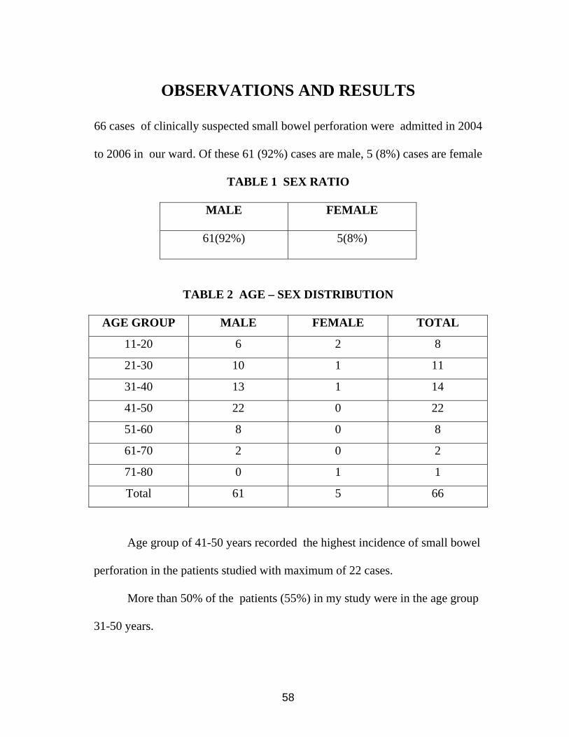
OBSERVATIONS AND RESULTS
66 cases of clinically suspected small bowel perforation were admitted in 2004
to 2006 in our ward. Of these 61 (92%) cases are male, 5 (8%) cases are female
TABLE 1 SEX RATIO
MALE FEMALE
61(92%) 5(8%)
TABLE 2 AGE – SEX DISTRIBUTION
AGE GROUP MALE FEMALE TOTAL
11-20 6 2 8
21-30 10 1 11
31-40 13 1 14
41-50 22 0 22
51-60 8 0 8
61-70 2 0 2
71-80 0 1 1
Total 61 5 66
Age group of 41-50 years recorded the highest incidence of small bowel
perforation in the patients studied with maximum of 22 cases.
More than 50% of the patients (55%) in my study were in the age group
31-50 years.
59
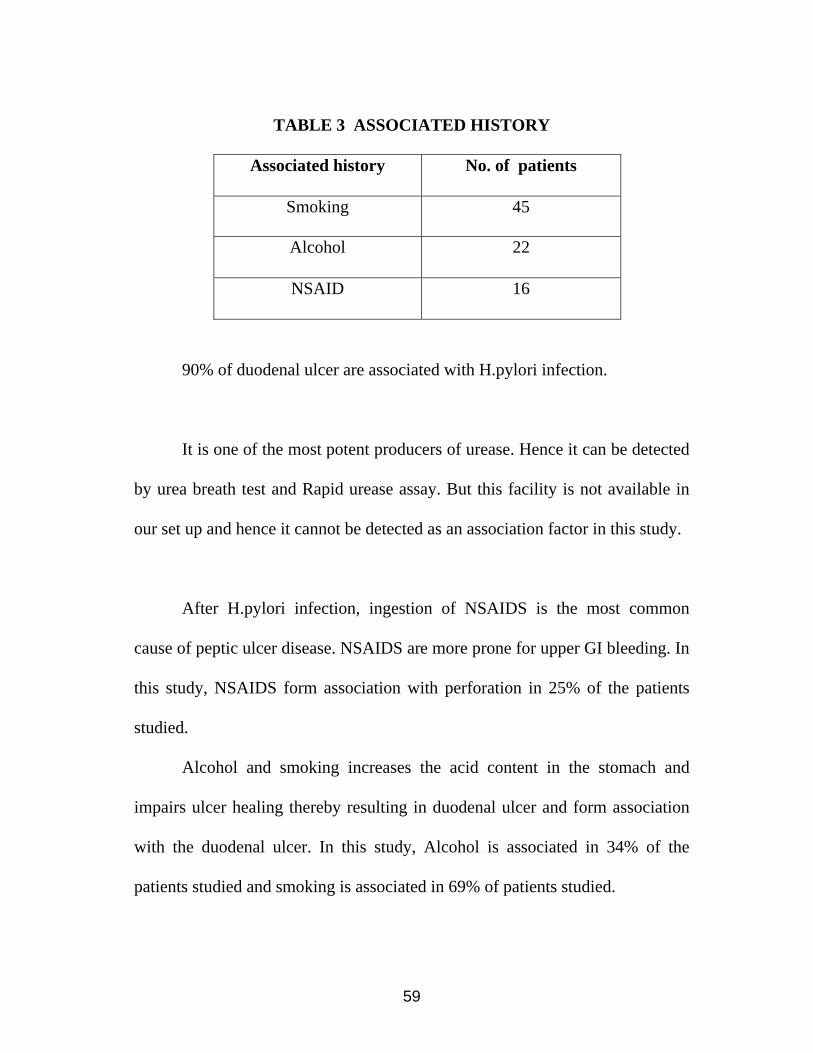
TABLE 3 ASSOCIATED HISTORY
Associated history No. of patients
Smoking 45
Alcohol 22
NSAID 16
90% of duodenal ulcer are associated with H.pylori infection.
It is one of the most potent producers of urease. Hence it can be detected
by urea breath test and Rapid urease assay. But this facility is not available in
our set up and hence it cannot be detected as an association factor in this study.
After H.pylori infection, ingestion of NSAIDS is the most common
cause of peptic ulcer disease. NSAIDS are more prone for upper GI bleeding. In
this study, NSAIDS form association with perforation in 25% of the patients
studied.
Alcohol and smoking increases the acid content in the stomach and
impairs ulcer healing thereby resulting in duodenal ulcer and form association
with the duodenal ulcer. In this study, Alcohol is associated in 34% of the
patients studied and smoking is associated in 69% of patients studied.
60
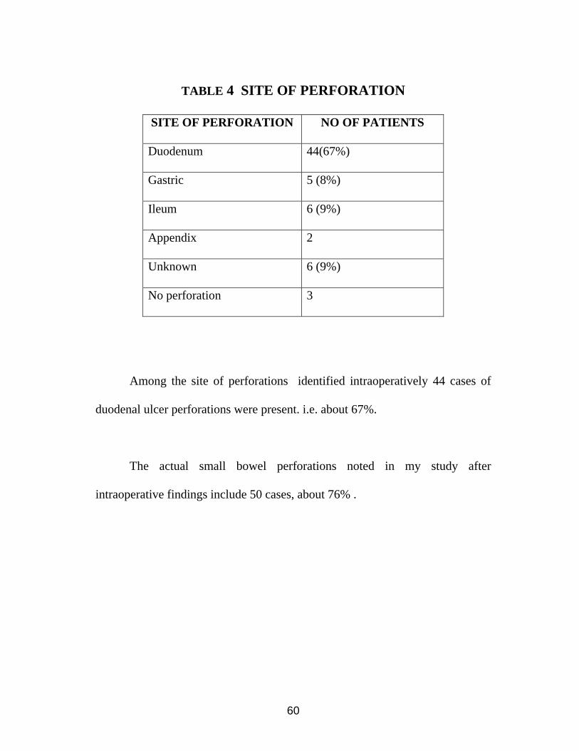
TABLE 4 SITE OF PERFORATION
SITE OF PERFORATION NO OF PATIENTS
Duodenum 44(67%)
Gastric 5 (8%)
Ileum 6 (9%)
Appendix 2
Unknown 6 (9%)
No perforation 3
Among the site of perforations identified intraoperatively 44 cases of
duodenal ulcer perforations were present. i.e. about 67%.
The actual small bowel perforations noted in my study after
intraoperative findings include 50 cases, about 76% .
61
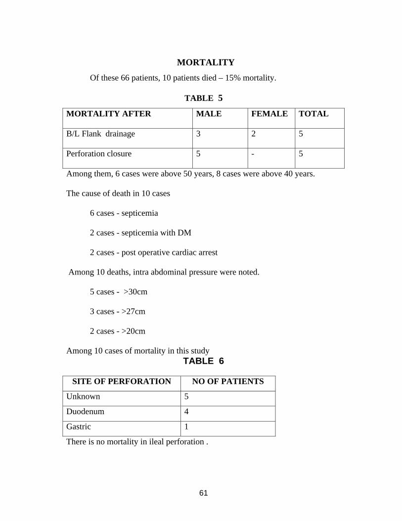
MORTALITY
Of these 66 patients, 10 patients died – 15% mortality.
TABLE 5
MORTALITY AFTER MALE FEMALE TOTAL
B/L Flank drainage 3 2 5
Perforation closure 5 - 5
Among them, 6 cases were above 50 years, 8 cases were above 40 years.
The cause of death in 10 cases
6 cases - septicemia
2 cases - septicemia with DM
2 cases - post operative cardiac arrest
Among 10 deaths, intra abdominal pressure were noted.
5 cases - >30cm
3 cases - >27cm
2 cases - >20cm
Among 10 cases of mortality in this study TABLE 6
SITE OF PERFORATION NO OF PATIENTS
Unknown 5
Duodenum 4
Gastric 1
There is no mortality in ileal perforation .
62
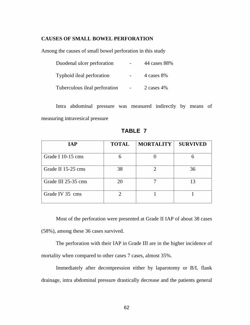
CAUSES OF SMALL BOWEL PERFORATION
Among the causes of small bowel perforation in this study
Duodenal ulcer perforation - 44 cases 88%
Typhoid ileal perforation - 4 cases 8%
Tuberculous ileal perforation - 2 cases 4%
Intra abdominal pressure was measured indirectly by means of
measuring intravesical pressure
TABLE 7
IAP TOTAL MORTALITY SURVIVED
Grade I 10-15 cms 6 0 6
Grade II 15-25 cms 38 2 36
Grade III 25-35 cms 20 7 13
Grade IV 35 cms 2 1 1
Most of the perforation were presented at Grade II IAP of about 38 cases
(58%), among these 36 cases survived.
The perforation with their IAP in Grade III are in the higher incidence of
mortality when compared to other cases 7 cases, almost 35%.
Immediately after decompression either by laparotomy or B/L flank
drainage, intra abdominal pressure drastically decrease and the patients general
63
condition was improved. The compression effects of increased intraabdominal
pressure were released. There is improvement in respiratory functions,
increased splanchnic and renal blood flow thereby increasing renal urine output
and decreases the cardiac overload and improves the circulation. Also the
septicemic foci was removed thereby improving the general condition of the
patients.
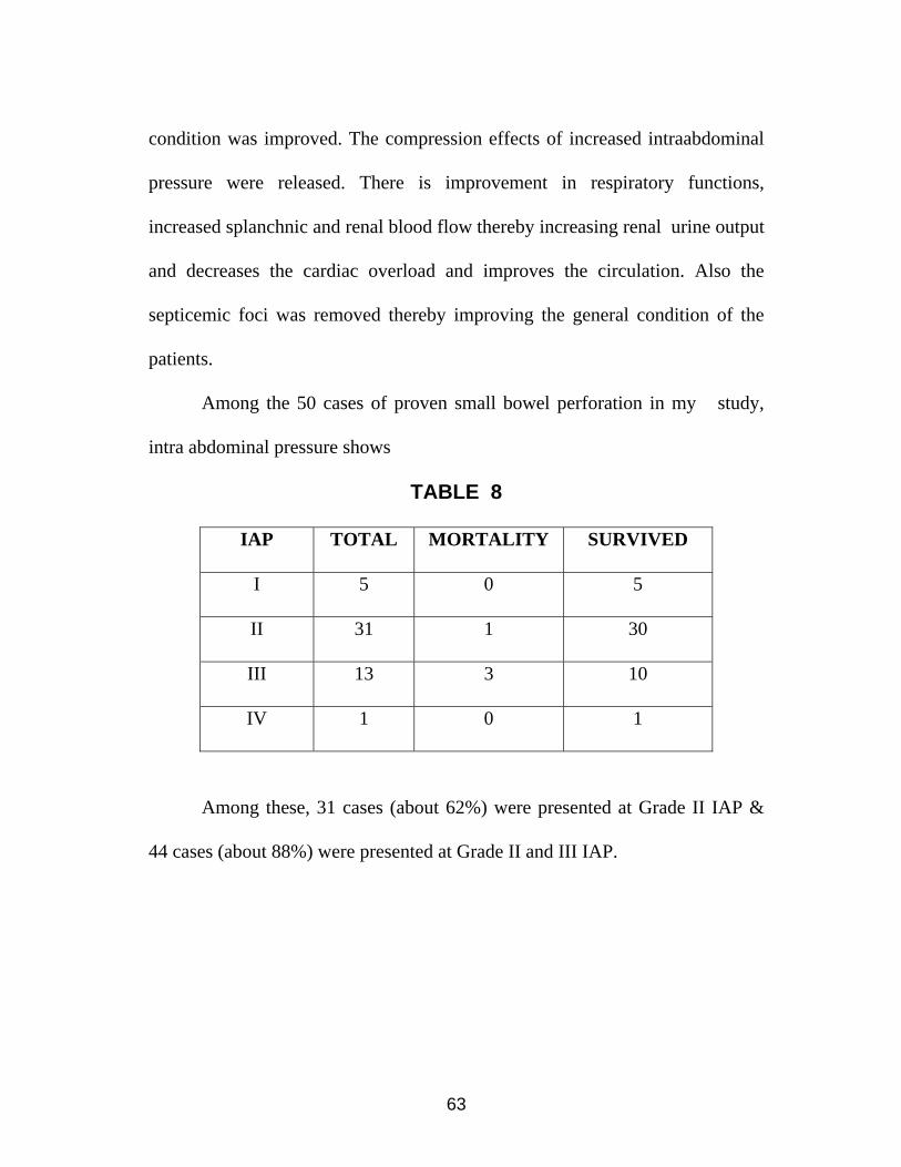
Among the 50 cases of proven small bowel perforation in my study,
intra abdominal pressure shows
TABLE 8
IAP TOTAL MORTALITY SURVIVED
I 5 0 5
II 31 1 30
III 13 3 10
IV 1 0 1
Among these, 31 cases (about 62%) were presented at Grade II IAP &
44 cases (about 88%) were presented at Grade II and III IAP.

64
COMPLICATIONS
Among 50 cases of proven small bowel perforation, 14 cases had
complications.
TABLE 9
No. of Cases Complications
7 cases Wound infection
2 cases Wound gapping
1 case B/L pneumonia
1 case Fecal fistula
1 case post operative adhesion
1 case Iatrogenic urethral injury
Of these 13 cases, 7 cases were duodenal ulcer perforation.
All the ileal perforation cases in my study had complications.
3 cases has wound infections treated conservatively.
2 cases had wound gapping treated with secondary
suturing.
1 case developed fecal fistula.
65
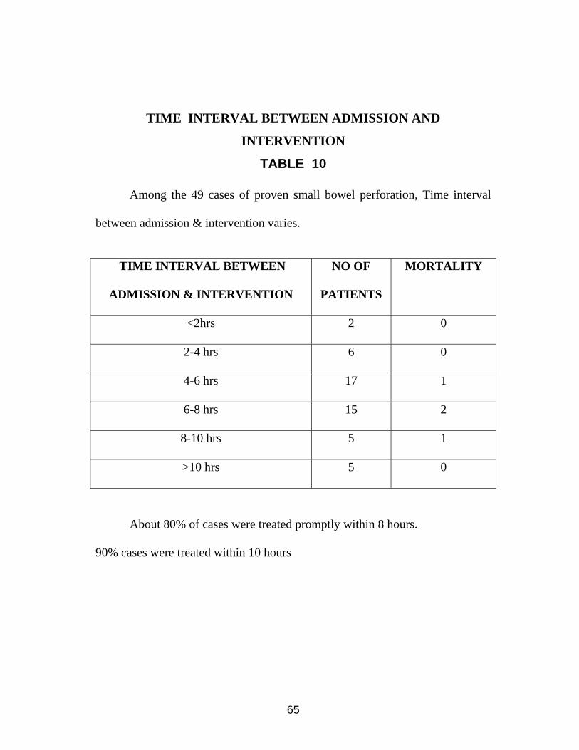
TIME INTERVAL BETWEEN ADMISSION AND
INTERVENTION
TABLE 10
Among the 49 cases of proven small bowel perforation, Time interval
between admission & intervention varies.
TIME INTERVAL BETWEEN
ADMISSION & INTERVENTION
NO OF
PATIENTS
MORTALITY
<2hrs 2 0
2-4 hrs 6 0
4-6 hrs 17 1
6-8 hrs 15 2
8-10 hrs 5 1
>10 hrs 5 0
About 80% of cases were treated promptly within 8 hours.
90% cases were treated within 10 hours
66
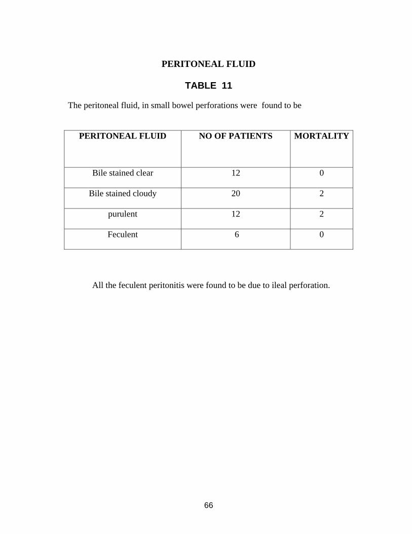
PERITONEAL FLUID
TABLE 11 The peritoneal fluid, in small bowel perforations were found to be
PERITONEAL FLUID NO OF PATIENTS MORTALITY
Bile stained clear 12 0
Bile stained cloudy 20 2
purulent 12 2
Feculent 6 0
All the feculent peritonitis were found to be due to ileal perforation.
67
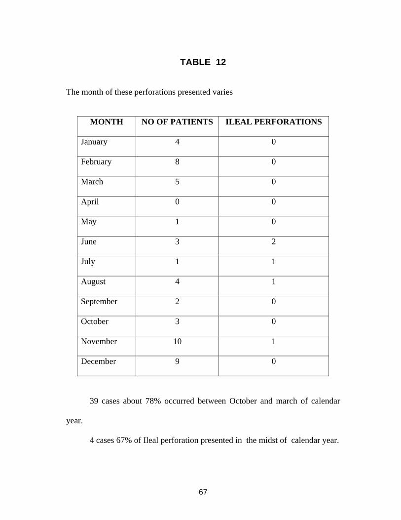
TABLE 12
The month of these perforations presented varies
MONTH NO OF PATIENTS ILEAL PERFORATIONS
January 4 0
February 8 0
March 5 0
April 0 0
May 1 0
June 3 2
July 1 1
August 4 1
September 2 0
October 3 0
November 10 1
December 9 0
39 cases about 78% occurred between October and march of calendar
year.
4 cases 67% of Ileal perforation presented in the midst of calendar year.

68
TREATMENT - TABLE 13
TREATMENT GIVEN SURVIVED MORTALITY TOTAL NO.
Laparotomy 52 5 57
B/L flank drainage alone 0 5 5
B/L flank drainage followed
by surgery
4 0 4
Even though the above table shows an apparent increase in mortality of
those patients who have undergone flank drainage, they were having associated
factor which increased the mortality independently.
69
DISCUSSION
Perforation has been found to be a major complication of peptic ulcer
disease with a mortality rate ranging form 6 to 31% worldwide. Several factor
might contribute to increased mortality in patients with duodenal ulcer
perforation. In this study, mortality rate is 15%.
Age of the patient with duodenal ulcer perforation has been increasing in
many western series. This is due to complex age cohort phenomenon. But in
this study maximal clustering of cases was found in fourth, fifth decades. Stress
factor may be an added factor.
Factor associated with increased morality were studied in detail. Older
age more than 50 years, presence of shock at the time of presentation, delayed
presentation more than 24 hour, presence of co-morbid conditions were found
to have significant impact on increasing the mortality rate.
All cases were having anterior perforation. Posterior perforation was not
encountered in this study. Awareness of this serious condition however is
important because the best chance for survival of patients lies in prompt and
thorough surgical exploration, drainage and when appropriate, definitive
surgery.
We could not undertake definitive surgery at the time of peptic ulcer
closure because majority of the cases was having risk factor which would have
70
increased the mortality considerably. It is also true that we are not able to
compare the efficacy of treatment by different surgical procedures (like omental
plugging vs patch) because of the poor matching of confounding factor in the
study group.
Simple closure alone was found to be effective in healing of the
perforated ulcer with medical therapy preventing recurrence of the disease on
follow up.
There was an increased death rate in patients treated with flank drain
above, because almost all of them had presence of two or more risk factors
which we found to increase the mortality. In this study, there is 100% mortality
in patients treated with flank drain alone.
The availability of effective medical treatment for acid peptic disease
and the introduction of H. pylori eradication therapy had made definitive
surgery unnecessary at the time of perforation closure.
Open surgery still remains the choice of treatment in majority of centres.
Laparoscopic technique though allows for perforation closure, does not allow
sufficient lavage of all quadrants to remove all sequelae of peritonitis. Minimal
access surgery may be useful for early cases. In this study, all have been treated
by open surgery.
Perforated peptic ulcer is likely to continue to be a surgical challenge
despite the medical treatment. In this study, 69% of patients were found to be
71
smoker. 25% of patients were taken NSAIDS prior to the symptoms and 34% of
patients were alcoholic.
In this study, there were 6 cases of ileal perforation.
All of the ileal perforation cases were managed with primary closure in 2
layers and all of them have been survived.
All the ileal perforations were presented with feculant peritonitis and all
of them had post operative complication. All the ileal perforation cases had
stormy postoperative period.
Among the 6 cases of ileal perforation, 4 cases were typhoid ileal
perforations and were treated with primary closure of perforation site, although
there are various modalities of treatment.
2 cases of rare tuberculous peritonitis were managed with primary
closure and started anti tuberculous drugs post operatively and they impoved on
follow up.
In this study, all typhoid ileal perforations were presented in the male.
The reason for this male predominance is yet to be identified, although it is
possible that men have an increased risk of exposure to typhoid fever.
The important investigations carried out to confirm the diagnosis are
plain x-ray abdomen erect view for evidence of pneumo –peritonium, widal test
and biopsy of the ileal at the site of perforation.
72
This study recommend, when general condition of the patients is good,
simple closure alone is adequate in uncomplicated solitary perforation. But
resection is necessary if there is bleeding or if multiple ulcers with more than
one perforation are present. But in this case study, no resection was carried out.
Bilateral flank drainage may be of help in those patients who are too ill
to with stand the laparotomy and after improving the patients general condition,
laparotomy can be carried out.
All of the ileal ulcers in my study were confined to the last 40 cm of
ileum.
Other than wound infection, the most common abdominal complication
is wound dehiscence in Forrest’s view31, and in my study next to the wound
infection is wound gapping. Wound gapping or dehiscence is the reflection of
both high incidence of infection and debility of the patients. Just like wound
dehiscence, the development of fecal fistula is catastrophic. This may be the
result of re perforation, perforation in another area or the result of suture line
break down. In this study, one case had fecal fistula.
When the intra abdominal pressure rises, there is shunting of blood away
from renal cortex into medulla, diminution of renal blood flow leading to anuria
and decreased cardiac output, these effects are returned back to normal when
the abdominal decompression was done.
73
By early intervention to decompress the abdomen either by bilateral
flank drainage or by laparotomy, patient’s general condition can be improved
by increasing the Cardiac output, relieving septicemic focus from abdominal
cavity and increasing urine output.
In this study, 80% of cases were treated prompty with in 8 hours.
74
CONCLUSION Among the small bowel perforation studied, Duodenal ulcer perforation forms
the majority of cases about 88%.
Typhoid ileal perforation compromise about 8% of all small bowel perforation.
i.e. 4 out of 50 cases. Tuberculous small bowel perforation comprise about 4% of all
small bowel perforation i.e. 2 out of 50 cases. More than half of the cases were
presented in the age group of 31- 50 years about 36 cases out of 66 cases.
Most of the small bowel perforations were presented at Grade II IAP. More
than 95% of these were survived.
The patients with their IAP in Grade III & IV are in higher incidence of
mortality 8 out of 14 cases, 36% has mortality .
In this study 80% of cases were managed within 8 hours of presentations with
a mortality of 4 cases i.e. 8%.
All the ileal perforation cases were presented with feculent peritonitis & all of
them have been survived.
High mortality occurs in small bowel perforations when the age is above 50
years i.e. 6 out of 10 cases about 60%.
About 78% of cases occured between October and March of calendar year.
So, by these methods, we will grade the small bowel perforation and assess
the prognosis of these patients.
BIBLIOGRAPHY
1. Bailey and love’s short practice of surgery – 24th Edition R.C.G. Russel ,
N.S. Williams , C.J.K. Bolstrode
2. Text book of surgery - Sabiston 17th Edition
3. Principles of Surgery - 8th Edition, Schwartz / Charles Brunicardi
4. Essential surgical practice - 4th Edition. A Cuscieri / J.C. Steele / A.R.
Moosa.
5. Farquharson’s Text book of operative surgery – 9th Edition, R. F. Rintoul.
6. Maingot’s Abdominal operations – 10th Edition , Rodney Maingot.
7. A manual on clinical surgery – 6th Edition: S. Das
8. Surgery Annual – 1991 – Lloyd M. Nyhus .
9. Recent advances in Gastroenterology – 7th Edition , R. R. Pounder
10. Oxford textbook of surgery – 2nd Edition, Peter J. Morris and William C.
Wood.
11. Christensen A, Bousfield R , Christensen J. Incidence of perforated and
bleeding peptic ulcers before and after the introduction of H2 – receptor
antagonist . Ann Surg 1988:207: 4-6.
12. Bliss DW, Stabile BE. The impact of ulcerogenic drugs on surgery for the
treatment of peptic ulcer disease . Arch Surg 1991: 126 : 609-612.
13. Lana WL, Leung KL, Kwong KH, Davey IC, Robertson C, Dawson JJ .
Chung SC, Li Ak. A randomized study comparing laparoscopic versus
open repair of perforated peptic ulcer using suture or sutureless technique
. Ann Surg 1996 ;224: 131- 138.
14. Boey J, Wong J, Ong GB . A prospective study of operative risk factors in
performed duodenal ulcers . Ann surg 1982: 195: 265- 269.
15. Evans JP, Smith R. Predicting poor outcome in perforated peptic ulcer
disease. Aust N Z /Surg 1997; 67 : 792 – 795.
16. Greiser WB, Burner BW, Shamoun JM, Jurkovich GJ , Ferrara JJ .
Factors affecting mortality in patients operated upon for complications of
peptic ulcer disease. Ann Surg 1989; 55: 7-11.
17. Irvin TT. Mortality and perforated peptic ulcer: a case for risk
stratification in elderly patients. Br J Surg 1989; 76:215-218.
18. Suter M. Surgical treatment of perforated peptic ulcer. Is there a need for a
change? Acta Chir Belg 1993;93: 83-87.
19. Lee Fy. Leung KL. Lai BS. Ng SS. Dexter S. Lau WY. Predicting
mortality and morbidity of patients operated on for perforated peptic
ulcers. Arch Surg 2001;139: 30-94.
20. Blomgren LGM. Perforated peptic ulcer: long-term results after simple
closure in the elderly. World/Surg 1997;21:412-415.
21. Gray’s Anatomy – 39th edition.
22. Last’s Anatomy – 9th Edition
23. Hamby L.S. Zweng TN, Strodel WE. Performed gastric and duodenal
ulcer: an analysis of prognostic factors. Am Surg 1993;59:319-324.
24. Matsuda M. et al., Laparoscopic omental patch repair for perforated peptic
ulcer. Ann Surg 1995;221:336-240.
25. Wakayama T, et al., Risk factors influencing short term results of
duodenal perforation. Surg Today 1994;24:681-687.
26. Celen Jones, “A rapid method of treatment of perforated peptic ulcer”
Gasroenterology 33:353,1957.
27. Graham R.R. “The treatment of perforated Duodenal ulcer” Sur Gyaence
obset. 64: 235, 1937.
28. Archampong E.Q (1976) Typhoid ileal perforations Br. J. Surg Vol 63
(1976) 317-321.
29. M.K. Chouhan and S.K. Pande (Rajasthan) Typhoid Enteric perforation.
Br. J. Surg. Vol-69 (1982) 173-175.
30. Kuruvilla M.J.F.R.C.S.F.R.C.S. Role of Resection in Typhoid Perforation
Ann. R. Coll. Durg., Eng Vol – 60 (1978).
31. Forrest C. Eggleston and Bannu Santoshi – Typhoid Perforation choice of
perforation. Br. J. Surg., Vol – 68 (1981) 341-342.
32. Kim J. P. Oh S.K. & Jarrett E. Management of ileal Perforation due to
Typhoid fever. Ann., Surg, 1975; 181: 88-91.
33. Venkataramani Sitaram M.S. (C.M.C.Vellore) Typhoid ileal perforations
a Retrospective study. Ann R. Col. Surg., Engl., Nov – 1990 Vol – 72.
No.6.
34. Purohit P.G. Surgical Treatment of Typhoid perforation Ind. J. Surg., 40:
227-238, 1978.
35. Egglesten F.C. Santoshi B & Singh C.M. typhoid perforation of the
bowel; Experiences in 78 cases. Ann. Surg. 1979; 190: 31-35.
36. Barnes GE, Laine GA, Giam PY, et al: Cardiovascular responses to
elevation of intraabdominal hydrostatic pressure. Am J physiol 248 :R208,
1985.
37. Bongard FB, Ryan M, Dubecz S, et al: Adverse consequences of
increased intraabdominal pressure on bowel tissue oxygen, J Trauma 39:
519,1995.
38. Burch JM, Moore EE, Moore FA, et al: The abdominal compartment
syndrome. Surg clin north Am 76:833, 1996.
39. Cullen DJ, Coyle JP, Teplick R, et al: Cardiovascular, pulmonary, renal
effects of massively increased intraabdominal pressure in critically ill
patients. Crit care Med 17: 118, 19889.
40. Morris JA Jr, Eddy VA, Blinman TA, et al: The staged celiotomy for
trauma. Issues in unpacking and reconstruction, Ann Surg. 217:576, 1993.
41. Kron IL, Harman PK, Nolan SP : The measurement of intraabdominal
pressure as a criterion for exploration. Ann Surg 199:28,1984.
42. Ian Aird – A companion of surgical studies – 3rd edition.
43. Rao R. Ivatury, Lawrence, M.Porter, J. Simon: Intraabdominal
hypertension and the abdominal compartment syndrome Surg. Clin North
Am. 77:783, 1997.
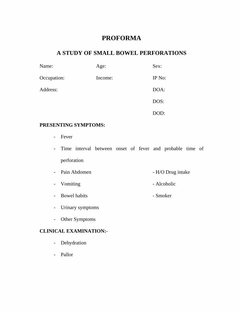
PROFORMA
A STUDY OF SMALL BOWEL PERFORATIONS
Name: Age: Sex:
Occupation: Income: IP No:
Address: DOA:
DOS:
DOD:
PRESENTING SYMPTOMS:
- Fever
- Time interval between onset of fever and probable time of
perforation
- Pain Abdomen - H/O Drug intake
- Vomiting - Alcoholic
- Bowel habits - Smoker
- Urinary symptoms
- Other Symptoms
CLINICAL EXAMINATION:-
- Dehydration
- Pallor
- P/A - Distension
Diffuse Rigidity / Guarding
Localized
Obliteration of liver dullness
Bowel sound
- P/R
- Pulse rate - Urinary output
- BP - Time interval between time of
admission and surgery
- Temperature
Associated Systemic illness, if any:
INVESTIGATION:
Hb
Blood – Urea Urine
Creatinine X-ray chest PA view
Sugar X-ray Abdomen erect view
Serum electrolytes other specific tests –
Widal test
Intraabdominal pressure
Pre procedure Post procedure
OPERATIVE FINDINGS:
o Amount of Peritoneal fluid
o Site of Perforation
o Size of Perforation
o Other findings
PROCEDURE DONE:
Post Operative Complications, if any:
Follow Up:
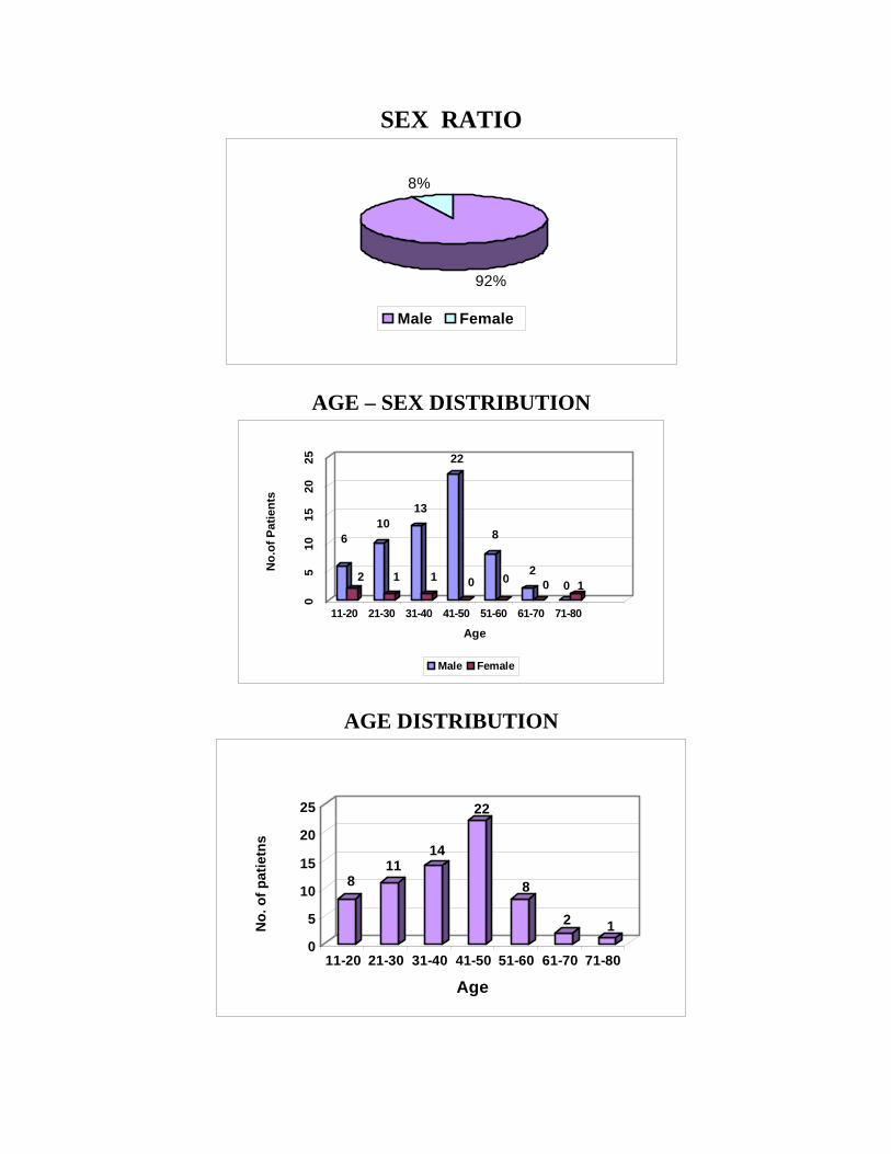
SEX RATIO
92%
8%
Male Female
AGE – SEX DISTRIBUTION
6
2
10
1
13
1
22
0
8
02
0 0 1
05
1015
2025
No.
of P
atie
nts
11-20 21-30 31-40 41-50 51-60 61-70 71-80
Age
Male Female
AGE DISTRIBUTION
811
14
22
8
2 10
5
10
15
20
25
No.
of p
atie
tns
11-20 21-30 31-40 41-50 51-60 61-70 71-80
Age
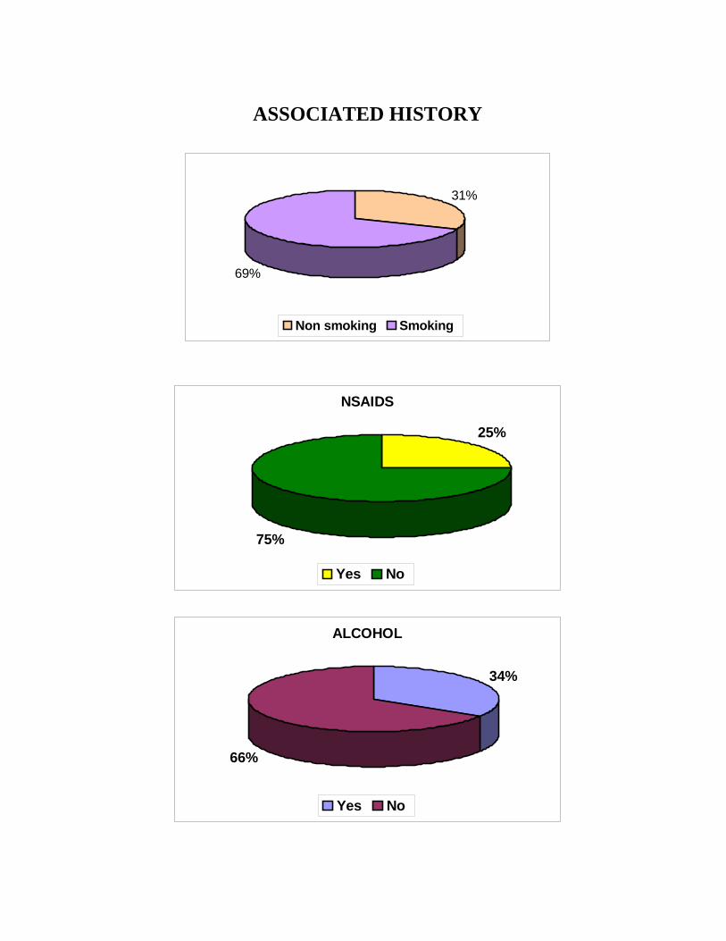
ASSOCIATED HISTORY
31%
69%
Non smoking Smoking
NSAIDS
25%
75%
Yes No
ALCOHOL
34%
66%
Yes No
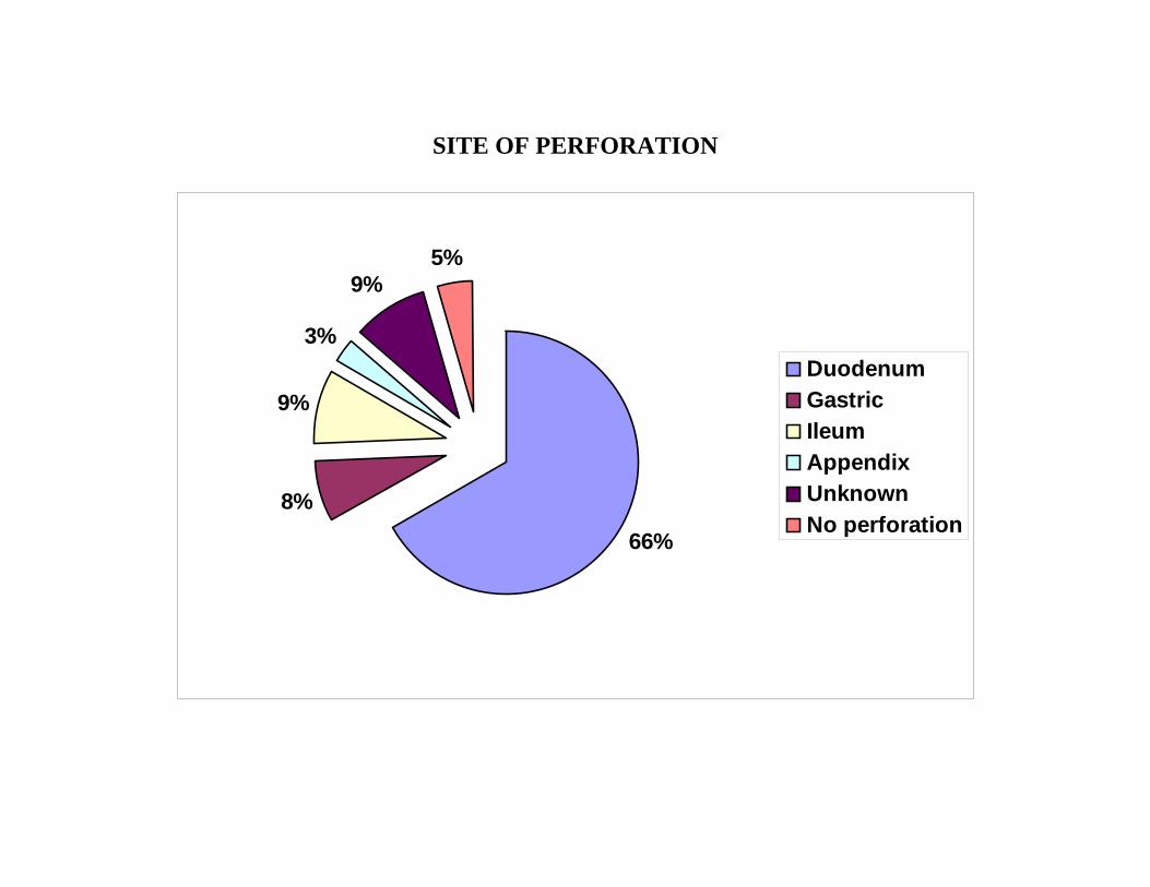
SITE OF PERFORATION
66%
8%
9%
3%
9%5%
DuodenumGastricIleumAppendixUnknownNo perforation
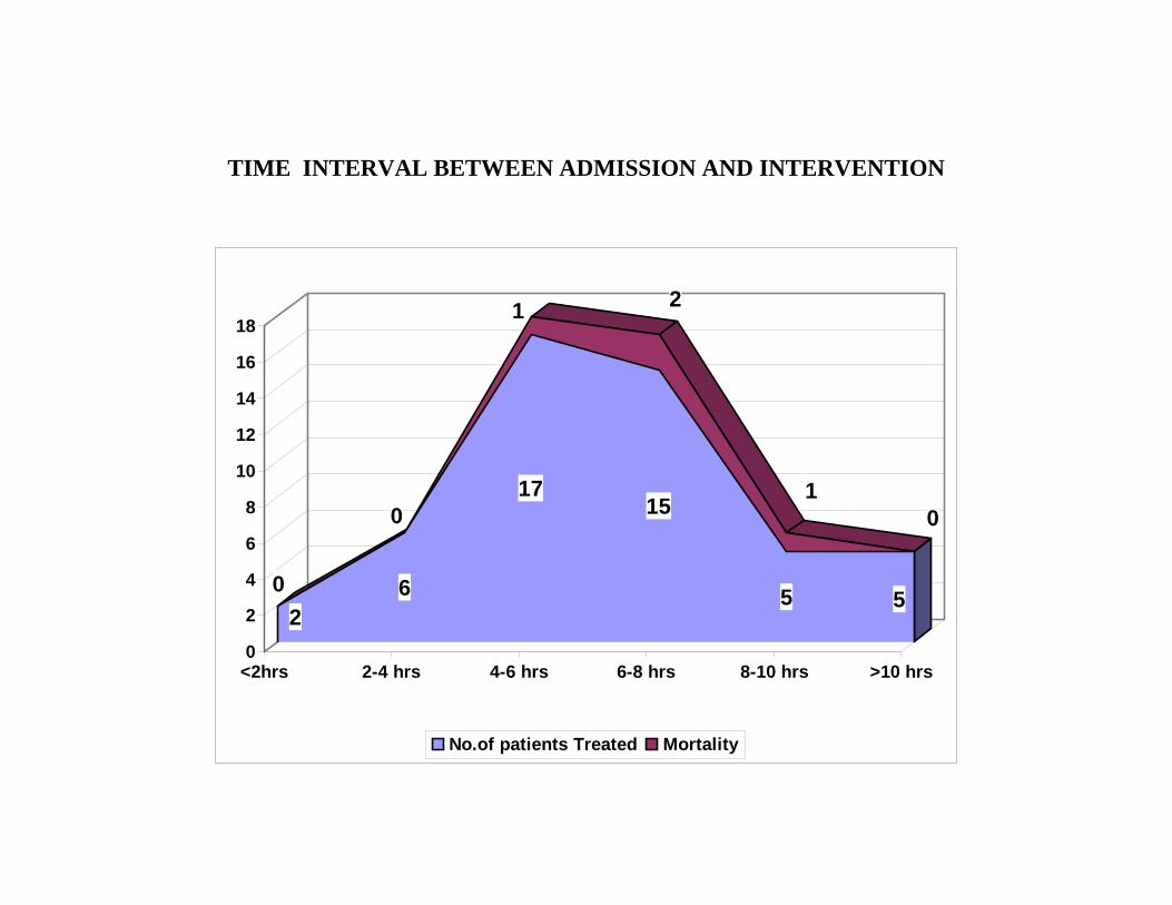
TIME INTERVAL BETWEEN ADMISSION AND INTERVENTION
20 6
017
1
15
2
5
1
5
0
0
2
4
6
8
10
12
14
16
18
<2hrs 2-4 hrs 4-6 hrs 6-8 hrs 8-10 hrs >10 hrs
No.of patients Treated Mortality

PERITONEAL FLUID SEEN IN SMALL BOWEL PERFORATIONS
02468
101214161820
No.
of p
atie
nts
Bile stained clear Bile stainedcloudy
purulent Feculent
Peritoneal fluid Total no. of patients Mortality

Sl. No. Name Age Sex I.P. No.
Time interval between
admissin & intervention
Peritoneal fluid
Site of Perforation Procedure done
Month of admission Outcome
Pre Procedure
(cm)
Post Procedure
(cm)
1 Alagu 45 M 315957 9 hours
Bile stained cloudy 1200ml
Duodenum I 0.5x0.5cm Simple closure 29 16 November Alive
2 Diraviam 55 M 317270 8 hoursPurulent 650ml
Duodenum I 0.5x0.5cm Simple closure 22 12 November Alive
3 Subramani 50 M 317246 6 hoursPurulent 350ml
Duodenum I 1x0.5cm Simple closure 29 14 November Alive
4 Vengaram 28 M 318462 8 hoursPurulent 800ml
Duodenum I 1x0.5cm Simple closure 28 13 December Alive
5 Saradammal 77 F 322905 8 hoursPurulent 350ml - B/L flank drainage 34 18 December Dead
6 Chellamani 18 M 324059 4 hoursBile stained clear 200ml Gastric 1x1cm Simple closure 16 9 December Alive
7 Nagaraj 22 M 325041 2 hoursBile stained
cloudy 200mlDuodenum I
1x0.5cm
B/L flank drainage followed by simple
closure 24 13 December Alive
8 Rajeshkannan 22 M 326114 6 hoursBile stained
cloudy 350mlDuodenum I
1x0.5cm Simple closure 30 14 January Alive
9 Palaniappan 40 M 336212 7 hoursPurulent 1250ml
Duodenum I 1x0.5cm Simple closure 27 13 February Alive
10 Markandeyan 45 M 336358 2 hoursPurulent 500ml - B/L flank drainage 33 18 February Dead
11 Murugan 36 M 337646 6 hoursBile stained
cloudy 150mlDuodenum I 0.5x0.5cm Simple closure 19 10 February Alive
12 Pandi 45 M 337620 10 hoursPurulent 750ml
Duodenum I 1x1cm Simple closure 25 12 February Alive
MASTER CHART
Intraabdominal pressure
pdfMachine - is a pdf writer that produces quality PDF files with ease! Get yours now!
“Thank you very much! I can use Acrobat Distiller or the Acrobat PDFWriter but I consider your product a lot easier to use and much preferable to Adobe's" A.Sarras - USA
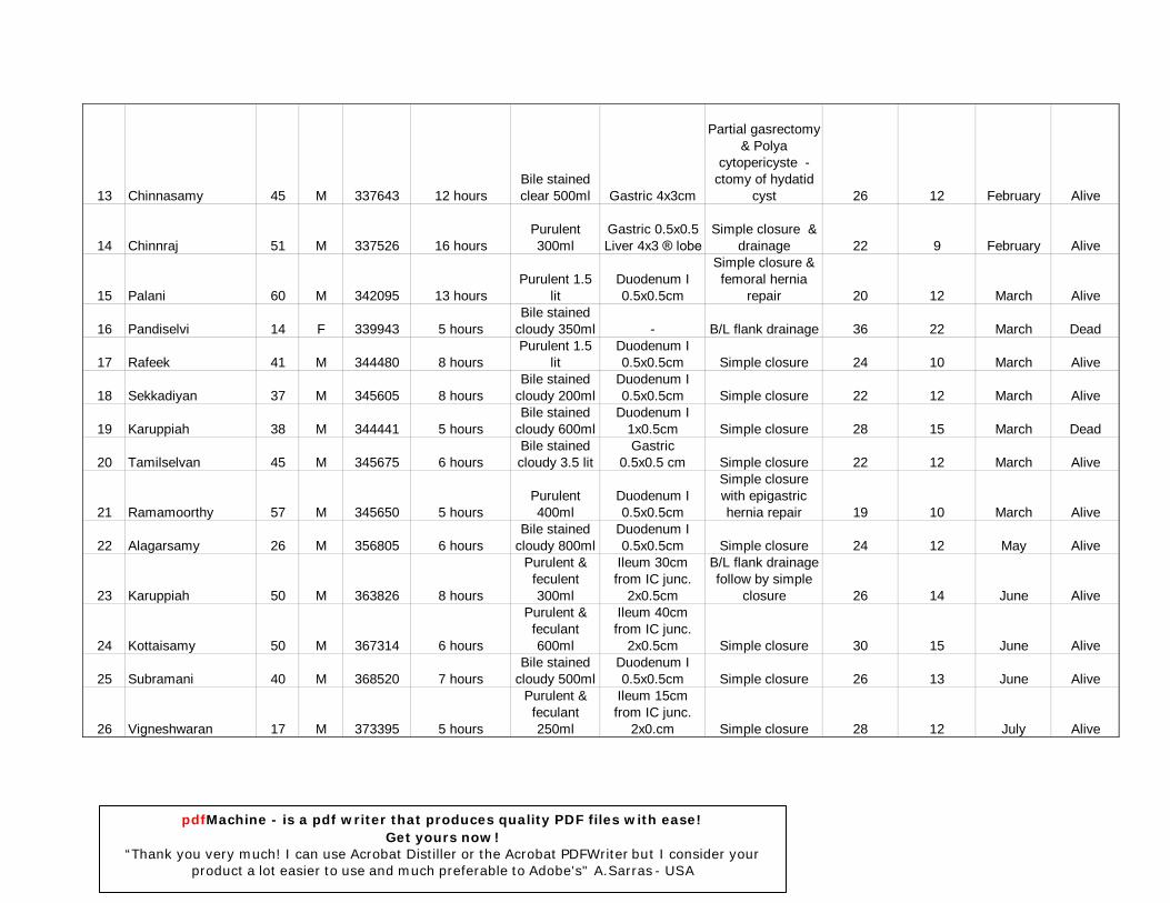
13 Chinnasamy 45 M 337643 12 hoursBile stained clear 500ml Gastric 4x3cm
Partial gasrectomy & Polya
cytopericyste - ctomy of hydatid
cyst 26 12 February Alive
14 Chinnraj 51 M 337526 16 hoursPurulent 300ml
Gastric 0.5x0.5 Liver 4x3 ® lobe
Simple closure & drainage 22 9 February Alive
15 Palani 60 M 342095 13 hoursPurulent 1.5
litDuodenum I 0.5x0.5cm
Simple closure & femoral hernia
repair 20 12 March Alive
16 Pandiselvi 14 F 339943 5 hoursBile stained
cloudy 350ml - B/L flank drainage 36 22 March Dead
17 Rafeek 41 M 344480 8 hoursPurulent 1.5
litDuodenum I 0.5x0.5cm Simple closure 24 10 March Alive
18 Sekkadiyan 37 M 345605 8 hoursBile stained
cloudy 200mlDuodenum I 0.5x0.5cm Simple closure 22 12 March Alive
19 Karuppiah 38 M 344441 5 hoursBile stained
cloudy 600mlDuodenum I
1x0.5cm Simple closure 28 15 March Dead
20 Tamilselvan 45 M 345675 6 hoursBile stained cloudy 3.5 lit
Gastric 0.5x0.5 cm Simple closure 22 12 March Alive
21 Ramamoorthy 57 M 345650 5 hoursPurulent 400ml
Duodenum I 0.5x0.5cm
Simple closure with epigastric hernia repair 19 10 March Alive
22 Alagarsamy 26 M 356805 6 hoursBile stained
cloudy 800mlDuodenum I 0.5x0.5cm Simple closure 24 12 May Alive
23 Karuppiah 50 M 363826 8 hours
Purulent & feculent 300ml
Ileum 30cm from IC junc.
2x0.5cm
B/L flank drainage follow by simple
closure 26 14 June Alive
24 Kottaisamy 50 M 367314 6 hours
Purulent & feculant 600ml
Ileum 40cm from IC junc.
2x0.5cm Simple closure 30 15 June Alive
25 Subramani 40 M 368520 7 hoursBile stained
cloudy 500mlDuodenum I 0.5x0.5cm Simple closure 26 13 June Alive
26 Vigneshwaran 17 M 373395 5 hours
Purulent & feculant 250ml
Ileum 15cm from IC junc.
2x0.cm Simple closure 28 12 July Alive
pdfMachine - is a pdf writer that produces quality PDF files with ease! Get yours now!
“Thank you very much! I can use Acrobat Distiller or the Acrobat PDFWriter but I consider your product a lot easier to use and much preferable to Adobe's" A.Sarras - USA
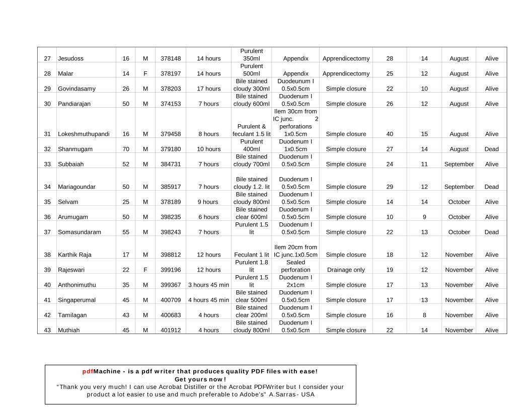
27 Jesudoss 16 M 378148 14 hoursPurulent 350ml Appendix Apprendicectomy 28 14 August Alive
28 Malar 14 F 378197 14 hoursPurulent 500ml Appendix Apprendicectomy 25 12 August Alive
29 Govindasamy 26 M 378203 17 hoursBile stained
cloudy 300mlDuodeunum I
0.5x0.5cm Simple closure 22 10 August Alive
30 Pandiarajan 50 M 374153 7 hoursBile stained
cloudy 600mlDuodenum I 0.5x0.5cm Simple closure 26 12 August Alive
31 Lokeshmuthupandi 16 M 379458 8 hoursPurulent &
feculant 1.5 lit
Ilem 30cm from IC junc. 2
perforations 1x0.5cm Simple closure 40 15 August Alive
32 Shanmugam 70 M 379180 10 hoursPurulent 400ml
Duodenum I 1x0.5cm Simple closure 27 14 August Dead
33 Subbaiah 52 M 384731 7 hoursBile stained
cloudy 700mlDuodenum I 0.5x0.5cm Simple closure 24 11 September Alive
34 Mariagoundar 50 M 385917 7 hoursBile stained
cloudy 1.2. litDuodenum I 0.5x0.5cm Simple closure 29 12 September Dead
35 Selvam 25 M 378189 9 hours Bile stained
cloudy 800mlDuodenum I 0.5x0.5cm Simple closure 14 14 October Alive
36 Arumugam 50 M 398235 6 hoursBile stained clear 600ml
Duodenum I 0.5x0.5cm Simple closure 10 9 October Alive
37 Somasundaram 55 M 398243 7 hoursPurulent 1.5
litDuodenum I 0.5x0.5cm Simple closure 22 13 October Dead
38 Karthik Raja 17 M 398812 12 hours Feculant 1 litIlem 20cm from IC junc.1x0.5cm Simple closure 18 12 November Alive
39 Rajeswari 22 F 399196 12 hoursPurulent 1.8
litSealed
perforation Drainage only 19 12 November Alive
40 Anthonimuthu 35 M 399367 3 hours 45 minPurulent 1.5
litDuodenum I
2x1cm Simple closure 17 13 November Alive
41 Singaperumal 45 M 400709 4 hours 45 minBile stained clear 500ml
Duodenum I 0.5x0.5cm Simple closure 17 13 November Alive
42 Tamilagan 43 M 400683 4 hoursBile stained clear 200ml
Duodenum I 0.5x0.5cm Simple closure 16 8 November Alive
43 Muthiah 45 M 401912 4 hoursBile stained
cloudy 800mlDuodenum I 0.5x0.5cm Simple closure 22 14 November Alive
pdfMachine - is a pdf writer that produces quality PDF files with ease! Get yours now!
“Thank you very much! I can use Acrobat Distiller or the Acrobat PDFWriter but I consider your product a lot easier to use and much preferable to Adobe's" A.Sarras - USA
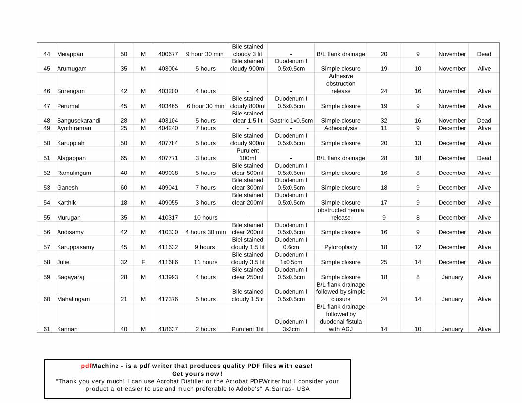
44 Meiappan 50 M 400677 9 hour 30 min Bile stained cloudy 3 lit - B/L flank drainage 20 9 November Dead
45 Arumugam 35 M 403004 5 hoursBile stained
cloudy 900mlDuodenum I 0.5x0.5cm Simple closure 19 10 November Alive
46 Srirengam 42 M 403200 4 hours - -
Adhesive obstruction
release 24 16 November Alive
47 Perumal 45 M 403465 6 hour 30 minBile stained
cloudy 800mlDuodenum I 0.5x0.5cm Simple closure 19 9 November Alive
48 Sangusekarandi 28 M 403104 5 hoursBile stained clear 1.5 lit Gastric 1x0.5cm Simple closure 32 16 November Dead
49 Ayothiraman 25 M 404240 7 hours - - Adhesiolysis 11 9 December Alive
50 Karuppiah 50 M 407784 5 hoursBile stained
cloudy 900mlDuodenum I 0.5x0.5cm Simple closure 20 13 December Alive
51 Alagappan 65 M 407771 3 hoursPurulent 100ml - B/L flank drainage 28 18 December Dead
52 Ramalingam 40 M 409038 5 hoursBile stained clear 500ml
Duodenum I 0.5x0.5cm Simple closure 16 8 December Alive
53 Ganesh 60 M 409041 7 hoursBile stained clear 300ml
Duodenum I 0.5x0.5cm Simple closure 18 9 December Alive
54 Karthik 18 M 409055 3 hoursBile stained clear 200ml
Duodenum I 0.5x0.5cm Simple closure 17 9 December Alive
55 Murugan 35 M 410317 10 hours - -obstructed hernia
release 9 8 December Alive
56 Andisamy 42 M 410330 4 hours 30 minBile stained clear 200ml
Duodenum I 0.5x0.5cm Simple closure 16 9 December Alive
57 Karuppasamy 45 M 411632 9 hours Biel stained cloudy 1.5 lit
Duodenum I 0.6cm Pyloroplasty 18 12 December Alive
58 Julie 32 F 411686 11 hoursBile stained cloudy 3.5 lit
Duodenum I 1x0.5cm Simple closure 25 14 December Alive
59 Sagayaraj 28 M 413993 4 hoursBile stained clear 250ml
Duodenum I 0.5x0.5cm Simple closure 18 8 January Alive
60 Mahalingam 21 M 417376 5 hoursBile stained cloudy 1.5lit
Duodenum I 0.5x0.5cm
B/L flank drainage followed by simple
closure 24 14 January Alive
61 Kannan 40 M 418637 2 hours Purulent 1litDuodenum I
3x2cm
B/L flank drainage followed by
duodenal fistula with AGJ 14 10 January Alive
pdfMachine - is a pdf writer that produces quality PDF files with ease! Get yours now!
“Thank you very much! I can use Acrobat Distiller or the Acrobat PDFWriter but I consider your product a lot easier to use and much preferable to Adobe's" A.Sarras - USA
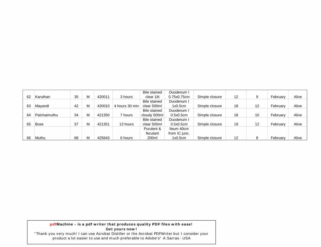
62 Karuthan 35 M 420011 3 hoursBile stained
clear 1litDuodenum I 0.75x0.75cm Simple closure 12 9 February Alive
63 Mayandi 42 M 420010 4 hours 30 minBile stained clear 500ml
Duodenum I 1x0.5cm Simple closure 18 12 February Alive
64 Patchaimuthu 34 M 421350 7 hoursBile stained
cloudy 500mlDuodenum I 0.5x0.5cm Simple closure 18 10 February Alive
65 Bose 37 M 421351 13 hoursBile stained clear 500ml
Duodenum I 0.5x0.5cm Simple closure 19 12 February Alive
66 Muthu 68 M 425643 6 hours
Purulent & feculant 200ml
Ileum 40cm from IC junc.
1x0.5cm Simple closure 12 8 February Alive
pdfMachine - is a pdf writer that produces quality PDF files with ease! Get yours now!
“Thank you very much! I can use Acrobat Distiller or the Acrobat PDFWriter but I consider your product a lot easier to use and much preferable to Adobe's" A.Sarras - USA


