


A Single Nucleotide Polymorphism Alters the Activity of ... · Despite the important role of TSC in...
46
A Single Nucleotide Polymorphism Alters the Activity of the Renal Na + :Cl - Cotransporter and Reveals a Role for Transmembrane Segment 4 in Chloride and Thiazide Affinity by Erika Moreno 1 , Claudia Tovar-Palacio 1 , Paola de los Heros 1 , Blanca Guzmán 2 , Norma A. Bobadilla 1 , Norma Vázquez 1 , Daniela Riccardi 3 , Esteban Poch 2 , & Gerardo Gamba 1 1 Molecular Physiology Unit Instituto de Investigaciones Biomédicas, Universidad Nacional Autónoma de México and Instituto Nacional de Ciencias Médicas y Nutrición Salvador Zubirán Tlalpan 14000, Mexico City, Mexico 2 Servicio de Nefrología, IDIBAPS, Hospital Clínic, 08036 Barcelona, SPAIN 3 School of Biological Sciences. University of Manchester, United Kingdom Short title: Renal Na-Cl cotransporter SNPs Address correspondence to: Gerardo Gamba MD Ph.D. Molecular Physiology Unit Vasco de Quiroga No. 15 JBC Papers in Press. Published on February 5, 2004 as Manuscript M400602200 Copyright 2004 by The American Society for Biochemistry and Molecular Biology, Inc. by guest on September 3, 2018 http://www.jbc.org/ Downloaded from
Transcript of A Single Nucleotide Polymorphism Alters the Activity of ... · Despite the important role of TSC in...

A Single Nucleotide Polymorphism Alters the Activity of the Renal
Na+:Cl- Cotransporter and Reveals a Role for Transmembrane Segment 4 in Chloride and Thiazide Affinity
by
Erika Moreno1, Claudia Tovar-Palacio1, Paola de los Heros1, Blanca Guzmán2,
Norma A. Bobadilla1, Norma Vázquez1, Daniela Riccardi3, Esteban Poch2, & Gerardo Gamba1
1Molecular Physiology Unit
Instituto de Investigaciones Biomédicas,
Universidad Nacional Autónoma de México and
Instituto Nacional de Ciencias Médicas y Nutrición Salvador Zubirán
Tlalpan 14000, Mexico City, Mexico
2 Servicio de Nefrología, IDIBAPS, Hospital Clínic, 08036 Barcelona, SPAIN
3School of Biological Sciences. University of Manchester, United Kingdom
Short title: Renal Na-Cl cotransporter SNPsAddress correspondence to: Gerardo Gamba MD Ph.D. Molecular Physiology UnitVasco de Quiroga No. 15
JBC Papers in Press. Published on February 5, 2004 as Manuscript M400602200
Copyright 2004 by The American Society for Biochemistry and Molecular Biology, Inc.
by guest on September 3, 2018
http://ww
w.jbc.org/
Dow
nloaded from

Tlalpan 14000, México City.Phone.: 5255-5513-3868 Fax: 5255-5655-0382e-mail: [email protected]
2
by guest on September 3, 2018
http://ww
w.jbc.org/
Dow
nloaded from
Abstract
The thiazide-sensitive Na+:Cl- cotransporter is the major salt transport pathway in the
distal convolute tubule of the kidney and a role of this cotransporter in blood pressure
homeostasis has been defined by physiological studies on pressure natriuresis and by its
involvement in monogenic diseases that feature arterial hypotension or hypertension. Database
analysis revealed that 135 single nucleotide polymorphisms along the human SLC12A3 gene that
encodes the Na+:Cl- cotransporter have been informed. Eight are located within the coding
region and one result in a single amino acid change: the residue glycine at the position 264 is
changed to alanine (G264A). This residue is located within the fourth transmembrane domain of
the predicted structure. Because G264 is a highly conserved residue, we studied the functional
properties of this polymorphism using in vitro mutagenesis and the heterologous expression
system in Xenopus laevis oocytes. G264A resulted in a significant and reproducible reduction
(~50%) in 22Na+ uptake when compared to the wild type cotransporter. The affinity for
extracellular Cl- and for thiazide diuretics was increased in G264A. Western blot analysis
showed similar immunoreactive bands between the wild type and the G264A cotransporters and
confocal images of oocytes injected with EGFP-tagged wild-type and G264A cotransporter
showed no differences in the protein surface expression level. These observations suggest that the
G264A polymorphism is associated with reduction in the substrate translocation rate of the
3
by guest on September 3, 2018
http://ww
w.jbc.org/
Dow
nloaded from

cotransporter, due to a decrease in the intrinsic activity. Our study also reveals a role of the
transmembrane segment 4 in defining the affinity for extracellular Cl- and thiazides diuretics.
Key words: thiazide, Na-Cl cotransporter, hypertension, single nucleotide polymorphism,
diuretics, structure, osteoporosis.
4
by guest on September 3, 2018
http://ww
w.jbc.org/
Dow
nloaded from

Introduction
The thiazide-sensitive Na+:Cl- cotransporter (TSC, gene symbol: SLC12A3. Locus ID 6559)
is the major NaCl transport pathway in the apical membrane of the mammalian distal convoluted
tubule (DCT) and the teleost urinary bladder (1)(2)(3-7). The fundamental role of the Na+:Cl-
cotransporter encoded by the SLC12A3 gene in preserving the extracellular fluid volume and
divalent cation homeostasis has been firmly established by the identification of inactivating
mutations of this gene as the cause of Gitelman’s disease (8-10), an inherited disorder featuring
arterial hypotension, hypokalemic metabolic alkalosis with hypocalciuria, hypomagnesemia and
renal salt wasting. TSC also serves as the target for the thiazide-type diuretics that are currently
recommended as the drug of choice for treatment of hypertension (11). Finally, a defect in TSC
regulation by the WNK1 and WNK4 kinases has been implicated in the pathogenesis of a salt-
dependent form of human hypertension known as pseudohyopaldosteronism type II (PHAII)
(12)(13), that features marked sensitivity to hydrochlorothiazide and a clinical picture that is a
mirror image of Gitelman’s disease (hypertension, hyperkalemia, and metabolic acidosis) (14).
Taken together, all these observations suggest that TSC molecular variants, resulting from single
nucleotide polymorphisms (SNPs), could contribute to the normal variations in blood pressure in
the population at large, to the inherited predisposition towards essential hypertension, and/or to
the differential response to diuretic therapy.
Despite the important role of TSC in cardiovascular physiology, pharmacology, and
5
by guest on September 3, 2018
http://ww
w.jbc.org/
Dow
nloaded from
pathophysiology, little is currently known about the structure-function relationships in this
cotransporter. Using [3H]metolazone binding to membrane preparations from rat renal cortex,
Tran et al. (15) proposed that thiazides and Cl- share the same binding site. Recent studies in
which the functional properties of the cloned cotransporter were determined, however, provided
evidence that metolazone competes with both Na+ and Cl- ions (16), suggesting that thiazide
binding site maybe shared by both ions and not only by Cl-, as suggested by Tran et al. (15). In
addition, nothing is known regarding domains or amino acid residues defining the TSC ion
transport kinetics or thiazide affinity. So far, within the family of electroneutral cotransporters,
some aspects of the structure-function relationships have been investigated only in the two
isoforms of the Na+:K+:2Cl- cotransporter, BSC1/NKCC2 and BSC2/NKCC1. Results between
both isoforms, however, have shown important differences suggesting that conclusions reached in
one member of the family cannot be extended to the other members. For example, in
BSC2/NKCC1, Isenring et al. (17)(18) have implicated transmembrane domains 4 and 7 in
defining Cl- transport affinity, while recent studies in BSC1/NKCC2 clearly showed that
transmembrane domain 2 contains affinity modifier residues for extracellular Cl- (19)(20)(21).
In the present study we show that a SNP that changes one amino acid residue in TSC
results in a dramatic decrease in TSC function, apparently secondary to a decrease in the intrinsic
activity of the cotransporter and reveals a role of transmembrane segment 4 in TSC affinity for
6
by guest on September 3, 2018
http://ww
w.jbc.org/
Dow
nloaded from

extracellular Cl- and for thiazides diuretics.
7
by guest on September 3, 2018
http://ww
w.jbc.org/
Dow
nloaded from

Methods
An extensive search of genome databases (lpgws.nci.nih.gov/cgi-bin/GeneViewer.cgi;
ncbi.nlm.nih.gov/SNP) was performed to find the SNPs that have been informed within the
SLC12A3 gene. The SNPs within the coding regions that were considered as potentially
important were incorporated into the rat TSC cDNA by using the Quickchange site directed
mutagenesis system (Stratagene) following the manufacturer’s recommendations. Automatic
DNA sequencing was used to corroborate all the mutations. All primers used for mutagenesis
were custom made (Sigma).
Genotyping of the G264A polymorphism. A restriction fragment length polymorphism (RFLP)
method was created for the G264A polymorphism to confirm it and to simplify its detection in
200 normal subjects. Total genomic DNA was extracted from whole blood according to standard
procedures. PCR was conducted using 125 ng of genomic DNA using the primer pair sense 5´-
AGACCGTGCGGGACCTGCTC-3´ and antisense 5´- CCTCCTCCATGGCCTCCTCACCTT-
3´. PCR was conducted for 34 cycles with denaturation at 96°C for 30 sec, annealing at 60°C for
30 sec and extension at 72°C for 30 sec, with a final extension step at 72°C for 5 min. The
G264A variant is recognized by RFLP by using Btg1 (New England Biolabs), the restriction
fragments separated on 7.5% polyacrylamide gel electrophoresis and visualized under ultraviolet
light after staining with ethidium bromide. The polymorphism was confirmed by automatic
sequencing (AbiPrism) in all positive cases.
8
by guest on September 3, 2018
http://ww
w.jbc.org/
Dow
nloaded from

Assessment of the Na+:Cl - cotransporter function. Oocytes were harvested from anesthetized
adult female Xenopus laevis frogs, defolliculated, and prepared for microinjection following our
standard procedure (16)(19). The next day mature oocytes were injected with 50 nl of water or
cRNA transcribed in vitro, using the T7 RNA polymerase mMESSAGE kit (Ambion), at a
concentration of 0.5 µg/µl. Oocytes were then incubated 3 days in ND96 with sodium pyruvate
and gentamicin and one day in Cl--free ND96 (16). The function of the Na+:Cl- cotransporter
was determined by assessing tracer 22Na+ uptake (New England Nuclear) in groups of at least 15
oocytes following our standard protocol (16): 30 min incubation in a Cl--free ND96 medium
containing 1 mM ouabain, 0.1 mM amiloride, and 0.1 mM bumetanide, followed by a 60-min
uptake period in a K+-free, NaCl medium containing the same drugs plus 2 µCi of 22Na+ per
ml. Gluconate was used as a Cl- substitute and N-Methyl-D-Glucamine (NMDG) as a Na+
substitute. At the end of the uptake period tracer activity was determined for each dissolved
oocyte by β-scintillation counting.
Western Blotting. Western blot analysis was used to compare WT with mutant protein in cRNA-
injected oocytes following our standard protocol (22). In brief, groups of 15 oocytes injected with
water or cRNA were homogenized in 2 µl/oocyte of homogenization buffer, centrifuged twice at
100 x g for 10 min at 4°C, and the supernatant was collected. Protein extracts from oocytes (four
oocytes per lane) were heated in sample buffer containing 6% SDS, 15% glycerol, 0.3%
bromophenol blue, 150 mM Tris pH 7.6 and 2% ²-mercaptoethanol, resolved by Laemmli SDS-
9
by guest on September 3, 2018
http://ww
w.jbc.org/
Dow
nloaded from
polyacrylamide (7.5%) gel electrophoresis, and transferred to a polyvinylidene difluoride (PVDF)
membrane. For immunodetection we used a rabbit polyclonal anti rat TSC antibody (generously
provided by Dr. Mark Knepper, NIH), diluted 1:1000 (23). The membrane was exposed to anti-
TSC antibody diluted in blocking buffer (TTBS, 0.2% tween-20) overnight at 4°C, subsequently
washed in TTBS and incubated for 60 min at room temperature with alkaline phosphatase-
conjugated secondary (anti-rabbit) antibody (BIO-RAD) diluted 1:2000 in blocking buffer and
washed again. Immunoreactive species were detected using Immun-Star Chemiluminescent
Protein Detection Systems (BIO-RAD).
Assessment of the TSC expression at the oocytes plasma membrane. The surface expression of
wild type or mutant TSC (see below) was determined by assessing the fluorescence in the
Xenopus oocytes using a TSC fusion construct that we have previously validated (24)(12). In this
construct, the enhanced green fluorescent protein (EGFP) was fused to the amino terminal
domain of TSC. Then, Xenopus oocytes were microinjected with water as control or with EGFP-
WT-TSC or EGFP-mutant-TSC cRNA. After four days of incubation, oocytes were monitored
for EGFP fluorescence in the oocytes surface using a Zeiss laser scanning confocal microscope
(objective lens x10, Nikon). Excitation and emission wavelengths used to visualize EGFP
fluorescence were 488 nm 515-565 nm, respectively. We have shown previously that EGFP-
TSC fluorescence in the oocytes surface co-localizes with the F-404 specific plasma membrane
dye and that oocytes injected with EGFP-TSC exhibit significant thiazide-sensitive 22Na+
10
by guest on September 3, 2018
http://ww
w.jbc.org/
Dow
nloaded from
uptake, indicating the EGFP-TSC fluorescence is located in the plasma membrane (24). For
densitometry analysis, the plasma membrane fluorescence was quantified by determining the
pixel intensity around the entire oocytes circumference using SigmaScan Pro image analysis
software.
Statistical analysis. Statistical significance is defined as two-tailed p<0.05 and the results are
presented as mean ± SEM. The significance of the differences between means was tested with the
Student’s t-test.
11
by guest on September 3, 2018
http://ww
w.jbc.org/
Dow
nloaded from

Results
Single nucleotide polymorphisms in the SLC12A3 gene. Up to 135 SNPs have been informed
within the SLC12A3 gene. 127 SNPs are located within intronic and only eight are within exonic
sequences. Figure 1 depicts the proposed TSC topology (25) and the localization of the eight
SNPs within the coding sequence. Six SNPs result in no change of the amino acid sequence.
These are the SNPs A122A, T465T, S628S, A714A, G875G, and I1017I, corresponding to the
NCBI SNP cluster IDs rs2304479, rs5801, rs55802, rs5803, rs5804, and rs2289113, respectively.
Two SNPs result in a single amino acid change. One is the R863K SNP (cluster ID rs8060046)
that was considered as irrelevant because this SNP located within the carboxyl terminal domain
results in a conserved substitution of the positively charged amino acid arginine, which is present
in the human cotransporter (8), for the positively charged residue lysine, which is present in the
TSC from rabbit, rat, mouse, and fish (26)(25)(27)(2). In contrast, the other SNP that alter the
primary sequence of human TSC (ID number rs1529927) predicts a change of the nonpolar
amino acid glycine at the position 264 for the residue alanine. The residue glycine is located
within the fourth transmembrane domain and is a conserved amino acid residue, not only in the
available TSC sequences from rat (25), mouse (27), rabbit (26), human (8), and flounder (2), but
also in all members of SLC12 family that include two genes encoding Na+:K+:2Cl-
cotransporters (28)(25) and four genes encoding K+:Cl- cotransporters (29)(30)(31). Thus, the
G264A SNP was considered as potentially important and therefore was introduced into TSC by
12
by guest on September 3, 2018
http://ww
w.jbc.org/
Dow
nloaded from

in vitro mutagenesis.
Allele frequency of the G264A polymorphism. A RFLP strategy was used to assess the presence
of G264A SNP by PCR. The PCR product (510 bp) contained a constant Btg1 restriction site and
therefore, when digested, two bands of 461 bp and 49 bp, respectively, are observed in GG
genotype (encoding glycine at position 264 in both alleles). In contrast, in GA genotype
heterozygotes (that is, one allele encoding glycine and the other encoding alanine at position
264), a new specific Btg1 restriction site is used and then four bands of 461 bp, 390 bp, 71 bp and
49 bp are observed. To test the allele frequency of the G264A polymorphism, 200 Caucasian
subjects were genotyped. The sample included 119 males and 81 females with the following
characteristics (mean ± SD): age 52 ± 16 years, systolic blood pressure 117 ± 11 mmHg, diastolic
blood pressure 69 ± 7 mmHg, and body mass index of 24 ± 4 kg/m2. None of the subjects had
present or past cardiovascular conditions including hypertension, coronary heart disease, stroke
or diabetes. The frequency of the GA genotype was 2% in the sample studied. No subjects with
AA phenotype were detected (that is, homozygotes encoding alanine at position 264 in both
alleles). Shown in Figure 2A is a representative gel containing the Btg 1 digested PCR fragment
from a normal subject and one heterozygous for the GA genotype. Fig. 2B and C illustrate
sequencing of this region in a GG and a GA phenotype (codons GGC and GCC, respectively).
Thus, our results suggest that G264A is a true SNP.
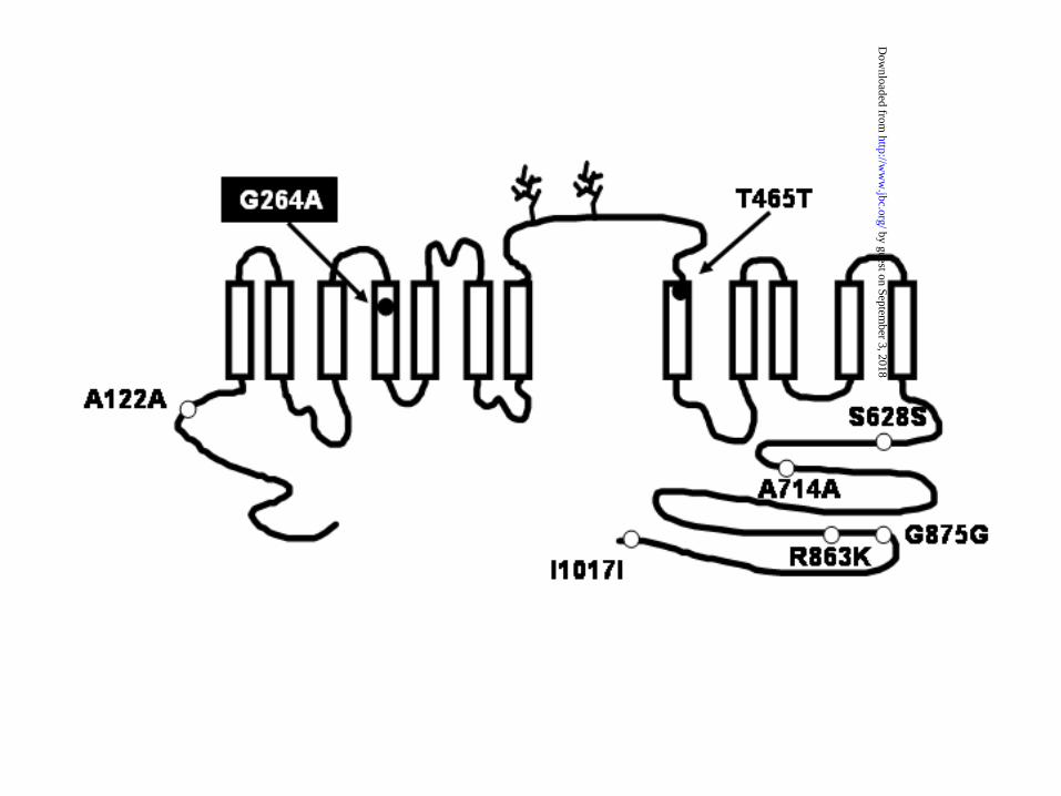
Functional consequences of the TSC G264A polymorphism. The functional consequences of the
13
by guest on September 3, 2018
http://ww
w.jbc.org/
Dow
nloaded from
G264A SNP were assessed using a heterologous expression system in X. laevis oocytes. This
expression system has shown to be an excellent tool for a robust and reproducible expression of
TSC in our hands (2)(25)(16)(32)(24)(12) and also in other laboratories (27)(13)(33)(34). In
contrast, TSC expression in transfected eukaryotic cells has not been successful in many
laboratories, including our own. The best expression so far observed in stably transfected
eukaryotic cells (MDCK cells) with human TSC cDNA consist of small increase (~25%) over
background (35). Thus, X. laevis oocytes were microinjected with cRNA transcribed from wild-
type TSC (WT) or from TSC harboring the G264A SNP (G264A). As shown in figure 3, WT or
G264A cRNA injection induced a significant increase in 22Na+ uptake in X. laevis oocytes.
However, the increase in G264A-injected oocytes was of a significantly lower magnitude that
the increase observed in WT oocytes. Uptake in WT-injected oocytes was 3448 ± 234
pmol–oocyte-1–h-1, while in G264A-injected oocytes was 1712 ± 366 pmol–oocyte-1–h-1 (p<0.01,
n=20). As shown in Figure 3, the uptake was due to the TSC activity since a complete inhibition
of uptake was observed in the absence of extracellular chloride or in the presence of the thiazide-
type diuretic, metolazone. A reduction of a similar magnitude in the G264A activity was
consistently observed in every single experiment. In order to assure that the reduction in 22Na+
uptake was not due to differences in the amount of cRNA injected or in the transcription rate of
the protein, cRNA concentration was determined by densitometry of the corresponding bands in
the ethidium bromide stained agarose gel and the transcribed protein was assessed by Western
14
by guest on September 3, 2018
http://ww
w.jbc.org/
Dow
nloaded from

blot analysis of the injected oocytes. No differences were found between WT and G264A in the
amount of cRNA injected (Fig. 3B), as well as in the TSC protein produced by the oocytes (Fig.
3C).
Surface expression analysis in EGFP-WT or EGFP-G264A. Because X. laevis oocytes injected
with WT or G264A exhibited similar immunoreactive proteins in the Western blot, we reasoned
that potential explanations for the reduction in functional expression of the TSC containing the
G264A could be a decrease in the amount of the transporter that reaches the plasma membrane, a
decrease in the affinity for the cotransported ions, or a decrease in the intrinsic activity of the
cotransporter. To study the first possibility, we assessed the surface expression of the WT and
G264A proteins by injecting X. laevis oocytes with the cRNA encoding the WT or G264A
cotransporters that has been previously tagged with the EGFP. To perform these experiments, we
used the EGFP-tagged TSC construct that we have previously characterized (24) in which the
EGFP was fused to the amino terminal domain of TSC. Then, the EGFP cDNA was subcloned
into the G264A TSC. The cRNA encoding the EGFP-WT or EGFP-G264A was injected into X.
laevis oocytes. As shown in Fig. 4A, densitometry of the corresponding cRNA bands in an
ethidum bromide agarose gel assured that similar amount of cRNA was injected. Four days after
injection, the surface expression was assessed by monitoring the EGFP fluorescence with a
confocal microscope. After oocytes were analyzed in the microscope, half of them were used for
protein extraction to assess the EGFP-tagged proteins by Western blot, using a rabbit polyclonal
antibody against TSC, and the other half were use in a functional expression assay to determine
15
by guest on September 3, 2018
http://ww
w.jbc.org/
Dow
nloaded from
the thiazide-sensitive 22Na+ uptake. Western blot revealed similar immunoreactive bands in
proteins extracted from EGFP-WT or EGFP-G264A injected oocytes (Fig. 4B) and surface
expression analysis revealed similar fluorescence intensity at the surface of oocytes injected with
EGFP-WT or EGFP-G264A, respectively (representative images are shown in Figures 4C and
4D). Figure 4E and 4F illustrate the plasma membrane fluorescence intensity analysis in 30
EGFP-WT or 30 EGFP-G264A-injected oocytes and the functional activity expressed as
22Na+ uptake in the same oocytes, respectively. While the surface expression of both clones was
comparable, the 22Na+ uptake was reduced in G264A-injected oocytes. Thus, the G264A
substitution results in a reduction in the cotransporter activity, which does not appear to be due to
a decrease in the surface expression rate.
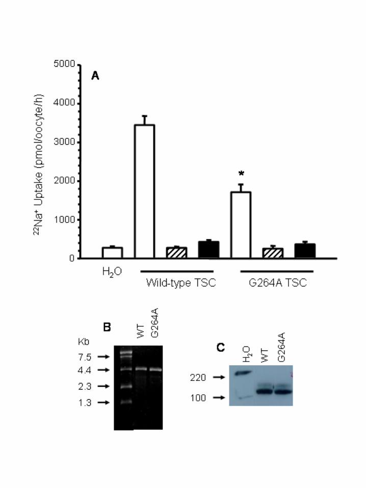
Ion transport kinetics. One potential source of reduction in the functional activity of a membrane
transporter is a reduction of the affinity for the transported ions or molecules, preventing that the
cotransporter reaches maximal transport capacity when incubated in regular uptake solutions.
Thus, we assessed the Na+ and Cl- transport kinetics in X. laevis oocytes injected with WT or
G264A cRNA. Shown in Fig. 5A and 5C are the Na+ transport kinetics analysis in WT and
G264A, respectively. The Km values for Na+ transport kinetics were 7.6 ± 1.6 and 5.7 ± 1.1 mM
in WT and G264A, respectively, with no significant difference between them. Shown in Fig. 5B
and 5D are the Cl- transport kinetic analysis. The apparent Km value for extracellular Cl-
16
by guest on September 3, 2018
http://ww
w.jbc.org/
Dow
nloaded from
uptake in WT (Fig. 5B) was 6.3 ± 1.1 mM. This value is similar to the previously reported for
the wild-type TSC (16). In contrast, the apparent Km in G264A-injected oocytes was 0.89 ± 0.2
mM (p<0.001, N=3). We repeated the same analysis in triplicate using three different batches of
oocytes and the results were similar. Thus, the G264A resulted in a significant increase in Cl-
affinity on the cotransporter. This increase in ion transport affinity, however, does not explain the
reduction in the cotransporter activity.
Diuretic inhibitory kinetics. Studies in which the kinetic analysis of [3H]metolazone to renal
cortex plasma membranes were assessed suggested that chloride and thiazides may compete for
the same site in the cotransporter (15). Supporting this possibility we have shown that thiazide
affinity in TSC is increased when oocytes are incubated in uptake solutions containing low Cl-
concentration (16). Because an increase in Cl- affinity in the G264A cotransporter was observed,
we assessed the dose-response simultaneously on TSC and G264A for the thiazide-type diuretic
metolazone in order to determine the thiazide affinity on each cotransporter. As shown in figure
6, when uptakes where performed in 96 mM NaCl (closed symbols and continuous lines), the
IC50 for metolazone-induced reduction in 22Na+ uptake was similar between WT and G264A. The
IC50 value in both was ~ 1 x 10⁻⁶ M, which is similar to the IC50 that we have previous
reported for rat TSC (16). However, because the Km for extracellular Cl- in WT TSC is below
10 mM and in G264A below 1 mM, it is possible that competition between chloride and
17
by guest on September 3, 2018
http://ww
w.jbc.org/
Dow
nloaded from

thiazides does not become apparent using 96 mM of extracellular Cl-. Thus, we performed the
metolazone dose-response inhibitory curve in the same experiment, but using another groups of
oocytes in which uptake was done using an extracellular Cl- concentration around the Km
values for this ion, that is, ~ 6 mM for TSC and 1 mM for G264A (open symbols and
discontinued lines). As we have shown before (16), the metolazone IC50 in the oocytes injected
with WT-TSC changed from ~1 x 10⁻⁶ M to ~3 x 10⁻⁷ M, suggesting that metolazone binding
to the cotransporter is enhanced when extracellular chloride is lower. In oocytes injected with
G264A cRNA, the shift to the left was more dramatic than in WT-TSC since metolazone IC50
changed from ~8 x 10⁻⁷ M in the presence of 96 mM Cl, to ~1 x 10⁻⁸ M when extracellular Cl-
concentration was 1 mM. To test the possibility that the G264A SNP not only affects the affinity
for thiazides, but also the diuretic inhibitory profile, we also assessed the dose-response to
several thiazides. This experiment was performed simultaneously in WT and G264A-injected
oocytes. Therefore, each of the thiazide dilutions that were used was the same for both WT and
G264A oocytes. As shown in Figure 7, the thiazide inhibitory profile of polythiazide >
bendroflumethiazide = trichloromethiazide = benzthiazide > hydrochlorothiazide = chlortalidone
was similar between WT and G264A TSC cotransporters, suggesting that the G264A substitution
increases the affinity of the cotransporter for thiazides, but does not affect the inhibitory profile.
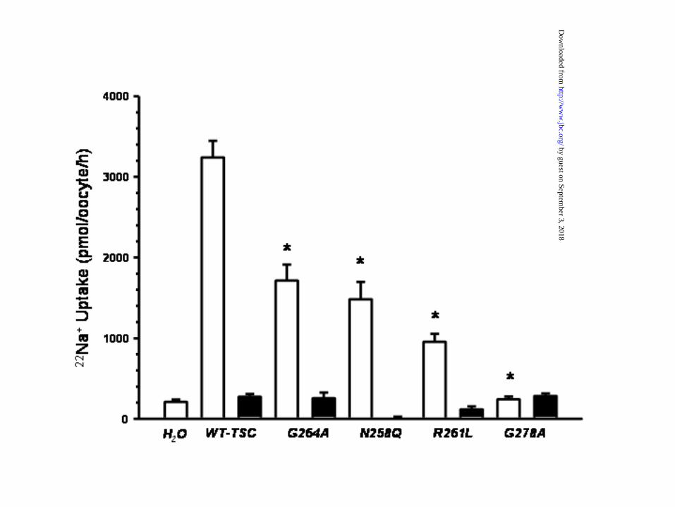
Role of the conserved residues in the transmembrane segment 4. As shown in Figure 8, the
alignment analysis of the transmembrane domain 4 in the electroneutral cotransporter family
18
by guest on September 3, 2018
http://ww
w.jbc.org/
Dow
nloaded from
members revealed that G264 is not the only residue that is conserved in all cotransporters. In
addition to G264, the residues N258, R261, and G278 are also conserved in all members of the
family. Thus we performed similar substitutions of these residues and analyzed the effects upon
the functional properties of the cotransporter. As shown in figure 9, substitution of these residues
resulted in different magnitudes of reduction in the TSC activity. The effect was similar between
G264A and N258Q (~50% reduction). Further reduction in the activity was observed in the
R261L transporter, while the G278A substitution resulted in a complete block of the
cotransporter activity. To find out if the decrease in activity was associated or not with a decrease
in the surface expression of the cotransporters, X. laevis oocytes were injected with similar
amounts of cRNA transcribed from the EGFP-tagged wild type TSC or the EGFP-tagged TSC
containing the substitutions and confocal microscopy analysis of the oocytes was performed four
days later for assessing the fluorescence intensity at the surface. Interestingly, as shown in Figure
10, there were no significant differences among all groups. Thus, mutations in TSC on any of the
conserved residues in the transmembrane segment 4 produce cotransporters in which the intrinsic
activity is reduced. Finally, the ion transport kinetics analysis revealed that in TSC harbouring
the N258Q and R261L substitutions, the Km values for extracellular Na+ were 7.3 ± 0.7 and 8.9
± 2.3 mM, respectively, and for extracellular Cl- were 3.2 ± 0.7 (p<0.05, N=4) and 3.2 ± 1.6
mM (p=NS, N=4), respectively. Thus, no change was observed in the affinity for extracellular
Na+, while a slight increase was observed in the affinity for Cl-.
19
by guest on September 3, 2018
http://ww
w.jbc.org/
Dow
nloaded from

Discussion
One hundred and thirty five SNPs have been informed along the SLC12A3 gene that
encodes the thiazide-sensitive Na+:Cl- cotransporter of the renal distal convoluted tubule. Only
eight of the 135 SNPs are located within exonic sequences and one of them, the SNP G264A,
results in a significant change of a single amino acid residue. We showed that in our population
the distribution of this SNP is 98% homozygous for G264G and 2% for heterozygous G264A.
This frequency is different to the previously shown by Melander et al. (36) in Sweden to be ~91%
for homozygous G264G, ~8% for heterozygous G264A, and ~1% for homozygous A264A.
When expressed in the heterologous system of X. laevis oocytes, G264A exhibited a reduced
maximal transport capacity to about 50% of that shown in simultaneous experiments with WT-
TSC. As shown by Western blot analysis, the lower activity of the TSC harboring the G264A
substitution does not appear to be due to reduced translation of the protein since densitometric
analysis demonstrated no differences in the amount of TSC protein produced with or without the
G264A substitution. The surface image analysis that was assessed by confocal microscopy in X.
laevis oocytes injected with EGFP tagged WT or G264A cRNA revealed that reduced
translocation of the cotransporter to the cell surface is not responsible for the lower activity in
G264A, since surface expression in the plasma membrane was comparable between EGFP-WT
and EGFP-G264A, while the 22Na+ uptake experiments performed with the EGFP-WT or
EGFP-G264A injected oocytes revealed a significant reduction in TSC activity in the presence of
20
by guest on September 3, 2018
http://ww
w.jbc.org/
Dow
nloaded from

the G264A substitution. Thus, taking all these data together; we propose that the lower activity in
TSC harboring the G264A substitution is due to a reduced ions translocation rate. That is, due to
a decrease in the intrinsic activity of the cotransporter. A similar negative effect on the intrinsic
activity or on the receptor signaling capacity has been documented for the SNP I89V and the
SNP S268P that occur in the human high affinity choline transporter (37) and in the human ¼-
opiod receptor (38), respectively.
The observation in the present study of one SNP in the renal Na+:Cl- cotransporter that
results in reduction of the cotransporter intrinsic activity suggests a number of testable
hypothesis. For example, this SNP may be probably less prevalent in hypertensive patients than
in normal subjects or individuals harboring this SNP probably are less sensitive to treatment with
thiazide drugs. In addition, it is well known that TSC activity inversely correlates with calcium
reabsorption in the distal tubule (39) and that chronic thiazide treatment is associated with
increased bone density (40) which is a protective factor against osteoporosis (41)(42). Therefore,
another hypothesis could be that SNP G264A is less prevalent in patients with osteoporosis than
in general population.
One study addressed the genotype frequency distribution on this G264A SNP in 264
normal and 292 hypertensive subjects from Sweden (36) and no difference was observed between
normotensive and hypertensive subjects. It is possible, however, that more selected hypertensive
patients will be necessary to be studied in order to reveal the association between hypertension
21
by guest on September 3, 2018
http://ww
w.jbc.org/
Dow
nloaded from

and certain SNPs. For example, Baker et al. (43) have shown that the T594M mutation in the β-
subunit of the epithelial sodium channel correlates with the development of hypertension only in
black patients with low renin hypertension and Zhu et al. (44) observed that in white patients, but
not in black hypertensive subjects, there is a significant association between the intron 2
conversion allele of the aldosterone synthase gene and the development of essential hypertension.
Regardless of the potential role in the disease discussed above, the G264A SNP reveals a
role of the transmembrane domain 4 in the affinity for extracellular Cl- and also for thiazides.
We observed that TSC harboring the G264A exhibits an increase in the affinity for extracellular
Cl-, since the apparent Km value for extracellular Cl- in G264A was almost 10 times lower than
in WT. This increase in affinity for Cl- was specific for this ion since no change was observed in
the affinity for extracellular Na+. Glycine is a non-hydrophobic residue at the position 264,
which is predicted to be located in the TSC putative transmembrane domain 4 and is conserved in
all species in which TSC has been sequenced, including human (8), rat (25), mouse (27), rabbit
(26), and flounder (2), as well as, among all members of the SLC12 family including TSC, two
Na+:K+:2Cl- and four K+:Cl- cotransporter isoforms. Previous studies in the basolateral
isoform of the Na+:K+:2Cl- cotransporter (45)(18) indicated that transmembrane helix 4
contains affinity-modifying residues for Cl- translocation. In addition to G264, there are three
other residues within the transmembrane domain 4 that are conserved among all members of the
22
by guest on September 3, 2018
http://ww
w.jbc.org/
Dow
nloaded from

SLC12 family (Fig. 8). We observed that substitution of each of these residues resulted in
Na+:Cl- cotransporters with reduced intrinsic activity, and one of them also exhibited increased affinity
for extracellular Cl-. Thus, as has been suggested by Isenring et al. in the Na+:K+:2Cl-
cotransporter BSC2/NKCC1, it is possible that transmembrane segment 4 in TSC also play a role
in defining the affinity for extracellular chloride. The G278A substitution resulted in a non-
functional protein, without affecting the surface expression rate, suggesting that this glycine is
completely necessary for the cotransporter to reach is functional conformation state.
The structural mechanisms by which glycine substitution in the TSC fourth
transmembrane domain produces the observed changes in its functional properties are not clear,
but some hypothesis can be proposed. Glycine is an amino acid residue that plays an important
structural role, because this residue allows unusual main chain conformations in proteins. This is
probably why glycine is one of the amino acid residues that show high proportion of conservation
among homologous protein sequences (46). Although not yet studied in cotransporters, it has
been shown in several enzymes that some glycine residues can be important to define the protein
flexibility because this residue can be part of a hinge (47)(48)(49)(50) and the extent of rigidity
or flexibility in a given hinge has been proposed to play a role in the affinity of the protein for its
ligand (51). Thus, one possibility is that the presence of glycine at position 264 provides certain
flexibility that in TSC might affect both, rates of transport and anion binding. Alternatively,
because the four residues that we studied, conserved among all members of the SLC12 family,
23
by guest on September 3, 2018
http://ww
w.jbc.org/
Dow
nloaded from
are hydrophilic (two glycines, one asparagine and one arginine), it is possible that these amino
acids mainly face the putative translocation pocket in the cotransporter and the conformational
change that results when these residues were substituted could render the putative translocation
pocket more accessible to Cl- ions, but with reduced rates of transport. Further studies will be
necessary to clarify these issues
Previous studies using the binding of tracer [3H]metolazone suggested that Cl- ions and
thiazide diuretics compete for the same site on the cotransporter (15). Supporting this hypothesis,
we have shown that in TSC the higher affinity for chloride is accompanied by a higher affinity
for thiazide diuretics. On one hand, the affinity for thiazides is shifted to the left when dose-
response curves are performed in low extracellular Cl- concentration (16)(32) and on the other
hand, the prevention of glycosylation in rat TSC increases the affinity for both extracellular Cl-
and thiazides (24). In the present study, we observed that G264A substitution produce a dramatic
increase in Cl- affinity, together with an increase in the affinity for the thiazide-type diuretic
metolazone. This observation also supports the hypothesis that in TSC, the affinity-modifying
residues for Cl- may also be involved in defining thiazide affinity, increasing the data supporting
that anions and diuretics compete for the same site on the cotransporter. Interestingly, while a
similar type of competence between Cl- and loop diuretics was proposed for the Na+:K+:2Cl-
cotransporter based on studies using [3H]Bumetanide (52), the functional analysis of chimeras
24
by guest on September 3, 2018
http://ww
w.jbc.org/
Dow
nloaded from

between the shark and human basolateral BSC2/NKCC1 revealed that changes in Cl- transport
kinetics are not accompanied by similar changes in bumetanide affinity (45).
In summary, we report here the functional characterization of a single nucleotide
polymorphism in the SLC12A3 gene that encodes the thiazide-sensitive Na+:Cl- cotransporter.
This membrane protein has been implicated in human diseases such as arterial hypertension and
osteoporosis and the pharmacological modulations of its function are currently used for treating
or preventing these disorders. The studied SNP is a substitution of a glycine for alanine in the
fourth transmembrane domain, which caused a significant reduction in the Na+:Cl- transport
rate, suggesting that people with this SNP could have an allele with reduced function of the
cotransporter. In addition to the effect of the SNP upon the TSC activity, the G264A substitution
produced an increase in the affinity of the cotransporter for extracellular Cl-, but nor for Na+,
that was accompanied by an increase in the affinity for thiazide diuretics. Thus, our study
represents the first detailed examination of genetic polymorphism in the SLC12A3 gene and
reveals a role of the TSC transmembrane segment 4 in anion and thiazide affinity.
25
by guest on September 3, 2018
http://ww
w.jbc.org/
Dow
nloaded from
ACKNOWLEDGMENTS
We are grateful to members of the Molecular Physiology Unit for their suggestions and
assistance.
This work was supported by research grant from the Mexican Council of Science and
Technology (CONACYT No. 36124 to GG), The Wellcome Trust (GR070159MA to GG and
DR), the Consorcio INMEGEN (to GG), and FIS (01/1151 to EP).
26
by guest on September 3, 2018
http://ww
w.jbc.org/
Dow
nloaded from

References
1. Stokes J B, Lee I, and D’Amico M (1984) J.Clin.Invest. 74, 7-16
2. Gamba G, Saltzberg S N, Lombardi M, Miyanoshita A, Lytton J, Hediger M A, Brenner B M,and Hebert S C (1993) Proc.Natl.Acad.Sci.USA 90, 2749-2753
3. Kunau R T, Weller D R, and Webb H L (1975) J.Clin.Invest. 56, 410-407
4. Velazquez H, Good D W, and Wright F S (1984) Am.J.Physiol.(Renal Fluid ElectrolytePhysiol.) 247, F904-F911
5. Costanzo L S (1985) Am.J.Physiol.(Renal Fluid Electrolyte Physiol.) 248, F527-F535
6. Ellison D H, Velazquez H, and Wright F S (1987) Am.J.Physiol.(Renal Fluid ElectrolytePhysiol.) 253, F546-F554
7. Plotkin M D, Kaplan M R, Verlander J M, Lee W-S, Brown D, Poch E, Gullans S R, andHebert S C (1996) Kidney Int. 50, 174-183
8. Simon D B, Nelson-Williams C, Johnson-Bia M, Ellison D, Karet F E, Morey-Molina A,Vaara I, Iwata F, Cushner H M, Koolen M, Gainza F J, Gitelman H J, and Lifton R P(1996) Nature Genetics 12, 24-30
9. Mastroianni N, DeFusco M, Zollo M, Arrigo G, Zuffardi O, Bettinelli A, Ballabio A, andCasari G (1996) Genomics 35, 486-493
10. Mastroianni N, Bettinelli A, Bianchetti M, Colussi G, de Fusco M, Sereni F, Ballabio A,and Casari G (1996) Am.J.Hum.Genet. 59, 1019-1026
11. Chobanian, A. V., Bakris, G. L., Black, H. R., Cushman, W. C., Green, L. A., Izzo, J. L.,Jr., Jones, D. W., Materson, B. J., Oparil, S., Wright, J. T., Jr., and Roccella, E. J. (2003) JAMA 289, 2560-2571
12. Wilson, F. H., Kahle, K. T., Sabath, E., Lalioti, M. D., Rapson, A. K., Hoover, R. S.,Hebert, S. C., Gamba, G., and Lifton, R. P. (2003) Proc.Natl.Acad.Sci.U.S.A 100, 680-684
13. Yang, C. L., Angell, J., Mitchell, R., and Ellison, D. H. (2003) J.Clin.Invest 111, 1039-1045
14. Mayan, H., Vered, I., Mouallem, M., Tzadok-Witkon, M., Pauzner, R., and Farfel, Z.
27
by guest on September 3, 2018
http://ww
w.jbc.org/
Dow
nloaded from
(2002) J.Clin.Endocrinol.Metab 87, 3248-3254
15. Tran J M, Farrell M A, and Fanestil D D (1990) Am.J.Physiol.(Renal Fluid ElectrolytePhysiol.) 258, F908-F915
16. Monroy, A., Plata, C., Hebert, S. C., and Gamba, G. (2000) Am J Physiol Renal Physiol 279, F161-F169
17. Isenring P and Forbush III B (1997) J.Biol.Chem. 272, 24556-24562
18. Isenring P, Jacoby S C, Chang J, and Forbush III B (1998) J.Gen.Physiol. 112, 549-558
19. Plata, C., Meade, P., Vazquez, N., Hebert, S. C., and Gamba, G. (2002) J Biol.Chem. 277, 11004-11012
20. Gimenez I, Isenring, P., and Forbush, B., III (2002) J Biol.Chem. 277, 8767-8770
21. Gagnon, E., Bergeron, M. J., Brunet, G. M., Daigle, N. D., Simard, C. F., and Isenring, P.(2003) J.Biol.Chem.
22. Meade, P., Hoover, R. S., Plata, C., Vazquez, N., Bobadilla, N. A., Gamba, G., andHebert, S. C. (2003) Am.J.Physiol Renal Physiol 284, F1145-F1154
23. Wang, X. Y., Masilamani, S., Nielsen, J., Kwon, T. H., Brooks, H. L., Nielsen, S., andKnepper, M. A. (2001) J Clin.Invest 108, 215-222
24. Hoover, R. S., Poch, E., Monroy, A., Vazquez, N., Nishio, T., Gamba, G., and Hebert, S.C. (2003) J.Am.Soc.Nephrol. 14, 271-282
25. Gamba G, Miyanoshita A, Lombardi M, Lytton J, Lee WS, Hediger MA, and Hebert SC(1994) J.Biol.Chem. 269, 17713-17722
26. Velazquez, H., Naray-Fejes-Toth, A., Silva, T., Andujar, E., Reilly, R. F., Desir, G. V.,and Ellison, D. H. (1998) Kidney Int. 54, 464-472
27. Kunchaparty, S., Palcso, M., Berkman, J., zquez, H., Desir, G. V., Bernstein, P., Reilly, R.F., and Ellison, D. H. (1999) Am.J.Physiol 277, F643-F649
28. Delpire E, Rauchman M I, Beier D R, Hebert S C, and Gullans S R (1994) J.Biol.Chem. 269, 25677-25683
29. Gillen C M, Brill S, Payne J A, and Forbush III B (1996) J.Biol.Chem. 271, 16237-16244
30. Payne J A, Stevenson T J, and Donaldson L F (1996) J.Biol.Chem. 271, 16245-16252
28
by guest on September 3, 2018
http://ww
w.jbc.org/
Dow
nloaded from

31. Mount D B, Mercado A, Song L, Xu J, Geroge Jr.A L, Delpire E, and Gamba G (1999) J.Biol.Chem. 274, 16355-16362
32. Vazquez, N., Monroy, A., Dorantes, E., Munoz-Clares, R. A., and Gamba, G. (2002) AmJ Physiol Renal Physiol 282, F599-F607
33. De Jong, J. C., Willems, P. H., Mooren, F. J., van den Heuvel, L. P., Knoers, N. V., andBindels, R. J. (2003) J.Biol.Chem. 278, 24302-24307
34. De Jong, J. C., Van Der Vliet, W. A., van den Heuvel, L. P., Willems, P. H., Knoers, N.V., and Bindels, R. J. (2002) J Am Soc.Nephrol. 13, 1442-1448
35. De Jong, J. C., Willems, P. H., van den Heuvel, L. P., Knoers, N. V., and Bindels, R. J.(2003) J.Am.Soc.Nephrol. 14, 2428-2435
36. Melander, O., Orho-Melander, M., Bengtsson, K., Lindblad, U., Rastam, L., Groop, L.,and Hulthen, U. L. (2000) Hypertension 36, 389-394
37. Okuda, T., Okamura, M., Kaitsuka, C., Haga, T., and Gurwitz, D. (2002) J.Biol.Chem. 277, 45315-45322
38. Befort, K., Filliol, D., Decaillot, F. M., Gaveriaux-Ruff, C., Hoehe, M. R., and Kieffer, B.L. (2001) J.Biol.Chem. 276, 3130-3137
39. Gesek F A and Friedman P A (1995) Am.J.Physiol.(Renal Fluid Electrolyte Physiol.) 268, F89-F98
40. Feskanich D, Willett W C, Stampfer M J, and Colditz G A (1997) Osteoporosis Int. 7, 79-84
41. Jones G, Nguyen T, Sambrook P N, and Eisman J A (1995) J.Bone Miner.Res. 10, 106-111
42. Schoofs, M. W., van der, K. M., Hofman, A., de Laet, C. E., Herings, R. M., Stijnen, T.,Pols, H. A., and Stricker, B. H. (2003) Ann.Intern.Med. 139, 476-482
43. Baker, E. H., Dong, Y. B., Sagnella, G. A., Rothwell, M., Onipinla, A. K., Markandu, N.D., Cappuccio, F. P., Cook, D. G., Persu, A., Corvol, P., Jeunemaitre, X., Carter, N. D., andMacGregor, G. A. (1998) Lancet 351, 1388-1392
44. Zhu, H., Sagnella, G. A., Dong, Y., Miller, M. A., Onipinla, A., Markandu, N. D., andMacGregor, G. A. (2003) J.Hypertens. 21, 87-95
29
by guest on September 3, 2018
http://ww
w.jbc.org/
Dow
nloaded from
45. Isenring, P. and Forbush, B. (2001) Comp Biochem.Physiol A Mol.Integr.Physiol 130, 487-497
46. Branden, C., and Tooze, J. (1991) Introduction to protrein structure, First Ed., GarlandPublishing Inc., New York
47. Cheng, B., Feng, J., Gadgil, S., and Tse-Dinh, Y. C. (2004) J.Biol.Chem. Jan 2004;10.1074/jbc.M312095200
48. Grant, G. A., Hu, Z., and Xu, X. L. (2001) J.Biol.Chem. 276, 17844-17850
49. Zhao, G. P. and Somerville, R. L. (1993) J.Biol.Chem. 268, 14921-14931
50. Rafferty, J. A., Wibley, J. E., Speers, P., Hickson, I., Margison, G. P., Moody, P. C., andDouglas, K. T. (1997) Biochim.Biophys.Acta 1342, 90-102
51. Vermersch, P. S., Tesmer, J. J., Lemon, D. D., and Quiocho, F. A. (1990) J.Biol.Chem. 265, 16592-16603
52. Haas M and McManus T J (1983) Am.J.Physiol (Cell Physiol) 245, C235-C240
53. Gerelsaikhan, T. and Turner, R. J. (2000) J Biol.Chem. 275, 40471-40477
30
by guest on September 3, 2018
http://ww
w.jbc.org/
Dow
nloaded from
Figures
Figure 1. Topological model of the thiazide-sensitive Na+:Cl- cotransporter and the localization
of the eight SNPs located within the coding region.
Figure 2. A) Example of a genotyping result for the Gly264Ala (G264A) polymorphism. The
polymorphism consists in a G to C transversion at codon 264 that changed the glycine- encoding
codon GGC to the alanine-encoding codon, GCC. The PCR products were digested with Btg1
and resolved on SDS-PAGE: lane 1 molecular weight marker, lane 2 GA heterozygous (codon
GCC); lane 3 GG homozygous (codon GGC), and lane 4 (undigested PCR product). B) Sequence of
wild-type and C) polymorphic variant G264A PCR products were excised from the gel and fully
sequenced.
Figure 3. Functional expression of WT and G264A cotransporters in X. laevis oocytes. A) 22Na+
uptake in oocytes that were injected with water, with 25 ng of cRNA from WT, or from G264A.
Uptake was assessed in control conditions (open bars), in the absence of extracellular Cl-
(hatched bars), or in the presence of 10-4 M of the inhibitor metolazone (closed bars). The
absence of endogenous thiazide-inhibitable 22Na+ uptake in X. laevis oocytes has been shown
before (2)(25)(16). * p<0.01 vs. WT cRNA oocytes in control conditions. N=20 oocytes per bar.
31
by guest on September 3, 2018
http://ww
w.jbc.org/
Dow
nloaded from

B) Ethidium bromide stained agarose gel showing 2 ¼l of 0.5 ¼g/¼l of WT and G264A cRNA as
stated. C) Autoradiograms of Western blot analysis of proteins extracted from WT or G264A
cRNA-injected oocytes, as stated. The analysis was performed using rabbit polyclonal anti-TSC
antibodies. Comparable immunoreactivities are observed in both lanes.
Figure 4. Surface and functional expression of EGFP-WT and EGFP-G264A cotransporters. A)
Ethidium bromide agarose gel showing 2 ¼l of 0.5 ¼g/¼l of EGFP-WT and EGFP-G264A
cRNAs as stated. B) Autoradiography of a Western blot analysis of proteins extracted from
EGFP-WT or EGFP-G264A cRNA-injected oocytes, as stated. C and D) representative
examples of surface fluorescence in X. laevis oocytes expressing EGFP-WT or EGFP-G264A,
as stated. E) Mean ± SEM of the surface expression analysis in arbitrary unit of oocytes injected
with EGFP-WT cRNA or EGFP-G264A cRNA, as stated (N=30 oocytes per bar). F) Four hours
after the confocal microscopy analysis was performed, the 22Na+ uptake was assessed using the
same oocytes from Fig. 4E, in the absence (open bars) or presence (hatched bars) of 10-4 M
metolazone. Thus, each bar represents the mean ± SEM of 15 oocytes. Uptake in the water
control group was 189 ± 21 pmol–oocyte-1–h-1. *p<0.05 vs uptake in EGFP-WT in control
conditions.
Figure 5. Kinetic analysis of 22Na+ uptake in oocytes injected with cRNA from WT (panel A
32
by guest on September 3, 2018
http://ww
w.jbc.org/
Dow
nloaded from

and B) or G264A (panels C and D). A) and C): Na+ dependency of 22Na+ uptake. B) and D):
Cl- dependency of 22Na+ uptake. Uptakes were performed with Na+ or Cl- fixed at 96 mM,
varying the concentration of the appropriate counterion from 0 to 40 mM, as indicated. Uptakes
were also measured in water-injected oocytes (data not shown) and the mean values for the
corresponding water groups were subtracted to analyze only the 22Na+ uptake due to each
injected cotransporter. Curve fitting was performed using the Michaelis-Menten equation. Data
are expressed as uptakes in pmol–oocyte-1–h-1, each point represents the mean of at least 15
oocytes.
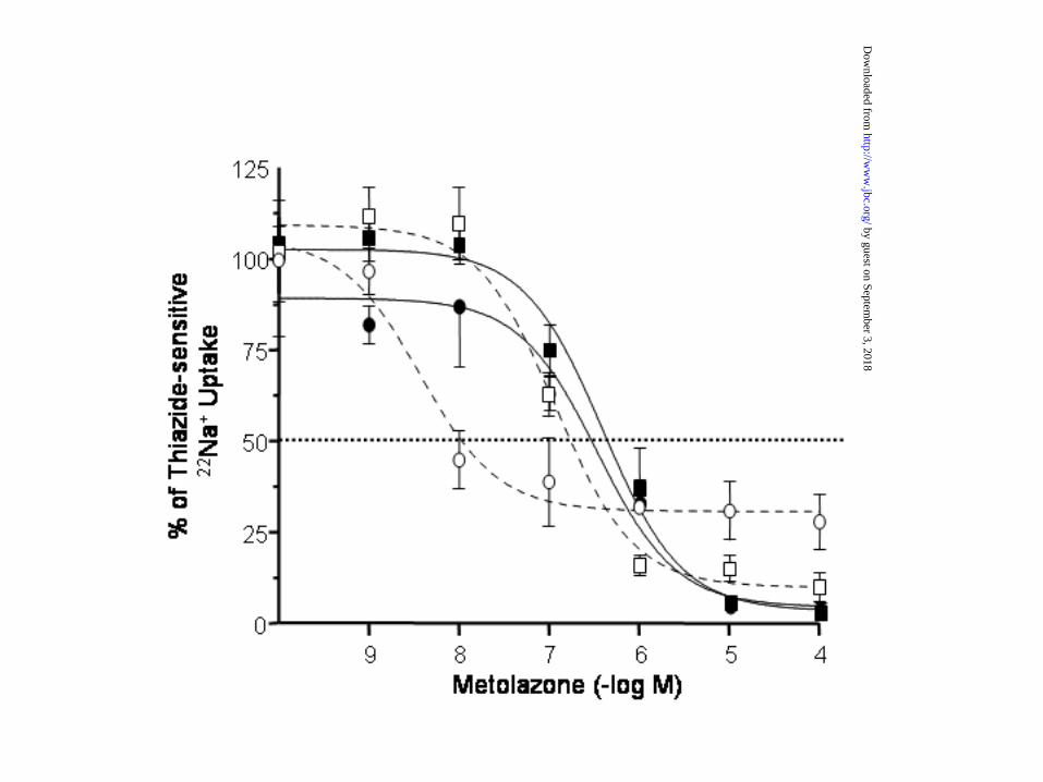
Figure 6. Concentration-response for inhibition of WT (squares) and G264A (circles) by
metolazone. Groups of 15 oocytes microinjected with WT or G264A were exposed to increased
concentrations of metolazone in the preincubation and uptake mediums, from 10-9 to 10-4 M.
Uptakes were performed in the presence of 96 mM of extracellular Cl- in both WT and G264A
(continuous lines and closed symbols) or in the presence of 6 mM of extracellular Cl- for WT
(open squares and discontinuous line), or 1 mM of extracellular Cl- for G264A (open circles and
discontinuous line). Data were normalized as the percentage of influx, taking 100% as the value
observed in oocytes in which uptake was performed in the absence of metolazone. Each point
represents the mean ± SEM of at least 15 oocytes.
33
by guest on September 3, 2018
http://ww
w.jbc.org/
Dow
nloaded from

Figure 7. Kinetic analyses of the inhibition of WT (upper panel) or G264A (lower panel) function
by several thiazide-type diuretics. All Na+ uptakes were preformed during 60 minutes with
thiazides tested at concentrations from 10-8 M to 10-4 M in uptake solution containing 40 mM
Na+ and 96 mM Cl-. Each point represents the mean ± SEM of at least 15 oocytes. The
inhibitory profile polythiazide (s) > metolazone (not shown) > bendroflumethiazide (¸) =
trichloromethiazide (r) = benzthiazide ( ) > hydrochlorothiazide (0) = chlortalidone () is similar
between WT and G264A.
Figure 8. Alignment of the amino acid residues that in the electroneutral cation-chloride
cotransporters correspond to the transmembrane domain 4, according with the topology analysis
that has been performed for BSC2 (53). Amino acid numbers correspond to the TSC sequence.
Figure 9. Functional expression of WT and TSC harboring the substitutions G264A, N258Q,
R261L, or G278A. The 22Na+ uptake was determined in groups of oocytes that were injected
with water or with 25 ng of cRNA from the WT or substituted mutants (as stated). Uptake was
assessed in control conditions (open bars) or in the presence of 10-4 M of the inhibitor
metolazone (closed bars). * p<0.01 vs. WT cRNA oocytes in control conditions. N=20 oocytes
per bar.
Figure 10. Surface abundance of EGFP-tagged WT, or EGFP-tagged TSC harbouring the
34
by guest on September 3, 2018
http://ww
w.jbc.org/
Dow
nloaded from
substitutions G264A, N258Q, R261L, or G278A, as stated. Oocytes were injected with
corresponding cRNA and green fluorescence was assessed by confocal microscopy as described
in Methods. The mean and standard error of fluorescence is shown for each set of eight oocytes
per injection in arbitrary units.
35
by guest on September 3, 2018
http://ww
w.jbc.org/
Dow
nloaded from
Bobadilla, Norma Vazquez, Daniela Riccardi, Esteban Poch and Gerardo GambaErika Moreno, Claudia Tovar-Palacio, Paola De los Heros, Blanca Guzman, Norma A.and reveals a role for transmembrane segment 4 in chloride and thiazide affinity
A single nucleotide polymorphism alters the activity of the renal Na+:Cl-cotransporter
published online February 5, 2004J. Biol. Chem.
10.1074/jbc.M400602200Access the most updated version of this article at doi:
Alerts:
When a correction for this article is posted•
When this article is cited•
to choose from all of JBC's e-mail alertsClick here
by guest on September 3, 2018
http://ww
w.jbc.org/
Dow
nloaded from