A simplified method to develop an interdisciplinary ...
53
76 | The International Journal of Esthetic Dentistry | Volume 16 | Number 1 | Spring 2021 CLINICAL RESEARCH A simplified method to develop an interdisciplinary treatment plan: an esthetically and functionally driven approach in three steps Stefano Gracis, DMD, MSD Private Practice, Milan, Italy Correspondence to: Dr Stefano Gracis Via Brera 28/a, 20121 Milan, Italy; Tel: +39 02 72094471; Email: [email protected]
Transcript of A simplified method to develop an interdisciplinary ...
76 | The International Journal of Esthetic Dentistry | Volume 16 | Number 1 | Spring 2021
CLINICAL RESEARCH
A simplified method to develop an
interdisciplinary treatment plan:
an esthetically and functionally
driven approach in three steps
Stefano Gracis, DMD, MSD
Private Practice, Milan, Italy
Correspondence to: Dr Stefano Gracis
Via Brera 28/a, 20121 Milan, Italy; Tel: +39 02 72094471; Email: [email protected]
GRACIS
7777The International Journal of Esthetic Dentistry | Volume 16 | Number 1 | Spring 2021 |
Abstract
Many clinicians are unsure of how to develop a com-
prehensive plan of treatment for patients who present
with multiple problems and pathologies. In order to
efficiently plan appropriate treatment for such com-
plex patient cases, the clinician needs to either have or
develop the necessary knowledge of evidence-based
information on the predictability of available clinical
procedures. The clinician also needs to understand
the correct sequence in which such treatment is ap-
plied, and perfect the skills required for carrying out
that treatment. Since most clinicians have not ac-
quired all the knowledge and skills necessary for this
task, an interdisciplinary approach to treatment is
typically required. This article provides a practical step-
by-step approach to planning comprehensive interdis-
ciplinary treatment focused primarily on the teeth as
they relate to each other and to the structures that
surround them. The approach is based on the answers
to six questions that are grouped into three steps: 1)
evaluation of the teeth relative to the face and lips;
2) assessment of anterior tooth dimensions; and 3)
analysis of the anteroposterior and maxillomandibular
relationships. The information obtained must then be
related to the patient’s skeletal framework, periodontal
status, caries susceptibility, and biomechanical risk as-
sessment in order to formulate a clear and complete
plan of treatment.
(Int J Esthet Dent 2021;16:76–128)
77
CLINICAL RESEARCH
78 | The International Journal of Esthetic Dentistry | Volume 16 | Number 1 | Spring 2021
on a referral basis. Some discussion of the
treatment to be provided will likely take
place, but there is no real coordinated ef-
fort in planning or providing treatment for
a patient. In contrast, an interdisciplinary
approach takes into account all available
patient-specific variables. It requires the
knowledge and expertise of all the treat-
ing clinicians from the disciplines involved
and coordinated communication between
them, not only with respect to the treat-
ment involved, but also to the planning,
sequencing, and timing of such treatment.
It also requires knowledge of available ev-
idence-based information on the predict-
ability of the various clinical procedures
being considered as well as an understand-
ing of their correct sequence. In essence,
interdisciplinary care is about teamwork
– the interconnectivity and ongoing inter-
action between clinicians from various dis-
ciplines involved in planning and providing
treatment.
The aim of this article is to provide a
relatively clear concept of comprehensive
interdisciplinary treatment planning for cli-
nicians faced with challenging clinical sit-
uations, and to do so in a simplified way.
However, in applying this simplified ap-
proach, one should recognize that it is pri-
marily focused on the teeth and surround-
ing structures with respect to function and
esthetics. Additionally, some patients will
present with problems that will require a
more definitive assessment in order to de-
sign a plan of treatment commensurate
with the number and/or extent of their
problems. Whether you use the simplified
approach to treatment planning present-
ed in this article, or a more traditional ap-
proach to comprehensive interdisciplinary
treatment planning, it is important to real-
ize that treatment planning is a process
that should begin with a vision of the end
result. As Stephen Covey put it, “Begin with
the end in mind.”5
Introduction
The goals of prosthetic treatment are to
rehabilitate a patient’s stomatognathic sys-
tem to be functionally sound, biologically
healthy, and esthetically pleasing while fol-
lowing accepted principles and using de-
pendable materials to ensure that the end
result will serve the patient for as long as
possible.1-4 To achieve these goals, it is im-
portant to adopt a systematic approach that
allows the clinician to identify the problems,
establish specific treatment goals for each
problem, determine which therapies will ac-
complish each goal, and develop an appro-
priate sequence of treatment that will lead
to the envisioned outcome, based on the
presenting conditions.
Patients often present with a multitude
of problems that pose varying degrees of
difficulty to manage. These include, but are
not limited to, esthetic expectations, degree
of periodontal compromise, tooth integrity,
tooth position, number of missing teeth,
occlusal relationships, functional demands,
and levels of risk associated with the treat-
ment of each problem (Figs 1 to 3). As the
complexity of a patient’s problems increas-
es, the clinician may need to acquire further
knowledge, develop additional skills, and
know when to involve clinicians from oth-
er disciplines to provide expertise and care
in order to design and execute a treatment
plan that will achieve the therapeutic goals
and the desired end result. This, then, would
establish the need for an interdisciplinary
approach to providing optimal care for the
patient.
There is a significant, but often over-
looked, difference between an interdisci-
plinary approach to managing a patient’s
problems and treatment compared with a
multidisciplinary one. Multidisciplinary care
simply refers to multiple clinicians of differ-
ent disciplines providing some level of care
or procedure in treating a patient, typically
GRACIS
79The International Journal of Esthetic Dentistry | Volume 16 | Number 1 | Spring 2021 |
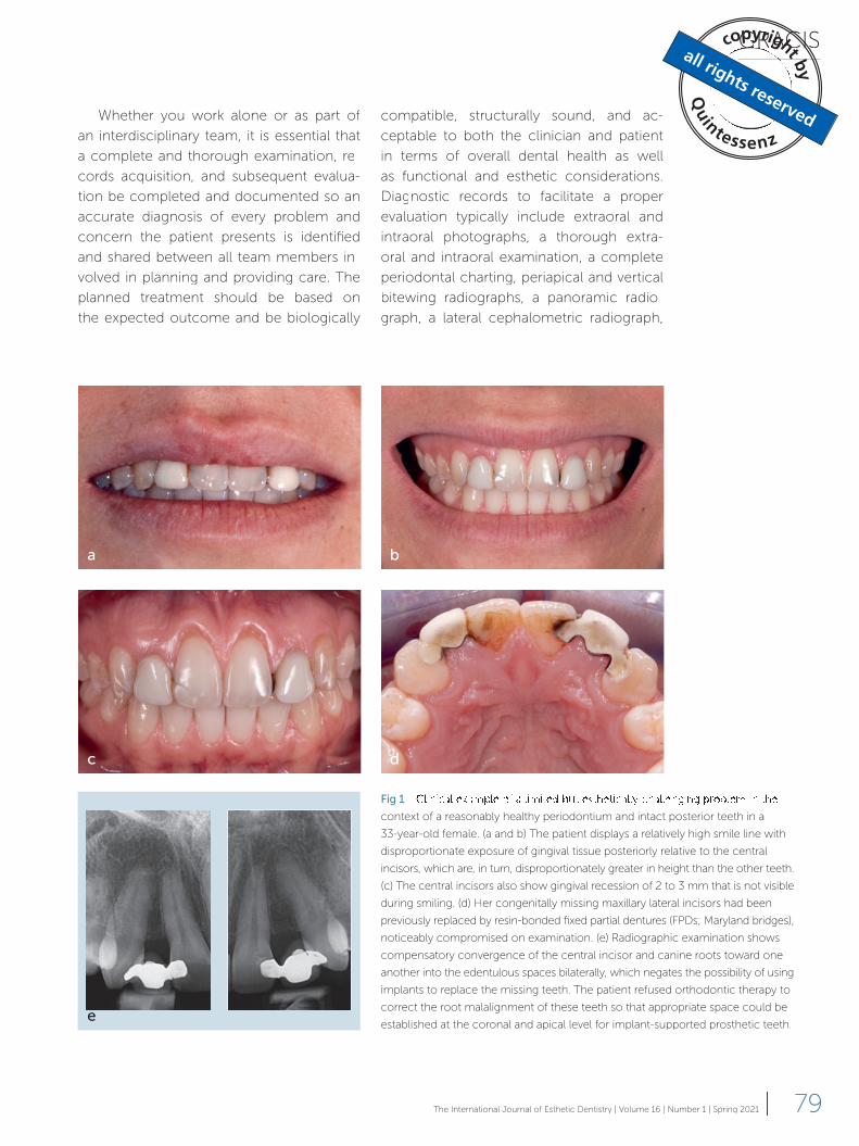
Fig 1
context of a reasonably healthy periodontium and intact posterior teeth in a
33-year-old female. (a and b) The patient displays a relatively high smile line with
disproportionate exposure of gingival tissue posteriorly relative to the central
incisors, which are, in turn, disproportionately greater in height than the other teeth.
(c) The central incisors also show gingival recession of 2 to 3 mm that is not visible
during smiling. (d) Her congenitally missing maxillary lateral incisors had been
previously replaced by resin-bonded fixed partial dentures (FPDs; Maryland bridges),
noticeably compromised on examination. (e) Radiographic examination shows
compensatory convergence of the central incisor and canine roots toward one
another into the edentulous spaces bilaterally, which negates the possibility of using
implants to replace the missing teeth. The patient refused orthodontic therapy to
correct the root malalignment of these teeth so that appropriate space could be
established at the coronal and apical level for implant-supported prosthetic teeth.
Whether you work alone or as part of
an interdisciplinary team, it is essential that
a complete and thorough examination, re-
cords acquisition, and subsequent evalua-
tion be completed and documented so an
accurate diagnosis of every problem and
concern the patient presents is identified
and shared between all team members in-
volved in planning and providing care. The
planned treatment should be based on
the expected outcome and be biologically
compatible, structurally sound, and ac-
ceptable to both the clinician and patient
in terms of overall dental health as well
as functional and esthetic considerations.
Diagnostic records to facilitate a proper
evaluation typically include extraoral and
intraoral photographs, a thorough extra-
oral and intraoral examination, a complete
periodontal charting, periapical and vertical
bitewing radiographs, a panoramic radio-
graph, a lateral cephalometric radiograph,
a b
c d
e
CLINICAL RESEARCH
80 | The International Journal of Esthetic Dentistry | Volume 16 | Number 1 | Spring 2021
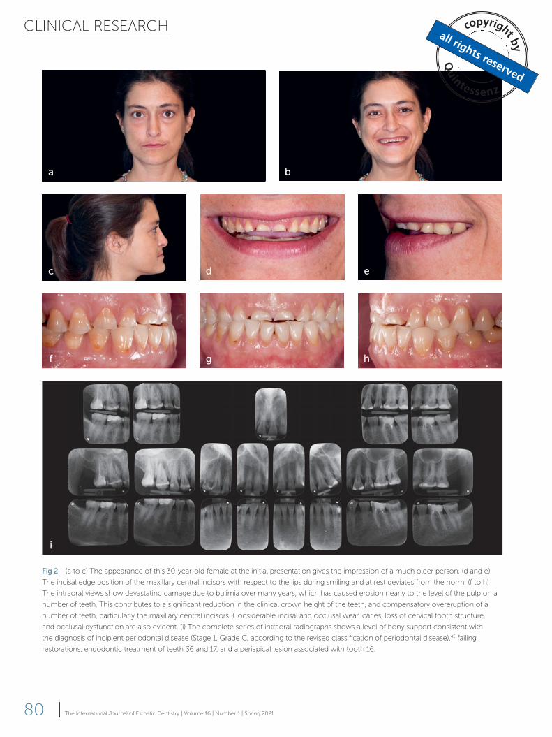
Fig 2 (a to c) The appearance of this 30-year-old female at the initial presentation gives the impression of a much older person. (d and e)
The incisal edge position of the maxillary central incisors with respect to the lips during smiling and at rest deviates from the norm. (f to h)
The intraoral views show devastating damage due to bulimia over many years, which has caused erosion nearly to the level of the pulp on a
number of teeth. This contributes to a signifi cant reduction in the clinical crown height of the teeth, and compensatory overeruption of a
number of teeth, particularly the maxillary central incisors. Considerable incisal and occlusal wear, caries, loss of cervical tooth structure,
and occlusal dysfunction are also evident. (i) The complete series of intraoral radiographs shows a level of bony support consistent with
the diagnosis of incipient periodontal disease (Stage 1, Grade C, according to the revised classifi cation of periodontal disease),45 failing
restorations, endodontic treatment of teeth 36 and 17, and a periapical lesion associated with tooth 16.
c d e
f g h
a b
i
GRACIS
81The International Journal of Esthetic Dentistry | Volume 16 | Number 1 | Spring 2021 |
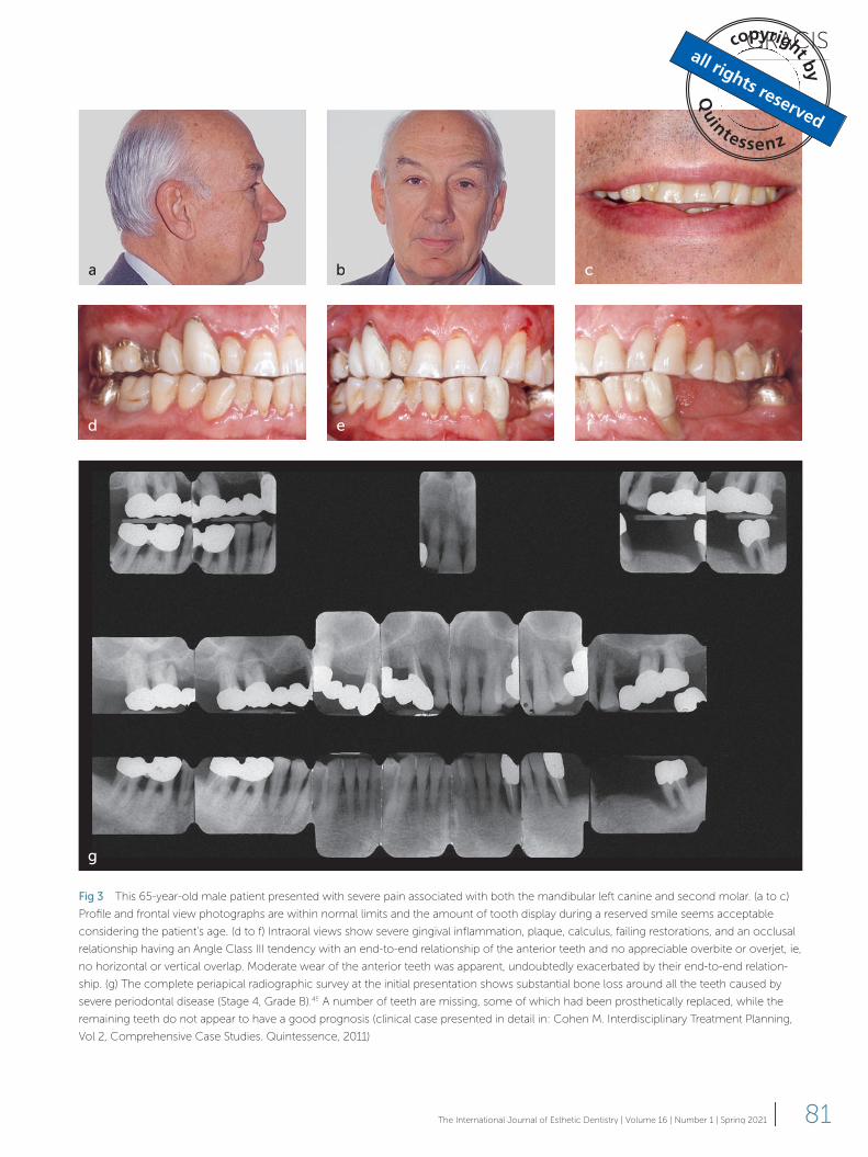
Fig 3 This 65-year-old male patient presented with severe pain associated with both the mandibular left canine and second molar. (a to c)
Profi le and frontal view photographs are within normal limits and the amount of tooth display during a reserved smile seems acceptable
considering the patient’s age. (d to f) Intraoral views show severe gingival infl ammation, plaque, calculus, failing restorations, and an occlusal
relationship having an Angle Class III tendency with an end-to-end relationship of the anterior teeth and no appreciable overbite or overjet, ie,
no horizontal or vertical overlap. Moderate wear of the anterior teeth was apparent, undoubtedly exacerbated by their end-to-end relation-
ship. (g) The complete periapical radiographic survey at the initial presentation shows substantial bone loss around all the teeth caused by
severe periodontal disease (Stage 4, Grade B).45 A number of teeth are missing, some of which had been prosthetically replaced, while the
remaining teeth do not appear to have a good prognosis (clinical case presented in detail in: Cohen M. Interdisciplinary Treatment Planning,
Vol 2, Comprehensive Case Studies. Quintessence, 2011).
a b c
d e f
g
CLINICAL RESEARCH
82 | The International Journal of Esthetic Dentistry | Volume 16 | Number 1 | Spring 2021
Step 1: Evaluation of the teeth relative to the face and lips
1. Are the patient’s lips and teeth symmet-
ric relative to the face and is the maxillary
dental midline coincident with the facial
midline?
2. Are the maxillary teeth ideally positioned
vertically relative to the lips at rest, during
smiling, talking, and laughing for the age,
sex, and race of the patient?
3. Are the mandibular teeth ideally posi-
tioned relative to the lower lip at rest as
well as during smiling and talking for the
age, sex, and race of the patient?
Step 2: Assessment of anterior tooth dimensions
4. Are the clinical crowns of the maxillary
and mandibular anterior teeth appropri-
ate with respect to size, shape, and pro-
portions (width to height) consistent with
established norms?
Step 3: Analysis of the antero-posterior and maxillomandibular relationships
5. Are the maxillary and mandibular curves
of Spee (in the sagittal plane) and Wilson
(in the frontal plane) appropriate?
6. Is the vertical dimension of occlusion
(VDO) acceptable relative to the face,
lips, and existing crown height of the
teeth?
The therapeutic possibilities inferred by
these questions will likely require evalua-
tion and potential treatment by a profes-
sional from more than one dental disci-
pline, including, in no particular order, oral
medicine, periodontics, endodontics, re-
storative dentistry, prosthodontics, ortho-
dontics, and oral surgery. If the answers to
any of the first five questions are negative,
CBCT when needed, impressions or scans
of the dental arches, and accurate maxillo-
mandibular records. The complexity of the
patient’s problems determines the volume
and extent of data to be collected. Diag-
nostic records do not necessarily need to
be collected in any specific order, although
many clinicians prefer to do so. However,
the evaluation or assessment of diagnostic
data with respect to the sequence of the
treatment planned and performed needs
to be carried out in a specific order to pro-
vide optimal care and to avoid completing
a procedure to correct one problem that
adversely affects the planned treatment of
another one.
Another component that is necessary in
order to effectively and efficiently plan the
most comprehensive and appropriate treat-
ment is to establish specific treatment goals
based on the patient’s particular problems
and desires. Unfortunately, most clinicians
do not perform this step, which is one rea-
son why efficient and optimal treatment
planning on a comprehensive basis seems
difficult to many colleagues.
The simplified method to develop an
interdisciplinary plan of treatment focused
on esthetics and function, as described in
this article, involves a sequential, stepwise
approach of answering six questions that
are grouped into three steps. The aim of the
author is to assist clinicians involved in such
planning to focus on specific diagnostic cri-
teria in a progressive manner, including the
relationship of patients’ teeth to the frame-
work of their lips and face, the size and ap-
pearance of their teeth, and the relationship
of their teeth with respect to function.
The three steps comprising the decision-making process
What follows are the six questions grouped
into three steps that comprise the deci-
sion-making process.
GRACIS
83The International Journal of Esthetic Dentistry | Volume 16 | Number 1 | Spring 2021 |
distant point at eye level. It is usually, but not
always, within a few degrees of the Frank-
fort horizontal plane, the previous standard
reference for head orientation. A reliable
way to establish the NHP is for the patient
to look at his or her own eyes in a mirror.
Alternatively, the patient could be standing,
but it is important that the clinician is able
to observe the patient at eye level with the
patient looking straight ahead. Bear in mind
that a height difference between the patient
and the clinician could hinder the acquisi-
tion of an accurate assessment in this stand-
ing position.
The facial analysis should always begin by
assessing the relative symmetry of the face,
noting any apparent asymmetries, some of
which may be slight deviations from the
norm that do not typically require or be con-
sidered for alteration. These include, but are
not limited to, a cant of the ears and a cant
of the eyes or interpupillary line (Fig 5a). The
former would unfavorably affect the posi-
tion of articulated casts if mounted with the
use of an earbow. The latter would not be
an appropriate reference to dictate the posi-
tion of other anatomical structures in terms
of the transverse orientation of the patient’s
occlusal plane.
The facial midline is the best and most
reliable landmark from the frontal perspec-
tive for evaluating the symmetry and bal-
ance of the face overall as well as the lips
and teeth14,15. This imaginary line runs ver-
tically centered through nasion (N), which
is the midpoint of the intersection of the
frontonasal suture and internasal suture that
joins the two nasal bones, the midpoint of
the nasal bridge (below N), the philtrum of
the upper lip, and the point of the philtrum
often referred to as the center of Cupid’s
bow.
It is also important to observe the pro-
portions of the face vertically and laterally.
Vertically, the face is divided into thirds by
landmarks in the midsagittal plane (Fig 5b):
the clinician then needs to decide, in
consultation with the patient, whether to
correct some or all of the problems or
discrepancies, and, if so, which therapeu-
tic alternatives would be appropriate or
recommended.
As has been suggested by several au-
thors,1,6-10 it is advisable to evaluate the
planned (or proposed) changes directly in
the patient’s mouth through a direct or in-
direct mock-up and/or provisional restor-
ations. Both these options will allow the pa-
tient and the clinician to visualize a potential
treatment result, especially the esthetic out-
come. Provisional restorations, additionally,
will facilitate further evaluation with respect
to function, speech, wear, and structural
considerations that include resistance and
retention form and the number of abut-
ments needed to support the replacement
of missing teeth, in lieu of implant-support-
ed prosthetic teeth, by monitoring the sta-
bility of the provisional prosthesis over a pe-
riod of time (Fig 4).
The steps and questions in detail
Step 1: Evaluation of the teeth relative to the face and lips
Question 1: Are the patient’s lips and teeth
symmetric relative to the face and is the
maxillary dental midline coincident with
the facial midline?
Many authors have pointed out the impor-
tance of an esthetic analysis or evaluation
to assess the position of the teeth relative to
the face and lips.11-13 To obtain an accurate
assessment, this should be performed with
the patient seated upright in a chair facing
the clinician, with the feet on the floor, and
the head in the normal upright position.
This is commonly referred to as the natu-
ral head position (NHP), which is, by defi-
nition, a reproducible position of the head
in space when the patient is focused on a
CLINICAL RESEARCH
84 | The International Journal of Esthetic Dentistry | Volume 16 | Number 1 | Spring 2021
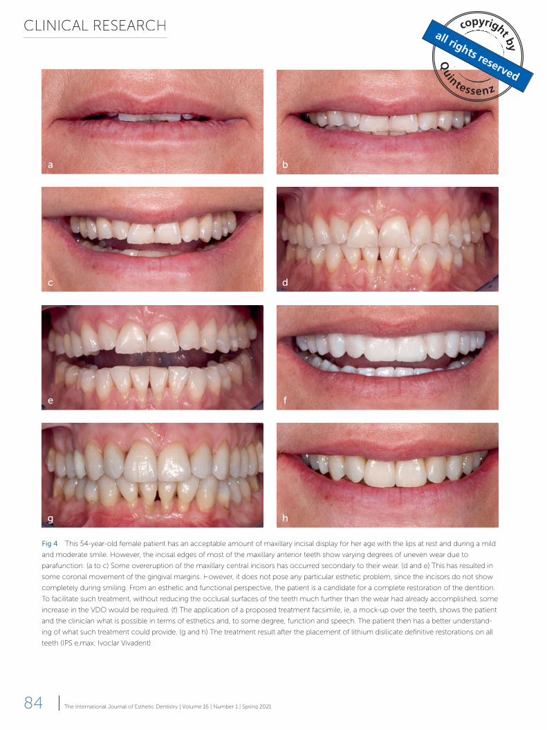
Fig 4 This 54-year-old female patient has an acceptable amount of maxillary incisal display for her age with the lips at rest and during a mild
and moderate smile. However, the incisal edges of most of the maxillary anterior teeth show varying degrees of uneven wear due to
parafunction. (a to c) Some overeruption of the maxillary central incisors has occurred secondary to their wear. (d and e) This has resulted in
some coronal movement of the gingival margins. However, it does not pose any particular esthetic problem, since the incisors do not show
completely during smiling. From an esthetic and functional perspective, the patient is a candidate for a complete restoration of the dentition.
To facilitate such treatment, without reducing the occlusal surfaces of the teeth much further than the wear had already accomplished, some
increase in the VDO would be required. (f) The application of a proposed treatment facsimile, ie, a mock-up over the teeth, shows the patient
and the clinician what is possible in terms of esthetics and, to some degree, function and speech. The patient then has a better understand-
ing of what such treatment could provide. (g and h) The treatment result after the placement of lithium disilicate definitive restorations on all
teeth (IPS e.max; Ivoclar Vivadent).
a b
c d
e f
g h
GRACIS
85The International Journal of Esthetic Dentistry | Volume 16 | Number 1 | Spring 2021 |
■ The top third extends downward from
trichion (Tr), which is the hairline, to gla-
bella (Gl), which is the most prominent
point of the forehead.
■ The middle third extends from Gl to sub-
nasale (Sn), which is the point where the
base of the nasal columelle meets the
upper lip.
■ The lower third extends from Sn to the
soft tissue menton (Me), the lowest point
of the chin.
Although the facial thirds imply equal size,
they vary to some extent, the lower third
often being greater in size than the middle
third, especially in males. The top third, on
the other hand, can increase in size due to a
receding hairline. Significant variations may
reveal an abnormality.
As dentists, we can effect a change in
the size of especially the lower third with
the treatment we can provide, in particular,
orthognathic surgery. Cephalometric land-
marks are typically used in the assessment
of facial heights in this regard, with the up-
per facial height measured from N to the
anterior nasal spine (ANS), and lower facial
height measured from the ANS to Me. Up-
per facial height is typically 40% to 46% of
the overall facial height from N to Me.
Knowledge of facial proportions is im-
portant in planning treatment for a patient
when changes to facial height are being
considered prosthodontically by increasing
the VDO or altering the position of the max-
illa and/or mandible through orthognathic
surgery for a number of disparities. These
include, but are not limited to, a maxillary
vertical deficiency expressed clinically with
a short face and limited or no tooth display,
with the lips at rest or smiling; and a max-
illary vertical excess from disproportionate
downward development of the maxilla,
seen clinically as a long face with excessive
display of gingival tissue during smiling and,
if severe, even with the lips at rest.
Transverse facial proportions should
also be evaluated. This is typically done by
dividing the face using vertical lines. One
perspective is the ‘rule of fifths,’ where the
face is divided into equal fifths (Fig 5c). The
size of each fifth should be about the width
of the patient’s eye. Bilateral assessment of
the face should also be performed for any
asymmetries that may exist, bearing in mind
that no person has a face where the two
sides are mirror images of each other. Man-
dibular asymmetry can occur due to asym-
metric growth of the mandible. Another
way to evaluate transverse facial propor-
tions is to mark the outer edge of the widths
of the face. Anteroposterior relationships of
the patient’s head should also be evaluated
for deficiencies or excesses in the growth
of the maxilla and mandible, as such growth
abnormalities generally affect how the teeth
fit together and the resultant malocclusions
that might occur.
Unfortunately, the recording of the fa-
cial midline to assess it relative to the ar-
ticulated casts of the patient’s teeth and
facilitate the fabrication of prosthetic or
restorative protheses on an articulator is
problematic. If the patient has a transverse
cant of the occlusal plane relative to the fa-
cial midline, it cannot be accurately repro-
duced on an articulator for evaluation and
correction (Fig 6). Conversely, if the casts
are mounted on an articulator with the use
of a hinge-axis recording facebow or an
earbow, particularly if it is used according
to the manufacturer’s recommendations,
and the resultant mounted casts, from the
frontal perspective, are canted on the ar-
ticulator, there is no easy or precise way
to know if the orientation of the mounted
casts reflects the existing situation in the
patient’s mouth. Some clinicians, knowing
the shortcomings of using an earbow to
mount casts, alter the recording by torque-
ing the horizontal component of the ear-
bow to make it either parallel with the eyes

CLINICAL RESEARCH
86 | The International Journal of Esthetic Dentistry | Volume 16 | Number 1 | Spring 2021
For optimal esthetics, the positions of
the maxillary and mandibular teeth are de-
pendent on where the maxillary central in-
cisors are or should be relative to the face
and lips. Ideally, the maxillary dental midline
should be in line with the facial midline, and
the smile should, in turn, be centered on the
dental midline. Although some movement
of the maxillary dental midline away from
the facial midline is tolerable as long as it
is parallel to the facial midline, any cant or
angle of the maxillary dental midline relative
to the facial midline will alter the balance of
the maxillary teeth relative to the surround-
ing structures in both the horizontal and ver-
tical planes. The commissural line of the lips
and the transverse orientation of the incisal
or perpendicular to the facial midline13,16-18
(Fig 7). Although this modifi cation in the
use of an earbow can improve the possi-
bility of obtaining a more accurate result,
it may still fall short of exactly reproduc-
ing the orientation of the patient’s occlu-
sal plane on the articulator. This problem
was resolved by the advent of the Kois
Dento-Facial Analyzer System (Panadent).19
Since the system records the facial midline
and the average distance of the maxillary
central incisal edges to the hinge axis as
well as the orientation of the occlusal plane
in all three planes of space, an accurate re-
production of the patient’s occlusal plane
from the frontal perspective can be made
on the articulator (Fig 8).
Fig 5 Diagrams showing horizontal and vertical landmarks that are typically used
to evaluate facial symmetry. (a) The analysis of the patient’s face, smile, and teeth
should start with an assessment of the symmetry relative to the facial midline
(white dashed line) – see text as to how this imaginary ‘white line’ runs. Next, the
eyes, nose, and mouth are observed and evaluated to determine whether they
are symmetric and proportionate to the facial midline. The interpupillary line is a
horizontal reference used by some to evaluate whether the lips and teeth are
level, but it is not particularly reliable as the eyes can be at diff erent heights
(canted) in some patients. With respect to symmetry, the teeth and smile as well
as the occlusal plane and the commissural line of the lips should ideally be
perpendicular to the facial midline, level with the face; otherwise, they would
appear to be at an angle or canted relative to the face. (b) It is also important to
observe the proportions of the face vertically and laterally (see text). (c) Transverse
facial proportions are evaluated through ‘the rule of fi fths,’ where the face is
divided into equal fi fths (see text).
a b
c
GRACIS
87The International Journal of Esthetic Dentistry | Volume 16 | Number 1 | Spring 2021 |
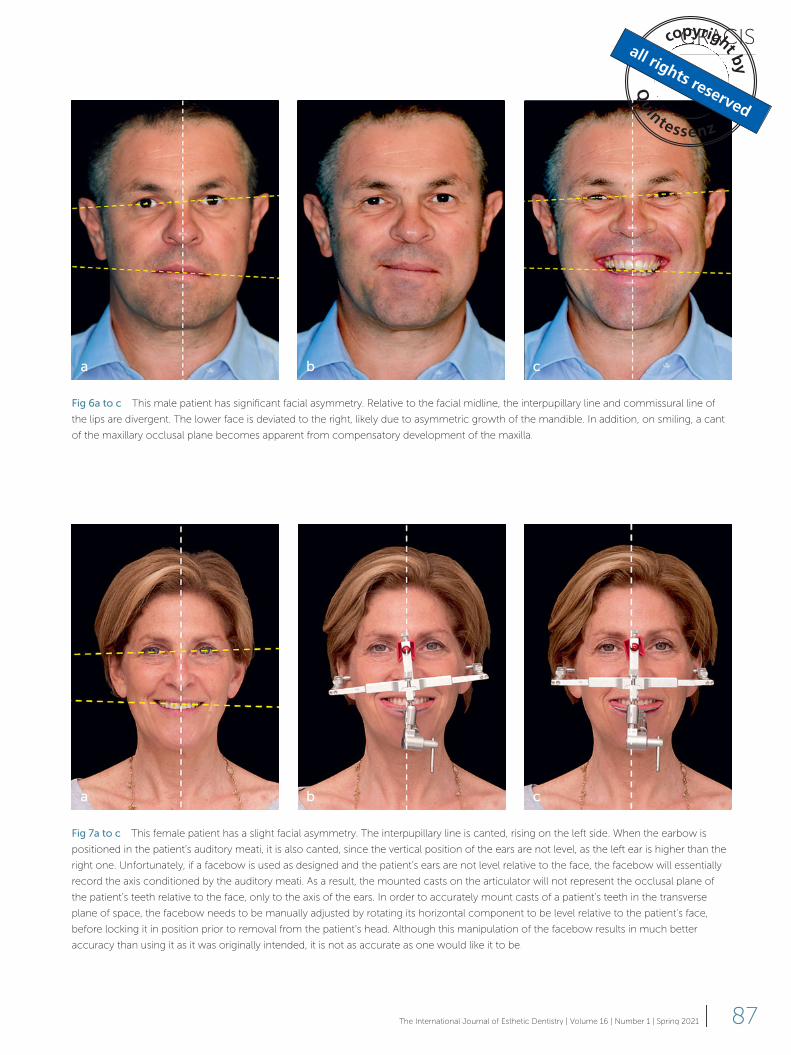
Fig 6a to c This male patient has significant facial asymmetry. Relative to the facial midline, the interpupillary line and commissural line of
the lips are divergent. The lower face is deviated to the right, likely due to asymmetric growth of the mandible. In addition, on smiling, a cant
of the maxillary occlusal plane becomes apparent from compensatory development of the maxilla.
Fig 7a to c This female patient has a slight facial asymmetry. The interpupillary line is canted, rising on the left side. When the earbow is
positioned in the patient’s auditory meati, it is also canted, since the vertical position of the ears are not level, as the left ear is higher than the
right one. Unfortunately, if a facebow is used as designed and the patient’s ears are not level relative to the face, the facebow will essentially
record the axis conditioned by the auditory meati. As a result, the mounted casts on the articulator will not represent the occlusal plane of
the patient’s teeth relative to the face, only to the axis of the ears. In order to accurately mount casts of a patient’s teeth in the transverse
plane of space, the facebow needs to be manually adjusted by rotating its horizontal component to be level relative to the patient’s face,
before locking it in position prior to removal from the patient’s head. Although this manipulation of the facebow results in much better
accuracy than using it as it was originally intended, it is not as accurate as one would like it to be.
a b c
a b c
CLINICAL RESEARCH
88 | The International Journal of Esthetic Dentistry | Volume 16 | Number 1 | Spring 2021
development of the maxilla. Asymmetric
skeletal growth typically leads to canted lips
and a canted dental midline.
Canted or asymmetric occlusal planes
can be corrected depending on a patient’s
desires, the severity of the asymmetry, and
the treatment needed to facilitate such a
correction. It may be possible to correct a
mild cant of the incisal-occlusal plane that
does not involve the lips or alteration of the
teeth by changing the position of the teeth
via orthodontics. If the teeth require treat-
ment or alteration due to periodontal and/
or restorative needs, a canted incisal-occlu-
sal plane may be corrected prosthetically or
in combination with orthodontic treatment.
More severe cants of the occlusal plane
that are associated with a facial skeletal
and occlusal planes should be perpendicu-
lar to the facial midline. The smile line of the
maxillary teeth should also be perpendicu-
lar to the facial midline and follow the cur-
vature of the lower lip. Disparities in these
relationships can initiate some visual dis-
cord and, depending on the severity of the
asymmetry, may be considered distracting
or unattractive.13,20 Only minor cants of up
to 3 degrees have been reported to be ac-
ceptable to dental professionals.21 Possible
causes for a canted incisal-occlusal plane
include, but are not limited to, the asymmet-
ric eruption of teeth following the wear or
loss of opposing teeth, the development of
various malocclusions (Fig 9), and condylar
dysplasia or asymmetric growth of the man-
dible, which contribute to the asymmetric
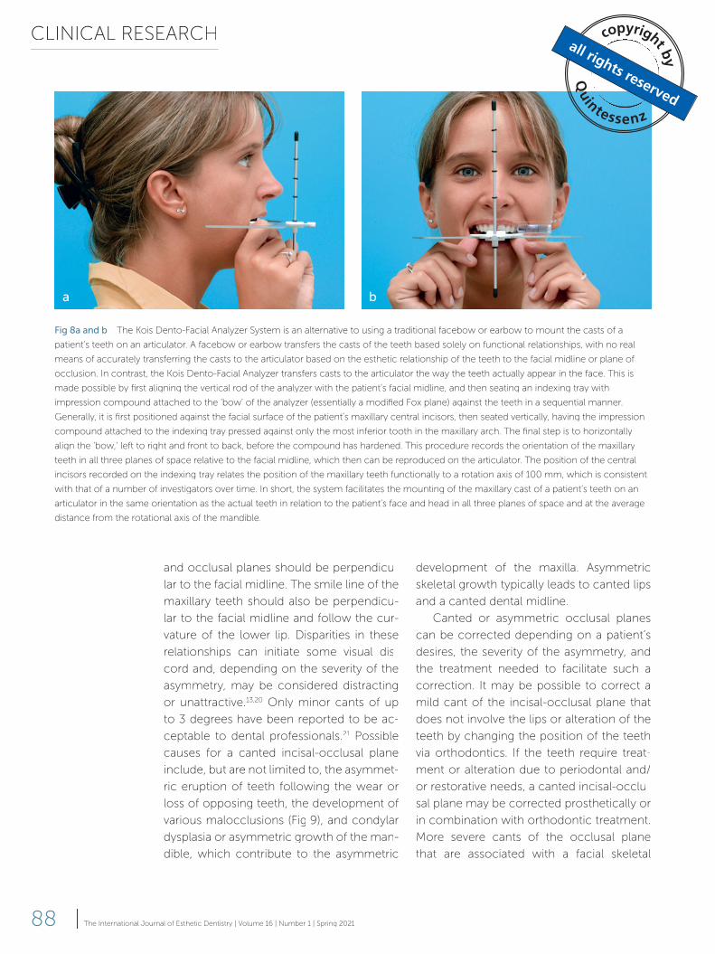
Fig 8a and b The Kois Dento-Facial Analyzer System is an alternative to using a traditional facebow or earbow to mount the casts of a
patient’s teeth on an articulator. A facebow or earbow transfers the casts of the teeth based solely on functional relationships, with no real
means of accurately transferring the casts to the articulator based on the esthetic relationship of the teeth to the facial midline or plane of
occlusion. In contrast, the Kois Dento-Facial Analyzer transfers casts to the articulator the way the teeth actually appear in the face. This is
made possible by first aligning the vertical rod of the analyzer with the patient’s facial midline, and then seating an indexing tray with
impression compound attached to the ‘bow’ of the analyzer (essentially a modified Fox plane) against the teeth in a sequential manner.
Generally, it is first positioned against the facial surface of the patient’s maxillary central incisors, then seated vertically, having the impression
compound attached to the indexing tray pressed against only the most inferior tooth in the maxillary arch. The final step is to horizontally
align the ‘bow,’ left to right and front to back, before the compound has hardened. This procedure records the orientation of the maxillary
teeth in all three planes of space relative to the facial midline, which then can be reproduced on the articulator. The position of the central
incisors recorded on the indexing tray relates the position of the maxillary teeth functionally to a rotation axis of 100 mm, which is consistent
with that of a number of investigators over time. In short, the system facilitates the mounting of the maxillary cast of a patient’s teeth on an
articulator in the same orientation as the actual teeth in relation to the patient’s face and head in all three planes of space and at the average
distance from the rotational axis of the mandible.
ba
GRACIS
89The International Journal of Esthetic Dentistry | Volume 16 | Number 1 | Spring 2021 |
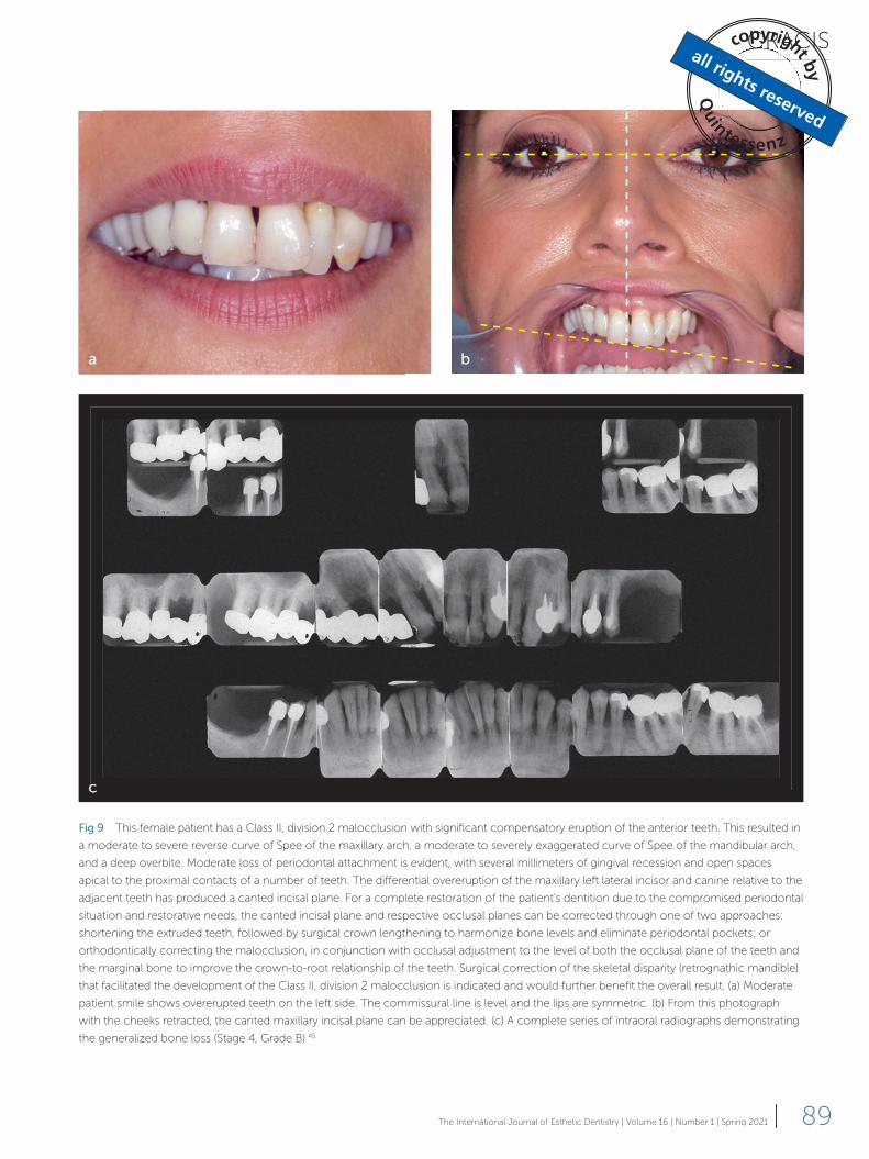
Fig 9 This female patient has a Class II, division 2 malocclusion with significant compensatory eruption of the anterior teeth. This resulted in
a moderate to severe reverse curve of Spee of the maxillary arch, a moderate to severely exaggerated curve of Spee of the mandibular arch,
and a deep overbite. Moderate loss of periodontal attachment is evident, with several millimeters of gingival recession and open spaces
apical to the proximal contacts of a number of teeth. The differential overeruption of the maxillary left lateral incisor and canine relative to the
adjacent teeth has produced a canted incisal plane. For a complete restoration of the patient’s dentition due to the compromised periodontal
situation and restorative needs, the canted incisal plane and respective occlusal planes can be corrected through one of two approaches:
shortening the extruded teeth, followed by surgical crown lengthening to harmonize bone levels and eliminate periodontal pockets; or
orthodontically correcting the malocclusion, in conjunction with occlusal adjustment to the level of both the occlusal plane of the teeth and
the marginal bone to improve the crown-to-root relationship of the teeth. Surgical correction of the skeletal disparity (retrognathic mandible)
that facilitated the development of the Class II, division 2 malocclusion is indicated and would further benefit the overall result. (a) Moderate
patient smile shows overerupted teeth on the left side. The commissural line is level and the lips are symmetric. (b) From this photograph
with the cheeks retracted, the canted maxillary incisal plane can be appreciated. (c) A complete series of intraoral radiographs demonstrating
the generalized bone loss (Stage 4, Grade B).45
ba
c
CLINICAL RESEARCH
90 | The International Journal of Esthetic Dentistry | Volume 16 | Number 1 | Spring 2021
teeth than other reference planes, they take
precedence. Similarly, if a patient presents
with a canted incisal-occlusal plane with lips
that are level, the smile does not appear to
be balanced. Therefore, the occlusal plane
should be leveled to be parallel with the lips,
and the cant eliminated.
Table 1 lists the possible causes of an
asymmetric incisal-occlusal plane in the
maxillary (or mandibular) arch and the thera-
peutic alternatives when the teeth are intact
and not in need of restorative treatment,
and when they are restored or present with
restorative needs.
Question 2: Are the maxillary teeth ideally
positioned vertically relative to the lips at
rest, during smiling, talking, and laughing
for the age, sex, and race of the patient?
The overall position of the teeth for optimal
esthetics relative to the smile, phonetics, and
function is dependent on and determined
by the vertical position of the maxillary cen-
tral incisal edges, as has been reported by a
number of authors.11-14 The incisal edges of
the maxillary central incisors are essential-
ly the cornerstones from which a patient’s
asymmetry involving the maxilla, mandible,
lips, and dental midline require a combina-
tion of several disciplines to correct, includ-
ing orthodontics, orthognathic surgery, and
general dentistry, depending on the patient’s
other dental needs (Fig 10). A determination
should be made early in the diagnostic and
treatment planning phases of these more
complex problems, with clinicians from
each discipline evaluating and formulating
the appropriate plan and sequence of treat-
ment for the patient.
If a patient presents with a canted occlu-
sal plane and canted lips, but without any
other appreciable skeletal asymmetry, and
requires a restoration or replacement of the
teeth, it is preferable to maintain the cant
of the overall occlusal plane and not to lev-
el it with the eyes or make it perpendicular
to the facial midline. The reason is that to
do so would create an occlusal plane that
is not parallel with the commissural line of
the lips. This would upset the balance of
the patient’s mouth relative to the teeth.
The closest reference that frames the teeth
is what dictates the treatment with respect
to esthetics. Since the lips are closer to the
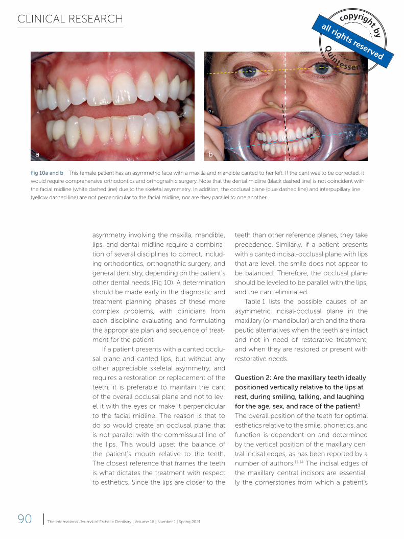
Fig 10a and b This female patient has an asymmetric face with a maxilla and mandible canted to her left. If the cant was to be corrected, it
would require comprehensive orthodontics and orthognathic surgery. Note that the dental midline (black dashed line) is not coincident with
the facial midline (white dashed line) due to the skeletal asymmetry. In addition, the occlusal plane (blue dashed line) and interpupillary line
(yellow dashed line) are not perpendicular to the facial midline, nor are they parallel to one another.
a b
GRACIS
91The International Journal of Esthetic Dentistry | Volume 16 | Number 1 | Spring 2021 |
of maxillary tooth display during smiling,
talking, and laughing (Figs 12 and 13).
Due to the relatively wider range of max-
illary central incisal edge exposure in rela-
tion to the maxillary lip line in repose, how-
ever, a preliminary study by Misch22 pointed
out that the canine incisal edge exposure
may be a better landmark to observe and
use clinically to determine anterior incisal
edge positions for edentulous patients. The
study compared the amount and range of
incisal edge display of the maxillary right
central incisor and right canine of 104
dentate Caucasian patients between 30 to
59 years of age. It reported that the average
smile, speech, tooth position, tooth form,
and occlusal relationship is derived. There-
fore, evaluation and management of the
incisal edge positions of the maxillary cen-
tral incisors is essential to the development
and execution of a treatment plan that will
achieve the desired end result in terms of
overall esthetics and function.
Initially, the vertical incisal edge positions
of the maxillary central incisors should be
evaluated with respect to the upper lip at
rest (Fig 11). Ideally, the range of incisal edge
display with the lips at rest is 2 to 4 mm,
depending on the patient’s age and sex. A
subsequent determination should be made
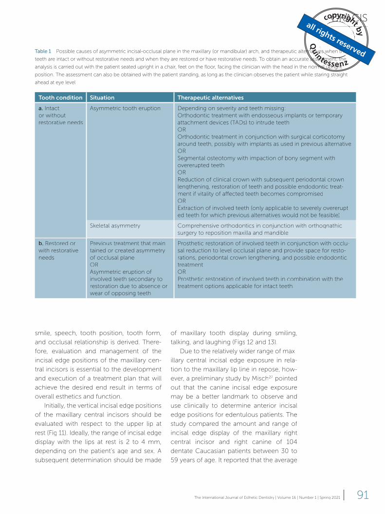
Table 1 Possible causes of asymmetric incisal-occlusal plane in the maxillary (or mandibular) arch, and therapeutic alternatives when the
teeth are intact or without restorative needs and when they are restored or have restorative needs. To obtain an accurate assessment, the
analysis is carried out with the patient seated upright in a chair, feet on the floor, facing the clinician with the head in the normal upright
position. The assessment can also be obtained with the patient standing, as long as the clinician observes the patient while staring straight
ahead at eye level
Tooth condition Situation Therapeutic alternatives
a. Intact
or without
restorative needs
Asymmetric tooth eruption Depending on severity and teeth missing:
Orthodontic treatment with endosseous implants or temporary
attachment devices (TATT Ds) to intrude teeth
OR
Orthodontic treatment in conjunction with surgical corticotomy
around teeth, possibly with implants as used in previous alternative
OR
Segmental osteotomy with impaction of bony segment with
overerupted teeth
OR
Reduction of clinical crown with subsequent periodontal crown
lengthening, restoration of teeth and possible endodontic treat-
ment if vitality of affected teeth becomes compromised
OR
Extraction of involved teeth (only applicable to severely overerupt-
ed teeth for which previous alternatives would not be feasible)
Skeletal asymmetry Comprehensive orthodontics in conjunction with orthognathic
surgery to reposition maxilla and mandible
b. Restored or
with restorative
needs
Previous treatment that main-
tained or created asymmetry
of occlusal plane
OR
Asymmetric eruption of
involved teeth secondary to
restoration due to absence or
wear of opposing teeth
Prosthetic restoration of involved teeth in conjunction with occlu-
sal reduction to level occlusal plane and provide space for resto-
rations, periodontal crown lengthening, and possible endodontic
treatment
OR
Prosthetic restoration of involved teeth in combination with the
treatment options applicable for intact teeth
CLINICAL RESEARCH
92 | The International Journal of Esthetic Dentistry | Volume 16 | Number 1 | Spring 2021
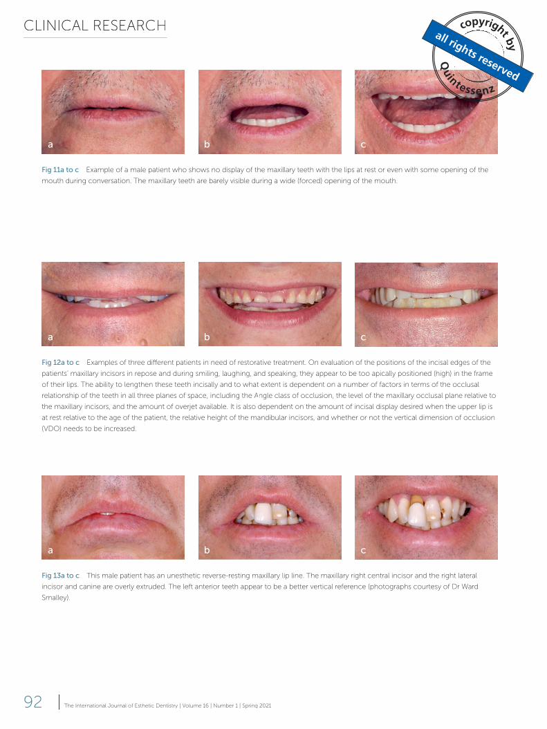
Fig 11a to c Example of a male patient who shows no display of the maxillary teeth with the lips at rest or even with some opening of the
mouth during conversation. The maxillary teeth are barely visible during a wide (forced) opening of the mouth.
Fig 12a to c Examples of three different patients in need of restorative treatment. On evaluation of the positions of the incisal edges of the
patients’ maxillary incisors in repose and during smiling, laughing, and speaking, they appear to be too apically positioned (high) in the frame
of their lips. The ability to lengthen these teeth incisally and to what extent is dependent on a number of factors in terms of the occlusal
relationship of the teeth in all three planes of space, including the Angle class of occlusion, the level of the maxillary occlusal plane relative to
the maxillary incisors, and the amount of overjet available. It is also dependent on the amount of incisal display desired when the upper lip is
at rest relative to the age of the patient, the relative height of the mandibular incisors, and whether or not the vertical dimension of occlusion
(VDO) needs to be increased.
Fig 13a to c This male patient has an unesthetic reverse-resting maxillary lip line. The maxillary right central incisor and the right lateral
incisor and canine are overly extruded. The left anterior teeth appear to be a better vertical reference (photographs courtesy of Dr Ward
Smalley).
a b c
a b c
a b c
GRACIS
93The International Journal of Esthetic Dentistry | Volume 16 | Number 1 | Spring 2021 |
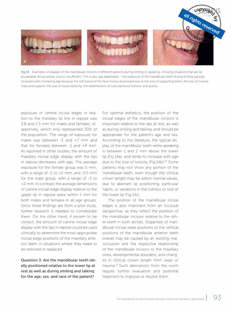
For optimal esthetics, the position of the
incisal edges of the mandibular incisors is
important relative to the lips at rest, as well
as during smiling and talking, and should be
appropriate for the patient’s age and sex.
According to the literature, the typical dis-
play of the mandibular teeth while speaking
is between 1 and 2 mm above the lower
lip (Fig 14a), and tends to increase with age
due to the loss of tonicity (Fig 14b).23 Some
patients may not show any portion of the
mandibular teeth, even though the clinical
crown length may be within normal values,
due to aberrant lip positioning, particular
habits, or variations in the fullness or size of
the lower lip (Fig 14c).
The position of the mandibular incisal
edges is also important from an occlusal
perspective, as they reflect the position of
the mandibular incisors relative to the oth-
er teeth in both arches. Disparities of man-
dibular incisal edge positions or the vertical
positions of the mandibular anterior teeth
overall may be caused by an existing mal-
occlusion and the respective relationship
of the mandibular incisors to the maxillary
ones, developmental disorders, and chang-
es in clinical crown length from wear or
trauma.23 Such aberrations from the norm
require further evaluation and potential
treatment to improve or resolve them.
exposure of central incisal edges in rela-
tion to the maxillary lip line in repose was
3.8 and 2.5 mm for males and females, re-
spectively, which only represented 30% of
the population. The range of exposure for
males was between -3 and +7 mm and
that for females between -1 and +8 mm.
As reported in other studies, the amount of
maxillary incisal edge display with the lips
in repose decreases with age. The average
exposure for the female group was 0 mm,
with a range of -2 to +2 mm, and -0.5 mm
for the male group, with a range of -3 to
+2 mm. In contrast, the average dimensions
of canine incisal edge display relative to the
upper lip in repose were within 1 mm for
both males and females in all age groups.
Since these findings are from a pilot study,
further research is needed to corroborate
them. On the other hand, if proven to be
correct, the amount of canine incisal edge
display with the lips in repose could be used
clinically to determine the most appropriate
incisal edge positions of the maxillary ante-
rior teeth in situations where they need to
be restored or replaced.
Question 3: Are the mandibular teeth ide-
ally positioned relative to the lower lip at
rest as well as during smiling and talking
for the age, sex, and race of the patient?
Fig 14 Examples of displays of the mandibular incisors in different patients during smiling or speaking, showing situations that are (a)
acceptable, (b) excessive, and (c) insufficient. This is also age dependent – the exposure of the mandibular teeth during smiling typically
increases with increasing age because the soft tissue of the face moves downward due to the loss of supporting bone, the loss of muscle
mass and support, the loss of tissue elasticity, the redistribution of subcutaneous fullness, and gravity.
a b c
CLINICAL RESEARCH
94 | The International Journal of Esthetic Dentistry | Volume 16 | Number 1 | Spring 2021
Although there are no norms for specific
tooth sizes for all individuals, average values
do exist within a range of norms for dimen-
sions in all three planes of space, as has been
reported in the literature by a number of
authors.24-30 Based on the findings of these
authors, these norms provide the average
and the range of measurements as well as
the average proportions of each tooth. As
a consequence, the size of patients’ teeth
can be evaluated as being within the norm
(Figs 15a and 16a), too narrow (ie, too long
relative to width; Figs 15b and 16b) or too
wide (ie, too short relative to width; Figs 15c
and 16c).
Applying the average tooth sizes or
shapes to all patients is inappropriate as
there is a range of tooth sizes and shapes
among people. Applying the average tooth
proportions to specific individuals is also
problematic. Two teeth may have the same
proportions and yet be completely different
in size. Worse yet, a worn tooth may have
optimal proportions with respect to height
and width yet be undersized relative to an
intact tooth.
Transverse assessment of the teeth rela-
tive to one another in terms of size, propor-
tion, and shape (or form) should also be per-
formed, as such parameters influence the
overall intercuspation of a patient’s teeth,
An appropriate treatment goal is to reduce
or increase the amount of incisal display, de-
pending on the etiology of the disparity, its
magnitude, and the benefit to be gained from
treatment. The etiology of a deficiency or ex-
cess in mandibular incisal display could be
related to the size of the incisors and/or their
position relative to the adjacent and oppos-
ing teeth. Treatment alternatives to correct
the disparate incisal display, depending on
the etiology, include orthodontic movement
of the incisors to more appropriate positions,
changing their clinical crown height, or both.
The degree of involvement will determine the
extent of the alteration of the incisal crown
height or the degree of tooth movement.
Correction may also require surgical crown
lengthening and/or intentional endodontic
treatment of the involved teeth. Such treat-
ment, which will alter the occlusal relation-
ship of the mandibular incisors relative to the
maxillary ones, will also require management.
Step 2: Assessment of anterior tooth dimensions
Question 4: Are the clinical crowns of the
maxillary and mandibular anterior teeth
appropriate with respect to size, shape,
and proportions (width to height) consis-
tent with established norms?
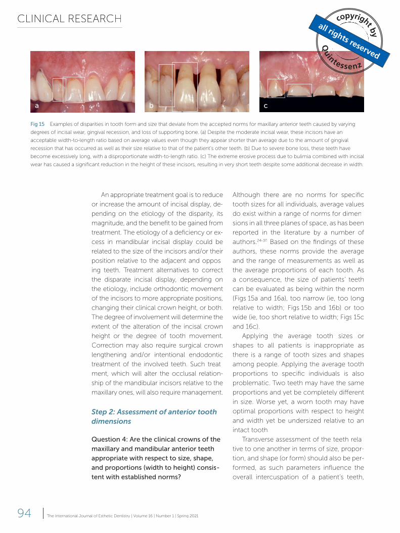
Fig 15 Examples of disparities in tooth form and size that deviate from the accepted norms for maxillary anterior teeth caused by varying
degrees of incisal wear, gingival recession, and loss of supporting bone. (a) Despite the moderate incisal wear, these incisors have an
acceptable width-to-length ratio based on average values even though they appear shorter than average due to the amount of gingival
recession that has occurred as well as their size relative to that of the patient’s other teeth. (b) Due to severe bone loss, these teeth have
become excessively long, with a disproportionate width-to-length ratio. (c) The extreme erosive process due to bulimia combined with incisal
wear has caused a significant reduction in the height of these incisors, resulting in very short teeth despite some additional decrease in width.
a b c
GRACIS
95The International Journal of Esthetic Dentistry | Volume 16 | Number 1 | Spring 2021 |
Orthodontists, using a tooth size analysis
commonly referred to as the Bolton anal-
ysis, use a similar approach to determine
whether the maxillary and mandibular teeth
can achieve maximum intercuspation (MI)
in a Class I relationship with normal values
of vertical and horizontal overlap of the an-
terior teeth, based on the sizes, specifically
the widths, of the teeth. The Bolton analysis
measures the sum total of the mesiodistal
widths of the six mandibular anterior teeth
and compares it with that of the six maxillary
anterior teeth to determine whether a dis-
crepancy exists between the total widths of
the anterior teeth in both jaws, which would
not allow the teeth to achieve MI. The per-
centage that represents the ideal ratio for
the anterior teeth is 77.2%, arrived at by divid-
ing the sum total of the mandibular anterior
tooth widths by that of the maxillary anterior
tooth widths, measured at the widest part
of the respective teeth, times 100.35 If the
number is lower, it means that either one or
more of the mandibular anterior teeth are
too small or that one or more of the maxil-
lary anterior teeth are too large. Conversely,
a higher number implies that one or more of
the mandibular anterior teeth are too large
or that one or more of the maxillary anterior
teeth are smaller than what they should be,
as in the case of peg laterals.
arch width, and certainly the smile. Howev-
er, not all assessments or recommended ap-
proaches in this regard have much validity.
One such recommendation advises that the
proportional widths of the maxillary anteri-
or teeth should mirror the proportional re-
lationship of the ‘golden proportion,’ which
is essentially a ratio of 1.618 to 1.0.31 Using
this ratio, the relative width of the central in-
cisor would be 1.618, the lateral incisor 1.0,
and the canine 0.618. It is important to re-
alize that the ratio only applies to the teeth
in alignment within the arch viewed from
the front, ie, the apparent width of the teeth
due to the curvature of the dental arch. It
does not apply to the actual widths of the
teeth. In reality, the golden proportion has
no validity for use in dentistry, despite the
recommendations of some authors.32 Even
Lombardi, who first considered the possibil-
ity that the golden proportion might have
some relevance to tooth sizes, has since de-
termined that it does not.33 Since the initial
publication, it has been reported that only
a relatively small percentage of people with
naturally arranged teeth fit the ratio of the
golden proportion.34 It has also been shown
that aligning teeth according to this rule cre-
ates relatively narrow or constricted dental
arches that are too narrow for the opposing
mandibular arch and are rather unesthetic.30
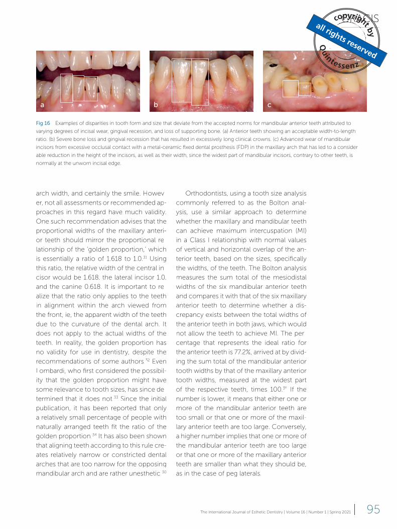
Fig 16 Examples of disparities in tooth form and size that deviate from the accepted norms for mandibular anterior teeth attributed to
varying degrees of incisal wear, gingival recession, and loss of supporting bone. (a) Anterior teeth showing an acceptable width-to-length
ratio. (b) Severe bone loss and gingival recession that has resulted in excessively long clinical crowns. (c) Advanced wear of mandibular
incisors from excessive occlusal contact with a metal-ceramic fixed dental prosthesis (FDP) in the maxillary arch that has led to a consider-
able reduction in the height of the incisors, as well as their width, since the widest part of mandibular incisors, contrary to other teeth, is
normally at the unworn incisal edge.
a b c
CLINICAL RESEARCH
96 | The International Journal of Esthetic Dentistry | Volume 16 | Number 1 | Spring 2021
If the incisal edges of the maxillary cen-
tral incisors are correctly positioned rela-
tive to the upper lip at rest, but their clinical
crowns are too long (Table 3, section I-a),
as viewed during smiling, the appropriate
treatment goal would be to correct the
crown length disparity without changing
their position. If the increased length of the
maxillary central incisors is due to gingival
recession, correction of the problem might
be achieved, depending on the overall con-
dition of the teeth, either through a peri-
odontal root coverage procedure or forced
eruption of the teeth with subsequent re-
duction of the incisal edges and restoration
(Fig 17a to d). If the teeth have a hopeless
prognosis due to significant loss of support-
ing bone, the best treatment might seem to
be the extraction of the affected teeth with
subsequent replacement. However, replac-
ing these failing teeth is likely to be challeng-
ing from an esthetic perspective due to the
additional bone loss that would occur sec-
ondary to their removal. Grafting bone to
restore vertical bone height is difficult and
not predictable. Another treatment modality
that would improve the situation rather than
make it worse is orthodontic eruption of the
compromised teeth to extraction. This alter-
native would not only preserve the existing
The Bolton analysis can also determine
whether an overall disparity exists by ex-
tending the measurements to the first mo-
lars. The ideal ratio then becomes 91.3%.
Note that the Bolton analysis, from a practi-
cal perspective, is beneficial only if the teeth
are in a Class I relationship. If a patient’s
teeth are in considerable disrepair, with a
significant alteration of their size and shape,
it can be beneficial to apply the analysis to
the widths of any planned prosthetic teeth.
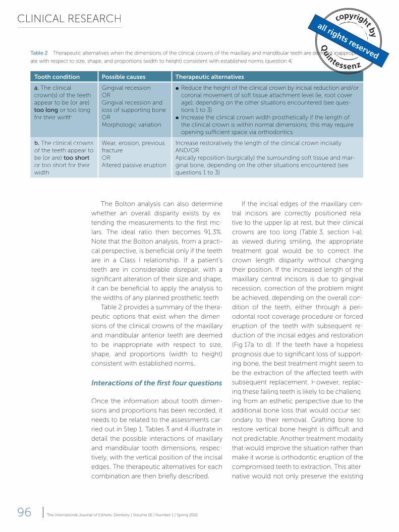
Table 2 provides a summary of the thera-
peutic options that exist when the dimen-
sions of the clinical crowns of the maxillary
and mandibular anterior teeth are deemed
to be inappropriate with respect to size,
shape, and proportions (width to height)
consistent with established norms.
Interactions of the first four questions
Once the information about tooth dimen-
sions and proportions has been recorded, it
needs to be related to the assessments car-
ried out in Step 1. Tables 3 and 4 illustrate in
detail the possible interactions of maxillary
and mandibular tooth dimensions, respec-
tively, with the vertical position of the incisal
edges. The therapeutic alternatives for each
combination are then briefly described.
Table 2 Therapeutic alternatives when the dimensions of the clinical crowns of the maxillary and mandibular teeth are deemed inappropri-
ate with respect to size, shape, and proportions (width to height) consistent with established norms (question 4)
Tooth condition Possible causes Therapeutic alternatives
a. The clinical
crown(s) of the teeth
appear to be (or are)
too long or too long
for their width
Gingival recession
OR
Gingival recession and
loss of supporting bone
OR
Morphologic variation
● Reduce the height of the clinical crown by incisal reduction and/or
coronal movement of soft tissue attachment level (ie, root cover-
age), depending on the other situations encountered (see ques-
tions 1 to 3)
● Increase the clinical crown width prosthetically if the length of
the clinical crown is within normal dimensions; this may require
opening sufficient space via orthodontics
b. The clinical crowns
of the teeth appear to
be (or are) too shortor too short for their
width
Wear, erosion, previous
fracture
OR
Altered passive eruption
Increase restoratively the length of the clinical crown incisally
AND/OR
Apically reposition (surgically) the surrounding soft tissue and mar-
ginal bone, depending on the other situations encountered (see
questions 1 to 3)
GRACIS
97The International Journal of Esthetic Dentistry | Volume 16 | Number 1 | Spring 2021 |
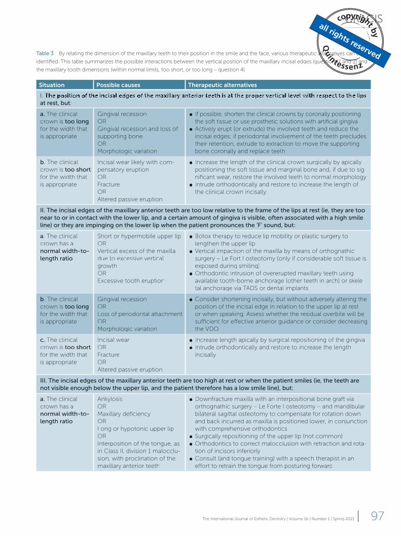
Table 3 By relating the dimension of the maxillary teeth to their position in the smile and the face, various therapeutic alternatives can be
identified. This table summarizes the possible interactions between the vertical position of the maxillary incisal edges (questions 1 and 2) and
the maxillary tooth dimensions (within normal limits, too short, or too long – question 4)
Situation Possible causes Therapeutic alternatives
at rest, but:
a. The clinical
crown is too longfor the width that
is appropriate
Gingival recession
OR
Gingival recession and loss of
supporting bone
OR
Morphologic variation
● If possible, shorten the clinical crowns by coronally positioning
the soft tissue or use prosthetic solutions with artificial gingiva
● Actively erupt (or extrude) the involved teeth and reduce the
incisal edges; if periodontal involvement of the teeth precludes
their retention, extrude to extraction to move the supporting
bone coronally and replace teeth
b. The clinical
crown is too shortfor the width that
is appropriate
Incisal wear likely with com-
pensatory eruption
OR
Fracture
OR
Altered passive eruption
● Increase the length of the clinical crown surgically by apically
positioning the soft tissue and marginal bone and, if due to sig-
nificant wear, restore the involved teeth to normal morphology
● Intrude orthodontically and restore to increase the length of
the clinical crown incisally
II. The incisal edges of the maxillary anterior teeth are too low relative to the frame of the lips at rest (ie, they are too near to or in contact with the lower lip, and a certain amount of gingiva is visible, often associated with a high smile line) or they are impinging on the lower lip when the patient pronounces the ‘F’ sound, but:
a. The clinical
crown has a
normal width-to-length ratio
Short or hypermobile upper lip
OR
Vertical excess of the maxilla
due to excessive vertical
growth
OR
Excessive tooth eruption
● Botox therapy to reduce lip mobility or plastic surgery to
lengthen the upper lip
● Vertical impaction of the maxilla by means of orthognathic
surgery – Le Fort I osteotomy (only if considerable soft tissue is
exposed during smiling)
● Orthodontic intrusion of overerupted maxillary teeth using
available tooth-borne anchorage (other teeth in arch) or skele-
tal anchorage via TATT DS or dental implants
b. The clinical
crown is too longfor the width that
is appropriate
Gingival recession
OR
Loss of periodontal attachment
OR
Morphologic variation
● Consider shortening incisally, but without adversely altering the
position of the incisal edge in relation to the upper lip at rest
or when speaking. Assess whether the residual overbite will be
sufficient for effective anterior guidance or consider decreasing
the VDO
c. The clinical
crown is too shortfor the width that
is appropriate
Incisal wear
OR
Fracture
OR
Altered passive eruption
● Increase length apically by surgical repositioning of the gingiva
● Intrude orthodontically and restore to increase the length
incisally
III. The incisal edges of the maxillary anterior teeth are too high at rest or when the patient smiles (ie, the teeth are not visible enough below the upper lip, and the patient therefore has a low smile line), but:
a. The clinical
crown has a
normal width-to-length ratio
Ankylosis
OR
Maxillary deficiency
OR
Long or hypotonic upper lip
OR
Interposition of the tongue, as
in Class II, division 1 malocclu-
sion, with proclination of the
maxillary anterior teeth
● Downfracture maxilla with an interpositional bone graft via
orthognathic surgery – Le Forte I osteotomy – and mandibular
bilateral sagittal osteotomy to compensate for rotation down
and back incurred as maxilla is positioned lower, in conjunction
with comprehensive orthodontics
● Surgically repositioning of the upper lip (not common)
● Orthodontics to correct malocclusion with retraction and rota-
tion of incisors inferiorly
● Consult (and tongue training) with a speech therapist in an
effort to retrain the tongue from posturing forward
CLINICAL RESEARCH
98 | The International Journal of Esthetic Dentistry | Volume 16 | Number 1 | Spring 2021
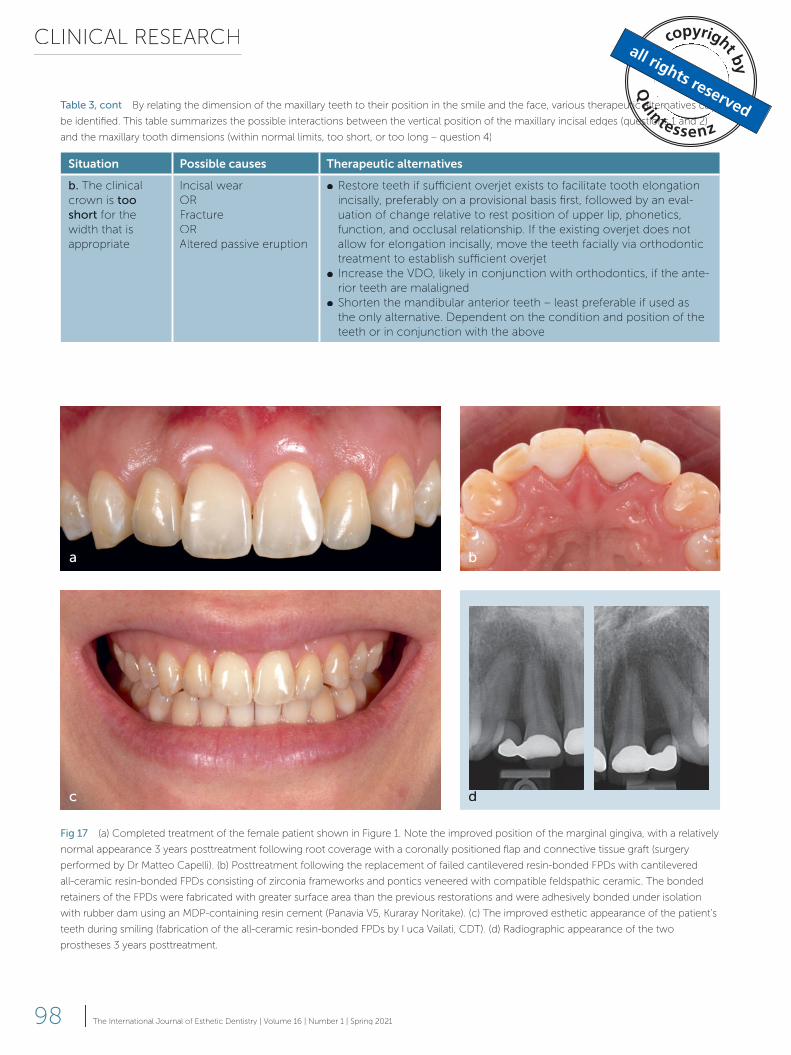
Table 3, cont By relating the dimension of the maxillary teeth to their position in the smile and the face, various therapeutic alternatives can
be identified. This table summarizes the possible interactions between the vertical position of the maxillary incisal edges (questions 1 and 2)
and the maxillary tooth dimensions (within normal limits, too short, or too long – question 4)
Situation Possible causes Therapeutic alternatives
b. The clinical
crown is too short for the
width that is
appropriate
Incisal wear
OR
Fracture
OR
Altered passive eruption
● Restore teeth if sufficient overjet exists to facilitate tooth elongation
incisally, preferably on a provisional basis first, followed by an eval-
uation of change relative to rest position of upper lip, phonetics,
function, and occlusal relationship. If the existing overjet does not
allow for elongation incisally, move the teeth facially via orthodontic
treatment to establish sufficient overjet
● Increase the VDO, likely in conjunction with orthodontics, if the ante-
rior teeth are malaligned
● Shorten the mandibular anterior teeth – least preferable if used as
the only alternative. Dependent on the condition and position of the
teeth or in conjunction with the above
Fig 17 (a) Completed treatment of the female patient shown in Figure 1. Note the improved position of the marginal gingiva, with a relatively
normal appearance 3 years posttreatment following root coverage with a coronally positioned flap and connective tissue graft (surgery
performed by Dr Matteo Capelli). (b) Posttreatment following the replacement of failed cantilevered resin-bonded FPDs with cantilevered
all-ceramic resin-bonded FPDs consisting of zirconia frameworks and pontics veneered with compatible feldspathic ceramic. The bonded
retainers of the FPDs were fabricated with greater surface area than the previous restorations and were adhesively bonded under isolation
with rubber dam using an MDP-containing resin cement (Panavia V5, Kuraray Noritake). (c) The improved esthetic appearance of the patient’s
teeth during smiling (fabrication of the all-ceramic resin-bonded FPDs by Luca Vailati, CDT). (d) Radiographic appearance of the two
prostheses 3 years posttreatment.
a
c
b
dd
GRACIS
99The International Journal of Esthetic Dentistry | Volume 16 | Number 1 | Spring 2021 |
bone, but would also develop new bone and
create a bony ridge that would facilitate sub-
sequent prosthodontic treatment, provided
that the periodontal situation of the relevant
teeth is favorable for orthodontic extrusion.
Forced eruption has the potential for
hard and/or soft tissue augmentation be-
cause orthodontic tooth movement affects
the periodontal anatomical structures, pro-
ducing a predictable biologic response.36
The periodontal ligament and the supracr-
estal gingival fibers connect the tooth to the
bone. These fibers stretch as the orthodon-
tic force moves the tooth coronally, thus
producing tension in the bone on a cellular
level, which causes bone deposition. New
bone is formed at the crestal aspect of the
alveolar bone and along the surface of the
root approximating the bone. On the oth-
er hand, the behavior of the overlying gin-
gival soft tissue during and after extrusion
depends on the evaluation of three param-
eters to assess whether the attached gin-
giva is connected to the root surface and/
or the periosteum: sulcus or pocket depth,
position of the mucogingival junction (MGJ)
relative to the crest of the bone, and deter-
mination of the location of the bone crest.
According to the classification proposed by
Hochman et al,36 when the extruded teeth
have the attached gingiva connected to
both the bone and the root surface (Type
1 classification), an increase in the width of
the attached gingiva is to be expected. If the
attached gingiva and the MGJ are connect-
ed to the root surface (Type 2), the gingi-
val tissue moves coronally with the tooth,
but an increase in the width of the attached
gingiva does not occur. Finally, when a peri-
odontal pocket is present (Type 3), during
orthodontic extrusion the free gingival mar-
gin does not move coronally, so that the
end result is a complete elimination of the
periodontal pocket.
When the incisal edges of the maxillary
central incisors are correctly positioned
relative to the upper lip at rest, but the clinical
crowns are short rather than long (Table 3,
section I-b), the therapeutic alternatives are
completely different. For this situation to oc-
cur, the teeth may have been worn down
or fractured in conjunction with compensa-
tory eruption. Treatment alternatives include
surgical lengthening of the clinical crown
to expose sufficient tooth length to facili-
tate restoration of the tooth and decrease
the excessive amount of gingival display or
orthodontic intrusion of the tooth, followed
by prosthetic restoration to reestablish nor-
mal clinical crown dimensions (see Fig 19).
The overall level of marginal bone of the
involved teeth relative to the adjacent unin-
volved teeth will likely have some influence
on the best procedure to choose. Another
situation is where the clinical crowns of a
patient’s teeth appear short, but the teeth
are intact due to altered passive eruption.
Excessive gingival tissue is exposed due to
the failure of the marginal gingiva to migrate
apically to the normal position of 1 mm
above the cementoenamel junction (CEJ)
of the involved teeth. Treatment would be to
surgically lengthen the crowns of the teeth
involved to correctly position the marginal
gingiva relative to the CEJ.
If the incisal edges of the maxillary ante-
rior teeth are too low relative to the frame of
the lips at rest (ie, they are near to or in con-
tact with the lower lip, and a certain amount
of gingiva is visible) or they are impinging on
the lower lip when the patient pronounces
the ‘F’ sound, different therapeutic alterna-
tives are available depending on the size of
the anterior clinical crowns (Table 3, section
II). When the clinical crown has a normal
width-to-length ratio, the excessive tooth
visibility (often accompanied by a so-called
gummy smile; see Fig 6c) may be due to a
short or hypermobile upper lip, exaggerated
vertical growth of the maxilla or excessive
tooth eruption (as in patients with Class II,
division 2 malocclusions). Depending on the
CLINICAL RESEARCH
100 | The International Journal of Esthetic Dentistry | Volume 16 | Number 1 | Spring 2021
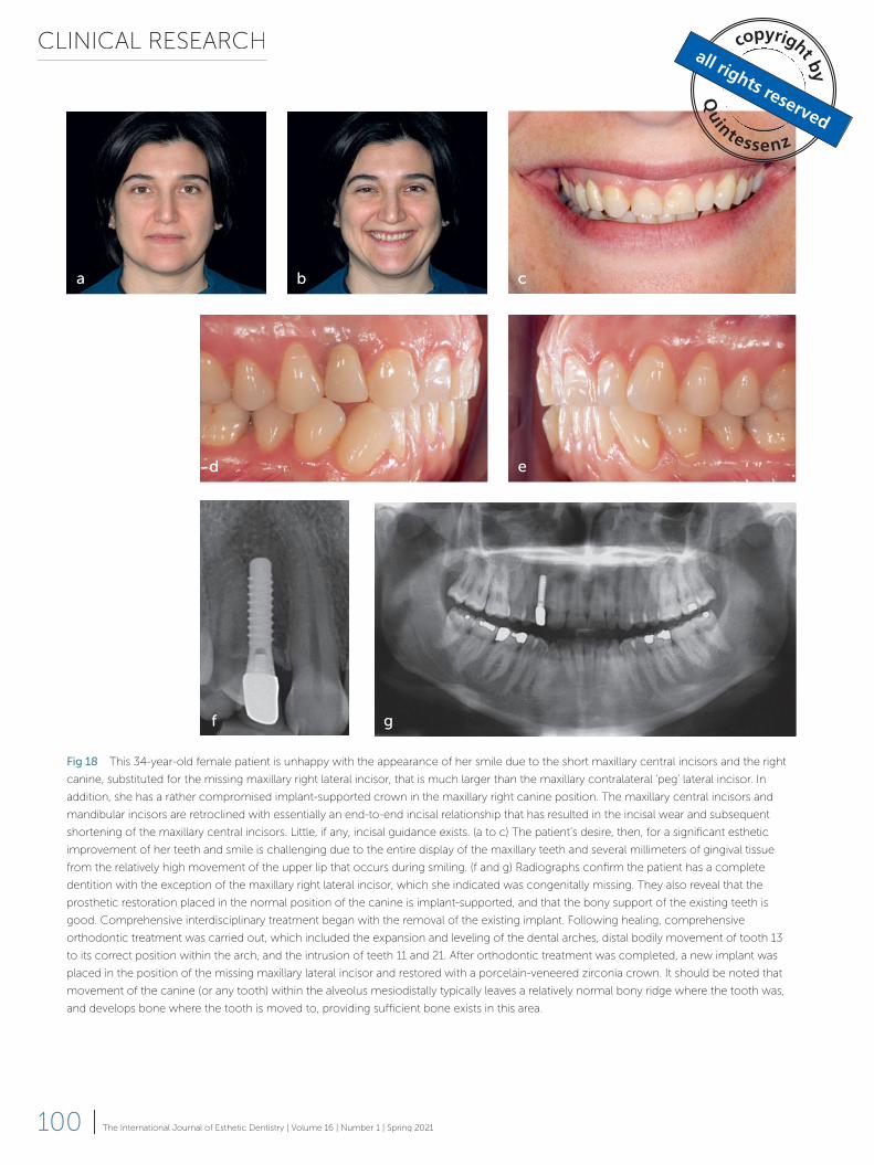
Fig 18 This 34-year-old female patient is unhappy with the appearance of her smile due to the short maxillary central incisors and the right
canine, substituted for the missing maxillary right lateral incisor, that is much larger than the maxillary contralateral ‘peg’ lateral incisor. In
addition, she has a rather compromised implant-supported crown in the maxillary right canine position. The maxillary central incisors and
mandibular incisors are retroclined with essentially an end-to-end incisal relationship that has resulted in the incisal wear and subsequent
shortening of the maxillary central incisors. Little, if any, incisal guidance exists. (a to c) The patient’s desire, then, for a significant esthetic
improvement of her teeth and smile is challenging due to the entire display of the maxillary teeth and several millimeters of gingival tissue
from the relatively high movement of the upper lip that occurs during smiling. (f and g) Radiographs confirm the patient has a complete
dentition with the exception of the maxillary right lateral incisor, which she indicated was congenitally missing. They also reveal that the
prosthetic restoration placed in the normal position of the canine is implant-supported, and that the bony support of the existing teeth is
good. Comprehensive interdisciplinary treatment began with the removal of the existing implant. Following healing, comprehensive
orthodontic treatment was carried out, which included the expansion and leveling of the dental arches, distal bodily movement of tooth 13
to its correct position within the arch, and the intrusion of teeth 11 and 21. After orthodontic treatment was completed, a new implant was
placed in the position of the missing maxillary lateral incisor and restored with a porcelain-veneered zirconia crown. It should be noted that
movement of the canine (or any tooth) within the alveolus mesiodistally typically leaves a relatively normal bony ridge where the tooth was,
and develops bone where the tooth is moved to, providing sufficient bone exists in this area.
f g
a b c
d e

GRACIS
101The International Journal of Esthetic Dentistry | Volume 16 | Number 1 | Spring 2021 |
Fig 18, cont (h) The patient was happy with the overall result of the
treatment and her much improved smile. (i) Extraoral smile
photograph of the final treatment result Teeth 11, 21, and 22 were
restored with ceramic veneers. At the completion of the rehabilita-
tion, appropriate overbite and overjet was established to provide an
acceptable envelope of function and anterior guidance to disclude
the posterior teeth during excursive movements of the mandible.
(j) Periapical radiographs of the maxillary anterior teeth at the
completion of treatment. (k to m) Excursive movements of the
dentition 3 years postsurgery (orthodontic treatment by Dr David De
Franco; implant surgeries by Dr Matteo Capelli; prosthetic laboratory
work by Luca Vailati, CDT).
h
i
j
k l
m
CLINICAL RESEARCH
102 | The International Journal of Esthetic Dentistry | Volume 16 | Number 1 | Spring 2021
affecting the occlusal and functional rela-
tionships as well as the overall stability of the
teeth, otherwise alternative treatments need
to be considered. These include increasing
the length of the mandibular anterior teeth
(see question 3), decreasing the VDO (see
question 6) or orthodontic correction of the
problem. It is important to maintain or es-
tablish sufficient overbite to provide disclu-
sion of the posterior teeth during excursive
movements of the mandible.
If the anterior teeth are too short (Ta-
ble 3, section II-c) and the goal of treatment
is to increase their length, one needs to de-
cide whether to lengthen them restorative-
ly, surgically or both. This treatment plan
depends on an assessment of the vertical
position of these teeth with respect to the
lips in repose, when smiling and talking, in
function, and in the patient’s face. This as-
sessment should always be based on the
dynamic movement of the lips (see Fig 21).
etiology, the proposed therapies, therefore,
range from Botox injections to orthognath-
ic surgery to orthodontic intrusion, respec-
tively (Table 3, section II-a). This last option
requires considerable anchorage provided
either by all the other maxillary teeth or skel-
etal anchorage via TATT DS or dental implants.
Once repositioning has been achieved, the
clinician, however, has to provide fixed re-
tention or a night guard to maintain the new
position of the teeth.
In cases where the clinical crowns are
assessed as being too long relative to the
other teeth, or because of recession, loss
of periodontal attachment or morphologic
variation (Table 3, section II-b), shortening of
the teeth is typically indicated (see Fig 20).
However, the patient should be made aware
that this may lead to endodontic treatment
and a preprosthetic reconstruction. One
needs to determine how much the involved
teeth can be shortened without adversely
Fig 19a to c Female patient with a Class II, division 2 malocclusion showing the typical dental compensations that occur: significant
overeruption of the maxillary incisors (usually upright or palatally inclined) and proclination of the lateral incisors, minimal or no overjet, and
the development of a reverse maxillary curve of Spee. Overeruption of the mandibular incisors also usually occurs, with exaggeration of the
mandibular curve of Spee, and often palatal impingement by the mandibular incisors. Wear of the incisors, if present, typically occurs on the
incised edges of the maxillary central incisors and mandibular incisors, the labial surfaces of the mandibular incisors, and the palatal surfaces
of the maxillary central incisors due to a constricted arc of closure during function and/or parafunction. As the clinical crown height of the
incisors decreases due to wear, the teeth take on a squarer appearance. In addition, the maxillary arch usually becomes constricted to some
degree to facilitate intercuspation of the maxillary and mandibular teeth posteriorly as the malocclusion develops. Ideally, this patient should
undergo comprehensive orthodontic treatment in conjunction with the surgical advancement of the mandible. Orthodontic treatment
presurgically would consist of decompensation of the teeth by aligning them, which includes proclination of the incisors, leveling the curves
of Spee, and opening spaces to facilitate the restoration of any worn teeth deficient in width, in conjuction with expanding the maxillary arch.
This enables the worn teeth to be optimally positioned vertically for the definitive restoration of the worn edges. During orthodontic
treatment, after the initial alignment of the teeth, they should be provisionally restored with an appropriate composite material to establish
the proper tooth width and height. This is particularly important to establish ideal tooth positions.
a b c
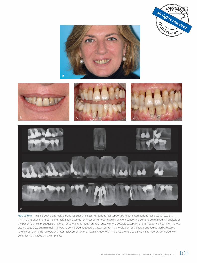
GRACIS
103The International Journal of Esthetic Dentistry | Volume 16 | Number 1 | Spring 2021 |
Fig 20a to h This 62-year-old female patient has substantial loss of periodontal support from advanced periodontal disease (Stage 4,
Grade C). As seen in the complete radiographic survey (e), most of her teeth have insufficient supporting bone to be retained. An analysis of
the patient’s smile (b) suggests that the maxillary anterior teeth are too long, with the possible exception of the maxillary left canine. The over-
bite is acceptable but minimal. The VDO is considered adequate as assessed from the evaluation of the facial and radiographic features
(lateral cephalometric radiograph). After replacement of the maxillary teeth with implants, a one-piece zirconia framework veneered with
ceramics was placed on the implants.
a
b c d
e
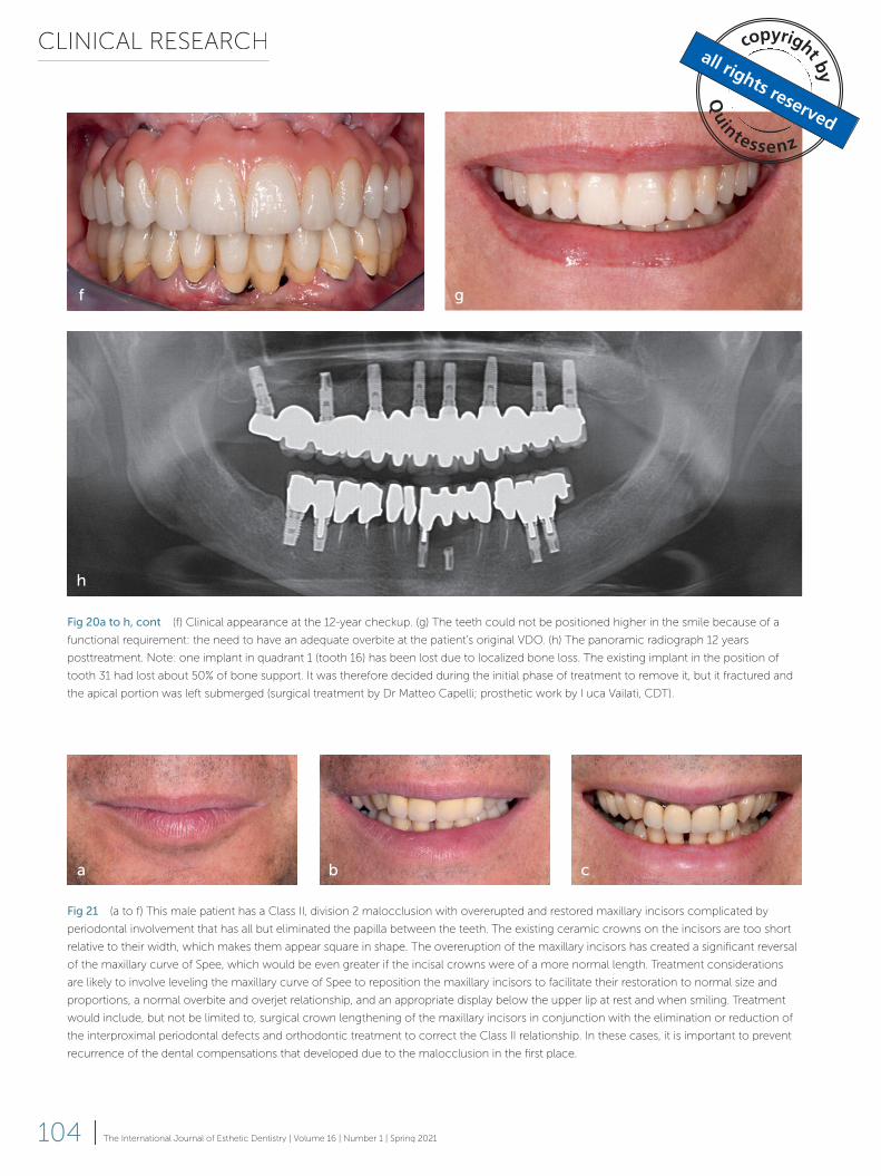
CLINICAL RESEARCH
104 | The International Journal of Esthetic Dentistry | Volume 16 | Number 1 | Spring 2021
f g
Fig 20a to h, cont (f) Clinical appearance at the 12-year checkup. (g) The teeth could not be positioned higher in the smile because of a
functional requirement: the need to have an adequate overbite at the patient’s original VDO. (h) The panoramic radiograph 12 years
posttreatment. Note: one implant in quadrant 1 (tooth 16) has been lost due to localized bone loss. The existing implant in the position of
tooth 31 had lost about 50% of bone support. It was therefore decided during the initial phase of treatment to remove it, but it fractured and
the apical portion was left submerged (surgical treatment by Dr Matteo Capelli; prosthetic work by Luca Vailati, CDT).
h
Fig 21 (a to f) This male patient has a Class II, division 2 malocclusion with overerupted and restored maxillary incisors complicated by
periodontal involvement that has all but eliminated the papilla between the teeth. The existing ceramic crowns on the incisors are too short
relative to their width, which makes them appear square in shape. The overeruption of the maxillary incisors has created a significant reversal
of the maxillary curve of Spee, which would be even greater if the incisal crowns were of a more normal length. Treatment considerations
are likely to involve leveling the maxillary curve of Spee to reposition the maxillary incisors to facilitate their restoration to normal size and
proportions, a normal overbite and overjet relationship, and an appropriate display below the upper lip at rest and when smiling. Treatment
would include, but not be limited to, surgical crown lengthening of the maxillary incisors in conjunction with the elimination or reduction of
the interproximal periodontal defects and orthodontic treatment to correct the Class II relationship. In these cases, it is important to prevent
recurrence of the dental compensations that developed due to the malocclusion in the first place.
a b c
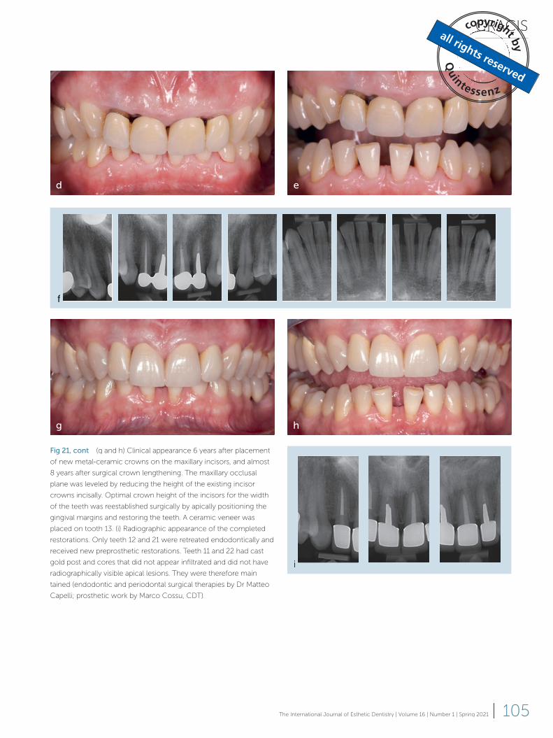
GRACIS
105The International Journal of Esthetic Dentistry | Volume 16 | Number 1 | Spring 2021 |
d e
g h
f
i
Fig 21, cont (g and h) Clinical appearance 6 years after placement
of new metal-ceramic crowns on the maxillary incisors, and almost
8 years after surgical crown lengthening. The maxillary occlusal
plane was leveled by reducing the height of the existing incisor
crowns incisally. Optimal crown height of the incisors for the width
of the teeth was reestablished surgically by apically positioning the
gingival margins and restoring the teeth. A ceramic veneer was
placed on tooth 13. (i) Radiographic appearance of the completed
restorations. Only teeth 12 and 21 were retreated endodontically and
received new preprosthetic restorations. Teeth 11 and 22 had cast
gold post and cores that did not appear infiltrated and did not have
radiographically visible apical lesions. They were therefore main-
tained (endodontic and periodontal surgical therapies by Dr Matteo
Capelli; prosthetic work by Marco Cossu, CDT).
CLINICAL RESEARCH
106 | The International Journal of Esthetic Dentistry | Volume 16 | Number 1 | Spring 2021
merely opening the patient’s bite, would not
be indicated. Correction requires orthog-
nathic surgery to downfracture the maxilla
via a Le Fort I osteotomy with an interposi-
tional bone graft between the floor of the
nose and the top of the maxilla, in combi-
nation with comprehensive orthodontic
treatment. Surgery of the mandible may be
required. A bilateral sagittal split osteotomy
of the mandible may need to be done at
the same time to compensate for any sig-
nificant rotation of the mandible down and
back as the maxilla is moved down. The re-
sult of such treatment is a considerable in-
crease in maxillary tooth display relative to
the lips and face, with an increase in the pa-
tient’s lower facial height. It should be not-
ed that opening the VDO slightly in less in-
volved situations to facilitate the restoration
does not bring about enough change in the
mandibular musculature to cause any signif-
icant disturbance to the patient’s musculo-
skeletal system.37,38 Whether to correct the
problem of deficient maxillary incisal display
restoratively with crowns or veneers, or-
thodontics, and/or surgery depends on the
cause and magnitude of the problem, the
condition of the teeth, the bite relationship,
the skeletal relationship of the jaws, and the
esthetic goals.
Table 4 addresses a variety of situations
concerning mandibular incisor visibility with
respect to the lower lip at rest and when
talking and smiling. Three situations are
possible relative to the age of the patient:
with respect to the lower lip, the mandib-
ular teeth are sufficiently visible (section I),
not visible enough (section II), and too visi-
ble (section III). This visibility, however, must
relate to the dimension of the anterior teeth,
as assessed intraorally.
If the mandibular anterior teeth are suf-
ficiently visible but the clinical crown is too
short due to incisal wear or fracture, likely
followed by compensatory eruption (Ta-
ble 4, section I-a), establishing the proper
If the maxillary incisal position is high
with respect to the frame of the lips (ie,
barely visible) or, worse yet, not visible at all
(ie, the patient has a low smile line – Table 3,
section III; see Figs 11 and 12), it would like-
ly be appropriate and desirable to increase
the display of the maxillary teeth. The mag-
nitude and etiology of the relationship as
well as the desires of the patient will have a
bearing on the appropriate treatment alter-
natives available to do this. One possibility
to increase the display of the maxillary inci-
sors as well as the other maxillary teeth is to
lengthen the teeth restoratively at the incisal
edges, especially if they are short for the
width that is appropriate (Table 3, section
III-b), provided that the change in length is
compatible from an occlusal perspective
with the opposing mandibular teeth. If not,
an increase in the VDO may be required
(see question 6). Increasing the display of
the maxillary teeth may also be accom-
plished through orthodontic movement of
the teeth (ie, buccal movement of the max-
illary anterior teeth and lingual movement
of the mandibular teeth). The teeth can be
lengthened incisally even though the width-
to-length ratio is acceptable, as the added
length will not be seen. The patient’s re-
sponse to such treatment with respect to
function, phonetics, and esthetics can be
evaluated with a provisional phase of treat-
ment before any definitive or irreversible
treatment is initiated to see whether the pa-
tient can tolerate the planned changes.
A significant deficiency or absence of
maxillary teeth seen during smiling is like-
ly related to a maxillary deficiency, provid-
ed that the heights of the clinical crowns
have not been compromised due to wear
(erosion, attrition or abrasion), ankyloses of
multiple teeth, or due to some other cause
(Table 3, section III-a). This phenomenon is
typically associated with decreased lower
facial height. Lengthening of the teeth, even
with an increase in the VDO achieved by
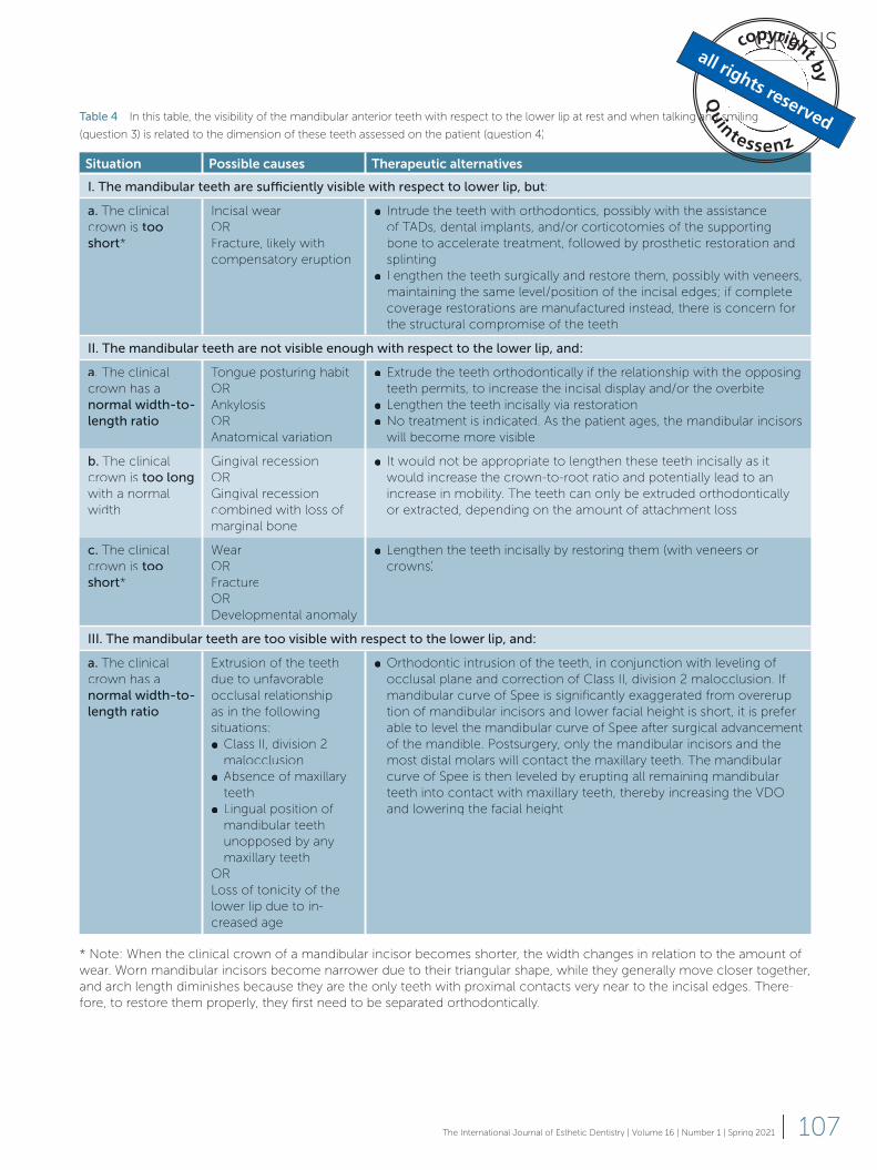
GRACIS
107The International Journal of Esthetic Dentistry | Volume 16 | Number 1 | Spring 2021 |
Table 4 In this table, the visibility of the mandibular anterior teeth with respect to the lower lip at rest and when talking and smiling
(question 3) is related to the dimension of these teeth assessed on the patient (question 4)
Situation Possible causes Therapeutic alternatives
I. The mandibular teeth are sufficiently visible with respect to lower lip, but:
a. The clinical
crown is too short*
Incisal wear
OR
Fracture, likely with
compensatory eruption
● Intrude the teeth with orthodontics, possibly with the assistance
of TATT Ds, dental implants, and/or corticotomies of the supporting
bone to accelerate treatment, followed by prosthetic restoration and
splinting
● Lengthen the teeth surgically and restore them, possibly with veneers,
maintaining the same level/position of the incisal edges; if complete
coverage restorations are manufactured instead, there is concern for
the structural compromise of the teeth
II. The mandibular teeth are not visible enough with respect to the lower lip, and:
a. The clinical
crown has a
normal width-to-length ratio
Tongue posturing habit
OR
Ankylosis
OR
Anatomical variation
● Extrude the teeth orthodontically if the relationship with the opposing
teeth permits, to increase the incisal display and/or the overbite
● Lengthen the teeth incisally via restoration
● No treatment is indicated. As the patient ages, the mandibular incisors
will become more visible
b. The clinical
crown is too longwith a normal
width
Gingival recession
OR
Gingival recession
combined with loss of
marginal bone
● It would not be appropriate to lengthen these teeth incisally as it
would increase the crown-to-root ratio and potentially lead to an
increase in mobility. The teeth can only be extruded orthodontically
or extracted, depending on the amount of attachment loss
c. The clinical
crown is too short*
Wear
OR
Fracture
OR
Developmental anomaly
● Lengthen the teeth incisally by restoring them (with veneers or
crowns)
III. The mandibular teeth are too visible with respect to the lower lip, and:
a. The clinical
crown has a
normal width-to-length ratio
Extrusion of the teeth
due to unfavorable
occlusal relationship
as in the following
situations:
● Class II, division 2
malocclusion
● Absence of maxillary
teeth
● Lingual position of
mandibular teeth
unopposed by any
maxillary teeth
OR
Loss of tonicity of the
lower lip due to in-
creased age
● Orthodontic intrusion of the teeth, in conjunction with leveling of
occlusal plane and correction of Class II, division 2 malocclusion. If
mandibular curve of Spee is significantly exaggerated from overerup-
tion of mandibular incisors and lower facial height is short, it is prefer-
able to level the mandibular curve of Spee after surgical advancement
of the mandible. Postsurgery, only the mandibular incisors and the
most distal molars will contact the maxillary teeth. The mandibular
curve of Spee is then leveled by erupting all remaining mandibular
teeth into contact with maxillary teeth, thereby increasing the VDO
and lowering the facial height
* Note: When the clinical crown of a mandibular incisor becomes shorter, the width changes in relation to the amount of
wear. Worn mandibular incisors become narrower due to their triangular shape, while they generally move closer together,
and arch length diminishes because they are the only teeth with proximal contacts very near to the incisal edges. There-
fore, to restore them properly, they first need to be separated orthodontically.
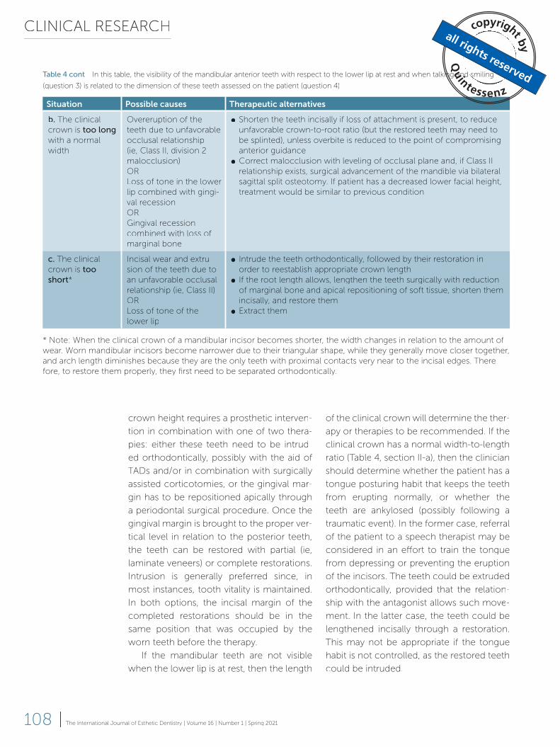
CLINICAL RESEARCH
108 | The International Journal of Esthetic Dentistry | Volume 16 | Number 1 | Spring 2021
of the clinical crown will determine the ther-
apy or therapies to be recommended. If the
clinical crown has a normal width-to-length
ratio (Table 4, section II-a), then the clinician
should determine whether the patient has a
tongue posturing habit that keeps the teeth
from erupting normally, or whether the
teeth are ankylosed (possibly following a
traumatic event). In the former case, referral
of the patient to a speech therapist may be
considered in an effort to train the tongue
from depressing or preventing the eruption
of the incisors. The teeth could be extruded
orthodontically, provided that the relation-
ship with the antagonist allows such move-
ment. In the latter case, the teeth could be
lengthened incisally through a restoration.
This may not be appropriate if the tongue
habit is not controlled, as the restored teeth
could be intruded.
crown height requires a prosthetic interven-
tion in combination with one of two thera-
pies: either these teeth need to be intrud-
ed orthodontically, possibly with the aid of
TATT Ds and/or in combination with surgically
assisted corticotomies, or the gingival mar-
gin has to be repositioned apically through
a periodontal surgical procedure. Once the
gingival margin is brought to the proper ver-
tical level in relation to the posterior teeth,
the teeth can be restored with partial (ie,
laminate veneers) or complete restorations.
Intrusion is generally preferred since, in
most instances, tooth vitality is maintained.
In both options, the incisal margin of the
completed restorations should be in the
same position that was occupied by the
worn teeth before the therapy.
If the mandibular teeth are not visible
when the lower lip is at rest, then the length
Situation Possible causes Therapeutic alternatives
b. The clinical
crown is too longwith a normal
width
Overeruption of the
teeth due to unfavorable
occlusal relationship
(ie, Class II, division 2
malocclusion)
OR
Loss of tone in the lower
lip combined with gingi-
val recession
OR
Gingival recession
combined with loss of
marginal bone
● Shorten the teeth incisally if loss of attachment is present, to reduce
unfavorable crown-to-root ratio (but the restored teeth may need to
be splinted), unless overbite is reduced to the point of compromising
anterior guidance
● Correct malocclusion with leveling of occlusal plane and, if Class II
relationship exists, surgical advancement of the mandible via bilateral
sagittal split osteotomy. If patient has a decreased lower facial height,
treatment would be similar to previous condition
c. The clinical
crown is too short*
Incisal wear and extru-
sion of the teeth due to
an unfavorable occlusal
relationship (ie, Class II)
OR
Loss of tone of the
lower lip
● Intrude the teeth orthodontically, followed by their restoration in
order to reestablish appropriate crown length
● If the root length allows, lengthen the teeth surgically with reduction
of marginal bone and apical repositioning of soft tissue, shorten them
incisally, and restore them
● Extract them
Table 4 cont In this table, the visibility of the mandibular anterior teeth with respect to the lower lip at rest and when talking and smiling
(question 3) is related to the dimension of these teeth assessed on the patient (question 4)
* Note: When the clinical crown of a mandibular incisor becomes shorter, the width changes in relation to the amount of
wear. Worn mandibular incisors become narrower due to their triangular shape, while they generally move closer together,
and arch length diminishes because they are the only teeth with proximal contacts very near to the incisal edges. There-
fore, to restore them properly, they first need to be separated orthodontically.
GRACIS
109The International Journal of Esthetic Dentistry | Volume 16 | Number 1 | Spring 2021 |
to the significant anteroposterior disparity
between the two arches. If the mandibu-
lar incisors are blocked out lingually by the
adjacent mandibular teeth, for whatever
reason, and lose occlusal contact with the
opposing maxillary teeth, they will typically
overerupt and become excessively exposed
above the lower lip.
What differs between these scenarios
are the therapeutic alternatives. When the
teeth have a normal width-to-length ra-
tio (Table 4, section III-a), the treatment of
choice would be to intrude (and likely pro-
cline) them orthodontically in conjunction
with leveling the occlusal plane and cor-
recting the malocclusion. If the mandibular
curve of Spee is significantly exaggerated
due to overeruption of the mandibular in-
cisors from compensation related to the
development of a Class II, division 2 maloc-
clusion in an adult with a short lower facial
height, leveling the exaggerated mandibular
curve of Spee is managed differently to the
way, and sequence in which, it is typically
leveled. Surgical advancement of the ret-
rognathic mandible is usually the best way
to facilitate the correction of Class II mal-
occlusions in an adult, especially when the
patent has a decreased lower facial height.
In the absence of a decreased lower facial
height, the exaggerated mandibular curve
of Spee is leveled presurgically, along with
leveling the maxillary arch and aligning the
teeth. The mandible is then advanced for-
ward, essentially horizontally, so that min-
imal change occurs to the patient’s lower
facial height. In a patient with a decreased
lower facial height, the mandibular curve
of Spee, in contrast, is maintained or even
exaggerated presurgically, which allows the
mandible to tip downward as it is advanced.
Postsurgery, only the mandibular incisors
and the most distal molars will contact the
maxillary teeth. The mandibular curve of
Spee is then leveled by erupting all the re-
maining mandibular teeth into contact with
When the clinical crown of a natural
tooth is too long (Table 4, section II-b), it
is usually related to soft tissue recession.
It is important to determine the reason for
the recession, which is usually related to a
traumatic tooth brushing habit facilitated by
dehiscence of the root or to the loss of peri-
odontal attachment from an inflammatory
process. In the former case, depending on
the severity of the problem, orthodontics
could assist in moving the affected tooth
or teeth to within the confines of the alve-
olar process (which is a prerequisite for a
predictable outcome of root-covering peri-
odontal procedures), and/or reducing the
root surface. In the latter case, if the attach-
ment loss is considerable, the only viable
solution may be extraction of the compro-
mised tooth or teeth and replacement with
implant-supported FDPs if sufficient bone
is available or a FDP on the available abut-
ment teeth. On the other hand, if the clinical
crown is short due to wear, fracture or a de-
velopmental anomaly (Table 4, section II-c),
the most appropriate solution is the (direct
or indirect) restoration of the involved tooth
or teeth.
The third possible scenario is when the
mandibular teeth are overly exposed above
the lower lip (Table 4, section III). Regard-
less of whether the teeth have a normal
width-to-length ratio or a clinical crown
that is either too long or too short, overex-
posure of the incisors above the lower lip
is esthetically unpleasing. It may also be
undesirable from a functional, occlusal or
stability perspective. Such overexposure of
the mandibular incisors gradually occurs
in most people as they age due to loss of
tonicity of the lower lip. It also occurs due
to the absence of contact that these teeth
would normally have with the maxillary
teeth. This is one characteristic of Class II,
division 2 malocclusions due to the den-
tal compensation or overeruption of the
mandibular (and maxillary) incisors related
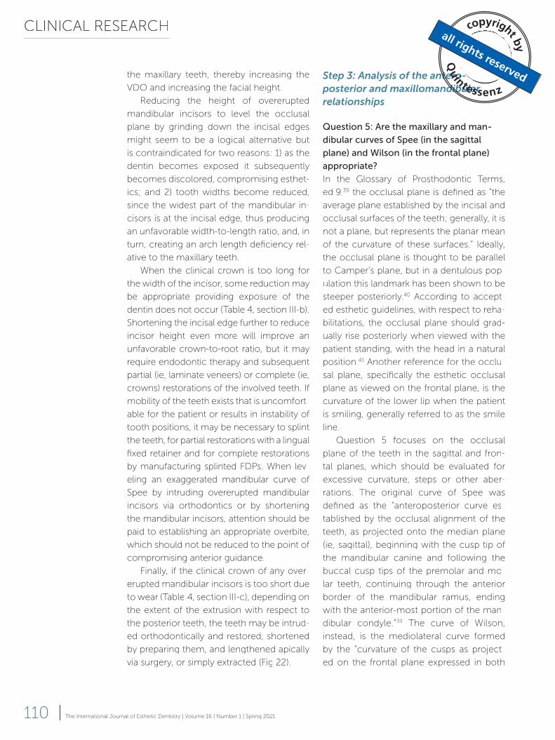
CLINICAL RESEARCH
110 | The International Journal of Esthetic Dentistry | Volume 16 | Number 1 | Spring 2021
Step 3: Analysis of the antero-posterior and maxillomandibular relationships
Question 5: Are the maxillary and man-
dibular curves of Spee (in the sagittal
plane) and Wilson (in the frontal plane)
appropriate?
In the Glossary of Prosthodontic Terms,
ed 9,39 the occlusal plane is defined as “the
average plane established by the incisal and
occlusal surfaces of the teeth; generally, it is
not a plane, but represents the planar mean
of the curvature of these surfaces.” Ideally,
the occlusal plane is thought to be parallel
to Camper’s plane, but in a dentulous pop-
ulation this landmark has been shown to be
steeper posteriorly.40 According to accept-
ed esthetic guidelines, with respect to reha-
bilitations, the occlusal plane should grad-
ually rise posteriorly when viewed with the
patient standing, with the head in a natural
position.41 Another reference for the occlu-
sal plane, specifically the esthetic occlusal
plane as viewed on the frontal plane, is the
curvature of the lower lip when the patient
is smiling, generally referred to as the smile
line.
Question 5 focuses on the occlusal
plane of the teeth in the sagittal and fron-
tal planes, which should be evaluated for
excessive curvature, steps or other aber-
rations. The original curve of Spee was
defined as the “anteroposterior curve es-
tablished by the occlusal alignment of the
teeth, as projected onto the median plane
(ie, sagittal), beginning with the cusp tip of
the mandibular canine and following the
buccal cusp tips of the premolar and mo-
lar teeth, continuing through the anterior
border of the mandibular ramus, ending
with the anterior-most portion of the man-
dibular condyle.”39 The curve of Wilson,
instead, is the mediolateral curve formed
by the “curvature of the cusps as project-
ed on the frontal plane expressed in both
the maxillary teeth, thereby increasing the
VDO and increasing the facial height.
Reducing the height of overerupted
mandibular incisors to level the occlusal
plane by grinding down the incisal edges
might seem to be a logical alternative but
is contraindicated for two reasons: 1) as the
dentin becomes exposed it subsequently
becomes discolored, compromising esthet-
ics; and 2) tooth widths become reduced,
since the widest part of the mandibular in-
cisors is at the incisal edge, thus producing
an unfavorable width-to-length ratio, and, in
turn, creating an arch length deficiency rel-
ative to the maxillary teeth.
When the clinical crown is too long for
the width of the incisor, some reduction may
be appropriate providing exposure of the
dentin does not occur (Table 4, section III-b).
Shortening the incisal edge further to reduce
incisor height even more will improve an
unfavorable crown-to-root ratio, but it may
require endodontic therapy and subsequent
partial (ie, laminate veneers) or complete (ie,
crowns) restorations of the involved teeth. If
mobility of the teeth exists that is uncomfort-
able for the patient or results in instability of
tooth positions, it may be necessary to splint
the teeth, for partial restorations with a lingual
fixed retainer and for complete restorations
by manufacturing splinted FDPs. When lev-
eling an exaggerated mandibular curve of
Spee by intruding overerupted mandibular
incisors via orthodontics or by shortening
the mandibular incisors, attention should be
paid to establishing an appropriate overbite,
which should not be reduced to the point of
compromising anterior guidance.
Finally, if the clinical crown of any over-
erupted mandibular incisors is too short due
to wear (Table 4, section III-c), depending on
the extent of the extrusion with respect to
the posterior teeth, the teeth may be intrud-
ed orthodontically and restored, shortened
by preparing them, and lengthened apically
via surgery, or simply extracted (Fig 22).
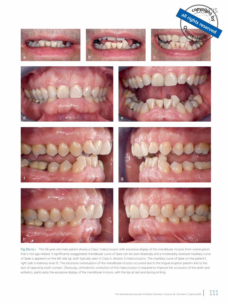
GRACIS
111The International Journal of Esthetic Dentistry | Volume 16 | Number 1 | Spring 2021 |
Fig 22a to i This 56-year-old male patient shows a Class I malocclusion with excessive display of the mandibular incisors from overeruption
that is not age related. A significantly exaggerated mandibular curve of Spee can be seen bilaterally and a moderately reversed maxillary curve
of Spee is apparent on the left side (g), both typically seen in Class II, division 2 malocclusions. The maxillary curve of Spee on the patient’s
right side is relatively level (f). The excessive overeruption of the mandibular incisors occurred due to the lingual eruption pattern and to the
lack of opposing tooth contact. Obviously, orthodontic correction of this malocclusion is required to improve the occlusion of the teeth and
esthetics, particularly the excessive display of the mandibular incisors, with the lips at rest and during smiling.
a b c
d e
f g
h i
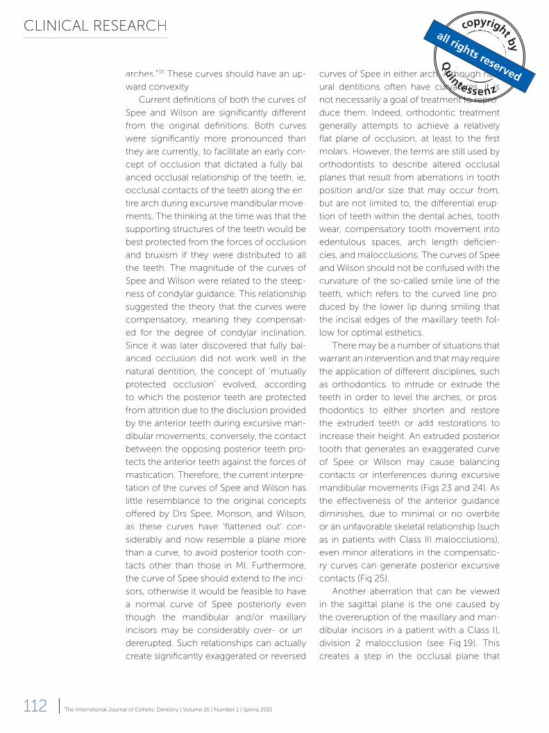
CLINICAL RESEARCH
112 | The International Journal of Esthetic Dentistry | Volume 16 | Number 1 | Spring 2021
curves of Spee in either arch. Although nat-
ural dentitions often have curvatures, it is
not necessarily a goal of treatment to repro-
duce them. Indeed, orthodontic treatment
generally attempts to achieve a relatively
flat plane of occlusion, at least to the first
molars. However, the terms are still used by
orthodontists to describe altered occlusal
planes that result from aberrations in tooth
position and/or size that may occur from,
but are not limited to, the differential erup-
tion of teeth within the dental aches, tooth
wear, compensatory tooth movement into
edentulous spaces, arch length deficien-
cies, and malocclusions. The curves of Spee
and Wilson should not be confused with the
curvature of the so-called smile line of the
teeth, which refers to the curved line pro-
duced by the lower lip during smiling that
the incisal edges of the maxillary teeth fol-
low for optimal esthetics.
There may be a number of situations that
warrant an intervention and that may require
the application of different disciplines, such
as orthodontics, to intrude or extrude the
teeth in order to level the arches, or pros-
thodontics to either shorten and restore
the extruded teeth or add restorations to
increase their height. An extruded posterior
tooth that generates an exaggerated curve
of Spee or Wilson may cause balancing
contacts or interferences during excursive
mandibular movements (Figs 23 and 24). As
the effectiveness of the anterior guidance
diminishes, due to minimal or no overbite
or an unfavorable skeletal relationship (such
as in patients with Class III malocclusions),
even minor alterations in the compensato-
ry curves can generate posterior excursive
contacts (Fig 25).
Another aberration that can be viewed
in the sagittal plane is the one caused by
the overeruption of the maxillary and man-
dibular incisors in a patient with a Class II,
division 2 malocclusion (see Fig 19). This
creates a step in the occlusal plane that
arches.”39 These curves should have an up-
ward convexity.
Current definitions of both the curves of
Spee and Wilson are significantly different
from the original definitions. Both curves
were significantly more pronounced than
they are currently, to facilitate an early con-
cept of occlusion that dictated a fully bal-
anced occlusal relationship of the teeth, ie,
occlusal contacts of the teeth along the en-
tire arch during excursive mandibular move-
ments. The thinking at the time was that the
supporting structures of the teeth would be
best protected from the forces of occlusion
and bruxism if they were distributed to all
the teeth. The magnitude of the curves of
Spee and Wilson were related to the steep-
ness of condylar guidance. This relationship
suggested the theory that the curves were
compensatory, meaning they compensat-
ed for the degree of condylar inclination.
Since it was later discovered that fully bal-
anced occlusion did not work well in the
natural dentition, the concept of ‘mutually
protected occlusion’ evolved, according
to which the posterior teeth are protected
from attrition due to the disclusion provided
by the anterior teeth during excursive man-
dibular movements; conversely, the contact
between the opposing posterior teeth pro-
tects the anterior teeth against the forces of
mastication. Therefore, the current interpre-
tation of the curves of Spee and Wilson has
little resemblance to the original concepts
offered by Drs Spee, Monson, and Wilson,
as these curves have ‘flattened out’ con-
siderably and now resemble a plane more
than a curve, to avoid posterior tooth con-
tacts other than those in MI. Furthermore,
the curve of Spee should extend to the inci-
sors, otherwise it would be feasible to have
a normal curve of Spee posteriorly even
though the mandibular and/or maxillary
incisors may be considerably over- or un-
dererupted. Such relationships can actually
create significantly exaggerated or reversed
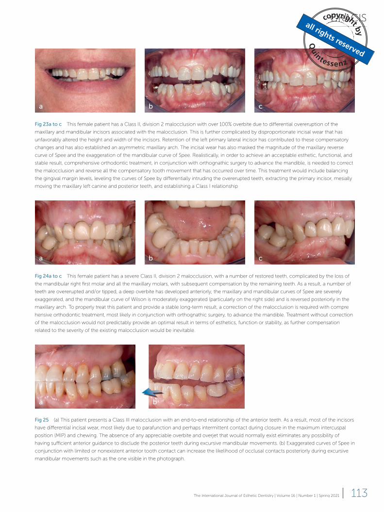
GRACIS
113The International Journal of Esthetic Dentistry | Volume 16 | Number 1 | Spring 2021 |
Fig 23a to c This female patient has a Class II, division 2 malocclusion with over 100% overbite due to differential overeruption of the
maxillary and mandibular incisors associated with the malocclusion. This is further complicated by disproportionate incisal wear that has
unfavorably altered the height and width of the incisors. Retention of the left primary lateral incisor has contributed to these compensatory
changes and has also established an asymmetric maxillary arch. The incisal wear has also masked the magnitude of the maxillary reverse
curve of Spee and the exaggeration of the mandibular curve of Spee. Realistically, in order to achieve an acceptable esthetic, functional, and
stable result, comprehensive orthodontic treatment, in conjunction with orthognathic surgery to advance the mandible, is needed to correct
the malocclusion and reverse all the compensatory tooth movement that has occurred over time. This treatment would include balancing
the gingival margin levels, leveling the curves of Spee by differentially intruding the overerupted teeth, extracting the primary incisor, mesially
moving the maxillary left canine and posterior teeth, and establishing a Class I relationship.
a b c
Fig 24a to c This female patient has a severe Class II, division 2 malocclusion, with a number of restored teeth, complicated by the loss of
the mandibular right first molar and all the maxillary molars, with subsequent compensation by the remaining teeth. As a result, a number of
teeth are overerupted and/or tipped, a deep overbite has developed anteriorly, the maxillary and mandibular curves of Spee are severely
exaggerated, and the mandibular curve of Wilson is moderately exaggerated (particularly on the right side) and is reversed posteriorly in the
maxillary arch. To properly treat this patient and provide a stable long-term result, a correction of the malocclusion is required with compre-
hensive orthodontic treatment, most likely in conjunction with orthognathic surgery, to advance the mandible. Treatment without correction
of the malocclusion would not predictably provide an optimal result in terms of esthetics, function or stability, as further compensation
related to the severity of the existing malocclusion would be inevitable.
Fig 25 (a) This patient presents a Class III malocclusion with an end-to-end relationship of the anterior teeth. As a result, most of the incisors
have differential incisal wear, most likely due to parafunction and perhaps intermittent contact during closure in the maximum intercuspal
position (MIP) and chewing. The absence of any appreciable overbite and overjet that would normally exist eliminates any possibility of
having sufficient anterior guidance to disclude the posterior teeth during excursive mandibular movements. (b) Exaggerated curves of Spee in
conjunction with limited or nonexistent anterior tooth contact can increase the likelihood of occlusal contacts posteriorly during excursive
mandibular movements such as the one visible in the photograph.
a b c
a b
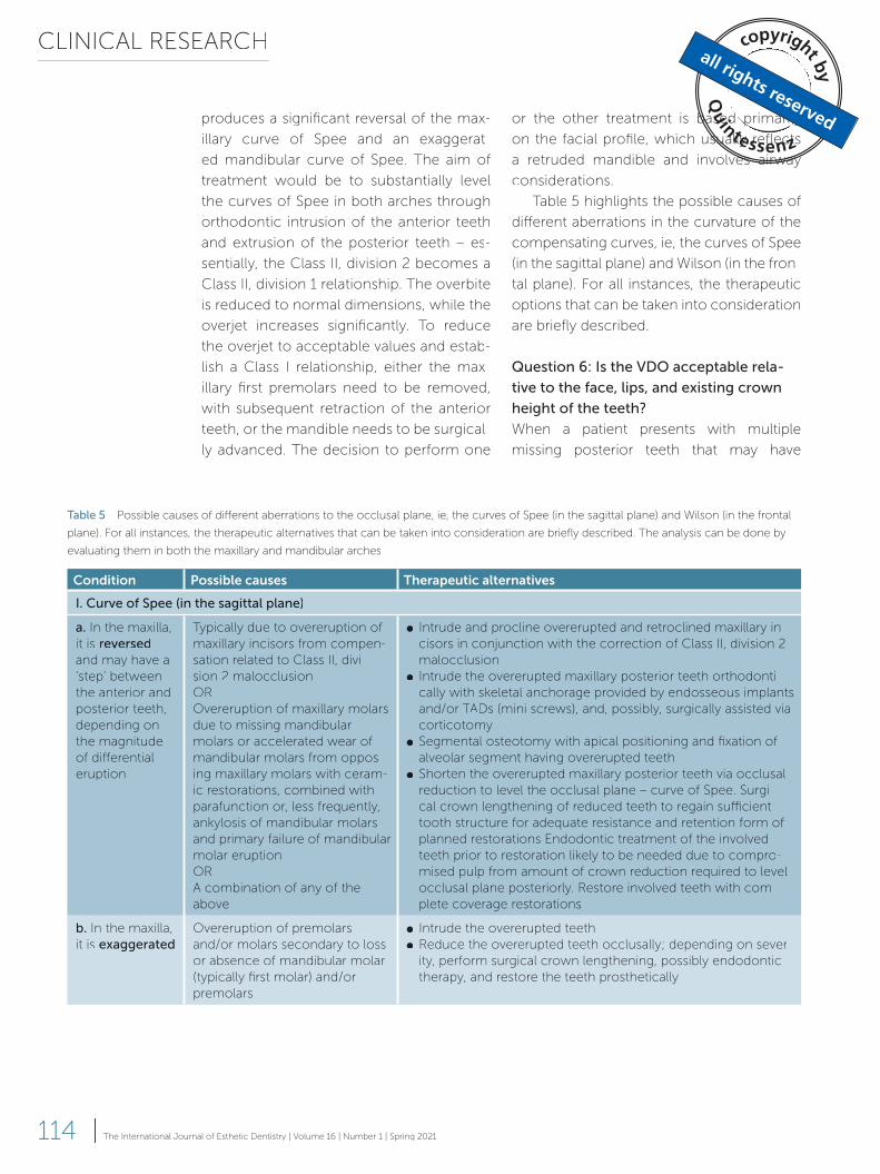
CLINICAL RESEARCH
114 | The International Journal of Esthetic Dentistry | Volume 16 | Number 1 | Spring 2021
or the other treatment is based primarily
on the facial profile, which usually reflects
a retruded mandible and involves airway
considerations.
Table 5 highlights the possible causes of
different aberrations in the curvature of the
compensating curves, ie, the curves of Spee
(in the sagittal plane) and Wilson (in the fron-
tal plane). For all instances, the therapeutic
options that can be taken into consideration
are briefly described.
Question 6: Is the VDO acceptable rela-
tive to the face, lips, and existing crown
height of the teeth?
When a patient presents with multiple
missing posterior teeth that may have
produces a significant reversal of the max-
illary curve of Spee and an exaggerat-
ed mandibular curve of Spee. The aim of
treatment would be to substantially level
the curves of Spee in both arches through
orthodontic intrusion of the anterior teeth
and extrusion of the posterior teeth – es-
sentially, the Class II, division 2 becomes a
Class II, division 1 relationship. The overbite
is reduced to normal dimensions, while the
overjet increases significantly. To reduce
the overjet to acceptable values and estab-
lish a Class I relationship, either the max-
illary first premolars need to be removed,
with subsequent retraction of the anterior
teeth, or the mandible needs to be surgical-
ly advanced. The decision to perform one
Table 5 Possible causes of different aberrations to the occlusal plane, ie, the curves of Spee (in the sagittal plane) and Wilson (in the frontal
plane). For all instances, the therapeutic alternatives that can be taken into consideration are briefly described. The analysis can be done by
evaluating them in both the maxillary and mandibular arches
Condition Possible causes Therapeutic alternatives
I. Curve of Spee (in the sagittal plane)
a. In the maxilla,
it is reversedand may have a
‘step’ between
the anterior and
posterior teeth,
depending on
the magnitude
of differential
eruption
Typically due to overeruption of
maxillary incisors from compen-
sation related to Class II, divi-
sion 2 malocclusion
OR
Overeruption of maxillary molars
due to missing mandibular
molars or accelerated wear of
mandibular molars from oppos-
ing maxillary molars with ceram-
ic restorations, combined with
parafunction or, less frequently,
ankylosis of mandibular molars
and primary failure of mandibular
molar eruption
OR
A combination of any of the
above
● Intrude and procline overerupted and retroclined maxillary in-
cisors in conjunction with the correction of Class II, division 2
malocclusion
● Intrude the overerupted maxillary posterior teeth orthodonti-
cally with skeletal anchorage provided by endosseous implants
and/or TATT Ds (mini screws), and, possibly, surgically assisted via
corticotomy
● Segmental osteotomy with apical positioning and fixation of
alveolar segment having overerupted teeth
● Shorten the overerupted maxillary posterior teeth via occlusal
reduction to level the occlusal plane – curve of Spee. Surgi-
cal crown lengthening of reduced teeth to regain sufficient
tooth structure for adequate resistance and retention form of
planned restorations Endodontic treatment of the involved
teeth prior to restoration likely to be needed due to compro-
mised pulp from amount of crown reduction required to level
occlusal plane posteriorly. Restore involved teeth with com-
plete coverage restorations
b. In the maxilla,
it is exaggeratedOvereruption of premolars
and/or molars secondary to loss
or absence of mandibular molar
(typically first molar) and/or
premolars
● Intrude the overerupted teeth
● Reduce the overerupted teeth occlusally; depending on sever-
ity, perform surgical crown lengthening, possibly endodontic
therapy, and restore the teeth prosthetically
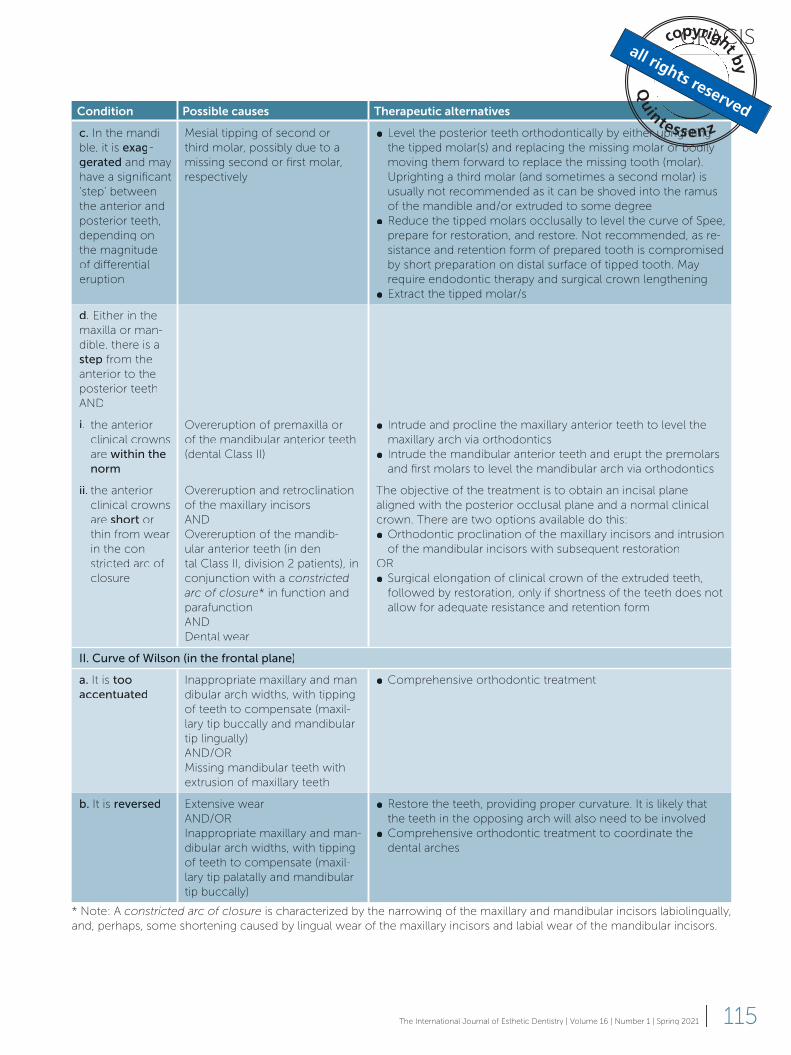
GRACIS
115The International Journal of Esthetic Dentistry | Volume 16 | Number 1 | Spring 2021 |
Condition Possible causes Therapeutic alternatives
c. In the mandi-
ble, it is exag-gerated and may
have a significant
‘step’ between
the anterior and
posterior teeth,
depending on
the magnitude
of differential
eruption
Mesial tipping of second or
third molar, possibly due to a
missing second or first molar,
respectively
● Level the posterior teeth orthodontically by either uprighting
the tipped molar(s) and replacing the missing molar or bodily
moving them forward to replace the missing tooth (molar).
Uprighting a third molar (and sometimes a second molar) is
usually not recommended as it can be shoved into the ramus
of the mandible and/or extruded to some degree
● Reduce the tipped molars occlusally to level the curve of Spee,
prepare for restoration, and restore. Not recommended, as re-
sistance and retention form of prepared tooth is compromised
by short preparation on distal surface of tipped tooth. May
require endodontic therapy and surgical crown lengthening
● Extract the tipped molar/s
d. Either in the
maxilla or man-
dible, there is a
step from the
anterior to the
posterior teeth
AND
i. the anterior
clinical crowns
are within the norm
Overeruption of premaxilla or
of the mandibular anterior teeth
(dental Class II)
● Intrude and procline the maxillary anterior teeth to level the
maxillary arch via orthodontics
● Intrude the mandibular anterior teeth and erupt the premolars
and first molars to level the mandibular arch via orthodontics
ii. the anterior
clinical crowns
are short or
thin from wear
in the con-
stricted arc of
closure
Overeruption and retroclination
of the maxillary incisors
AND
Overeruption of the mandib-
ular anterior teeth (in den-
tal Class II, division 2 patients), in
conjunction with a constricted arc of closure* in function and
parafunction
AND
Dental wear
The objective of the treatment is to obtain an incisal plane
aligned with the posterior occlusal plane and a normal clinical
crown. There are two options available do this:
● Orthodontic proclination of the maxillary incisors and intrusion
of the mandibular incisors with subsequent restoration
OR
● Surgical elongation of clinical crown of the extruded teeth,
followed by restoration, only if shortness of the teeth does not
allow for adequate resistance and retention form
II. Curve of Wilson (in the frontal plane)
a. It is too accentuated
Inappropriate maxillary and man-
dibular arch widths, with tipping
of teeth to compensate (maxil-
lary tip buccally and mandibular
tip lingually)
AND/OR
Missing mandibular teeth with
extrusion of maxillary teeth
● Comprehensive orthodontic treatment
b. It is reversed Extensive wear
AND/OR
Inappropriate maxillary and man-
dibular arch widths, with tipping
of teeth to compensate (maxil-
lary tip palatally and mandibular
tip buccally)
● Restore the teeth, providing proper curvature. It is likely that
the teeth in the opposing arch will also need to be involved
● Comprehensive orthodontic treatment to coordinate the
dental arches
* Note: A constricted arc of closure is characterized by the narrowing of the maxillary and mandibular incisors labiolingually,
and, perhaps, some shortening caused by lingual wear of the maxillary incisors and labial wear of the mandibular incisors.
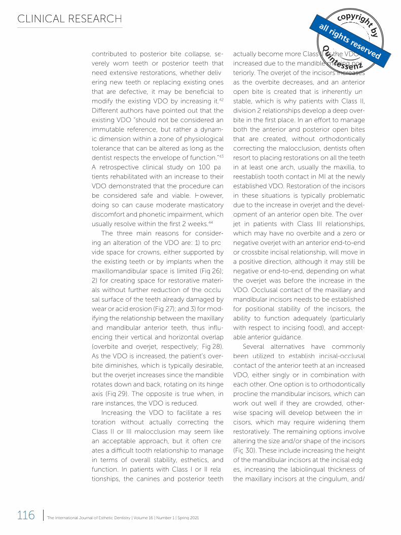
CLINICAL RESEARCH
116 | The International Journal of Esthetic Dentistry | Volume 16 | Number 1 | Spring 2021
contributed to posterior bite collapse, se-
verely worn teeth or posterior teeth that
need extensive restorations, whether deliv-
ering new teeth or replacing existing ones
that are defective, it may be beneficial to
modify the existing VDO by increasing it.42
Different authors have pointed out that the
existing VDO “should not be considered an
immutable reference, but rather a dynam-
ic dimension within a zone of physiological
tolerance that can be altered as long as the
dentist respects the envelope of function.”43
A retrospective clinical study on 100 pa-
tients rehabilitated with an increase to their
VDO demonstrated that the procedure can
be considered safe and viable. However,
doing so can cause moderate masticatory
discomfort and phonetic impairment, which
usually resolve within the first 2 weeks.44
The three main reasons for consider-
ing an alteration of the VDO are: 1) to pro-
vide space for crowns, either supported by
the existing teeth or by implants when the
maxillomandibular space is limited (Fig 26);
2) for creating space for restorative materi-
als without further reduction of the occlu-
sal surface of the teeth already damaged by
wear or acid erosion (Fig 27); and 3) for mod-
ifying the relationship between the maxillary
and mandibular anterior teeth, thus influ-
encing their vertical and horizontal overlap
(overbite and overjet, respectively; Fig 28).
As the VDO is increased, the patient’s over-
bite diminishes, which is typically desirable,
but the overjet increases since the mandible
rotates down and back, rotating on its hinge
axis (Fig 29). The opposite is true when, in
rare instances, the VDO is reduced.
Increasing the VDO to facilitate a res-
toration without actually correcting the
Class II or III malocclusion may seem like
an acceptable approach, but it often cre-
ates a difficult tooth relationship to manage
in terms of overall stability, esthetics, and
function. In patients with Class I or II rela-
tionships, the canines and posterior teeth
actually become more Class II as the VDO is
increased due to the mandible moving pos-
teriorly. The overjet of the incisors increases
as the overbite decreases, and an anterior
open bite is created that is inherently un-
stable, which is why patients with Class II,
division 2 relationships develop a deep over-
bite in the first place. In an effort to manage
both the anterior and posterior open bites
that are created, without orthodontically
correcting the malocclusion, dentists often
resort to placing restorations on all the teeth
in at least one arch, usually the maxilla, to
reestablish tooth contact in MI at the newly
established VDO. Restoration of the incisors
in these situations is typically problematic
due to the increase in overjet and the devel-
opment of an anterior open bite. The over-
jet in patients with Class III relationships,
which may have no overbite and a zero or
negative overjet with an anterior end-to-end
or crossbite incisal relationship, will move in
a positive direction, although it may still be
negative or end-to-end, depending on what
the overjet was before the increase in the
VDO. Occlusal contact of the maxillary and
mandibular incisors needs to be established
for positional stability of the incisors, the
ability to function adequately (particularly
with respect to incising food), and accept-
able anterior guidance.
Several alternatives have commonly
been utilized to establish incisal-occlusal
contact of the anterior teeth at an increased
VDO, either singly or in combination with
each other. One option is to orthodontically
procline the mandibular incisors, which can
work out well if they are crowded, other-
wise spacing will develop between the in-
cisors, which may require widening them
restoratively. The remaining options involve
altering the size and/or shape of the incisors
(Fig 30). These include increasing the height
of the mandibular incisors at the incisal edg-
es, increasing the labiolingual thickness of
the maxillary incisors at the cingulum, and/
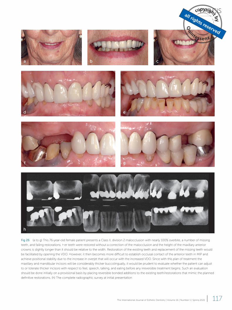
GRACIS
117The International Journal of Esthetic Dentistry | Volume 16 | Number 1 | Spring 2021 |
a b c
d e
f g
Fig 26 (a to g) This 76-year-old female patient presents a Class II, division 2 malocclusion with nearly 100% overbite, a number of missing
teeth, and failing restorations. Her teeth were restored without a correction of the malocclusion and the height of the maxillary anterior
crowns is slightly longer than it should be relative to the width. Restoration of the existing teeth and replacement of the missing teeth would
be facilitated by opening the VDO. However, it then becomes more difficult to establish occlusal contact of the anterior teeth in MIP and
achieve positional stability due to the increase in overjet that will occur with the increased VDO. Since with this plan of treatment the
maxillary and mandibular incisors will be considerably thicker buccolingually, it would be prudent to evaluate whether the patient can adjust
to or tolerate thicker incisors with respect to feel, speech, talking, and eating before any irreversible treatment begins. Such an evaluation
should be done initially on a provisional basis by placing reversible bonded additions to the existing teeth/restorations that mimic the planned
definitive restorations. (h) The complete radiographic survey at initial presentation.
h
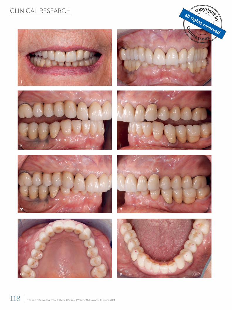
CLINICAL RESEARCH
118 | The International Journal of Esthetic Dentistry | Volume 16 | Number 1 | Spring 2021
i j
k l
m n
o p
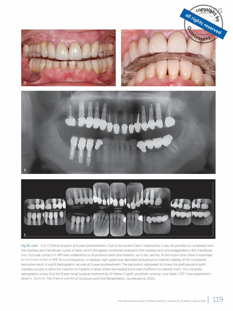
GRACIS
119The International Journal of Esthetic Dentistry | Volume 16 | Number 1 | Spring 2021 |
q r
s
t
Fig 26, cont (i to r) Clinical situation at 9 years posttreatment. Due to the severe Class II relationship, it was not possible to completely level
the maxillary and mandibular curves of Spee, which still appear somewhat reversed in the maxillary arch and exaggerated in the mandibular
one. Occlusal contacts in MIP were established on all posterior teeth and implants, up to the canines. At the incisor level, there is essentially
no occlusal contact in MIP. As a consequence, a maxillary night guard was fabricated and placed to maintain stability of the completed
restorative result. (s and t) Radiographic records at 9 years posttreatment. The panoramic radiograph (s) shows the graft placed in both
maxillary sinuses to allow the insertion of implants in areas where the residual bone was insufficient to stabilize them. The complete
radiographic survey (t) at the 9-year recall (surgical treatment by Dr Matteo Capelli; prosthetic work by Luca Vailati, CDT. Case presented in
detail in: Gross M. The Science and Art of Occlusion and Oral Rehabilitation. Quintessence, 2015).
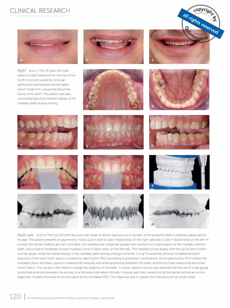
CLINICAL RESEARCH
120 | The International Journal of Esthetic Dentistry | Volume 16 | Number 1 | Spring 2021
Fig 27, cont (d to h) The loss of tooth structure with areas of dentin exposure on a number of the posterior teeth is relatively advanced for
his age. The patient presents an asymmetric malocclusion with a Class I relationship on the right side and a Class II relationship on the left. As
a result, the dental midlines are not coincident, the overbite and overjet are greater than normal from overeruption of the maxillary anterior
teeth, and a mild to moderate reverse maxillary curve of Spee exists on the left side. The maxillary incisal display with the lips at rest is within
normal values, while the overall display of the maxillary teeth during smiling is minimal. (i to q) To avoid the removal of additional tooth
structure of the worn teeth, space is created by opening the VDO according to prosthetic convenience. Since opening the VDO rotates the
mandible down and back, space is created both vertically and anteroposteriorly between the teeth, and the occlusal relationship becomes
more Class II. This results in the need to change the anatomy of the teeth. A centric relation record was obtained with the aid of a leaf gauge
positioned anteriorly between the arches, of a thickness estimated clinically. A waxup was then carried out by the dental technician on the
diagnostic models mounted on an articulator at this increased VDO. The objective was to replace the missing tooth structure while
a b c
f g h
d e
i j kkkkkkkkkkkkkkkkkkkkkkk
l mmmm n
Fig 27 (a to c) This 42-year-old male
patient sought treatment for the loss of his
tooth structure caused by occlusal
dysfunction and parafunctional habits,
which made him concerned about the
future of his teeth. The patient was also
concerned about the limited visibility of his
maxillary teeth during smiling.
k
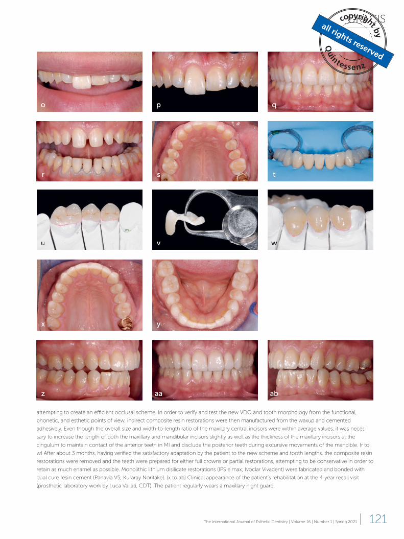
GRACIS
121The International Journal of Esthetic Dentistry | Volume 16 | Number 1 | Spring 2021 |
attempting to create an efficient occlusal scheme. In order to verify and test the new VDO and tooth morphology from the functional,
phonetic, and esthetic points of view, indirect composite resin restorations were then manufactured from the waxup and cemented
adhesively. Even though the overall size and width-to-length ratio of the maxillary central incisors were within average values, it was neces-
sary to increase the length of both the maxillary and mandibular incisors slightly as well as the thickness of the maxillary incisors at the
cingulum to maintain contact of the anterior teeth in MI and disclude the posterior teeth during excursive movements of the mandible. (r to
w) After about 3 months, having verified the satisfactory adaptation by the patient to the new scheme and tooth lengths, the composite resin
restorations were removed and the teeth were prepared for either full crowns or partial restorations, attempting to be conservative in order to
retain as much enamel as possible. Monolithic lithium disilicate restorations (IPS e.max; Ivoclar Vivadent) were fabricated and bonded with
dual cure resin cement (Panavia V5; Kuraray Noritake). (x to ab) Clinical appearance of the patient’s rehabilitation at the 4-year recall visit
(prosthetic laboratory work by Luca Vailati, CDT). The patient regularly wears a maxillary night guard.
aa
o p q
r s t
uuu v wwwwwwwwww
z ab
x y
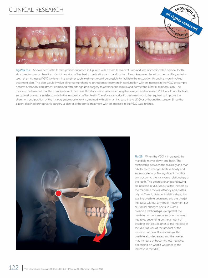
CLINICAL RESEARCH
122 | The International Journal of Esthetic Dentistry | Volume 16 | Number 1 | Spring 2021
Fig 28a to c Shown here is the female patient discussed in Figure 2 with a Class III malocclusion and loss of considerable coronal tooth
structure from a combination of acidic erosion of her teeth, mastication, and parafunction. A mock-up was placed on the maxillary anterior
teeth at an increased VDO to determine whether such treatment would be possible to facilitate the restoration through a more involved
treatment plan. The plan would involve either comprehensive orthodontic treatment in conjunction with an increase in the VDO or compre-
hensive orthodontic treatment combined with orthognathic surgery to advance the maxilla and correct the Class III malocclusion. The
mock-up determined that the combination of the Class III malocclusion, associated negative overjet, and increased VDO would not facilitate
an optimal or even a satisfactory definitive restoration of her teeth. Therefore, orthodontic treatment would be required to improve the
alignment and position of the incisors anteroposteriorly, combined with either an increase in the VDO or orthognathic surgery. Since the
patient declined orthognathic surgery, a plan of orthodontic treatment with an increase in the VDO was initiated.
Fig 29 When the VDO is increased, the
mandible moves down and back. The
relationship between the maxillary and man-
dibular teeth changes both vertically and
anteroposteriorly. No significant modifica-
tions occur to the transverse relationships of
the teeth. The greatest changes following
an increase in VDO occur at the incisors as
the mandible moves inferiorly and posteri-
orly. In Class II, division 2 relationships, the
existing overbite decreases and the overjet
increases without any tooth movement per
se. Similar changes occur in Class II,
division 1 relationships, except that the
overbite can become nonexistent or even
negative, depending on the amount of
overbite that existed prior to the increase in
the VDO as well as the amount of the
increase. In Class III relationships, the
overbite also decreases, and the overjet
may increase or becomes less negative,
depending on what it was prior to the
increase in the VDO.
a b c
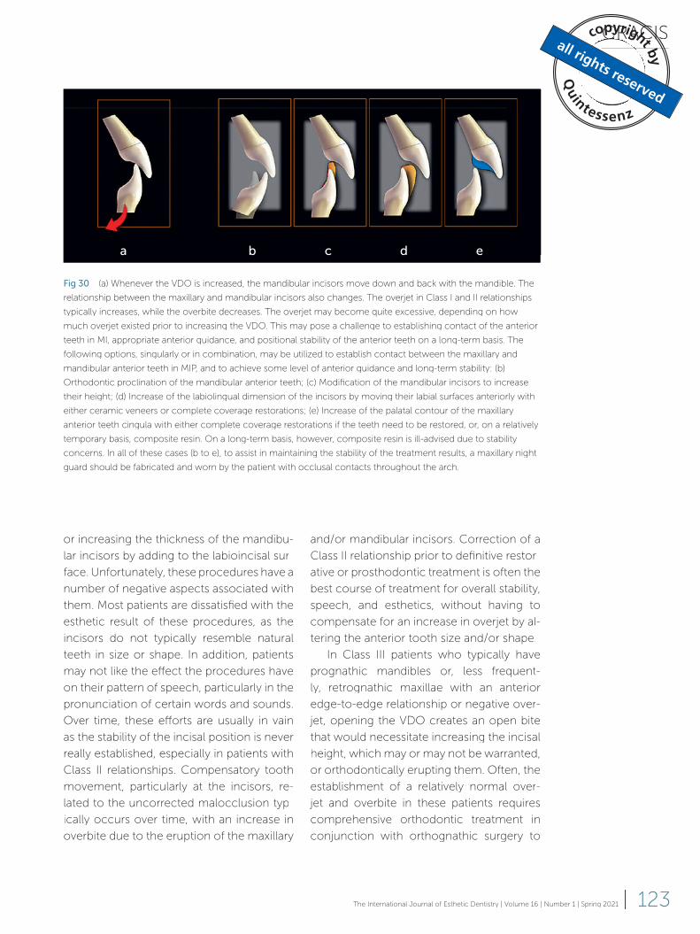
GRACIS
123The International Journal of Esthetic Dentistry | Volume 16 | Number 1 | Spring 2021 |
and/or mandibular incisors. Correction of a
Class II relationship prior to definitive restor-
ative or prosthodontic treatment is often the
best course of treatment for overall stability,
speech, and esthetics, without having to
compensate for an increase in overjet by al-
tering the anterior tooth size and/or shape.
In Class III patients who typically have
prognathic mandibles or, less frequent-
ly, retrognathic maxillae with an anterior
edge-to-edge relationship or negative over-
jet, opening the VDO creates an open bite
that would necessitate increasing the incisal
height, which may or may not be warranted,
or orthodontically erupting them. Often, the
establishment of a relatively normal over-
jet and overbite in these patients requires
comprehensive orthodontic treatment in
conjunction with orthognathic surgery to
or increasing the thickness of the mandibu-
lar incisors by adding to the labioincisal sur-
face. Unfortunately, these procedures have a
number of negative aspects associated with
them. Most patients are dissatisfied with the
esthetic result of these procedures, as the
incisors do not typically resemble natural
teeth in size or shape. In addition, patients
may not like the effect the procedures have
on their pattern of speech, particularly in the
pronunciation of certain words and sounds.
Over time, these efforts are usually in vain
as the stability of the incisal position is never
really established, especially in patients with
Class II relationships. Compensatory tooth
movement, particularly at the incisors, re-
lated to the uncorrected malocclusion typ-
ically occurs over time, with an increase in
overbite due to the eruption of the maxillary
Fig 30 (a) Whenever the VDO is increased, the mandibular incisors move down and back with the mandible. The
relationship between the maxillary and mandibular incisors also changes. The overjet in Class I and II relationships
typically increases, while the overbite decreases. The overjet may become quite excessive, depending on how
much overjet existed prior to increasing the VDO. This may pose a challenge to establishing contact of the anterior
teeth in MI, appropriate anterior guidance, and positional stability of the anterior teeth on a long-term basis. The
following options, singularly or in combination, may be utilized to establish contact between the maxillary and
mandibular anterior teeth in MIP, and to achieve some level of anterior guidance and long-term stability: (b)
Orthodontic proclination of the mandibular anterior teeth; (c) Modification of the mandibular incisors to increase
their height; (d) Increase of the labiolingual dimension of the incisors by moving their labial surfaces anteriorly with
either ceramic veneers or complete coverage restorations; (e) Increase of the palatal contour of the maxillary
anterior teeth cingula with either complete coverage restorations if the teeth need to be restored, or, on a relatively
temporary basis, composite resin. On a long-term basis, however, composite resin is ill-advised due to stability
concerns. In all of these cases (b to e), to assist in maintaining the stability of the treatment results, a maxillary night
guard should be fabricated and worn by the patient with occlusal contacts throughout the arch.
a b c d e
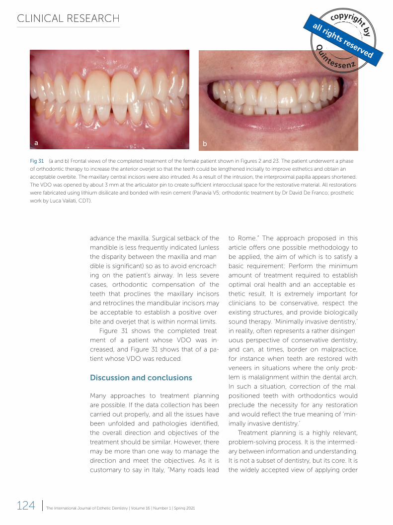
CLINICAL RESEARCH
124 | The International Journal of Esthetic Dentistry | Volume 16 | Number 1 | Spring 2021
to Rome.” The approach proposed in this
article offers one possible methodology to
be applied, the aim of which is to satisfy a
basic requirement: Perform the minimum
amount of treatment required to establish
optimal oral health and an acceptable es-
thetic result. It is extremely important for
clinicians to be conservative, respect the
existing structures, and provide biologically
sound therapy. ‘Minimally invasive dentistry,’
in reality, often represents a rather disingen-
uous perspective of conservative dentistry,
and can, at times, border on malpractice,
for instance when teeth are restored with
veneers in situations where the only prob-
lem is malalignment within the dental arch.
In such a situation, correction of the mal-
positioned teeth with orthodontics would
preclude the necessity for any restoration
and would reflect the true meaning of ‘min-
imally invasive dentistry.’
Treatment planning is a highly relevant,
problem-solving process. It is the intermedi-
ary between information and understanding.
It is not a subset of dentistry, but its core. It is
the widely accepted view of applying order
advance the maxilla. Surgical setback of the
mandible is less frequently indicated (unless
the disparity between the maxilla and man-
dible is significant) so as to avoid encroach-
ing on the patient’s airway. In less severe
cases, orthodontic compensation of the
teeth that proclines the maxillary incisors
and retroclines the mandibular incisors may
be acceptable to establish a positive over-
bite and overjet that is within normal limits.
Figure 31 shows the completed treat-
ment of a patient whose VDO was in-
creased, and Figure 31 shows that of a pa-
tient whose VDO was reduced.
Discussion and conclusions
Many approaches to treatment planning
are possible. If the data collection has been
carried out properly, and all the issues have
been unfolded and pathologies identified,
the overall direction and objectives of the
treatment should be similar. However, there
may be more than one way to manage the
direction and meet the objectives. As it is
customary to say in Italy, “Many roads lead
Fig 31 (a and b) Frontal views of the completed treatment of the female patient shown in Figures 2 and 23. The patient underwent a phase
of orthodontic therapy to increase the anterior overjet so that the teeth could be lengthened incisally to improve esthetics and obtain an
acceptable overbite. The maxillary central incisors were also intruded. As a result of the intrusion, the interproximal papilla appears shortened.
The VDO was opened by about 3 mm at the articulator pin to create sufficient interocclusal space for the restorative material. All restorations
were fabricated using lithium disilicate and bonded with resin cement (Panavia V5; orthodontic treatment by Dr David De Franco; prosthetic
work by Luca Vailati, CDT).
a b
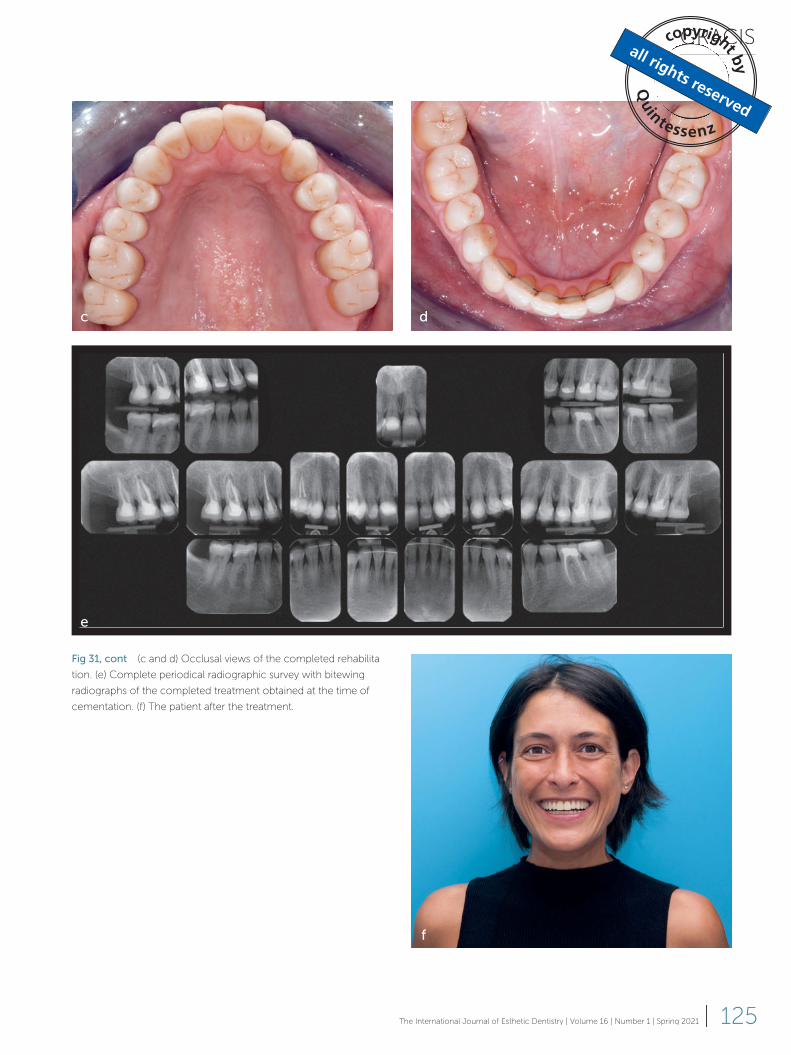
GRACIS
125The International Journal of Esthetic Dentistry | Volume 16 | Number 1 | Spring 2021 |
Fig 31, cont (c and d) Occlusal views of the completed rehabilita-
tion. (e) Complete periodical radiographic survey with bitewing
radiographs of the completed treatment obtained at the time of
cementation. (f) The patient after the treatment.
c d
e
f
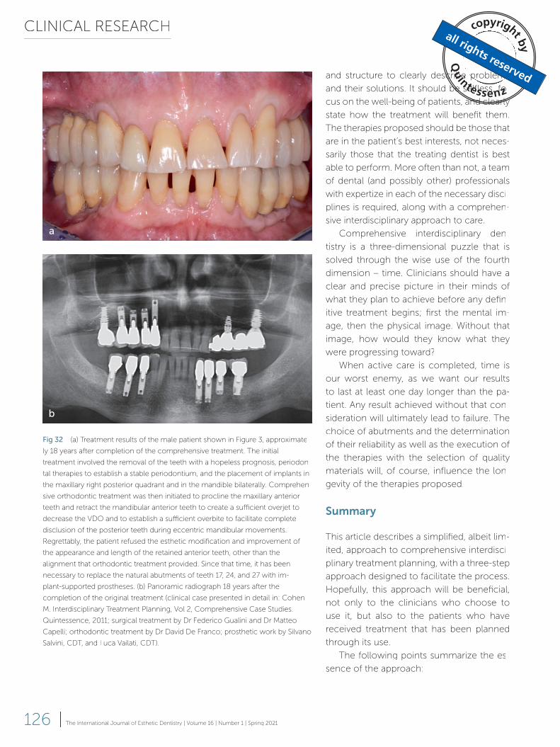
CLINICAL RESEARCH
126 | The International Journal of Esthetic Dentistry | Volume 16 | Number 1 | Spring 2021
and structure to clearly describe problems
and their solutions. It should be selfless, fo-
cus on the well-being of patients, and clearly
state how the treatment will benefit them.
The therapies proposed should be those that
are in the patient’s best interests, not neces-
sarily those that the treating dentist is best
able to perform. More often than not, a team
of dental (and possibly other) professionals
with expertize in each of the necessary disci-
plines is required, along with a comprehen-
sive interdisciplinary approach to care.
Comprehensive interdisciplinary den-
tistry is a three-dimensional puzzle that is
solved through the wise use of the fourth
dimension – time. Clinicians should have a
clear and precise picture in their minds of
what they plan to achieve before any defin-
itive treatment begins; first the mental im-
age, then the physical image. Without that
image, how would they know what they
were progressing toward?
When active care is completed, time is
our worst enemy, as we want our results
to last at least one day longer than the pa-
tient. Any result achieved without that con-
sideration will ultimately lead to failure. The
choice of abutments and the determination
of their reliability as well as the execution of
the therapies with the selection of quality
materials will, of course, influence the lon-
gevity of the therapies proposed.
Summary
This article describes a simplified, albeit lim-
ited, approach to comprehensive interdisci-
plinary treatment planning, with a three-step
approach designed to facilitate the process.
Hopefully, this approach will be beneficial,
not only to the clinicians who choose to
use it, but also to the patients who have
received treatment that has been planned
through its use.
The following points summarize the es-
sence of the approach:
Fig 32 (a) Treatment results of the male patient shown in Figure 3, approximate-
ly 18 years after completion of the comprehensive treatment. The initial
treatment involved the removal of the teeth with a hopeless prognosis, periodon-
tal therapies to establish a stable periodontium, and the placement of implants in
the maxillary right posterior quadrant and in the mandible bilaterally. Comprehen-
sive orthodontic treatment was then initiated to procline the maxillary anterior
teeth and retract the mandibular anterior teeth to create a sufficient overjet to
decrease the VDO and to establish a sufficient overbite to facilitate complete
disclusion of the posterior teeth during eccentric mandibular movements.
Regrettably, the patient refused the esthetic modification and improvement of
the appearance and length of the retained anterior teeth, other than the
alignment that orthodontic treatment provided. Since that time, it has been
necessary to replace the natural abutments of teeth 17, 24, and 27 with im-
plant-supported prostheses. (b) Panoramic radiograph 18 years after the
completion of the original treatment (clinical case presented in detail in: Cohen
M. Interdisciplinary Treatment Planning, Vol 2, Comprehensive Case Studies.
Quintessence, 2011; surgical treatment by Dr Federico Gualini and Dr Matteo
Capelli; orthodontic treatment by Dr David De Franco; prosthetic work by Silvano
Salvini, CDT, and Luca Vailati, CDT).
a
b
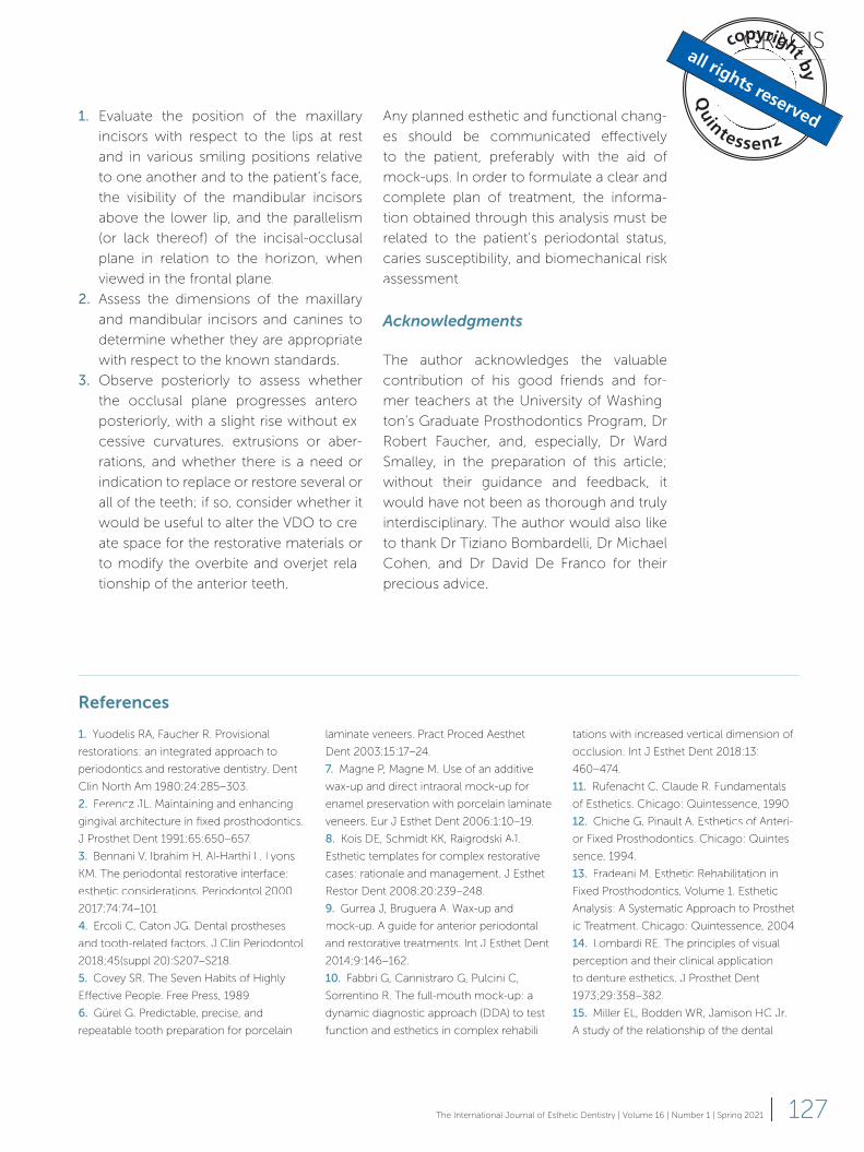
GRACIS
127The International Journal of Esthetic Dentistry | Volume 16 | Number 1 | Spring 2021 |
Any planned esthetic and functional chang-
es should be communicated effectively
to the patient, preferably with the aid of
mock-ups. In order to formulate a clear and
complete plan of treatment, the informa-
tion obtained through this analysis must be
related to the patient’s periodontal status,
caries susceptibility, and biomechanical risk
assessment.
Acknowledgments
The author acknowledges the valuable
contribution of his good friends and for-
mer teachers at the University of Washing-
ton’s Graduate Prosthodontics Program, Dr
Robert Faucher, and, especially, Dr Ward
Smalley, in the preparation of this article;
without their guidance and feedback, it
would have not been as thorough and truly
interdisciplinary. The author would also like
to thank Dr Tiziano Bombardelli, Dr Michael
Cohen, and Dr David De Franco for their
precious advice.
1. Evaluate the position of the maxillary
incisors with respect to the lips at rest
and in various smiling positions relative
to one another and to the patient’s face,
the visibility of the mandibular incisors
above the lower lip, and the parallelism
(or lack thereof) of the incisal-occlusal
plane in relation to the horizon, when
viewed in the frontal plane.
2. Assess the dimensions of the maxillary
and mandibular incisors and canines to
determine whether they are appropriate
with respect to the known standards.
3. Observe posteriorly to assess whether
the occlusal plane progresses antero-
posteriorly, with a slight rise without ex-
cessive curvatures, extrusions or aber-
rations, and whether there is a need or
indication to replace or restore several or
all of the teeth; if so, consider whether it
would be useful to alter the VDO to cre-
ate space for the restorative materials or
to modify the overbite and overjet rela-
tionship of the anterior teeth.
References
1. Yuodelis RA, Faucher R. Provisional
restorations: an integrated approach to
periodontics and restorative dentistry. Dent
Clin North Am 1980;24:285–303.
2. Ferencz JL. Maintaining and enhancing
gingival architecture in fixed prosthodontics.
J Prosthet Dent 1991;65:650–657.
3. Bennani V, Ibrahim H, Al-Harthi L, Lyons LL
KM. The periodontal restorative interface:
esthetic considerations. Periodontol 2000
2017;74:74–101.
4. Ercoli C, Caton JG. Dental prostheses
and tooth-related factors. J Clin Periodontol
2018;45(suppl 20):S207–S218.
5. Covey SR. The Seven Habits of Highly
Effective People. Free Press, 1989.
6. Gürel G. Predictable, precise, and
repeatable tooth preparation for porcelain
laminate veneers. Pract Proced Aesthet
Dent 2003;15:17–24.
7. Magne P, Magne M. Use of an additive
wax-up and direct intraoral mock-up for
enamel preservation with porcelain laminate
veneers. Eur J Esthet Dent 2006;1:10–19.
8. Kois DE, Schmidt KK, Raigrodski AJ.
Esthetic templates for complex restorative
cases: rationale and management. J Esthet
Restor Dent 2008;20:239–248.
9. Gurrea J, Bruguera A. Wax-up and
mock-up. A guide for anterior periodontal
and restorative treatments. Int J Esthet Dent
2014;9:146–162.
10. Fabbri G, Cannistraro G, Pulcini C,
Sorrentino R. The full-mouth mock-up: a
dynamic diagnostic approach (DDA) to test
function and esthetics in complex rehabili-
tations with increased vertical dimension of
occlusion. Int J Esthet Dent 2018;13:
460–474.
11. Rufenacht C, Claude R. Fundamentals
of Esthetics. Chicago: Quintessence, 1990.
12. Chiche G, Pinault A. Esthetics of Anteri-
or Fixed Prosthodontics. Chicago: Quintes-
sence, 1994.
13. Fradeani M. Esthetic Rehabilitation in
Fixed Prosthodontics, Volume 1. Esthetic
Analysis: A Systematic Approach to Prosthet-
ic Treatment. Chicago: Quintessence, 2004.
14. Lombardi RE. The principles of visual
perception and their clinical application
to denture esthetics. J Prosthet Dent
1973;29:358–382.
15. Miller EL, Bodden WR, Jamison HC Jr.
A study of the relationship of the dental
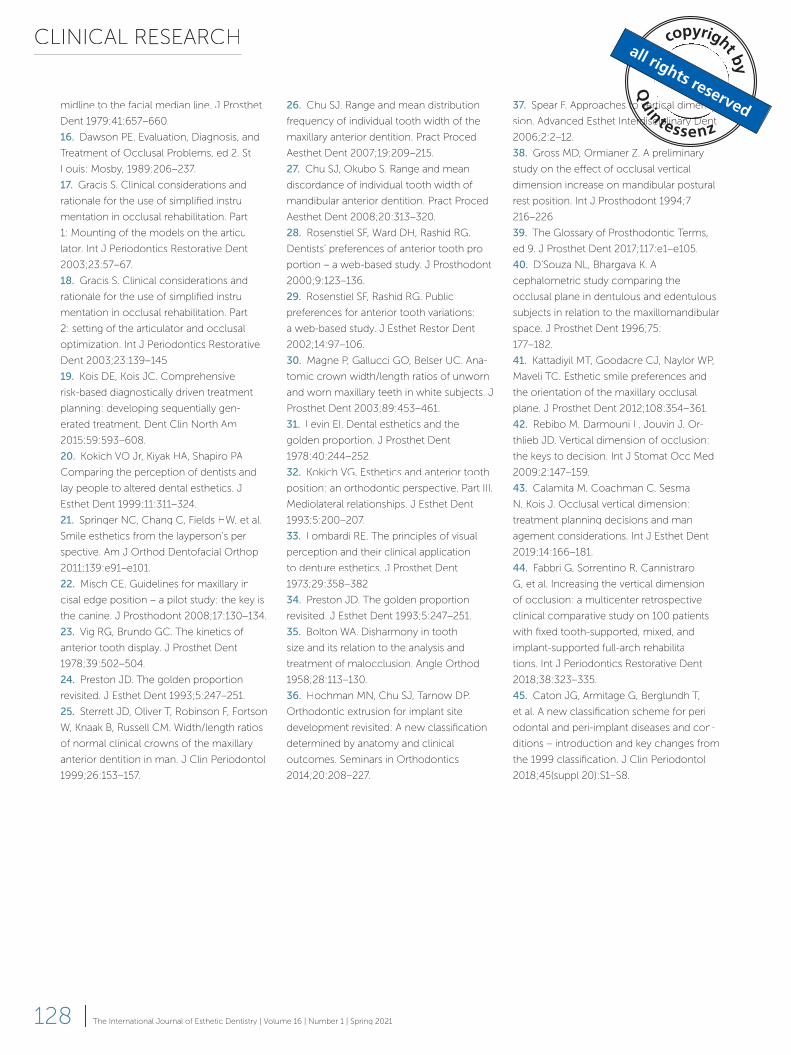
CLINICAL RESEARCH
128 | The International Journal of Esthetic Dentistry | Volume 16 | Number 1 | Spring 2021
midline to the facial median line. J Prosthet
Dent 1979;41:657–660.
16. Dawson PE. Evaluation, Diagnosis, and
Treatment of Occlusal Problems, ed 2. St
Louis: Mosby, 1989:206–237.
17. Gracis S. Clinical considerations and
rationale for the use of simplified instru-
mentation in occlusal rehabilitation. Part
1: Mounting of the models on the articu-
lator. Int J Periodontics Restorative Dent
2003;23:57–67.
18. Gracis S. Clinical considerations and
rationale for the use of simplified instru-
mentation in occlusal rehabilitation. Part
2: setting of the articulator and occlusal
optimization. Int J Periodontics Restorative
Dent 2003;23:139–145.
19. Kois DE, Kois JC. Comprehensive
risk-based diagnostically driven treatment
planning: developing sequentially gen-
erated treatment. Dent Clin North Am
2015;59:593–608.
20. Kokich VO Jr, Kiyak HA, Shapiro PA.
Comparing the perception of dentists and
lay people to altered dental esthetics. J
Esthet Dent 1999;11:311–324.
21. Springer NC, Chang C, Fields HW, et al.
Smile esthetics from the layperson’s per-
spective. Am J Orthod Dentofacial Orthop
2011;139:e91–e101.
22. Misch CE. Guidelines for maxillary in-
cisal edge position – a pilot study: the key is
the canine. J Prosthodont 2008;17:130–134.
23. Vig RG, Brundo GC. The kinetics of
anterior tooth display. J Prosthet Dent
1978;39:502–504.
24. Preston JD. The golden proportion
revisited. J Esthet Dent 1993;5:247–251.
25. Sterrett JD, Oliver T, Robinson F, Fortson
W, Knaak B, Russell CM. Width/length ratios
of normal clinical crowns of the maxillary
anterior dentition in man. J Clin Periodontol
1999;26:153–157.
26. Chu SJ. Range and mean distribution
frequency of individual tooth width of the
maxillary anterior dentition. Pract Proced
Aesthet Dent 2007;19:209–215.
27. Chu SJ, Okubo S. Range and mean
discordance of individual tooth width of
mandibular anterior dentition. Pract Proced
Aesthet Dent 2008;20:313–320.
28. Rosenstiel SF, Ward DH, Rashid RG.
Dentists’ preferences of anterior tooth pro-
portion – a web-based study. J Prosthodont
2000;9:123–136.
29. Rosenstiel SF, Rashid RG. Public
preferences for anterior tooth variations:
a web-based study. J Esthet Restor Dent
2002;14:97–106.
30. Magne P, Gallucci GO, Belser UC. Ana-
tomic crown width/length ratios of unworn
and worn maxillary teeth in white subjects. J
Prosthet Dent 2003;89:453–461.
31. Levin EI. Dental esthetics and the
golden proportion. J Prosthet Dent
1978;40:244–252.
32. Kokich VG. Esthetics and anterior tooth
position: an orthodontic perspective. Part III.
Mediolateral relationships. J Esthet Dent
1993;5:200–207.
33. Lombardi RE. The principles of visual
perception and their clinical application
to denture esthetics. J Prosthet Dent
1973;29:358–382.
34. Preston JD. The golden proportion
revisited. J Esthet Dent 1993;5:247–251.
35. Bolton WA. Disharmony in tooth
size and its relation to the analysis and
treatment of malocclusion. Angle Orthod
1958;28:113–130.
36. Hochman MN, Chu SJ, Tarnow DP.
Orthodontic extrusion for implant site
development revisited: A new classification
determined by anatomy and clinical
outcomes. Seminars in Orthodontics
2014;20:208–227.
37. Spear F. Approaches to vertical dimen-
sion. Advanced Esthet Interdisciplinary Dent
2006;2:2–12.
38. Gross MD, Ormianer Z. A preliminary
study on the effect of occlusal vertical
dimension increase on mandibular postural
rest position. Int J Prosthodont 1994;7:
216–226.
39. The Glossary of Prosthodontic Terms,
ed 9. J Prosthet Dent 2017;117:e1–e105.
40. D’Souza NL, Bhargava K. A
cephalometric study comparing the
occlusal plane in dentulous and edentulous
subjects in relation to the maxillomandibular
space. J Prosthet Dent 1996;75:
177–182.
41. Kattadiyil MT, Goodacre CJ, Naylor WP,
Maveli TC. Esthetic smile preferences and
the orientation of the maxillary occlusal
plane. J Prosthet Dent 2012;108:354–361.
42. Rebibo M, Darmouni L, Jouvin J, Or-
thlieb JD. Vertical dimension of occlusion:
the keys to decision. Int J Stomat Occ Med
2009;2:147–159.
43. Calamita M, Coachman C, Sesma
N, Kois J. Occlusal vertical dimension:
treatment planning decisions and man-
agement considerations. Int J Esthet Dent
2019;14:166–181.
44. Fabbri G, Sorrentino R, Cannistraro
G, et al. Increasing the vertical dimension
of occlusion: a multicenter retrospective
clinical comparative study on 100 patients
with fixed tooth-supported, mixed, and
implant-supported full-arch rehabilita-
tions. Int J Periodontics Restorative Dent
2018;38:323–335.
45. Caton JG, Armitage G, Berglundh T,
et al. A new classification scheme for peri-
odontal and peri-implant diseases and con-
ditions – introduction and key changes from
the 1999 classification. J Clin Periodontol
2018;45(suppl 20):S1–S8.