A simple probe for DNA accessibility in chromatin
8
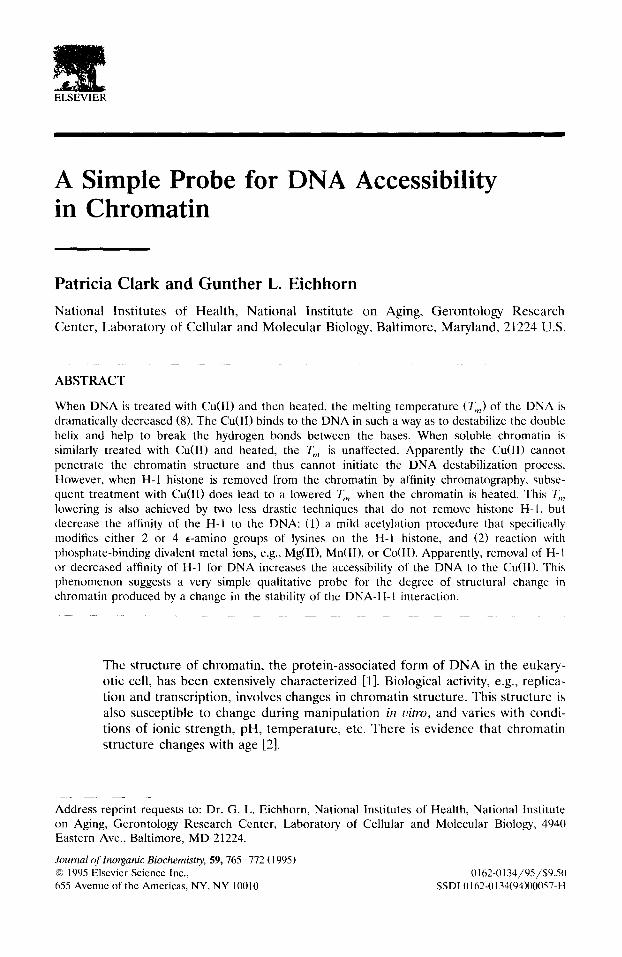
: • . ,,, ELSEVIER A Simple Probe for DNA Accessibility in Chromatin Patricia Clark and Gunther L. Eichhorn National Institutes of Health, National Institute on Aging, Gerontology Research Center, Laboratory of Cellular and Molecular Biology, Baltimore, Maryland, 21224 U.S. ABSTRACT When DNA is treated with Cu(II) and then heated, the melting temperature (T m) of thc DNA is dramatically decreased (8). The Cu(II) binds to the DNA in such a way as to destabilize the double helix and help to break the hydrogen bonds between the bases. When soluble chromatin is similarly treated with Cu(II) and heated, the Tm is unaffected. Apparently the Cu(ll) cannot penetrate the chromatin structure and thus cannot initiate the DNA destabilization process. However, when H-1 histone is removed from the chromatin by affinity chromatography, subse- quent treatment with Cu(II) does lead to a lowered 7,, when the chromatin is heated. This T,,, lowering is also achieved by two less drastic techniques that do not remove histonc H-l, but decrease the affinity of the H-1 to the DNA: (1) a mild acetylation procedure that specifically modifies either 2 or 4 e-amino groups of lysines on the H-I histone, and (2) reaction with phosphate-binding divalent metal ions, e.g., Mg(II), Mn(I1), or Co(II). Apparently, removal of H-1 or decreased affinity of H-1 for DNA increases the accessibility of the DNA to the Cu(lI). This phenomenon suggests a very simple qualitative probe for the degree of structural change in chromatin produced by a change in the stability of the DNA-H-1 interaction. The structure of chromatin, the protein-associated form of DNA in the eukary- otic cell, has been extensively characterized [1]. Biological activity, e.g., replica- tion and transcription, involves changes in chromatin structure. This structure is also susceptible to change during manipulation in tqtro, and varies with condi- tions of ionic strength, pH, temperature, etc. There is evidence that chromatin structure changes with age [2]. Address reprint requests to: Dr. G. L. Eichhorn, National Institutes of Health, National Institute on Aging, Gerontology Research Center, Laboratory of Cellular and Molecular Biology, 494(I Eastern Ave., Baltimore, MD 21224. Journal of Inorganic Biochemistry, 59, 765-772 (1995) © 1995 Elsevier Science Inc., l)162-0134/95/$9.50 655 Avenue of the Americas, NY, NY 10010 SSDI 0162-0134(94)00057-H
-
Upload
patricia-clark -
Category
Documents
-
view
212 -
download
0
Transcript of A simple probe for DNA accessibility in chromatin
: • . , , ,
ELSEVIER
A Simple Probe for DNA Accessibility in Chromatin
Patricia Clark and Gunther L. Eichhorn
National Institutes of Health, Nat ional Institute on Aging, Geron to logy Research Center, Labora tory of Cellular and Molecular Biology, Balt imore, Maryland, 21224 U.S.
ABSTRACT
When DNA is treated with Cu(II) and then heated, the melting temperature (T m) of thc DNA is dramatically decreased (8). The Cu(II) binds to the DNA in such a way as to destabilize the double helix and help to break the hydrogen bonds between the bases. When soluble chromatin is similarly treated with Cu(II) and heated, the T m is unaffected. Apparently the Cu(ll) cannot penetrate the chromatin structure and thus cannot initiate the DNA destabilization process. However, when H-1 histone is removed from the chromatin by affinity chromatography, subse- quent treatment with Cu(II) does lead to a lowered 7,, when the chromatin is heated. This T,,, lowering is also achieved by two less drastic techniques that do not remove histonc H-l, but decrease the affinity of the H-1 to the DNA: (1) a mild acetylation procedure that specifically modifies either 2 or 4 e-amino groups of lysines on the H-I histone, and (2) reaction with phosphate-binding divalent metal ions, e.g., Mg(II), Mn(I1), or Co(II). Apparently, removal of H-1 or decreased affinity of H-1 for DNA increases the accessibility of the DNA to the Cu(lI). This phenomenon suggests a very simple qualitative probe for the degree of structural change in chromatin produced by a change in the stability of the DNA-H-1 interaction.
The structure of chromatin , the protein-associated form of D N A in the eukary- otic cell, has been extensively character ized [1]. Biological activity, e.g., replica- tion and transcription, involves changes in chromat in structure. This structure is also susceptible to change during manipulat ion in tqtro, and varies with condi- tions of ionic strength, pH, temperature , etc. There is evidence that chromatin structure changes with age [2].
Address reprint requests to: Dr. G. L. Eichhorn, National Institutes of Health, National Institute on Aging, Gerontology Research Center, Laboratory of Cellular and Molecular Biology, 494(I Eastern Ave., Baltimore, MD 21224.
Journal of Inorganic Biochemistry, 59, 765-772 (1995) © 1995 Elsevier Science Inc., l)162-0134/95/$9.50 655 Avenue of the Americas, NY, NY 10010 SSDI 0162-0134(94)00057-H

766 P. Clark and G. L. Eichhorn
The accessibility of the DNA to various agents is obviously affected by its association with proteins; the DNA is most accessible without such association, and the loss of accessibility in various forms of chromatin is an indication of the degree of association of protein and DNA.
Sophisticated techniques are available for studying chromatin structure and the changes in it. In this paper, we describe a very simple technique that can be a coarse guide in following changes in chromatin structure based on changes in DNA accessibility. The technique consists of the reaction of Cu(II) with the DNA, monitored by changes in the melting profile of the system.
EXPERIMENTAL
Reagents
The salts, NaCI, sodium tetra borate, Na cacodylate, the Mg(II), Co(II), Zn(II), Cu(II), Mn(II), and Ca(II) chlorides were all analytical reagent grade materials. EDTA, ethylene diamine tetraacetic acid disodium salt, and PMSF, phenyl methyl sulfonyl fluoride, were manufactured by the Sigma Chemical Co., St. Louis, Missouri, U.S. Acetic anhydride, A.C.S. certified, was manufactured by the Fisher Chemical Co., Fair Lawn, New Jersey, U.S.
Preparation of Chromatin
Chromatin was prepared by the modified procedures of Clark [3a] and Axel [3b] using the livers of eight-nine month old Wistar male rats from the Animal Research Facility of the Gerontology Research Center. 2 mg of RNase A were added to each 500 mL of the solvent containing 0.08 M NaCI, 0.02 M EDTA, pH 7.2. The washed nuclei were sheared for 2 min at 80 V in a Virtis Homogenizer, Model 45. The final solvent was 5 × 10 -4 M cacodylate, pH 6.0. The concentra- tion of the chromatin was determined by the diphenylamine procedure of G. M. Richards [4]. The concentrations used in the melting experiments were 5 × 10 -5 M(P) chromatin, 5 × 10 -3 M NaC1, 5 × 10 -4 M cacodylate, pH 6.0.
UV Spectra and Melting Curves
The UV spectra and melting curves were run on a Cary 219, to which a Lauda K 2 / R water bath was attached for heating and cooling, and a Tektronix 4052 computer for processing the melting curve data. A Gilford Spectrophotometer 2400, with a Lauda K2 water bath attached for heating, was also used for melting curves.
Analysis of Salts by Atomic Absorption
The solutions of metal ions used in this paper, Mg(II), Cu(II), Mn(II), Ca(II), Co(II), and Zn(II) chlorides, were analyzed by atomic absorption spectrometry using a Perkin-Elmer Model 306 instrument.
Acetylation of Chromatin [5]
To 10 mL of 5 × 10 -4 M(P) soluble chromatin in 0.01 M sodium borate, pH 8.'2, in 0.1 M NaCI, 0.1 mL of 1.4 × 10 -2 M acetic anhydride in benzene was added with stirring in an ice bath for 20 min. The sample was then dialyzed overnight against a solution of 5 × 10 -3 M NaC1, 5 × 10 -4 M cacodylate, pH 6.0 (2 L) that was changed twice. A second sample of 10 mL of chromatn with the same salt

P R O B E FOR DNA ACCESSIBILITY IN C H R O M A T I N 767
concentrations was acetylated with 0.7 x 10 -2 M acetic anhydride in 0.1 mL of benzene with stirring in an ice bath. It was then dialyzed overnight against a solution of 5 X 10 -3 M NaC1, 5 x 10 -4 M cacodylate, pH 6.0. The dialysis solvent was changed twice. A control sample of 5.0 mL of chromatin was treated similarly, but with no acetylation.
Removal of HI Histone from Chromatin
This procedure is taken from Allen et al. [6] with the following modifications. The buffer in which the chromatin is circulated over the cellose-DNA column is 10 mM NaC1, 10 mM tris, pH 7.5, 0.1 mM EDTA, and 0.1 mM PMSF. The DNA-cellulose was prepared by the method of Litman [7].
RESULTS AND DISCUSSION
Effect of Metals on DNA
The differential effect of metal ions on the melting temperature of DNA was discovered over 30 years ago; specifically, it was shown that some metals increase Tin, while others decrease it. In Figure 1, we have shown derivative melting curves of the effects of Mg(II) and Cu(II) on DNA, originally reported in the nonderivative mode [8]. Mg(II) binds to the phosphate and stabilizes the double helix by neutralization of the negative charges on the phosphate, thus increasing T m. Cu(II) binds to the bases, competes with H-bonds, and unravels the double helix with a marked reduction in T m.
Effect of Cu(II) on Chromatin
The derivative melting curve of chromatin is shown as curve a of Figure 2, and it can be seen that T,n is not only higher than T m of free D N A at the same
I-- "0
" 0
.09 - Cu(ll)
.06
.03 I I M(lil f~'~x - No Mg(ll)
I I / ....... .,../ "-
20 40 60 80 100
TEMPERATURE, °C FIGURE 1. Derivatives of melting curves of DNA, DNA + Mg(II), or Cu(II). a. 5 × 10- 5 M(P) DNA in 5 × 10 -3 M NaNO3, • • • -. b. DNA, same as curve a, + 10 -4 M Mg(NO3)2, . . . . . c. DNA, same as curve a, + 10 4 M Cu(NO3) 2, - - .

768 P. Clark and G. L. E ichhorn
t-.-.
. 0 1 5
.01 -
. 0 0 5 -
0
2 0
I I I
I I
40 60 80 100
TEMPERATURE, °C
FIGURE 2. Derivatives of melting curves of chromatin with Cu(II). Curve a: Chro- matin, 5 x 10 -5 M (P), with 5 x 10 -3 M NaC1, 5 x 10 -4 cacodylate, pH 6, - - . Curve b: Same as curve a, with 6.25 x 10 -6 M CuCI2, .. • -. Curve c: Same as curve a, 1.25 x 10 -5 M CuCI2, ---- . Curve d: Same as curve a, 1.875 x 10 -5 M CuC12, - . . . . . . . .
concentration (Figure 1), but that it is also higher than T m of D N A stabilized by Mg(II) (Figure 1). The protein bound to the D N A in chromatin evidently stabilizes the D N A more than does Mg(II); it does so through charge neutraliza- tion by positively charged lysines and arginines on the histones. The higher stabilization is undoubtedly due to the compact binding of the histones. The shoulder around 75 ° corresponds to the melting of the linker region.
The addition of Cu(II) to the chromatin up to a ratio of ~ 0.4 M C u ( I I ) / M DNA(P) has no effect on the T m (0.5 mole Cu(II) leads to precipitation). In fact, concentrations of Cu(II) f rom 0.1 to 0.4 (Figure 2b-d) produce virtually identical melting curves with chromatin (while T m varies considerably when such concen- trations are added to free D N A [9]). There is a slight increase in absorbance, probably due to nonspecific interaction with chromatin.
We have established, therefore, that Cu(II) reacts very dramatically with free DNA, as shown by the decrease in T m, but does not react at all with chromatin in this way. The apparent explanation is that the DNA bases in chromatin are protected by the association of the D N A with the chromatin proteins, so that these bases are not accessible to the Cu(II).
We next increase D N A accessibility in chromatin by removal of histone H-1 to determine whether Cu(II) now reacts with the DNA.
Effect of Cu(II) on Chromatin with H-1 Histone Removed
The H-1 histone is the agent that helps to hold the chromosomal nucleosomes together by binding to the linker D N A region as well as to the nucleosomes through its globular regions. Thus, the removal of H-1 produces an unprotected (stripped) chromatin structure. It is not surprising, therefore, that H-1 removal affects the D N A melting profile, and curve a in Figure 3 demonstrates the lowering of the temperature for the melting of the linker D N A from 75 ° (in Figure 2) to 65 °. The nucleosomal region, however, still melts at 82-83 ° , in Figure 3, as in Figure 2. Thus, the major effect of H-1 removal is, as expected, on the linker region, which the H-1 protected.
Removal of H-1 has a much more dramatic effect on the interaction of Cu(II) with chromatin. Figure 3b shows that all the DNA now melts in the 65 ° region, both the nucleosomal and the linker DNA. Thus, all of the chromatin DNA,

PROBE FOR DNA ACCESSIBILITY IN CH RO MA TIN 769
"0
.o1(,,,,,,b a .005 ~ \
0 20 40 60 80
TEMPERATURE, °C
FIGURE 3. Derivative melting curves of chromatin with the H-1 histone removed. Curve a: Chro- matin with H-1 histone removed, 5 x 10 -5 M (P), with 5 x 10 - 3 M NaC1, 5 x 10 ~ M cacodylate, pH 6, ----. Curve b: Same as curve a, 2.5 x 10 -5 M, CuCI 2, - -
inaccessible in the native state, has now become accessible to reaction with Cu(II). The removal of H-1 histone has freed the linker DNA so that the Cu(II) ion can react with it. The removal of the globular portion of the H-1 from the DNA entering and exiting the nucleosomal component probably permits the Cu(II) to react first with some of the nucleosomal DNA, to unravel it, and eventually to bind to all of it. The nucleosomal and linker DNA regions have become indistinguishable in terms of DNA melting.
Apparently, the addition of Cu(II) greatly enhances the difference in the melting curves of chromatin before and after the removal of H-1. The accessibil- ity of Cu(II) to DNA in stripped chromatin and the consequent change in the melting curve, particularly the decrease in T m and the removal of the high peak, appears to be a qualitative indicator of the degree of binding of DNA to proteins in chromatin. The difference in the presence of Cu(II) is much greater than the difference in its absence.
Since reaction with Cu(II) dramatically distinguishes between the tightly bound protein-nucleic acid system and the more loosely bound "stripped" system, the question arises whether Cu(II) ion can be more generally used as a very simple qualitative probe of the tightness of binding.
Acetylated Chromatin
The chromatin structure can be subjected to a milder perturbation by changing the H-1 histone without actually removing it. A mild acetylation procedure [5] was used to produce two chromatin modifications, in which either two or four e-amino groups of the lysines on H-1 had been acetylated. This relatively mild procedure had minor effects on the chromatin melting curve and the T,,, was unaffected (compare curves b and c with curve a of Figure 4).
The addition of 0.25 mole Cu(I I ) /DNA(P) to the acetylated chromatins lowered the T m of the nucleosomal DNA as well as the linker DNA quite considerably, and the Cu(II) reaction also differentiates between the doubly and quadruply acetylated chromatin (curves d and e of Figure 4). Clearly, the Cu(II) is a sensitive probe of changes in chromatin structure that are not very evident in the absence of the metal.
Chromatin Treated with Mg(II) and Other Divalent Ions Binding DNA Phosphate
We have previously noted the greater effect of histones, compared to Mg(II), in stabilizing DNA. It is of interest, therefore, to determine what effect Mg(II) has on chromatin. In Figure 5, it can be seen that the T m of the nucleosomal peak of native chromatin is decreased (although not greatly), and that there is an

770 ~ C ~ r k a n d G. L. Eichhorn
I - - "O
"O
.02,
.01
20 100
O
..<.-." /7 'd • C . ' " . ¢ / I.'t" / . , ; ......
40 60 80
TEMPERATURE,°C
FIGURE 4. Derivative melting curves of acetylation of chromatin, a. Unacety- lated chromatin 5 x 10 -5 M (P), with 5 x 10 -3 M NaC1, 5 X 10 -4 M cacody- late, pH 6, - - . b. Chromatin, same con- centration as in a. Low level acetylation, see Exerimental, 2 lysines acetylated . . . . . c. Chromatin, same concentration as curve a. High level acetylation, see Ex- perimental, 4 lysines acetylated . . . . . d. Sample b plus 1.25 x 10 -5 M CuC12 . . . . . . . . e. Sample c plus 1.25 x 10⁻⁵ M CuCI 2 . . . . . .
increase in the mel t ing o f the l inker D N A . A p p a r e n t l y , the Mg(I I ) loosens the b ind ing o f the C- te rmina l and N- t e rmina l a rms o f the H-1 h i s tone by compe t ing with the posi t ively cha rged a m i n o acids for the D N A phospha tes , thus making the l inker D N A m o r e accessible. Th rough mass act ion, an excess of Mg(I I ) will d isp lace the m o r e s t rongly b o u n d his tone.
But the add i t i on o f Cu( I I ) to the ch roma t in p r e t r e a t e d with Mg(I I ) leads to much m o r e extensive p e r t u r b a t i o n (F igure 6). The nuc leosomal p e a k has been d i sp laced to a much lower t e m p e r a t u r e at the h igher Cu(I I ) concent ra t ions , and appa ren t l y the bu lk o f the D N A has b e c o m e access ible to r eac t ion with Cu(II) .
S tud ies with o t h e r d iva len t ions tha t b ind to D N A phospha te , i.e., Co(II) , Ca(II) , Zn( I I ) , and Mn(I I ) , l ed to resul ts smi la r to those with Mg(II) .
D I S C U S S I O N
Cu(H) as a Probe of P ro te in -Nuc le ic Acid Binding in C h r o m a t i n
A compar i son o f F igures 1 and 2 suggests tha t the Cu(I I ) r eac t ion d e p e n d s on the accessibi l i ty of D N A - - i t is very d rama t i c in f ree D N A and vir tual ly nonexis- ten t in chromat in . A f t e r r emova l of H - l , thus s t r ipping the ch roma t in s t ructure ,
" D
.025
.02
0.015 b !
.01 r-
.005 L
0 20
I I I I I I l
. . S- .- ._i - I
40 60 80 100
TEMPERATURE, °C FIGURE 5. Derivative melting curves of chromatin with Mg(II) or Co(II). a. Chromatin, 5 x 10 -5 M (P), with 5 x 10 -3 M NaCI, 5 x 10 -5 M cacodylate, pH 6. b. Same as a, with 5 x 10 -5 M MgC12, • • - .. c. Same as a, with 1 x 10 -4 M MgC12, - - - - . d. Same as a, with 1.5 x 10 -4 M MgCI2, - - - -. e. Same as ~i, with 2.0 x 10 -4 M MgCI 2, - . . . . . . . f. Same as a, with 2.5 x 10 -4 M MgC12, - - - -

PROBE FOR DNA ACCESSIBILITY IN CHROMATIN 771
"0
.02
.015
.01
.005
0 2O
Z
.... b t ~ k
40 60 80
~¢,a
100
TEMPERATURE, °C FIGURE 6. Derivative melting curves of the effects of adding increasing amounts of Cu(II) to solutions of 4Mg(II) chromatin, a. Same as curve e in Figure 5 --. b. Same conditions as curve a, plus 1.24 x 10 -5 M CuCI 2 . . . . . . . . c. Same conditions as curve a, plus 5 x 10 -5 M CuC12 . . . . .
Cu(II) extensively lowers the T,, of all the DNA, whether originally in the nucleosomal or linker region. Acetylation of histone-H-1, which has a lesser effect on chromatin structure than removal of that histone, also has a lesser, although significant, effect on the Cu(II) induced change in the melting behav- ior. Treatment with divalent ions, such as Mg(II), Zn(II), Ca(II), Co(II), and Mn(II), also loosens the histone-DNA bonds, and therefore leads to the increased accessibility to Cu(II).
The effect of Cu(II) on DNA structure eventually leads to the breaking of all the hydrogen bonds of the DNA double helix. We have previously called this process unwinding, but it must be understood that the unwinding is more than the partial unwinding produced by reaction of milder reagents with DNA. Melting curves such as those in Figure 1 probably involve breaking of all the H-bonds. The reaction is reversible because Cu(II) atoms can hold the strands together in the denatured state, and renaturation can occur when Cu(II) is removed by complexing or competition with high electrolyte (10,11).
The beginning of the unwinding process probably occurs when a large enough segment of the double helix has broken H-bonds to make it possible for at least one Cu(II) to fit into the hole left by the H-bond breaks. The binding of this first Cu(II) sets up a cooperative phenomenon so that more Cu(II) ions bind, and eventually all the hydrogen bonds become severed. We picture the Cu(II) in dynamic equilibrium with the Cu(II) complex of DNA, and the Cu(II) ions as moving between the DNA chains, rather than being permanently attached at specific sites.
Obviously, the DNA must be accessible for this process to be initiated and to be continued. It is thus reasonable that varying degrees of accessibility of the DNA in chromatin lead to varying degrees of reaction with Cu(II), so that the Cu(II) reaction becomes a qualitative index of the looseness of binding of DNA and protein. We do not believe that it can be quantitative, since there are different types of bonds between DNA and protein in the chromatin structure, and structural changes in different regions of chromatin may lead to different decreases in DNA accessibility.

772 P. Clark and G. L. Eichhorn
On this basis, we place the DNAs studied here into the following sequence of increased accessibility to Cu(II), and therefore decreased tightness of binding of proteins to DNA:
Native Acetylated < Chromatin Treated Chromatin with < free D N A Chromatin < Chromatin with M(II) binding < H-1 histone
to phosphate removed
We suggest that the Cu(II) reaction could be used as a very simple test of p r o t e i n - D N A binding in chromatin preparations.
REFERENCES
1. (a) R. Kornberg, Science 184, 868-871 (1974). (b) R. Kornberg, Ann. Reu. Biochem. 46, 931 (1977). (c) J. D. McGhee and G. Felsenfeld, Ann. Rec. Biochem. 49, 1115-1156 (1980). (d) T. Igo-Kemenes, W. Horz, and H. G. Zachau, Ann. Rev. Biochem. 51, 89-121 (1982). (e) G. Felsenfeld and J. D. McGhee, Cell 44, 375 (1986). (f) J. Widom, Ann. Reu. Biophys. Chem. 18, 365-395 (1989). These references are only a few reviews of the many papers on the structure of chromatin in the literature.
2. (a) G. D. Bardyshev, "Age-dependent changes of in citro transcription activities of animals," in R. G. Cutler, Ed., Interdisciplinary Topics in Gerontology, Vol. 9, Karger Press, Basel, 1976. (b) C. W. Dingman and M. B. Sporn, J. of Biol. Chem. 239, 3483-3492 (1964). (c) D. I. Kurtz, A. P. Russell, and F. M. Sinex, Mech. of Aging & Devel. 3, 37-49 (1974). (d) G. D. Bardyshev and S. Zhelabovskaya, Exp. Get. 7, 321-330 (1972). (e) R. G. Cutler, "Alterations with age in the information storage and flow systems of the mammalian cell," in D. Bargsma, Ed., Genetic Effects on Aging, Alan R. Liss, Inc., New York, 1978, pp. 463-498. (f) F. P. von Hahn and E. Fritz, Gerontologia 12, 237-260 (1966). (g) H. P. yon Hahn, Exp. Get. 5, 323-334 (1970). (h) A. R. O'Meara and R. L. Herrmann, Biochem. Biophysica Acta 269, 419-427 (1972). (i) J. S. Salser and M. E. Baltis, J. of Ger. 27, 1-9 (1972).
3. (a) R. J. Clark and G. Felsenfeld, Nature New Biology 229, 101 (1971). (b) R. Axel, Biochem. 14, 2921 (1975).
4. G. M. Richards, Anal. Biochem. 57, 369-376 (1974). 5. T. K. Wong and K. Marushige, Biochem. 15, 2041-2046 (1976). 6. J. Allen, D. Z. Staynov, and H. Gould, Proc. Natl. Acad. Sci. U.S.A. 77, 885-889
(1980). 7. R. M. Litman, J. Biol. Chem. 243, 6222-6233 (1968). 8. G. L. Eichhorn, Nature 194, 474 (1962). 9. G. L. Eichhorn, Ed., Inorganic Biochemistry, Elsevier Scientific Publ. Co., New York,
1975, Vol. 2, Chapt. 34, pp. 1210-1243. 10. G. L. Eichhom and P. Clark, Proc. Natl. Acad. Sci. U.S.A. 53, 586-593 (1965). 11. S. Hiai, J. Mol. Biol. 11, 672-691 (1965).
Received August 18, 1994; accepted September 1, 1994