A Short Chromosomal Region With Major Roles in Yeast ...
11
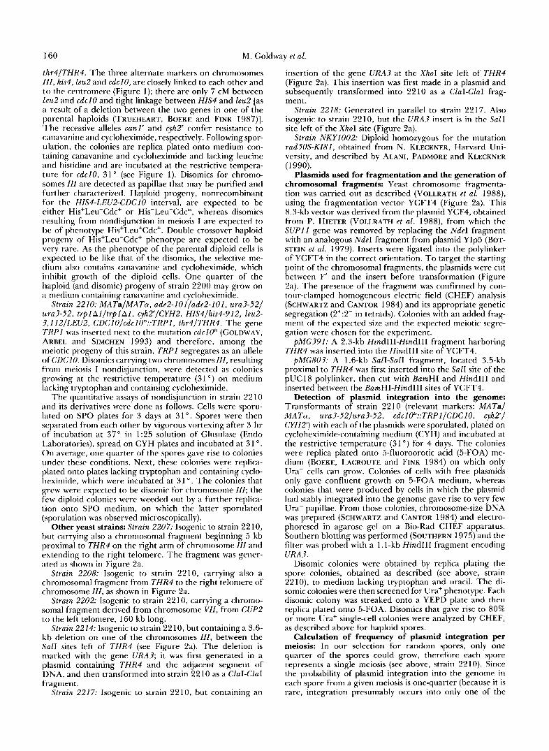
Copyright 0 1993 by the Genetics Society of America A Short Chromosomal Region With Major Roles in Yeast Chromosome ZZZ Meiotic Disjunction, Recombination and Double Strand Breaks Martin Goldway, Amir Sherman, Drora Zenvirth, Tamar Arbel and Giora Simchen Department of Genetics, The HebrewUniversity of Jerusalem, Jerusalem 91904, Israel Manuscript received July 29, 1992 Accepted for publication October 16, 1992 ABSTRACT A multicopy plasmid was isolated from a yeast genomiclibrary, whose presence resulted in a twofold increase in meiotic nondisjunction of chromosome III. T h e plasmid contains a 7.5-kb insert from the middle of the right armof chromosome 111, including the gene THR4. Using chromosomal fragments derived from chromosome 111, we determined that the cloned region caused a significant, specific, cis-acting increase in chromosome 111 nondisjunction in the first meiotic division. The plasmid containing this segment exhibited high spontaneous meiotic integration into chromosome 111 (in 2.4% of thenormal meiotic divisions) and a sixfold increase (15.5%) in integration in nondisjunctant meioses. Genetic analysis of the cloned region revealed that it contains a “hot spot” for meiotic recombination. In DNA of rad50S mutant cells, a strong meiosis-induced double strand break (DSB) signal was detected in this region. We discuss the possible relationshipsbetween meiosis-induced DSBs, recombination and chromosome disjunction, and propose that recombinational hot spots may be “pairingsites” for homologous chromosomes in meiosis. M EIOSIS consists of DNA synthesis followed by two cycles of nuclear divisions, the reductional division (meiosis I) and the equational division (meiosis 11), by which a diploid cell gives rise to four haploid products. Meiosis I1 resembles mitosis, whereas in meiosis I thereare distinct phenomena. The most outstanding of these are homologous chromosome pairing accompanied by synaptonemal complex (SC) formation (reviewed by VON WETTSTEIN et al. 1984) and high levels of homologous recombination leading to formation of chiasmata. Pairing and recombination are crucial forproperreductionalsegregation. Im- proper segregation of the chromosomes results in unbalanced meiotic products that may be inviable or severely impaired. It has been shown for Drosophila melanogaster (PARRY 1973; CARPENTER and SANDLER 1974; HALL 1972) and Saccharomyces cerevisiae (EN- GELRRECHT and ROEDER 1990; ROCKMILL and ROE- DER 1988; HOLLINGSWORTH, GOETSCH and BYERS 1990) thatmutations which cause deficientpairing and recombination in meiosis lead to malsegregation of chromosomes. In the yeast S. cerevisiae, the rad50 mutation was found to block both recombination and SC formation (ALANI, PADMORE and KLECKNER 1990). Moreover, meiotic double strand breaks (DSBs), which are interpreted as an initial stage of recombination (NICOLAS et al. 1989; CAO, ALANI and Kleckner 1990; SUN, TRECO and SZOSTAK 199 l), ap- pear early in meiosis (SUN et al. 1989), before and at the same time as the first appearance of synaptonemal complex (PADMORE, CAO and KLECKNER 199 1). These Genetics 133: 159-169 (February, 1992) findings suggest that the above processes are closely related. Here we report the molecular cloning and charac- terization of an element from chromosome ZZZ of the yeast S. cerevisiae, that has a profound effect on the meiotic disjunction of its chromosome of origin. The presence of the element on an added chromosomal fragment (VOLLRATH et al. 1988) led to increased meiotic nondisjunction of chromosomes ZZZ. More- over, a multicopy plasmid carrying the element inte- grated at a high frequency into chromosome ZZZ dur- ing meiosis,especiallyinmeiosesinwhich chromo- some ZZZ failed to disjoin correctly. We also present evidence for a “hot spot” for meiotic recombination and a strong DSB site in the element. On the basis of our findings, we discuss the possible relationships be- tween recombination and chromosome disjunction during meiosis. MATERIALS AND METHODS Media for yeast: YEPD medium (1% yeast extract, 2% glucose, 2% Bacto-peptone plus, 1.5% agar for solid me- dium) was used for nonselective growth. For selective growth,complete synthetic media (0.67% yeast nitrogen base without amino acids, 2% glucose, 1.5% agar, uracil, adenine and amino acids) lacking the components selected for, were used (SHERMAN, FINK and HICKS 1986). SPO solid sporulation medium has been described (HICKS and HER- SKOWITZ 1976). CYH medium is YEPD with 1.8 mg/liter cycloheximide. Yeast strains for the detection and measurement of meiotic nondisjunction of chromosome ZZk Strain 2200: MATalMATcU, ura3-52/ura3-52, trplltrpl, canl‘/CANI, cyhT/CYH2, HIS4/his4-912, leu2/LEU2, CDCIO/cdclOts,
-
Upload
nguyenxuyen -
Category
Documents
-
view
216 -
download
0
Transcript of A Short Chromosomal Region With Major Roles in Yeast ...

Copyright 0 1993 by the Genetics Society of America
A Short Chromosomal Region With Major Roles in Yeast Chromosome ZZZ Meiotic Disjunction, Recombination and Double Strand Breaks
Martin Goldway, Amir Sherman, Drora Zenvirth, Tamar Arbel and Giora Simchen Department of Genetics, The Hebrew University of Jerusalem, Jerusalem 91904, Israel
Manuscript received July 29, 1992 Accepted for publication October 16, 1992
ABSTRACT A multicopy plasmid was isolated from a yeast genomic library, whose presence resulted in a
twofold increase in meiotic nondisjunction of chromosome III . The plasmid contains a 7.5-kb insert from the middle of the right arm of chromosome 111, including the gene THR4. Using chromosomal fragments derived from chromosome 111, we determined that the cloned region caused a significant, specific, cis-acting increase in chromosome 111 nondisjunction in the first meiotic division. The plasmid containing this segment exhibited high spontaneous meiotic integration into chromosome 111 (in 2.4% of the normal meiotic divisions) and a sixfold increase (15.5%) in integration in nondisjunctant meioses. Genetic analysis of the cloned region revealed that it contains a “hot spot” for meiotic recombination. In DNA of rad50S mutant cells, a strong meiosis-induced double strand break (DSB) signal was detected in this region. We discuss the possible relationships between meiosis-induced DSBs, recombination and chromosome disjunction, and propose that recombinational hot spots may be “pairing sites” for homologous chromosomes in meiosis.
M EIOSIS consists of DNA synthesis followed by two cycles of nuclear divisions, the reductional
division (meiosis I) and the equational division (meiosis 11), by which a diploid cell gives rise to four haploid products. Meiosis I1 resembles mitosis, whereas in meiosis I there are distinct phenomena. The most outstanding of these are homologous chromosome pairing accompanied by synaptonemal complex (SC) formation (reviewed by VON WETTSTEIN et al. 1984) and high levels of homologous recombination leading to formation of chiasmata. Pairing and recombination are crucial for proper reductional segregation. Im- proper segregation of the chromosomes results in unbalanced meiotic products that may be inviable or severely impaired. It has been shown for Drosophila melanogaster (PARRY 1973; CARPENTER and SANDLER 1974; HALL 1972) and Saccharomyces cerevisiae (EN- GELRRECHT and ROEDER 1990; ROCKMILL and ROE- DER 1988; HOLLINGSWORTH, GOETSCH and BYERS 1990) that mutations which cause deficient pairing and recombination in meiosis lead to malsegregation of chromosomes. In the yeast S. cerevisiae, the rad50 mutation was found to block both recombination and SC formation (ALANI, PADMORE and KLECKNER 1990). Moreover, meiotic double strand breaks (DSBs), which are interpreted as an initial stage of recombination (NICOLAS et al. 1989; CAO, ALANI and Kleckner 1990; SUN, TRECO and SZOSTAK 199 l), ap- pear early in meiosis (SUN et al. 1989), before and at the same time as the first appearance of synaptonemal complex (PADMORE, CAO and KLECKNER 199 1). These
Genetics 133: 159-169 (February, 1992)
findings suggest that the above processes are closely related.
Here we report the molecular cloning and charac- terization of an element from chromosome ZZZ of the yeast S. cerevisiae, that has a profound effect on the meiotic disjunction of its chromosome of origin. The presence of the element on an added chromosomal fragment (VOLLRATH et al. 1988) led to increased meiotic nondisjunction of chromosomes ZZZ. More- over, a multicopy plasmid carrying the element inte- grated at a high frequency into chromosome ZZZ dur- ing meiosis, especially in meioses in which chromo- some ZZZ failed to disjoin correctly. We also present evidence for a “hot spot” for meiotic recombination and a strong DSB site in the element. On the basis of our findings, we discuss the possible relationships be- tween recombination and chromosome disjunction during meiosis.
MATERIALS AND METHODS
Media for yeast: YEPD medium ( 1 % yeast extract, 2% glucose, 2% Bacto-peptone plus, 1.5% agar for solid me- dium) was used for nonselective growth. For selective growth, complete synthetic media (0.67% yeast nitrogen base without amino acids, 2% glucose, 1.5% agar, uracil, adenine and amino acids) lacking the components selected for, were used (SHERMAN, FINK and HICKS 1986). SPO solid sporulation medium has been described (HICKS and HER- SKOWITZ 1976). CYH medium is YEPD with 1.8 mg/liter cycloheximide.
Yeast strains for the detection and measurement of meiotic nondisjunction of chromosome ZZk Strain 2200: MATalMATcU, ura3-52/ura3-52, trplltrpl, canl‘/CANI, cyhT/CYH2, HIS4/his4-912, leu2/LEU2, CDCIO/cdclOts,
160 M. Goldwav et al.
thr4/THR4. The three alternate markers on chromosomes 111, his4,leu2 and cdcl0, are closely linked to each other and to the centromere (Figure 1); there are only 7 cM between leu2 and cdclO and tight linkage between HIS4 and leu2 [as ;I result of a deletion between the two genes in one of the parental haploids (TRUEHEART, BOEKE and FINK 1987)]. The recessive alleles canl' and cyhT confer resistance to canavanine and cycloheximide, respectively. Following spor- ulation, the colonies are replica plated onto medium con- taining canavanine and cycloheximide and lacking leucine and histidine and are incubated at the restrictive tempera- ture for cdcl0, 31' (see Figure 1). Disomics for chromo- somes III are detected as papillae that may be purified and further characterized. Haploid progeny, nonrecombinant for the HZS4-LEU2-CDClO interval, are expected to be either His+Leu-Cdc+ or His-Leu+Cdc", whereas disomics resulting from nondisjunction in meiosis I are expected to be of phenotype His+Leu+Cdc+. Double crossover haploid progeny of His+Leu+Cdc+ phenotype are expected to be very rare. As the phenotype of the parental diploid cells is expected to be like that of the disomics, the selective me- d i u m also contains canavanine and cycloheximide, which inhibit growth of the diploid cells. One quarter of the haploid (and disomic) progeny of strain 2200 may grow on ;I medium containing canavanine and cycloheximide.
Strain 2210: MATaIMATa, ade2-lOl/ade2-101, ura3-521 ura3-52, trpl Alltrpl A l , cyh2'/CYH2, HIS4/his4-9 12, leu2- ?,112/LEU2, C D C ~ O / C ~ C ~ ~ ~ ~ : : T R P I , thr4/THR4. The gene TRPl was inserted next to the mutation cdcl0" (GOLDWAY, ARREL and SIMCHEN 1993) and therefore, among the meiotic progeny of this strain, TRPl segregates as an allele ofCDC10. Disomics carrying two chromosomes I I I , resulting from meiosis I nondisjunction, were detected as colonies growing at the restrictive temperature (31 ") on medium lacking tryptophan and containing cycloheximide.
The quantitative assays of nondisjunction in strain 22 10 and its derivatives were done as follows. Cells were sporu- lated on SPO plates for 3 days at 3 1 O . Spores were then separated from each other by vigorous vortexing after 3 hr of incubation at 37" in 1:25 solution of Glusulase (Endo Laboratories), spread on CYH plates and incubated at 3 1 O .
O n average, one quarter of the spores gave rise to colonies under these conditions. Next, these colonies were replica- plated onto plates lacking tryptophan and containing cyclo- heximide, which were incubated at 3 1 O . The colonies that grew were expected to be disomic for chromosome IZI; the few diploid colonies were weeded out by a further replica- tion onto SPO medium, on which the latter sporulated (sporulation was observed microscopically).
Other yeast strains: Strain 2207: Isogenic to strain 22 10, but carrying also a chromosomal fragment beginning 5 kb proximal to THR4 on the right arm of chromosome I I I and extending to the right telomere. The fragment was gener- ated as shown in Figure 2a.
Strain 2208: Isogenic to strain 2210, carrying also a chromosomal fragment from THR4 to the right telomere of chromosome I I I , as shown in Figure 2a.
Strain 2202: Isogenic to strain 2210, carrying a chromo- somal fragment derived from chromosome VII , from CUP2 to the left telomere, 160 kb long.
Strain 2214: Isogenic to strain 2210, but containinga 3.6- kb deletion on one of the chromosomes ZII, between the Sal1 sites left of THR4 (see Figure 2a). The deletion is marked with the gene URA?; it was first generated in a plasmid containing THR4 and the adjacent segment of DNA, and then transformed into strain 22 10 as a ClaI-ClaI fragment.
Strain 2217: Isogenic to strain 2210, but containing an
insertion of the gene URA3 at the XhoI site left of THR4 (Figure 24 . This insertion was first made in a plasmid and subsequently transformed into 2210 as a ClaI-ClaI frag- ment.
Strain 2218: Generated in parallel to strain 2217. Also isogenic to strain 2210, but the URA? insert is in the Sal1 site left of the XhoI site (Figure 24.
Strain NKY1002: Diploid homozygous for the mutation rad50S-Kl81, obtained from N. KLECKNER, Harvard Uni- versity, and described by ALANI, PADMORE and KLECKNER
Plasmids used for fragmentation and the generation of chromosomal fragments: Yeast chromosome fragmenta- tion was carried out as described (VOLLRATH et al. 1988), using the fragmentation vector YCFT4 (Figure 2a). This 8.3-kb vector was derived from the plasmid YCF4, obtained from P. HIETER (VOLLRATH et al. 1988), from which the SUP1 1 gene was removed by replacing the NdeI fragment with an analogous NdeI fragment from plasmid YIp5 (BOT- STEIN et al. 1979). Inserts were ligated into the polylinker of YCFT4 in the correct orientation. T o target the starting point of the chromosomal fragments, the plasmids were cut between Y' and the insert before transformation (Figure 2a). The presence of the fragment was confirmed by con- tour-clamped homogeneous electric field (CHEF) analysis (SCHWARTZ and CANTOR 1984) and its appropriate genetic segregation (2+:2- in tetrads). Colonies with an added frag- ment of the expected size and the expected meiotic segre- gation were chosen for the experiment.
pMG391: A 2.3-kb HindIII-Hind111 fragment harboring THR4 was inserted into the HindIII site of YCFT4.
pMG803: A 1.6-kb SalI-Sal1 fragment, located 3.5-kb proximal to THR4 was first inserted into the Sal1 site of the pUC18 polylinker, then cut with BamHI and HindIII and inserted between the BamHI-Hind111 sites of YCFT4.
Detection of plasmid integration into the genome: Transformants of strain 2210 (relevant markers: MATaI MATa, ura?-52/ura?-52, cdc1O1"::TRP1/CDClO, c y h l l CYHY) with each of the plasmids were sporulated, plated on cycloheximide-containing medium (CYH) and incubated at the restrictive temperature (31 ") for 4 days. The colonies were replica plated onto 5-fluoroorotic acid (5-FOA) me- dium (BOEKE, LACROUTE and FINK 1984) on which only Ura- cells can grow. Colonies of cells with free plasmids only gave confluent growth on 5-FOA medium, whereas colonies that were produced by cells in which the plasmid had stably integrated into the genome gave rise to very few Ura- papillae. From those colonies, chromosome-size DNA was prepared (SCHWARTZ and CANTOR 1984) and electro- phoresed in agarose gel on a Bio-Rad CHEF apparatus. Southern blotting was performed (SOUTHERN 1975) and the filter was probed with a 1. I-kb HindIII fragment encoding URA?.
Disomic colonies were obtained by replica plating the spore colonies, obtained as described (see above, strain 2210), to medium lacking tryptophan and uracil. The di- somic colonies were then screened for Ura+ phenotype. Each disomic colony was streaked onto a YEPD plate and then replica plated onto 5-FOA. Disomics that gave rise to 80% or more Ura+ single-cell colonies were analyzed by CHEF, as described above for haploid spores.
Calculation of frequency of plasmid integration per meiosis: In our selection for random spores, only one quarter of the spores could grow, therefore each spore represents a single meiosis (see above, strain 2210). Since the probability of plasmid integration into the genome in each spore from a given meiosis is one-quarter (because it is rare, integration presumably occurs into only one of the
(1990).
Disjunction and Recombination Site 161
Strain 2200:
C A N P - V z"--
cyh2 ' - 7
HIS4 leu2 CDClO
Ill
hrs4 LEU2 cdcl0
Disomic haploid papillae:
- - VI1
C YH.@
I Meiosis on SPO plates
Replica on medium containing canavanine and cycloheximlde and lacking leucine and histidine; incubation at 31OC
can1' cyh2
four chromatids present at meiosis), we multiply the ob- served number of spore colonies that contain an integrated plasmid by four, to obtain the frequency of integrations per meiosis. For disomic spores following plasmid integration, the probability of containing the integrated plasmid is twice that for haploids, as the disomic harbors two of the four chromatids. Therefore, the frequency of integrations ob- served is multiplied only by two to obtain the frequency of events per meiosis.
Calculation of genetic distances: Calculation of genetic distances, in cM, from tetrad analysis data is based on PERKINS' (1949) formula, 50(T + GNPD)/(PD + NPD + T).
DNA extraction from meiotic cells, Southern blotting and hybridization with randomly primed "P-labeled probes: These procedures were performed as described by CAO, ALANI and KLECKNER (1 990).
RESULTS
A plasmid and chromosome fragments that in- crease meiotic nondisjunction of chromosome ZZZ: T h e yeast strain 2200 was designed for detecting first meiotic division nondisjunction of chromosomes ZZZ. Following transformation of a genomic yeast library in the multicopy 2p vector YEp24 (CARLSON and BOTSTEIN 1982), transformants were sporulated and replica plated onto selective plates that allowed growth of haploids that were disomic for chromosome IZZ (Figure 1). Screening for increased production of disomic spores, by the papilation screen described in MATERIALS AND METHODS, was performed for 4500 Ura+ transformants (among this number of transform- ants the yeast genome is represented at least once, with a probability of more than 95%) (CLARKE and CARBON 1976). Several rounds of screening yielded only two isolates. These transformants carried plas- mids with overlapping inserts whose presence elevated the number of papillae seen on a colony by a factor of two. T h e plasmids were shown to be responsible for the phenotype by isolation and retransformation
FIGURE I.-Strain 2200 and the method for screening for disomic haploid progeny. Three chromo- somes are shown, namely I I I , V and VII, with the markers relevant to the screening.
into the same strain. The smaller plasmid, pSG315, harbored a 7.5-kb insert. T h e insert was mapped to the right arm of chromosome ZZZ by 2~ plasmid inte- gration and subsequent loss of chromosome ZZZ mark- ers (FALCO and BOTSTEIN 1983). It was shown to contain the gene THR4 by complementation of a thr4 mutation, as well as 5 kb proximal to this gene (toward MAT) . Since the insert on the multicopy plasmid pSG315 maps to chromosome ZZZ, the same chromo- some on which it exerts its nondisjunction effect, we postulated that the effect of the plasmid might be due to interaction between the plasmid and the chromo- some. To explore this possibility we examined the influence of the insert, when present on an added chromosomal fragment (CF), on meiotic nondisjunc- tion of chromosomes ZZZ. A CF (VOLLRATH et al. 1988) is a duplication of a chromosomal segment that is attached to a vector containing a centromere, a marker and a telomere. T h e duplication consists of a part of a chromosomal arm running from a site of choice to the distal telomere on the same arm. Fur- thermore, we explored the possibility that there is a physical interaction of plasmid pSG3 15 with chromo- somes ZZZ by testing for its chromosomal integration during meiosis (see below).
Three strains with CFs were constructed: (1) strain 2208, which carries a 1 1 O-kb long CF extending from THR4 on the right arm of chromosome ZZZ through the right telomere; (2) strain 2207, with a 1 15-kb fragment which starts 5-kb proximal to and including THR4, thereby including the entire region cloned in pSG3 15 (Figure 2a); and (3) strain 2202, which served as a control, with a 160-kb fragment from chromo- some VZI, starting at the gene CUP2 (ARBEL 1992).
T h e fragmentation was performed in strain 2210, a strain related to 2200, in which it is easier to assay

162 M. Goldwav et al.
FIGURE 2.-Nondisjunction in meiosis I in isogenic strains carrying added chromosomal fragments. (a) Construction of chronlosomd liagments. The fragmentation pl;wnids contained subclones from the insert i n pSGSl.5, a 15-kb 2 p plasmid obtained from the YEp24 yeast genomic Iibrxy. For the flagnlentation i n strain 2207, the Sall-Sal1 segment was first inserted into the Sal1 site of the pUCI X polvlinker. I t HUS then cut out with I:’coKI and Hindlll and inserted between these sites in YCFT4. The plasmid was digested with the enzyme Nofl before transformation of strain 2210. For the fragmentation in strain 2208, the HindllI-Hindlll segment from pSGS1.5 was inserted into the IIindlll site of YCFT4 and the resulting plasmid was digested and used for transformation as described above. Dotted bars: fragments from pSGS1.5 subcloned into YCFT4; B, BamHI; C, Clal; G . Bglll; H . HindllI: S, SalI: X, Xhol. (b) CHEF gel showing DNA h;mtls of the strains
Disjunction and
meiosis 1 nondisjunction of chromosomes I I I . In strain 2210 the gene TRPl is inserted next to the centrom- ere of chromosome III and the mutation cdcl0". Thus, among haploid progeny of 2210, TRPl and CDClO are tightly linked and segregate as alleles of each other (the progeny are either Trp+CdctS or Trp-Cdc+). Di- somics carry both wild-type genes and are therefore recognized by their unusual Trp+Cdc+ phenotype. I'ulse-field gel electrophoresis profiles of the CF bear- ing strains, as well as of the parental 2210, are shown in Figure 2b.
The frequencies of meiosis I nondisjunction of chro- mosome III i n strain 2210 and its three derivatives are shown i n Figure 2c. The two strains that carry (:Fs derived from chromosome 111 show marked in- creases in nondisjunction, compared to the parental strain. On the other hand, the longer, chromosome VII-derived fragment i n strain 2202 has only a slight (and nonsignificant) effect on nondisjunction of chro- nlosolnes I I I . This fragment had a significant effect on chromosome VII nondisjunction (ARBEL 1992).
Nondisjunction was significantly higher in strain 2207 than in strain 2208 (x2 = 16.4, 1 d.f., P < 0.001). Whereas the 110-kb CF in strain 2208 ~7as responsible for 30.7 events of nondisjunction per 1000 meioses (0.28 x events per kb of CF), strain 2207, carrying the extra 5 kb on a 115-kb CF, gave 38 .3 nondisjunction events per 1000 meioses. Thus the additional 5 kb on the 11 5-kb CF in strain 2207 were responsible for 7.6 x additional events (1.5 X 1 0-3 per kb), an extremely high relative effect (more tllan fivefold higher than the effect, per kb, of the rest of the CF).
l h e nature of the effect of the chromosomal frag- ments on meiosis I nondisjunction is illuminated by the distribution of CFs among the haploid and disomic progeny. About one-half of haploid progeny contain the CF, as indeed tetrad analysis has shown the frag- ments to segregate 2:2 i n each tetrad (GOLDWAY 199 1); i . e . , the CF is present in two sister spores and is absent i n the other two spores. I n contrast, less than 2% ofdisomic spore progeny were Ura+and therefore contained the fragments (1 1/572 for strain 2207 and 1 1/660 for 2208). This frequency is consistent with the spontaneous number of disomics expected for chromosome I I I (Figure 2c), half of which should contain the CF. These results indicate that in the fragment-induced nondisjunctant meioses, the two cI1romosome I I I homologs segregated to the same pole
Reconlbination Site 163
TABLE 1
Integration of plasmids into the genome during meiosis
Haoloid mores Disomic swres
Size Integration Plasmid Insert (kb) spores Per meiosis spores Per meiosis
Integration
p21.4 LYS2 11.0 0/1338 0 pMG73 CUP2 7.5 0/1053 0 0/9 1 0 pSG315 THR4 7.5 6/1018 2.4% 8/103 15.5%
Calculation of integration frequency per meiosis for random spore data. The observed frequency of spores with integrated plasnlids is multiplied by four in order to generate this frequency. We assume that integration during meiosis has occurred into one of the four chromatids. Hence it may be observed only in one of the four spores produced by that particular meiosis. For disomic spores, the frequency of integrations observed is multiplied by two to obtain the frequency of events per meiosis. In this case we assume that there are two such spores in a nondisjunction ascus and that the other two are nullisomic and inviable. We further assume that only one of the disomic spores contains the plasmid integrated into one of the two chromosonles I l l .
i n the first division, while the fragment segregated to the other pole.
Frequent integration of plasmid pSG315 into the genome in nondisjunctant chromosome ZZZ meioses: I'hysical interaction between the insert in plasmid pSG3 15 and chromosome 111 is strongly implicated by the high frequency with which the plasmid integrates into chromosome 111 during meiosis. Integration of pSG3 1 .i and two control plasmids into the genome during meiosis was investigated i n the yeast strain 22 10. The control plasmids were also derived from the YEp24 genomic library (CARLSON and BOTSTEIN 1982): pMG73 contains the CUP2 gene on a 7.5-kb insert (GOLDWAY 1984) and p2L4 contains the LYS2 gene on an 1 I-kb fragment (S. C . FALCO, personal communication). Plasmid integration was examined by first testing colonies for plasmid marker stability: cells with free plasmids lose the plasmid markers more frequently than cells with the plasmid integrated into the genome and therefore colonies with integrated plasmid produce only a few papillae when replica plated onto 5-FOA medium, whereas colonies with free plasmids give confluent growth on 5-FOA me- dium (see MATERIALS AND METHODS). Next, the chro- mosome karyotype of colonies which exhibited plas- mid stability was determined by pulse-field gel electro- phoresis.
Integration of plasmid pSG315 into the genome occurred in 2.4% of the normal meiotic divisions (Table 1). This meiotic integration was at least one order of magnitude higher than that of the control
with cllron1osonxtl fragments. Lane 1, strain 2208; lane 2, strain 2207; lane 3, strain 2202; lane 4, strain 2210. Arrows point at the c.hromosoma1 fragments. (See Figure 2c for schematic presentation of the fragments.) Electrophoresis conditions: 1.3% agarose, 200 v, pulse switch every 20 sec, 22 hr, 17". (c) Levels of chromosome I l l first-division nondisjunction. Each value represents data from at least two colonies of each type. The rates of nondisjunction for 2207 and 2208 were found to differ significantly from each other (x' = 16.4, 1 d.f., P < 0,001). Open bars, chromosomes III and CFs derived from them; solid bar, CF derived from chromosome VII; full circle, centromere of chromosome I l l ; open circle, centromere of chromosome IV. The arrow marks the insert of T R P l next to the cdcl0" allele.

164 M. Goldway et al.
genomic average of 0.34 cM/kb (MORTIMER et al. 1989). The physical distance between THR4 and MAT is 15-18 kb (RAY, WHITE and HARER 1991; OLIVER et al. 1992) and the genetic distance between these two loci is 22 CM (MORTIMER et al. 1989) to 44 CM (Figure 4). In our analysis, genetic recombination between the inserts of URA3 in this region and THR4, and between the inserts and MAT, gave values of 2- 2.5 cM/kb (Figure 4). This is a sixfold increase over the genomic average. A 3.6-kb deletion within the 5- kb interval next to THR4 reduced the genetic distance between MAT and THR4 by half, from 44 to 22.1 CM. T h u s the region immediately proximal to THR4 ap- pears to be highly recombinogenic. The insertion of
FIGURE 3.-Integration of plasmid pSG3l5 into chromosome URA3 does not appear to be the CauSe Of elevated 111. CHEF analysis of chromosome-size DNA extracted from paren- recombination because strains 22 10, 22 1 7 and 22 18 I;II strains and from meiotic progeny for which genetic tests have give similar MAT-THR4 genetic distances. shown stable integration of the URA3 marker into the genome. An additional observation that should be noted is Electrophoresis conditions as in Figure 2b, except that length of run was 28 hr. Chromosomes 111 in the parental strain, 2210. are the high frequency of gene conversion that was found polymorphic; one chromosome 111 is 325 kb long and the other is for the inserted URA3 in strain 22 1 8 (two tetrads Of 340 kb. lane I , strain 2210 (polymorphism in chronlosomel isalso 3 Ura+: 1 Ura- and six with 1 uTd+:3 Ura-, Out of a seen). I.;tne 2, a haploid spore colony, disomic for chromosome I l l , total of 1 15 tetrads), whereas strain 22 17 with URA3 without plasmid integration. Lane 3, a haploid spore colony with inserted 800 bp ,-loser to THR4 did not yield any caSeS plasmid pSG315 integrated into the 340-kb chromosome Ill, in- creasing its size to 355 kb. Lanes 4 and 5, DNA from disomic spore of gene conversion for URA3 (out of 106 tetrads; colonies with a pSG3 15 integration. In lane 4. integration of the Table 2). This finding suggests that close to the point plasmid is into the 340-kb chromosome, increasing its size to 355 of insertion of URA3 in strain 2218 there might be a kb illld in lane 5 the integration is into the 325-kb chromosome, recombination initiation site similar to the one found increasing it to 340 kb. Note that the 340-kb band in this lane is brighter compared with the other chromosomal bands, as it contains in the promoter region of the gene ARG4 (NICOLAS et Imth. the native 340-kb chromosome 111 and the 325-kb chromo- a1* l 989)* some into which the plasmid was integrated. A weak band is seen A new site for meiosis-specific DSBs: We exam- at the original position of the chromosomes in lanes 4 and 5, ined the region proximal to THR4 for meiosis-induced probably due to “popout” of the plasmid. DSBs. Such DSBs can be easily visualized in rad5OS
plasmids. In meioses in which chromosomes 111 failed to disjoin correctly, integration of plasmid pSG315 was even higher, occurring in 15.5% of all cases. As shown by pulse-field gel electrophoresis, integration was into one of chromosomes 111, with an increase in size of that chromosome by about 15 kb, consistent with integration of one complete plasmid (Figure 3).
Integration of pSG315 into the genome during vegetative (mitotic) growth was not found to occur among 1700 colonies that contained the plasmid, all of which gave confluent growth on 5-FOA medium. We conclude that the high frequency integration of plasmid pSG3 15 is meiosis-specific.
The chromosomal region proximal to THR4 is a meiotic recombination “hot spot”: In view of the enhanced meiotic integration of pSG3 15 into the ge- nome, we decided to investigate the recombinational ability of the THRCadjacent segment. To test recom- bination in this region, the gene URA3 was inserted i n two positions (strains 22 17 and 22 18), and a 3.6-kb segment was deleted and replaced by URA3 (strain 22 14), as shown in Figure 4.
On average, meiotic recombination in chromosome 111 gives a value of 0.43-0.51 cM/kb (NEWLON et al. 199 1 ; OLIVER et al. 1992), somewhat higher than the
mutants as meiosis-induced hybridization bands on Southern blots (ALANI, PADMORE and KLECKNER 1990; CAO, ALANI and KLECKNER 1990), because in these strains the DSBs in DNA are not processed into recombination structures, and molecules containing DSBs accumulate in meiotic cells (which are arrested at this stage). DNA was extracted during meiosis from strain NKY 1002, which is homozygous for the rad50S-Kl81 mutation (ALANI, PADMORE and KLECK- NER 1990), digested with various restriction enzymes and hybridized with two overlapping probes derived from plasmid pSG3 15 (Figure 5) . The hybridization signals observed with DNA extracted at time 0, before meiosis, were consistent with the restriction map of the region (Figure 5b) and with the origins of the probes. Newly generated meiosis-induced bands were already visible after three hours in sporulation me- dium, but were most pronounced at 5.5 hr. Two BglII fragments, 5.0 and 6.0-kb in size, were generated by this DSB near the center of the 11.0-kb long BglII segment. The 5.0-kb fragment gave a stronger signal than the 6.0-kb fragment because the former shares 4.6 kb of homology with probe 1 , whereas the latter shares only 0.3 kb of homology with the probe. The other three digests shown gave only one new band

Strain 2 2 1 0
Disjunction and Recombination Site
Genetlc Distance No. of Markers cM Tetrads - MAT THR4 MAT-THR4 44.2 121 = X' C s s C
\ I \ \ I
Strain 221 4 \ 1-1.2-kb-
MA T ' . URA3 MAT-URA3 2 0 0 192
P- URA3-THR4 2.2 192 THR4
c S I X SAX C
Strain 221 7
MA T uRAJ t- 3.8-kb -
THR4 MAT-URA3 30.0 105
URA3-THR4 10 2 103 " c s x X C s s C
Strain 221 8 MAT URA3 - 4.6-kb - MAT-URA3 24 3 109
URA3-THR4 8.1 105 " THR4
C vx vx x C s s C
165
FIGURE 4.-Genetic analysis of the 5-kb interval proximal to THR4. Three strains isogenic to strain 2210 were constructed. Strains 2217 and 2218 are heterozygous for an insertion of the gene LIRA3 at positions 3.8 and 4 .6 ' kb proximal to THR4, respectively (marked by asterisks). Strain 2214 is heterozygous for a deletion of 3.6 kb, 1 kb proximal to THR4. All plasmids used for the constructions were deriv- atives of pSG315, into which the gene URA? was inserted at appropriate sites. In all cases the fragments were tar- geted into the chromosome by trans- formation with Clal digested plasmids. Integrations were confirmed by South- ern analysis. C, Clal; S, Sall; X, XhoI. Genetic distances were calculated from tetrad analysis data, based on PERKINS' (1949) formula: 50(T + GNPD)/(PD + NPD + T).
TABLE 2
Gene conversion in isogenic strains with URA3 inserts proximal to THR4
Meiotic gene conversion events on chromosome III
Strain HIS4 LEU2 TRPl* MAT URAP THR4 4-spore tetrads
2210 1 1 1 2 1 121 221 7 (with U R A ~ " ) 1 3 2 1 0 3 106 2218 (with LIRA?) 1 5 2 2 8 2 115
" Genes implanted in ectopic positions on chromosomes III (see Figure 4 for sites of insertion).
each. The PstI and EcoRV digests each gave a DSB- truncated fragment that was just a few hundred base pairs shorter than the band at time 0. Presumably the second new band generated by the DSB was too short and ran off the gel. In the EcoRI digest only the major trucated fragmented can be seen in Figure 4 because the other fragment (2.3 kb) shares too little homology with the probe to be detected at the given exposure. The sizes of all the new fragments are best interpreted by assuming that a DSB was meiotically induced at a specific position 4.2-kb upstream (proximal) to THR4.
The relative intensities of the meiosis-induced bands (compared to the original bands) suggest that the DSB site described here is engaged in DNA break- age in many of the cells. Similar intensities were obtained when the blots were stripped and rehybrid- ized with a labeled ARC4 probe (data not shown), suggesting that the new THRI-adjacent DSB defined i n this work is at least as active a breakage site as the DSB site i n the promoter region of ARG4 (SUN et al. 1989; CAO, ALANI and KLECKNER 1990).
We compared the sequence around the DSB prox- imal to THR4 (M. JACQUET, L. GRIVELL, personal communications; OLIVER et al. 1992) with sequences around the other two previously identified DSBs (SUN et al. 1989; CAO, ALANI and KLECKNER 1990). The
DSB from ARC4 shares two stretches of DNA homol- ogy with the THR4 DSB site: one, a poly(dA0dT) tract (a 15-mer in the THR4 site and a 14-mer in the ARC4 site), 30-40 bp from the estimated locations of the DSBs; the other homology is in a stretch of 16 bp, 15 of which are identical in the two regions, which is 15 bp away from the poly(dA*dT) in the THR4 site and immediately adjacent to the poly(dA*dT) in the ARC4 site (data not shown). No such homology was found with the DSB site that was created by the LEU2 insertion near HIS4 (CAO, ALANI and KLECKNER 1990; N. KLECKNER, personal communication). Sequences from other DSB sites need to be compared to these sites in order to arrive at conclusions about motifs common to DSBs.
DISCUSSION
A region in the center of the right arm of chro- mosome ZZZ affects its meiotic disjunction: We have characterized a region proximal to the gene THR4 on chromosome ZZZ, which plays an active role in meiotic recombination and segregation of chromosomes ZZZ. This region was originally isolated from a yeast ge- nomic library by screening for a plasmid with the ability to cause increased meiotic nondisjunction of chromosomes ZZZ. The plasmid was found to contain

1 66 M. Goldway et al.
1 2 3 4 5 6 7 8 910 I l l 2 kb I I
7 6-9 - kb
+ 5.6
k b O 1 2 3 4 5 6 7 8 9 1 0 1 1 1 2 I I I I I I I I I I I I I
DSB I I Illt I I I I
THR4 I1
I G I I
RI R v P ( s x Rv C S S C
Probe 1
Probe 2
G digest (1 ) - P digest ( 1 ) ._
RI digest ( 2 )
b Rv digest ( 2 ) .-- FIGURE 5.-Mapping of a DSB site near THRI. (a) Strain
NKY 1002, homozygous for the mutation rad5US-Kl81, was grown and tlansferred into sporulation medium, as described by CAO, ALANI and KLECKNER (1990). At times 0, 3, 4 and 5.5 hr after transfer to sporulation conditions, DNA was extracted, digested with various restriction enzymes and electrophoresed: The gels were blotted, and the membranes were hybridized to one of the following probes: a 4.9-kb Pstl-Pstl fragment (probe 1, Figure 5b) o r an EcoRV-EccoRV 2.2-kb fragment (probe 2). Lanes 1-4 contain DNA digested with Bglll, lanes 5-8 contain DNA digested with I’stl; these blots were hybridized with probe 1. Lanes 9-10 contain DNA digested with EcoRl and lanes 11-12 with EcoRV, and hy- bridized with probe 2. Lanes 1, 5, 9 and 1 1 samples were taken at 0 hr; lanes 2 and 6 at 3 hr; lanes 3 and 7 at 4 hr; and lanes 4, 8, 10 ;und 12 at 5.5 hr. A 4.6-kb band in lane 8 is covered by the strong signal of the 4.9-kb major band. @) Map of the region proximal to THRI. The restriction fragments hybridizing to the probes, as well a s the restriction fragments truncated by the DSB are shown. Also shown are the two fragments used as probes. The figure in brackets marks the probe used for that particular digest. Restriction sites: C , Clal; G , Bgll l ; P, Pstl; RI, EcoRI; Rv, EcoRV; S , Sall; X. Xhol.
;I 7.5-kb insert, including the gene THR4 and the 5- kb proximal to THR4, toward MAT. The effect on meiotic segregation of this region was even more apparent when two isogenic strains, carrying CFs con- sisting of the distal half of the right arm of chromo- some III (Figure 2) and differing only by the presence of the 5 kb proximal to THR4, were compared. The additional 5 kb were found to be responsible for fivefold more nondisjunction events per kb than the remaining DNA on the fragment.
The influence of the THR4-adjacent region on meiotic nondisjunction suggests that this DNA seg- ment acts as a chromosome domain with a profound role in meiotic disjunction and that it is not due to a dosage effect of some gene in the region. A genic
effect would be incompatible with the finding that the effect of the single-copy CF carrying this region is considerably stronger than that of plasmid pSG315, which probably exists in 20-50 copies per cell. More- over, a heterozygous deletion of most of the THR4 adjacent region (strain 22 14; see Figure 4) also results in a small (twofold) increase in nondisjunction (data not shown).
Previous observations are consistent with a pairing interaction between a CF and its homologs, that influ- ences the latter’s normal disjunction (SUROSKY and TYE 1988; GOLDWAY, ARBEL and SIMCHEN 1993). Our studies show that CFs derived from chromosome III affect meiotic nondisjunction of that chromosome but not of other chromosomes (GOLDWAY 199 1 ; AR- BEL 1992). Furthermore, a long CF derived from chromosome VI1 does not lead to a marked increase in meiotic nondisjunction of chromosomes III (Figure 2), but leads to a sevenfold increase in chromosome VII nondisjunction (ARBEL 1992). Thus we may con- clude that the effect of CFs on meiotic disjunction is chromosome-specific.
In meiotic nondisjunction of cells containing a CF, the fragment usually segregates away from the two nondisjoined chromosomes. This is concluded from the finding that less than 2% of the disomics carried fragments (these few disomics were presumably due to spontaneous nondisjunction), whereas 50% of the normal (nondisomic) haploid progeny carried frag- ments. The simplest explanation for the segregation pattern of the fragments in disomics-producing meioses is that the fragment pairs with at least one of the two homologs of chromosome III , and then seg- regates away from that homolog in anaphase I . If the second homolog segregates to the same pole as the first, nondisjunction occurs, and the fragment is ex- cluded from the disomic products. Pairing between the fragments and their homologs is also evident from the high frequency of recombination between frag- ment and homologs found in tetrad analysis of frag- ment-bearing strains (GOLDWAY 199 1 ; ARREL 1992).
Taken together, these results imply that the region proximal to THR4 has a major role in the disjunction of chromosome III in meiosis I , and that this role is chromosome-specific. The region may be regarded as a “disjunction region” and we suggest that other re- gions of the genome have a similar role, thus ensuring together the proper meiotic segregation of all the chromosomes.
Physical interaction in meiosis between the dis- junction region on a plasmid and its homologous chromosomes: I t is remarkable that plasmid pSG3 15, which contains the THR4-adjacent disjunction region, integrates into the yeast genome in 2.4% of cells undergoing meiosis. This is a high frequency of spon- taneous integration, which was not observed for other

Disjunction and Recombination Site 167
cpisomal plasmids in cells undergoing meiosis. No integration of pSG315 was found in vegetatively di- viding cells. Moreover, this meiosis-specific integra- tion of the plasmid is also chromosome-specific: inte- gration was exclusively into chromosome ZZZ, from which the insert in pSG3 15 originated. The increase in length of chromosome ZZZ upon plasmid integration, ;is determined from CHEF gels, was approximately 15 kb, the size of the plasmid. The most plausible interpretation of these findings is that the plasmid integrated into chromosome ZZZ during meiosis follow- ing a single crossover event in the region of shared homology (the insert).
Among disomics, integration of pSG3 15 into chro- mosome ZZZ was even more frequent than among nondisomic haploid progeny. We estimate that in 15.5% of the meioses in which nondisjunction of chromosomes ZZZ has occurred, such integration has also taken place. This suggests a functional relation- ship between plasmid integration into chromosome ZZZ and nondisjunction of that chromosome. We pro- pose that the plasmid pairs and recombines (or vice versa) with one chromosome ZZZ in the region of shared homology. This interaction between plasmid and chromosome interferes with proper segregation o f the two homologs, leading to noncoordinate seg- regation of the latter, that is nondisjunction of and disomy of chromosomes ZZZ. In the disomics, plasmid integration may remain as evidence of the earlier physical interaction between the plasmid and the chro- 1nosome.
An alternative interpretation of the association be- tween plasmid integration and meiotic nondisjunction is that the chromosomal integration of the plasmid sequences ( 2 ~ and pBR322) is the primary destabiliz- ing event, which results in chromosomes ZZZ nondis- ,junction. This interpretation is unlikely because the meiotic effects of the THR4-adjacent region, when present on a chromosomal fragment (see previous section), cannot be explained by this mechanism.
The disjunction region is also a recombination hot spot: The meiosis-specific integration of plasmid pSG315 into chromosome ZZZ probably occurs by a reciprocal recombination event in the region of shared homology. We find that the 5 kb of DNA proximal to THR4 also undergo meiotic recombination at a very high level in their native location in the genome, 2- 2.5 cM/kb (Figure 4), a sixfold increase over the genomic average (MORTIMER et al. 1989).
We present evidence for the existence of a pre- ferred site for meiosis-induced DSBs in the disjunction region. DSBs are believed to be intermediate products i n the process of meiotic recombination (SUN et al. 1989; CAO, ALANI and KLECKNER 1990). The inten- sity of the signals in Southern blots suggests that breaks occur at the DSB site proximal to THR4 in
many of the cells undergoing meiosis. Analysis of DSBs in whole-chromosome blots during meiosis shows that breaks at this particular site are more frequent than at eleven other DSB sites on chromo- some ZZZ (ZENVIRTH et al. 1992). Associations between a hot spot for genetic recombination and a DSB site were found for the promoter region of ARG4 (NICO- LAS et al. 1989; SUN et al. 1989), for an insert of LEU2 near HIS4 (CAO, ALANAI and KLECKNER 1990) and for three other regions on chromosome ZZZ (ZENVIRTH et al. 1992). As for ARC4 (NICOLAS et al. 1989), we find a high frequency of gene conversion next to the DSB site, which is 4.2 kb proximal to THR4, but not further away (Table 2). This suggests that recombi- nation in the THR4-adjacent region may initiate at the DSB site and then spread toward THR4. It should also be noted that the DSB site maps (Figure 5) to a position just outside the region that was shown to be a recombination hot spot (Figure 4). This effect of a DSB site on adjacent regions may also be explained by the movement of recombination structures away from the initiation (DSB) site, which are later resolved at some distance away.
The entire region between THR4 and MAT shows marked “map expansion” of the genetic distance rel- ative to the physical length of DNA, compared with other regions of chromosome ZZZ (YOSHIKAWA and ISONO 1990; RAY, WHITE and HABER 199 1 ; OLIVER et al. 1992). This might be due to the presence of the DSB site identified in this work proximal to THR4, which is the strongest of all DSBs found on chromo- some ZZZ (ZENVIRTH et al. 1992). The strength of this DSB site also explains why in our initial screen for high copy-number plasmids which increase chromo- some ZZZ nondisjunction, we were able to identify only plasmids carrying this region, and no others.
What is the relationship between the recombino- genic nature of the region proximally adjacent to THR4 and its roles in chromosome pairing and dis- junction? There are two basic ways to look at these meiotic processes. In one, recombination initiates pair- ing of homologs, and in the other it stabilizes their association. The first view regards recombination events as early steps in the homology search between chromosomes (CARPENTER 1987; ALANI, PADMORE and KLECKNER 1990; PADMORE, CAO and KLECKERN 199 1 ; SUN, TRECO and SCHULTES 199 l), which leads to pairing between the homologs. Thus initiation of recombination, through DSBs that are subsequently processed into mature recombinant DNA molecules, is also the initiation of chromosome pairing. A DNA sequence, which contains a preferred site for meiotic DSBs and adjacent regions in which recombination events resolve, will serve as a pairing region. Compet- itive pairing by such a sequence, whether on a plasmid or on a CF, and the associated recombination events

168 M . Goldway et al.
between the sequence and its homologous region on one of the chromosomes, are expected to interfere with comparable pairing interactions between the two homologs. This may lead to noncoordinate segrega- tion in meiosis I and to nondisjunction.
The alternative view is not concerned with initiation o f pairing between homologs. It presumes that recom- bination events occur at preferred sites, the “hot spots,” which later materialize as exchanges. The ex- changes result in chiasmata, which hold together the two homologs until early anaphase I and are then required for proper segregation (ROEDER 1990). One way to explain why chiasmata are required for proper segregation is that the tension generated by the at- tachment of the two centromeres (of a bivalent with one or more chiasmata) to spindle fibers from opposite poles signals proper disjunction in meiosis I. Thus the presence of a CF with a recombination hot spot may lead to a chiasma between the CF and one of the homologs. The CF and the homolog will segregate fi-0~11 each other, with the second homolog sometimes segregating to the same pole as the first, resulting in nondisjunction. I t is difficult to explain how the pres- ence of a multicopy plasmid containing a recombina- tion hot spot increases nondisjunction according to this hypothesis, as this plasmid does not contain a centromere and therefore will not generate mislead- ing tension in anaphase I . However, one could assume competition by the plasmids for recombination events at the THR4-adjacent region. This ..vould increase the number of meioses in which the two chromosomes ZZZ Ilave failed to recombine and are therefore not held together by a chiasma.
These two views are not mutually exclusive. One could imagine that recombination plays a role in ho- mology search in early meiosis as well as in stabilizing chromosome segregation at anaphase I . The distinc- tion between the various possibilities will not be easy. I t should be emphasized in this context that we have originally cloned the THR4-adjacent region due to its effects on meiotic disjunction of chromosomes ZZZ. O n l y later was it also shown to be recombinogenic and to harbor a strong DSB site. Will regions with other strong DSB sites also turn out to affect meiotic disjunction? Easy access to such sites through the analysis of whole yeast chromosomes separated by pulse-field gel electrophoresis (ZENVIRTH et al. 1992) makes these experiments feasible and may provide further insight into the relationship between meiotic recombination and chromosome segregation.
We thank AVI MORALI for technical assistance, MICHEL JACQUET
and l ~ s GRIVELL for sending us unpublished DNA sequences, and SHOSHANA KLEIN, NANCY KLECKNER, FORREST SPENCER and PHIL HIETER for critical reading of various versions of the manuscript. This work was supported by grants 87-66 and 90-83 from the US.- Israel Binational Science Foundation (BSF).
L I T E R A T U R E C I T E D
ALANI, E., R. PADMORE and N. KLECKNER, 1990 Analysis of wild- type and rad50 mutapts of yeast suggests an intimate relation- ship between meiotic chromosome synapsis and recombination. Cell 61: 419-436.
ARREL, T., 1992 Meiotic nondisjunction of homologous chromo- somes in the yeast Saccharomyces cerevisiae. Ph.D. Thesis, The Hebrew University, Jerusalem, Israel.
BOEKE, J. D., F. LACROUTE and G. R. FINK, 1984 A positive selection for mutants lacking orotidine 5”phosphate decabox- ylase activity in yeast. Mol. Gen. Genet. 197: 345-346.
BOTSTEIN, I)., S . C. FALCO, S. E. STEWART, M. BRENNAN, S . SCHERER, D. 1‘. STINCHOMR, K. STRUHL and R. W. DAVIS, 1979 Sterile host yeast (SHY): a eukaryotic system of biolog- ical containment for recombinant DNA experiments. Gene 8: 17-24.
CAO, L., E. ALANI and N. KLECKNER, 1990 A pathway of gener- ation and processing of double-strand breaks during meiotic recombination in S. cereuisiae. Cell 61: 1089- 1 10 1 .
CARLSON, M., and D. BOTSTEIN, 1982 Two differentially regu- lated mRNAs with different 5’ ends encode secreted and intracellular forms of yeast invertase. Cell 28: 145-154.
CARPENTER, A. T. C., 1987 Gene conversion recombination nod- ules and the initiation of meiotic synapsis. BioEssays 6: 232- 236.
CARPENTER, A. T . C., and L. SANDLER, 1974 On recombination- defective meiotic exchange in Drosophila melanogaster. Genetics 76: 453-473.
CLARKE, L., and J. CARBON, 1976 A colony bank containing synthetic ColEl hybrid plasmids representative of the entire E . coli genome. Cell 9: 91-99.
ENGELRRECHT, J., and G. S. ROEDER, 1990 M E R I : a yeast gene required for chromosome pairing and recombination, is in- duced in meiosis. Mol. Cell. Biol. 10: 2378-2389.
FALCO, S. C., and D. BOTSTEIN, 1983 A rapid chromosome map- ping method for cloned segments of yeast DNA. Genetics 105: 857-872.
GOLDWAY, M., 1984 Cloning and mapping of three copper-resist- ance genes in yeast. M.Sc. Thesis, The Hebrew University of Jerusalem, Israel.
GOLDWAY, M., 1991 Meiotic nondisjunction of chromo~o~nes in Saccharomyces cerevisiae. Ph.D. Thesis, The Hebrew University, Jerusalem, Israel.
GOLDWAY, M., T. ARREL and G. SIMCHEN, 1993 Meiotic nondis- junction and recombination of chromosomes I I I and homolo- gous fragments in Saccharomyces cereuisiae. Genetics 133: 149- 158.
HALL, J., 1972 Chromosome segregation influenced by two alleles of the meiotic mutant c(jr)G in Drosophila melanogaster. Genetics
HICKS, J. B., and I . HERSKOWITZ, 1976 Interconversion of yeast mating types. I . Direct observation of the homothally ( H O ) gene. Genetics 83: 3445-462.
HOLLINGSWORTH, N. M., L. GOETSCH and B. BYERS, 1990 The HOPI gene encodes a meiosis-specific component of yeast chromosomes. Cell 61: 73-84.
MORTIMER, R. K., D. SCHILD, C. R. CONTOPOULOU and J. A. KANS, 1989 Genetic map of Saccharomyces cereuisiae, Edition 10.
NEWLON, S. C., I,. R. LIPCHITZ, I . COLLINS, A. DESHPANDE, R. J. DEVENISH, R. P. GREEN, H. L. KLEIN, T. G . PALZKILL, R. REN, S. SYNN and S. T . WOODY, 1991 Analysis of a circular deriv- ative of S. cerevisiae chromosome III a physical map and iden- tification and location of ARS elements. Genetics 129: 343- 357.
NICOLAS, A,, D. TRECO, N. P. SCHULTES and J. W. SZOSTAK, 1989 An initiation site for meiotic gene conversion in the
71: 367-400.
Yeast 5: 321-403.

Disjunction and Recombination Site 169
yeast Saccharomyces cereuisiae. Nature 338: 35-40. OLIVER, G. S., J. M. VAN DER AART, M. L. ACOSTONI-CARBONE, M.
AICLE et a l . 1992 The complete DNA sequence of the yeast chromosome III. Nature 357: 38-46.
PADMORE, R., L. CAO and N. KLECKNER, 1991 Temporal com- parison of recombination and synaptonemal complex forma- tion during meiosis in S. cereuisiae. Cell 6 6 1239-1256.
PARRY, D. M., 1973 A meiotic mutant affecting recombination in female Drosophila melanogaster. Genetics 73: 465-486.
PERKINS, D., 1949 Biochemical mutants in the smut fungus Usti- lago maydis. Genetics 34: 607-626.
KAY, B. L., C. I. WHITE and J. E. HABER, 1991 The TSMZ gene of Saccharomyces cerevisiae overlaps the MAT locus. Curr. Genet.
KOEDER, G. S., 1990 Chromosome synapsis and genetic recombi-
KOCKMILL, B., and G. S. ROEDER, 1988 REDI: a yeast gene required for segregation of chromosomes during the reduc- tional division of meiosis. Proc. Natl. Acad. Sci. USA 85: 6057- 606 1.
2 0 25-32.
nation. Trends Genet. 6 385-389.
SCHWARTZ, D. C., and C. R. CANTOR, 1984 Separation of yeast chron~osome-sized DNAs by pulse field gradient gel electro- phoresis. Cell 37: 67-75.
SHERMAN, F., G. R. FINK and J. B. HICKS, 1986 Methods in Yeast Genetics: Laboratory Manual. Cold Spring Harbor Laboratory, Cold Spring Harbor, N.Y.
SOUTHERN, E. M., 1975 Detection of specific sequences among DNA fragments by gel electrophoresis. J. Mol. Biol. 98: 503- 517.
SUN, H., D. TRECO and J. W. SZOSTAK, 1991 Extensive 3”over- hanging, single strand DNA, associated with the meiosis specific double-strand breaks at the ARC4 recombination initiation site. Cell 6 4 1155-1 161.
SUN, H., D. TRECO, N. P. SCHULTES and J. W. SZOSTAK, 1989 Double strand breaks at an initiation site for meiotic gene conversions. Nature 3 3 8 87-90.
SUROSKY, R. T.: and B.-K. TYE, 1988 Meiotic disjunction of homologs in Saccharomyces cereuisiae is directed by pairing and recombination of chromosome arms but not by pairing of the centromeres. Genetics 1 1 9 273-287.
TRUEHEART, J.. J. D. BOEKE and G. R. FINK, 1987 Two genes required for cell fusion during yeast conjugation: evidence for phenomone-induced surface protein. Mol. Cell. Biol. 7: 23 16- 2328.
VOLLRATH, D., R. W. DAVIS, C. CONNELLY and P. HIETER, 1988 Physical mapping of large DNA by chromosome frag- mentation. Proc. Natl. Acad. Sci. USA 85: 6027-603 1.
VON WETTSTEIN, D., S. W. RASMUSSEN and P. B. HOLM, 1984 The synaptonemal complex in genetic recombination. Annu. Rev. Genet. 18: 331-413.
YOSHIKAWA, A,, and K. ISONO, 1990 Chromosome I l l of S. cervi- siae: an ordered cloned bank, a detailed restriction map and analysis of transcripts suggest of 160 genes. Yeast 6: 383-402.
ZENVIRTH, D., T . ARBEL, A. SHERMAN, M. GOLDWAY, S. KLEIN and G. SIMCHEN, 1992 Multiple sites for double-strand breaks in whole meiotic chromosomes of Saccharomyces cerevisiae. EMBO J. 11: 3441-3447.
Communicating editor: F. WINSTON