A Review of Efforts to Improve Lipid Stability during ...
14
REVIEW A Review of Efforts to Improve Lipid Stability during Sample Preparation and Standardization Efforts to Ensure Accuracy in the Reporting of Lipid Measurements Candice Z. Ulmer 1 · Jeremy P. Koelmel 2 · Christina M. Jones 3 · Timothy J. Garrett 4 · Juan J. Aristizabal-Henao 5 · Hubert W. Vesper 1 · John A. Bowden 5 Received: 9 February 2020 / Revised: 3 May 2020 / Accepted: 19 May 2020 © 2020 AOCS Abstract Lipidomics is a rapidly growing field, fueled by developments in analytical instrumentation and bioinfor- matics. To date, most researchers and industries have employed their own lipidomics workflows without a con- sensus on best practices. Without a community-wide con- sensus on best practices for the prevention of lipid degradation and transformations through sample collection and analysis, it is difficult to assess the quality of lipidomics data and hence trust results. Clinical studies often rely on samples being stored for weeks or months until they are analyzed, but inappropriate sampling tech- niques, storage temperatures, and analytical protocols can result in the degradation of complex lipids and the genera- tion of oxidized or hydrolyzed metabolite artifacts. While best practices for lipid stability are sample dependent, it is generally recommended that strategies during sample prep- aration capable of quenching enzymatic activity and preventing oxidation should be considered. In addition, after sample preparation, lipid extracts should be stored in organic solvents with antioxidants at −20 C or lower in an airtight container without exposure to light or oxygen. This will reduce or eliminate sublimation, and chemically and physically induced molecular transformations such as oxi- dation, enzymatic transformation, and photon/heat-induced degradation. This review explores the available literature on lipid stability, with a particular focus on human health and/or clinical lipidomic applications. Specifically, this includes a description of known mechanisms of lipid degra- dation, strategies, and considerations for lipid storage, as well as current efforts for standardization and quality insur- ance of protocols. Keywords Lipid standardization Lipid storage and handling Lipidomics Metabolite stability Sample preservation Lipids (2020). Abbreviations apo A-I apolipoprotein A-I apo B apolipoprotein B BHA butylated hydroxyanisole BHT butylated hydroxytoluene CDC Centers for Disease Control and Prevention CGMP current good manufacturing practices CRMLN Cholesterol Reference Method Laboratory Network FAQAP fatty acids in human serum and plasma quality assurance program FDA Food and Drug Administration HAMQAP * John A. Bowden john.bowden@ufl.edu 1 Division of Laboratory Sciences, National Center for Environmental Health, Centers for Disease Control and Prevention, 4770 Buford Hwy NE, MS F25, Atlanta, GA 30341, USA 2 Department of Environmental Health Sciences, Yale School of Medicine, Yale University, 60 College Street, Room 510, New Haven, CT, 06520, USA 3 Chemical Sciences Division, Organic Chemical Metrology Group, National Institute of Standards and Technology, 100 Bureau Drive, Gaithersburg, MD 20899, USA 4 Department of Pathology, Immunology and Laboratory Medicine, University of Florida, Gainesville, FL 32610, USA 5 Center for Environmental and Human Toxicology & Department of Physiological Sciences, College of Veterinary Medicine, University of Florida, Gainesville, FL 32611, USA Lipids (2020) DOI 10.1002/lipd.12263 Lipids (2020)
Transcript of A Review of Efforts to Improve Lipid Stability during ...
REVIEW
A Review of Efforts to Improve Lipid Stability during SamplePreparation and Standardization Efforts to Ensure Accuracy inthe Reporting of Lipid Measurements
Candice Z. Ulmer1 · Jeremy P. Koelmel2 · Christina M. Jones3 · Timothy J. Garrett4 ·Juan J. Aristizabal-Henao5 · Hubert W. Vesper1 · John A. Bowden5
Received: 9 February 2020 / Revised: 3 May 2020 / Accepted: 19 May 2020© 2020 AOCS
Abstract Lipidomics is a rapidly growing field, fueled bydevelopments in analytical instrumentation and bioinfor-matics. To date, most researchers and industries haveemployed their own lipidomics workflows without a con-sensus on best practices. Without a community-wide con-sensus on best practices for the prevention of lipiddegradation and transformations through sample collectionand analysis, it is difficult to assess the quality oflipidomics data and hence trust results. Clinical studiesoften rely on samples being stored for weeks or monthsuntil they are analyzed, but inappropriate sampling tech-niques, storage temperatures, and analytical protocols canresult in the degradation of complex lipids and the genera-tion of oxidized or hydrolyzed metabolite artifacts. Whilebest practices for lipid stability are sample dependent, it isgenerally recommended that strategies during sample prep-aration capable of quenching enzymatic activity and
preventing oxidation should be considered. In addition,after sample preparation, lipid extracts should be stored inorganic solvents with antioxidants at −20 �C or lower in anairtight container without exposure to light or oxygen. Thiswill reduce or eliminate sublimation, and chemically andphysically induced molecular transformations such as oxi-dation, enzymatic transformation, and photon/heat-induceddegradation. This review explores the available literatureon lipid stability, with a particular focus on human healthand/or clinical lipidomic applications. Specifically, thisincludes a description of known mechanisms of lipid degra-dation, strategies, and considerations for lipid storage, aswell as current efforts for standardization and quality insur-ance of protocols.
Keywords Lipid standardization � Lipid storage andhandling � Lipidomics � Metabolite stability � Samplepreservation
Lipids (2020).
Abbreviationsapo A-I apolipoprotein A-Iapo B apolipoprotein BBHA butylated hydroxyanisoleBHT butylated hydroxytolueneCDC Centers for Disease Control and PreventionCGMP current good manufacturing practicesCRMLN Cholesterol Reference Method Laboratory
NetworkFAQAP fatty acids in human serum and plasma quality
assurance programFDA Food and Drug AdministrationHAMQAP
* John A. [email protected]
1 Division of Laboratory Sciences, National Center forEnvironmental Health, Centers for Disease Control andPrevention, 4770 Buford Hwy NE, MS F25, Atlanta, GA30341, USA
2 Department of Environmental Health Sciences, Yale School ofMedicine, Yale University, 60 College Street, Room 510, NewHaven, CT, 06520, USA
3 Chemical Sciences Division, Organic Chemical MetrologyGroup, National Institute of Standards and Technology, 100Bureau Drive, Gaithersburg, MD 20899, USA
4 Department of Pathology, Immunology and Laboratory Medicine,University of Florida, Gainesville, FL 32610, USA
5 Center for Environmental and Human Toxicology & Departmentof Physiological Sciences, College of Veterinary Medicine,University of Florida, Gainesville, FL 32611, USA
Lipids (2020)DOI 10.1002/lipd.12263
Lipids (2020)
health assessment measurements qualityassurance program
HDL-C high-density lipoprotein cholesterolIROA isotopic ratio outlier analysisLILY lipidome isotope labeling of yeastLSP lipids standardization programNIH National Institutes of HealthNIST National Institute of Standards and
TechnologyODS Office of Dietary SupplementsPEt phosphatidylethanolPG propyl gallatePLA phospholipasePLA1 phospholipase A1
PMe phosphatidylmethanolPMSF phenylmethanesulfonyl fluoridePUFA polyunsaturated fatty acidsQA quality assuranceQC quality controlSOP standard operating proceduresSRM standard reference materialTBHQ tertiary butylhydroquinoneTC total cholesterolTAG triacylglycerol
Overview of Lipid Stability
Lipids are ubiquitous and structurally complex moleculeswith diverse biological functions. The comprehensive anal-ysis of lipids (lipidomics) has been recently useful in aplethora of plant, animal, and human studies. More notably,lipids in humans and animals have been shown over thepast decade to be valuable for unmasking disease etiology(e.g., serving as biomarkers for eating patterns (Cana-kci, 2007) as well as several diseases, including cardiovas-cular disease (De Caterina, 2011; Fung et al., 2001Halliwell, 2000; Hinterwirth et al., 2014 Massaroet al., 2008; Ridker et al., 2005; Stegemann et al., 2014;Watson, 2006), chronic kidney disease (Kwan et al., 2007;Oberg et al., 2004), specific cancers (Aiello et al., 2005;Bozza and Viola, 2010 Fernandis and Wenk, 2009; Furberget al., 2005 Han et al., 2005; Lonning et al., 2005; Sutphenet al., 2004; Xu et al., 2007; Zhou et al., 2012), and as gen-eral markers for oxidative damage (Dalle-Donneet al., 2006)). Despite the diversity in lipidomics applica-tions, comprehensive and standardized studies that addresshow lipid stability is influenced by (1) sample collectionconditions, (2) pre-analytical steps, and (3) matrix-specificsample preparation procedures are lacking in the literature(Abuja et al., 2015; Zivkovic et al., 2009). Lipid stability inthis context is defined as the resistance of a lipid species tochange or degrade during sample collection, preparation,
handling, storage, and/or analysis (pre-analytical and ana-lytical phase of the measurement process) through enzy-matic or chemical processes (i.e., in the presence ofoxygen, water, light, and/or extreme temperatures).During these processes, lipids can undergo hydrolysis,oxidation, or interspecies conversion. Although lipidmarkers such as sphingadienine 1-phosphate (Kamlageet al., 2014; Liu et al., 2018), spingosine-1-phosphate(Kamlage et al., 2014), and lysophosphatidylcholine 18:2(Anton et al., 2015) have been proposed as markers toassess sample quality and pre-analytical variation, thesemarkers have not been assessed in larger cohort studies, ina panel with other lipid markers for lipid stability, or inother matrices apart from human serum and plasma.Therefore, lipid stability studies should be incorporatedinto the method development and validation process andmatrix-dependent quality control measurements should beperformed during lipidomics studies, as the degradation orinterconversion of lipid species could potentially lead tomisleading and/or irreproducible results.
Forms of Lipid Degradation
Lipid instability can be caused by a variety of factors suchas chemical changes (e.g., oxidation) biochemical/micro-bial enzymatic actions (Jones et al., 2007; Wanget al., 2015), or nonoxidative heating (e.g., thermal decom-position) (Lorenz et al., 2011; Nawar, 1969). Chemicalchanges can trigger the release of hydrolyzed or oxidizedlipid species. Hydrolysis is the enzymatic or nonenzymaticbreakdown of lipids due to the presence of water.
Lipid Instability by Oxidation
Oxidation is considered a major source of lipid degradationduring sample collection, preparation, and storage. Oxi-dized lipid species are generated via several mechanisms(e.g., autooxidation with free radicals, photooxidation, andlipoxygenase activity). The rate of oxidation is influencedby both the lipid substrate structure and extraction environ-ment. Lipid structural components affecting the rate andtypes of oxidation products include the degree and locationof the unsaturations on the fatty acyl chains (Yun andSurh, 2012) and lipid class (Shen and Wijesundera, 2009).During sample collection, the sample origin, the presenceof water, (Yun and Surh, 2012), the temperature (Hess andO’Hare, 1950; Liu et al., 2019), light, oxidants, and antioxi-dants can all affect oxidation products and the rate of oxi-dation. In in vivo studies, the rate of oxidation is influencedby the method of anesthesia (Mohamed et al., 2020; Zhanget al., 2013) or euthanasia (Hennebelle et al., 2019;
Lipids
Lipids (2020)

Trépanier et al., 2017). It is important to note that this dif-ference could be tissue specific. For example, brain tissuemay be affected differently than adipose tissue as suggestedby Overmyer et al. (2015) in the analysis of metabolites ina mouse model.As previously mentioned, oxidation rates are influenced
by the quantity and location of double bonds in lipid spe-cies. Literature reports that double bonds located on fattyacyl chains in the sn-2 position of the glycerol backbone oftriacylglycerols are less susceptible to oxidation (Shen andWijesundera, 2009). It is postulated that the location of gly-cerophospholipids in membranes, the increased proportionof polyunsaturated fatty acids (PUFA) for membrane fluid-ity, and the relative proximity to oxidative enzymes makeglycerophospholipids more susceptible to oxidation com-pared to triacylglycerols.(Ademowo et al., 2017) In addi-tion, oxygen and other nonpolar oxidants can concentratein the nonpolar region of the membrane where highly oxi-dizable polyunsaturated fatty acids reside. PUFA-containing cholesteryl esters have also been observed to besusceptible to oxidation when exposed to ambient air(Bowden et al., 2011), and several PUFA-derived bioactivelipids such as prostaglandin D2, peptidoleukotrienes, and 5(6)epoxyeicosatrienoic acid are highly unstable and canundergo spontaneous nonenzymatic conversion into othermetabolites (Carmella et al., 2019; Dorow et al., 2016;Maddipati and Zhou, 2011; Maskrey andO’Donnell, 2008). Conversely, many lipids that do not con-tain PUFA such as steroid hormones and those with satu-rated fatty acyl moieties appear to be less labile (Hollet al., 2008; Jane Ellis et al., 2003). Hydrogens on methy-lene groups adjacent to a double bond (allylic hydrogens)or two double bonds (bis-allylic hydrogens) have muchlower C–H bond energies than those in unsaturated lipids,and hence can readily be abstracted by free radicals. Bis-allylic hydrogens have the weakest C–H bond and are hun-dreds of times more reactive with free radicals than allylichydrogens (Min and Ahn, 2005). Therefore, an increase inthe degrees of unsaturation results in a higher susceptibilityto oxidation, making lipid species of marine origin moreunstable due to the high number of PUFA.Oxidized lipids can abstract hydrogens from adjacent
bis-allylic hydrogens, inducing a chain oxidation reactionas lipids are often found in aggregates such as micelles andbilayers. This mechanism is termed lipid peroxidation orautoxidation (Fig. 1) and has been discussed in detail in aprevious review (Metherel and Stark, 2016). Autooxidationis the oxidation of lipid species via a three-step free radicalmechanism (initiation, propagation, and termination) due tothe presence of oxygen and/or metals. The initial productsof autooxidation (peroxides) can be further transformedinto secondary oxidation products such as long-chainedoxidized species (e.g., ketone, hydroxy, hydroperoxyl, and
epoxy containing species) and short-chained oxidized spe-cies (e.g., species ending in a carboxylic acid or aldehyde).Photooxidation type I involves the abstraction of a hydro-gen or electron from a triple state sensitizer, which yields afree radical (Shahidi and Zhong, 2010). Photooxidationtype II involves the excitation of oxygen to a more reactive,excited singlet state via an energy transfer from a tripletsensitizer. Lastly, lipoxygenase involves the enzymaticconversion of polyunsaturated fatty acids to conjugateddienes, which then react with oxygen to form peroxyl radi-cals and hydroperoxides.
Lipid Instability by Enzymatic Activity
Based on the matrix, lipids can become subject to enzy-matic degradation prior to analysis by enzymes such as lec-ithin cholesterol acyltransferase and phospholipases. Inaddition, dehydration and hydrolysis reactions under enzy-matic conditions can be catalyzed by lipase activity. Com-mon enzymatic reactions involving glycerophospholipidsand glycerolipids during sample handling have been dis-cussed elsewhere. Phospholipase A1 (PLA1) and PLA2 areresponsible for the hydrolysis of glycerophospholipids inthe sn-1 and sn-2 position of the glycerol backbone, respec-tively (Fig. 2). Failure to inhibit the activity of PLA1 andPLA2 during sample preparation results in elevated levelsof lysoglycerophospholipids and free fatty acids. In addi-tion, phospholipase D is responsible for the cleavage ofglycerophospholipids into phosphatidic acids, which areconverted to diacylglycerols via phosphatidic acidphosphohydrolase (Fig. 2). Alcohols such as methanol andethanol can be used during extraction, and these alcohols inthe presence of phospholipase D can act as acceptors intransphosphatidylation, leading to the ethylation of methyl-ated lipid species (Roughan et al., 1978) (Fig. 2). The gen-eration of transphosphatidylation products such asphosphatidylethanol (PEt) and phosphatidylmethanol(PMe) can occur at low concentrations (i.e., the generationof PEt species during alcohol consumption (Hill-Kapturczak et al., 2018)) as well as in the case of extractionwhere the solvent extraction mainly consists of methanol(Koelmel et al., 2018). Therefore, even trace levels of sol-vent contamination may generate unwanted lipidbyproducts. Literature has shown lower temperatures toplay a substantial role in the inactivation of enzymes,whereas samples undergoing sample preparation at ambienttemperatures are subject to enzymatic activity (HjmJansen, 2014; Lu et al., 2017; Yang et al., 2013). Heat treat-ment, further discussed below, can also reduce enzymaticdegradation.The use of structurally similar internal standards that are
spiked prior to lipid extraction can help to compensate forthe loss of certain lipid classes during sample preparation
Lipids
Lipids (2020)

as well as variability due to the lipid extraction. Limitationsof this approach include the lack of reliable internal stan-dards covering all lipids of interest, differences in degrada-tion within a lipid class (e.g., based on unsaturationlocation), and the high cost of standards.(Koelmelet al., 2019) In addition, reliable internal standards for
certain lipid groups such as oxidized lipid species are lac-king commercially. Therefore, lipid degradation should belimited to the greatest extent possible. One such way toreduce lipid degradation is through quenching enzymaticactivity, which potentially halts the metabolism of lipids inan effort to maintain the original concentration of cellular
Fig 1 One possible mechanism of lipid peroxidation and chain propagation. Further reactions can occur, which create short-chain products (alde-hydes and carboxylic acids) and other long-chain products (ketones and epoxies)
Fig 2 Major enzymatic degradation pathway that commonly occurs during sample processing for glycerophospholipids. The enzymes, phospho-lipase A1, A2, C, and D (PLA1, PLA2, PLC, and PLD, respectively) are responsible for cleaving phospholipids (PL) at ester linkages (PLA1 andPLA2), and different positions on the phosphate-containing headgroup (PLC and PLD). PLA1 and PLA2 cleave off fatty acyl chains resulting inboth fatty acids (FA) and lysophospholipids (LPL). PLC and PLD generate diacylglycerols (DAG) from PL, with a phosphatidic acid intermedi-ate in the case of PLD. DAG can also be generated via kinases and PLC from phosphatidyl inositol as well as by triacylglycerol lipases (TGL)for triacylglycerols (TAG). During extraction procedures in the presence of different alcohols (e.g., methanol or ethanol), PLD can lead to theaddition of carbons onto the phosphate group as well as the removal of the other attached functional groups, diacylglycerols (DAG), generatingphosphatidylmethanol (PMe), phosphatidylethanol (PEt), or other nonendogenous species
Lipids
Lipids (2020)
lipid species. Recommendations in literature support therapid use of cold organic solvents, such as methanol, toquench enzymatic activity early during sample preparationand to prevent the enzymatic degradation of lipid species(Kirkwood et al., 2013; Lu et al., 2017). Quenching canalso be accomplished with a rapid change in temperature toeither low (below −40 �C) or high temperature (above80 �C) conditions, as well as by implementing extreme pHconditions. It should be noted that the process implementedfor quenching is sample-dependent and careful consider-ation must be placed on the process to avoid the possiblechemical degradation of certain lipid species (Gilet al., 2019) or the production of artifacts.Various reports have highlighted the importance of uti-
lizing plasma instead of serum in lipidomic studies throughthe collection of blood in the presence of EDTA, heparin,or other anticoagulants. Plasma profiles are believed to pro-vide the most reliable representation of profiles in vivo, assignificant differences in lipidomic profiles between plasmaand serum have been shown (Aoki et al., 2002; Aokiet al., 2008; Aristizabal-Henao et al., 2019; Ishikawaet al., 2014). There are multiple explanations for this obser-vation. Plasma is typically processed faster than serum,which is obtained from coagulated blood, as no clottingtime is needed. Parameters that affect the clotting processin serum include the clotting time, temperature, and thetype of blood collection tube (e.g., with clot activators con-taining glass or siliceous materials, and thrombin or withgel separation) (Arzu et al., 2019; Bowen andRemaley, 2014; Burla et al., 2018; Ng and Yeo, 2013). Theeffect of these parameters on clinical testing has beenexplained in detail elsewhere (Bowen and Remaley, 2014).Certain changes in lipid metabolism may not be detected inserum because the blood clotting process elevates the levelsof particular lipids such as lysophosphatidylcholines anddiacylglycerols (Ishikawa et al., 2014). It has been pro-posed that during the clotting process, lipids and lipid-altering enzymes are released, which can result in the gen-eration or degradation of various lipid species such aslysoglycerophospholipids, sphingosine-1-phosphates(S1P), prostaglandins, and oxylipins (Burla et al., 2018;Ishikawa et al., 2014). It is generally recommended forlipidomic studies to obtain plasma from venous blood asopposed to capillary blood collected from a finger prick,which is often used in point-of-care testing and screening,to avoid hemolysis as well as contamination from cos-metics, skin tissue fluid, and antiseptics (Burla et al., 2018).More specifically, cosmetics and skin tissues contain lipidspecies that could skew the lipid profile of interest, couldcause interferences, and could potentially result in ion sup-pression. Plasma preparation from whole blood collected intubes containing an anticoagulant is preferred, asanticlotting agents such as EDTA can capture heavy metals
(Ferrero, 2016; Kensova et al., 2014), which are pro-oxi-dants. However, it has been reported that the type of antico-agulant (e.g., EDTA, lithium heparin, and citrate) used canimpact lipid extraction, create interferences, or result in thedegradation of certain lipid classes (Burla et al., 2018).There are currently no recommendations on which antico-agulant should be used for lipidomics studies, as there islimited research available on the effects of specific antico-agulants on the lipidome. A primary finding has suggestedthat the calcium-chelating effects of EDTA and citrate, butnot heparin, may inhibit the calcium-dependent ex vivo for-mation or degradation of certain lipid classes (Gonzalez-Covarrubias et al., 2013). Therefore, the same anticoagu-lant should be utilized for all samples within a study andamong studies that will be compared since each anticoagu-lant can impact the lipidome differently.
Lipid Instability by Nonoxidative Heating
While enzymatic activity can be controlled or decreased,for example, by quenching using organic solvents follow-ing sample collection and/or handling the sample at lowtemperatures, nonenzymatic activity regulation such aschemical degradation due to pH and temperature should begiven an equal amount of attention (Gil et al., 2015; Haidet al., 2018; Sohaib et al., 2015). Nonenzymatic, non-oxidative chemical degradation ultimately affects the qual-ity of the lipidomic datasets and the ability to inferbiological responses to physiological changes because itcould lead to the complete disappearance of certain lipidspecies or their conversion into another chemical species.For example, sample preparation procedures traditionallyused for lipid analysis often involve acid/base hydrolysisunder high temperatures for an extended period, which canlead to the dimerization and polymerization of unsaturatedfatty acids (Nawar, 1969, 1984). In addition, very high tem-peratures can result in the nonoxidative decomposition ofsaturated fatty acids (Nawar, 1984). The effects of non-oxidative thermal degradation have been explained in detailelsewhere (Nawar, 1969).Unfortunately, nonenzymatic chemical degradation and
other types of degradation, which occur prior to extractionof the sample, cannot be corrected for via the use of spikedinternal standards and thus, degradation and conversionmust be prevented or accounted for prior to extraction (Gilet al., 2015). For cell culture studies, an isotopically labeledgrowth medium incorporated within cells (i.e., fully isoto-pically labeled reference material similar to that offered bythe isotopic ratio outlier analysis (IROA) quantitation kit(Qiu et al., 2016; Stupp et al., 2013)), is useful in account-ing for effects of degradation as it would account for thesame environmental conditions across time as the endoge-nous lipid species. In addition to the IROA approach,
Lipids
Lipids (2020)

lipidome isotope labeling of yeast (LILY) has been pro-posed as an in vivo 13C labeling technique to produce isoto-pically labeled eukaryotic lipid standards in yeast (Rampleret al., 2018). While these techniques have significantadvantages, limitations include that this technique can beexpensive, the incorporation of labels into lipids is oftenincomplete, and not all untargeted lipidomics studies needstable isotopes to identify biomarkers, which makes theiruse less common in metabolomics.
Strategies for Improving Lipid Stability
As previously mentioned, lipid stability is influenced bythe sample type, sample collection workflow, sample prep-aration procedure, and sample storage conditions. Table 1contains suggested recommendations that can be employedto address these lipid stability concerns. The National Insti-tute of Standards and Technology (NIST) conducted alipidomics survey that included 125 respondents from labo-ratories across 5 continents and 32 countries (Bowdenet al., 2018). The survey questions targeted information onemployed lipidomic methodologies, quantitation practices,and protocols related to quality controls. There was onlyone question that probed responses on lipid stability, “Whatstrategies (if any) does your laboratory employ for enhanc-ing/monitoring lipid stability (select those that apply)?”Respondents suggested the following approaches to addresslipid stability: the use of internal/recovery standards(n = 96), sample preparation performed on ice (n = 76),flash freezing (n = 62), antioxidant addition (n = 54),derivatization (n = 27), the use of inhibitors (n = 14), andthe use of heat treatment (n = 6). Based on the responses, itappeared that most laboratories incorporated one or moreof the techniques mentioned above during a single samplepreparation workflow to avoid lipid degradation. However,this same survey highlighted that only 25 out of 122 respon-dents had standard operating procedures (SOP) for moni-toring lipid stability. It is important to include and followoptimized steps to ensure lipid stability in a laboratorySOP, while limiting the implementation of unnecessaryand/or impractical preventative strategies if the lipid(s) thatare being interrogated do not require it.
Flash Freezing
Flash freezing in liquid nitrogen at −196 �C, one of themost commonly employed techniques for sample preserva-tion, drastically reduces physical, chemical, and enzymaticdegradation/transformation. Ideally, sample handling at alow temperature (−40 �C) would be maintained throughoutall steps of sample storage, sample preparation, and data
acquisition to prevent degradation. However, lipid solubil-ity could be impacted even when working with organic sol-vents such as chloroform, causing some lipids to precipitateout of solution. Additionally, temperature regulation at lowtemperature that ensures quenching of enzymatic activitythroughout the entire workflow is impractical for most lab-oratories employing common strategies or available tech-nologies. Companies that provided cryogenic samplepreparation and sample introduction are less prominent,owing to the lack of demand from the community, the highprice of implementation, and the lack of awareness in thecommunity about the impact of sample preparation onresults. Therefore, additives or pre-analytical techniquesthat enhance lipid stability are useful as an additional stepto cryogenic sample storage and handling.
Additives
Additives can be applied to reduce both physical degrada-tion (e.g., oxidation) and enzymatic degradation. As anexample of reducing enzymatic degradation, Wang et al.proposed the use of 5 mM phenylmethanesulfonyl fluoride(PMSF) as a sample pretreatment, prior to sample extrac-tion, to increase the stability of glycerophospholipids,glycerolipids, and sphingolipids (Wang et al., 2015). Theauthors demonstrated higher levels of phosphatidylcholineand phosphatidylethanolamine compared to the respectivelysoglycerophospholipids, thus suggesting the inhibition ofphospholipase (PLA) activity. In addition, elevated levelsof triacylglycerols compared to the downstream products ofdiacylglycerols and free fatty acids were reported,suggesting reduced hydrolysis. Reduced degradation oflipids in the presence of PMSF is likely due to the deactiva-tion of serine hydrolase activity in proteins and enzymesvia covalent binding to these active sites. Serine hydrolaseis incorporated into numerous lipases, including phospholi-pases, owing to its nucleophilic serine residue, which canhydrolyze lipids and other molecules. Therefore, the deacti-vation of serine leads to a reduction in lipid enzyme degra-dation via hydrolysis.
Antioxidants
In addition to additives for the reduction of enzymaticactivity, various antioxidants have also been reported asadditive options used to decrease the incidence of oxidationduring sample preparation and storage, depending on theapplication. Additionally, a recent primary study showeddietary supplementation with antioxidants in birds toimprove the stability of lipids ex vivo (Sohaib et al., 2015).Antioxidants reduce or prevent oxidation via various mech-anisms as quenchers of oxidation products and/or reactivesinglet oxygen, scavengers of free radicals, metal–ion
Lipids
Lipids (2020)
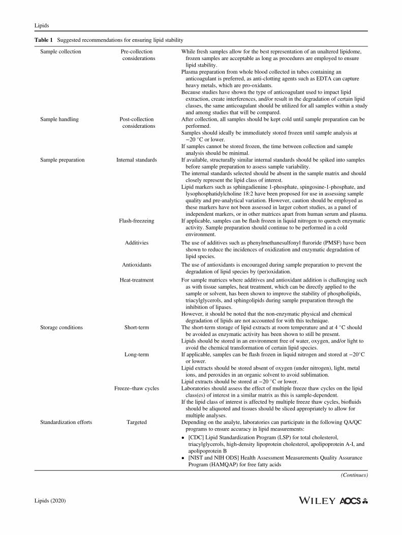
Table 1 Suggested recommendations for ensuring lipid stability
Sample collection Pre-collectionconsiderations
While fresh samples allow for the best representation of an unaltered lipidome,frozen samples are acceptable as long as procedures are employed to ensurelipid stability.
Plasma preparation from whole blood collected in tubes containing ananticoagulant is preferred, as anti-clotting agents such as EDTA can captureheavy metals, which are pro-oxidants.
Because studies have shown the type of anticoagulant used to impact lipidextraction, create interferences, and/or result in the degradation of certain lipidclasses, the same anticoagulant should be utilized for all samples within a studyand among studies that will be compared.
Sample handling Post-collectionconsiderations
After collection, all samples should be kept cold until sample preparation can beperformed.
Samples should ideally be immediately stored frozen until sample analysis at−20 �C or lower.
If samples cannot be stored frozen, the time between collection and sampleanalysis should be minimal.
Sample preparation Internal standards If available, structurally similar internal standards should be spiked into samplesbefore sample preparation to assess sample variability.
The internal standards selected should be absent in the sample matrix and shouldclosely represent the lipid class of interest.
Lipid markers such as sphingadienine 1-phosphate, spingosine-1-phosphate, andlysophosphatidylcholine 18:2 have been proposed for use in assessing samplequality and pre-analytical variation. However, caution should be employed asthese markers have not been assessed in larger cohort studies, as a panel ofindependent markers, or in other matrices apart from human serum and plasma.
Flash-freezeing If applicable, samples can be flash frozen in liquid nitrogen to quench enzymaticactivity. Sample preparation should continue to be performed in a coldenvironment.
Additivies The use of additives such as phenylmethanesulfonyl fluroride (PMSF) have beenshown to reduce the incidences of oxidization and enzymatic degradation oflipid species.
Antioxidants The use of antioxidants is encouraged during sample preparation to prevent thedegradation of lipid species by (per)oxidation.
Heat-treatment For sample matrices where additives and antioxidant addition is challenging suchas with tissue samples, heat treatment, which can be directly applied to thesample or solvent, has been shown to improve the stability of phospholipids,triacylglycerols, and sphingolipids during sample preparation through theinhibition of lipases.
However, it should be noted that the non-enzymatic physical and chemicaldegradation of lipids are not accounted for with this technique.
Storage conditions Short-term The short-term storage of lipid extracts at room temperature and at 4 �C shouldbe avoided as enzymatic activity has been shown to still be present.
Lipids should be stored in an environment free of water, oxygen, and/or light toavoid the chemical transformation of certain lipid species.
Long-term If applicable, samples can be flash frozen in liquid nitrogen and stored at −20�Cor lower.
Lipid extracts should be stored absent of oxygen (under nitrogen), light, metalions, and peroxides in an organic solvent to avoid sublimation.
Lipid extracts should be stored at −20 �C or lower.Freeze–thaw cycles Laboratories should assess the effect of multiple freeze thaw cycles on the lipid
class(es) of interest in a similar matrix as this is sample-dependent.If the lipid class of interest is affected by multiple freeze thaw cycles, biofluidsshould be aliquoted and tissues should be sliced appropriately to allow formultiple analyses.
Standardization efforts Targeted Depending on the analyte, laboratories can participate in the following QA/QCprograms to ensure accuracy in lipid measurements:
• [CDC] Lipid Standardization Program (LSP) for total cholesterol,triacylglycerols, high-density lipoprotein cholesterol, apolipoprotein A-I, andapolipoprotein B
• [NIST and NIH ODS] Health Assessment Measurements Quality AssuranceProgram (HAMQAP) for free fatty acids
(Continues)
Lipids
Lipids (2020)
chelators, and enzyme inhibitors (Blanco andBlanco, 2017; Lü et al., 2010; Nimse and Pal, 2015). Anti-oxidants are generally introduced during sample prepara-tion or added for long-term storage due to their low costand high effectiveness. Examples of antioxidants reportedin literature include butylated hydroxyanisole (BHA),butylated hydroxytoluene (BHT), ascorbic acid, caroten-oids, transferrin, deferoxamine, methyl silicone, phosphoricacid, propyl gallate (PG), tocopherols, quercetin, and ter-tiary butylhydroquinone (TBHQ).
Heat Treatment
While additives are useful chemical methods for increasinglipid stability in plasma and urine, it is challenging toimplement additives in tissue samples prior to pulveriza-tion, homogenization, and extraction. Heat treatment, atechnique employed since the 1940s, has been shown topromote the stability of certain lipid species includingphospholipids, neutral lipids (e.g., triacylglycerols), andsphingolipids during sample preparation via inhibition oflipases, including phospholipase A, C, and D activity(Koelmel et al., 2018; Rose et al., 2008). Heat treatmenthas the advantage of being applied at the time of collection,such as in environmental field studies, where access to liq-uid nitrogen or securing options for flash freezing is diffi-cult. Heat treatment can be directly applied to tissues,whole animals, or serum/plasma in a vacuum-sealed cham-ber without solvent. In addition, the solvent can be rapidlyheated during extraction (Koelmel et al., 2018). Generally,if the technology is available, the prior is desirable in that itreduces enzymatic activity earlier on during sample prepa-ration and hence any associated enzymatic transformationof lipids. It is important to note that nonenzymatic physicaland chemical transformation/degradation of lipids are notaccounted for by heat treatment. While heat treatmentreduces enzymatic activity, physical degradation, for exam-ple via oxidation, can still occur even though the applica-tion of heat treatment under vacuum may not increase theoxidation of lipids (Rose et al., 2008). It is often enzymaticdegradation that is most significant at the small intervals
between thawing and extraction, especially in tissues,whole blood, or cell cultures where the release of calciumupon cell pulverization activates enzymes to begin todegrade and transform lipids. Therefore, heat treatment isan effective approach for drastically increasing lipid stabil-ity during extraction and short-term sample handling.
Considerations for Lipid Storage
The abovementioned NIST lipidomics survey questioned122 laboratories on temperatures used to store lipidextracts, resulting in 174 responses. Most respondents indi-cated that lipid extracts were stored in either a –80 �Cfreezer (n = 96) or −20 �C freezer (n = 55). However, afew laboratories mentioned the storage of lipids at roomtemperature (n = 2) or in a refrigerator (n = 14).The stability and interconversion of lipids can be heavily
impacted by external factors such as storage and freeze–thaw cycles. The type of sample that is to be stored willcertainly influence the storage procedure employed toensure lipid stability (Burtis et al., 2013). It is still unknownhow storage and freeze–thaw cycles affect the entirelipidome as there appears to be sample-dependent effects(Stevens et al., 2019; Zivkovic et al., 2009). While the gen-eral idea for sample preservation is to freeze samplesquickly and store at a low temperature, this principle doesnot consider the exact storage temperature or the physicalstate in which the sample should be stored (e.g., in solution,dried under nitrogen, with preservatives/additives, etc.).While literature has shown the rate of autoxidation todecrease at lower temperatures and hence cryopreservationor freezing can be used to store lipids (Hess andO’Hare, 1950; Liu et al., 2019; Velasco andDobarganes, 2002), an exception can occur when autoxida-tion is induced during freezing. For example, in the case ofred blood cell-containing samples, lysis can occur due towater expansion, and the resulting release of iron can driveautoxidation (Metherel et al., 2013; Metherel andStark, 2015).For long-term storage after sample preparation, lipid
extracts should generally be stored in an airtight container
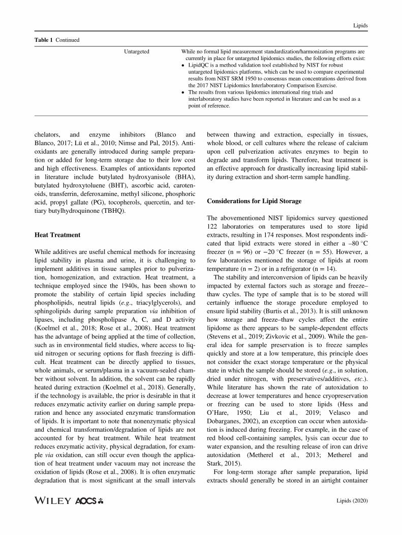
Table 1 Continued
Untargeted While no formal lipid measurement standardization/harmonization programs arecurrently in place for untargeted lipidomics studies, the following efforts exist:
• LipidQC is a method validation tool established by NIST for robustuntargeted lipidomics platforms, which can be used to compare experimentalresults from NIST SRM 1950 to consensus mean concentrations derived fromthe 2017 NIST Lipidomics Interlaboratory Comparison Exercise.
• The results from various lipidomics international ring trials andinterlaboratory studies have been reported in literature and can be used as apoint of reference.
Lipids
Lipids (2020)
at −20 �C or lower in organic solvent to avoid sublimation,an area free of light and oxygen, and in the presence ofantioxidants for liquids. Lipids stored as a lyophilizedmaterial are more prone to hydrolysis and/or oxidation dueto their hygroscopic behavior. There is also a lack of clearguidance between the recommendations for lipid extractstorage at −20 �C or temperatures lower than −20 �C. It issuggested that organic solutions of phospholipids shouldnot be stored at temperatures lower than −20 �C unlessthey are stored in glass. It is also recommended that lipidclasses, such as glycerophospholipids, not be stored inaqueous solutions for extended periods of time due to thepotential for hydrolysis. Plastic and/or polymer-based con-tainers should not be used to store organic solutions of lipidextracts as these organic solvents can leach the plastics.The number of freeze–thaw cycles should remain limited asliterature has shown up to a 37% variability in HDL- andLDL-cholesterol from one freeze–thaw cycle. An environ-ment rich in water, oxygen, and/or light may cause thechemical transformation of certain lipid species despitestorage at a low temperature (−20 �C or lower) (HjmJansen, 2014).Literature has shown that the temperature, duration of
storage, and presence of enzymatic activity all affect lipidstability differently depending on the lipid class and sampletype (Hjm Jansen, 2014; Roszkowska et al., 2018; Zivkovicet al., 2009). Jansen et al. demonstrated that while HDL-and LDL-cholesterol, triacylglycerols, and apolipoprotein-A1 and B were fairly stable at −20 �C, fatty acids showedlevels of degradation as high as 80% (Hjm Jansen, 2014).The authors also reported no significant differencesbetween lipid storage at −70 and −196 �C over the courseof 12 months, suggesting that a temperature lower than−20 �C may be more ideal. This observation was reflectedin a fatty acid stability study (Metherel et al., 2013), wherethe concentrations of eicosapentaenoic acid and doco-sahexaenoic acid (two omega-3 PUFA) in dried-bloodspots decreased more rapidly when stored at −20 �C ascompared with room temperature, 4, and −80 �C. In addi-tion, studies on the lipid profiles in human milk showedthat enzymatic activity was more reduced at −70 �C or−80 �C compared to −20 �C (Fusch et al., 2015; Levet al., 2014). Laboratories should consider an in-housefreeze–thaw stability and short-term/long-term storage sta-bility evaluation for each sample-type.Extreme care should be taken for the lipidomic analysis
of stored cell cultures. While cells can be stored at a muchlower temperature in liquid nitrogen to reduce lipid insta-bility, cells would then have to be introduced to acryopreservant solution containing DMSO, which maycause a high background in mass spectrometry-basedstudies.
While written for a wide range of metabolites, the Cen-ters for Disease Control and Prevention (CDC) has pro-vided a resource, Improving the Collection andManagement of Human Samples Used for Measuring Envi-ronmental Chemicals and Nutrition Indicators, thatdescribes the best practices for the collection and storage(e.g., whole blood, blood cells, serum, saliva, and urine) toensure analyte integrity (Centers for Disease Control andPrevention, 2018). The considerations presented can cer-tainly be transferable to the design of lipidomics studies.
Standardization Efforts for Accuracy in LipidMeasurements
Quality assurance (QA) and quality control (QC) measuresare necessary to ensure the harmonization of lipid measure-ments (Burla et al., 2018). Efforts are ongoing to establishexternal lipid QA and standardization programs. The CDCestablished the Lipids Standardization Program (LSP) toensure the analytical accuracy and precision of select lipidmeasurements reported in research and clinical laboratories(Warnick et al., 2008). Blinded high-quality pooled serasamples with target values determined by generally recog-nized reference methods for certain lipids such as total cho-lesterol (TC), triacylglycerols (TAG), high-densitylipoprotein cholesterol (HDL-C), apolipoprotein A-I (apoA-I), and apolipoprotein B (apo B) are provided to partici-pants of the LSP program over a 3-month period. A statisti-cal report that provides information about measurementconsistency and whether established analytical performancecriteria were met is provided to the LSP participants. Usingthis approach, laboratories can test the accuracy of theirmeasurements from an in-house established protocol toensure that these lipid measurements over time are notaffected by pre-analytical factors such as storage and stabil-ity. CDC provides similar programs for other analytes suchas steroid hormones and vitamin D.In addition, a clinical or research laboratory with a lab-
developed test can seek a 6-month certification by the CDCfor TC through a collaboration with a Cholesterol Refer-ence Method Laboratory Network (CRMLN) member labo-ratory (Myers et al., 2000). Briefly, the laboratories analyzesix high-quality serum samples over a pre-determined con-centration range to ensure that specific analytical criteriaare met (i.e., �3.0% maximum allowable bias to the refer-ence method and a ≤ 3.0% CV maximum allowable impre-cision) and traceability is established to the NationalReference System for Cholesterol (NRS/CHOL). Aftermeeting the certification criteria, the clinical laboratory orresearch laboratory is issued a 6-month valid Certificate ofTraceability, which states that the analytical system (e.g.,instrument model, reagent, lot, calibrator lot, and matrix)
Lipids
Lipids (2020)

successfully demonstrated traceability to the NRS/CHOLunder the tested conditions. In all of these CDC programs,procedures are used that ensure the stability of the specificlipids during processing, storage, and transport.NIST, in collaboration with the National Institutes of
Health (NIH) Office of Dietary Supplements (ODS), hasbeen actively involved in harmonizing fatty acid measure-ments in biological samples, particularly blood plasmaand serum. NIST and NIH ODS established Fatty Acidsin Human Serum and Plasma Quality Assurance Program(FAQAP) that administered interlaboratory comparisonexercises for the measurement of 24 fatty acids in selectedfreeze-dried and frozen plasma and serum matrices. Forthese studies, participants utilized their typical analyticalworkflows to analyze samples; the main goal was toassess laboratory performance and gain a better under-standing of fatty acid measurement variability across labo-ratories. Moreover, NIST helped participants troubleshoottheir analytical methods when requested. For the firstexercise (Schantz et al., 2013), a collaboration with theCDC conducted in 2012, participants measured fatty acidconcentrations in NIST SRM 2378 – Fatty Acids inHuman Serum, which is composed of serum from(1) donors who had not consumed fish or flaxseed oil sup-plements for 1 month prior to sample collection,(2) donors who consumed flaxseed oil supplements for1 month minimum before sample collection, and(3) donors who consumed fish oil supplements for 1 monthminimum before sample collection. This first effort dem-onstrated the urgent need for quality control materials thatcan be used to increase lipid measurement comparabilityacross laboratories. Although FAQAP formally ended in2017, NIST and NIH ODS has since formed the HealthAssessment Measurements Quality Assurance Program(HAMQAP) (Barber et al., 2018), which continues toserve the fatty acid measurement community. HAMQAPadministers interlaboratory comparison exercises whereparticipants measure a host of analytes, including fattyacids, in samples issued by NIST that represent bothhuman intake (e.g., food, dietary supplements) and output(e.g., plasma, serum, urine) so that laboratories can assesstheir in-house measurements. Participation can be utilizedto fulfill requirements established by accreditation bodiesor to show compliance with the Food and Drug Adminis-tration (FDA) Current Good Manufacturing Prac-tices (CGMP).While the abovementioned approaches by the CDC and
NIST/NIH ODS ensure accuracy for targeted lipid applica-tions, NIST established a method validation tool for robustuntargeted lipidomics platforms using LipidQC (Ulmeret al., 2017). Users can visually compare experimentalresults from the NIST Standard Reference Material (SRM)
1950, “Metabolites in Frozen Human Plasma”, againstbenchmark consensus mean concentrations derived fromthe NIST Lipidomics Interlaboratory Comparison Exercise(Bowden et al., 2017a; Bowden et al., 2017b), which waspublished in 2017. It is important to note that the consensusvalues reported are based on measurements across laborato-ries that as a whole demonstrated much variation due to thedifferent methodologies employed. Therefore, consensusvalues may not be synonymous with a high level of mea-surement accuracy and instead should be used as a point ofreference. Furthermore, other community-wide efforts suchas the Lipidomics Standards Initiative (https://lipidomics-standards-initiative.org/), Lipid MAPS (http://lipidmaps.org/), and the newly formed International Lipidomics Soci-ety (https://lipidomicssociety.org/), along with variousreports on pre-analytical processing, especially that fromthe International Ring Trial (Thompson et al., 2019), havehighlighted recommendations for sample storage andfreeze–thaw cycling, as well as other critical considerationsfor the standardization of lipidomics workflows (Burlaet al., 2018; Heiskanen et al., 2013; Kirwan et al., 2018;O’Donnell et al., 2019). New bioinformatics resourcescapable of identifying oxidized lipids and enzymatic prod-ucts such as phosphatidylmethanol lipid species inlipidomics datasets are emerging (Koelmel et al., 2017; Niet al., 2017; Tsugawa et al., 2015) and could become animportant complement in quality assurance/quality controlin both targeted and untargeted analyses.Community-wide guidelines are needed to establish best
practices to reduce lipid degradation during sample prepa-ration and storage, as there is limited consensus within thelipidomics field. In addition, rigorous studies that scrutinizethe advantages and disadvantages of approaches to ensurelipid stability are needed. Further, the lipidomics commu-nity should expand the concept of lipid stability over theentire analytical workflow, which includes more compre-hensive investigations into the gas phase instability of somelipid species, leading to in-source fragmentation and poten-tially skewed lipid profiles.To this point, tt is important to include optimized steps
across the entire lipidomics workflow (including massspectrometric parameters) in laboratory SOP to ensure lipidstability, while limiting the implementation of unnecessaryand/or impractical preventative strategies if the lipid(s) thatare being interrogated do not require it. Nevertheless, par-ticipation in programs offered by the CDC, such as the LSPthat ensure the accuracy and precision of select lipid mea-surements can aid in evaluating existing sample preparationmethodologies. The procedures and approaches success-fully used in the CDC and NIST programs can be adoptedin untargeted lipidomics applications to ensure accuracy,reliability, and comparability in lipid measurements.
Lipids
Lipids (2020)
Disclaimer
Certain commercial equipment, instruments, or materialsare identified in this paper to adequately specify the experi-mental procedures. Such identification does not imply rec-ommendation or endorsement by the National Institute ofStandards and Technology; nor does it imply that the mate-rials or equipment identified are necessarily the best for thepurpose. Furthermore, the content is solely the responsibil-ity of the authors and does not necessarily represent theofficial views of the National Institute of Standards andTechnology.The findings and conclusions in this report are those of
the author(s) and do not necessarily represent the officialposition of the Centers for Disease Control and Prevention/the Agency for Toxic Substances and Disease Registry.Use of trade names is for identification only and does notimply endorsement by the Centers for Disease Control andPrevention, the Public Health Service and the US Depart-ment of Health and Human Services.
Conflict of Interest
The authors declare that they have no conflict of interest.
References
Abuja, P. M., Ehrhart, F., Schoen, U., Schmidt, T., Stracke, F.,Dallmann, G., … Zatloukal, K. (2015) Alterations in human livermetabolome during prolonged cryostorage. Journal of ProteomeResearch, 14:2758–2768.
Ademowo, O. S., Dias, H. K. I., Burton, D. G. A., & Griffiths, H. R.(2017) Lipid (per) oxidation in mitochondria: An emerging targetin the ageing process? Biogerontology, 18:859–879.
Aiello, E. J., Tworoger, S. S., Yasui, Y., Stanczyk, F. Z., Potter, J.,Ulrich, C. M., … McTiernan, A. (2005) Associations among circu-lating sex hormones, insulin-like growth factor, lipids, and mam-mographic density in postmenopausal women. CancerEpidemiology, Biomarkers & Prevention, 14:1411–1417.
Anton, G., Wilson, R., Yu, Z.-H., Prehn, C., Zukunft, S., Adamski, J.,… Waldenberger, M. (2015) Pre-analytical sample quality: Metab-olite ratios as an intrinsic marker for prolonged room temperatureexposure of serum samples. PLoS One, 10:e0121495.
Aoki, J., Inoue, A., & Okudaira, S. (2008) Two pathways forlysophosphatidic acid production. Biochimica et Biophysica Acta,1781:513–518.
Aoki, J., Taira, A., Takanezawa, Y., Kishi, Y., Hama, K.,Kishimoto, T., … Arai, H. (2002) Serum lysophosphatidic acid isproduced through diverse phospholipase pathways. The Journal ofBiological Chemistry, 277:48737–48744.
Aristizabal-Henao, J. J., Fernandes, M. F., Duncan, R. E., &Stark, K. D. (2019) Development of a rapid ultra high-performanceliquid chromatography/tandem mass spectrometry method for theanalysis of sn-1 and sn-2 lysophosphatidic acid Regioisomers inmouse plasma. Lipids, 54:479–486.
Arzu, K., Canan, T., Sevilay, S., Simal Köksal, C., Ezgi Coskun, Y.,Fatih, D., & Turan, T. (2019) Comparison of some biochemicaltests in different blood collection tubes in hemodialysis patients.Turkish Journal of Biochemistry, 45:26–36.
Barber, C. A., Benner, B. A., Thomas, J. B., Burdette, C. Q.,Camara, J., Long, S., Murray, J. A., Phillips, M. M., Place, B. J.,Rimmer, C. A., Wood, L. J., Yu, L., Chinthalapati, S. K. R., &Tai, S. S.-C. (2018) Health assessment measurements qualityassurance program: Exercise 1 final report. NISTIR 8237.pp. 1–330
Blanco, A., & Blanco, G. (2017) Antioxidants. In A. Blanco &G. Blanco (Eds.), Medical Biochemistry (PP. 205–214). England:Academic Press.
Bowden, J. A., Albert, C. J., Barnaby, O. S., & Ford, D. A. (2011)Analysis of cholesteryl esters and diacylglycerols using lithiatedadducts and electrospray ionization-tandem mass spectrometry.Analytical Biochemistry, 417:202–210.
Bowden, J. A., Heckert, A., Ulmer, C. Z., & Jones, C. M. (2017b)Lipid concentrations in standard reference material (SRM) 1950:Results from an Interlaboratory Comparison Exercise forLipidomics. NISTIR 8185: 1–451
Bowden, J. A., Heckert, A., Ulmer, C. Z., Jones, C. M.,Koelmel, J. P., Abdullah, L., … Zhou, S. (2017a) Harmonizinglipidomics: NIST interlaboratory comparison exercise forlipidomics using SRM 1950-metabolites in frozen human plasma.Journal of Lipid Research, 58:2275–2288.
Bowden, J. A., Ulmer, C. Z., Jones, C. M., Koelmel, J. P., &Yost, R. A. (2018) NIST lipidomics workflow questionnaire: Anassessment of community-wide methodologies and perspectives.Metabolomics, 14:53.
Bowen, R. A. R., & Remaley, A. T. (2014) Interferences from bloodcollection tube components on clinical chemistry assays. BiochemiaMedica (Zagreb), 24:31–44.
Bozza, P. T., & Viola, J. P. (2010) Lipid droplets in inflammation andcancer. Prostaglandins, Leukotrienes, and Essential Fatty Acids,82:243–250.
Burla, B., Arita, M., Arita, M., Bendt, A. K., Cazenave-Gassiot, A.,Dennis, E. A., … Wenk, M. R. (2018) MS-based lipidomics ofhuman blood plasma: A community-initiated position paper todevelop accepted guidelines. Journal of Lipid Research, 59:2001–2017.
Burtis, C. A., Ashwood, E. R., Bruns, D. E., & Tietz, N. W. (2013)Tietz textbook of clinical chemistry and molecular diagnostics. St.Louis, MO: Saunders.
Canakci, M. (2007) The potential of restaurant waste lipids as biodie-sel feedstocks. Bioresource Technology, 98:183–190.
Carmella, S. G., Heskin, A. K., Tang, M. K., Jensen, J., Luo, X.,Le, C. T., … Hecht, S. S. (2019) Longitudinal stability in cigarettesmokers of urinary eicosanoid biomarkers of oxidative damage andinflammation. PLoS One, 14:e0215853.
Centers for Disease Control and Prevention. (2018) Improving theCollection and Management of Human Samples Used for Measur-ing Environmental Chemicals and Nutrition Indicators - Version1.3, Sciences USDoHaH, Editor. National Center for Environmen-tal Health: Division of Laboratory Sciences. pp. 1–24.
Dalle-Donne, I., Rossi, R., Colombo, R., Giustarini, D., & Milzani, A.(2006) Biomarkers of oxidative damage in human disease. ClinicalChemistry, 52:601–623.
De Caterina, R. (2011) N-3 fatty acids in cardiovascular disease. TheNew England Journal of Medicine, 364:2439–2450.
Dorow, J., Becker, S., Kortz, L., Thiery, J., Hauschildt, S., &Ceglarek, U. (2016) Preanalytical investigation of polyunsaturatedfatty acids and eicosanoids in human plasma by liquidchromatography-tandem mass spectrometry. Biopreservation andBiobanking, 14:107–113.
Lipids
Lipids (2020)
Fernandis, A. Z., & Wenk, M. R. (2009) Lipid-based biomarkers forcancer. Journal of Chromatography. B, Analytical Technologies inthe Biomedical and Life Sciences, 877:2830–2835.
Ferrero, M. E. (2016) Rationale for the successful management ofEDTA chelation therapy in human burden by toxic metals. BioMedResearch International, 2016:8274504–8274504.
Fung, T. T., Rimm, E. B., Spiegelman, D., Rifai, N., Tofler, G. H.,Willett, W. C., & Hu, F. B. (2001) Association between dietary pat-terns and plasma biomarkers of obesity and cardiovascular diseaserisk. The American Journal of Clinical Nutrition, 73:61–67.
Furberg, A. S., Jasienska, G., Bjurstam, N., Torjesen, P. A.,Emaus, A., Lipson, S. F., … Thune, I. (2005) Metabolic and hor-monal profiles: HDL cholesterol as a plausible biomarker of breastcancer risk. The Norwegian EBBA Study. Cancer Epidemiology,Biomarkers & Prevention, 14:33–40.
Fusch, G., Rochow, N., Choi, A., Fusch, S., Poeschl, S., Ubah, A. O.,… Fusch, C. (2015) Rapid measurement of macronutrients in breastmilk: How reliable are infrared milk analyzers? Clinical Nutrition(Edinburgh, Scotland), 34:465–476.
Gil, A., Siegel, D., Permentier, H., Reijngoud, D. J., Dekker, F., &Bischoff, R. (2015) Stability of energy metabolites-an often over-looked issue in metabolomics studies: A review. Electrophoresis,36:2156–2169.
Gil, A., Zhang, W., Wolters, J. C., Permentier, H., Horvatovich, P.,Rebecca Heiner-Fokkema, M., … Bischoff, R. (2019) Omics |Lipdomics and its pitfalls during the pre-analytical stage. InP. Worsfold, C. Poole, A. Townshend, & M. Miró (Eds.), Encyclo-pedia of analytical science (3rd ed.). Oxford, England: AcademicPress.
Gonzalez-Covarrubias, V., Dane, A., Hankemeier, T., & Vreeken, R.(2013) The influence of citrate, EDTA, and heparin anticoagulantsto human plasma LC–MS lipidomic profiling. Metabolomics, 9:337–348.
Haid, M., Muschet, C., Wahl, S., Romisch-Margl, W., Prehn, C.,Moller, G., & Adamski, J. (2018) Long-term stability of humanplasma metabolites during storage at −80 degrees C. Journal ofProteome Research, 17:203–211.
Halliwell, B. (2000) Lipid peroxidation, antioxidants and cardiovascu-lar disease: How should we move forward? CardiovascularResearch, 47:410–418.
Han, C., Zhang, H. T., Du, L., Liu, X., Jing, J., Zhao, X., … Tian, B.(2005) Serum levels of leptin, insulin, and lipids in relation tobreast cancer in China. Endocrine, 26:19–24.
Heiskanen, L. A., Suoniemi, M., Ta, H. X., Tarasov, K., & Ekroos, K.(2013) Long-term performance and stability of molecular shotgunlipidomic analysis of human plasma samples. Analytical Chemistry,85:8757–8763.
Hennebelle, M., Metherel, A. H., Kitson, A. P., Otoki, Y., Yang, J.,Lee, K. S. S., … Taha, A. Y. (2019) Brain oxylipin concentrationsfollowing hypercapnia/ischemia: Effects of brain dissection and dis-section time. Journal of Lipid Research, 60:671–682.
Hess, P. S., & O’Hare, G. A. (1950) Oxidation of linseed oil. Indus-trial & Engineering Chemistry, 42:1424–1431.
Hill-Kapturczak, N., Dougherty, D. M., Roache, J. D., Karns-Wright, T. E., & Javors, M. A. (2018) Differences in the synthesisand elimination of phosphatidylethanol 16:0/18:1 and 16:0/18:2after acute doses of alcohol. Alcoholism, Clinical and ExperimentalResearch, 42:851–860.
Hinterwirth, H., Stegemann, C., & Mayr, M. (2014) Lipidomics:Quest for molecular lipid biomarkers in cardiovascular disease. Cir-culation. Cardiovascular Genetics, 7:941–954.
Hjm Jansen, E. (2014) Long term stability of parameters of lipidmetabolism in frozen human serum: Triglycerides, free fatty acids,Total-, HDL- and LDL-cholesterol, apolipoprotein-A1 and B. Jour-nal of Molecular Biomarkers & Diagnosis, 5:1–5.
Holl, K., Lundin, E., Kaasila, M., Grankvist, K., Afanasyeva, Y.,Hallmans, G., … Lukanova, A. (2008) Effect of long-term storageon hormone measurements in samples from pregnant women: Theexperience of the Finnish maternity cohort. Acta Oncologica, 47:406–412.
Ishikawa, M., Maekawa, K., Saito, K., Senoo, Y., Urata, M.,Murayama, M., … Saito, Y. (2014) Plasma and serum lipidomicsof healthy white adults shows characteristic profiles by subjects’gender and age. PLoS One, 9:e91806.
Jane Ellis, M., Livesey, J. H., & Evans, M. J. (2003) Hormone stabil-ity in human whole blood. Clinical Biochemistry, 36:109–112.
Jones, M. E., Folkerd, E. J., Doody, D. A., Iqbal, J., Dowsett, M.,Ashworth, A., & Swerdlow, A. J. (2007) Effect of delays inprocessing blood samples on measured endogenous plasma sex hor-mone levels in women. Cancer Epidemiology, Biomarkers & Pre-vention, 16:1136–1139.
Kamlage, B., Maldonado, S. G., Bethan, B., Peter, E., Schmitz, O.,Liebenberg, V., & Schatz, P. (2014) Quality markers addressingpreanalytical variations of blood and plasma processing identifiedby broad and targeted metabolite profiling. Clinical Chemisty, 60:399–412.
Kensova, R., Hynek, D., Kynicky, J., Konecna, M., Eckschlager, T.,Adam, V., … Kizek, R. (2014) Determination of metal ions in theplasma of children with tumour diseases by differential pulsevoltammetry. International Journal of Electrochemical Science, 9:4675–4691.
Kirkwood, J. S., Maier, C., & Stevens, J. F. (2013) Simultaneous,untargeted metabolic profiling of polar and nonpolar metabolites byLC-Q-TOF mass spectrometry. Current Protocols in Toxicology, 4:4.39–34.39.
Kirwan, J. A., Brennan, L., Broadhurst, D., Fiehn, O., Cascante, M.,Dunn, W. B., … Velagapudi, V. (2018) Preanalytical processingand biobanking procedures of biological samples for metabolomicsresearch: A white paper, community perspective (for "precisionmedicine and Pharmacometabolomics task group"-the met-abolomics society initiative). Clinical Chemistry, 64:1158–1182.
Koelmel, J. P., Cochran, J. A., Ulmer, C. Z., Levy, A. J.,Patterson, R. E., Olsen, B. C., … Garrett, T. J. (2019) Software toolfor internal standard based normalization of lipids, and effect ofdata-processing strategies on resulting values. BMC Bioinformatics,20:217.
Koelmel, J. P., Jones, C. M., Ulmer, C. Z., Garrett, T. J., Yost, R. A.,Schock, T. B., & Bowden, J. A. (2018) Examining heat treatmentfor stabilization of the lipidome. Bioanalysis, 10:291–305.
Koelmel, J. P., Kroeger, N. M., Ulmer, C. Z., Bowden, J. A.,Patterson, R. E., Cochran, J. A., … Yost, R. A. (2017) LipidMatch:An automated workflow for rule-based lipid identification usinguntargeted high-resolution tandem mass spectrometry data. BMCBioinformatics, 18:331.
Kwan, B. C., Kronenberg, F., Beddhu, S., & Cheung, A. K. (2007)Lipoprotein metabolism and lipid management in chronic kidneydisease. Journal of the American Society of Nephrology, 18:1246–1261.
Lev, H. M., Ovental, A., Mandel, D., Mimouni, F. B., Marom, R., &Lubetzky, R. (2014) Major losses of fat, carbohydrates and energycontent of preterm human milk frozen at −80�C. Journal of Perina-tology, 34:396–398.
Liu, K., Liu, Y., & Chen, F. (2019) Effect of storage temperature onlipid oxidation and changes in nutrient contents in peanuts. FoodScience & Nutrition, 7:2280–2290.
Liu, X., Hoene, M., Yin, P., Fritsche, L., Plomgaard, P., Hansen, J. S.,… Lehmann, R. (2018) Quality control of serum and plasma byquantification of (4E,14Z)-sphingadienine-C18-1-phosphateuncovers common Preanalytical errors during handling of whole.Blood, 64:810–819.
Lipids
Lipids (2020)

Lonning, P. E., Geisler, J., Krag, L. E., Erikstein, B., Bremnes, Y.,Hagen, A. I., … Massimini, G. (2005) Effects of exemestaneadministered for 2 years versus placebo on bone mineral density,bone biomarkers, and plasma lipids in patients with surgicallyresected early breast cancer. Journal of Clinical Oncology: OfficialJournal of the American Society of Clinical Oncology, 23:5126–5137.
Lorenz, M. A., Burant, C. F., & Kennedy, R. T. (2011) Reducing timeand increasing sensitivity in sample preparation for adherent mam-malian cell metabolomics. Analytical Chemistry, 83:3406–3414.
Lü, J.-M., Lin, P. H., Yao, Q., & Chen, C. (2010) Chemical andmolecular mechanisms of antioxidants: Experimental approachesand model systems. Journal of Cellular and Molecular Medicine,14:840–860.
Lu, W., Su, X., Klein, M. S., Lewis, I. A., Fiehn, O., &Rabinowitz, J. D. (2017) Metabolite measurement: Pitfalls to avoidand practices to follow. Annual Review of Biochemistry, 86:277–304.
Maddipati, K. R., & Zhou, S. L. (2011) Stability and analysis ofeicosanoids and docosanoids in tissue culture media. Prostaglan-dins & Other Lipid Mediators, 94:59–72.
Maskrey, B. H., & O’Donnell, V. B. (2008) Analysis of eicosanoidsand related lipid mediators using mass spectrometry. BiochemicalSociety Transactions, 36:1055–1059.
Massaro, M., Scoditti, E., Carluccio, M. A., & De Caterina, R. (2008)Basic mechanisms behind the effects of n-3 fatty acids on cardio-vascular disease. Prostaglandins, Leukotrienes, and Essential FattyAcids, 79:109–115.
Metherel, A. H., Aristizabal Henao, J. J., & Stark, K. D. (2013) EPAand DHA levels in whole blood decrease more rapidly when storedat −20�C as compared with room temperature, 4 and −75�C.Lipids, 48:1079–1091.
Metherel, A. H., & Stark, K. D. (2015) Cryopreservation preventsiron-initiated highly unsaturated fatty acid loss during storage ofhuman blood on chromatography paper at −20�C. The Journal ofNutrition, 145:654–660.
Metherel, A. H., & Stark, K. D. (2016) The stability of blood fattyacids during storage and potential mechanisms of degradation: Areview. Prostaglandins, Leukotrienes, and Essential Fatty Acids,104:33–43.
Min, B., & Ahn, D. U. (2005) Mechanism of lipid peroxidation inmeat and meat products—A review. Food Science and Biotechnol-ogy, 14:152–163.
Mohamed, A. S., Hosney, M., Bassiony, H., Hassanein, S. S.,Soliman, A. M., Fahmy, S. R., & Gaafar, K. (2020) Sodium pento-barbital dosages for exsanguination affect biochemical, molecularand histological measurements in rats. Scientific Reports, 10:378.
Myers, G. L., Kimberly, M. M., Waymack, P. P., Smith, S. J.,Cooper, G. R., & Sampson, E. J. (2000) A reference method labora-tory network for cholesterol: A model for standardization andimprovement of clinical laboratory measurements. Clinical Chemis-try, 46:1762–1772.
Nawar, W. W. (1969) Thermal degradation of lipids. Journal of Agri-cultural and Food Chemistry, 17:18–21.
Nawar, W. W. (1984) Chemical changes in lipids produced by ther-mal processing. Journal of Chemical Education, 61:299.
Ng, W.-Y., & Yeo, C.-P. (2013) Thrombin-accelerated quick clottingserum tubes: An evaluation with 22 common biochemical analytes.Advances in Hematology, 2013:769479.
Ni, Z., Angelidou, G., Hoffmann, R., & Fedorova, M. (2017)LPPtiger software for lipidome-specific prediction and identifica-tion of oxidized phospholipids from LC-MS datasets. ScientificReports, 7:15138.
Nimse, S. B., & Pal, D. (2015) Free radicals, natural antioxidants, andtheir reaction mechanisms. RSC Advances, 5:27986–28006.
Oberg, B. P., McMenamin, E., Lucas, F. L., McMonagle, E.,Morrow, J., Ikizler, T. A., & Himmelfarb, J. (2004) Increased prev-alence of oxidant stress and inflammation in patients with moderateto severe chronic kidney disease. Kidney International, 65:1009–1016.
O’Donnell, V. B., Ekroos, K., Liebisch, G., & Wakelam, M. (2019)Lipidomics: Current state of the art in a fast moving field. WileyInterdisciplinary Reviews: Systems Biology and Medicine, 12:e1466.
Overmyer, K. A., Thonusin, C., Qi, N. R., Burant, C. F., &Evans, C. R. (2015) Impact of anesthesia and euthanasia on met-abolomics of mammalian tissues: Studies in a C57BL/6J mousemodel. PLoS One, 10:e0117232.
Qiu, Y., Moir, R., Willis, I., Beecher, C., Tsai, Y. H., Garrett, T. J., …Kurland, I. J. (2016) Isotopic ratio outlier analysis of theS. cerevisiae metabolome using accurate mass gaschromatography/time-of-flight mass spectrometry: A new methodfor discovery. Analytical Chemistry, 88:2747–2754.
Rampler, E., Criscuolo, A., Zeller, M., El Abiead, Y., Schoeny, H.,Hermann, G., … Koellensperger, G. (2018) A novel Lipidomicsworkflow for improved human plasma identification and quantifica-tion using RPLC-MSn methods and isotope dilution strategies.Analytical Chemistry, 90:6494–6501.
Ridker, P. M., Rifai, N., Cook, N. R., Bradwin, G., & Buring, J. E.(2005) Non-HDL cholesterol, apolipoproteins A-I and B100, stan-dard lipid measures, lipid ratios, and CRP as risk factors for cardio-vascular disease in women. JAMA, 294:326–333.
Rose, D. J., Ogden, L. V., Dunn, M. L., & Pike, O. A. (2008)Enhanced lipid stability in whole wheat flour by lipase inactivationand antioxidant retention. Cereal Chemistry Journal, 85:218–223.
Roszkowska, A., Yu, M., Bessonneau, V., Bragg, L., Servos, M., &Pawliszyn, J. (2018) Tissue storage affects lipidome profiling incomparison to in vivo microsampling approach. Scientific Reports,8:6980.
Roughan, P. G., Slack, C. R., & RJL, H. (1978) Generation of phos-pholipid artefacts during extraction of developing soybean seedswith methanolic solvents. Lipids, 13:497–503.
Schantz, M. M., Powers, C. D., & Schleicher, R. L. (2013) Inter-laboratory analytical comparison study of total fatty acid concen-trations in human serum: results for Exercise 01: QA12FASER01.NISTIR7953, pp. 1–82.
Shahidi, F., & Zhong, Y. (2010) Lipid oxidation and improving theoxidative stability. Chemical Society Reviews, 39:4067–4079.
Shen, Z., & Wijesundera, C. (2009) Effects of docosahexaenoic acidpositional distribution on the oxidative stability of modeltriacylglycerol in water emulsion. Journal of Food Lipids, 16:62–71.
Sohaib, M., Butt, M. S., Shabbir, M. A., & Shahid, M. (2015) Lipidstability, antioxidant potential and fatty acid composition of broilersbreast meat as influenced by quercetin in combination with alpha-tocopherol enriched diets. Lipids in Health and Disease, 14:61.
Stegemann, C., Pechlaner, R., Willeit, P., Langley, S. R.,Mangino, M., Mayr, U., … Mayr, M. (2014) Lipidomics profilingand risk of cardiovascular disease in the prospective population-based Bruneck study. Circulation, 129:1821–1831.
Stevens, V. L., Hoover, E., Wang, Y., & Zanetti, K. A. (2019) Pre-analytical factors that affect metabolite stability in human urine,plasma, and serum: A review. Metabolites, 9:156.
Stupp, G. S., Clendinen, C. S., Ajredini, R., Szewc, M. A.,Garrett, T., Menger, R. F., … Edison, A. S. (2013) Isotopic ratiooutlier analysis global metabolomics of Caenorhabditis elegans.Analytical Chemistry, 85:11858–11865.
Sutphen, R., Xu, Y., Wilbanks, G. D., Fiorica, J., Grendys, E. C.,LaPolla, J. P., … Krischer, J. P. (2004) Lysophospholipids arepotential biomarkers of ovarian cancer. Cancer Epidemiology Bio-markers & Prevention, 13:1185–1191.
Lipids
Lipids (2020)
Thompson, J. W., Adams, K. J., Adamski, J., Asad, Y., Borts, D.,Bowden, J. A., … Moseley, M. A. (2019) International ring trial ofa high resolution targeted metabolomics and Lipidomics platformfor serum and plasma analysis. Analytical Chemistry, 91:14407–14416.
Trépanier, M.-O., Eiden, M., Morin-Rivron, D., Bazinet, R. P., &Masoodi, M. (2017) High-resolution lipidomics coupled with rapidfixation reveals novel ischemia-induced signaling in the rat neurol-ipidome. Journal of Neurochemistry, 140:766–775.
Tsugawa, H., Cajka, T., Kind, T., Ma, Y., Higgins, B., Ikeda, K., …Arita, M. (2015) MS-DIAL: Data-independent MS/MSdeconvolution for comprehensive metabolome analysis. NatureMethods, 12:523–526.
Ulmer, C. Z., Ragland, J. M., Koelmel, J. P., Heckert, A.,Jones, C. M., Garrett, T. J., … Bowden, J. A. (2017) LipidQC:Method validation tool for visual comparison to SRM 1950 usingNIST Interlaboratory comparison exercise lipid consensus meanestimate values. Analytical Chemistry, 89:13069–13073.
Velasco, J., & Dobarganes, C. (2002) Oxidative stability of virginolive oil. European Journal of Lipid Science and Technology, 104:661–676.
Wang, X., Gu, X., Song, H., Song, Q., Gao, X., Lu, Y., & Chen, H.(2015) Phenylmethanesulfonyl fluoride pretreatment stabilizesplasma lipidome in lipidomic and metabolomic analysis. AnalyticaChimica Acta, 893:77–83.
Warnick, G. R., Kimberly, M. M., Waymack, P. P., Leary, E. T., &Myers, G. L. (2008) Standardization of measurements for choles-terol, triglycerides, and major lipoproteins. Laboratory Medicine,39:481–490.
Watson, A. D. (2006) Thematic review series: Systems biologyapproaches to metabolic and cardiovascular disorders. Lipidomics:a global approach to lipid analysis in biological systems. Journal ofLipid Research, 47:2101–2111.
Xu, Y., Zhao, Z., Xiao, Y., Elson, P., Tan, H., Plummer, S., …Graham, C. (2007) Plasma lysophosphatidylcholine levels: poten-tial biomarkers for colorectal cancer. Journal of Clnical Oncology,67:1687–1687.
Yang, W., Chen, Y., Xi, C., Zhang, R., Song, Y., Zhan, Q., …Abliz, Z. (2013) Liquid chromatography-tandem massspectrometry-based plasma metabonomics delineate the effect ofmetabolites’ stability on reliability of potential biomarkers. Analyti-cal Chemistry, 85:2606–2610.
Yun, J.-M., & Surh, J. (2012) Fatty acid composition as a predictorfor the oxidation stability of Korean vegetable oils with or withoutinduced oxidative stress. Preventive Nutrition and Food Science,17:158–165.
Zhang, W. L., Liu, M. Y., Zhang, Z. C., & Duan, C. Y. (2013) Effectof different anesthesia methods on erythrocyte immune function inmice. Asian Pacific Journal of Tropical Medicine, 6:995–998.
Zhou, X., Mao, J., Ai, J., Deng, Y., Roth, M. R., Pound, C., …Bigler, S. A. (2012) Identification of plasma lipid biomarkers forprostate cancer by lipidomics and bioinformatics. PLoS One, 7:e48889.
Zivkovic, A. M., Wiest, M. M., Nguyen, U. T., Davis, R.,Watkins, S. M., & German, J. B. (2009) Effects of sample handlingand storage on quantitative lipid analysis in human serum. Met-abolomics, 5:507–516.
Lipids
Lipids (2020)